Embed Size (px)
Citation preview
PCDU – Lecture
izmb.uni-bonn.de izmb.uni-bonn.de
Plant Cell, Development & Ultrastructure
Plant Cell Biology Labs
Download at:
http://goo.gl/111Tha
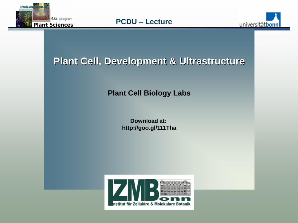
Plant Actin-Binding Proteins izmb.uni-bonn.de izmb.uni-bonn.de
Number of gene copies in A. thaliana
• Fimbrin (F-actin crosslinking) 3 genes
• Profilin (G-actin sequestering) 5 genes
• ADF/cofilin (F-actin destabilizing) 11 genes
• AiP1 (barbed end binder, co-factor of ADF) 2 genes
• Villin (F-actin crosslinking) 5 genes
• Gelsolin (F-actin destabilizing) single copy gene
• Arp2/3 (F-actin branching) single copy genes
• Formin (F-actin binding, polymerization) 21 genes
izmb.uni-bonn.de izmb.uni-bonn.de
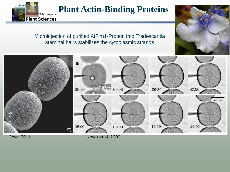
Microinjection of purified AtFim1-Protein into Tradescantia
staminal hairs stabilizes the cytoplasmic strands
Kovar et al. 2000
Tradescantia virginiana
Chwil 2011
Plant Actin-Binding Proteins
izmb.uni-bonn.de izmb.uni-bonn.de
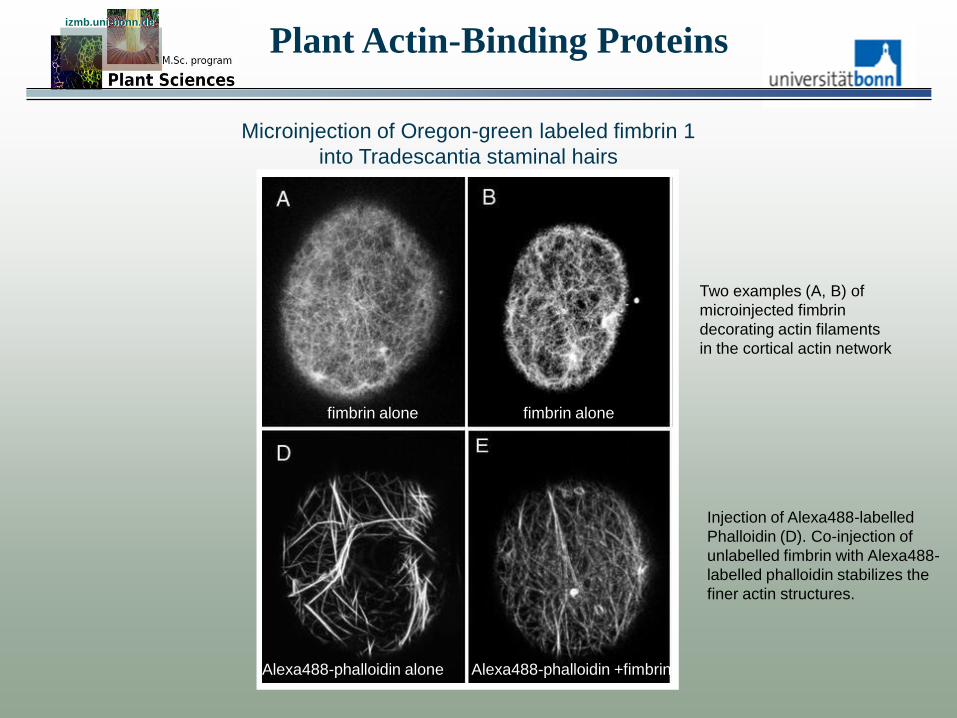
Microinjection of Oregon-green labeled fimbrin 1
into Tradescantia staminal hairs
fimbrin alone fimbrin alone
Alexa488-phalloidin alone Alexa488-phalloidin +fimbrin
Two examples (A, B) of
microinjected fimbrin
decorating actin filaments
in the cortical actin network
Injection of Alexa488-labelled
Phalloidin (D). Co-injection of
unlabelled fimbrin with Alexa488-
labelled phalloidin stabilizes the
finer actin structures.
Plant Actin-Binding Proteins

Calponin-Homology Domains of Fimbrin izmb.uni-bonn.de izmb.uni-bonn.de
ABD1 ABD2 Calponin is a smooth muscle
protein. it binds to F-actin and
blocks Actin-Myosin interaction.
Ca2+-dependent Phosphory-
ation releases Calponin from the
Actin filament.
Banuelos et al. 1998
CH1 CH2 CH3 CH4 Fimbrin
Klein et al. 2004
ABD1
ABD2
Ca-binding
EF-hand domain
helix F
helix E
loop

Calponin-Homology Domain Proteins izmb.uni-bonn.de izmb.uni-bonn.de
ABD1 ABD2
Calponin is a smooth muscle
protein. it binds to F-actin and
blocks Actin-Myosin interaction.
Ca2+-dependent Phosphory-
ation releases Calponin from the
Actin filament. Banuelos et al. 1998
Sr, spectrin repeat
PH, pleckstrin homology domain
EF, calmodulin-like EF-hand
Igr, immunoglobulin-like repeat
coiled-coil, a region with a propensity
to form a coiled-coils
PR, plectin repeat;
CR, calponin repeat;
DH, Dbl homology domain;
DAG, diacylglycerolbindingdomain
PR, Prolin-rich repeat
Calponin-Homology Domain izmb.uni-bonn.de izmb.uni-bonn.de
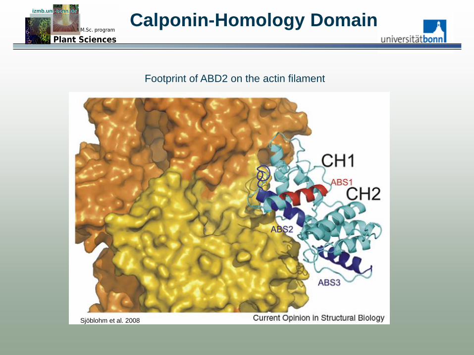
ABD1 ABD2
Footprint of ABD2 on the actin filament
Sjöblohm et al. 2008
ABD2-GFP izmb.uni-bonn.de izmb.uni-bonn.de
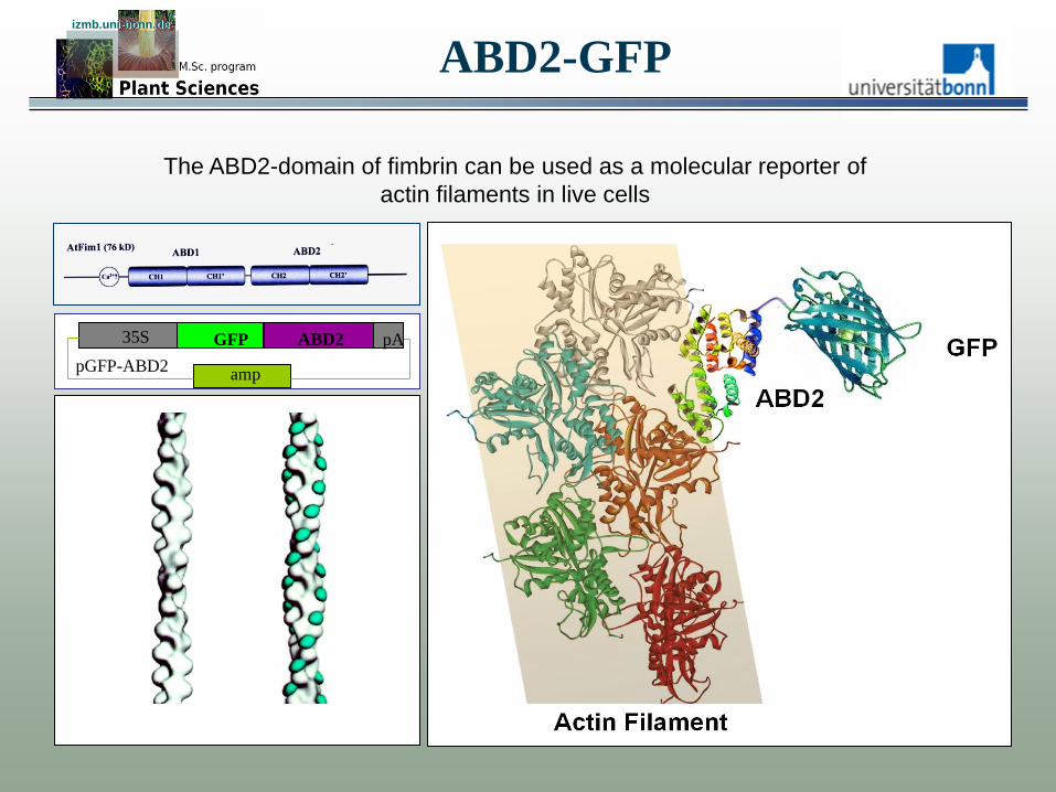
The ABD2-domain of fimbrin can be used as a molecular reporter of
actin filaments in live cells
pGFP-ABD2
35S GFP ABD2 pA
amp

ABD2-GFP as Reporter for F-Actin izmb.uni-bonn.de izmb.uni-bonn.de
Voigt et al. 2005
EUR J Cell Biol 84: 595-608
mouse monoclonal
Actin at the polar crosswalls in the root transition zone
A. thaliana
Zea mays
GFP-ABD2
Markus Schlicht, unpublished
10µm 20µm
50µm Xylem Primanen

Plant Actin-Binding Proteins izmb.uni-bonn.de izmb.uni-bonn.de
Number of gene copies in A. thaliana
• Fimbrin (F-actin crosslinking) 3 genes
• Profilin (G-actin sequestering) 5 genes
• ADF/cofilin (F-actin destabilizing) 11 genes
• AiP1 (barbed end binder, co-factor of ADF) 2 genes
• Villin (F-actin crosslinking) 5 genes
• Gelsolin (F-actin destabilizing) single copy gene
• Arp2/3 (F-actin branching) single copy genes
• Formin (F-actin binding, polymerization) 21 genes
Plant Actin Sequestering Proteins izmb.uni-bonn.de izmb.uni-bonn.de
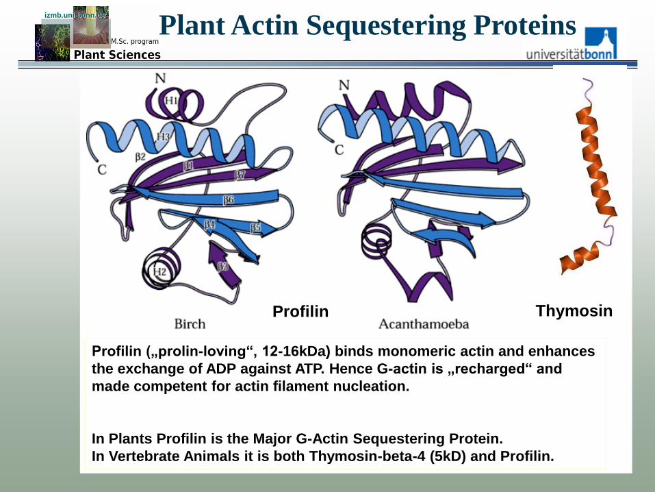
Profilin („prolin-loving“, 12-16kDa) binds monomeric actin and enhances
the exchange of ADP against ATP. Hence G-actin is „recharged“ and
made competent for actin filament nucleation.
In Plants Profilin is the Major G-Actin Sequestering Protein.
In Vertebrate Animals it is both Thymosin-beta-4 (5kD) and Profilin.
Profilin Thymosin
CAP1 (Cyclase Associated Protein)
izmb.uni-bonn.de izmb.uni-bonn.de
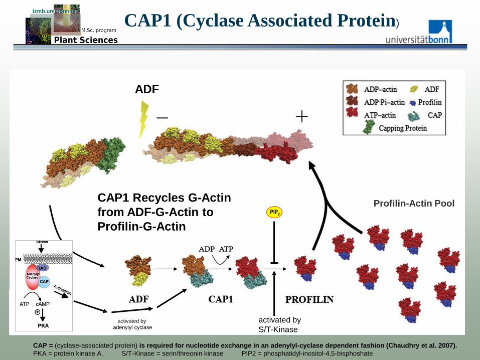
activated by
S/T-Kinase
CAP = (cyclase-associated protein) is required for nucleotide exchange in an adenylyl-cyclase dependent fashion (Chaudhry et al. 2007).
PKA = protein kinase A. S/T-Kinase = serin/threonin kinase PIP2 = phosphatidyl-inositol-4,5-bisphoshate
activated by
adenylyl cyclase
PIP2
ADF
Profilin-Actin Pool CAP1 Recycles G-Actin
from ADF-G-Actin to
Profilin-G-Actin
izmb.uni-bonn.de izmb.uni-bonn.de
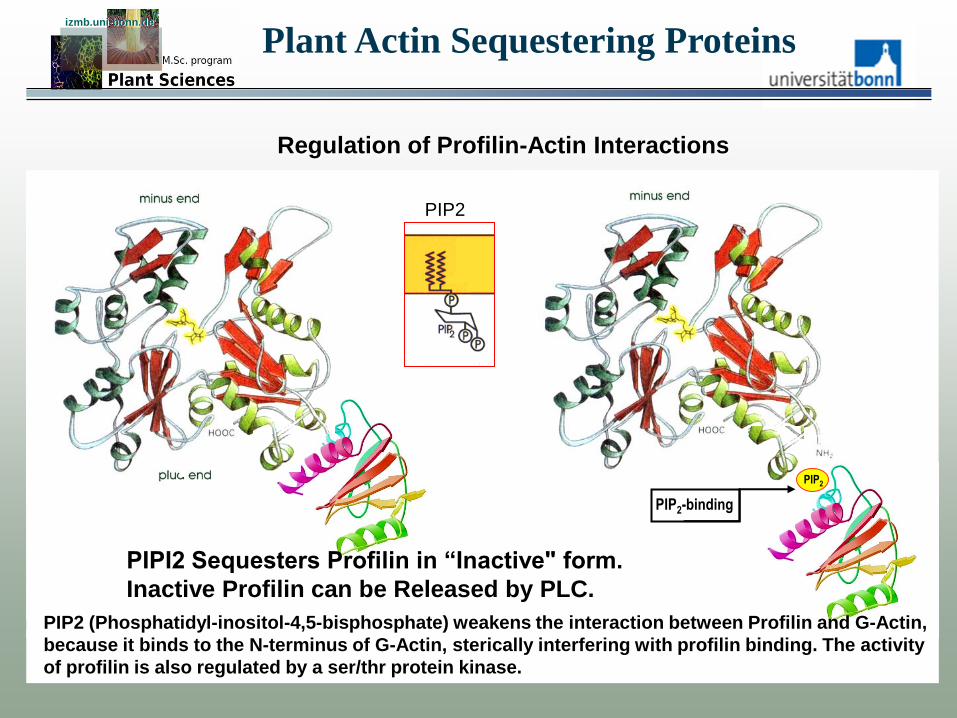
Regulation of Profilin-Actin Interactions
PIP2-binding
PIP2 (Phosphatidyl-inositol-4,5-bisphosphate) weakens the interaction between Profilin and G-Actin,
because it binds to the N-terminus of G-Actin, sterically interfering with profilin binding. The activity
of profilin is also regulated by a ser/thr protein kinase.
Plant Actin Sequestering Proteins
PIP2
PIP2
PIPI2 Sequesters Profilin in “Inactive" form.
Inactive Profilin can be Released by PLC.
Profilin izmb.uni-bonn.de izmb.uni-bonn.de
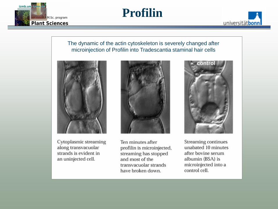
The dynamic of the actin cytoskeleton is severely changed after
microinjection of Profilin into Tradescantia staminal hair cells
control

Profilin izmb.uni-bonn.de izmb.uni-bonn.de
Completion of cytokinesis (fusion of the cell plate with the plasma membrane)
is inhibited after microinjection with profilin
control

Plant Actin-Binding Proteins izmb.uni-bonn.de izmb.uni-bonn.de
Number of gene copies in A. thaliana
• Fimbrin (F-actin crosslinking) 3 genes
• Profilin (G-actin sequestering) 5 genes
• ADF/cofilin (F-actin destabilizing) 11 genes
• AiP1 (barbed end binder, co-factor of ADF) 2 genes
• Villin (F-actin crosslinking) 5 genes
• Gelsolin (F-actin destabilizing) single copy gene
• Arp2/3 (F-actin branching) single copy genes
• Formin (F-actin binding, polymerization) 21 genes
Archer et al. 2005
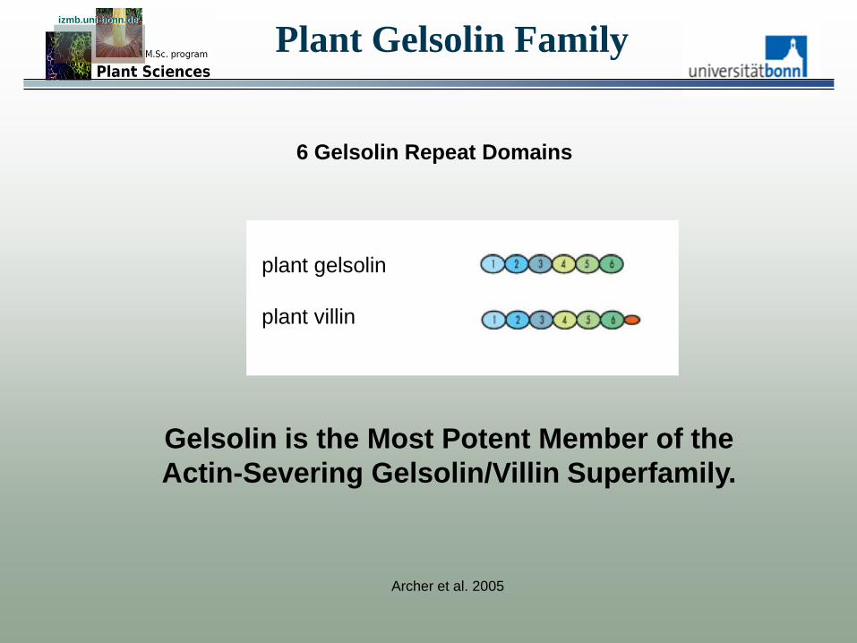
Plant Gelsolin Family izmb.uni-bonn.de izmb.uni-bonn.de
plant gelsolin
plant villin
6 Gelsolin Repeat Domains
Gelsolin is the Most Potent Member of the
Actin-Severing Gelsolin/Villin Superfamily.
Archer et al. 2005
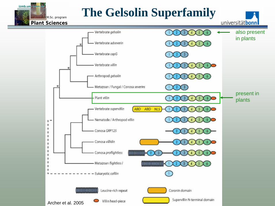
The Gelsolin Superfamily izmb.uni-bonn.de izmb.uni-bonn.de
also present
in plants
present in
plants
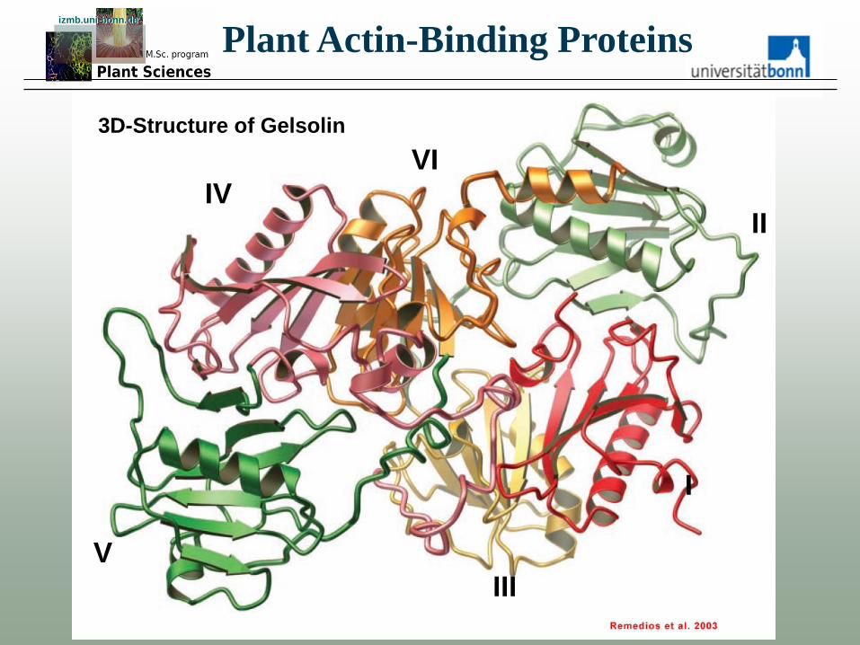
Plant Actin-Binding Proteins izmb.uni-bonn.de izmb.uni-bonn.de
I
II IV
V
VI
III
3D-Structure of Gelsolin

In vitro Assay of Plant Gelsolin Functions izmb.uni-bonn.de izmb.uni-bonn.de
Actin Actin plus gelsolin
Actin plus gelsolin plus 15nM Ca2+ Actin plus gelsolin plus 160nM Ca2+
Huang et al. 2004
F-actin is labelled with Rh-Phalloidin plant gelsolin = 80kDa protein
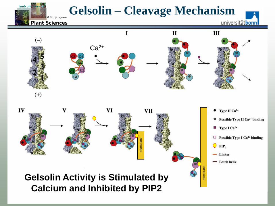
Gelsolin – Cleavage Mechanism izmb.uni-bonn.de izmb.uni-bonn.de
mem
bra
ne
mem
bra
ne
Ca2+
Gelsolin Activity is Stimulated by
Calcium and Inhibited by PIP2

Plant Actin-Binding Proteins izmb.uni-bonn.de izmb.uni-bonn.de
Number of gene copies in A. thaliana
• Fimbrin (F-actin crosslinking) 3 genes
• Profilin (G-actin sequestering) 5 genes
• ADF/cofilin (F-actin destabilizing) 9 genes
• AiP1 (barbed end binder, co-factor of ADF) 2 genes
• Villin (F-actin crosslinking) 5 genes
• Gelsolin (F-actin destabilizing) single copy gene
• Arp2/3 (F-actin branching) single copy genes
• Formin (F-actin binding, polymerization) 21 genes
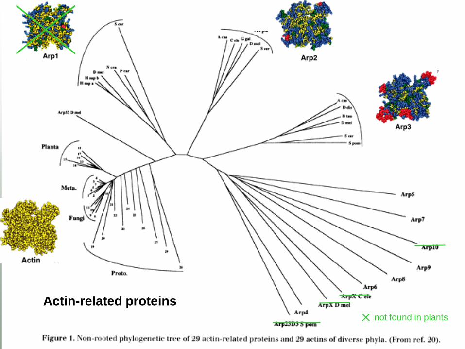
not found in plants
Actin-related proteins

Actin Filament Branching izmb.uni-bonn.de izmb.uni-bonn.de
P16 = crooked, Arabidopsis mutant
Activator of Arp2/3

izmb.uni-bonn.de izmb.uni-bonn.de
Svitkina & Borisy 1999
Actin Filament Branching
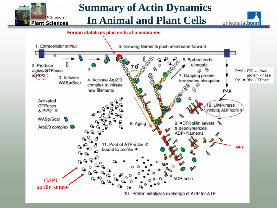
Summary of Actin Dynamics
In Animal and Plant Cells
izmb.uni-bonn.de izmb.uni-bonn.de
Pollard & Borisy
AiP1
Formin stabilizes plus ends at membranes
CAP1
ser/thr kinase
PAK = P21-activated
protein kinase
P21 = Rho-GTPase
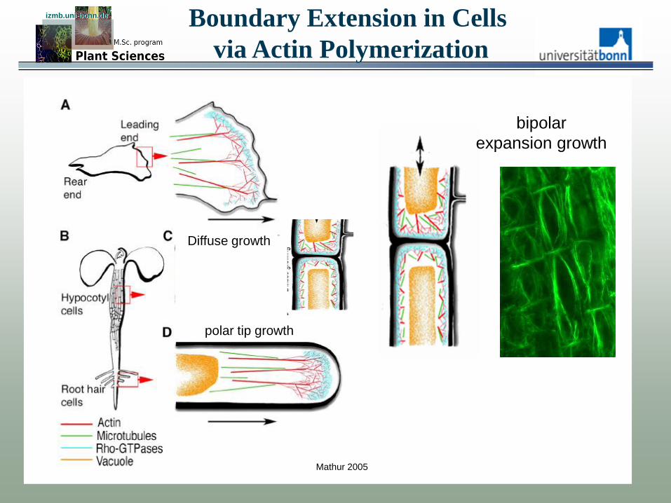
Boundary Extension in Cells
via Actin Polymerization
izmb.uni-bonn.de izmb.uni-bonn.de
Mathur 2005
bipolar
expansion growth
Diffuse growth
polar tip growth

izmb.uni-bonn.de izmb.uni-bonn.de
Mutations in proteins
of the ARP-complex
cause defects in cellular
morphogenesis
Boundary Extension in Cells
via Actin Polymerization
izmb.uni-bonn.de izmb.uni-bonn.de
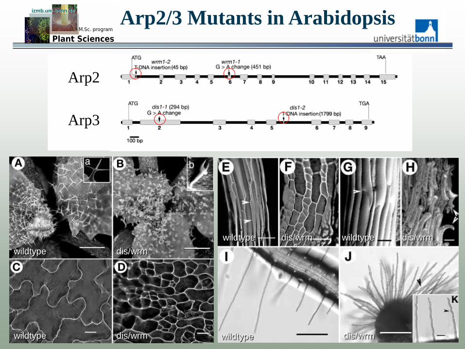
Arp2/3 Mutants in Arabidopsis
wildtype
wildtype wildtype
wildtype wildtype
dis/wrm
dis/wrm dis/wrm
dis/wrm dis/wrm
Arp2
Arp3

Arp2/3 Mutants in Arabidopsis izmb.uni-bonn.de izmb.uni-bonn.de
wrm
dis1-1
wt
wt
Actin cytoskeleton
in wild type leaf
trichomes
Arp2 mutant
Arp3 mutant

The brick Mutant in Zea mays izmb.uni-bonn.de izmb.uni-bonn.de
brick mutant wild type
brick mutant wild type Frank et al 2003
cell walls not interlocking
actin cytoskeleton
brick is an activator of the Arp2/3 complex
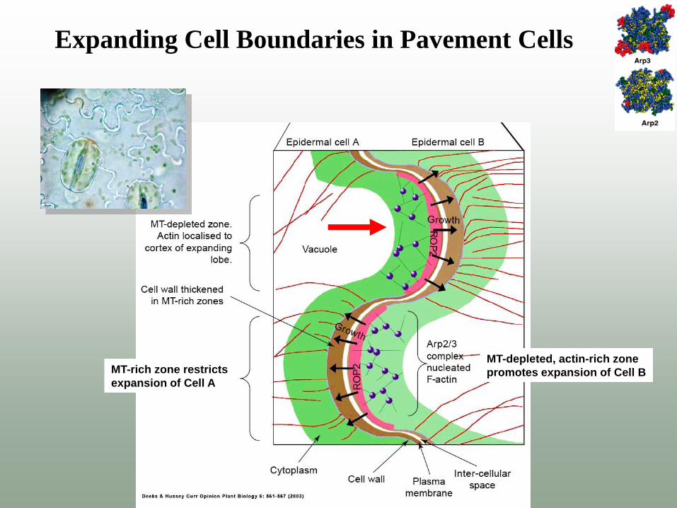
Expanding Cell Boundaries in Pavement Cells
MT-rich zone restricts
expansion of Cell A
MT-depleted, actin-rich zone
promotes expansion of Cell B
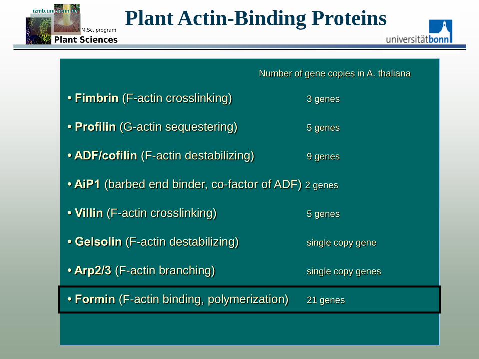
Plant Actin-Binding Proteins izmb.uni-bonn.de izmb.uni-bonn.de
Number of gene copies in A. thaliana
• Fimbrin (F-actin crosslinking) 3 genes
• Profilin (G-actin sequestering) 5 genes
• ADF/cofilin (F-actin destabilizing) 9 genes
• AiP1 (barbed end binder, co-factor of ADF) 2 genes
• Villin (F-actin crosslinking) 5 genes
• Gelsolin (F-actin destabilizing) single copy gene
• Arp2/3 (F-actin branching) single copy genes
• Formin (F-actin binding, polymerization) 21 genes
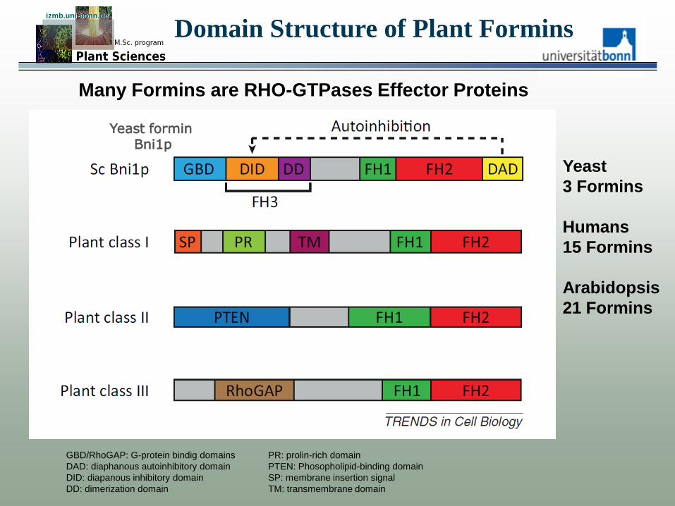
Domain Structure of Plant Formins izmb.uni-bonn.de izmb.uni-bonn.de
GBD/RhoGAP: G-protein bindig domains
DAD: diaphanous autoinhibitory domain
DID: diapanous inhibitory domain
DD: dimerization domain
Yeast formin Bni1p
PR: prolin-rich domain
PTEN: Phosopholipid-binding domain
SP: membrane insertion signal
TM: transmembrane domain
Many Formins are RHO-GTPases Effector Proteins
Yeast
3 Formins
Humans
15 Formins
Arabidopsis
21 Formins
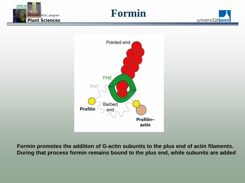
Formin izmb.uni-bonn.de izmb.uni-bonn.de
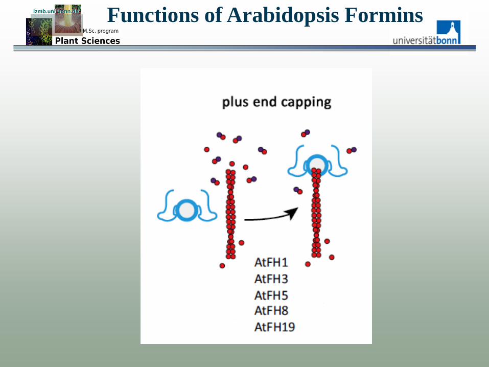
Formin promotes the addition of G-actin subunits to the plus end of actin filaments.
During that process formin remains bound to the plus end, while subunits are added

Formin izmb.uni-bonn.de izmb.uni-bonn.de
Tertiary Structure of the FH2-Domain
N
Association with the actin filament plus end
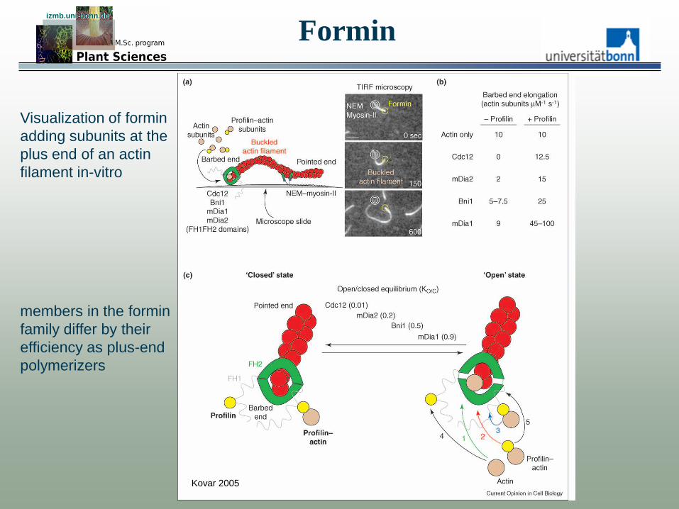
Visualization of formin
adding subunits at the
plus end of an actin
filament in-vitro
izmb.uni-bonn.de izmb.uni-bonn.de
Formin
Kovar 2005
members in the formin
family differ by their
efficiency as plus-end
polymerizers
Formin izmb.uni-bonn.de izmb.uni-bonn.de
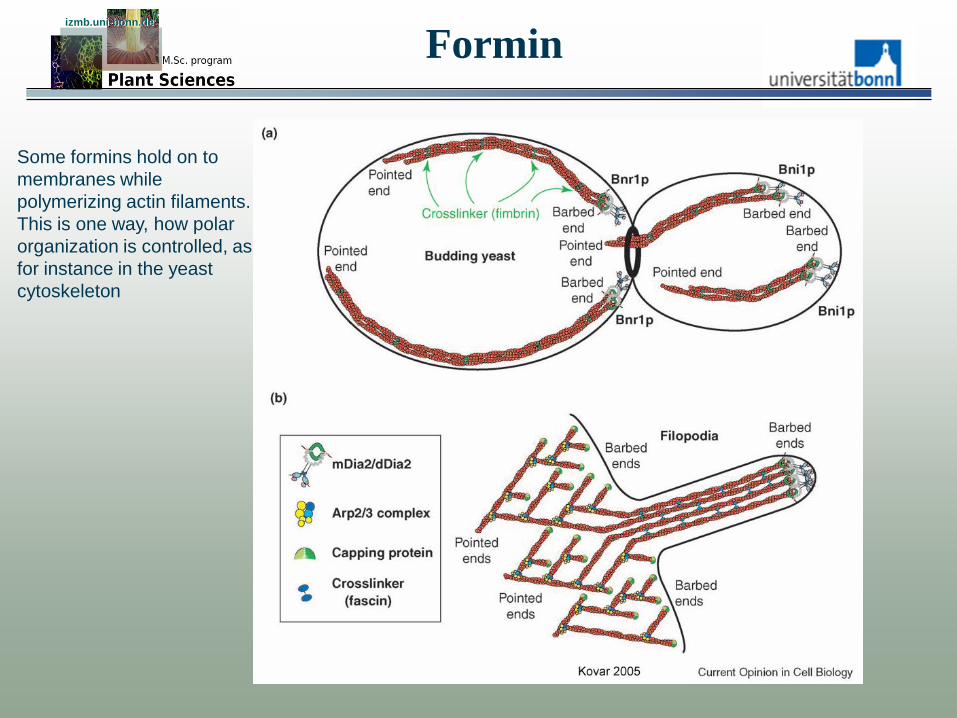
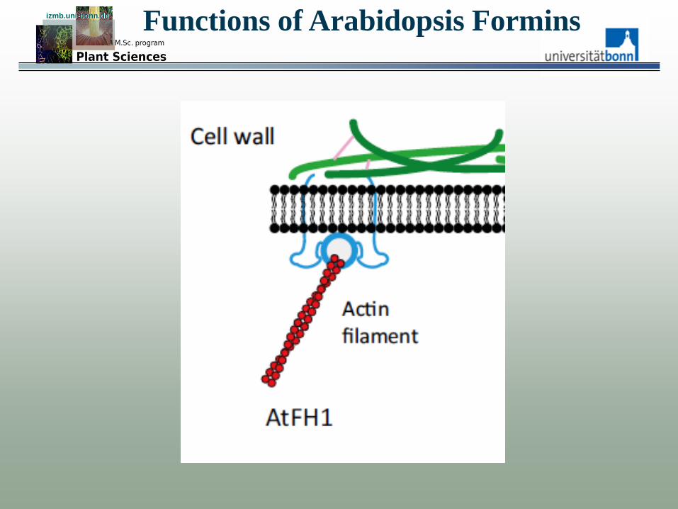
Some formins hold on to
membranes while
polymerizing actin filaments.
This is one way, how polar
organization is controlled, as
for instance in the yeast
cytoskeleton
Formin izmb.uni-bonn.de izmb.uni-bonn.de
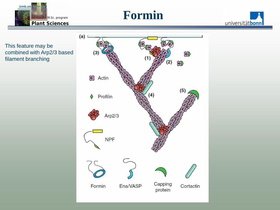
This feature may be
combined with Arp2/3 based
filament branching
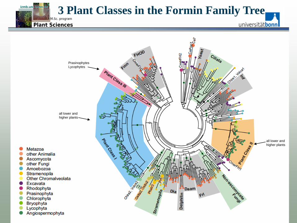
3 Plant Classes in the Formin Family Tree izmb.uni-bonn.de izmb.uni-bonn.de
Prasinophytes
Lycophytes
all lower and
higher plants
all lower and
higher plants

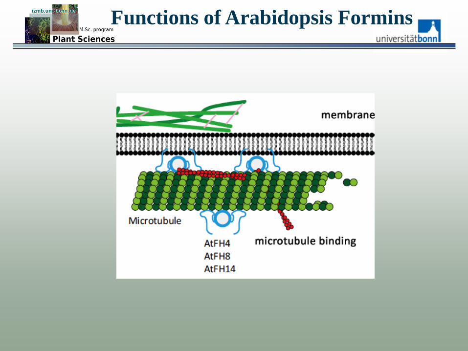
Functions of Arabidopsis Formins izmb.uni-bonn.de izmb.uni-bonn.de
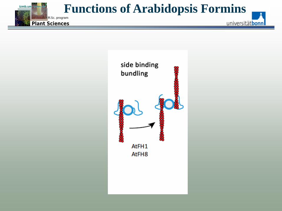
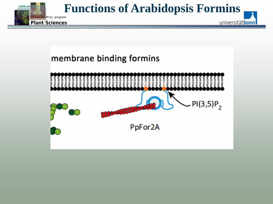
Functions of Arabidopsis Formins izmb.uni-bonn.de izmb.uni-bonn.de
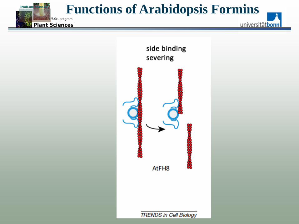
Functions of Arabidopsis Formins izmb.uni-bonn.de izmb.uni-bonn.de
Functions of Arabidopsis Formins izmb.uni-bonn.de izmb.uni-bonn.de
Functions of Arabidopsis Formins izmb.uni-bonn.de izmb.uni-bonn.de
Functions of Arabidopsis Formins izmb.uni-bonn.de izmb.uni-bonn.de
Functions of Arabidopsis Formins izmb.uni-bonn.de izmb.uni-bonn.de

Functions of Arabidopsis Formins izmb.uni-bonn.de izmb.uni-bonn.de
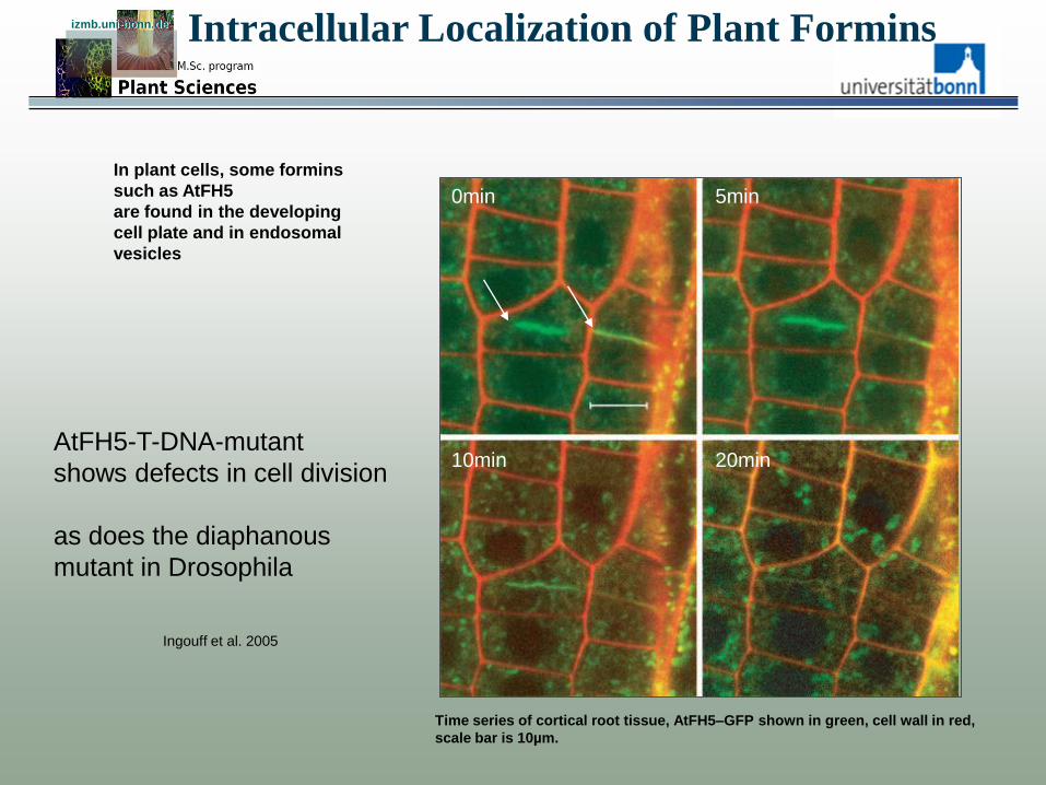
Intracellular Localization of Plant Formins izmb.uni-bonn.de izmb.uni-bonn.de
In plant cells, some formins
such as AtFH5
are found in the developing
cell plate and in endosomal
vesicles
Ingouff et al. 2005
Time series of cortical root tissue, AtFH5–GFP shown in green, cell wall in red,
scale bar is 10µm.
5min 0min
10min 20min AtFH5-T-DNA-mutant
shows defects in cell division
as does the diaphanous
mutant in Drosophila
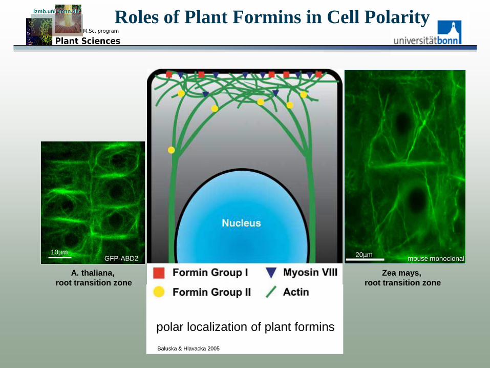
Roles of Plant Formins in Cell Polarity izmb.uni-bonn.de izmb.uni-bonn.de
polar localization of plant formins
Baluska & Hlavacka 2005
mouse monoclonal
A. thaliana,
root transition zone
GFP-ABD2 10µm 20µm
Zea mays,
root transition zone