Embed Size (px)
Citation preview
Phytotaxa 416 (1): 014–024https://www.mapress.com/j/pt/Copyright © 2019 Magnolia Press Article PHYTOTAXA
ISSN 1179-3155 (print edition)
ISSN 1179-3163 (online edition)
14 Accepted by Rajesh Jeewon: 25 Aug. 2019; published: 9 Sept. 2019
https://doi.org/10.11646/phytotaxa.416.1.2
Licensed under a Creative Commons Attribution License http://creativecommons.org/licenses/by/3.0
Taxonomy and biology of Cordyceps qingchengensis sp. nov. and its allies
LING-SHENG ZHA1,2, TING-CHI WEN3*, SHI-KE HUANG2, SARANYAPHAT BOONMEE2 & PRAPASSORN D. EUNGWANICHAYAPANT2
1School of Life Sciences, Huaibei Normal University, Huaibei 235000, China2Center of Excellence in Fungal Research, Mae Fah Luang University, Chiang Rai 57100, Thailand3Engineering Research Center of Southwest Bio–Pharmaceutical Resources, Ministry of Education, Guizhou University, Guiyang 550025, China*Corresponding author: [email protected]
Abstract
Cordyceps qingchengensis sp. nov., growing on a cocooned pupa of a silk moth (Lepidoptera: Bombycidae) and collected from southwestern China, is described, illustrated and compared with allied taxa. The species is morphologically similar to C. bifusispora and C. tenuipes, but can be easily separated from the latter two by the unique host and by branched and thicker stroma. Phylogenetic analyses of single ITS and combined SSU, LSU and TEF1-α datasets indicate that it is closely related to C. bifusispora, C. cicadae (Miq.) Massee (Chanhua) and C. tenuipes, but C. qingchengensis has distinct nucleotide differences which support it as new. Taxonomy of C. tenuipes and C. pruinosa is reviewed and C. ninchukispora (≡ Phytocordyceps ninchukispora) is considered as a synonym of C. pruinosa. Ecology and life cycles of C. qingchengensis, C. tenuipes, C. pruinosa and C. ningxiaensis are recorded and inferred. We provide important biological information for C. qingchengensis and its allies.
Keywords: Cordyceps ningxiaensis, Cordyceps pruinosa, Cordyceps tenuipes, life cycle, host, revision
Introduction
Cordyceps (Cordyceps sensu lato) fungi have always been highly regarded for their important edible and medicinal values and applications in biological control. Cordyceps can grow predominantly on insects, but also occur on spiders, nematodes, other cordyceps, the fungi Elaphomyces and even inhabit soil and plant tissues (Sung et al. 2007, Vega et al. 2009). This group currently has more than 900 species that belong to three families (Cordycipitaceae, Ophiocordycipitaceae and partial Clavicipitaceae) in the order Hypocreales (Zha et al. 2018). More than 140 species have been reported in China (Wen et al. 2017). The genus Cordyceps Fr. emend. G.H. Sung et al. (Cordycipitaceae) comprises 172 accepted species (Roskov et al. 2019). Due to a lack of molecular evidence or inconclusive morphology and ecology, more than 100 species are retained from the previous Cordyceps Fr. (Sung et al. 2007). Also, due to confusion of sexual and asexual morphs, many cordyceps species still have two or more names. Revision of sexual and asexual names and classification of undetermined Cordyceps species still need to be worked on. Occurrence of a cordyceps is closely related with ecological environment and the life cycle of its host. Factors affecting distribution are humidity (air and soil), temperature, light, rainfall, elevation, biogeography and the occurrence season (both the cordyceps and its host). Host information includes host name/group, the instar of death (egg, nymph/larva, pupae/cocoon, adult), the instar that becomes infected, locality of collection (in/on soil, humus layer, rotten wood, tree root or trunk, leaf or twig of plant, etc.) and the plant on which the insects reside (Wen et al. 2016). All this biological information is useful for cordyceps research. Unfortunately, biology of cordyceps has always been poorly studied, and this affects its identification, application and exploitation. We recently collected a new Cordyceps species from southwestern China. It is described as Cordyceps qingchengensis sp. nov., and is compared to allied species. In addition, we give taxonomic notes and biological information for related Cordyceps species.
CORDYCEPS QINGCHENGENSIS SP. NOV. AND ITS ALLIES Phytotaxa 416 (1) © 2019 Magnolia Press • 15
Materials and methods
SpecimensExplorations were made in China. Specimens were observed in the wild and brought to the laboratory for identification. Fungal specimens were examined and photographed using a Nikon Coolpix P520 camera, an Optec SZ660 stereo dissecting microscope and a Nikon Eclipse 80i compound microscope connected with a Cannon EOS 600D camera. Voucher specimens are deposited in Centre of Excellence in Fungal Research, Mae Fah Luang University (MFLU), Chiang Rai, Thailand, and the Herbarium of Guizhou University (GACP), Guiyang, China.
DNA extraction and sequencing Total DNA was extracted from specimens dried over silica-gel using a CTAB procedure (Doyle 1987). The ribosomal small and large subunits (SSU and LSU), internal transcribed spacers (ITS) and elongation factor 1α (TEF1-α) genes were amplified and sequenced using the primers detailed by White et al. (1990) and Ban et al. (2015). Amplification reactions were performed in an ABI 2720 thermal cycler (Applied Biosystems, Foster City, CA, USA) and PCR programs followed those of Ban et al. (2015). PCR products were purified using the Bioteke’s Purification Kit (Bioteke Corporation, Beijing, China), and were sequenced using an ABI 3730 DNA analyzer and an ABI BigDye 3.1 terminator cycle sequencing kit (Sangon Co., Ltd., Shanghai, China). Sequences were aligned and assembled visually and manually using Clustalx1.81 (Larkin et al. 2007), Chromas230 and ContigExpress software.
Construction of phylogenetic treePhylogenetic trees were constructed using sequences of Cordyceps qingchengensis sp. nov. and voucher sequences of its allies obtained from GenBank (Sung et al. 2007, Wang et al. 2008, Yan & Tolgor 2015, Kepler et al. 2017) (Table 1). Phylogeny was reconstructed using the single gene datasets of ITS, TEF1-α and finally a combined SSU, LSU and TEF1-α sequence dataset. Ophiocordyceps sinensis (Berk.) G.H. Sung et al. (EFCC 7287, Sung et al. 2007) was used as the outgroup taxon.
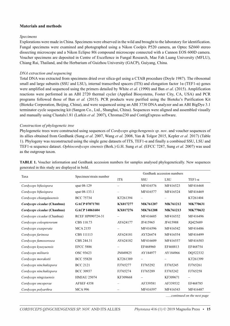
TABLE 1. Voucher information and GenBank accession numbers for samples analysed phylogenetically. New sequences generated in this study are displayed in bold.
Taxa Specimen/strain numberGenBank accession numbers
ITS SSU LSU TEF1-α
Cordyceps bifusispora spat 08-129 – MF416576 MF416523 MF416468
Cordyceps bifusispora spat 08-133.1 – MF416577 MF416524 MF416469
Cordyceps chiangdaoensis BCC 75734 KT261394 – – KT261404
Cordyceps cicadae (Chanhua) GACP 07071701 KX017277 MK761207 MK761212 MK770631
Cordyceps cicadae (Chanhua) GACP 14061604 KX017276 MK761208 MK761213 MK770632
Cordyceps cicadae (Chanhua) RCEF HP090724-31 – MF416605 MF416552 MF416496
Cordyceps coleopterorum CBS 110.73 AY624177 JF415965 JF415988 JQ425689
Cordyceps exasperata MCA 2155 – MF416596 MF416542 MF416486
Cordyceps farinosa CBS 111113 AY624181 AY526474 MF416554 MF416499
Cordyceps fumosorosea CBS 244.31 AY624182 MF416609 MF416557 MF416503
Cordyceps kyusyuensis EFCC 5886 – EF468960 EF468813 EF468754
Cordyceps militaris OSC 93623 JN049825 AY184977 AY184966 DQ522332
Cordyceps morakotii BCC 55820 KT261389 – – KT261399
Cordyceps ninchukispora BCC 2121 FJ765277 FJ765292 FJ765245 FJ765261
Cordyceps ninchukispora BCC 30937 FJ765274 FJ765289 FJ765242 FJ765258
Cordyceps ningxiaensis HMJAU 25074 KF309668 – KF309671 –
Cordyceps oncoperae AFSEF 4358 – AF339581 AF339532 EF468785
Cordyceps polyarthra MCA 996 – MF416597 MF416543 MF416487
......continued on the next page
ZHA ET AL.16 • Phytotaxa 416 (1) © 2019 Magnolia Press
TABLE 1. (Continued)
Taxa Specimen/strain numberGenBank accession numbers
ITS SSU LSU TEF1-α
Cordyceps pruinosa ARSEF 5413 JN049826 AY184979 AY184968 DQ522351
Cordyceps qingchengensis MFLU 17-1022 KY423506 MK761206 MK761211 MK770630
Cordyceps rosea spat 09-053 – MF416590 MF416536 MF416480
Cordyceps roseostromata ARSEF 4871 – AF339573 AF339523 –
Cordyceps roseostromata ARSEF 4870 EF368022 – – –
Cordyceps tenuipes MCA 1806 – MF416595 MF416541 MF416485
Cordyceps tenuipes ARSEF 5135 AY624196 MF416612 JF415980 JF416020
Cordyceps tenuipes GACP 16063004 KY423509 MK761209 MK761214 MK770633
Ophiocordyceps sinensis (outgroup) EFCC 7287 JN049854 EF468971 EF468827 EF468767
The single ITS and TEF1-α sequence datasets were analyzed using neighbor-joining (NJ) method (Saitou & Nei 1987), and the combined SSU, LSU and TEF1-α sequence dataset using maximum parsimony (MP) and maximum likelihood (ML) methods, respectively. NJ trees were performed with MEGA6 (Tamura et al. 2013) using the uncorrected p-distance method (Nei & Kumar 2000) with a bootstrap test of 1,000 replicates. ML tree was generated using RAxML v.8.2.8 employing a GTRGAMMA model of nucleotide substitution with other details described in Jeewon et al. (2002, 2003) and Hongsanan et al. (2017). MP tree was reconstructed with PAUP* 4.0b10 (Swofford 2002) and using the heuristic search option with TBR branch swapping, bootstrap of 1,000 replicates, and other details as outlined in Cai et al. (2006) and Tang et al. (2007).
Results
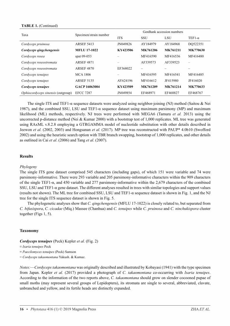
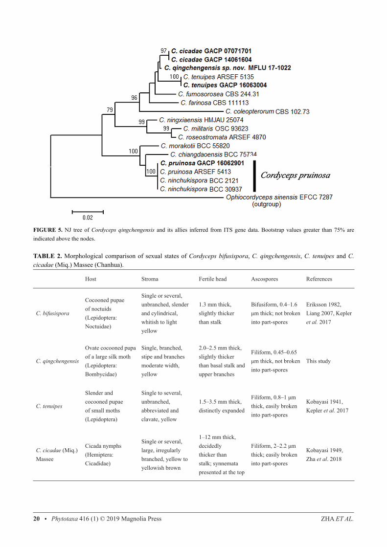
PhylogenyThe single ITS gene dataset comprised 545 characters (including gaps), of which 151 were variable and 74 were parsimony-informative. There were 293 variable and 205 parsimony-informative characters within the 909 characters of the single TEF1-α, and 450 variable and 277 parsimony-informative within the 2,679 characters of the combined SSU, LSU and TEF1-α gene dataset. The different analyses resulted in trees with similar topologies and support values (results not shown). The ML tree for combined SSU, LSU and TEF1-α sequence dataset is shown in Fig. 1, and the NJ tree for the single ITS sequence dataset is shown in Fig. 5. The phylogenetic analyses show that C. qingchengensis (MFLU 17-1022) is closely related to, but separated from C. bifusispora, C. cicadae (Miq.) Massee (Chanhua) and C. tenuipes while C. pruinosa and C. ninchukispora cluster together (Figs 1, 5).
Taxonomy
Cordyceps tenuipes (Peck) Kepler et al. (Fig. 2)≡ Isaria tenuipes Peck≡ Paecilomyces tenuipes (Peck) Samson= Cordyceps takaomontana Yakush. & Kumaz.
Notes:—Cordyceps takaomontana was originally described and illustrated by Kobayasi (1941) with the type specimen from Japan. Kepler et al. (2017) provided a photograph of C. takaomontana co-occurring with Isaria tenuipes. According to the information of the two reports above, C. takaomontana should grow on slender cocooned pupae of small moths (may represent several groups of Lepidoptera), its stromata are single to several, abbreviated, clavate, unbranched and yellow, and its fertile heads are distinctly expanded.
CORDYCEPS QINGCHENGENSIS SP. NOV. AND ITS ALLIES Phytotaxa 416 (1) © 2019 Magnolia Press • 17
FIGURE 1. ML tree of Cordyceps qingchengensis sp. nov. and its allies inferred from a combined SSU, LSU and TEF1-α dataset. Bootstrap support values greater than 70% are indicated above the nodes.
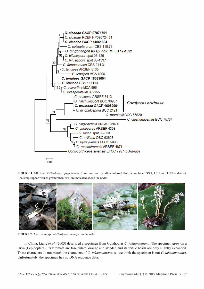
FIGURE 2. Asexual morph of Cordyceps tenuipes in the wild.
In China, Liang et al. (2003) described a specimen from Guizhou as C. takaomontana. The specimen grew on a larva (Lepidoptera), its stromata are fasciculate, orange and slender, and its fertile heads are only slightly expanded. These characters do not match the characters of C. takaomontana, so we think the specimen is not C. takaomontana. Unfortunately, the specimen has no DNA sequence data.
ZHA ET AL.18 • Phytotaxa 416 (1) © 2019 Magnolia Press
Another description of C. takaomontana from China was provided by Li et al. (2007) who collected a specimen from Anhui. The specimen grew on a large and nearly rounded (slightly ovate) cocooned pupae (Lepidoptera), its stromata were fasciculate, slender and light yellow, and its fertile heads were also only slightly expanded. Due to these different characters, we think this specimen is also not C. takaomontana. The specimen also lacks DNA sequence data. Though C. takaomontana is scarce, its asexual morph, I. tenuipes is quite common (Fig. 2). In the wild, I. tenuipes can readily be found on leaf litter or humus layer in humid environments. It generally grows on slender and cocooned pupae of numerous small moths (Lepidoptera), such as Arctiidae (Fig. 2). Old Arctiidae larvae move into shallow soil layer, or to soil surface and hide in dead leaves to pupate. During the process of pupation, probably due to continuous rainfall or very humid environment, these old larvae are easily infected by conidia of I. tenuipes that attach to their body surface. Isaria tenuipes grows rapidly and under suitable humidity, temperature and light, synnemata will soon be produced on the slender and cocooned pupae instead of the previous larvae. The insects are infected as old larvae and then die as cocooned pupae.
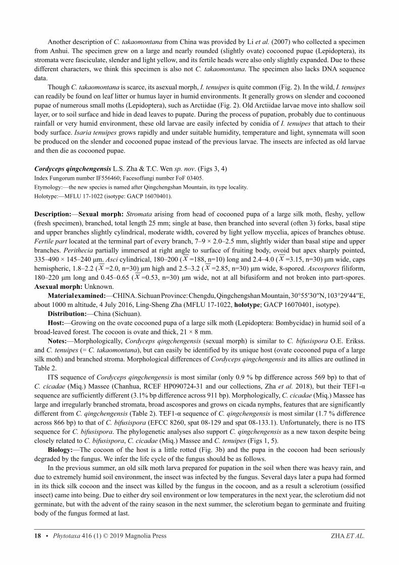
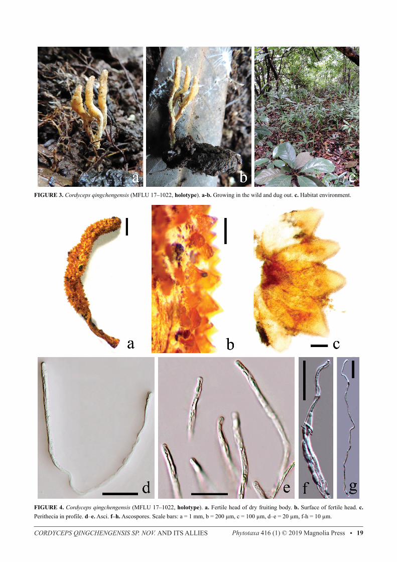
Cordyceps qingchengensis L.S. Zha & T.C. Wen sp. nov. (Figs 3, 4)Index Fungorum number IF556460; Facesoffungi number FoF 03405.Etymology:—the new species is named after Qingchengshan Mountain, its type locality.Holotype:—MFLU 17-1022 (isotype: GACP 16070401). Description:—Sexual morph: Stromata arising from head of cocooned pupa of a large silk moth, fleshy, yellow (fresh specimen), branched, total length 25 mm; single at base, then branched into several (often 3) forks, basal stipe and upper branches slightly cylindrical, moderate width, covered by light yellow mycelia, apices of branches obtuse. Fertile part located at the terminal part of every branch, 7–9 × 2.0–2.5 mm, slightly wider than basal stipe and upper branches. Perithecia partially immersed at right angle to surface of fruiting body, ovoid but apex sharply pointed, 335–490 × 145–240 μm. Asci cylindrical, 180–200 ( x =188, n=10) long and 2.4–4.0 ( x =3.15, n=30) μm wide, caps hemispheric, 1.8–2.2 ( x =2.0, n=30) μm high and 2.5–3.2 ( x =2.85, n=30) μm wide, 8-spored. Ascospores filiform, 180–220 μm long and 0.45–0.65 ( x =0.53, n=30) μm wide, not at all bifusiform and not broken into part-spores. Asexual morph: Unknown. Material examined:—CHINA. Sichuan Province: Chengdu, Qingchengshan Mountain, 30°55′30′′N, 103°29′44′′E, about 1000 m altitude, 4 July 2016, Ling-Sheng Zha (MFLU 17-1022, holotype; GACP 16070401, isotype). Distribution:—China (Sichuan). Host:—Growing on the ovate cocooned pupa of a large silk moth (Lepidoptera: Bombycidae) in humid soil of a broad-leaved forest. The cocoon is ovate and thick, 21 × 8 mm. Notes:—Morphologically, Cordyceps qingchengensis (sexual morph) is similar to C. bifusispora O.E. Erikss. and C. tenuipes (= C. takaomontana), but can easily be identified by its unique host (ovate cocooned pupa of a large silk moth) and branched stroma. Morphological differences of Cordyceps qingchengensis and its allies are outlined in Table 2. ITS sequence of Cordyceps qingchengensis is most similar (only 0.9 % bp difference across 569 bp) to that of C. cicadae (Miq.) Massee (Chanhua, RCEF HP090724-31 and our collections, Zha et al. 2018), but their TEF1-α sequence are sufficiently different (3.1% bp difference across 911 bp). Morphologically, C. cicadae (Miq.) Massee has large and irregularly branched stromata, broad ascospores and grows on cicada nymphs, features that are significantly different from C. qingchengensis (Table 2). TEF1-α sequence of C. qingchengensis is most similar (1.7 % difference across 866 bp) to that of C. bifusispora (EFCC 8260, spat 08-129 and spat 08-133.1). Unfortunately, there is no ITS sequence for C. bifusispora. The phylogenetic analyses also support C. qingchengensis as a new taxon despite being closely related to C. bifusispora, C. cicadae (Miq.) Massee and C. tenuipes (Figs 1, 5). Biology:—The cocoon of the host is a little rotted (Fig. 3b) and the pupa in the cocoon had been seriously degraded by the fungus. We infer the life cycle of the fungus should be as follows. In the previous summer, an old silk moth larva prepared for pupation in the soil when there was heavy rain, and due to extremely humid soil environment, the insect was infected by the fungus. Several days later a pupa had formed in its thick silk cocoon and the insect was killed by the fungus in the cocoon, and as a result a sclerotium (ossified insect) came into being. Due to either dry soil environment or low temperatures in the next year, the sclerotium did not germinate, but with the advent of the rainy season in the next summer, the sclerotium began to germinate and fruiting body of the fungus formed at last.
CORDYCEPS QINGCHENGENSIS SP. NOV. AND ITS ALLIES Phytotaxa 416 (1) © 2019 Magnolia Press • 19
FIGURE 3. Cordyceps qingchengensis (MFLU 17–1022, holotype). a-b. Growing in the wild and dug out. c. Habitat environment.
FIGURE 4. Cordyceps qingchengensis (MFLU 17–1022, holotype). a. Fertile head of dry fruiting body. b. Surface of fertile head. c. Perithecia in profile. d–e. Asci. f–h. Ascospores. Scale bars: a = 1 mm, b = 200 μm, c = 100 μm, d–e = 20 μm, f-h = 10 μm.
ZHA ET AL.20 • Phytotaxa 416 (1) © 2019 Magnolia Press
FIGURE 5. NJ tree of Cordyceps qingchengensis and its allies inferred from ITS gene data. Bootstrap values greater than 75% are indicated above the nodes.
TABLE 2. Morphological comparison of sexual states of Cordyceps bifusispora, C. qingchengensis, C. tenuipes and C. cicadae (Miq.) Massee (Chanhua).
Host Stroma Fertile head Ascospores References
C. bifusispora
Cocooned pupae of noctuids (Lepidoptera: Noctuidae)
Single or several, unbranched, slender and cylindrical, whitish to light yellow
1.3 mm thick, slightly thicker than stalk
Bifusiform, 0.4–1.6 μm thick; not broken into part-spores
Eriksson 1982, Liang 2007, Kepler et al. 2017
C. qingchengensis
Ovate cocooned pupa of a large silk moth(Lepidoptera: Bombycidae)
Single, branched, stipe and branches moderate width, yellow
2.0–2.5 mm thick, slightly thicker than basal stalk and upper branches
Filiform, 0.45–0.65 μm thick, not broken into part-spores
This study
C. tenuipes
Slender and cocooned pupae of small moths (Lepidoptera)
Single to several, unbranched, abbreviated and clavate, yellow
1.5–3.5 mm thick, distinctly expanded
Filiform, 0.8–1 μm thick, easily broken into part-spores
Kobayasi 1941, Kepler et al. 2017
C. cicadae (Miq.) Massee
Cicada nymphs (Hemiptera: Cicadidae)
Single or several, large, irregularly branched, yellow to yellowish brown
1–12 mm thick, decidedly thicker than stalk; synnemata presented at the top
Filiform, 2–2.2 μm thick; easily broken into part-spores
Kobayasi 1949, Zha et al. 2018
CORDYCEPS QINGCHENGENSIS SP. NOV. AND ITS ALLIES Phytotaxa 416 (1) © 2019 Magnolia Press • 21
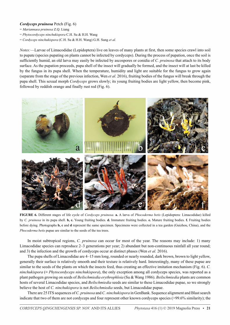
Cordyceps pruinosa Petch (Fig. 6)= Mariannaea pruinosa Z.Q. Liang= Phytocordyceps ninchukispora C.H. Su & H.H. Wang= Cordyceps ninchukispora (C.H. Su & H.H. Wang) G.H. Sung et al.
Notes:—Larvae of Limacodidae (Lepidoptera) live on leaves of many plants at first, then some species crawl into soil to pupate (species pupating on plants cannot be infected by cordyceps). During the process of pupation, once the soil is sufficiently humid, an old larva may easily be infected by ascospores or conidia of C. pruinosa that attach to its body surface. As the pupation proceeds, pupa shell of the insect will gradually be formed, and the insect will at last be killed by the fungus in its pupa shell. When the temperature, humidity and light are suitable for the fungus to grow again (separate from the stage of the previous infection, Wen et al. 2016), fruiting bodies of the fungus will break through the pupa shell. This sexual morph Cordyceps grows slowly; its young fruiting bodies are light yellow, then become pink, followed by reddish orange and finally rust red (Fig. 6).
FIGURE 6. Different stages of life cycle of Cordyceps pruinosa. a. A larva of Phocoderma betis (Lepidoptera: Limacodidae) killed by C. pruinosa in its pupa shell. b, c. Young fruiting bodies. d. Immature fruiting bodies. e. Mature fruiting bodies. f. Fruiting bodies before dying. Photographs b, c and d represent the same specimen. Specimens were collected in a tea garden (Guizhou, China), and the Phocoderma betis pupae are similar to the seeds of the tea trees.
In moist subtropical regions, C. pruinosa can occur for most of the year. The reasons may include: 1) many Limacodidae species can reproduce 2–3 generations per year; 2) abundant but non-continuous rainfall all year round; and 3) the infection and the growth of cordyceps occur at distinct phases (Wen et al. 2016). The pupa shells of Limacodidae are 4–15 mm long, rounded or nearly rounded, dark brown, brown to light yellow, generally their surface is relatively smooth and their texture is relatively hard. Interestingly, many of these pupae are similar to the seeds of the plants on which the insects feed, thus creating an effective imitation mechanism (Fig. 6). C. ninchukispora (≡ Phytocordyceps ninchukispora), the only exception among all cordyceps species, was reported as a plant pathogen growing on seeds of Beilschmiedia erythrophloia (Su & Wang 1986). Beilschmiedia plants are common hosts of several Limacodidae species, and Beilschmiedia seeds are similar to these Limacodidae pupae, so we strongly believe the host of C. ninchukispora is not Beilschmiedia seeds, but Limacodidae pupae. There are 25 ITS sequences of C. pruinosa and C. ninchukispora in GenBank. Sequence alignment and Blast search indicate that two of them are not cordyceps and four represent other known cordyceps species (>99.6% similarity); the
ZHA ET AL.22 • Phytotaxa 416 (1) © 2019 Magnolia Press
remaining 19 sequences (including one from Taiwan) are >99.5% similar and are probably the same species (the <0.5% base difference mainly comes from base insertions or gaps, which we think are errors during the process of sequence assembly). Cordyceps pruinosa was originally described from Sri Lanka (Petch 1924) and subsequently reported from Japan (Kobayasi 1941), China (Liang 2007), Korea and Thailand (Sung et al. 2007). Cordyceps ninchukispora was originally reported from Taiwan (Su & Wang 1986) and this epithet has been commonly used in Thailand. All these collections have the same macroscopic characters, the same hosts (Limacodidae pupae) and the same geographical distribution (south Asia). The phylogenetic analyses also support that the two species are conspecific (Figs 1, 5). For these reasons and following the suggestion of Sung et al. (2007), we herein synonymize C. ninchukispora to C. pruinosa. Their apparently different ascospores may be due to observations made at different stages of ascospore development — disarticulated (Petch 1924, Kobayasi 1941), bifusiform (Sung et al. 2007) and filiform at first then broken into disarticulated and bifusiform (Liang 2007).
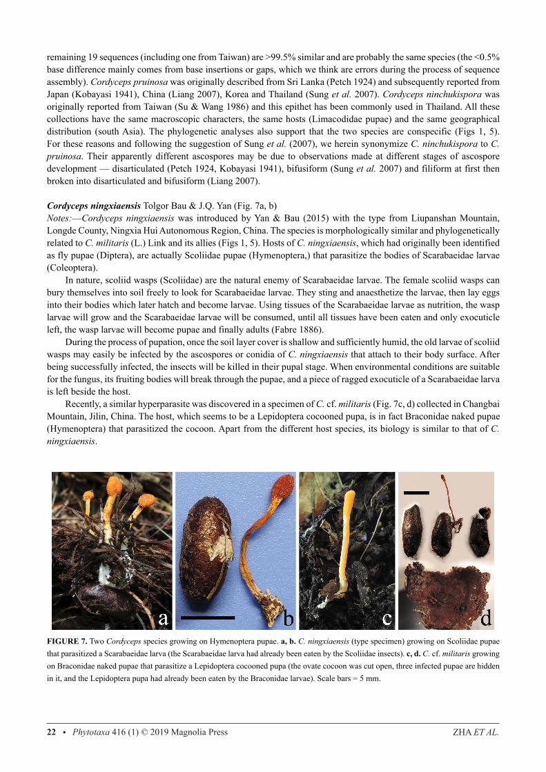
Cordyceps ningxiaensis Tolgor Bau & J.Q. Yan (Fig. 7a, b)Notes:—Cordyceps ningxiaensis was introduced by Yan & Bau (2015) with the type from Liupanshan Mountain, Longde County, Ningxia Hui Autonomous Region, China. The species is morphologically similar and phylogenetically related to C. militaris (L.) Link and its allies (Figs 1, 5). Hosts of C. ningxiaensis, which had originally been identified as fly pupae (Diptera), are actually Scoliidae pupae (Hymenoptera,) that parasitize the bodies of Scarabaeidae larvae (Coleoptera). In nature, scoliid wasps (Scoliidae) are the natural enemy of Scarabaeidae larvae. The female scoliid wasps can bury themselves into soil freely to look for Scarabaeidae larvae. They sting and anaesthetize the larvae, then lay eggs into their bodies which later hatch and become larvae. Using tissues of the Scarabaeidae larvae as nutrition, the wasp larvae will grow and the Scarabaeidae larvae will be consumed, until all tissues have been eaten and only exocuticle left, the wasp larvae will become pupae and finally adults (Fabre 1886). During the process of pupation, once the soil layer cover is shallow and sufficiently humid, the old larvae of scoliid wasps may easily be infected by the ascospores or conidia of C. ningxiaensis that attach to their body surface. After being successfully infected, the insects will be killed in their pupal stage. When environmental conditions are suitable for the fungus, its fruiting bodies will break through the pupae, and a piece of ragged exocuticle of a Scarabaeidae larva is left beside the host. Recently, a similar hyperparasite was discovered in a specimen of C. cf. militaris (Fig. 7c, d) collected in Changbai Mountain, Jilin, China. The host, which seems to be a Lepidoptera cocooned pupa, is in fact Braconidae naked pupae (Hymenoptera) that parasitized the cocoon. Apart from the different host species, its biology is similar to that of C. ningxiaensis.
FIGURE 7. Two Cordyceps species growing on Hymenoptera pupae. a, b. C. ningxiaensis (type specimen) growing on Scoliidae pupae that parasitized a Scarabaeidae larva (the Scarabaeidae larva had already been eaten by the Scoliidae insects). c, d. C. cf. militaris growing on Braconidae naked pupae that parasitize a Lepidoptera cocooned pupa (the ovate cocoon was cut open, three infected pupae are hidden in it, and the Lepidoptera pupa had already been eaten by the Braconidae larvae). Scale bars = 5 mm.
CORDYCEPS QINGCHENGENSIS SP. NOV. AND ITS ALLIES Phytotaxa 416 (1) © 2019 Magnolia Press • 23
Conflict of interest
The authors declare no conflicts of interest to disclose.
Acknowledgements
We sincerely thank Prof. Tolgor Bau (Institute of Mycology, Jilin Agriculture University, China) who provided specimens in the Herbarium for us to check. This work was supported by the National Natural Science Foundation of China (No. 31760014), and the Science and Technology Foundation of Guizhou Province (KY[2018]039, No. [2016]2863).
References
Ban, S., Sakane, T. & Nakagiri, A. (2015) Three new species of Ophiocordyceps and overview of anamorph types in the genus and the family Ophiocordyceptaceae. Mycological Progress 14: e1017.
https://doi.org/10.1007/s11557-014-1017-8Cai, L., Jeewon, R. & Hyde, K.D. (2006) Molecular systematics of Zopfiella and allied genera: evidence from multigene sequence
analyses. Mycological Research 110: 359–368. https://doi.org/10.1016/j.mycres.2006.01.007Doyle, J.J. (1987) A rapid DNA isolation procedure for small quantities of fresh leaf tissue. Phytochemical Bulletin 19 (1): 11–15. Eriksson, O. (1982) Cordyceps bifusispora spec. nov. Mycotaxon 15: 185–188. Fabre, J.H. (1886) Souvenirs Entomologiques, Livre III. Étude sur l’instinct et les mœurs des insectes. Hongsanan, S., Maharachchikumbura, S.S.N., Hyde, K.D., Samarakoon, M.C., Jeewon, R., Zhao, Q., Al-Sadi, A.M. & Bahkali, A.H.
(2017) An updated phylogeny of Sordariomycetes based on phylogenetic and molecular clock evidence. Fungal Diversity 84: 25–41.
https://doi.org/10.1007/s13225-017-0384-2Jeewon, R., Liew, E.C.Y., & Hyde, K.D. (2002) Phylogenetic relationships of Pestalotiopsis and allied genera inferred from ribosomal
DNA sequences and morphological characters. Molecular Phylogenetics and Evolution 25: 378-392. https://doi.org/10.1016/S1055-7903(02)00422-0Jeewon, R., Liew, E.C.Y., Simpson, J.A., Hodgkiss, I.J. & Hyde, K.D. (2003) Phylogenetic significance of morphological characters in the
taxonomy of Pestalotiopsis species. Molecular Phylogenetics and Evolution 27: 372–383. https://doi.org/10.1016/S1055-7903(03)00010-1Kepler, R.M., Luangsa-ard, J.J., Hywel-Jones, N.L., Quandt, C.A., Sung, G.H., Rehner, S.A., Aime, M.C., Henkel, T.W., Sanjuan, T.,
Zare, R., Chen, M., Li, Z., Rossman, A.Y., Spatafora, J.W. & Shrestha, B. (2017) A phylogenetically-based nomenclature for Cordycipitaceae (Hypocreales). IMA Fungus 8: 335–353.
https://doi.org/10.5598/imafungus.2017.08.02.08Kobayasi, Y. (1941) The genus Cordyceps and its allies. Science Reports of the Tokyo Bunrika Daigaku (Section B, No. 84) 5: 53–260.Kobayasi, Y. (1949) Several species of the genus Cordyceps and their conidial forms. Journal of Japanese Botany 24: 176–180.Larkin, M.A., Blackshields, G., Brown, N.P., Chenna, R., McGettigan, P.A., McWilliam, H. & Valentin, F. (2007) Clustal W and Clustal
X version 2.0. Bioinformatics 23: 2947–2948. https://doi.org/10.1093/bioinformatics/btm404Li, C.R., Zuo, D.P., Nam, S.H., Pu, S.C., Fan, M.Z. & Li, Z.Z. (2007) Cordyceps takaomontana and its anamorph Paecilomyces tenuipes.
Mycosystema 26 (2): 217–220. [in Chinese] https://doi.org/10.13346/j.mycosystema.2007.02.012Liang, Z.Q. (2007) Flora Fungorum Sinicorum, Vol. 32, Cordyceps. Science Press, Beijing. [in Chinese]Liang, Z.Q., Liu, A.Y., Liu, M.H. & Kang, J.C. (2003) The genus Cordyceps and its allies from the Kuankuoshui Reserve in Guizhou III.
Fungal Diversity 14: 95–101.Nei, M. & Kumar, S. (2000) Molecular Evolution and Phylogenetics. Oxford University Press, New York.Petch, T. (1924) Studies in entomogenous fungi, IV, some Ceylon Cordyceps. Transactions of the British Mycological Society 10 (1–2):
28–45. https://doi.org/10.1016/S0007-1536(24)80005-0
ZHA ET AL.24 • Phytotaxa 416 (1) © 2019 Magnolia Press
Roskov, Y., Ower, G., Orrell, T., Nicolson, D., Bailly, N., Kirk, P.M., Bourgoin, T., DeWalt, R.E., Decock, W., Nieukerken, E. van, Zarucchi, J. & Penev, L. (Eds.) (2019) Species 2000 & ITIS Catalogue of Life, 25th March 2019. Species 2000: Naturalis, Leiden, the Netherlands. Digital resource available from: www.catalogueoflife.org/col. (accessed 9 September 2019) [ISSN 2405-8858]
Saitou, N. & Nei, M. (1987) The neighbor-joining method: a new method for reconstructing phylogenetic trees. Molecular Biology and Evolution 4: 406–425.
https://doi.org/10.1093/oxfordjournals.molbev.a040454Samson, R.A. (1974) Paecilomyces and some allied hyphomycetes. Studies in Mycology 6: 1–119.Su, C.H. & Wang, H.H. (1986) Phytocordyceps, a new genus of the Clavicipitaceae. Mycotaxon 26: 337–344.Sung, G.H., Hywel-Jones, N.L., Sung, J.M., Luangsa-ard, J.J., Shrestha, B. & Spatafora, J.W. (2007) Phylogenetic classification of
Cordyceps and the clavicipitaceous fungi. Studies in Mycology 57: 5–69. https://doi.org/10.3114/sim.2007.57.01Swofford, D.L. (2002) PAUP*: Phylogenetic Analysis Using Parsimony (*and Other Methods), version 4.0b10. Sinauer Associates,
Sunderland.Tamura, K., Stecher, G., Peterson, D., Filipski, A. & Kumar, S. (2013) MEGA6: molecular evolutionary genetics analysis version 6.0.
Molecular Biology and Evolution 30: 2725–2729. https://doi.org/10.1093/molbev/mst197Tang, A.M.C., Jeewon, R. & Hyde, K.D. (2007) Phylogenetic utility of protein (RPB2, β-tubulin) and ribosomal (LSU, SSU) gene
sequences in the systematics of Sordariomycetes (Ascomycota, Fungi). Antonie van Leeuwenhoek 91: 327–349. https://doi.org/10.1007/s10482-006-9120-8Vega, F.E., Goettel, M.S., Blackwell, M., Chandler, D., Jackson, M.A., Keller, S., Koike, M., Maniania, N.K., Monzon, A., Ownley,
B.H., Pell, J.K., Rangel, D.E.N. & Roy, H.E. (2009) Fungal entomopathogens: new insights on their ecology. Fungal Ecology 2: 149–159.
https://doi.org/10.1016/j.funeco.2009.05.001Wang, L., Zhang, W.M., Hu, B., Chen, Y.Q. & Qu, L.H. (2008) Genetic variation of Cordyceps militaris and its allies based on phylogenetic
analysis of rDNA ITS sequence data. Fungal Diversity 31: 147–155.Wen, T.C., Zha, L.S., Hyde, K.D. & Kang, J.C. (2016) Some entomological issues in studying entomogenous fungi. Mycosystema 35 (11):
1303–1309. [in Chinese] https://doi.org/10.13346/j.mycosystema.150144Wen, T.C., Xiao, Y.P., Han, Y.F., Huang, S.K., Zha, L.S., Hyde, K.D. & Kang, J.C. (2017) Multigene phylogeny and morphology reveal
that the Chinese medicinal mushroom ‘Cordyceps gunnii’ is Metacordyceps neogunnii sp. nov.. Phytotaxa 302 (1): 27–39. https://doi.org/10.11646/phytotaxa.302.1.2White, T.J., Bruns, T., Lee, S. & Taylor, J.W. (1990) Amplification and direct sequencing of fungal ribosomal RNA genes for phylogenetics.
In: Innis, M.A., Gelfand, D.H., Sninsky, J.J. & White, T.J. (Eds.) PCR Protocols: a Guide to Methods and Applications. Academic Press, New York.
https://doi.org/10.1016/B978-0-12-372180-8.50042-1Yan, J.Q. & Tolgor, B. (2015) Cordyceps ningxiaensis sp. nov., a new species from dipteran pupae in Ningxia Hui Autonomous Region of
China. Nova Hedwigia 100 (1–2): 251–258. https://doi.org/10.1127/nova_hedwigia/2014/0222Zha, L.S., Huang, S.K., Xiao, Y.P., Boonmee, S., Eungwanichayapant, P.D., McKenzie, E.H.C., Kryukov, V., Wu, X.L., Hyde, K.D. &
Wen, T.C. (2018) An evaluation of common Cordyceps (Ascomycetes) species found in Chinese markets. International Journal of Medicinal Mushrooms 20 (12): 1149–1162.
https://doi.org/10.1615/IntJMedMushrooms.2018027330