Embed Size (px)
Citation preview

Phytotaxa 220 (3): 224–238www.mapress.com/phytotaxa/ Copyright © 2015 Magnolia Press Article PHYTOTAXA
ISSN 1179-3155 (print edition)
ISSN 1179-3163 (online edition)
224 Accepted by Kevin Hyde: 6 May 2015; published: 24 Jul. 2015
http://dx.doi.org/10.11646/phytotaxa.220.3.2
Licensed under a Creative Commons Attribution License http://creativecommons.org/licenses/by/3.0
Unravelling the diversity behind the Ophiocordyceps unilateralis (Ophiocordycipitaceae) complex: Three new species of zombie-ant fungi from the Brazilian Amazon
JOÃO P. M. ARAÚJO1*, HARRY C. EVANS2, DAVID M. GEISER3, WILLIAM P. MACKAY4 & DAVID P. HUGHES1, 5*
1 Department of Biology, Penn State University, University Park, Pennsylvania, United States of America.2 CAB International, E-UK, Egham, Surrey, United Kingdom3 Department of Plant Pathology, Penn State University, University Park, Pennsylvania, United States of America.4 Department of Biological Sciences, University of Texas at El Paso, 500 West University Avenue, El Paso, Texas, United States of America.5 Department of Entomology, Penn State University, University Park, Pennsylvania, United States of America. * email: [email protected]; [email protected]
Abstract
In tropical forests, one of the most commonly encountered relationships between parasites and insects is that between the fungus Ophiocordyceps (Ophiocordycipitaceae, Hypocreales, Ascomycota) and ants, especially within the tribe Campono-tini. Here, we describe three newly discovered host-specific species, Ophiocordyceps camponoti-atricipis, O. camponoti-bispinosi and O. camponoti-indiani, on Camponotus ants from the central Amazonian region of Brazil, which can readily be separated using morphological traits, in particular the shape and behavior of the ascospores. DNA sequence data support inclusion of these species within the Ophiocordyceps unilateralis complex.
Introduction
In tropical forests, social insects (ants, bees, termites and wasps) are the most abundant land-dwelling arthropods. Although they represent only 2% of the nearly 900,000 known insect species on Earth, they are estimated to compose more than half of the biomass (Fittkau & Klinge 1973; Höldobler & Wilson 2009). One of the better known members within this group are the ants, which form a single family (Formicidae), with close to 13,000 species described (Agosti & Johnson 2009). Ants occupy a wide range of habitats from high canopy to the leaf litter, forming huge colonies comprising tens to hundreds of thousands to millions of individuals. Ants are associated with and susceptible to a variety of parasites. Amongst these, one group is particularly well adapted to live in tropical forests and to exploit this ant abundance, the entomopathogenic fungi of the genus Ophiocordyceps (Hypocreales; Ophiocordycipitaceae), currently comprising around 160 species (Robert et al. 2005; Sung et al. 2007). These parasites infect many different insects with a wide range of ecologies, from solitary wandering beetles to highly-organized ant societies. The orders infected include Blattaria, Coleoptera, Dermaptera, Diptera, Hemiptera, Hymenoptera, Isoptera, Lepidoptera, Mantodea, Odonata and Orthoptera (Araújo & Hughes bioRxiv). The functional morphology of Ophiocordyceps is equally diverse and has been linked to the host’s ecology and biology (Evans et al. 2011). Species of Ophiocordyceps were originally placed within Cordyceps, a genus established to accommodate fungal pathogens of arthropods bearing the sexual spore-producing structures (ascoma) on conspicuous stalks (stroma), arising from the host cadaver (Evans et al. 2011). However, due to the polyphyletic nature of Cordyceps—as evidenced by recent phylogenetic studies—species formerly assigned to the genus have now been reorganized into four genera (Cordyceps, Elaphocordyceps (currently Tolypocladium, see Quandt et al. 2014), Metacordyceps and Ophiocordyceps), within three families (Cordycipitaceae, Clavicipitaceae and Ophiocordycipitaceae) (Sung et al. 2007). Within the Formicidae, Ophiocordyceps infections have been reported from the basal primitive groups (Ponerines) through to modern genera, such as Camponotus (Evans & Samson, 1982; Sanjuan et al. 2001; Evans et al. 2011), and often occur as epizootics, killing large number of ants in small patches of forest (Andersen et al. 2009; Pontoppidan et al. 2009). Such events are pan-tropical with records from Asia, Australasia, Africa and the Americas (Evans 1974;
THREE NEW SPECIES FROM THE BRAZI Phytotaxa 220 (3) © 2015 Magnolia Press • 225
Andrade 1980; Evans & Samson 1982, 1984; Evans 1988a, 2001; Kepler et al. 2011; Luangsa-ard et al. 2011; Kobmoo et al. 2012). Entomopathogenic fungi infect their hosts following spore contact and subsequent germination on the cuticle. Typically, an adhesive pad (appressorium) is formed, whereby a germ tube penetrates the exoskeleton of the host. Upon reaching the haemocoel, the fungus proliferates in the form of yeast-like cells (Evans 1988b). In the O. unilateralis complex, a series of synchronized events are triggered within the ant host in order to make it leave the colony, climb understorey shrubs to die in an elevated position—characteristically, biting the underside or edge of a leaf—and, once there, a spore-producing structure arises from the back of its head from which the ascospores are forcibly released at maturity (Andersen et al. 2009). The taxonomy and evolutionary relationships of this important group of pathogens remain unclear. For many years it was suspected that O. unilateralis, originally described as Torrubia unilateralis (Tulasne & Tulasne 1865), represents a complex of species, based on macro-morphological variation within collections worldwide (Petch 1931; Kobayasi 1941; Mains 1958; Samson et al. 1982; Evans & Samson 1984). However, it was not until recently that species delimitation was proposed formally, when four new taxa were described from the Minas Gerais state of Brazil (Evans et al. 2011), and it was posited that each ant within the Camponotini would host a different species of Ophiocordyceps. Subsequent descriptions of new taxa, from Thailand and Japan on both Camponotus and Polyrhachis hosts, are lending support to this hypothesis (Luangsa-ard et al. 2011; Kepler et al. 2011; Kobmoo et al. 2012)—and, even more recently, from the USA (de Bekker et al. 2014)—with the clear indication that there are still many species to be discovered. During field surveys in the central Brazilian Amazon, we collected a range of Camponotus species killed by Ophiocordyceps, often in large numbers. Based on macro-morphological characters, all the specimens fell within O. unilateralis sensu lato: typically, comprising a stalk (stroma) arising from the dorsal anterior part of the pronotum. A Hirsutella-like asexual morph occupies the terminal region and the sexual morph (ascoma) occurring as lateral cushions or plates. Here, we describe and characterize three new species from field-collected material, which have distinct micro-morphological characters (Kobayasi 1941; Evans et al. 2011). We also performed PCR and ribosomal DNA (rDNA) sequence analyses of DNA extracted directly from the field collected material. Overall these analyses yielded inferences consistent with placement of these species in the O. unilateralis clade, but different rDNA regions produced different phylogenetic results.
Materials and Methods
Sampling
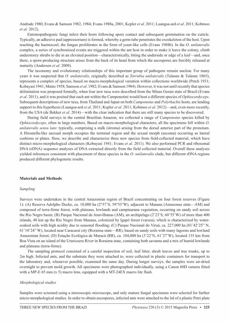
Surveys were undertaken in the central Amazonian region of Brazil concentrating on four forest reserves (Figure 1): (A) Reserva Adolpho Ducke, ca. 10,000 ha (2°57’S, 59°55’W), adjacent to Manaus (Amazonas state—AM) and composed of terra-firme forest, with plateaus, lowlands and campinarana vegetation, occurring on sandy soil across the Rio Negro basin; (B) Parque Nacional de Anavilhanas (AM), an archipelago (2˚23’S, 60˚55’W) of more than 400 islands, 40 km up the Rio Negro from Manaus, colonized by Igapó forest (varzea), which is characterized by water-soaked soils with high acidity due to seasonal flooding; (C) Parque Nacional do Viruá, ca. 227,000 ha (01˚42’25” N, 61˚10’24” W), located near Caracaraí city (Roraima state—RR), based on sandy soils with many lagoons and lowland Amazonian forest; (D) Estação Ecológica de Maracá (RR), ca. 104,000 ha (3˚22’N, 61˚27’W), located 135 km from Boa Vista on an island of the Uraricoera River in Roraima state, containing both savanna and a mix of humid lowlands and plateaus (terra-firme). The sampling protocol consisted of a careful inspection of soil, leaf litter, shrub leaves and tree trunks, up to 2m high. Infected ants, and the substrata they were attached to, were collected in plastic containers for transport to the laboratory and, whenever possible, examined the same day. During longer surveys, the samples were air-dried overnight to prevent mold growth. All specimens were photographed individually, using a Canon 60D camera fitted with a MP-E 65 mm (x 5) macro lens, equipped with a MT-24EX macro lite flash.
Morphological studies
Samples were screened using a stereoscopic microscope, and only mature fungal specimens were selected for further micro-morphological studies. In order to obtain ascospores, infected ants were attached to the lid of a plastic Petri plate
ARAÚJO ET AL.226 • Phytotaxa 220 (3) © 2015 Magnolia Press
(9-cm diam) using petroleum jelly, and suspended above a plate containing either distilled water agar (DWA) or potato-dextrose agar (PDA). Plates were transferred to covered stands placed on the forest, subject to natural temperature and light fluctuations and examined daily for the presence of ascospores, which, after ejection from the ascomata, formed sub-hyaline halos on the agar surface. Freshly deposited ascospores were removed with a sterile hypodermic needle under a stereoscopic microscope, and mounted on a slide in lactofuchsin (0.1g of acid fuchsin in 100 mL of lactic acid) for light microscopy (Olympus BX61). A minimum of 50 naturally-released (mature) ascospores were measured for morphological comparison (Table 1). The remaining ascospores were left in situ on the agar surface and examined over a number of days in order to monitor germination events. For micro-morphology of the ascomata, either free-hand or cryo-sectioning (Leica CM1950 Cryostat) was used. Permanent slides were deposited in the Entomological Collection at INPA (Instituto Nacional de Pesquisas da Amazônia, Manaus), with isotype collections held in Frost Entomological Museum at PSU (Penn State University). Permits for collecting and export were provided by SISBIO to JPMA (Authorizations 40496-1; 32435-1; 32410-1).
FIGURE 1. Map showing the forest reserves sampled in the central Brazilian Amazon: A) Reserva Adolpho Ducke; B) Parque Nacional de Anavilhanas; C) Parque Nacional do Viruá; D) Estação Ecológica de Maracá.
DNA extraction, PCR, sequencing and phylogenetic analyses
DNA was extracted from small pieces of the ascoma/stroma. The tissue was placed with two metal balls (1/8’’) in a 2 mL Eppendorf tube, frozen in liquid nitrogen and broken into powder using a Tissuelyzer (Qiagen) for 1 min. Afterwards, with the fungal tissue still frozen, 0.8 ml of DNA extraction buffer at 50°C (1% SDS, 0.024 g/mL PAS, 0.2 mL/mL RNB, Distilled water) and 0.8 mL phenol-chloroform was added, incubated at 60°C for 10 min and centrifuged for 10 min at 10,000 rpm. The water phase was transferred to a new 2 mL tube (about 0.8 mL), 0.5 mL of chloroform
THREE NEW SPECIES FROM THE BRAZI Phytotaxa 220 (3) © 2015 Magnolia Press • 227
was added, mixed by inverting and centrifuged for 10 min at 10,000 rpm. The supernatant was transferred to a new tube, to which an 80% volume of isopropanol added, mixed by inverting and centrifuged for 10 min at 14,000 rpm. The supernatant was removed and the pellet was washed with 0.5 mL of 70% ethanol (Koptec), air-dried and resuspended in 30 μL of TE buffer. Approximately 1,030 bp of nu-SSU (using 2 overlapping sets of primers, NS1/SR7 and NS4/NS3; White et al. 1990), 770 bp of nu-LSU (primer pair LR5/LR0R; Vilgalys and Sun 1994) and 550 bp of ITS (primers ITS1F/ITS4; Gardes and Bruns, 1993) were amplified by PCR. PCR was performed using a T-3000 Thermocycler (Biometra GmbH, Göttingen, Germany), in 25 uL reactions on the final concentration of: 1 × PCR buffer, 0.2 μM dNTP (each), 1.5 mM MgCl2, 0.2 μM each primer, 1 μL genomic DNA template (concentration varies according to different samples), 1 unit Platinum Taq DNA Polymerase (Invitrogen) and 18.4 μL Gibco UltraPure Distilled Water. PCR products were cleaned using the QIAquick PCR Purification Kit (Qiagen, Venlo, Netherlands) following the manufacturer’s instructions and sequenced by Sanger DNA sequencing (Applied Biosystems 3730XL) at Genomics Core Facility service at Penn State University. The raw ABI chromatograms were manually edited using Sequencher version 4.7 (Gene Codes Corp., Ann Arbor, MI, USA). Sequences generated for this study were compared with closely related species sequences identified in GenBank by BLAST. The alignment was obtained using MUSCLE in MEGA v.5.X (Kumar et al. 2012), and gaps were treated as missing data. All phylogenetic analyses were performed using MEGA v.5.X (Kumar et al. 2012). The Tamura-Nei (Tamura & Nei, 1992) model of molecular evolution was selected for ML analyses. ML analyses were performed using ten random taxon-additions and Nearest Neighbor Interchange (NNI) branch swapping. The MP analyses were run using ten random taxon-addition replicates and Subtree-Pruning-Regrafting (SPR) branch swapping. Clade support was assessed by bootstrapping, employing 500 pseudoreplicates of the data on both ML and MP analyses. The GenBank assession numbers and Herbarium voucher information for each taxon are provided in Table 2.
TABLE 1. Comparison of main morphological characters of the new Ophiocordyceps species and closely related species (*New species; **Septation).
Species Host Stroma (mm)Capilliconidiophore length (μm)
Ascospore dimension (μm)
Hirsutella Type
A B C
O. camponoti-atricipis* Camponotus atriceps 15–20 (–25) 55 80–85 × 3 **(5) + - -
O. camponoti-bispinosi* C. bispinosus 5–7 65 70–75 × 4.5 (4–5)
+ - -
O. camponoti-indiani* C. indianus 8–10 (–16) 130 75 × 4.5 (5) + - +
O. camponoti-rufipedis C. rufipes 5–8 (–15) 60–70 (–80) 80–95–2–3 (4–7) + - -
O. camponoti-balzani C. balzani similar to O.c-rufipedis - 135–175 × 4–5 (14–22)
+ - +
O. camponoti-melanotici
C. melanoticus similar to O.c-rufipedis - 170–210 × 4–5 (27–35)
+ - -
O. camponoti-novogranadensis
C. novogranadensis similar to O.c-rufipedis 20–25 75–95 × 2.5–3.5 (5–10)
+ + -
O. halabalaensis C. gigas 6.5–18 - 60–75 × 3–5(“multi-septate”)
+ - -
Results
Taxonomy
Ophiocordyceps camponoti-atricipis Araújo, H.C. Evans & D.P. Hughes, sp. nov. (Fig.2)IF 550743.
Differs from other members of the O. unilateralis clade by the host (always Camponotus atriceps), ascospore morphol-ogy and germination. Stroma usually velvety at the basal part when mature.
Type:—BRAZIL. Amazonas: Reserva Adolpho Ducke, 2° 57’ 42”S, 59° 55’ 40” W, 100 m, on Camponotus atriceps F. Smith, 10 January 2012 J.P.M. Araújo & H.C. Evans A-56, (INPA 261937, holotype!,FEM 90326, isotype!).
ARAÚJO ET AL.228 • Phytotaxa 220 (3) © 2015 Magnolia Press
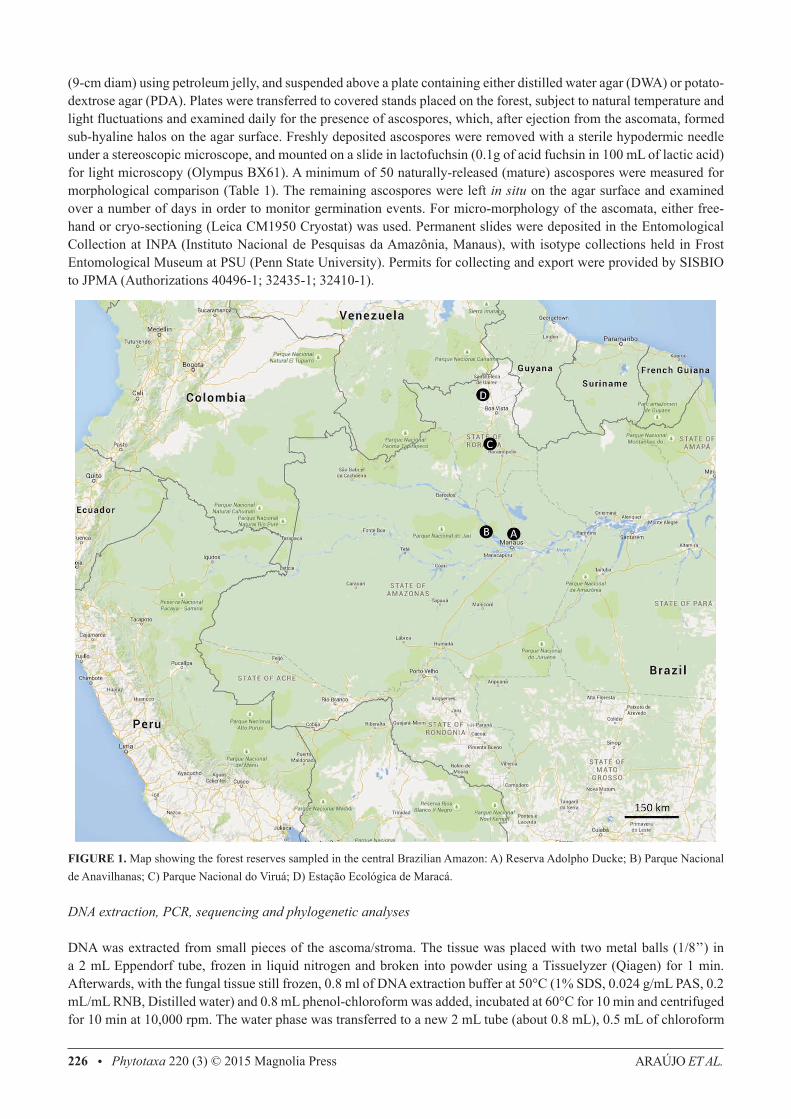
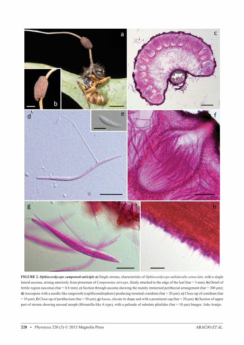
FIGURE 2. Ophiocordyceps camponoti-atricipis a) Single stroma, characteristic of Ophiocordyceps unilateralis sensu lato, with a single lateral ascoma, arising anteriorly from pronotum of Camponotus atriceps, firmly attached to the edge of the leaf (bar = 3 mm); b) Detail of fertile region (ascoma) (bar = 0.8 mm); c) Section through ascoma showing the mainly immersed perithecial arrangement (bar = 200 μm); d) Ascospore with a needle-like outgrowth (capilliconidiophore) producing terminal conidium (bar = 20 μm); e) Close-up of conidium (bar = 10 μm); f) Close-up of perithecium (bar = 50 μm); g) Ascus, clavate in shape and with a prominent cap (bar = 20 μm); h) Section of upper part of stroma showing asexual morph (Hirsutella-like A type), with a palisade of subulate phialides (bar = 10 μm) Images: João Araújo.
THREE NEW SPECIES FROM THE BRAZI Phytotaxa 220 (3) © 2015 Magnolia Press • 229
External mycelium abundant, covering most of the host, produced from all orifices and sutures; initially white, turning light-brown. Stroma single, produced from dorsal pronotum, averaging 15–20 mm, up to 25 mm in length, cylindrical, velvety and ginger brown at the base, becoming cream-pinkish towards apex; fertile region of lateral cushions, 1–2, hemispherical, chocolate brown, darkening with age, variable in size, averaging 1.5 × 0.5-0.8 mm. Perithecia immersed to partially erumpent, flask-shaped, 240–280 × 100–150 μm, with short, exposed neck or ostiole. Asci 8-spored, hyaline, cylindrical to clavate, 110–140 × (4.5–) 6–6.5 (–8) μm; prominent cap, (3.5–) 5 × 5.5 (–6.5) μm. Ascospores hyaline, thin-walled, vermiform (75–) 80–85 (–100) × (2–) 3 (–3.5) μm, 5-septate, sinuous to curved, never straight at maturity, rounded to acute apex. Etymology: Named after the ant host, Camponotus (Myrmothrix) atriceps F. Smith. Asexual-morph:— Hirsutella-like A type only, produced on the upper stromatal surface; phialides cylindrical to lageniform, 5–7 × 2–3 μm, tapering to a long neck, 5–11 μm; conidia not seen. This asexual morph occurs in all the species included here and is not considered to be critical for species separation. Hence, it is not analyzed in detail. Germination process:—The released ascospores germinated within 24–48 h, producing 1–2, uniformly straight, thread-like structures (capilliconidiophores); typically of uniform length and averaging 55 μm; bearing a single terminal spore (capilliconidium), hyaline, smooth-walled, allantoid and tapering at the ends at maturity, 10–11 × 2–2.5 μm, narrowing apically. Habitat:—Brazilian Central Amazon. Hosts commonly found biting the apical part of palm-fronds, but also on dicot leaves, rarely on palm-spines; abundant mycelial growth from the mouthparts helping to fix the host to the leaf, in addition to the locked jaws. Additional specimens examined (paratypes):—BRAZIL. Reserva Adolpho Ducke: Manaus, 2° 57’ 42”S, 59° 55’ 40” W, 100m elevation, 12 January 2012, Araújo, H.C. Evans & D.P. Hughes, A35 (INPA 261940!).
Ophiocordyceps camponoti-bispinosi Araújo, H.C. Evans & D.P. Hughes, sp. nov. (Fig.3)IF 550744.
Differs from other members of the O. unilateralis clade by the host (always Camponotus bispinosus), ascospore mor-phology and germination. Club-shaped terminal part of the stroma.
Type:—BRAZIL. Amazonas: Reserva Adolpho Ducke, 2° 57’ 42” S, 59° 55’ 40” W, 100 m, on Camponotus bispinosus Mayr, 15 January 2012, J.P.M. Araújo & H. C. Evans B-77, (INPA (261936, holotype!, FEM 90327, isotype!).
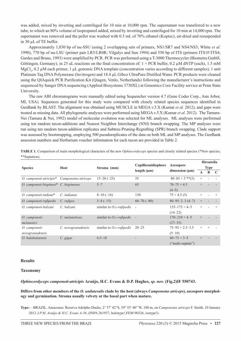
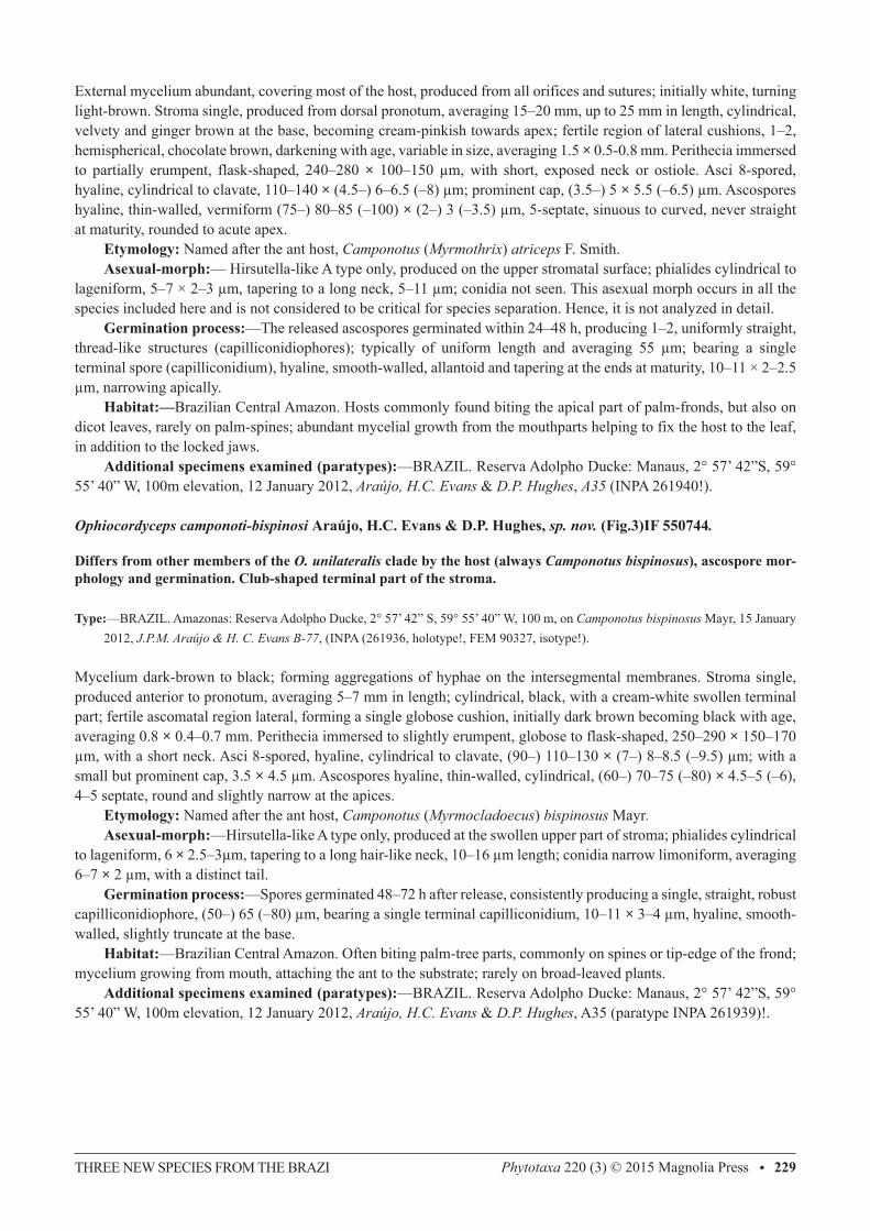
Mycelium dark-brown to black; forming aggregations of hyphae on the intersegmental membranes. Stroma single, produced anterior to pronotum, averaging 5–7 mm in length; cylindrical, black, with a cream-white swollen terminal part; fertile ascomatal region lateral, forming a single globose cushion, initially dark brown becoming black with age, averaging 0.8 × 0.4–0.7 mm. Perithecia immersed to slightly erumpent, globose to flask-shaped, 250–290 × 150–170 μm, with a short neck. Asci 8-spored, hyaline, cylindrical to clavate, (90–) 110–130 × (7–) 8–8.5 (–9.5) μm; with a small but prominent cap, 3.5 × 4.5 μm. Ascospores hyaline, thin-walled, cylindrical, (60–) 70–75 (–80) × 4.5–5 (–6), 4–5 septate, round and slightly narrow at the apices. Etymology: Named after the ant host, Camponotus (Myrmocladoecus) bispinosus Mayr. Asexual-morph:—Hirsutella-like A type only, produced at the swollen upper part of stroma; phialides cylindrical to lageniform, 6 × 2.5–3μm, tapering to a long hair-like neck, 10–16 μm length; conidia narrow limoniform, averaging 6–7 × 2 μm, with a distinct tail. Germination process:—Spores germinated 48–72 h after release, consistently producing a single, straight, robust capilliconidiophore, (50–) 65 (–80) μm, bearing a single terminal capilliconidium, 10–11 × 3–4 μm, hyaline, smooth-walled, slightly truncate at the base. Habitat:—Brazilian Central Amazon. Often biting palm-tree parts, commonly on spines or tip-edge of the frond; mycelium growing from mouth, attaching the ant to the substrate; rarely on broad-leaved plants. Additional specimens examined (paratypes):—BRAZIL. Reserva Adolpho Ducke: Manaus, 2° 57’ 42”S, 59° 55’ 40” W, 100m elevation, 12 January 2012, Araújo, H.C. Evans & D.P. Hughes, A35 (paratype INPA 261939)!.
ARAÚJO ET AL.230 • Phytotaxa 220 (3) © 2015 Magnolia Press
FIGURE 3. Ophiocordyceps camponoti-bispinosi a) Infected Camponotus bispinosus biting into tip of palm leaf; b) Close-up showing biting behavior; c) Section through ascoma showing perithecial arrangement (bar = 100 μm); d) Ascospore after 48 h germinating with a single capilliconidiophore with a capilliconidium at the tip (bar = 10 μm), upper right corner shows the capilliconidium in close-up (bar = 10 μm); e) Section of perithecium showing the arrangement of asci (bar = 50 μm); f-g) Detail showing the prominent ascus cap (bar = 10 μm); h) Section of the swollen stromatal tip with the asexual morph (Hirsutella-like A) (bar = 10 μm). Images: João Araújo.
THREE NEW SPECIES FROM THE BRAZI Phytotaxa 220 (3) © 2015 Magnolia Press • 231
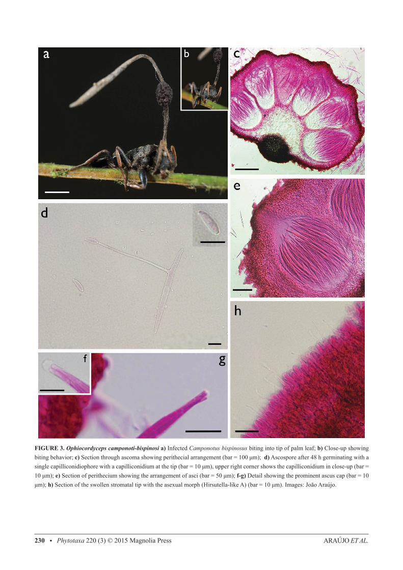
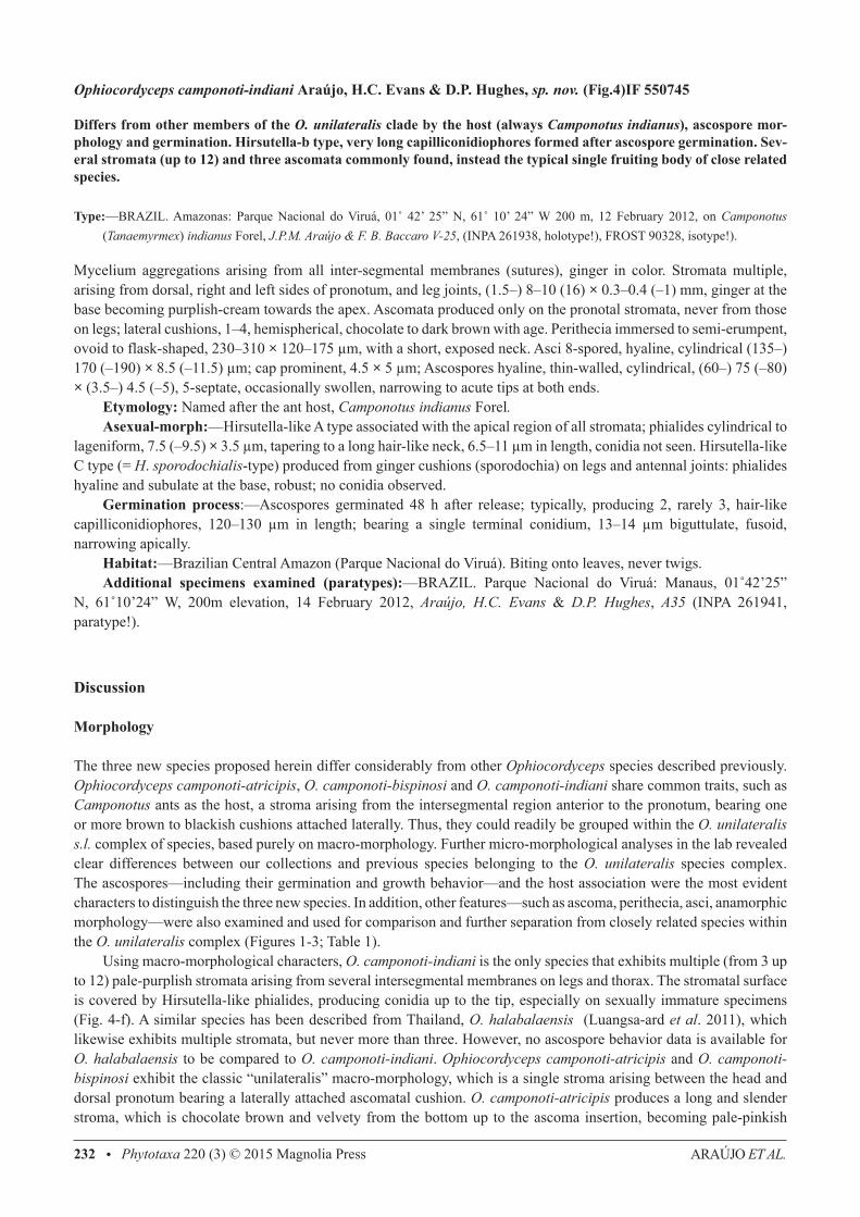
FIGURE 4. Ophiocordyceps camponoti-indiani a) Camponotus indianus biting into a leaf, several stromata arising from dorsal pronotum, mesonotum and leg joints, with a characteristic purplish coloration. a-1) lateral, fertile cushion (ascoma) ; a-2) Close up of the dead ant’s head showing the biting behavior. b) Section through ascoma showing perithecial arrangement (bar = 500 μm); c) Ascospore after 24 h, with very long capilliconidiophores (1-3) with capilliconidia at the tip (bar = 50 μm); c-1) Detail of fusoid capilliconidium (bar = 10 μm); d) Close up of perithecia showing asci arrangement and the semi-erumpent ostiole (bar = 50 μm); e) Ascus showing the spiral arrangement of ascospores (bar = 20 μm); e-1) Ascus cap detail (bar = 5 μm); f) Section of upper part of stroma showing asexual morph (Hirsutella-like A type), with long-necked phialides (bar = 10 μm); g) Phialides formed as mycelial cushions (sporodochia) on leg joints and antenna (Hirsutella-like C type) (bar = 10 μm). Images: João Araújo.
ARAÚJO ET AL.232 • Phytotaxa 220 (3) © 2015 Magnolia Press
Ophiocordyceps camponoti-indiani Araújo, H.C. Evans & D.P. Hughes, sp. nov. (Fig.4)IF 550745
Differs from other members of the O. unilateralis clade by the host (always Camponotus indianus), ascospore mor-phology and germination. Hirsutella-b type, very long capilliconidiophores formed after ascospore germination. Sev-eral stromata (up to 12) and three ascomata commonly found, instead the typical single fruiting body of close related species.
Type:—BRAZIL. Amazonas: Parque Nacional do Viruá, 01˚ 42’ 25” N, 61˚ 10’ 24” W 200 m, 12 February 2012, on Camponotus (Tanaemyrmex) indianus Forel, J.P.M. Araújo & F. B. Baccaro V-25, (INPA 261938, holotype!), FROST 90328, isotype!).
Mycelium aggregations arising from all inter-segmental membranes (sutures), ginger in color. Stromata multiple, arising from dorsal, right and left sides of pronotum, and leg joints, (1.5–) 8–10 (16) × 0.3–0.4 (–1) mm, ginger at the base becoming purplish-cream towards the apex. Ascomata produced only on the pronotal stromata, never from those on legs; lateral cushions, 1–4, hemispherical, chocolate to dark brown with age. Perithecia immersed to semi-erumpent, ovoid to flask-shaped, 230–310 × 120–175 μm, with a short, exposed neck. Asci 8-spored, hyaline, cylindrical (135–) 170 (–190) × 8.5 (–11.5) μm; cap prominent, 4.5 × 5 μm; Ascospores hyaline, thin-walled, cylindrical, (60–) 75 (–80) × (3.5–) 4.5 (–5), 5-septate, occasionally swollen, narrowing to acute tips at both ends. Etymology: Named after the ant host, Camponotus indianus Forel. Asexual-morph:—Hirsutella-like A type associated with the apical region of all stromata; phialides cylindrical to lageniform, 7.5 (–9.5) × 3.5 μm, tapering to a long hair-like neck, 6.5–11 μm in length, conidia not seen. Hirsutella-like C type (= H. sporodochialis-type) produced from ginger cushions (sporodochia) on legs and antennal joints: phialides hyaline and subulate at the base, robust; no conidia observed. Germination process:—Ascospores germinated 48 h after release; typically, producing 2, rarely 3, hair-like capilliconidiophores, 120–130 μm in length; bearing a single terminal conidium, 13–14 μm biguttulate, fusoid, narrowing apically. Habitat:—Brazilian Central Amazon (Parque Nacional do Viruá). Biting onto leaves, never twigs. Additional specimens examined (paratypes):—BRAZIL. Parque Nacional do Viruá: Manaus, 01˚42’25” N, 61˚10’24” W, 200m elevation, 14 February 2012, Araújo, H.C. Evans & D.P. Hughes, A35 (INPA 261941, paratype!).
Discussion
Morphology
The three new species proposed herein differ considerably from other Ophiocordyceps species described previously. Ophiocordyceps camponoti-atricipis, O. camponoti-bispinosi and O. camponoti-indiani share common traits, such as Camponotus ants as the host, a stroma arising from the intersegmental region anterior to the pronotum, bearing one or more brown to blackish cushions attached laterally. Thus, they could readily be grouped within the O. unilateralis s.l. complex of species, based purely on macro-morphology. Further micro-morphological analyses in the lab revealed clear differences between our collections and previous species belonging to the O. unilateralis species complex. The ascospores—including their germination and growth behavior—and the host association were the most evident characters to distinguish the three new species. In addition, other features—such as ascoma, perithecia, asci, anamorphic morphology—were also examined and used for comparison and further separation from closely related species within the O. unilateralis complex (Figures 1-3; Table 1). Using macro-morphological characters, O. camponoti-indiani is the only species that exhibits multiple (from 3 up to 12) pale-purplish stromata arising from several intersegmental membranes on legs and thorax. The stromatal surface is covered by Hirsutella-like phialides, producing conidia up to the tip, especially on sexually immature specimens (Fig. 4-f). A similar species has been described from Thailand, O. halabalaensis (Luangsa-ard et al. 2011), which likewise exhibits multiple stromata, but never more than three. However, no ascospore behavior data is available for O. halabalaensis to be compared to O. camponoti-indiani. Ophiocordyceps camponoti-atricipis and O. camponoti-bispinosi exhibit the classic “unilateralis” macro-morphology, which is a single stroma arising between the head and dorsal pronotum bearing a laterally attached ascomatal cushion. O. camponoti-atricipis produces a long and slender stroma, which is chocolate brown and velvety from the bottom up to the ascoma insertion, becoming pale-pinkish
THREE NEW SPECIES FROM THE BRAZI Phytotaxa 220 (3) © 2015 Magnolia Press • 233
and glabrous above and gradually forming a Hirsutella-like A layer, producing conidia up to the tip (Fig. 2-j). O. camponoti-bispinosi produces a much shorter black stroma, often swelling and becoming pale-cream in the last third (Figure 3-a), with a Hirsutella-like A layer producing conidia (Fig. 3-h). Microscopic features are also markedly different between the species. As with other taxa in the O. unilateralis clade proposed by Sung et al. (2007), the ascospores of all the new species do not disarticulate into part-spores, but they can readily be delimited on shape, size and septation (Table 1). In addition, their germination behavior can also be used to separate them, as reported for other species within the complex (Evans et al. 2011). O. camponoti-indiani ascospores germinate with one to three long capilliconidiophores that reach up to 130 μm (Figure 4-c), contrasting with the fewer and shorter capillioconidiophores in O. camponoti-atricipis (producing 1-2, up to 55 μm long) and O. camponoti-bispinosi (single and robust, averaging 65 μm in length) (Figures 2-d; 3-d). In addition, the shapes of the capilliconidia are also distinct between the three species (Figures. 2-e; 3-d; 4-c1). Capilliconidiophore variation among the species, especially the length, may reflect an adaptation to the host biology and further studies aim to address this question. Our results confirm that it is justifiable to split O. unilateralis s.l. into many taxa based on host specificity, potentially, unraveling a huge diversity of undocumented species.
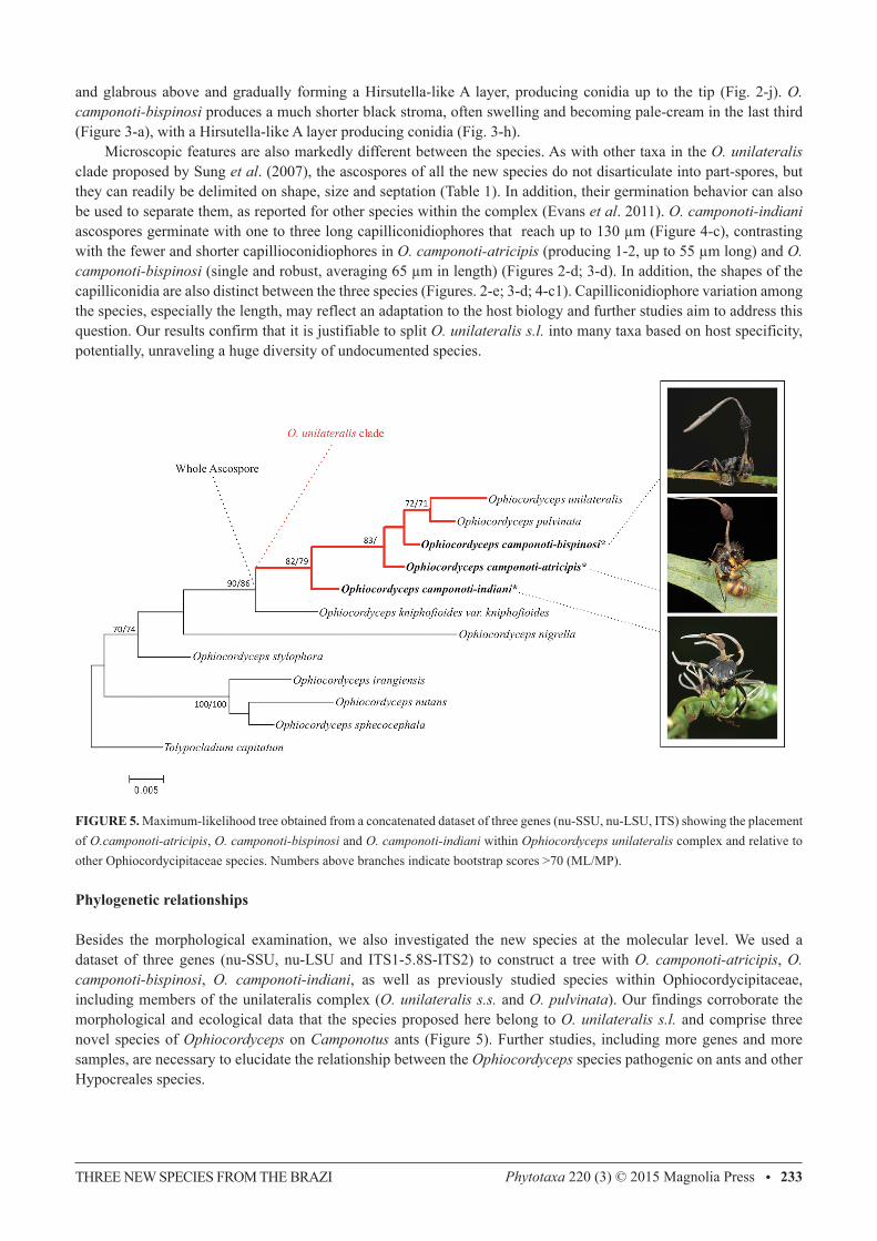
FIGURE 5. Maximum-likelihood tree obtained from a concatenated dataset of three genes (nu-SSU, nu-LSU, ITS) showing the placement of O.camponoti-atricipis, O. camponoti-bispinosi and O. camponoti-indiani within Ophiocordyceps unilateralis complex and relative to other Ophiocordycipitaceae species. Numbers above branches indicate bootstrap scores >70 (ML/MP).
Phylogenetic relationships
Besides the morphological examination, we also investigated the new species at the molecular level. We used a dataset of three genes (nu-SSU, nu-LSU and ITS1-5.8S-ITS2) to construct a tree with O. camponoti-atricipis, O. camponoti-bispinosi, O. camponoti-indiani, as well as previously studied species within Ophiocordycipitaceae, including members of the unilateralis complex (O. unilateralis s.s. and O. pulvinata). Our findings corroborate the morphological and ecological data that the species proposed here belong to O. unilateralis s.l. and comprise three novel species of Ophiocordyceps on Camponotus ants (Figure 5). Further studies, including more genes and more samples, are necessary to elucidate the relationship between the Ophiocordyceps species pathogenic on ants and other Hypocreales species.
ARAÚJO ET AL.234 • Phytotaxa 220 (3) © 2015 Magnolia Press
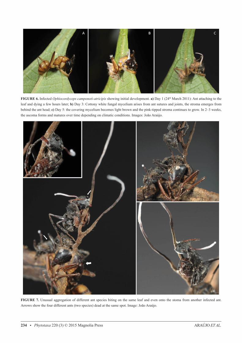
FIGURE 6. Infected Ophiocordyceps camponoti-atricipis showing initial development. a) Day 1 (24th March 2011): Ant attaching to the leaf and dying a few hours later; b) Day 3: Cottony white fungal mycelium arises from ant sutures and joints, the stroma emerges from behind the ant head; c) Day 5: the covering mycelium becomes light brown and the pink-tipped stroma continues to grow. In 2–3 weeks, the ascoma forms and matures over time depending on climatic conditions. Images: João Araújo.
FIGURE 7. Unusual aggregation of different ant species biting on the same leaf and even onto the stoma from another infected ant. Arrows show the four different ants (two species) dead at the same spot. Image: João Araújo.
THREE NEW SPECIES FROM THE BRAZI Phytotaxa 220 (3) © 2015 Magnolia Press • 235
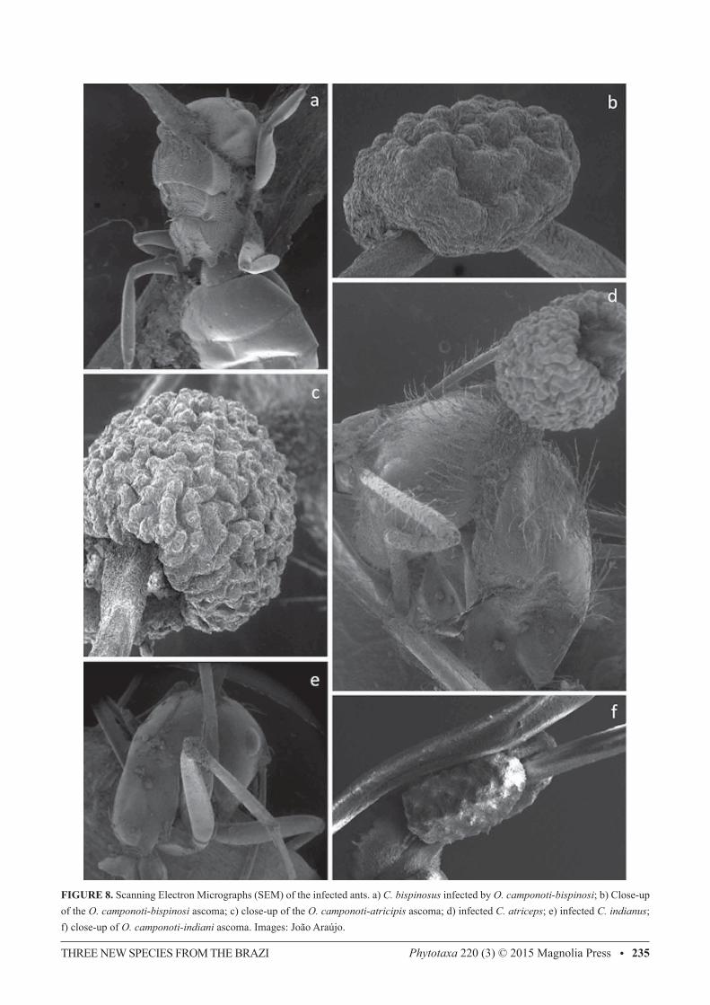
FIGURE 8. Scanning Electron Micrographs (SEM) of the infected ants. a) C. bispinosus infected by O. camponoti-bispinosi; b) Close-up of the O. camponoti-bispinosi ascoma; c) close-up of the O. camponoti-atricipis ascoma; d) infected C. atriceps; e) infected C. indianus; f) close-up of O. camponoti-indiani ascoma. Images: João Araújo.
ARAÚJO ET AL.236 • Phytotaxa 220 (3) © 2015 Magnolia Press
Ecological aspects
The species described in the present study are currently known only from the Brazilian Amazon. Infected dead ants were collected biting onto shrub leaves and palm fronds, especially near the tips, as well as on palm spines and epiphytes, and, occasionally, on slender climbing stems or lianas. We observed that early in the morning, droplets of dew are formed on the leaf tips (especially of palm leaves and spines), perhaps providing a daily supply of water for fungal development, even during the dry season. Thus, the adaptation of dying in this specific location may be advantageous for continual and consistent fungal development along the year. The same death position and substrate was also reported for O. halabalaensis in Thailand (Luangsa-ard et al. 2011). O. camponoti-atricipis and O. camponoti-bispinosi were commonly found at multiple sites in all the four areas visited during this study, as well as in other areas previously visited across the Brazilian Amazon. The clusters of infections were composed of either one or multiple species of O. unilateralis s.l. in the same area, varying from just a few (i.e. 3-5) up to dozens of individuals. In some areas, the density was so high that it was possible to find multiple ants biting into the same leaf or even into other infected ants (Figure 7). In contrast, the 19 O. camponoti-indiani specimens were all collected at the Parque Nacional do Viruá on a single occasion from one small site (~10 m2).
Conclusions
Ants of the genus Camponotus are parasitized by specialist fungal pathogens of the genus Ophiocordyceps, and this parasite-host association is especially common in tropical and sub-tropical forests. Based on macro-morphology, these pathogens—or, zombie-ant fungi—can all be assigned to O. unilateralis s.l. However, it has been posited that each species of Camponotus is parasitized by a host-specific species of the fungus and that these taxa can be separated on micro-morphology (Evans et al. 2011). Here, based on collections from central Amazonia, we show that this hypothesis is robust, particularly when the form and function of mature and living ascospores are compared. This has also been supported by molecular evidence and, therefore, we are confident that many more (perhaps, hundreds) new Camponotus-Ophiocordyceps associations remain to be discovered worldwide.
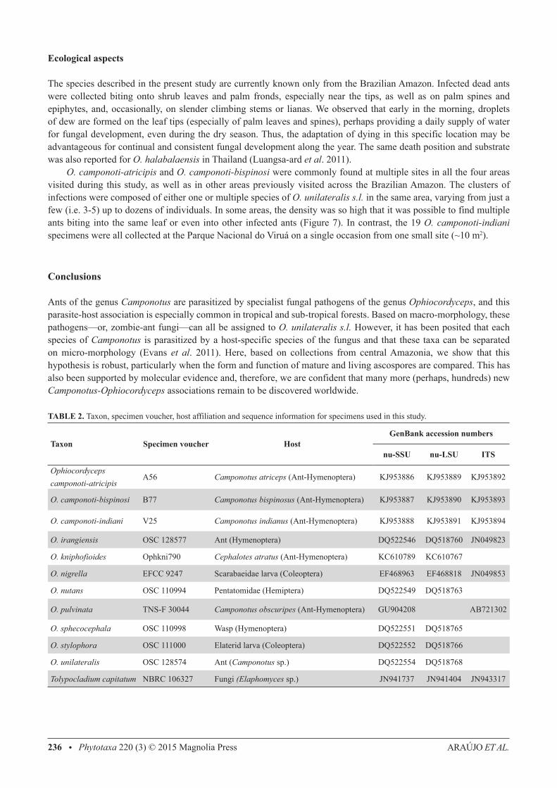
TABLE 2. Taxon, specimen voucher, host affiliation and sequence information for specimens used in this study.
Taxon Specimen voucher HostGenBank accession numbers
nu-SSU nu-LSU ITS
Ophiocordyceps camponoti-atricipis
A56 Camponotus atriceps (Ant-Hymenoptera) KJ953886 KJ953889 KJ953892
O. camponoti-bispinosi B77 Camponotus bispinosus (Ant-Hymenoptera) KJ953887 KJ953890 KJ953893
O. camponoti-indiani V25 Camponotus indianus (Ant-Hymenoptera) KJ953888 KJ953891 KJ953894
O. irangiensis OSC 128577 Ant (Hymenoptera) DQ522546 DQ518760 JN049823
O. kniphofioides Ophkni790 Cephalotes atratus (Ant-Hymenoptera) KC610789 KC610767
O. nigrella EFCC 9247 Scarabaeidae larva (Coleoptera) EF468963 EF468818 JN049853
O. nutans OSC 110994 Pentatomidae (Hemiptera) DQ522549 DQ518763
O. pulvinata TNS-F 30044 Camponotus obscuripes (Ant-Hymenoptera) GU904208 AB721302
O. sphecocephala OSC 110998 Wasp (Hymenoptera) DQ522551 DQ518765
O. stylophora OSC 111000 Elaterid larva (Coleoptera) DQ522552 DQ518766
O. unilateralis OSC 128574 Ant (Camponotus sp.) DQ522554 DQ518768
Tolypocladium capitatum NBRC 106327 Fungi (Elaphomyces sp.) JN941737 JN941404 JN943317
THREE NEW SPECIES FROM THE BRAZI Phytotaxa 220 (3) © 2015 Magnolia Press • 237
Acknowledgements
We wish to thank Fabrício Baccaro (UFAM), Ricardo Braga-Neto (INPA) and Carlos Nogueira (INPA) for assistance in the field and INPA for the logistic help. Our special thanks to Márcio Oliveira and Thiago Mahlmann for the assistance with the permissions at INPA Entomological Collection, John Cantolina for the help with microscopy. Robert Weingart Barreto (UFV) for the support in this project. We are also very grateful to Priscila Santos (Parque Nacional de Anavilhanas), Benjamin da Luz (Estação Ecológica de Maracá) and Antônio Lisboa (Parque Nacional do Viruá) to help with support offered for the fieldtrips. This research was supported by CNPq (Science without borders scholarship granted to JPMA) and funds from Anavilhanas National Park/ICMBio and Programa de Áreas Protegidas na Amazônia/MMA with partnership with Instituto IPÊ granted to JPMA.
References
Agosti, D. & Johnson, N.F. (2009) Antbase: World Wide Web electronic publication. Available from: http://antbase.org (accessed 25 March 2014)Andrade, C.F.S. (1980) Epizootia natural causada por Cordyceps unilateralis (Hypocreales, Euascomycetes) em adultos de Camponotus
sp. (Hymenoptera, Formicidae) na região de Manaus, Amazonas, Brasil. Acta Amazonica 10: 671–677. Araújo, J.P.M. & Hughes, D.P. (2014) Diversity of Entomopathogenic Fungi: Which groups conquered the insect body? bioRxiv.de Bekker, C., Quevillon, L.E., Philip, B.S., Fleming, K.R., Ghosh, D., Patterson, A.D. & Hughes, D.P. (2014) Species-specific ant brain
manipulation by a specialized fungal parasite. BMC Evolutionay Biology 14: 166–178. http://dx.doi.org/10.1186/s12862-014-0166-3Dawkins, R. (1982) The extended phenotype. Oxford University Press, Oxford, 307 pp.Evans, H.C. (1974) Natural control of arthropods, with special reference to ants (Formicidae), by fungi in the tropical high forest of Ghana.
Journal of Applied Ecology 11: 37–49. http://dx.doi.org/10.2307/2402003Evans, H.C. (1988a) Mycopathogens of insects of epigeal and aerial habitats. In: Wilding, N., Hammond, P.M. & Webber, J. (Eds.) Insect-
fungus interactions. Academic Press, London, pp. 205–238.Evans, H.C. (1988b) Coevolution of entomogenous fungi and their insect hosts. In: Pirozynski, K.A. & Hawksworth, D.L. (Eds.) Coevolution
of fungi with plants and animals. Academic Press, London, pp. 149–171.Evans, H.C. (2001) Entomopathogenic fungi associated with ants (Formicidae): A review. In: Misra, J.K. & Horn, B.W. (Eds.) Trichomycetes
and other fungal groups. Science Publishers, Enfield, pp. 119–144.Evans, H.C., Elliot, S.L. & Hughes, D.P. (2011) Hidden diversity behind the zombie-antfungus Ophiocordyceps unilateralis: Four new
species described from carpenter antsin Minas Gerais. Brazil. PLoS ONE 6: e17024. http://dx.doi.org/10.1371/journal.pone.0017024Evans, H.C. & Samson, R.A. (1982) Cordyceps species and their anamorphs pathogenic on ants (Formicidae) in tropical forest ecosystems.
I. The Cephalotes (Myrmicinae) complex. Transactions of the British Mycological Society 79: 431–453. http://dx.doi.org/10.1016/S0007-1536(82)80037-5Evans, H.C. & Samson, R.A. (1984) Cordyceps species and their anamorphs pathogenic of ants (Formicidae) in tropical forest ecosystems
II. The Camponotus (Formicinae) complex. Transactions of the British Mycological Society 82: 127–150. http://dx.doi.org/10.1016/S0007-1536(84)80219-3Fittkau, E.J. & Klinge, H. (1973) On biomass and trophic structure of the Central Amazonian rain forest ecosystem. Biotropica 5: 2–14. http://dx.doi.org/10.2307/2989676Hölldobler, B. & Wilson, E.O. (2009) The superorganism: The beauty, elegance, and strangeness of insect societies. W.W. Norton, New
York, 522 pp.Kepler, R.M., Kaitsu, Y., Tanaka, E., Shimano, S. & Spatafora, J.W. (2011) Ophiocordyceps pulvinata sp. nov., a pathogen of ants with a
reduced stroma. Mycoscience 52: 39–47. http://dx.doi.org/10.1007/S10267-010-0072-5Kobayasi, Y. (1941) The genus Cordyceps and its allies. Science Reports of the Tokyo Bunrika Daigaku 84: 53–260.Kobmoo, N., Mongkolsamrit, S., Tasanathai, K., Thanakitpipattana, D. & Luangsa-ard, J.J. (2012) Molecular phylogenies reveal host-
specific divergence of Ophiocordyceps unilateralis sensu lato following its host ants. Molecular Ecology 21: 3022–3031. http://dx.doi.org/10.1111/j.1365-294X.2012.05574.xLuangsa-ard, J.J., Ridkaew, R., Tasanathai, K., Thanakitpipattana, D. & Hywel-Jones, N. (2011) Ophiocordyceps halabalaensis: a new
species of Ophiocordyceps pathogenic to Camponotus gigas in Hala Bala Wildlife Sanctuary, Southern Thailand. Fungal Biology
ARAÚJO ET AL.238 • Phytotaxa 220 (3) © 2015 Magnolia Press
115: 608–614. http://dx.doi.org/10.1016/j.funbio.2011.03.002Mains, E.B. (1958) North American entomogenous species of Cordyceps. Mycologia 50: 169–222. http://dx.doi.org/10.2307/3756193Petch, T. (1931) Notes on entomogenous fungi. Transactions of the British Mycologial Society 16: 55–75. http://dx.doi.org/10.1016/S0007-1536(31)80006-3Quandt, C.A., Kepler, R.M., Gams, W., Araújo, J.P.M., Ban, S., Evans, H.C., Hughes, D., Humber, R., Hywel-Jones, N., Li, Z., Luangsa-
ard, J.J., Rehner, S.A., Sanjuan, T., Sato, H., Shrestha, B., Sung, G., Yao, Y., Zare, R. & Spatafora, J.W. (2014) Phylogenetic-based nomenclatural proposals for Ophiocordycipitaceae (Hypocreales) with new combinations in Tolypocladium. IMA Fungus 5: 121–134.
Robert, V., Stegehuis, G. & Stalpers, J. (2005) The MycoBank engine and related databases. Available from: http://www.mycobank.org (accessed 28 March 2014)
Samson, R.A., Evans, H.C. & Hoekstra, E.S. (1982) Notes on entomogenous fungi from Ghana. VI. The genus Cordyceps. Proceedings of the Koninklijke Nederlandse Akademie van Wetenschappen 85: 589–605.
Sung, G.-H., Hywel-Jones, N.L., Sung, J.-M., Luangsa-ard, J.J., Shrestha, B. & Spatafora, J.W. (2007) Phylogenetic classification of Cordyceps and the clavicipitaceous fungi. Studies in Mycology 57: 5–59.
http://dx.doi.org/10.3114/sim.2007.57.01Tamura, K. & Nei, M. (1993) Estimation of the number of nucleotide substitutions in the control region of mithocondrial DNA in Humans
and Chimpanzees. Molecular Biology and Evolution 10: 512–526.Tamura, K., Peterson, D., Peterson, N., Stecher, G., Nei, M. & Kumar, S. (2011) MEGA5: Molecular Evolutionary Genetics Analysis
using Maximum Likelihood, Evolutionary Distance, and Maximum Parsimony Methods. Molecular Biology and Evolution 28: 2731–2739.
http://dx.doi.org/10.1093/molbev/msr121Tulasne, L.R. & Tulasne C. (1865) Selecta Fungorum Carpologia III. Paris Museum, Paris, 221 pp.Underwood, E. & Fisher, B. (2006) The role of ants in conservation monitoring: If, when, and how. Biological Conservation 132: 166–182. http://dx.doi.org/10.1016/j.biocon.2006.03.022Vilgalys, R. & Sun, B.L. (1994) Ancient and recent patterns of geographic speciation in the oyster mushroom Pleurotus revealed by
phylogenetic analysis of ribosomal DNA sequences. Proceedings of National Academy of Sciences 91: 4599–4603. http://dx.doi.org/10.1073/pnas.91.10.4599White, T.J., Bruns, T., Lee, S. & Taylor, J.W. (1990) Amplification and direct sequencing of fungal ribosomal RNA genes for phylogenetics.
In: Innis, M.A., Gelfand, D.H., Sninsky, J.J. & White, T.J. (Eds.) PCR Protocols: A Guide to Methods and Applications. Academic Press, New York, pp. 315–322.
http://dx.doi.org/10.1016/b978-0-12-372180-8.50042-1