Embed Size (px)
Citation preview
Phytotaxa 253 (4): 285–292http://www.mapress.com/j/pt/Copyright © 2016 Magnolia Press Article PHYTOTAXA
ISSN 1179-3155 (print edition)
ISSN 1179-3163 (online edition)
Accepted by Sajeewa Maharachchikumbura: 3 Mar. 2016; published: 30 Mar. 2016
http://dx.doi.org/10.11646/phytotaxa.253.4.4
285Licensed under a Creative Commons Attribution License http://creativecommons.org/licenses/by/3.0
Cryptosporella platyphylla, a new species associated with Betula platyphylla in China
XIN-LeI FAN1, Zhuo Du 1, KevIN D. hYDe 3, YINg-MeI LIANg 2, YAN-PIINg PAN 4 & CheNg-MINg TIAN1*1The Key Laboratory for Silviculture and Conservation of Ministry of Education, Beijing Forestry University, Beijing 100083, China2Museum of Beijing Forestry University, Beijing 100083, China3Center of Excellence in Fungal Research, Mae Fah Luang University, Chiang Rai 57100, Thailand4Beijing Municipal Forestry Protection Station, Beijing 100029, China*Correspondence author: [email protected]
Abstract
Members of Cryptosporella are well-known as common endophytes, and occasionally, as pathogens on a narrow range of hosts in Betulaceae, Tiliaceae and ulmaceae. Two fresh specimens associated with canker and dieback of Betula platyphylla were made in Beijing, China in 2015. Morphological and multi-gene, combined, phylogenetic analyses (ITS, tef1-α and β-tub) support these speciemens as a distinct and new species of Cryptosporella, from a unique host, Betula platyphylla. Cryptosporella platyphylla sp. nov. is introduced with an illustrated account and differs from similar species in its host as-sociation and multigene phylogeny.
Key words: Diaporthales, Disculina, gnomoniaceae, systematics, taxonomy
Introduction
Cryptosporella was introduced by Saccardo (1877) and was distinguished from Cryptospora based on the morphology of the ascospores. The genus subsequently entered a long period of confusion with the gnomoniaceous genera Ophiovalsa Petr. and Winterella (Sacc.) o. Kuntze. Mejía et al. (2008) regarded Ophiovalsa and Winterella as synonyms of Cryptosporella, which is based on type species C. hypodermia (Fr.) Sacc., and accepted nine species in this genus. Mejía et al. (2011) added a further ten species to the genus supported by DNA data from three genes (ITS, tef1-α and β-tub). Most Cryptosporella species are distributed in temperate regions, e.g., europe, Japan, and North America as endophytes, and occasionally as saprobes and pathogens on hardwood trees, i.e., Betulaceae, Tiliaceae and ulmaceae (Saccardo 1877, Kobayashi 1970, Barr 1978, Mejía et al. 2011). Cryptosporella is characterized by aggregated ascomata below the bark surface, with converging necks and ellipsoid to elongated, aseptate or rarely 1-septate ascospores. Additionally, their Disculina höhn. asexual morph is characterized by hyaline, aseptate conidia, arising from proliferating conidiogenous cells (Rossman et al. 2007, Mejía et al. 2008, 2011). Cryptosporella is presently placed in gnomoniaceae (Castlebury et al. 2002, Maharachchikumbura et al. 2015). During an investigation of forest pathogens that cause canker or dieback disease in China, two Cryptosporella specimens were collected from Betula platyphylla Sukaczev in Beijing, China. These specimens are characterized by slightly erumpent, conidiomatal stromata with conical central column and dark brown ectostromatic disc, and hyaline, cylindrical to ellipsoidal, aseptate conidia (51 × 4 µm), arising from narrowly cylindrical, conidiogenous cells. Phylogenetic analysis of combined ITS, tef1-α and β-tub sequence data suggested that these specimens represent a new species, supported by high bootstrap values. Therefore, the objective of this study is to introduce Cryptosporella platyphylla sp. nov. with a description and illustrations and compare it with other species in the genus.

FAN ET AL.286 • Phytotaxa 253 (4) © 2016 Magnolia Press
Materials and Methods
IsolatesTwo specimens of gnomoniaceous fungi were collected from infected branches or twigs of Betula platyphylla during collecting trips in Beijing, China. Two strains of Cryptosporella were obtained following the method of Chomnunti et al. (2014), where a mucoid spore mass was removed from the conidiomata, and transferred to the surface of 1.8 % potato dextrose agar (PDA) in a Petri-dish, and incubated at 25 °C for up to 24 h. Single germinating conidia were transferred to fresh PDA plates. The representative strains were deposited and now maintained at the China Forestry Culture Collection Center (CFCC) and Beijing Forestry university (BJFu) under strain numbers CFCC 50465 and CFCC 50466. Specimens are deposited in the Museum of the Beijing Forestry university (BJFC) under collection numbers BJFC-S1313 and BJFC-S1314.
DNA amplification, sequencing and phylogenygenomic DNA was obtained from colonies grown on PDA using a CTAB method (Doyle and Doyle 1990). The ITS region was amplified using primers ITS1 and ITS4 (White et al. 1990). The partial translation elongation factor 1-alpha (tef1-α) gene region was amplified using primers EF1-728F and EF1-986R (Carbone and Kohn 1999). The partial β-tubulin (β-tub) was amplified using primers Bt2a and Bt2b (Glass and Donaldson 1995). DNA sequencing was performed using an ABI PRISM® 3730XL DNA Analyzer with BigDye® Terminator Kit v.3.1 (Invitrogen) at the Shanghai Invitrogen Biological Technology Company Limited (Beijing, China). DNA sequences were deposited in genBank (Table 1) and a data matrix (expanded from the work of Mejía et al. 2011) were deposited in TreeBASe (www.treebase.org) as accession S18571. The current sequences and ex-type reference sequences selected from recent studies and genBank were aligned in MAFFT v.7 (Katoh and Standley 2013) and checked manually.
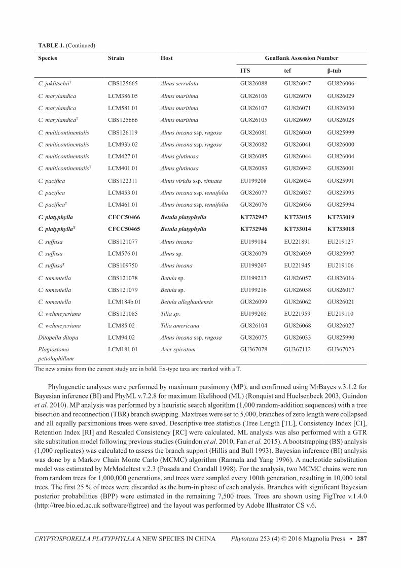
TAble 1. Strains of Cryptosporella used in the molecular analyses in this study.
Species Strain Host Genbank Assession Number
ITS tef β-tub
C. alnicola CBS121074 Corylus cornuta eu199204 eu199160 eu219138
C. alni-rubrae CBS126120 Alnus rubra gu826092 gu826051 gu826010
C. alni-rubrae LCM411 Alnus rubra gu826090 gu826049 gu826008
C. alni-rubraeT LCM499.01 Alnus rubra gu826096 gu826055 gu826014
C. alni-sinuatae AR4200 Alnus viridis ssp. sinuata gu826086 gu826045 gu825989
C. alni-sinuataeT CBS125662 Alnus viridis ssp. sinuata gu826087 gu826046 gu826005
C. alni-tenuifoliaeT CBS125663 Alnus incana ssp. tenuifol gu826097 gu826056 gu826015
C. amistadensis LCM618.01 Alnus acuminata gu826109 gu826073 gu826032
C. amistadensisT CBS125664 Alnus acuminata gu826108 gu826072 gu826031
C. betulae CBS121078 Betula pendula eu199213 gu826057 gu826016
C. betulae LCM477.01 Betula pendula gu826098 gu826059 gu826018
C. betulae CBS121079 Betula pendula eu199216 gu826058 gu826017
C. confusa CBS121063 Betula papyrifera eu199219 - -
C. corylina LCM391.04 Corylus avellana gu826100 gu826063 gu826022
C. femoralis LCM196.04 Alnus incana ssp. rugosa gu826102 gu826067 gu826025
C. femoralisT CBS121076 Alnus incana ssp. rugosa eu199220 eu219139 eu221951
C. hypodermia CBS109753 Ulmus minor eu199224 gu826064 gu826023
C. hypodermiaT CBS122593 Ulmus minor eu199181 gu826066 gu826024
C. jaklitschii LCM112.01 Alnus serrulata gu826089 gu826048 gu826007
...Continued on next page
CRyPTOSPORELLA PLATyPhyLLA A NeW SPeCIeS IN ChINA Phytotaxa 253 (4) © 2016 Magnolia Press • 287
TAble 1. (Continued)
Species Strain Host Genbank Assession Number
ITS tef β-tub
C. jaklitschiiT CBS125665 Alnus serrulata gu826088 gu826047 gu826006
C. marylandica LCM386.05 Alnus maritima gu826106 gu826070 gu826029
C. marylandica LCM581.01 Alnus maritima gu826107 gu826071 gu826030
C. marylandicaT CBS125666 Alnus maritima gu826105 gu826069 gu826028
C. multicontinentalis CBS126119 Alnus incana ssp. rugosa gu826081 gu826040 gu825999
C. multicontinentalis LCM93b.02 Alnus incana ssp. rugosa gu826082 gu826041 gu826000
C. multicontinentalis LCM427.01 Alnus glutinosa gu826085 gu826044 gu826004
C. multicontinentalisT LCM401.01 Alnus glutinosa gu826083 gu826042 gu826001
C. pacifica CBS122311 Alnus viridis ssp. sinuata eu199208 gu826034 gu825991
C. pacifica LCM453.01 Alnus incana ssp. tenuifolia gu826077 gu826037 gu825995
C. pacificaT LCM461.01 Alnus incana ssp. tenuifolia gu826076 gu826036 gu825994
C. platyphylla CFCC50466 Betula platyphylla KT732947 KT733015 KT733019
C. platyphyllaT CFCC50465 Betula platyphylla KT732946 KT733014 KT733018
C. suffusa CBS121077 Alnus incana eu199184 eu221891 eu219127
C. suffusa LCM576.01 Alnus sp. gu826079 gu826039 gu825997
C. suffusaT CBS109750 Alnus incana eu199207 eu221945 eu219106
C. tomentella CBS121078 Betula sp. eu199213 gu826057 gu826016
C. tomentella CBS121079 Betula sp. eu199216 gu826058 gu826017
C. tomentella LCM184b.01 Betula alleghaniensis gu826099 gu826062 gu826021
C. wehmeyeriana CBS121085 Tilia sp. eu199205 eu221959 eu219110
C. wehmeyeriana LCM85.02 Tilia americana gu826104 gu826068 gu826027
Ditopella ditopa LCM94.02 Alnus incana ssp. rugosa gu826075 gu826033 gu825990
Plagiostoma petiolophillum
LCM181.01 Acer spicatum gu367078 gu367112 gu367023
The new strains from the current study are in bold. ex-type taxa are marked with a T.
Phylogenetic analyses were performed by maximum parsimony (MP), and confirmed using MrBayes v.3.1.2 for Bayesian inference (BI) and PhyML v.7.2.8 for maximum likelihood (ML) (Ronquist and huelsenbeck 2003, guindon et al. 2010). MP analysis was performed by a heuristic search algorithm (1,000 random-addition sequences) with a tree bisection and reconnection (TBR) branch swapping. Maxtrees were set to 5,000, branches of zero length were collapsed and all equally parsimonious trees were saved. Descriptive tree statistics (Tree Length [TL], Consistency Index [CI], Retention Index [RI] and Rescaled Consistency [RC] were calculated. ML analysis was also performed with a gTR site substitution model following previous studies (guindon et al. 2010, Fan et al. 2015). A bootstrapping (BS) analysis (1,000 replicates) was calculated to assess the branch support (hillis and Bull 1993). Bayesian inference (BI) analysis was done by a Markov Chain Monte Carlo (MCMC) algorithm (Rannala and Yang 1996). A nucleotide substitution model was estimated by MrModeltest v.2.3 (Posada and Crandall 1998). For the analysis, two MCMC chains were run from random trees for 1,000,000 generations, and trees were sampled every 100th generation, resulting in 10,000 total trees. The first 25 % of trees were discarded as the burn-in phase of each analysis. Branches with significant Bayesian posterior probabilities (BPP) were estimated in the remaining 7,500 trees. Trees are shown using FigTree v.1.4.0 (http://tree.bio.ed.ac.uk software/figtree) and the layout was performed by Adobe Illustrator CS v.6.
FAN ET AL.288 • Phytotaxa 253 (4) © 2016 Magnolia Press
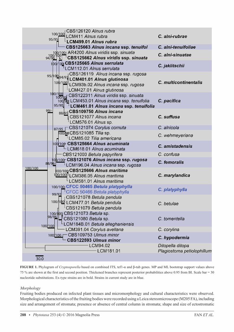
FIGURe 1. Phylogram of Cryptosporella based on combined ITS, tef1-α and β-tub genes. MP and ML bootstrap support values above 75 % are shown at the first and second position. Thickened branches represent posterior probabilities above 0.95 from BI. Scale bar = 30 nucleotide substitutions. ex-type strains are in bold. Strains in current study are in blue.
MorphologyFruiting bodies produced on infected plant tissues and micromorphology and cultural characteristics were observed. Morphological characteristics of the fruiting bodies were recorded using a Leica stereomicroscope (M205 FA), including size and arrangement of stromata; presence or absence of central column in stromata; shape and size of ectostromatic
CRyPTOSPORELLA PLATyPhyLLA A NeW SPeCIeS IN ChINA Phytotaxa 253 (4) © 2016 Magnolia Press • 289
disc and ostiole. Micromorphological observations included the size and shape of conidiophores and conidia using a Leica compound microscope (DM 2500). More than 20 conidiomata were sectioned and 50 conidia were selected randomly for measurement. Cultural characteristics of strains incubated on PDA in the dark at 25 °C were recorded, including the colony colour and conidiomata distribution. Taxonomic novelties were deposited in MycoBank (Crous et al. 2004) with numbers MB 815811 and Faces of fungi numbers (FoF 01898) obtained as in Jayasiri et al. (2015).
Results
The combined ITS, tef1-α and β-tub dataset from 38 ingroup strains (sequences of two strains from this study and sequences of 36 strains available in genBank mostly from Mejía et al. 2011) clustered in 18 clades representing species of Cryptosporella (Table 1). The alignment including gaps comprised 2864 characters of which 2062 characters were constant, 369 variable characters were parsimony-uninformative, and 433 were parsimony informative. The heuristic search generated one parsimonious tree (TL= 1238, CI = 0.823, RI = 0.883, RC = 0.726) as shown in Fig. 1. Strains CFCC 50465 and CFCC 50466, sequenced in this study, formed a well-supported clade (MP/ML/BI = 100/100/1) representing a new phylogenetic species.
Taxonomy
Cryptosporella platyphylla C.M. Tian, X.L. Fan & K.D. hyde, sp. nov., Fig. 2MycoBank MB 815811; Facesoffungi number: FoF 01898
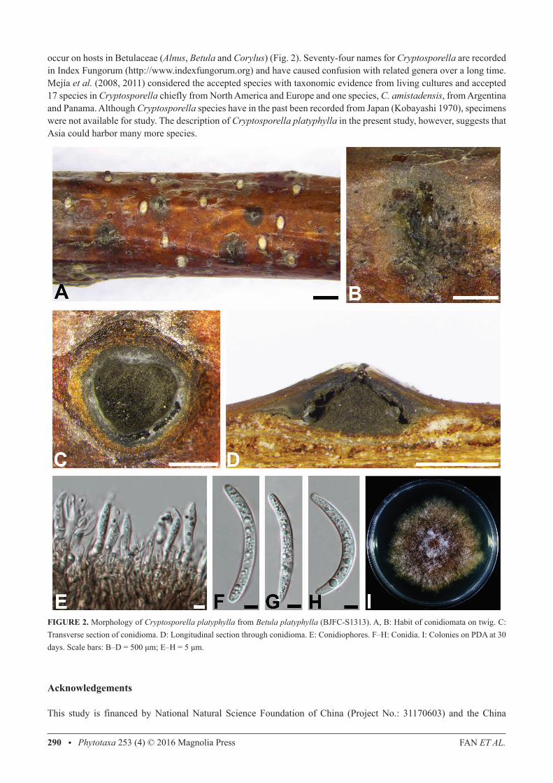
holotype:—BJFC-S1313. ChINA, Beijing: Tongzhou District, Jingdong Forest Park, 39°59'44.37"N, 116°39'29.16"e, 26 m asl, on twigs and branches of Betula platyphylla, coll. X.L. Fan, 16 May 2015 (BJFC-S1313, holotype), ex-type culture, CFCC 50465. Etymology:—platyphylla, referring to Betula platyphylla, the only host known for this species. host/Distribution:—from Betula platyphylla in China. Original description:—Sexual morph: undetermined. Asexual morph: Conidiomatal stromata immersed, slightly erumpent from surface of host branches, separate, discoid to conical, (880–)950–1330(–1460) μm (av. = 1130 μm, n = 20) diam. Stromatic zones lacking. Central column beneath the disc more or less conical, grey to black. Ectostromatic disc dark brown, circular to ovoid (400–)480–610(–790) μm (av. = 560 μm, n = 20) diam. Ostiole one per disc, emerging in central position in the ectostromatic disc. Conidiophores reduced to conidiogenous cells. Conidiogenous cells narrowly cylindrical, smooth, hyaline, producing a conidium at apex. Conidia hyaline, aseptate, cylindrical to clavate, gently curved, (45–)46.5–53(–59) × (3–)3.5–4.5 μm (av. = 51 × 4 µm, n = 50). Cultures:—Colony growth on PDA initially white, becoming yellowish brown to brownish after 7–10 days. Colony flat, felt-like, with a uniform texture, extending to ca. 66 mm diam at room temperature. Conidiomata absent.Material examined:—ChINA, Beijing: Tongzhou District, Song village, 39°59’40”N, 116°39’29.31”e, 10 m asl, on twigs and branches of Betula platyphylla, coll. X.L. Fan, 20 March 2015 (BJFC-S1314, paratype), ex-paratype culture, CFCC 50466.
Discussion
In this study two strains of Cryptosporella associated with Betula platyphylla dieback or canker disease in China were identified. Species identification was supported by morphology and multigene DNA data (ITS, tef1-α and β-tub), which indicated that Cryptosporella platyphylla represents a distinct species (Fig. 1). Although Cryptosporella platyphylla represents a closely related sister group to Cryptosporella betulae, the two taxa are distinguished based on their unique host and multigene phylogenetic data (Fig. 2). Cryptosporella is a well-defined genus which seems to have taxa with narrow host ranges, frequently limited to a single host species, especially in the host family Betulaceae (Castlebury et al. 2002, Mejía et al. 2008, 2011, Sogonov et al. 2008). except for C. wehmeyeriana on Tilia spp. and type species C. hypodermia on Ulmus spp. all these species
FAN ET AL.290 • Phytotaxa 253 (4) © 2016 Magnolia Press
occur on hosts in Betulaceae (Alnus, Betula and Corylus) (Fig. 2). Seventy-four names for Cryptosporella are recorded in Index Fungorum (http://www.indexfungorum.org) and have caused confusion with related genera over a long time. Mejía et al. (2008, 2011) considered the accepted species with taxonomic evidence from living cultures and accepted 17 species in Cryptosporella chiefly from North America and europe and one species, C. amistadensis, from Argentina and Panama. Although Cryptosporella species have in the past been recorded from Japan (Kobayashi 1970), specimens were not available for study. The description of Cryptosporella platyphylla in the present study, however, suggests that Asia could harbor many more species.
FIGURe 2. Morphology of Cryptosporella platyphylla from Betula platyphylla (BJFC-S1313). A, B: habit of conidiomata on twig. C: Transverse section of conidioma. D: Longitudinal section through conidioma. e: Conidiophores. F–h: Conidia. I: Colonies on PDA at 30 days. Scale bars: B–D = 500 μm; E–H = 5 μm.
Acknowledgements
This study is financed by National Natural Science Foundation of China (Project No.: 31170603) and the China
CRyPTOSPORELLA PLATyPhyLLA A NeW SPeCIeS IN ChINA Phytotaxa 253 (4) © 2016 Magnolia Press • 291
Scholarship Council (CSC). We are grateful to Chungen Piao and Minwei guo (China Forestry Culture Collection Center (CFCC), Chinese Academy of Forestry, Beijing) for support of strain preservation during this study.
References
Barr, M.e. (1978) The Diaporthales in North America with emphasis on Gnomonia and its segregates. Mycologia Memoir 7: 1–232.Carbone, I. & Kohn, L.M. (1999) A method for designing primer sets for speciation studies in filamentous ascomycetes. Mycologia 91:
553–556. http://dx.doi.org/10.2307/3761358Castlebury, L.A., Rossman, A.Y., Jaklitsch, W.J. & vasilyeva, L.N. (2002) A preliminary overview of the Diaporthales based on large
subunit nuclear ribosomal DNA sequences. Mycologia 94: 1017–1031. http://dx.doi.org/10.2307/3761867Chomnunti, P., hongsanan, S., Aguirre-hudson, B., Tian, Q., Peršoh, D., Dhami, M.K., Alias, A.S., Xu, J.C., Liu, X., Stadler, M., hyde,
K.D. (2014) The sooty moulds. Fungal Diversity 66: 1–6. http://dx.doi.org/10.1007/s13225-014-0278-5Crous, P.W., gams, W., Stalpers, J.A., Robert, v. & Stegehuis, g. (2004) MycoBank: an online initiative to launch mycology into the 21st
century. Studies in Mycology 50: 19–22.Doyle, J.J. & Doyle, J.L. (1990) Isolation of plant DNA from fresh tissue. Focus 12: 13–15.Fan, X.L., hyde, K.D., Liu, M., Liang, Y.M. & Tian, C.M. (2015) Cytospora species associated with walnut canker disease in China, with
description of a new species C. gigalocus. Fungal Biology 119: 310–319. http://dx.doi.org/10.1016/j.funbio.2014.12.011guindon, S., Dufayard, J.F., Lefort, v., Anisimova, M., hordijk, W. & gascuel, o. (2010) New algorithms and methods to estimate
maximum-likelihood phylogenies: assessing the performance of PhyML 3.0. Systematic Biology 59: 307–321. http://dx.doi.org/10.1093/sysbio/syq010glass, N.L. & Donaldson, g.C. (1995) Development of primer sets designed for use with the PCR to amplify conserved genes from
filamentous ascomycetes. Applied and Environmental Microbiology 61: 1323–1330.hillis, D.M. & Bull, J.J. (1993) An empirical test of bootstrapping as a method for assessing confidence in phylogenetic analysis. Systematic
Biology 42: 182–192. http://dx.doi.org/10.1093/sysbio/42.2.182Jayasiri, S.C., hyde, K.D., Ariyawansa, h.A., Bhat, D.J., Buyck, B., Cai, L., Dai, Y.C., Abd-elsalam, K.A., ertz, D., hidayat, I., Jeewon,
R., Jones, e.B.g., Bahkali, A.h., Karunarathna, S.C., Liu, J.K., Luangsa-ard, J.J., Lumbsch, h.T., Maharachchikumbura, S.S.N., McKenzie, e.h.C., Moncalvo, J.M., ghobad-Nejhad, M., Nilsson, h., Pang, K.L., Pereira, o.L., Phillips, A.J.L., Raspé, o., Rollins, A.W., Romero, A.I., etayo, J., Selçuk, F., Stephenson, S.L., Suetrong, S., Taylor, J.e., Tsui, C.K.M., vizzini, A., Abdel-Wahab, M.A., Wen, T.C., Boonmee, S., Dai, D.Q., Daranagama, D.A., Dissanayake, A.J., ekanayaka, A.h., Fryar, S.C., hongsanan, S., Jayawardena, R.S., Li, W.J., Perera, R.h., Phookamsak, R., de Silva, N.I., Thambugala, K.M., Tian, Q., Wijayawardene, N.N., Zhao, R.L., Zhao, Q., Kang, J.C. & Promputtha, I. (2015) The Faces of Fungi database: fungal names linked with morphology, phylogeny and human impacts. Fungal Diversity 74: 3–18.
http://dx.doi.org/10.1007/s13225-015-0351-8Katoh, K. & Standley, D.M. (2013) MAFFT multiple sequence alignment software version 7: improvements in performance and usability.
Molecular Biology and Evolution 30: 772–780. http://dx.doi.org/10.1093/molbev/mst010Kobayashi, T. (1970) Taxonomic studies of Japanese Diaporthaceae with special reference to their life-histories. Tokyo, Japan.Maharachchikumbura, S.S.N., hyde, K.D., Jones, e.B.g., McKenzie, e.h.C., huang, S.K., Abdel-Wahab, M.A., Daranagama, D.A.,
Dayarathne, M.C., D’ souza, M.J., goonasekara, I.D., hongsanan, S., Jayawardena, R.S., Kirk, P.M., Konta, S., Liu, J.K., Liu, Z.Y., Norphanphoun, C., Pang, K.L., Perera, R.h., Senanayake, I.C., Shang, Q.J., Shenoy, B.D., Xiao, Y., Bahkali, A.h., Kang, J., Somrothipol, S., Suetrong, S., Wen, T. & Xu, J.C. (2015) Towards a natural classification and backbone tree for Sordariomycetes. Fungal Diversity 72: 199–301.
http://dx.doi.org/10.1007/s13225-015-0331-zMejía, L.C., Castlebury, L.A., Rossman, A.Y., Sogonov, M.v. & White, J.F. (2008) Phylogenetic placement and taxonomic review of
the genus Cryptosporella and its synonyms Ophiovalsa and Winterella (gnomoniaceae, Diaporthales). Mycological Research 112: 23–35.
http://dx.doi.org/10.1016/j.mycres.2007.03.021
FAN ET AL.292 • Phytotaxa 253 (4) © 2016 Magnolia Press
Mejía, L.C., Rossman, A.Y., Castlebury, L.A. & White, J.F. (2011) New species, phylogeny, host-associations and geographic distribution of genus Cryptosporella (gnomoniaceae, Diaporthales). Mycologia 103: 379–399.
http://dx.doi.org/10.3852/10-134Posada, D. & Crandall, K.A. (1998) Modeltest: Testing the model of DNA substitution. Bioinformatics 14: 817–818. http://dx.doi.org/10.1093/bioinformatics/14.9.817Rannala, B. & Yang, Z. (1996) Probability distribution of molecular evolutionary trees: a new method of phylogenetic inference. Journal
of Molecular Evolution 43: 304–311. http://dx.doi.org/10.1007/BF02338839Ronquist, F. & huelsenbeck, J.P. (2003) MrBayes 3: Bayesian phylogenetic inference under mixed models. Bioinformatics 19: 1572–
1574. http://dx.doi.org/10.1093/bioinformatics/btg180Rossman, A.Y., Farr, D.F. & Castlebury, L.A. (2007) A review of the phylogeny and biology of the Diaporthales. Mycoscience 48: 135–
144. http://dx.doi.org/10.1007/S10267-007-0347-7Saccardo, P.A. (1877) Commentarium mycologicum fungos in primis italicos illustrans. Michelia 1: 30.Sogonov, M.v., Castlebury, L.A., Rossman, A.Y., Mejía, L.C. & White, J.F. (2008) Leaf-inhabiting genera of the gnomoniaceae,
Diaporthales. Studies in Mycology 62: 1–77. http://dx.doi.org/10.3114/sim.2008.62.01Swofford, D.L. (2003) PAUP*: Phylogenetic Analysis Using Parsimony, * and Other Methods, Version 4.0b10. Sinauer Associates,
Sunderland.White, T.J., Bruns, T., Lee, S. & Taylor, J. (1990) Amplification and direct sequencing of fungal ribosomal RNA genes for phylogenetics.
PCR protocols: a guide to methods and applications 18: 315–322. http://dx.doi.org/10.1016/b978-0-12-372180-8.50042-1Zhuang, W.Y. (2005) Fungi of northwestern China, New York, uSA.