Embed Size (px)
Citation preview
2017.10.03.
1
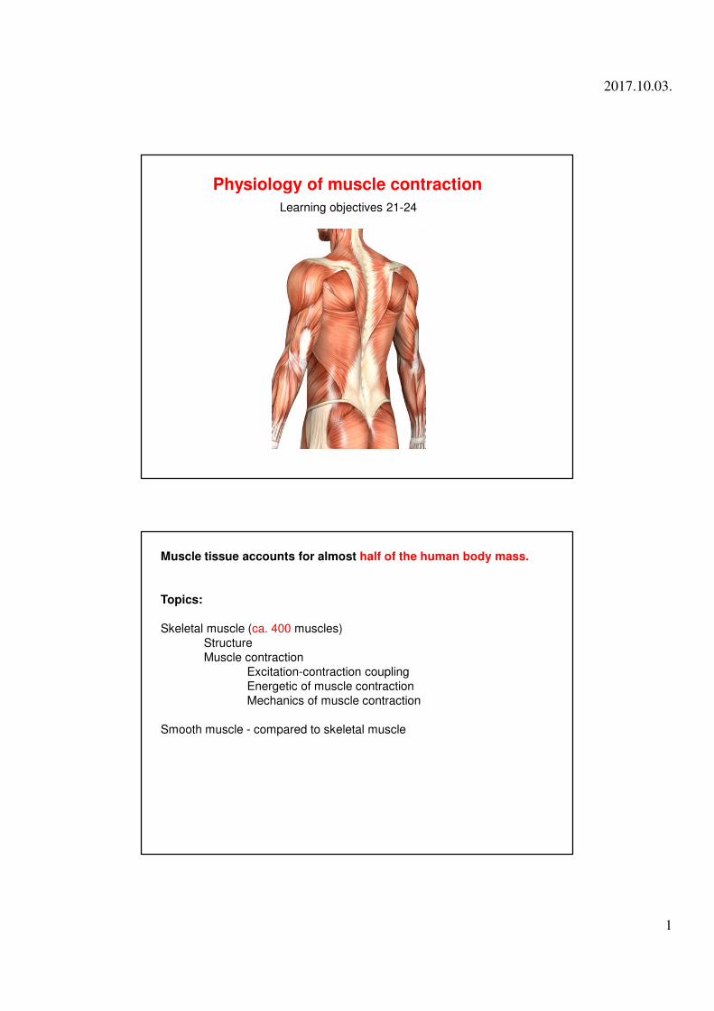
Physiology of muscle contraction
Learning objectives 21-24
Muscle tissue accounts for almost half of the human body mass.
Topics:
Skeletal muscle (ca. 400 muscles)
Structure
Muscle contraction
Excitation-contraction coupling
Energetic of muscle contraction
Mechanics of muscle contraction
Smooth muscle - compared to skeletal muscle
2017.10.03.
2
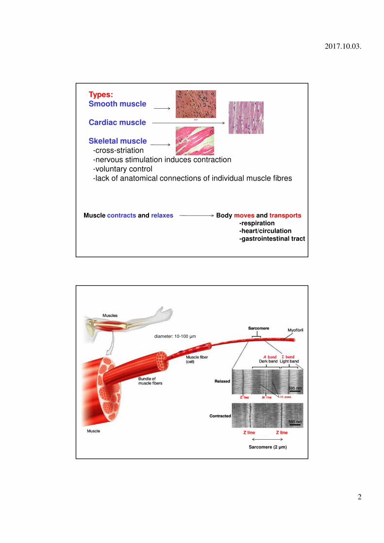
Types:Smooth muscle
Cardiac muscle
Skeletal muscle-cross-striation-nervous stimulation induces contraction-voluntary control-lack of anatomical connections of individual muscle fibres
Muscle contracts and relaxes Body moves and transports
-respiration
-heart/circulation
-gastrointestinal tract
Sarcomere (2 µm)
diameter: 10-100 µm
Z line Z line
2017.10.03.
3
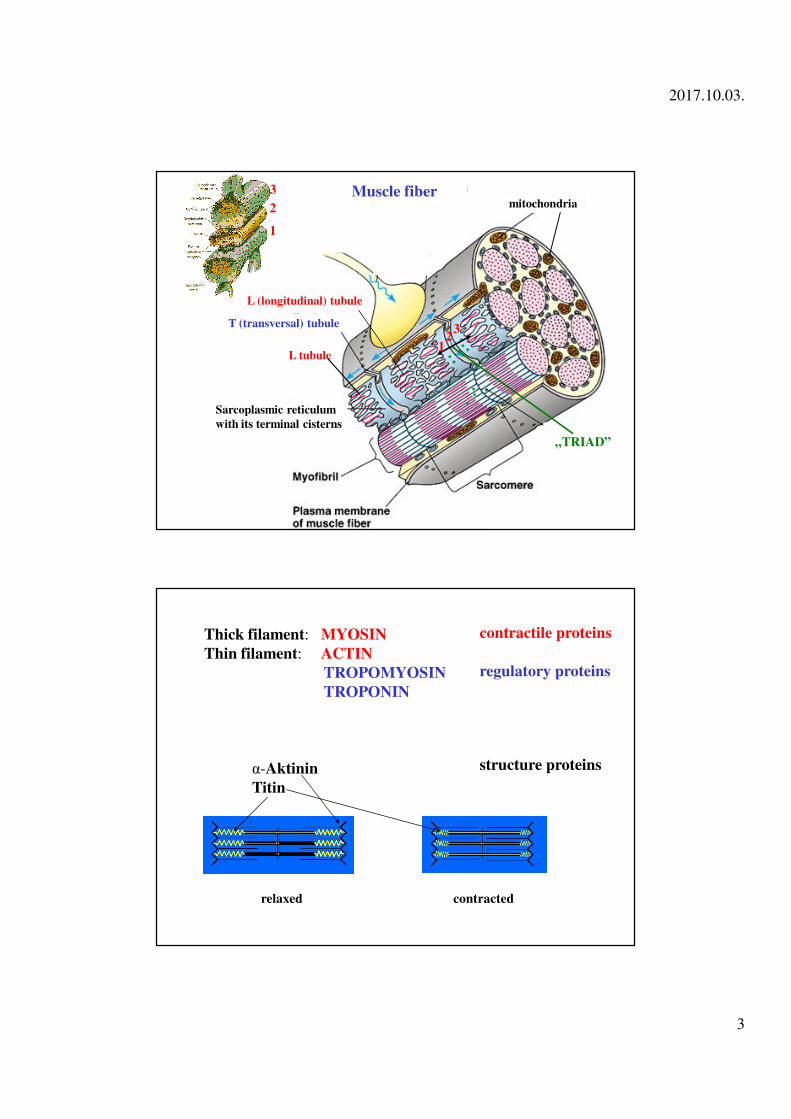
L tubule
„TRIAD”
T (transversal) tubule
12
3
Sarcoplasmic reticulum
with its terminal cisterns
mitochondriaMuscle fiber
L (longitudinal) tubule
2
3
1
Thick filament: MYOSIN
Thin filament: ACTIN
TROPOMYOSIN
TROPONIN
α-Aktinin
Titin
contractile proteins
regulatory proteins
relaxed contracted
structure proteins
2017.10.03.
4
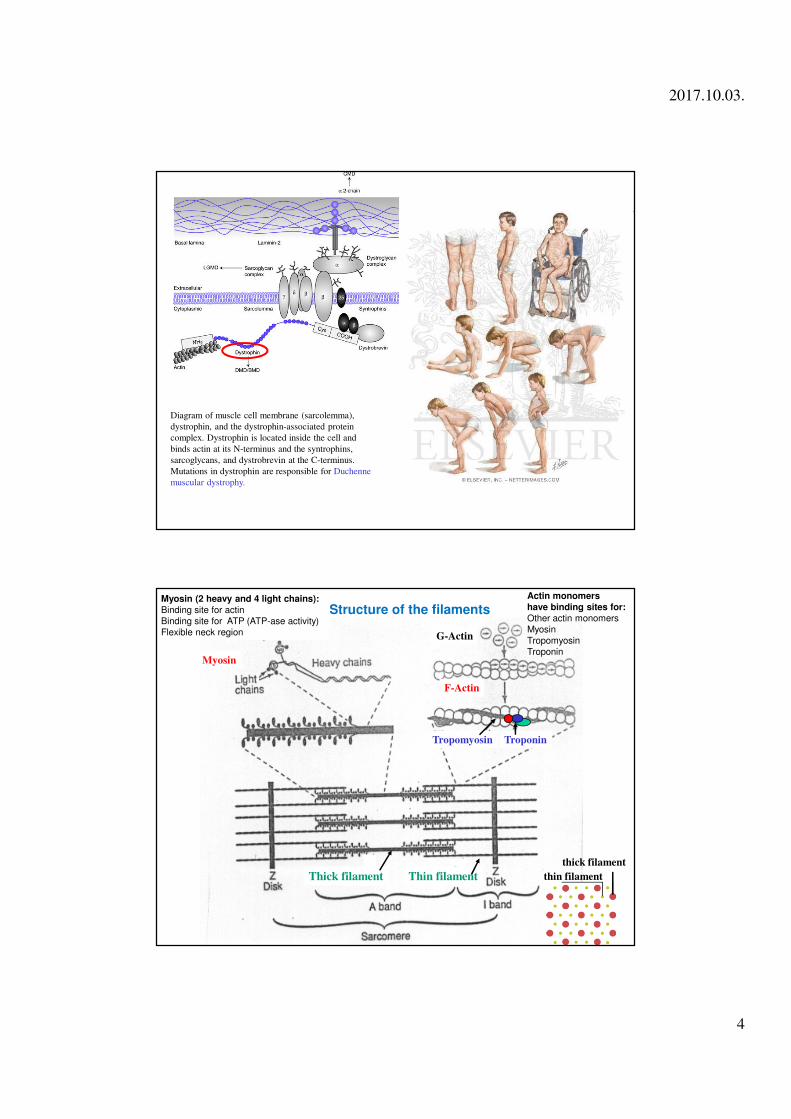
Diagram of muscle cell membrane (sarcolemma),
dystrophin, and the dystrophin-associated protein
complex. Dystrophin is located inside the cell and
binds actin at its N-terminus and the syntrophins,
sarcoglycans, and dystrobrevin at the C-terminus.
Mutations in dystrophin are responsible for Duchenne
muscular dystrophy.
G-Actin
F-Actin
Tropomyosin Troponin
Thick filament Thin filament thin filament
thick filament
Structure of the filamentsMyosin (2 heavy and 4 light chains):
Binding site for actin
Binding site for ATP (ATP-ase activity)
Flexible neck region
Actin monomers
have binding sites for:
Other actin monomers
Myosin
Tropomyosin
TroponinMyosin
2017.10.03.
5
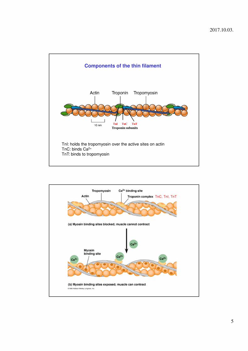
Components of the thin filament
Troponin subunits
TnI TnC TnT
TnI: holds the tropomyosin over the active sites on actin
TnC: binds Ca2+
TnT: binds to tropomyosin
2017.10.03.
6
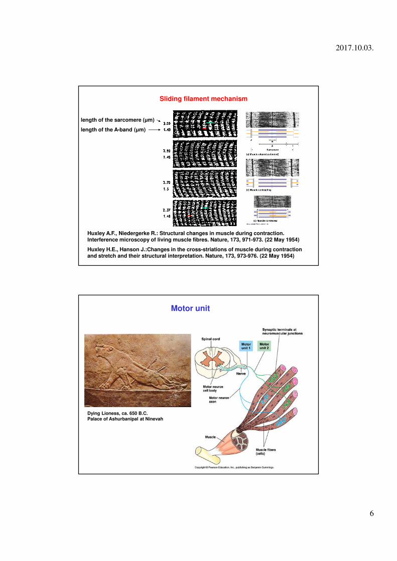
length of the sarcomere (µm)
length of the A-band (µm)
Sliding filament mechanism
Huxley A.F., Niedergerke R.: Structural changes in muscle during contraction. Interference microscopy of living muscle fibres. Nature, 173, 971-973. (22 May 1954)
Huxley H.E., Hanson J.:Changes in the cross-striations of muscle during contraction and stretch and their structural interpretation. Nature, 173, 973-976. (22 May 1954)
Dying Lioness, ca. 650 B.C.
Palace of Ashurbanipal at Ninevah
Motor unit
2017.10.03.
7
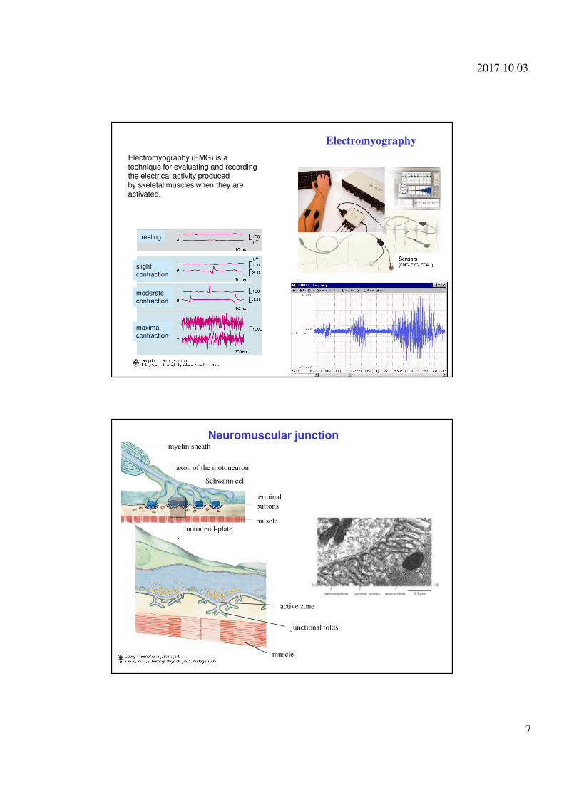
Electromyography
Electromyography (EMG) is a technique for evaluating and recording the electrical activity produced by skeletal muscles when they are activated.
resting
slight
contraction
moderate
contraction
maximal
contraction
myelin sheath
axon of the motoneuron
Schwann cell
motor end-plate
junctional folds
muscle
active zone
terminal
buttons
muscle
Neuromuscular junction
2017.10.03.
8
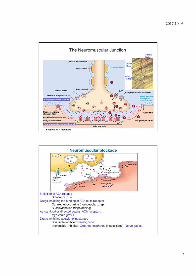
end-plate potential
nicotinic ACh receptors
Neuromuscular blockade
Inhibition of ACh releaseBotulinum toxin
Drugs inhibiting the binding of ACh to its receptorCurare, tubocurarine (non-depolarizing)Succinylcholine (depolarizing)
Autoantibodies directed against ACh receptorsMyastenia gravis
Drugs inhibiting acetylcholinesterasereversible inhibitor: Neostigmineirreversible inhibitor: Organophosphates (insecticides), Nerve gases
Acetat
2017.10.03.
9
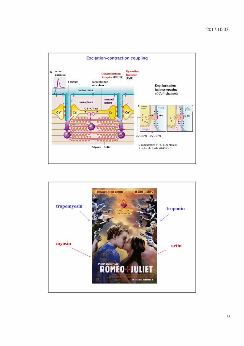
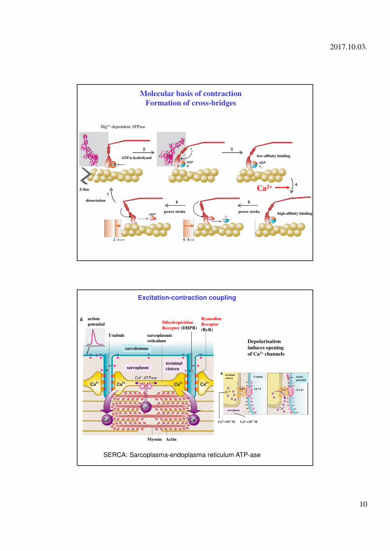
Excitation-contraction coupling
T-tubule
sarcoplasm
Ryanodine
Receptor
(RyR)
Dihydropiridine
Receptor (DHPR)
Depolarisation
induces opening
of Ca2+ channels
ActinMyosin
sarcolemma
action
potential
sarcoplasmic
reticulum
terminal
cistern
T-tubule terminal
cistern
sarcoplasm
action
potential
Ca2+<10-7 MCa2+≈10-3 M
Calsequestrin: 44-67 kDa protein
1 molecule binds 40-45 Ca2+
actinmyosin
troponintropomyosin
2017.10.03.
10
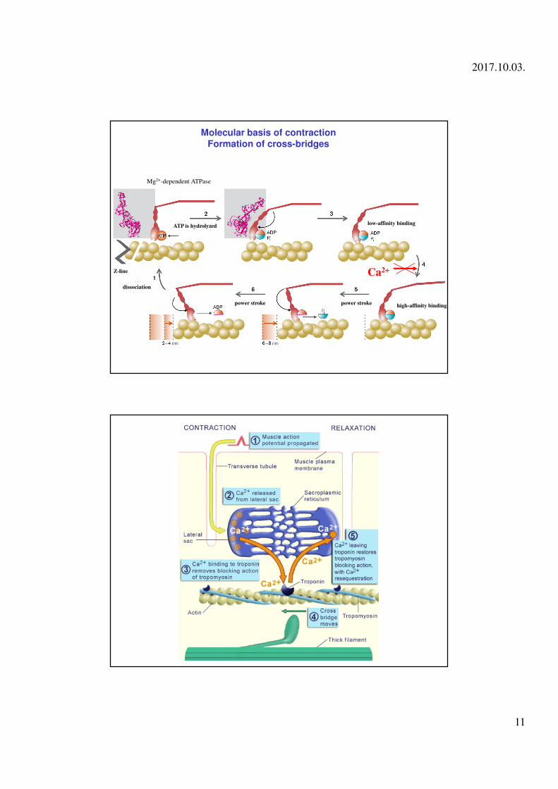
Molecular basis of contraction
Formation of cross-bridges
Z-line
ATP is hydrolyzedlow-affinity binding
high-affinity bindingpower strokepower stroke
dissociation
Ca2+
Mg2+-dependent ATPase
Excitation-contraction coupling
T-tubule
sarcoplasm
Ryanodine
Receptor
(RyR)
Dihydropiridine
Receptor (DHPR)
Depolarisation
induces opening
of Ca2+ channels
ActinMyosin
sarcolemma
action
potential
sarcoplasmic
reticulum
terminal
cistern
T-tubule terminal
cistern
sarcoplasm
action
potential
Ca2+<10-7 MCa2+≈10-3 M
SERCA: Sarcoplasma-endoplasma reticulum ATP-ase
2017.10.03.
11
Molecular basis of contraction
Formation of cross-bridges
Z-line
ATP is hydrolyzedlow-affinity binding
high-affinity bindingpower strokepower stroke
dissociation
Ca2+
Mg2+-dependent ATPase
2017.10.03.
12
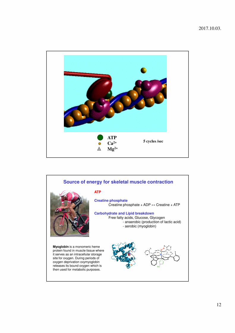
ATP
Ca2+
Mg2+
5 cycles /sec
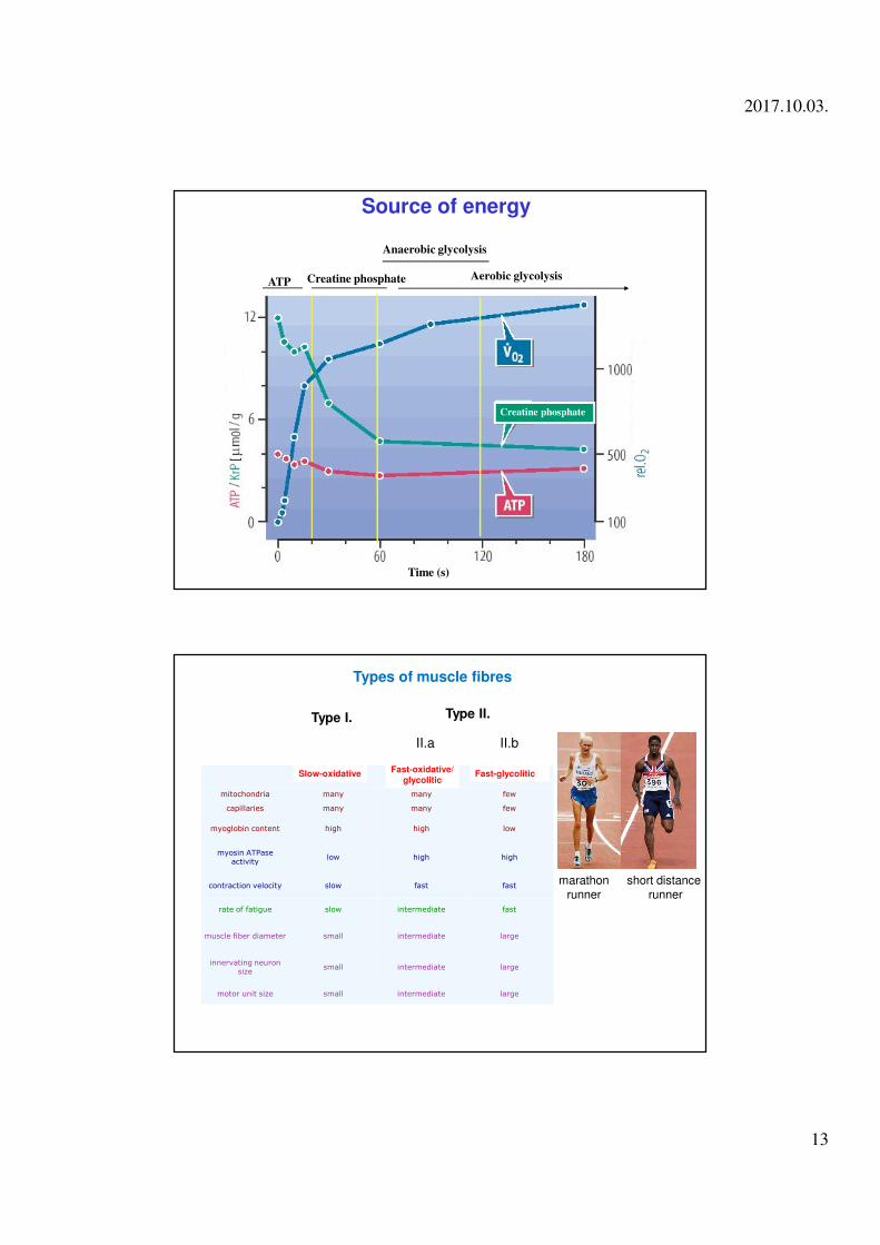
Source of energy for skeletal muscle contraction
ATP
Creatine phosphateCreatine phosphate + ADP ↔ Creatine + ATP
Carbohydrate and Lipid breakdownFree fatty acids, Glucose, Glycogen
- anaerobic (production of lactic acid) - aerobic (myoglobin)
Myoglobin is a monomeric heme protein found in muscle tissue where it serves as an intracellular storage site for oxygen. During periods of oxygen deprivation oxymyoglobin releases its bound oxygen which is then used for metabolic purposes.
2017.10.03.
13
Source of energy
ATP
Creatine phosphate
Anaerobic glycolysis
Aerobic glycolysis
Time (s)
Creatine phosphate
Slow-oxidative Fast-oxidative Fast-glycolytic
mitochondria many many few
capillaries many many few
myoglobin content high high low
myosin ATPase activity
low high high
contraction velocity slow fast fast
rate of fatigue slow intermediate fast
muscle fiber diameter small intermediate large
innervating neuron size
small intermediate large
motor unit size small intermediate large
short distancerunner
marathonrunner
Types of muscle fibres
Type I. Type II.
II.a II.b
Slow-oxidative Fast-oxidative/glycolitic
Fast-glycolitic
2017.10.03.
14
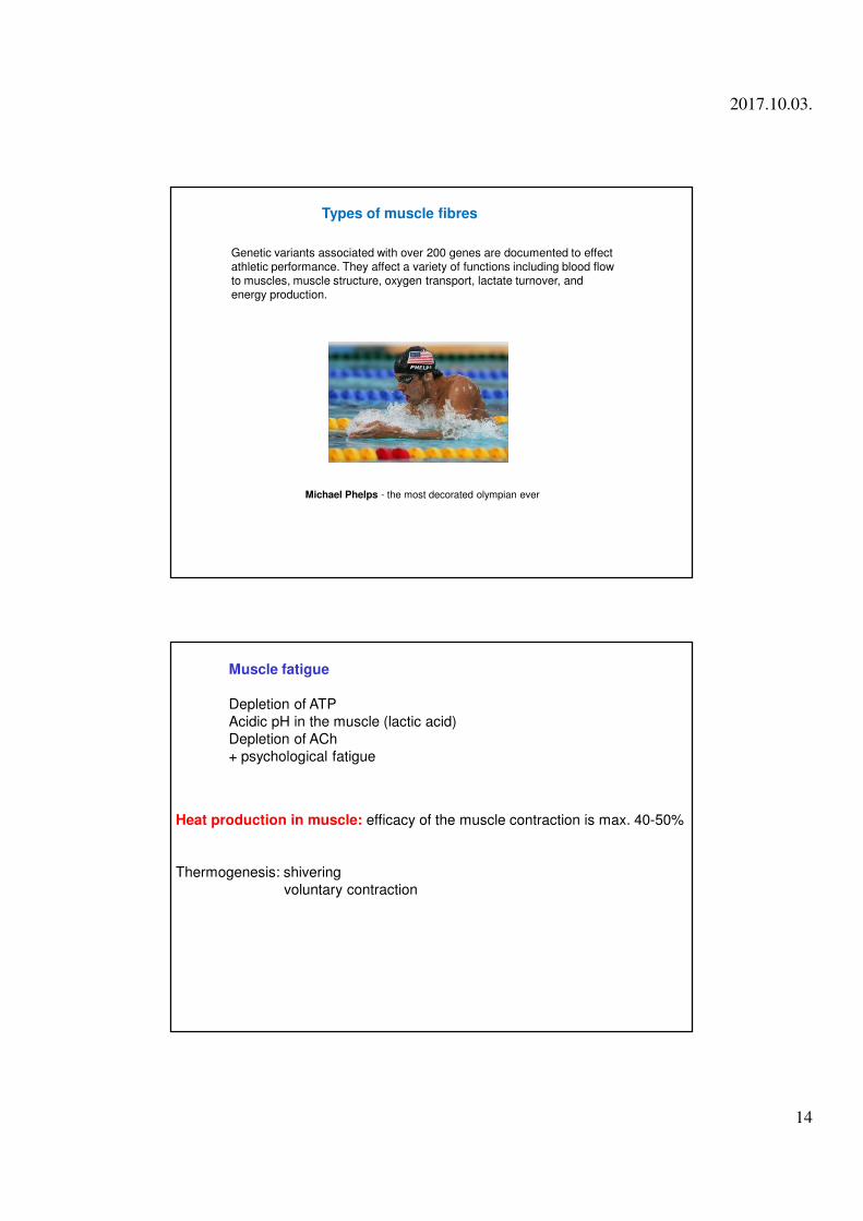
Michael Phelps - the most decorated olympian ever
Genetic variants associated with over 200 genes are documented to effect athletic performance. They affect a variety of functions including blood flow to muscles, muscle structure, oxygen transport, lactate turnover, and energy production.
Types of muscle fibres
Muscle fatigue
Depletion of ATP
Acidic pH in the muscle (lactic acid)
Depletion of ACh
+ psychological fatigue
Heat production in muscle: efficacy of the muscle contraction is max. 40-50%
Thermogenesis: shivering
voluntary contraction
2017.10.03.
15
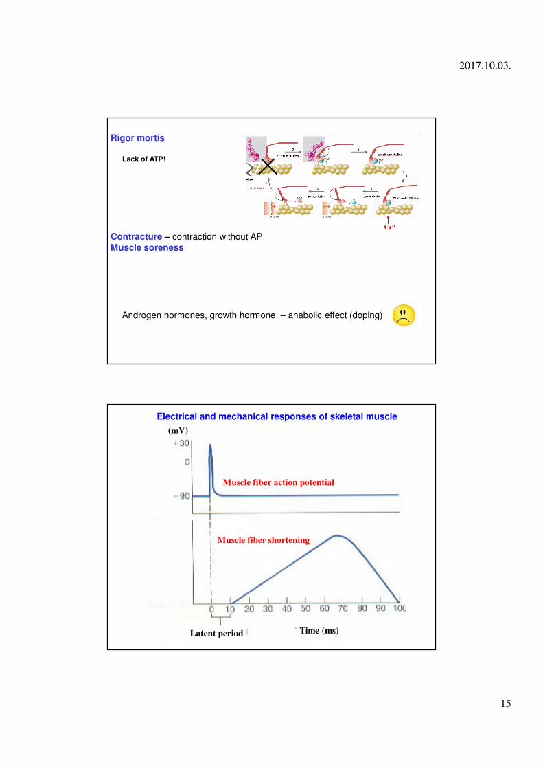
Rigor mortis
Contracture – contraction without AP
Muscle soreness
Lack of ATP!
Androgen hormones, growth hormone – anabolic effect (doping)
Muscle fiber action potential
Muscle fiber shortening
Latent period Time (ms)
(mV)
Electrical and mechanical responses of skeletal muscle
2017.10.03.
16
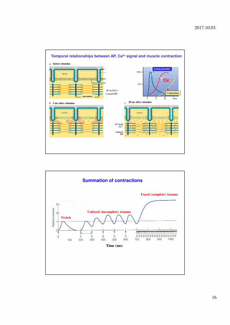
before stimulus
sarcomere
5 ms after stimulus 20 ms after stimulus
Action potential
Contraction
Temporal relationships between AP, Ca2+ signal and muscle contraction
Twitch
Unfused (incomplete) tetanus
Fused (complete) tetanus
Time (ms)
Summation of contractions
2017.10.03.
17
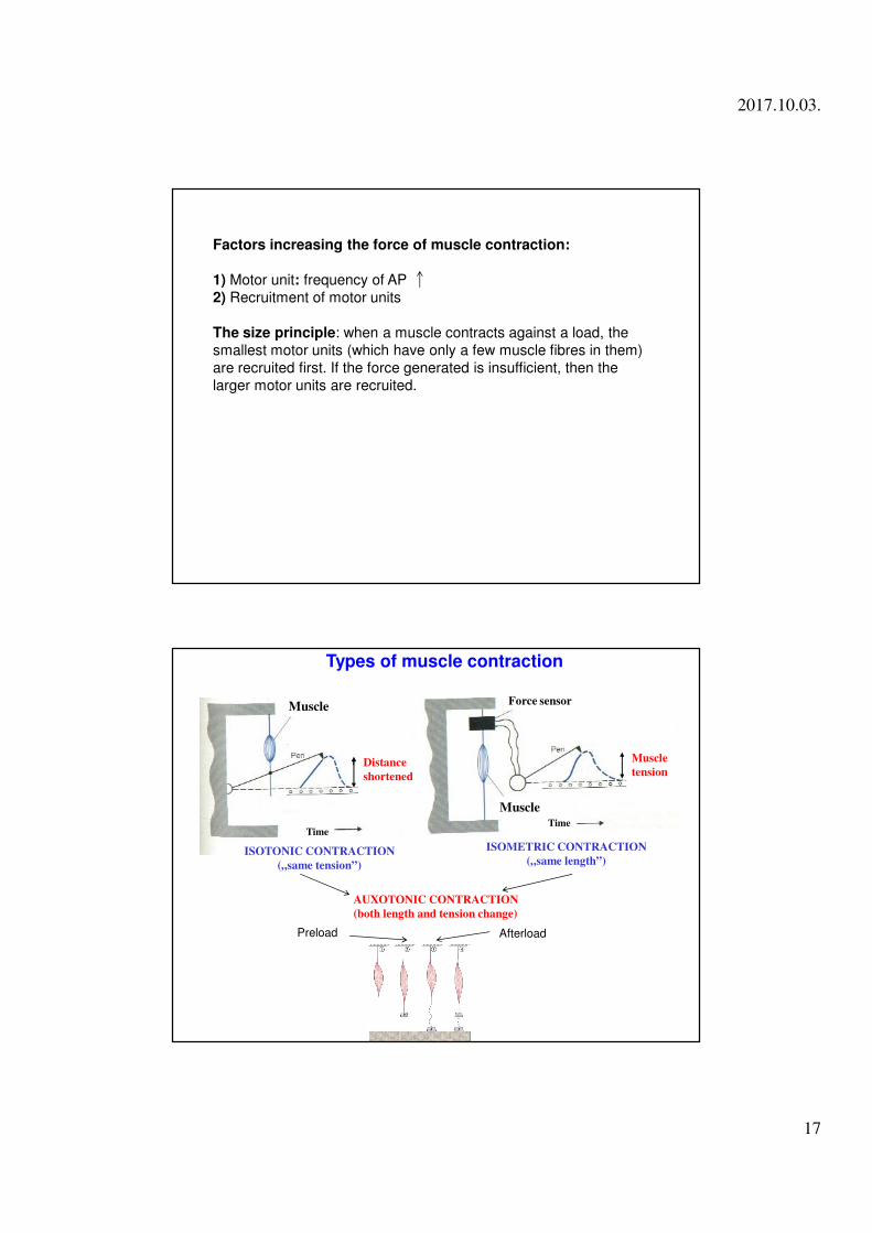
Factors increasing the force of muscle contraction:
1) Motor unit: frequency of AP
2) Recruitment of motor units
The size principle: when a muscle contracts against a load, the
smallest motor units (which have only a few muscle fibres in them)
are recruited first. If the force generated is insufficient, then the
larger motor units are recruited.
Distance
shortened
Muscle
tension
Muscle
TimeTime
Muscle
Force sensor
Types of muscle contraction
ISOTONIC CONTRACTION
(„same tension”)
ISOMETRIC CONTRACTION
(„same length”)
AUXOTONIC CONTRACTION
(both length and tension change)
Preload Afterload
2017.10.03.
18
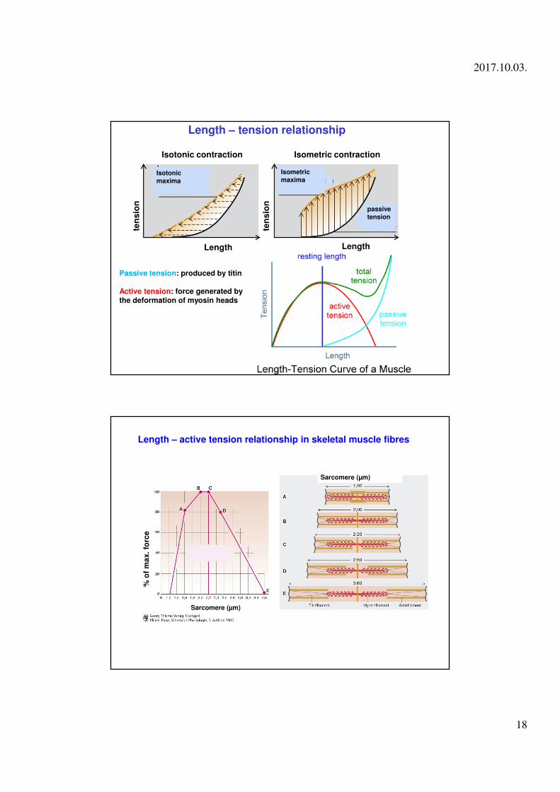
Length – tension relationship
ten
sio
n
passive
tension
Isotonic
maxima
Isometric
maxima
ten
sio
n
Length
Isotonic contraction Isometric contraction
Passive tension: produced by titin
Active tension: force generated by the deformation of myosin heads
Length
Sarcomere (µm)
% o
f m
ax. fo
rce
Length – active tension relationship in skeletal muscle fibres
Sarcomere (µm)
2017.10.03.
19
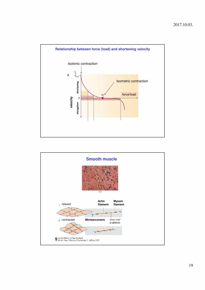
Relationship between force (load) and shortening velocity
Isotonic contraction
ve
loc
ity
elo
ng
ati
on
sh
ort
en
ing
force/load
Isometric contraction
relaxed
contracted
Actinfilament
Myosinfilament
Minisarcomere
Smooth muscle
α-aktinin
2017.10.03.
20
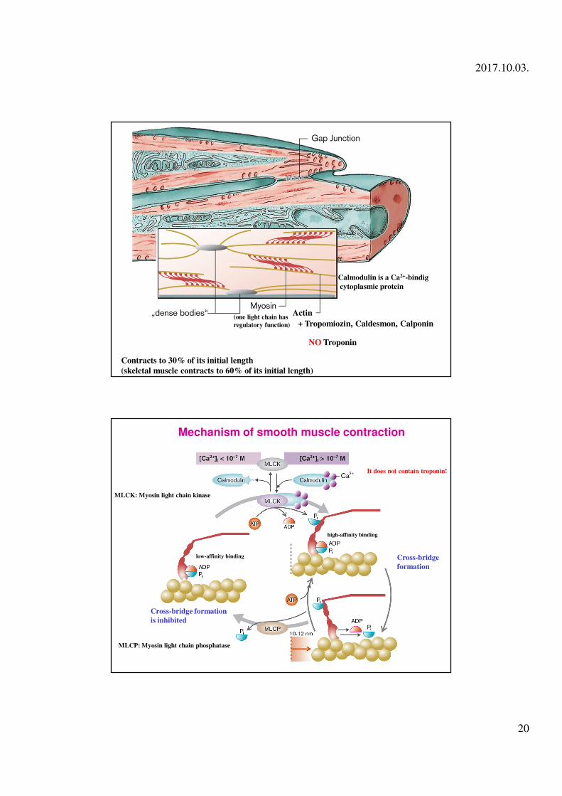
Contracts to 30% of its initial length
(skeletal muscle contracts to 60% of its initial length)
Actin
+ Tropomiozin, Caldesmon, Calponin
NO Troponin
Calmodulin is a Ca2+-bindig
cytoplasmic protein
(one light chain has
regulatory function)
Mechanism of smooth muscle contraction
MLCK: Myosin light chain kinase
Cross-bridge
formation
Cross-bridge formation
is inhibited
MLCP: Myosin light chain phosphatase
low-affinity binding
high-affinity binding
It does not contain troponin!
2017.10.03.
21
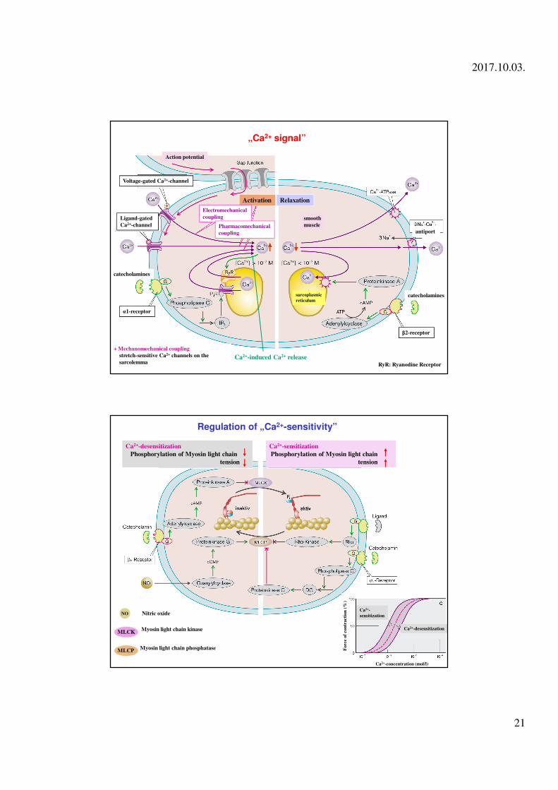
Action potential
Voltage-gated Ca2+-channel
Ligand-gated
Ca2+-channel
catecholamines
α1-receptor
Activation Relaxation
Electromechanical
coupling
Pharmacomechanical
coupling
smooth
muscle
antiport
catecholamines
β2-receptor
sarcoplasmic
reticulum
„Ca2+ signal”
Ca2+-induced Ca2+ release
+ Mechanomechanical coupling
stretch-sensitive Ca2+ channels on the
sarcolemma RyR: Ryanodine Receptor
Regulation of „Ca2+-sensitivity”
Ca2+-desensitization
Phosphorylation of Myosin light chain
tension
Ca2+-sensitization
Phosphorylation of Myosin light chain
tension
Myosin light chain kinase
Myosin light chain phosphatase
Ca2+-desensitization
Ca2+-
sensitization
Force o
f con
tra
cti
on
(%
)
Ca2+-concentration (mol/l)
Nitric oxideNO
MLCK
MLCP
2017.10.03.
22
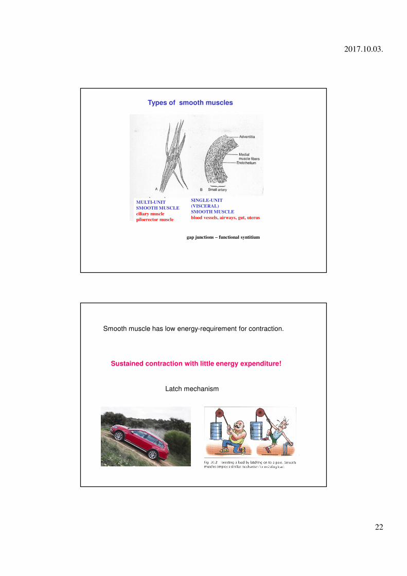
MULTI-UNIT
SMOOTH MUSCLE
ciliary muscle
piloerector muscle
SINGLE-UNIT
(VISCERAL)
SMOOTH MUSCLE
blood vessels, airways, gut, uterus
Types of smooth muscles
gap junctions – functional syntitium
Smooth muscle has low energy-requirement for contraction.
Latch mechanism
Sustained contraction with little energy expenditure!
2017.10.03.
23
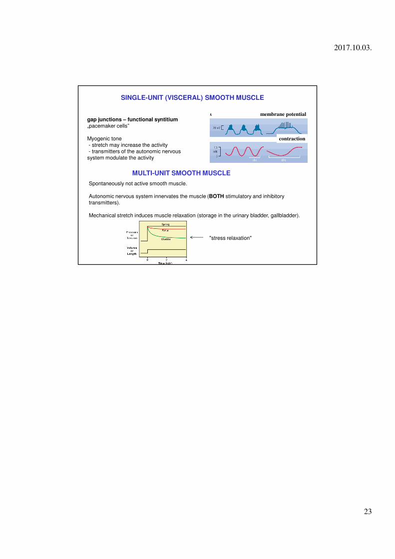
gap junctions – functional syntitium„pacemaker cells”
Myogenic tone- stretch may increase the activity- transmitters of the autonomic nervous system modulate the activity
SINGLE-UNIT (VISCERAL) SMOOTH MUSCLE
membrane potential
contraction
MULTI-UNIT SMOOTH MUSCLE
Spontaneously not active smooth muscle.
Autonomic nervous system innervates the muscle (BOTH stimulatory and inhibitory transmitters).
Mechanical stretch induces muscle relaxation (storage in the urinary bladder, gallbladder).
"stress relaxation"