Embed Size (px)
Citation preview
Plant Physiol. (1989) 90, 11 5-011200032-0889/89/90/111 5/06/$01 .00/0
Received for publication December 5, 1988and in revised form March 13, 1989
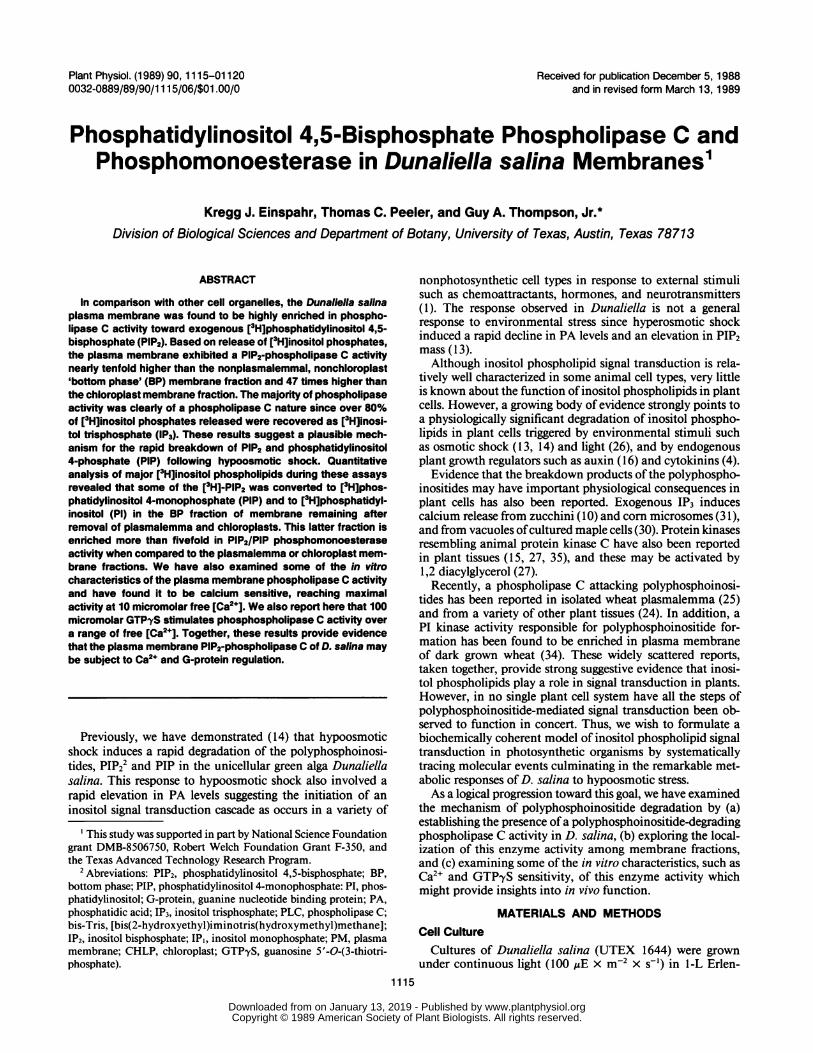
Phosphatidylinositol 4,5-Bisphosphate Phospholipase C andPhosphomonoesterase in Dunaliella salina Membranes1
Kregg J. Einspahr, Thomas C. Peeler, and Guy A. Thompson, Jr.*Division of Biological Sciences and Department of Botany, University of Texas, Austin, Texas 78713
ABSTRACT
In comparison with other cell organelles, the Dunaliella salinaplasma membrane was found to be highly enriched in phospho-lipase C activity toward exogenous [3H]phosphatidylinositol 4,5-bisphosphate (PIP2). Based on release of [3H]inositol phosphates,the plasma membrane exhibited a PIP2-phospholipase C activitynearly tenfold higher than the nonplasmalemmal, nonchloroplast'bottom phase' (BP) membrane fraction and 47 times higher thanthe chloroplast membrane fraction. The majority of phospholipaseactivity was clearly of a phospholipase C nature since over 80%of [3H]inositol phosphates released were recovered as [3H]inosi-tol trisphosphate (IP3). These results suggest a plausible mech-anism for the rapid breakdown of PIP2 and phosphatidylinositol4-phosphate (PIP) following hypoosmotic shock. Quantitativeanalysis of major [3H]inositol phospholipids during these assaysrevealed that some of the [3H1-PIP2 was converted to [3H]phos-phatidylinositol 4-monophosphate (PIP) and to [3H]phosphatidyl-inositol (PI) in the BP fraction of membrane remaining afterremoval of plasmalemma and chloroplasts. This latter fraction isenriched more than fivefold in PIP2/PIP phosphomonoesteraseactivity when compared to the plasmalemma or chloroplast mem-brane fractions. We have also examined some of the in vitrocharacteristics of the plasma membrane phospholipase C activityand have found it to be calcium sensitive, reaching maximalactivity at 10 micromolar free [Ca2+J. We also report here that 100micromolar GTP'yS stimulates phosphospholipase C activity overa range of free [Ca2+]. Together, these results provide evidencethat the plasma membrane PIP2-phospholipase C of D. salina maybe subject to Ca2+ and G-protein regulation.
Previously, we have demonstrated (14) that hypoosmoticshock induces a rapid degradation of the polyphosphoinosi-tides, PIP22 and PIP in the unicellular green alga Dunaliellasalina. This response to hypoosmotic shock also involved a
rapid elevation in PA levels suggesting the initiation of an
inositol signal transduction cascade as occurs in a variety of
' This study was supported in part by National Science Foundationgrant DMB-8506750, Robert Welch Foundation Grant F-350, andthe Texas Advanced Technology Research Program.
2Abreviations: PIP2, phosphatidylinositol 4,5-bisphosphate; BP,bottom phase; PIP, phosphatidylinositol 4-monophosphate: PI, phos-phatidylinositol; G-protein, guanine nucleotide binding protein; PA,phosphatidic acid; IP3, inositol trisphosphate; PLC, phospholipase C;bis-Tris, [bis(2-hydroxyethyl)iminotris(hydroxymethyl)methane];IP2, inositol bisphosphate; IPI, inositol monophosphate; PM, plasmamembrane; CHLP, chloroplast; GTPyS, guanosine 5'-O-(3-thiotri-phosphate).
nonphotosynthetic cell types in response to external stimulisuch as chemoattractants, hormones, and neurotransmitters(1). The response observed in Dunaliella is not a generalresponse to environmental stress since hyperosmotic shockinduced a rapid decline in PA levels and an elevation in PIP2mass (13).Although inositol phospholipid signal transduction is rela-
tively well characterized in some animal cell types, very littleis known about the function ofinositol phospholipids in plantcells. However, a growing body of evidence strongly points toa physiologically significant degradation of inositol phospho-lipids in plant cells triggered by environmental stimuli suchas osmotic shock (13, 14) and light (26), and by endogenousplant growth regulators such as auxin (16) and cytokinins (4).
Evidence that the breakdown products of the polyphospho-inositides may have important physiological consequences inplant cells has also been reported. Exogenous IP3 inducescalcium release from zucchini (10) and corn microsomes (31),and from vacuoles ofcultured maple cells (30). Protein kinasesresembling animal protein kinase C have also been reportedin plant tissues (15, 27, 35), and these may be activated by1,2 diacylglycerol (27).Recently, a phospholipase C attacking polyphosphoinosi-
tides has been reported in isolated wheat plasmalemma (25)and from a variety of other plant tissues (24). In addition, aPI kinase activity responsible for polyphosphoinositide for-mation has been found to be enriched in plasma membraneof dark grown wheat (34). These widely scattered reports,taken together, provide strong suggestive evidence that inosi-tol phospholipids play a role in signal transduction in plants.However, in no single plant cell system have all the steps ofpolyphosphoinositide-mediated signal transduction been ob-served to function in concert. Thus, we wish to formulate abiochemically coherent model of inositol phospholipid signaltransduction in photosynthetic organisms by systematicallytracing molecular events culminating in the remarkable met-abolic responses of D. salina to hypoosmotic stress.As a logical progression toward this goal, we have examined
the mechanism of polyphosphoinositide degradation by (a)establishing the presence ofa polyphosphoinositide-degradingphospholipase C activity in D. salina, (b) exploring the local-ization of this enzyme activity among membrane fractions,and (c) examining some of the in vitro characteristics, such asCa2' and GTPyS sensitivity, of this enzyme activity whichmight provide insights into in vivo function.
MATERIALS AND METHODSCell Culture
Cultures of Dunaliella salina (UTEX 1644) were grownunder continuous light (100 ,E x m-2 x s-') in l-L Erlen-
1115
www.plantphysiol.orgon January 13, 2019 - Published by Downloaded from Copyright © 1989 American Society of Plant Biologists. All rights reserved.
Plant Physiol. Vol. 90, 1989
meyer flasks containing 500 mL of synthetic medium bubbledwith 0.5% C02-enriched air at 30°C (13). Under these condi-tions, the cells grew with a generation time of 20 h. Culturesroutinely used for experiments were grown to a density of 0.8to 1.2 x 10⁶ cells/mL. Cell population density was measuredusing a Coulter Counter model ZB.
Chemicals
[Inositol-2-3H(N)]phosphatidylinositol 4,5-bisphosphate,2.8 Ci/mmol, was obtained from DuPont NEN, (Boston,MA). Nonradiolabeled PIP2 and PIP (bovine brain) standardswere purchased from Sigma (St. Louis, MO). AGlX8 anionexchange resin, 200 to 400 mesh, formate form, was fromBio-Rad (Richmond, CA).
Membrane Fractionation
Membrane fractions incuding plasmalemma were preparedas described in detail by (29). Cells were harvested and frac-tionated in a disruption buffer containing 400 mm mannitol,2 mm EDTA, 1 mm MgCl, 100 mM NaPO4 (pH 8.0). Afterdisruption in a Parr bomb, the suspension ofbroken cells wascentrifuged at 2,600g for 3.5 min to pellet intact chloroplasts.The supernatant over the chloroplast pellet was then addedto a dextran-PEG aqueous two-phase system to partition theplasma membrane away from the remainder ofthe organelles.The final concentrations of each of the components of thetwo-phase system were: 6.7% PEG, 6.7% dextran T500 (w/w), 178 mM mannitol, 0.89 mM EDTA, 0.44 mM MgCl2, 44.6mM NaPO4 (pH 8.0). In addition, approximately 30 mM NaClwas added to improve the partitioning of plasma membrane(29). In a typical experiment, 1.5 x 10⁹ cells were lysed in 40mL of phosphate disruption buffer, and the subsequent su-pernatant was divided between two separate tubes of thedextran-PEG system, so that 17.5 mL of supernatant wereadded to 21.7 mL of the dextran-PEG system. The completetwo-phase system was mixed by shaking and centrifuged at660g for 10 min in a Sorvall HB-4 swinging bucket rotor. Theupper phase (PEG-rich) was removed and centrifuged at1 50,000g for 1 h in a Beckman SW-4 1 swinging bucket rotor.The resulting pellet was the plasma membrane fraction. Theremaining lower phase (dextran-rich) was diluted with bufferand also centrifuged at 150,000g for 1 h. This BP pelletcontained all cellular membranes other than the plasma mem-brane and chloroplasts. All steps of this isolation procedurewere carried out at 4°C. As assayed and measured by themethods described previously (29), the purified plasma mem-brane was free of Chl, free of Cyt c oxidase activity, andenriched in an ATPase activity that was stimulated by K+ andinhibited by vanadate.
PLC Assay
The assay for PLC activity in D. salina membrane fractionswas slightly modified from previously published methods (33).Essentially, this is an in vitro assay using exogenous radiola-beled inositol phospholipid as the substrate for enzyme pre-sent in cell fractions isolated as described above. Approxi-mately 18 h prior to the assay, stock [3H]inositol phospholipid
solution, 0.10 gCi or 0.05 ,uCi per assay, was added to anacidwashed glass tube and dried under a steady stream of N2.After this, 5 AsL per assay of buffer consisting of 0.1 M bis-Trisbuffer (pH 6.5), 100 gM CaCl2, 5% sodium cholate was thenadded and vortexed for about 1 min. This was then incubatedat room temperature overnight to completely dissolve thephospholipid after which an equal volume of assay bufferlacking sodium cholate was added and vortex mixed. Theassay, started by adding [3H]phospholipid substrate, containedfinal concentrations of 0.1 M bis-Tris (pH 6.5), 100 uM CaCl2,10 to 50 ,ug membrane protein, and 5.5 mm sodium cholatein a final volume of 100 ,ul at 30°C. For experiments in whichcalcium sensitivity of PLC was measured, 1.0 mM EGTA wasincluded in the assay buffer, and the total calcium added forgiven free calcium concentrations was determined using bind-ing and stability constants of Martell and Smith (23). Sampleswere quenched with 500 ,uL of ice-cold chloroform:methanol:conc. HC1 (200:100:1.5). Control samples wereboiled for 5 min and cooled to 30°C prior to addition ofradiolabeled phospholipid substrate. After adding 250 AL ofH20 to each sample, they were centrifuged at 1060g for 4min and the organic phase removed to another tube. Theaqueous phase was reextracted with 250 uL of chloroform,and the organic phase was removed and combined with theprevious organic phase.
Lipids of the organic phase were analyzed by TLC, andradioactivity of individual phospholipids was quantified asdescribed previously (13, 14). Inositol phosphates were quan-tified by sequential elution from Bio-Rad AG1X8 anionexchange resin columns utilizing a slightly modified methoddescribed by Irvine (20). Anion exchange columns were madewith 0.5 g of resin (200-400 mesh, formate form) packed intoglass Pasteur pipettes plugged with a small amount of glasswool. The aqueous phase from PLC assays was neutralizedwith KOH, brought to a volume of 1 mL with H20, andapplied to the column. Fractions were eluted with the follow-ing sequence of solutions. (a) 25 mL H20, (b) 10 mL 5 mMdisodium tetraborate/60 mm sodium formate, (c) 10 mL 0.1M formic acid/0.2 M ammonium formate, (d) 10 mL 0.1 Mformic acid/0.4 M ammonium formate, (e) 10 mL 0.1 Mformic acid/0.8 M ammonium formate, (f) 10 mL 0.1 M formicacid/1.0 M ammonium formate. As reported previously (21),and supported by paper chromatography of radioactiveeluates run with standards in the solvent system of Desjobertand Petek (6) with the detection spray of Clarke and Dawson(3), the order of elution was inositol, glycerophosphoinositol,inositol monophosphate, IP2, IP3, and inositol tetrakisphos-phate. Two aliquots of 2 mL each from each fraction wereadded to 13 mL ofscintillation fluor and radioactivity countedas described previously (13, 14). In some experiments theaqueous phase containing the combined [3H]inositol phos-phates was counted directly in scintillation fluor. The validityof results utilizing this method were confirmed by comparisonwith results from identical experiments in which samples wereanalyzed by the anion exchange method already described.
RESULTS
When Dunaliella salina cellular membranes were fraction-ated and assayed for PIP2-PLC activity using exogenous ra-
1116 EINSPAHR ET AL.
www.plantphysiol.orgon January 13, 2019 - Published by Downloaded from Copyright © 1989 American Society of Plant Biologists. All rights reserved.
PIP2 PHOSPHOLIPASE C AND PHOSPHOMONOESTERASE
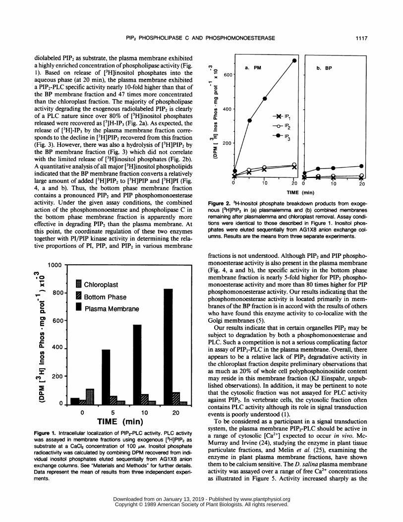
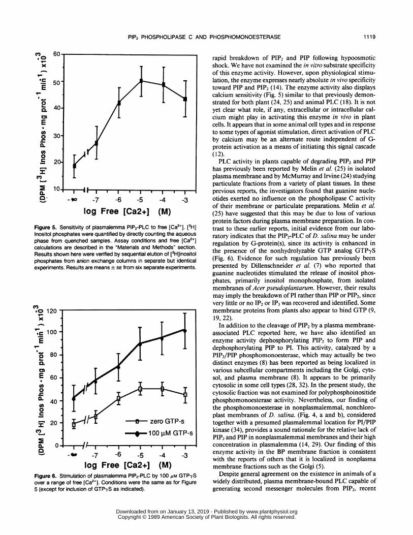
diolabeled PIP2 as substrate, the plasma membrane exhibiteda highly enriched concentration ofphospholipase activity (Fig.1). Based on release of [3H]inositol phosphates into theaqueous phase (at 20 min), the plasma membrane exhibiteda PIP2-PLC specific activity nearly 10-fold higher than that ofthe BP membrane fraction and 47 times more concentratedthan the chloroplast fraction. The majority of phospholipaseactivity degrading the exogenous radiolabeled PIP2 is clearlyof a PLC nature since over 80% of [3H]inositol phosphatesreleased were recovered as [3]H-IP3 (Fig. 2a). As expected, therelease of [3H]-IP3 by the plasma membrane fraction corre-sponds to the decline in [3H]PIP2 recovered from this fraction(Fig. 3). However, there was also a hydrolysis of [3H]PIP2 bythe BP membrane fraction (Fig. 3) which did not correlatewith the limited release of [3H]inositol phosphates (Fig. 2b).A quantitative analysis of all major [3H]inositol phospholipidsindicated that the BP membrane fraction converts a relativelylarge amount of added [3H]PIP2 to [3H]PIP and [3H]PI (Fig.4, a and b). Thus, the bottom phase membrane fractioncontains a pronounced PIP2 and PIP phosphomonoesteraseactivity. Under the given assay conditions, the combinedaction of the phosphomonoesterase and phospholipase C inthe bottom phase membrane fraction is apparently moreeffective in degrading PIP2 than the plasma membrane. Atthis point, the coordinate regulation of these two enzymestogether with PI/PIP kinase activity in determining the rela-tive proportions of PI, PIP, and PIP2 in various membrane
1000
.0oCI-0
a-
O-f
r
cm0
a.
800
600
400.
200
0.
0 5 10 20
TIME (min)Figure 1. Intracellular localization of PIP2-PLC activity. PLC activitywas assayed in membrane fractions using exogenous [3H]PIP2 assubstrate at a CaCI2 concentration of 100 gM. Inositol phosphateradioactivity was calculated by combining DPM recovered from indi-vidual inositol phosphates eluted sequentially from AG1X8 anionexchange columns. See "Materials and Methods" for further details.Data represent the mean of results from three independent experi-ments.
I 0
-
0.
0)
E
0m
cn0
0.
CZ2
b. BP
10 20TIME (min)
Figure 2. 3H-Inositol phosphate breakdown products from exoge-nous [3H]PIP2 in (a) plasmalemma and (b) combined membranesremaining after plasmalemma and chloroplast removal. Assay condi-tions were identical to those described in Figure 1. Inositol phos-phates were eluted sequentially from AG1X8 anion exchange col-umns. Results are the means from three separate experiments.
fractions is not understood. Although PIP2 and PIP phospho-monoesterase activity is also present in the plasma membrane(Fig. 4, a and b), the specific activity in the bottom phasemembrane fraction is nearly 5-fold higher for PIP2 phospho-monoesterase activity and more than 80 times higher for PIPphosphomonoesterase activity. Our results indicating that thephosphomonoesterase activity is located primarily in mem-branes ofthe BP fraction is in accord with the results of otherswho have found this enzyme activity to co-localize with theGolgi membranes (5).Our results indicate that in certain organelles PIP2 may be
subject to degradation by both a phosphomonoesterase andPLC. Such a competition is not a serious complicating factorin assay of PIP2-PLC in the plasma membrane. Overall, thereappears to be a relative lack of PIP2 degradative activity inthe chloroplast fraction despite preliminary observations thatas much as 20% of whole cell polyphosphoinositide contentmay reside in this membrane fraction (KJ Einspahr, unpub-lished observations). In addition, it may be pertinent to notethat the cytosolic fraction was not assayed for PLC activityagainst PIP2. In vertebrate cells, the cytosolic fraction oftencontains PLC activity although its role in signal transductionevents is poorly understood (1).To be considered as a participant in a signal transduction
system, the plasma membrane PIP2-PLC should be active ina range of cytosolic [Ca2+] expected to occur in vivo. Mc-Murray and Irvine (24), studying the enzyme in plant tissueparticulate fractions, and Melin et al. (25), examining theenzyme in plant plasma membrane fractions, have shownthem to be calcium sensitive. The D. salina plasma membraneactivity was assayed over a range of free Ca2+ concentrationsas illustrated in Figure 5. Activity increased sharply as the
1117
0
www.plantphysiol.orgon January 13, 2019 - Published by Downloaded from Copyright © 1989 American Society of Plant Biologists. All rights reserved.

Plant Physiol. Vol. 90, 1989
100
I-
C)I-
0
0
C.
E
0
0
80
60
40
20
00 10 20
TIME (min)Figure 3. Decline in [3H]PIP2 recovered from membrane fractionsassayed for PLC activity. Assay conditions were identical to thosedescribed in Figure 1. After extraction of phospholipids, [3H]PIP2 wasanalyzed by TLC as described previously (14). Results represent themeans from three independent experiments.
[Ca2+] reached 1 ,M and became maximal at 10 Mm. Thus,PIP2-PLC was activated at the upper levels of free Ca2+ thatare thought to be physiological (1).We have also begun initial experiments to explore the
potential involvement of GTP-binding proteins (G-proteins)in the regulation D. salina PLC. Such G-proteins appear toplay an important role in some vertebrate cells in couplingreceptor activation to PLC activation (17). We have foundthat over a range of free [Ca2+], 100 AM GTPyS stimulatesplasma membrane PLC (Fig. 6). This provides initial evidencethat the plasmalemmal PLC may be under G-protein regula-tion as it appears to be in other cell systems (17, 18, 33).
DISCUSSION
Our overriding purpose in examining inositol phospholipidmetabolism in Dunaliella salina is to utilize the experimentaladvantages of this organism to systematically formulate abiochemically coherent model of inositol phospholipid signaltransduction in photosynthetic organisms. Previously, wehave identified, quantified, localized, and demonstrated thephysiologicial responsiveness of D. salina polyphosphoinosi-tides to two selected environmental stimuli (13, 14). Thus, asa logical progression toward biochemically characterizing ino-sitol phospholipid signal transduction in D. salina, the im-
tvv
CE,)'0I-
x
I-l
* __lo0L..
0.E* 100-(L
cE
%-
a0-,
* Plasma Membr* Bottom Phase
N Chloroplast
0 5
aane
10 20
TIME (min)
CE,)0I-
x
I-
E0
.-I
a.
0 5 10 20
TIME (min)Figure 4. a, Formation of [3H]PIP from [3H]PIP2 during PLC assaysof membrane fractions as described in Figure 1. Phospholipids wereanalyzed as described in Figure 3. Data represent the means fromthree independent experiments. b, Formation of [3H]PI from [3H]PIP2during PLC assays. Conditions were the same as those for panel a.
mediate aim of the present study was to examine the mecha-nism(s) by which the polyphosphoinositides may be degraded,as observed after hypoosmotic shock in D. salina (14).
In the present report, we have demonstrated that a PIP2-degrading PLC is localized in the plasma membrane of D.salina. This finding identifies a mechanism underlying the
'Jlll'I
1118 EINSPAHR ET AL.
www.plantphysiol.orgon January 13, 2019 - Published by Downloaded from Copyright © 1989 American Society of Plant Biologists. All rights reserved.
PIP2 PHOSPHOLIPASE C AND PHOSPHOMONOESTERASE
c.o 60 rapid breakdown of PIP2 and PIP following hypoosmoticx shock. We have not examined the in vitro substrate specificity
of this enzyme activity. However, upon physiological stimu-'- 50 lation, the enzyme expresses nearly absolute in vivo specificityE / ^ \ toward PIP and PIP2 (14). The enzyme activity also displays
calcium sensitivity (Fig. 5) similar to that previously demon-o j f . , strated for both plant (24, 25) and animal PLC ( 18). It is not
yet clear what role, if any, extracellular or intracellular cal-cium might play in activating this enzyme in vivo in plant
E cells. It appears that in some animal cell types and in response°O 30-1 1/ | to some types of agonist stimulation, direct activation ofPLC0 3m . f by calcium may be an alternate route independent of G-CL /,protein activation as a means of initiating this signal cascade(0 (12).0e 2 PLC activity in plants capable of degrading PIP2 and PIP
has previously been reported by Melin et al. (25) in isolatedcV) .oplasma membrane and by McMurray and Irvine (24) studying
&-.a particulate fractions from a variety of plant tissues. In theseE4l0 *
| ^ | . | W | -.* previous reports, the investigators found that guanine nucle-- o -7 -6 -5 -4 -3 otides exerted no influence on the phospholipase C activity
of their membrane or particulate preparations. Melin et al.log Free [Ca2+J (M) (25) have suggested that this may be due to loss of various
protein factors during plasma membrane preparation. In con-Figure 5. Sensitivity of plasmalemma PIP2-PLC to free [Ca2+]. [3H] trast to these earlier reports, initial evidence from our labo-lnositol phosphates were quantified by directly counting the aqueous ratory indicates that the PIP2-PLC of D. salina may be underphase from quenched samples. Assay conditions and free [Ca2+] regulation by G-protein(s), since its activity is enhanced incalculations are described in the "Materials and Methods" section.Results shown here were verified by sequential elution of [3H]inositol the presence of the nonhydrolyzable GTP analog GTPySphosphates from anion exchange columns in separate but identical (Fig. 6). Evidence for such regulation has previously beenexperiments. Results are means + SE from six separate experiments. presented by Dillenschneider et al. (7) who reported that
guanine nucleotides stimulated the release of inositol phos-phates, primarily inositol monophosphate, from isolatedmembranes of Acer pseudoplantarum. However, their resultsmay imply the breakdown of PI rather than PIP or PIP2, sincevery little or no IP2 or IP3 was recovered and identified. Some
0 120 membrane proteins from plants also appear to bind GTP (9,x . . 19, 22)._ . In addition to the cleavage of PIP2 by a plasma membrane-=- 100oo l T I associated PLC reported here, we have also identified an
E enzyme activity dephosphorylating PIP2 to form PIP anddephosphorylating PIP to PI. This activity, catalyzed by a
80 PIP2/PIP phosphomonoesterase, which may actually be two&, distinct enzymes (8) has been reported as being localized inam . ,various subcellular compartments including the Golgi, cyto-E 60 sol, and plasma membrane (8). It appears to be primarilyrn 4 T j < L ;cytosolic in some cell types (28, 32). In the present study, theo 1 cytosolic fraction was not examined for polyphosphoinositideX 40Q phosphomonoesterase activity. Nevertheless, our finding ofCa the phosphomonoesterase in nonplasmalemmal, nonchloro-c 2 plast membranes of D. salina. (Fig. 4, a and b), consideredI20d.i ~- zero GTP-s together with a presumed plasmalemmal location for P1/PIP
C< --- 100 .tM GTP-s | kinase (34), provides a sound rationale for the relative lack of1 00 gM GTP-s PIP2 and PIP in nonplasmalemmal membranes and their high
o. 0- J1 o *I I I* concentration in plasmalemma (14, 29). Our finding of this- o 7 -6 5 -4 -3 enzyme activity in the BP membrane fraction is consistent
with the reports of others that it is localized in nonplasmalog Free [Ca2+J (M) membrane fractions such as the Golgi (5).
Figure 6. Stimulation of plasmalemma PIP2-PLC by 100 jM GTP7S Despite general agreement on the existence in animals of aover a range of free [Ca2+]. Conditions were the same as for Figure widely distributed, plasma membrane-bound PLC capable of5 (except for inclusion of GTP-yS as indicated). generating second messenger molecules from PIP2, recent
1119
www.plantphysiol.orgon January 13, 2019 - Published by Downloaded from Copyright © 1989 American Society of Plant Biologists. All rights reserved.
Plant Physiol. Vol. 90, 1989
attempts to confirm this pathway in plants have produced
equivocal results. For example, Boss et al. (2) have been
unable to demonstrate PLC activity toward PIP2 in cultured
carrot cells or membranes. Likewise, Dr0bak et al. (11) and
Connett and Hanke (4) were able to identify PIP but not PIP2
in cultured tomato cells and cultured soybean cells, respec-
tively. While these reports appear to be the exception rather
than the rule (see introduction), such irregularities demon-
strate an urgent need for clarification, preferably with a single
model plant system in which all the components thought to
participate in signal transduction can be identified. Our dem-
onstration of a hypoosmotic shock-induced, preferential hy-
drolysis of PIP2 in living D. salina cells (14), and a plasma
membrane-localized, Ca24-sensitive, PLC activity in vitro
which appears to be stimulated by guanine nucleotides (pres-
ent report), taken together with the detection in D. salina of
a characteristic protein kinase C (T Lim, R Wayne, S Roux,
G Thompson, unpublished observations), and osmotically
induced phosphorylation of a specific plasma membrane pro-
tein (U Pick, personal communication), create an exciting
opportunity for deciphering the mechanism of transmem-
brane signaling in a photosynthetic cell.
LITERATURE CITED
1. Abdel-Latif AA (1986) Calcium mobilizing receptors, polyphos-
phoinositides, and the generation of second messengers. Phar-
macol Rev 38: 227-2722. Boss WF, Chen Q, Dengler LA, Hendrix KW, Rincon M,
Wheeler JJ (1988) Activation of phospholipase A but not
phospholipase C in plasma membranes (abstract 502). Plant
Physiol 86: S-843. Clarke NG, Dawson RMC (1981) Alkaline O-N-transacylation.
Biochem J 195: 301-3064. Connett RJA, Hanke DE (1987) Changes in the pattern of
phospholipid synthesis during the induction by cytokinin of
cell division in soybean suspension cultures. Planta 170: 161-
1675. Cooper PH, Hawthorne JN (1975) Phosphomonoesterase hy-
drolysis of PPI in rat kidney: properties and subcellular locali-
sation of the enzyme system. Biochem J 150: 537-551
6. Desjobert A, Petek F (1956) Chromatographie sur papier des
esters phosphorique de l'inositol. Application al'etude de la
degradation hydrolytique de l'inositolhexaphosphate. Bul Soc
Chim Biol 38: 871-8837. Dillenschneider M, Hetherington A, Graziana A, Alibert G, Berta
P, Haiech J(1986) The formation of inositol phosphate deriv-
atives by isolated membranes from Acer pseudoplantarum is
stimulated by guanine nucleotides. FEBS Lett 208: 413-417
8. Downes CP, Michell RH (1985) Inositol phospholipid break-
down as a receptor-controlled generator of second messengers.
In P Cohen, M Houslay, eds, Molecular Mechanisms of Trans-
membrane Signalling. Elsevier, Amsterdam, pp 3-56
9. Dr0bak BK, Allan EF, Comerford JG, Roberts K, Dawson AP
(1988) Presence of guanine nucleotide binding proteins in a
plant hypocotyl microsomal fraction. Biochem Biophys Res
Comm 150: 899-90310. Dr0bak BK, FergusonIB (1985) Release of Ca2" from plant
hypocotyl microsomes by inositol 1,4,5-trisphosphate.Biochem Biophys Res Comm 130: 1241-1246
11. Dr0bak BK, Ferguson IB, Dawson AP, Irvine RF (1988) Inositol-
containing lipids in suspension-cultured plant cells. Plant Phys-
iol 87: 217-22212. Eberhard DA, Holz RW (1988) Intracellular Ca24 activates phos-
pholipase C. Trends Neurosci 11: 517-52013. Einspahr KJ, Maeda M, Thompson GA Jr (1988) Concurrent
changes in Dunaliella salina ultrastructure and membrane
phospholipid metabolism following hyperosmotic shock. J CellBiol 107: 529-538
14. Einspahr KJ, Peeler TC, Thompson GA Jr (1988) Rapid changesin polyphosphoinositide metabolism associated with the re-sponse of Dunaliella salina to hypoosmotic shock. J Biol Chem263: 5775-5779
15. Elliot DC, Skinner JD (1986) Calcium dependent, phospholipidactivated protein kinase in plants. Phytochemistry 25: 39-44
16. Ettlinger C, Lehle L (1988) Auxin induces rapid changes inphosphatidylinositol metabolites. Nature 331: 176-178
17. Fain JN, Wallace MA, Wojcikiewicz RJH (1988) Evidence forinvolvement of guanine nucleotide-binding regulatory proteinsin the activation of phospholipases by hormones. FASEB J 2:2569-2574
18. Gonzales RA, Crews FT (1985) Guanine nucleotides stimulateproduction of inositol trisphosphate in rat cortical membranes.Biochem J 232: 799-804
19. Hasunuma K, Furukawa K, Tomita K, Mukai C, Nakamura T(1987) GTP-Binding proteins in etiolated epicotyls of Pisumsativum. Biochem Biophys Res Commun 148: 133-139
20. Irvine RF (1986) The structure, metabolism, and analysis ofinositol lipids and inositol phosphates. In JW Putney, ed,Phosphoinositides and Receptor Mechanisms. Alan R Liss,New York, pp 89-107
21. Irvine RF, Letcher AJ, Lander DJ, Dawson RMC (1986) Thecontrol and function of inositide metabolizing enzymes. In LAHorrocks, L Freysz, G Toffano, eds, Phospholipid Researchand the Nervous System. Biochemical and Molecular Phar-macology. Liviana Press, Podova, pp 161-180
22. Jacobs M, Thelen MP, Farndale RW, Astle MC, Rubery PH(1988) Specific guanine nucleotide binding by membranesfrom Cucurbita pepo seedlings. Biochem Biophys Res Com-mun 155: 1478-1484
23. Martell AE, Smith RM (1974) Critical Stability Constants, Vol1. Plenum, New York, pp 269-272
24. McMurray WC, Irvine RF (1988) Phosphatidylinositol 4,5-bis-phosphate phosphodiesterase in higher plants. Biochem J 249:877-881
25. Melin PM, Sommarin M, Sandelius AS, Jergil B (1987) Identi-fication of Ca2"-stimulated polyphosphoinositide phospholi-pase C in isolated plant plasma membranes. FEBS Lett 223:87-91
26. Morse MJ, Crain RC, Satter RL (1987) Light-stimulated inositolphospholipid turnover in Samanea saman leaf pulvini. ProcNatl Acad Sci USA 84: 7075-7078
27. Olah Z, Kiss Z (1986) Occurrence of lipid and phorbol esteractivated protein kinase in wheat cells. FEBS Lett 195: 33-37
28. Palmer FB St C (1981) The phosphatidyl-myo-inositol-4,5-bis-phosphate phosphatase from Crithidia fasciculata. Can JBiochem 59: 469-476
29. Peeler TC, Stephenson MB, Einspahr KJ, Thompson GA Jr(1989) Lipid characterization of an enriched plasma membranefraction of Dunaliella salina grown in media of varying salinity.Plant Physiol 89: 970-976
30. Ranjeva R, Carrasco A, Boudet AM(1988) Inositol trisphosphatestimulates the release of calcium from intact vacuoles fromAcer cells. FEBS Lett 230: 137-141
31. Reddy ASN, Poovaiah BW (1987) Inositol 1,4,5-trisphosphateinduced calcium release from corn coleoptile microsomes. JBiochem 101: 569-573
31. Roach PD, Palmer FB St C (1981) Human erythrocyte cytosolphosphatidyl-inositol-bisphosphate phosphatase. Biochim Bio-phys Acta 661: 323-333
33. Rock CO, JackowskiS (1987) Thrombin and nucleotide-acti-vated phosphatidylinositol 4,5-bisphosphate phospholipase Cin human platelet membranes. J Biol Chem 262: 5492-5495
34. Sandelius SA, Sommarin M (1986) Phosphorylation of phospha-tidylinositol in isolated plant membranes. FEBS Lett 201: 282-286
35. Schaffer A, Bygrave F, MatzenauerS, Marme D (1985) Identi-fication of a calcium and phospholipid dependent proteinkinase in plant tissues. FEBS Lett 187: 25-28
1120 EINSPAHR ET AL.
www.plantphysiol.orgon January 13, 2019 - Published by Downloaded from Copyright © 1989 American Society of Plant Biologists. All rights reserved.


![Chloroplast phylogenomic analysis of chlorophyte green algae ......nas reinhardtii [29], Volvox carteri f. nagariensis [30], Chlamydomonas moewusii [5], Dunaliella salina [31] and](https://img.pdfslide.us/doc/110x75/60eef2527f1f704a697716f6/chloroplast-phylogenomic-analysis-of-chlorophyte-green-algae-nas-reinhardtii.jpg)