Embed Size (px)
Citation preview
Plant Physiol. (1989) 91, 788-7940032-0889/89/91 /0787/07/$01 .00/0
Received for publication February 21, 1989and in revised form May 30, 1989
Selection and Characterization of Dunaliella salina MutantsDefective in Haloadaptation'
Edith Chitlaru and Uri Pick*
Department of Biochemistry, The Weizmann Institute of Science, Rehovot 76100, Israel
ABSTRACT
A technique for selection of Dunaliella mutants defective intheir capacity to recover from osmotic shocks has been devel-oped. The selection is based on physical separation of mutantson density gradients. This technique takes advantage of the factthat DunalielIa cells, when exposed to osmotic shocks, initiallychange volume and density due to water gain or loss and sub-sequently recover their volume and density by readjusting theirintracellular glycerol. Eight mutants that do not recover theiroriginal density following hyperosmotic shocks have been iso-lated. The mutants grow similar to wild type cells in I molar NaCI,and recover like the wild type from hypotonic shocks but aredefective in recovering from hypertonic shocks. A partial char-acterization of one of the mutants is described.
The unicellular green alga Dunaliella has the remarkablecapacity to grow and adapt to media ranging in salinity from50 mM to 5 M NaCl. The major means of osmoregulation ofthis wall-less alga are by production of intracellular glycerolat concentrations that are proportional to the external NaClconcentration.The response of Dunaliella to changes in the extracellular
osmotic pressure occurs in two distinct phases. In the firstphase the cells rapidly shrink or swell under hypertonic orhypotonic conditions, respectively. The second phase of ad-aptation is slower (2-3 h) and involves synthesis or elimina-tion of glycerol. By the end of this period the cells recovertheir original volume.
Although it is clear that recovery of Dunaliella cells fromhypertonic shocks involves glycerol production, and a fewnovel enzymes that seem to be involved in glycerol metabo-lism have been identified (2, 5), it is still uncertain whattriggers glycerol production or elimination in response toosmotic shocks. Changes in Na+ content (6, 13) pH level (9),phosphate (8), inositol phospholipids (7), and in ultrastructure(1 1) have been observed following osmotic shocks and weresuggested to be involved in triggering glycerol production orelimination.A valuable approach to study the mechanism of osmo-
regulation could be characterization of mutants defective inosmoregulation. Towards this aim we have developed amethod for the separation of Dunaliella mutants defective intheir osmotic response. Here, we describe the use of this
'This research was supported by a grant from the MINERVAfoundation.
method for the isolation of several different mutants defectivein their response to hyperosmotic shocks and present thepartial characterization of one of them.
MATERIAL AND METHODS
Growth Conditions
Dunaliella salina was obtained from the culture collectionof Dr. Thomas (Fisheries Institute, La Jolla, CA). The cellswere grown in batch cultures, with periodic dilutions (to bemaintained in a logarithmic growth phase).The growth-medium contained 1 M NaCl, 50 mm NaHCO3,
5mM KNO3, 5 mM MgSO4, 0.3 mm CaCl2, 0.2 mM KH2PO4,0. 185 mM H3BO3, 7 AM MnCl2, 6 AM Na2EDTA, 1.55M FeCl3,0.8 gM ZnC12, 20 nM CoCl2, and 0.2 nm CuCl2; the initial pHwas 8.0. Cell suspensions in low-form culture flasks wereshaken (80 rpm) in a New Brunswick controlled environmentincubator shaker (model G-27) at 26°C. Continuous illumi-nation of 300 to 350 foot-candles was supplied from cool-white fluorescent tubes (Sylvania). Under these conditions thedoubling time was 7 to 8 h.
Cell concentration and volume was determined using aCoulter Counter (model F) with a 100 gm orifice.
Mutagenesis
Chemical mutagenesis: 10 mL of 4 x 10' cells/mL weretreated for 60 min at room temperature with 0.3 mg of N-methyl-N-nitro-N-nitrosoguanidine. Under these conditionsthe survival score was 1.5%. The culture was transferred tofresh 1 M NaCl growth media and allowed to recover for afew (4-8) d. UV irradiation: 100 mL containing 106 cells/mLwere irradiated with a UV lamp (10) at light intensity of 6erg/cm2/min. Following irradiation, the cells were kept in thedark for 24 h to avoid DNA photorepair processes.
Osmotic Shocks
Hypotonic shocks were induced by transferring cells grownat 2.5 M NaCl to 1 M NaCl madia. Hypertonic shocks wereinduced either by transferring cells grown in 1 M NaCl to 2.5M NaCl or by a twofold dilution with growth media containing4 M NaCl.
Separation on Density Gradients
To create discontinuous density gradients, 1 mL aliquotsof isoosmotic gradient media were layered in 9 mL glasscentrifuge tubes. The density of each gradient step was ob-
788
www.plantphysiol.orgon July 31, 2020 - Published by Downloaded from Copyright © 1989 American Society of Plant Biologists. All rights reserved.
DUNALIELLA MUTANTS DEFECTIVE IN HALOADAPTATION
tained by weighing 1 mL of each layer. The gradients con-tained 7 layers. A sample of 0.5 mL media containing 5 X10⁶ to 107 cells (at 00C) were layered on the top of eachgradient. For separation of cells after mutagenesis similargradients of 50 mL were formed, and samples of 5 x 10⁷ cellswere loaded on each gradient. The gradients were centrifugedfor 30 min, at 3000 rpm at 00C. The cells, identified as greenbands, were carefully removed with a syringe, and their den-sity was determined by weight.
Measurements of Glycerol Content
Glycerol was determined as follows: the cells were washedtwice in isotonic media and filtered through miracloth paper.To 200 ,L of the cell culture 1 mL of periodate reagent wasadded (65 mg NaIO4, 10 mL acetic acid, 7.7 g ammoniumacetate), and 2.5 mL of acetylacetone reagent (2.5 mL acetyl-acetone, 247.5 mL isopropanol) was added and mixed. Thesamples were incubated at 45TC for 20 min. Optical densitywas determined at 410 nm and compared to calibrationstandards.
Measurements of Carbohydrate Content
Two mL of 1 N HCO were added to 2 x I07 cells. Themixture was boiled for 20 min, cooled, and centrifuged ( 5min, 2000 rpm). Samples of 0.5 mL were mixed with 0.5 mLof 5% phenol; 2.5 mL of H2SO4 were added to the samplesand the optical density was determined at 488 nm and com-pared to calibration standards.
ATP Measurements
Osmotic shocks were induced by a twofold dilution withgrowth media containing 4 M NaCl. Samples of 20 ,uL werefrozen immediately in liquid nitrogen to avoid ATP hydrolysisand to facilitate lysis of the cells. For the ATP measurementsthe cells were thawed and solubilized with 50 AL detergent(NRB Reagent 3M, Lumac, catalog No. 9225). Luciferaseand luciferin were automatically injected into the lysate, andthe ATP content was determined from the fluorescence inten-sity by comparison to ATP standards in a Lumac 3M Bicoun-ter luminometer.
RESULTS
Distribution of Dunaliella salina Cells followingHyperosmotic Shocks on Stepwise Sucrose Gradients
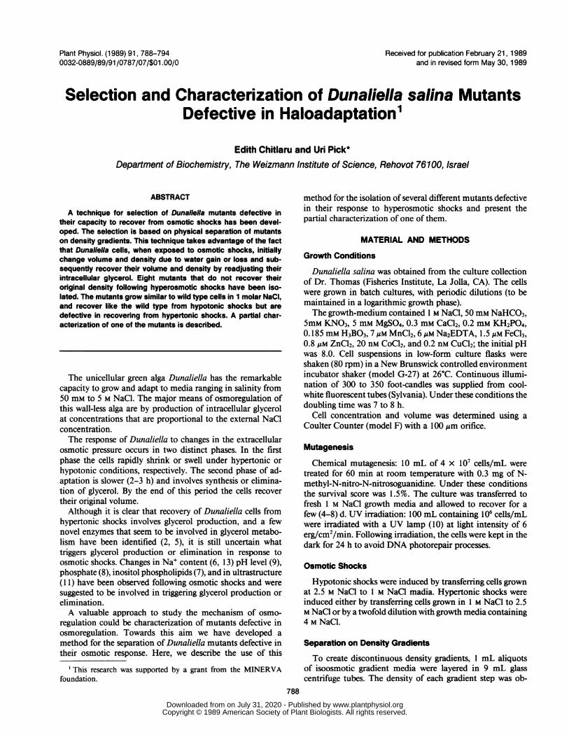
Remarkable changes in volume occur in Dunaliella cellsfollowing hypertonic shocks. Since these volume changesresult from water influx or efflux, they should be accompaniedalso by changes in cell density. As is demonstrated in Figure1 the density of D. salina cells indeed increases immediatelyfollowing hypertonic shocks (from 1-2.5 M NaCl, Fig. 1B),but cells resume their original density during a recovery periodof 2 h (Fig. IC). It should be noted that the density in samplesB and C in Figure 1 are higher than the corresponding layersin sample A due to the contribution of the higher NaClconcentration. The density of cells grown at 1 M NaCl isbetween 1.18 and 1.22 g/mL (Fig. IA); it increases to 1.25 to
Figure 1. Density changes in D. salina cells following hypertonicshocks. A (left), Cells grown at 1 M NaCI were layered on a sucrosegradient containing 1 M NaCI. The densities of the layers (from thetop) are: 1 .14, 1.16, 1.18, 1.20, 1.22, 1.24, and 1.26 g/mL. B (center),Immediately after osmotic shock, cells were layered on a gradientcontaining 2.5 M NaCI. The densities of the layers (from the top) are:1.19, 1.21, 1.23, 1.25, 1.27, 1.29, 1.31 g/mL. C (right), Two h afterosmotic shock, cells were layered on a gradient with the samedensities and NaCI content as in B.
1.27 g/mL immediately after the shock (Fig. 1B), and itreturns to 1. 19 to 1.23 g/mL 2h following the shock (Fig. IC).It can be concluded, therefore, that the change in density ofthe cells during the osmotic response, can be followed usingsucrose gradients.The observation that cells resume almost exactly their
original density following recovery from hyperosmotic shocksis somewhat surprising in view of the high specific weight ofglycerol which is accumulated inside the cells. We have com-pared densities of D. salina cells adapted to 0.2 to 4 M NaCland observed that at higher salinities cells indeed have higherdensities than low salt adapted cells, consistent with the higherglycerol contents of the former. However, it appears thatadditional factors, and in particular the starch content, con-tribute to the overall cell density. It is possible that the decreasein starch content compensates the effect of accumulated glyc-erol on cell density following hyperosmotic shocks to preservethe original density.
Since the sucrose concentrations needed to obtain the den-sities required for cell separation are high, the osmotic contri-bution of sucrose, on top of NaCl becomes significant andshould cause further cell shrinkage and an increase in density.Therefore, a comparison was made between different chemi-cals commonly used for density gradient separation of cells,including polysaccharides which have negligible osmoticcontribution.As is demonstrated in Table I the apparent densities ob-
tained before and after hypertonic shocks with ficoll, dextran,
789
www.plantphysiol.orgon July 31, 2020 - Published by Downloaded from Copyright © 1989 American Society of Plant Biologists. All rights reserved.
Plant Physiol. Vol. 91, 1989
sucrose, and metrizamide density gradients are quite similar,indicating only a slight increase in density in sucrose at 1 MNaCl, compared to ficoll and dextran. It may be noted thatseparation on ficoll and dextran density gradients requireslonger and faster centrifugation times due to their higherviscosity and often results in cell aggregation, while separationon sucrose density gradients is fast, and the viability of theseparated cells is excellent. Therefore, for most practical pur-poses we have used sucrose density gradients.To exemplify the usefulness of this technique we followed
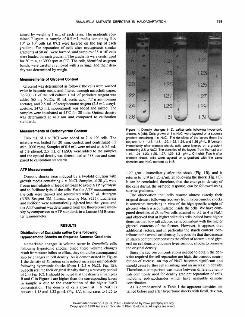
the kinetics of density changes in Dunaliella cells after hypo-tonic and hypertonic shocks by sucrose density gradients (Fig.2). Dunaliella cells grown either at 1 or 2.5 M NaCl weretransferred to 2.5 or 1 M NaCl, respectively. After hypertonicshock, the cells shrink and their density increases from 1.19to 1.20 to 1.23 to 1.25 g/mL (Fig. 2A). The density of thecells decreases gradually and reaches its original value 90 minafter osmotic shock. After hypotonic shock, the cells swelland their density decreases from 1.19 to 1.20 g/mL to 1.14to 1.18 g/mL (Fig. 2B). During the adaptation period of 90min, the density of the cells resumes its original value. Inboth, hypertonic or hypotonic shocks, the recovery of thedensity under dark conditions is slower. It may be concludedthat density changes as well as volume or glycerol contentmay be used to follow the recovery of D. salina cells fromosmotic shocks.
Isolation of Mutants Defective in the Osmotic Response
The observation that recovery from hyperosmotic shocksis associated with changes in cell density may be used forselection of mutants defective in their osmotic response, therationale being that mutants should not resume their lowdensity during the recovery period and therefore can bephysically separated from normal cells. The isolation proce-dure was as follows: D. salina cells were exposed either to UVlight or to a chemical mutagen. After mutagenesis, cells weresubjected to a hypertonic shock by a 2.5-fold increase in NaClconcentration, followed by incubation for 4 h in the light toallow recovery from the osmotic shock and applied to discon-tinuous sucrose gradients containing 2.5 M NaCl. After cen-trifugation, 1 mL of the 35 to 40% interphase, correspondingto shrunken cells (most of the cells recovered from the shock
Table I. Density of D. salina Cells during Osmotic Shock andRecovery Measured on Different Density Gradients
Cells grown in 1 M NaCI were subjected to hypertonic shock byraising the salt concentration to 2.5 M. Numbers indicate the densitiesat the interphase where the cells migrate in step-wise gradients. Thegradients were made with isoosmotic NaCI solutions. After applicationof cell samples, the gradients were centrifuged for 30 min at 3000rpm (sucrose or metrizamide) or at 5000 rpm (ficoll or dextran).Densities are expressed in g/mL.
Gradient Control OsmoticallyComposition Cells Shocked Cells
Sucrose 1.18-1.20 1.23-1.24Ficoll 1.17 1.24Dextran 1.15-1.17Metrizamide 1.16-1.19 1.23-1.24
1 B2.3E
1.21I
v% Light Dark
0)
.E 1201.19
1.4
0 5 1530(90 0 5 153090Time after osmotic shock
(minutes)Figure 2. Kinetics of density changes in D. salina following hypertonicor hypotonic shocks in light or dark conditions. Cells grown at 1 or2.5 M NaCI were transferred to 2.5 or 1 M NaCI growth media,respectively. The cells were allowed to recover from both hypertonicor hypotonic shocks either in light or dark. Samples of cells werecentrifuged 0, 5, 15, 30, or 90 min after the osmotic shocks onsucrose stepwise gradients composed of seven layers of sucrose (1mL each) containing either 2.5 or 1 M NaCI (hypertonic or hypotonicshocks, respectively). The density of cells grown at either 1 or 2.5 MNaCI was 1.19 to 1.20 g/mL.
and were in the 20-25% interphase), was collected from thegradients with a syringe. The cells were washed from thesucrose and transferred to 1 M NaCl growth media.
Cloning of the Mutants
With the purpose of obtaining single cell cultures, the cellswere plated in 96 well microplates in high dilution (1 cell/4wells). To avoid evaporation, the plates were sealed withmasking tape. After 10 culturing days in continuous illumi-nation, 350 colonies were observed in 18 plates. Samples fromeach colony were transferred to microplates containing 2.5 MNaCl growth media. Growth was monitored by the increasein absorption at 450 nm. The colonies which failed to growwere selected and cultured in 1 M NaCl media.
Following chemical mutagenesis, 7 out of 350 coloniesfailed to respond normally to upshocks. They are denoted:D3, D4, D6, D7, D8, D9, and DIO (see Fig. 3). FollowingUV irradiation, 1 out of 100 colonies was unable to recoverafter upshock. It is denoted U22.
790 CHITLARU AND PICK
www.plantphysiol.orgon July 31, 2020 - Published by Downloaded from Copyright © 1989 American Society of Plant Biologists. All rights reserved.
DUNALIELLA MUTANTS DEFECTIVE IN HALOADAPTATION
6038 ]
I0 ~~I> ~~wt wt
) 90 1 wt1I0
66
80-
10 6
70
60
80 160 80 160 240 80 160- 240Time after osmotic shock
(minutes)
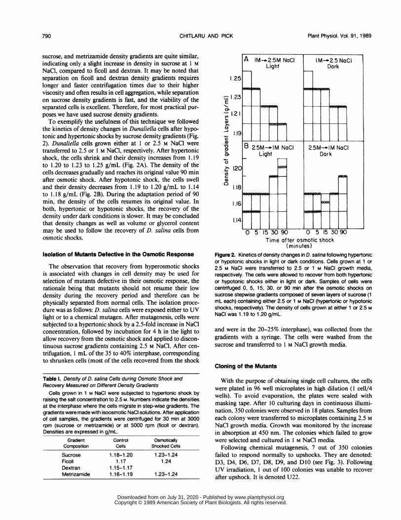
Figure 3. Volume changes in D. salina mutants following hypertonicshocks in different salt concentrations. The mutants D3, D4, D6, D7,D8, D9, Dl 0, and wild type, adapted to 0.4, 1, or 1.75 M NaCI weretransferred to 1, 2.5, or 3.5 M NaCI, respectively. Samples of cellswere taken at the indicated times following hypertonic shock and thevolume measured using a coulter counter. Note that the factor ofshock for cells adapted to 0.4 and 1 M NaCI is higher (2.5-fold) thanfor cells adapted to 1.75 M NaCI (2-fold).
Response to Hyperosomotic Shocks of DunaliellaOsmotic Mutants, Adapted to Different NaCIConcentrations
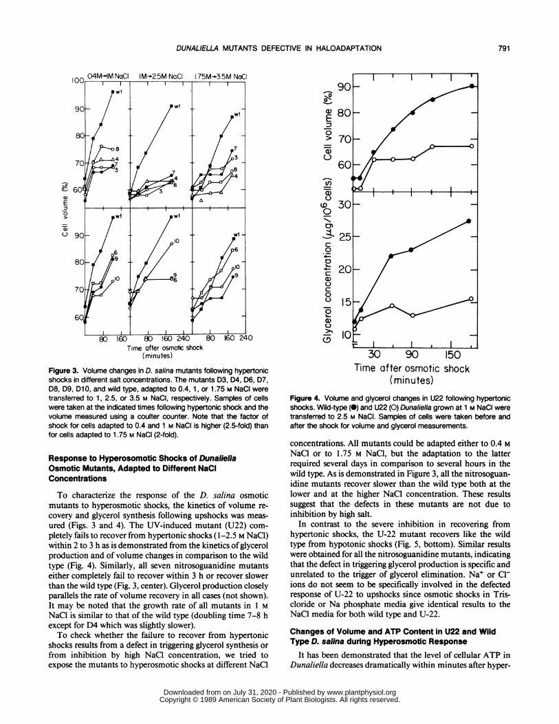
To characterize the response of the D. salina osmoticmutants to hyperosmotic shocks, the kinetics of volume re-covery and glycerol synthesis following upshocks was meas-ured (Figs. 3 and 4). The UV-induced mutant (U22) com-pletely fails to recover from hypertonic shocks ( 1-2.5 M NaCl)within 2 to 3 h as is demonstrated from the kinetics ofglycerolproduction and of volume changes in comparison to the wildtype (Fig. 4). Similarly, all seven nitrosoguanidine mutantseither completely fail to recover within 3 h or recover slowerthan the wild type (Fig. 3, center). Glycerol production closelyparallels the rate of volume recovery in all cases (not shown).It may be noted that the growth rate of all mutants in 1 MNaCl is similar to that of the wild type (doubling time 7-8 hexcept for D4 which was slightly slower).To check whether the failure to recover from hypertonic
shocks results from a defect in triggering glycerol synthesis orfrom inhibition by high NaCl concentration, we tried toexpose the mutants to hyperosmotic shocks at different NaCl
a)=
0
0
c00
0w
0
C:
a)
00
0
0
Time after osmotic shock(minutes)
Figure 4. Volume and glycerol changes in U22 following hypertonicshocks. Wild-type (0) and U22 (0) Dunaliella grown at 1 M NaCI weretransferred to 2.5 M NaCI. Samples of cells were taken before andafter the shock for volume and glycerol measurements.
concentrations. All mutants could be adapted either to 0.4 MNaCl or to 1.75 M NaCl, but the adaptation to the latterrequired several days in comparison to several hours in thewild type. As is demonstrated in Figure 3, all the nitrosoguan-idine mutants recover slower than the wild type both at thelower and at the higher NaCl concentration. These resultssuggest that the defects in these mutants are not due toinhibition by high salt.
In contrast to the severe inhibition in recovering fromhypertonic shocks, the U-22 mutant recovers like the wildtype from hypotonic shocks (Fig. 5, bottom). Similar resultswere obtained for all the nitrosoguanidine mutants, indicatingthat the defect in triggering glycerol production is specific andunrelated to the trigger of glycerol elimination. Na+ or C1lions do not seem to be specifically involved in the defectedresponse of U-22 to upshocks since osmotic shocks in Tris-cloride or Na phosphate media give identical results to theNaCl media for both wild type and U-22.
Changes of Volume and ATP Content in U22 and WildType D. salina during Hyperosmotic Response
It has been demonstrated that the level of cellular ATP inDunaliella decreases dramatically within minutes after hyper-
791
www.plantphysiol.orgon July 31, 2020 - Published by Downloaded from Copyright © 1989 American Society of Plant Biologists. All rights reserved.

Plant Physiol. Vol. 91, 1989
A
E0
Uz
B
a,20
a)
A
ai
oJE0
Uai
B
a,r-Li
=
N.,Q-
0
0C-
C-a,oic0v
Time after osmotic shock(minutes)
10030 60 90 120
Time after osmotic shock(minutes)
Figure 5. Volume changes in D. salina wild type (0) and U22 (0)following hypertonic or hypotonic shocks. A, Cells were tested fortheir response to hypertonic shock as in Figure 4. B, Wild-typeDunaliella and U22 adapted to 1 M NaCI were transferred to 0.5 MNaCI. Samples of cells were taken following the hypotonic shock forvolume measurements.
osmotic shocks. Furthermore, it appears that the capacity torecover from hyperosmotic shocks is closely correlated withATP production and is perturbed by metabolic inhibitors(12). It seemed possible, therefore, that the defect in U-22may be related to ATP supply and utilization in the responseto upshocks. However, as is demonstrated in Figure 6, thetransient decrease in cellular ATP level in wild type and U-22 D. salina following upshocks is similar, suggesting that thedefect in U-22 is not related to ATP production or utilization.
Starch Utilization of U22 and Wild Type Dunaliella duringHyperosmotic Response in the Dark and in the Light
One of the possible defects in U-22, as well as the othermutants, is a metabolic deficiency in triggering starch mobi-lization to glycerol. To check this possibility, we searched forconditions under which recovery from hypertonic shocks willbe solely dependent on starch reserves. Glycerol synthesis ofDunaliella can be driven either through starch breakdown orvia photosynthetic CO2 assimilation through the Calvin cycle.Cells recovering in the dark from hypertonic shock will utilizeonly starch for glycerol synthesis. Therefore, recovery from
Time after osmotic shock (minutes)
Figure 6. Changes in ATP content following hypertonic shock in D.salina wild type and U22. U22 (0) and wild-type (@) D. salina grownin 1 M NaCI are osmoticaly shocked in 2.5 M NaCI. Samples of cellswere taken for volume (A) and ATP (B) measurements.
hypertonic shocks in the dark is more efficient in cells withhigh starch reserves. Recovery of wild type and U-22 fromupshocks was compared in the dark in cells subjected tonitrate starvation, a treatment that causes an increase ofstarchconcentration in the cells (1).As is demonstrated in Table II, the mutant U22 cannot
degrade starch needed for glycerol accumulation followingosmotic upshocks either in the dark or in the light. However,the mutant appears to be capable of utilizing starch for othermetabolic purposes, since the starch content of U22 cellsdecreases in the dark and the cells duplicate normally insynchronized culturing conditions, in the dark (not shown).These results may indicate that the mutation in U22 may
be in the triggering of starch utilization in response to hyper-tonic shock. An alternative metabolic defect that could blockstarch conversion to glycerol is the inactivation of one of theterminal enzymes leading to glycerol production from dihy-droxyacetone phosphate. Such a defect could lead to accu-mulation of precursors of glycerol following upshocks, suchas glycerol-3-phosphate or dihydroxyacetonephosphate. Wehave, therefore, compared the soluble carbon metabolites inwild type and U22 D. salina following osmotic upshocks,using 13C NMR (3), but observed no difference in their
792 CHITLARU AND PICK
www.plantphysiol.orgon July 31, 2020 - Published by Downloaded from Copyright © 1989 American Society of Plant Biologists. All rights reserved.

DUNALIELLA MUTANTS DEFECTIVE IN HALOADAPTATION
Table II. Starch, Glycerol, and Volume Changes followingHypertonic Shock in D. salina Wild Type and U22 in the Light or inthe Dark
Cells cultured in low-nitrate for starch accumulation (A) or in regulargrowth medium (B) were osmoticaly shocked (1-2.5 M NaCI) in thedark (A) or in the light (B). Starch or glycerol contents were determinedeither before the shock or after a recovery period of 2 h (A) or 45 min(B) as described under "Materials and Methods."
Cells State of Cells Cell Starch GlycerolVolume Content Content
% lglls/1O cells(A)Wild type Before shock 100 39.5 NTa
After shock 61Recovery period 78 21.0 NT
U22 Before shock 100 42.0 NTAfter shock 63Recovery period 68 43.0 NT
(B)Wild type Before shock 100 12.6 11.5
After shock 52Recovery period 76 8.0 18.8
U22 Before shock 100 23.0 14.0After shock 52Recovery period 60 24.0 15.0
a Not tested.
metabolite contents except for the absence of glycerol accu-mulation in U22 (E. Chitlaru, M. Shamir, unpublished ob-servations). From these results it may be concluded that thedefect in U22 is most probably in triggering one of theosmoregulatory metabolic steps leading to glycerol produc-tion, such as starch degradation.
DISCUSSION
Little is known about the mechanism which triggers glycerolproduction in Dunaliella in response to hypertonic shocks.Mutants which are specifically defective in triggering glycerolproduction are in this context of special importance. How-ever, so far there has been only one report about a Dunaliellamutant defective in responding to osmotic shocks, which isin fact a metabolic mutant, with an abnormal carbonicanhydrase (4).
Theoretically a minimum of four types of osmotic mutantsmay be expected: (a) mutants in which one of the enzymesinvolved in glycerol biosynthesis becomes rate limiting inrecovery from hyperosmotic shocks; (b) mutants defective inthe triggering mechanism of glycerol production, which ispresumably located in the plasma membrane (7, 11); (c)metabolic mutants in which the production of ATP or
NAD(P)H needed for glycerol production becomes rate lim-iting for the recovery from hyperosmotic shocks; (d) mutantswhich are particularly sensitive to high salt.The selection method which is described here has several
advantages over a general screening technique: (a) the physicalseparation of nonrecovering from recovering cells, followingosmotic upshocks, avoids the need of screening a large num-
ber of mutants; (b) the fact that the permissive mediumcontains 1 M NaCl lowers the probability of selection ofmetabolic mutants or of mutants with major defects in glyc-erol production, since such mutants will not survive in 1 MNaCl. In some experiments, we have incubated cells followingmutagenesis under light-dark cycles (8-16 h, respectively) toeliminate metabolic mutants defective in starch mobilization.The final elimination of metabolic mutants was made byavoiding mutants which failed to grow like wild type in thepermissive medium. Nevertheless, metabolic mutants cannotbe excluded altogether by this selection since a minor meta-bolic defect that may not be manifested in steady state growthmay become a limiting factor under stringent osmotic stressconditions.The possibility of salt-sensitive mutants seem unlikely in
view of the observations that all the selected mutants can beadapted to high salt following prolonged incubation andrespond poorly to osmotic upshocks in different salt concen-tration ranges (Fig. 3).Although at present we have not identified the specific
defect in any ofthe mutants, several conclusions can be drawnalready from their preliminary characterization: (a) The ob-servation that all the mutants respond like the wild type tohypotonic shocks suggests that the triggering mechanisms ofglycerol production and elimination are different regulatoryand metabolic elements. This conclusion is consistent withrecent observations of different changes in plasma membranelipids, notably inositol phospholipids, and ultrastructure fol-lowing upshocks and downshocks in Dunaliella (7, 1 1) andwith our recent observation that certain H+-ATPase inhibitorsspecificaly inhibit recovery from upshocks but not fromdownshocks in D. salina (12). (b) We have noticed severaldifferences in the response to osmotic upshocks among themutants; for example, the rate of recovery differs in differentsalt concentrations (Fig. 3). We have preliminary indicationssuggesting that some of the osmotic mutants may fall into thecategory of metabolic mutants, other are limited in starchmobilization, and the rest are blocked at different stages.These differences suggest that the triggering of glycerol pro-duction is a sequence of steps involving several differentenzymes. It is our hope that further characterization of thesemutants will contribute to the understanding of the triggeringmechanism of glycerol production in response to hypertonicshocks in Dunaliella.
ACKNOWLEDGMENTS
We are grateful to Prof. M. Avron and to Prof. E. Padan for helpfulsuggestions and discussions, and to Mrs. M. Weiss for technicalassistance.
LITERATURE CITED
1. Ben-Amotz A (1987) Effect of irradiance and nutrient deficiencyon the chemical composition of Dunaliella bardawil. J PlantPhysiol 131: 479-487
2. Ben-Amotz A, Sussman I, Avron M (1982) Glycerol productionby Dunaliella. Experientia 38: 49-52
3. Bental M, Oren-Shamir M, Avron M, Degani H (1988) 3'P and'3C-NMR studies of the phosphorus and carbon metabolitesin halotolerant alga Dunaliella salina. Plant Physiol 87: 320-324
4. Brown AD, Goyal A, Larsen H, Lilley RMcC (1987) A salt-
793
www.plantphysiol.orgon July 31, 2020 - Published by Downloaded from Copyright © 1989 American Society of Plant Biologists. All rights reserved.
CHITLARU AND PICK
sensitive mutant ofDunaliella tertiolecta. Arch Microbiol 147:309-314
5. Brown AD, Lilley RMcC, Marengo T (1982) Osmorregulationin Dunaliella. Intracellular distribution of enzymes of glycerolmetabolism. Plant Physiol 53: 628-631
6. Ehrenfeld J, Cousin JL (1982) Ionic regulation of the unicellulargreen alga Dunaliella tertiolecta. J. Membr Biol. 70: 47-57
7. Einspahr EK, Peeler TC, Thompson GA Jr (1988) Rapid changesin polyphosphoinositide metabolism associated with the re-sponse ofDunaliella salina to hypoosmotic shock. J Biol Chem263: 5775-5779
8. Gimmler H, Moller EM (1981) Salinity dependent regulation ofstarch and glycerol metabolism in Dunaliella parva. Plant CellEnviron 3: 367-375
9. Goyal A, Brown AD, Gimmler H (1986) Regulation of salt-induced starch degradation in Dunaliella tertiolecta. J Plant
Plant Physiol. Vol. 91, 1989
Physiol 127: 67-7610. Harris EH, Boynton JE, Gillham NW (1982) Induction of nu-
clear and chloroplast mutations which affect the chloroplast inChlamydomonas reinhardtii. In M Edelman, ed, Methods inChloroplast Molecular Biology. Elsevier Biochemical Press,Amsterdam, pp 3-24
11. Maeda M. Thompson GA Jr (1986) On the mechanism of rapidplasma membrane and chloroplast envelope expansion in Dun-aliella salina exposed to hypoosmotic shock. J Cell Biol 102:289-297
12. Oren-Shamir M, Pick U, Avron M (1989) Involvement of theplasma membrane ATPase in the osmoregulatory mechanismof the alga Dunaliella salina. Plant Physiol 89: 1258-1263
13. Pick U, Karni L, Avron M (1986) Determination of ion contentand ion fluxes in the halotolerant alga Dunaliella salina. PlantPhysiol 81: 92-96
794
www.plantphysiol.orgon July 31, 2020 - Published by Downloaded from Copyright © 1989 American Society of Plant Biologists. All rights reserved.


![omonas gracilis and Dunaliella sp.] in laboratory batch](https://img.pdfslide.us/doc/110x75/61bd177961276e740b0f4389/omonas-gracilis-and-dunaliella-sp-in-laboratory-batch-.jpg)