Embed Size (px)
Citation preview
International Union of Pharmacology LVII:Recommendations for the Nomenclature of Receptors
for Relaxin Family PeptidesROSS A. BATHGATE, RICHARD IVELL, BARBARA M. SANBORN, O. DAVID SHERWOOD, AND ROGER J. SUMMERS
Howard Florey Institute, University of Melbourne, Melbourne, Victoria, Australia (R.A.B.); School of Molecular and Biomedical Science,The University of Adelaide, South Australia, Australia (R.I.); Department of Biological Sciences, Colorado State University, Fort Collins,
Colorado (B.M.S.); Department of Molecular and Integrative Physiology and College of Medicine, University of Illinois at Urbana-Champaign, Urbana, Illinois (O.D.S.); Department of Pharmacology, Monash University, Clayton, Victoria, Australia (R.J.S.)
Abstract . . . . . . . . . . . . . . . . . . . . . . . . . . . . . . . . . . . . . . . . . . . . . . . . . . . . . . . . . . . . . . . . . . . . . . . . . . . . . . . . . 8I. Brief historical background of relaxin family peptides and their receptors . . . . . . . . . . . . . . . . . . . . . . 8
II. Receptor distribution and function. . . . . . . . . . . . . . . . . . . . . . . . . . . . . . . . . . . . . . . . . . . . . . . . . . . . . . . . . 11A. Reproductive tissues . . . . . . . . . . . . . . . . . . . . . . . . . . . . . . . . . . . . . . . . . . . . . . . . . . . . . . . . . . . . . . . . . . 11B. Brain. . . . . . . . . . . . . . . . . . . . . . . . . . . . . . . . . . . . . . . . . . . . . . . . . . . . . . . . . . . . . . . . . . . . . . . . . . . . . . . . 13C. Cardiovascular and renal systems . . . . . . . . . . . . . . . . . . . . . . . . . . . . . . . . . . . . . . . . . . . . . . . . . . . . . . 14D. Other sites of action of relaxin family peptides . . . . . . . . . . . . . . . . . . . . . . . . . . . . . . . . . . . . . . . . . . 15E. Roles of relaxin family peptides determined from studies in receptor knockout mice . . . . . . . . . 16
III. Structure of relaxin family peptides . . . . . . . . . . . . . . . . . . . . . . . . . . . . . . . . . . . . . . . . . . . . . . . . . . . . . . . 17A. Structural features of relaxin . . . . . . . . . . . . . . . . . . . . . . . . . . . . . . . . . . . . . . . . . . . . . . . . . . . . . . . . . . 17B. Structural features of other relaxin family peptides . . . . . . . . . . . . . . . . . . . . . . . . . . . . . . . . . . . . . . 17
IV. Structure-activity relationships . . . . . . . . . . . . . . . . . . . . . . . . . . . . . . . . . . . . . . . . . . . . . . . . . . . . . . . . . . . 17A. Relaxin . . . . . . . . . . . . . . . . . . . . . . . . . . . . . . . . . . . . . . . . . . . . . . . . . . . . . . . . . . . . . . . . . . . . . . . . . . . . . . 17B. INSL3 . . . . . . . . . . . . . . . . . . . . . . . . . . . . . . . . . . . . . . . . . . . . . . . . . . . . . . . . . . . . . . . . . . . . . . . . . . . . . . . 18
V. Binding of relaxin and relaxin family peptides . . . . . . . . . . . . . . . . . . . . . . . . . . . . . . . . . . . . . . . . . . . . . . 18A. Relaxin binding . . . . . . . . . . . . . . . . . . . . . . . . . . . . . . . . . . . . . . . . . . . . . . . . . . . . . . . . . . . . . . . . . . . . . . 18B. Binding of other relaxin family peptides . . . . . . . . . . . . . . . . . . . . . . . . . . . . . . . . . . . . . . . . . . . . . . . . 19
VI. Relaxin family peptide receptors . . . . . . . . . . . . . . . . . . . . . . . . . . . . . . . . . . . . . . . . . . . . . . . . . . . . . . . . . . 19A. RXFP1 and RXFP2 . . . . . . . . . . . . . . . . . . . . . . . . . . . . . . . . . . . . . . . . . . . . . . . . . . . . . . . . . . . . . . . . . . . 19B. RXFP3 and RXFP4 . . . . . . . . . . . . . . . . . . . . . . . . . . . . . . . . . . . . . . . . . . . . . . . . . . . . . . . . . . . . . . . . . . . 21
VII. Functional domains of receptors for relaxin family peptides . . . . . . . . . . . . . . . . . . . . . . . . . . . . . . . . . . 22A. General features of leucine-rich repeat-containing receptors . . . . . . . . . . . . . . . . . . . . . . . . . . . . . . 22B. Functional domains of RXFP1 and RXFP2 (LGR7 and LGR8) . . . . . . . . . . . . . . . . . . . . . . . . . . . . . 23C. Functional domains of RXFP3 and RXFP4 (GPCR135 and GPCR142) . . . . . . . . . . . . . . . . . . . . . . 24
VIII. Signaling pathways activated by relaxin family peptides. . . . . . . . . . . . . . . . . . . . . . . . . . . . . . . . . . . . . 24A. Signaling in response to relaxin . . . . . . . . . . . . . . . . . . . . . . . . . . . . . . . . . . . . . . . . . . . . . . . . . . . . . . . . 24B. Signaling in response to other relaxin family peptides . . . . . . . . . . . . . . . . . . . . . . . . . . . . . . . . . . . . 27
IX. Regulation of relaxin family peptide receptors . . . . . . . . . . . . . . . . . . . . . . . . . . . . . . . . . . . . . . . . . . . . . . 27X. Nomenclature issues for relaxin family peptides and their receptors . . . . . . . . . . . . . . . . . . . . . . . . . . 27
Acknowledgments. . . . . . . . . . . . . . . . . . . . . . . . . . . . . . . . . . . . . . . . . . . . . . . . . . . . . . . . . . . . . . . . . . . . . . . . 28References . . . . . . . . . . . . . . . . . . . . . . . . . . . . . . . . . . . . . . . . . . . . . . . . . . . . . . . . . . . . . . . . . . . . . . . . . . . . . . 28
Address correspondence to: Professor R. J. Summers, P.O. Box 13E, Department of Pharmacology, Monash University, Victoria 3800,Australia. E-mail: [email protected]
This work was supported in part by a National Health and Medical Research Council (NH&MRC) block grant to the Howard FloreyInstitute (Reg Key 983001), NH&MRC project grants to R.A.D.B. and R.J.S. (300012) and R.I (349502), Australian Research Council LinkageGrant LP0211545 to R.J.S., and Grant Iv7/11 from the Deutsche Forschungsgemeinschaft to R.I.
Article, publication date, and citation information can be found at http://pharmrev.aspetjournals.org.doi:10.1124/pr.58.1.9.
0031-6997/06/5801-7–31$7.00PHARMACOLOGICAL REVIEWS Vol. 58, No. 1Copyright © 2006 by The American Society for Pharmacology and Experimental Therapeutics 60101/3095449Pharmacol Rev 58:7–31, 2006 Printed in U.S.A
7
by guest on Novem
ber 23, 2021D
ownloaded from
Abstract——Although the hormone relaxin was dis-covered 80 years ago, only in the past 5 years have thereceptors for relaxin and three other receptors thatrespond to related peptides been identified with allfour receptors being G-protein-coupled receptors. Inthis review it is suggested that the receptors for re-laxin (LGR7) and those for the related peptides insu-lin-like peptide 3 (LGR8), relaxin-3 (GPCR135), andinsulin-like peptide 5 (LGPCR142) be named the re-laxin family peptide receptors 1 through 4 (RXFP1–4).RXFP1 and RXFP2 are leucine-rich repeat-containingG-protein-coupled receptors with complex bindingcharacteristics involving both the large ectodomainand the transmembrane loops. RXFP1 activates ade-nylate cyclase, protein kinase A, protein kinase C,phosphatidylinositol 3-kinase, and extracellular sig-naling regulated kinase (Erk1/2) and also interactswith nitric oxide signaling. RXFP2 activates adenylatecyclase in recombinant systems, but physiological re-sponses are sensitive to pertussis toxin. RXFP3 and
RXFP4 resemble more conventional peptide ligandedreceptors and both inhibit adenylate cyclase, and inaddition RXFP3 activates Erk1/2 signaling. Physiolog-ical studies and examination of the phenotypes oftransgenic mice have established that relaxin hasroles as a reproductive hormone involved in uterinerelaxation (some species), reproductive tissue growth,and collagen remodeling but also in the cardiovascu-lar and renal systems and in the brain. The connectivetissue remodeling properties of relaxin acting atRXFP1 receptors have potential for the developmentof agents effective for the treatment of cardiac andrenal fibrosis, asthma, and scleroderma and for orth-odontic remodelling. Agents acting at RXFP2 recep-tors may be useful for the treatment of cryptorchidismand infertility, whereas antagonists may be used ascontraceptives. The brain distribution of RXFP3 re-ceptors suggests that actions at these receptors havethe potential for the development of antianxiety andantiobesity drugs.
I. Brief Historical Background of Relaxin FamilyPeptides and Their Receptors
The circulating hormone relaxin was discovered whenHisaw (1926) observed that the injection of serum frompregnant guinea pigs or rabbits into virgin guinea pigsshortly after estrus induced a relaxation of the pubicligament. Between the mid 1970s and 1990s the primarysequence of relaxin was determined for the pig (Schwabeet al., 1976; Schwabe and McDonald, 1977a), rat (Johnet al., 1981), mouse (Evans et al., 1993), human (Hudsonet al., 1983, 1984), and �20 other species (Bryant-Greenwood and Schwabe, 1994; Sherwood, 1994; Bath-gate et al., 2002b). In the human and four great apespecies, there are two forms of relaxin that are encodedby separate genes RLN11 and RLN2 (Hudson et al.,
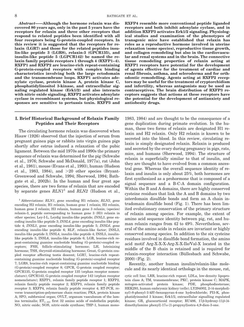
1983, 1984) and are thought to be the consequence of agene duplication during primate evolution. In the hu-man, these two forms of relaxin are designated H1 re-laxin and H2 relaxin. Only H2 relaxin is known to besecreted into the blood. In this review, circulating re-laxin is simply designated relaxin. Relaxin is producedand secreted by the ovary during pregnancy in pigs, rats,mice, and humans (Sherwood, 1994). The structure ofrelaxin is superficially similar to that of insulin, andthey are thought to have evolved from a common ances-tral gene. Whereas the overall sequence identity of re-laxin and insulin is only about 25%, both hormones arefirst synthesized as a prohormone that is composed of asignal sequence and a B-C-A domain configuration.Within the B and A domains, there are highly conservedcysteine residues that link the A and B domains by twointerdomain disulfide bonds and form an A chain in-tradomain disulfide bond (Fig. 1). There has been lim-ited evolutionary conservation of the primary structureof relaxin among species. For example, the extent ofamino acid sequence identity between pig, rat, and hu-man relaxin ranges from 42 to 49%. Nevertheless, sev-eral of the amino acids in relaxin are invariant or highlyconserved among species. In addition to the six cysteineresidues involved in disulfide bond formation, the aminoacid motif Arg-X-X-X-Arg-X-X-Ile/Val-X located in themiddle of the B chain is retained and is required forrelaxin-receptor interaction (Bullesbach and Schwabe,2000) (Fig. 2).
Recently, another human insulin/relaxin-like mole-cule and its nearly identical orthologs in the mouse, rat,
1 Abbreviations: RLN1, gene encoding H1 relaxin; RLN2, geneencoding H2 relaxin; H1 relaxin, human gene 1 relaxin; H2 relaxin,human gene 2 relaxin; H3 relaxin, human gene 3 relaxin; (species)relaxin-3, peptide corresponding to human gene 3 (H3) relaxin inother species; Ley-I-L, Leydig insulin-like peptide; INSL3, gene en-coding insulin-like peptide 3; INSL4, gene encoding insulin-like pep-tide 4; INSL5, gene encoding insulin-like peptide 5; INSL6, geneencoding insulin-like peptide 6; RLF, relaxin-like factor; INSL3,insulin-like peptide 3; INSL4, insulin-like peptide 4; INSL5, insulin-like peptide 5; INSL6, insulin-like peptide 6; LGR, leucine-rich re-peat-containing guanine nucleotide binding (G-protein)-coupled re-ceptors; FSH, follicle-stimulating hormone; LH, luteinizinghormone; TSH, thryroid-stimulating hormone; Great, G-protein cou-pled receptor affecting testis descent; LGR7, leucine-rich repeat-containing guanine nucleotide binding (G-protein)-coupled receptor7; LGR8, leucine-rich repeat-containing guanine nucleotide binding(G-protein)-coupled receptor 8; GPCR, G-protein coupled receptor;GPCR135, G-protein coupled receptor 135 (orphan receptor nomen-clature); GPCR142, G-protein coupled receptor 142 (orphan receptornomenclature); RXFP1, relaxin family peptide receptor 1; RXFP2,relaxin family peptide receptor 2; RXFP3, relaxin family peptidereceptor 3; RXFP4, relaxin family peptide receptor 4; RT-PCR, re-verse transcription-polymerase chain reaction; PKA, protein kinaseA; SFO, subfornical organ; OVLT, organum vasculosum of the lam-ina terminalis; ET1–32, first 32 amino acids of endothelin peptide;NO, nitric oxide; NOS, nitric-oxide synthase; THP-1, human mono-
cytic cell line; LRR, leucine-rich repeat; LDLa, low-density lipopro-tein class A; TM, transmembrane; PKC, protein kinase C; MAPK,mitogen-activated protein kinase; PDE, phosphodiesterase;HEK293, human embryonic kidney (cells); LY294002, 2-(4-morpholi-nyl)-8-phenyl-1(4H)-benzopyran-4-one hydrochloride; PI3-K, phos-phatidyinositol 3 kinase; Erk1/2, extracellular signalling regulatedkinase; GR, glucocorticoid receptor; RU486, 17�-hydroxy-11�-[4-dimethylamino phenyl]-17�-[1-propynyl]estra-4,9-dien-3-one.
8 BATHGATE ET AL.
and pig were discovered (Bathgate et al., 2002a; Burazinet al., 2002; Kizawa et al., 2003). Because this peptidehas the amino acid motif Arg-X-X-X-Arg-X-X-Ile re-quired for relaxin bioactivity in the middle of the B chainand it activates the relaxin receptor (Sudo et al., 2003),it was designated relaxin-3 or in the case of the humanrelaxin-3 peptide, H3 relaxin (Fig. 2). Relaxin-3 is ex-pressed in greatest levels in the ventromedial dorsaltegmental nucleus [also called nucleus incertus (Goto etal., 2001)] in the rat and mouse brain, where it has beenpostulated to act locally as a neuropeptide (Burazin etal., 2002; Liu et al., 2003b).
Another insulin-like protein was discovered by differ-ential screening of a boar testis cDNA library (Adham etal., 1993). Because the mRNA for this molecule wasfound in Leydig cells of the testis, it was originallydesignated Leydig insulin-like peptide (Ley-I-L). Thegene for Ley-I-L was designated INSL3 (Burkhardt etal., 1994). Ley-I-L contains a portion (Arg-X-X-X-Arg) ofthe putative relaxin receptor-binding region Arg-X-X-X-Arg-X-X-Ile, which is offset by four amino acid residuestoward the C terminus relative to its position in relaxin.Subsequent reports (Bullesbach and Schwabe, 1995) re-vealed that synthetic human Ley-I-L augmented relaxinactivity in a mouse interpubic ligament bioassay, lead-ing to the suggestion that the term relaxin-like-factor(RLF) was the more appropriate term for this hormone.Although both INSL3 and RLF are used in the litera-ture, for the purposes of this review the hormone will bedesignated insulin-like peptide 3 (INSL3). INSL3 ex-pression is not confined to the male; it is also expressedin the ovary within both the follicular theca interna cellsand the corpora lutea (Bathgate et al., 1996; Roche et al.,1996; Kawamura et al., 2004), as well as in the placenta(Hombach-Klonisch et al., 2001), breast (Hombach-Klonisch et al., 2000), and thyroid gland (Hombach-Klonisch et al., 2003).
Shortly after the discovery of INSL3, a cDNA clone foranother member of the relaxin peptide family was iden-tified independently by two groups during screening ofcDNA libraries of first-trimester human placenta (Chas-sin et al., 1995; Koman et al., 1996). The protein productof this gene was named early placenta insulin-like pep-
tide (Chassin et al., 1995) and placentin (Koman et al.,1996), respectively. The gene has been named insulin-like 4 (INSL4) based on the nomenclature used for theINSL3 gene. The INSL4 gene emerged before the diver-gence of New and Old World monkeys (Bieche et al.,2003), predating the emergence of the RLN1 gene. TheINSL4 gene is highly expressed in the human placentabut its function is unknown.
More recently, two novel genes were independentlydiscovered by three groups via searching of the Ex-pressed Sequence Tags database using the conservedcysteine pattern of this peptide family. Hence, insulin-like peptide-5 (INSL5) (Conklin et al., 1999; Hsu, 1999)and insulin-like peptide-6 (INSL6) (Hsu, 1999; Lok etal., 2000) were added to the relaxin peptide family (Fig.2). Both genes are present in rodent genomes andevolved from the ancestral RLN3 gene (Wilkinson et al.,2005). Their functions are currently unknown, althoughINSL5 has been identified as the cognate ligand forGPCR142 (RXFP4) (see below). The occurrence of therelaxin family peptides in human, rat and mouse issummarized in Table 1.
The receptors for relaxin, relaxin-3, INSL3, andINSL5 were identified recently (Table 2). Relaxin andINSL3 receptors are a subgroup (type C) of the family ofleucine-rich repeat-containing guanine nucleotide bind-ing (G-protein)-coupled receptors designated LGRs, thatinclude the receptors for FSH, LH, and TSH. By usinginferences from similar phenotypic expression followingmutation and inactivation of INSL3 (Nef and Parada,1999; Zimmermann et al., 1999) and a transgenic inser-tional mutation in mouse chromosome 5, an orphan LGRdesignated either G-protein-coupled receptor affectingtestis descent (Great) (Overbeek et al., 2001) or LGR8(Hsu et al., 2002; Kumagai et al., 2002) was identified asthe INSL3 receptor. The discovery that the orphan re-ceptor LGR7 is the relaxin receptor was largely attrib-utable to the pursuit of a hunch raised by the combina-tion of the similarity of the structure of LGR7 to LGR8and the similarity of the structure of relaxin to INSL3(Hsu et al., 2002). LGR7 and LGR8, which are 757 (Hsuet al., 2002) and 737 (Overbeek et al., 2001) amino acidsin length, respectively, share about 60% amino acid se-quence identity and contain 10 leucine-rich repeats intheir large N-terminal extracellular domain. Two or-phan G-protein-coupled receptors designated GPCR135and GPCR142 were recently proposed as putative recep-tors for relaxin-3 (Liu et al., 2003a,b). Both receptorsbelong to the type I family of GPCRs. Unlike LGR7 andLGR8, GPCR135 and GPCR142 have short N-terminalextracellular domains, and they contain only 469 and374 amino acid residues, respectively. Chinese hamsterovary-K1 cells transfected with orphan receptorGPCR135, which is also known as somatostatin andangiotensin-like peptide receptor (Matsumoto et al.,2000) were used to identify relaxin-3 as a ligand inporcine brain extracts (Liu et al., 2003a). GPCR142, the
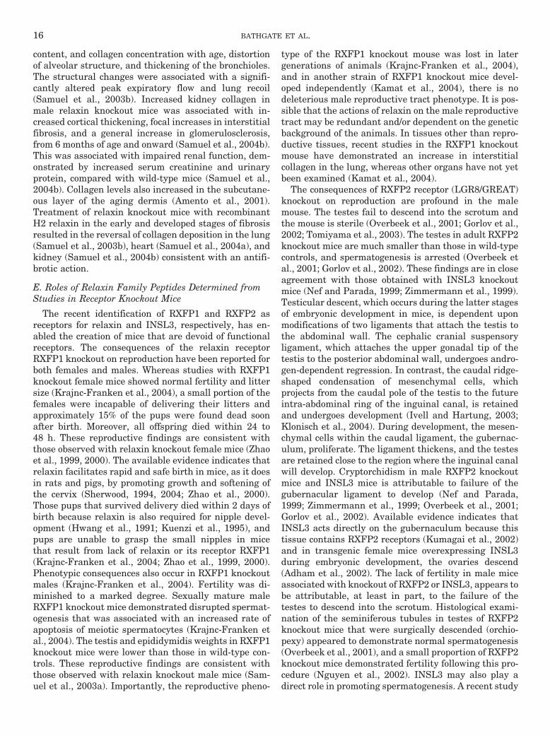
FIG. 1. Representation of the structure of H2 relaxin based on thecrystal structure (Eigenbrot et al., 1991). The conserved residues incor-porating the relaxin receptor binding motif (Arg-X-X-X-Arg-X-X-Ile/Val)are indicated as are the separate A and B chains. Reproduced fromBathgate et al. (2006), with permission; copyright © 2006 Elsevier, Inc.
RECEPTORS FOR RELAXIN FAMILY PEPTIDES 9
remaining orphan GPCR receptor that shares the closestamino acid sequence identity to GPC135 (43%), was alsoshown to be a receptor for relaxin-3 (Liu et al., 2003a).
More recent experiments have demonstrated thatGPCR142 is probably the receptor for INSL5 (Liu et al.,2005). Thus, most of the receptors and cognate ligands
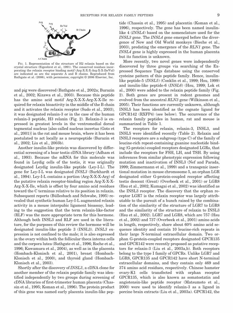
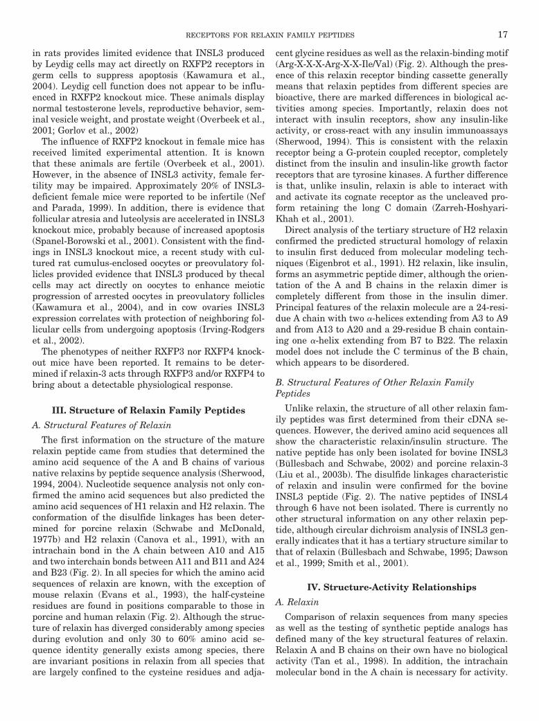
FIG. 2. Alignment of A and B chain sequences of all human relaxin-like peptides. Conserved amino acid residues are boxed in black, andconservative amino acid substitutions are boxed and shaded. Gaps have been introduced to aid the alignment where necessary. The chains arenumbered according to the H2 relaxin sequence. A, alignment showing the relationship between relaxin family peptides in human, rat, and mouse.B, alignment showing the relationship between each peptide across species.
10 BATHGATE ET AL.
for the relaxin family peptides have been identified. Nospecific receptors have yet been identified for INSL4 andINSL6. It is recommended that LGR7, LGR8, GPCR135,and GPCR142 now be known as relaxin family peptidereceptors 1 through 4 (RXFP1–4), respectively (Table 3).
II. Receptor Distribution and Function
Receptors for relaxin family peptides have been local-ized by their mRNA expression patterns using RT-PCR,Northern blotting, or in situ hybridization or as proteinby immunohistochemistry or receptor autoradiography(Table 4). Several studies have been conducted to iden-tify the tissues that contain receptors for relaxin(RXFP1, LGR7), INSL3 (RXFP2, LGR8), relaxin-3(RXFP3, GPCR135), and INSL5 (RXFP4, GPCR142). AsTable 4 indicates, the receptor for relaxin has receivedthe most experimental attention. LGR7 (RXFP1) hasbeen identified in female and male reproductive tissues,brain, and numerous other nonreproductive tissues suchas kidney, heart, and lung. Neither the cell types within
tissues that express LGR7 (RXFP1) nor the factors thatregulate the relaxin receptor are well characterized.
A. Reproductive Tissues
A few investigators have used immunohistochemicalanalysis to locate RXFP1 (LGR7) protein in female re-productive tissues. In the rat uterus, RXFP1 was re-ported in both myometrial cells and epithelial cells (Hsuet al., 2002). RXFP1 has been reported in human endo-metrial stroma, as well as in endometrial epithelial cells(Luna et al., 2004), albeit in the latter cell type to dif-fering extents. Another study demonstrated specific re-laxin binding localized to the epithelium of the endome-trial glands and uterine lumen and that both bindingand RXFP1 mRNA levels increased markedly in theearly secretory phase of the menstrual cycle comparedwith the proliferative phases (Bond et al., 2004). Weakerbinding was observed in the endometrial stromal tissueand little in the myometrium (Bond et al., 2004). In twoother primates, the marmoset and macaque monkeys,
TABLE 3Agonists and antagonists at receptors for relaxin and relaxin-related peptides
LDLa is the unique N-terminal region found in type C-glycoprotein receptors; the RXFP1-truncate is a naturally occurring splice variant of the receptor that includes theLDLa region and acts as a functional antagonist at the RXFP1 receptor (Scott et al., 2005c).
Receptor Previous Name Agonist Antagonist Radioligand
RXFP1 LGR7 H2 relaxin � H1 relaxin � H3relaxin �� INSL3
RXFP1-truncate 33P-H2 relaxin, 125I-H2 relaxin
RXFP2 LGR8 INSL3 � H2 relaxin � H1 relaxin�� H3 relaxin
INSL3 B chain analoga;(des 1–8) A chainINSL3 analogb
33P-H2 relaxin, 125I-H2 relaxin,125I-INSL3
RXFP3 GPCR135, SALPR H3 relaxin � H3 relaxin B chain INSL5 125I-H3 relaxin, 125I-H3-B/INSL5 Achimera
RXFP4 GPCR142, GPR100 INSL5 � H3 relaxin � H3 relaxinB chain
125I-H3 relaxin, 125I-H3-B/INSL5 Achimera
a Del Borgo et al. (2004); b Büllesbach and Schwabe (2005a).
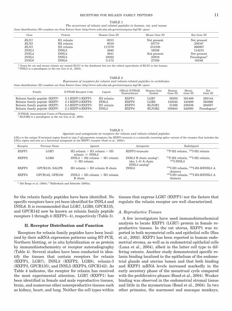
TABLE 1The occurrence of relaxin and related peptides in human, rat, and mouse
Gene identification (ID) numbers are from Entrez Gene (http://www.ncbi.nlm.nih.gov/entrez/query.fcgi?db�gene).
Gene Protein Human Gene ID Mouse Gene ID Rat Gene ID
RLN1 H1 relaxin 6013 Not present Not presentRLN2 H2 relaxin 6019 19773a 25616a
RLN3 H3 relaxin 117579 212108 266997INSL3 INSL3 3640 16336 114215INSL4 INSL4 3641 Not present Not presentINSL5 INSL5 10022 23919 Pseudogeneb
INSL6 INSL6 11172 27356 50546a Genes for rat and mouse relaxin are named RLN1 in the databases but are the rodent equivalents of RLN2 in the human.b INSL5 is a pseudogene in the rat (Liu et al., 2005).
TABLE 2Expression of receptors for relaxin and relaxin-related peptides in vertebrates
Gene identification (ID) numbers are from Entrez Gene (http://www.ncbi.nlm.nih.gov/entrez/query.fcgi?db�gene).
Family IUPHAR Receptor Code Ligand Official IUPHARNomenclature
Human GeneSymbol
HumanGene ID
MouseGene ID
RatGene ID
Relaxin family peptide (RXFP) 2.1:RXFP:1:RXFP1: H2 relaxin RXFP1 LGR7 59350 381489 295144Relaxin family peptide (RXFP) 2.1:RXFP:2:RXFP2: INSL3 RXFP2 LGR8 122042 140498 363866Relaxin family peptide (RXFP) 2.1:RXFP:3:RXFP3: H3 relaxin RXFP3 RLN3R1 51289 239336 294807Relaxin family peptide (RXFP) 2.1:RXFP:4:RXFP4: INSL5 RXFP4 RLN3R2 339403 242093 Pseudogenea
IUPHAR, International Union of Pharmacology.a RLN3R2 is a pseudogene in the rat (Liu et al., 2005).
RECEPTORS FOR RELAXIN FAMILY PEPTIDES 11
strong RXFP1 immunoreactivity was found in the endo-metrial stroma and weaker staining was found in theepithelium (Ivell et al., 2003). RXFP1 immunostaininghas also been reported in rat vagina and cervix smoothmuscle (Hsu et al., 2002) and human breast subepithe-lial stroma (Ivell et al., 2003).
Histological studies confirm the many detailed func-tional reports establishing that relaxin has importantroles in pregnancy and parturition. Established targettissues include pubic symphysis, cervix, uterus, nipples,and mammary glands although the relative importanceof the functions varies with species. In many speciesincluding humans there is growth and an increase inelasticity of pubic joint cartilage during pregnancy (forreviews, see Sherwood, 1994, 2004; Bathgate et al.,2005). There is strong evidence from animal studies thatthese effects are caused by relaxin (Hisaw, 1926; Zhao etal., 1999; Sherwood, 1994). The softening and increasein size of the cervix during the second half of pregnancyis a general phenomenon in mammals and is thought toinvolve effects of relaxin on the collagen, elastin, proteo-glycans, and glycosaminoglycans that are responsiblefor the tensile properties of the cervix. The mechanismsinvolved are complex and involve interactions with ste-roid hormones and prostaglandins. In humans, although
it has been demonstrated that relaxins can promoteripening of the cervix, clinical trials of topically appliedrelaxin failed, probably due to poor pharmacokineticproperties of the preparation (see Bathgate et al., 2005).The uterus is another target tissue for relaxin, althoughthe effects on myometrial contractile activity are highlyspecies-dependent. Relaxin clearly inhibits uterine con-tractility in rats, mice, and pigs but not in sheep, cows,or humans (Sherwood, 1994, 2004; Bathgate et al.,2005). In rats, the relaxation is associated with in-creases in intracellular levels of cAMP, PKA activity andstimulation of Ca2�-activated K� channels (Sanborn,2001). Relaxin also has a uterotrophic effect in somespecies that is associated with increased vasculariza-tion. In pigs but not rats this may have a role in thedevelopment of the uterus in pregnancy. The vagina,rather like the cervix, increases in size and softens dur-ing pregnancy, and there is good evidence from experi-ments in rats and mice that these effects involve relaxin(Bathgate et al., 2005).
Relaxin also has an important role in the developmentof the mammary nipple in rats and mice and in thedevelopment of the mammary gland in pigs. Ratstreated with monoclonal relaxin antibody during thesecond half of pregnancy display a lack of nipple devel-
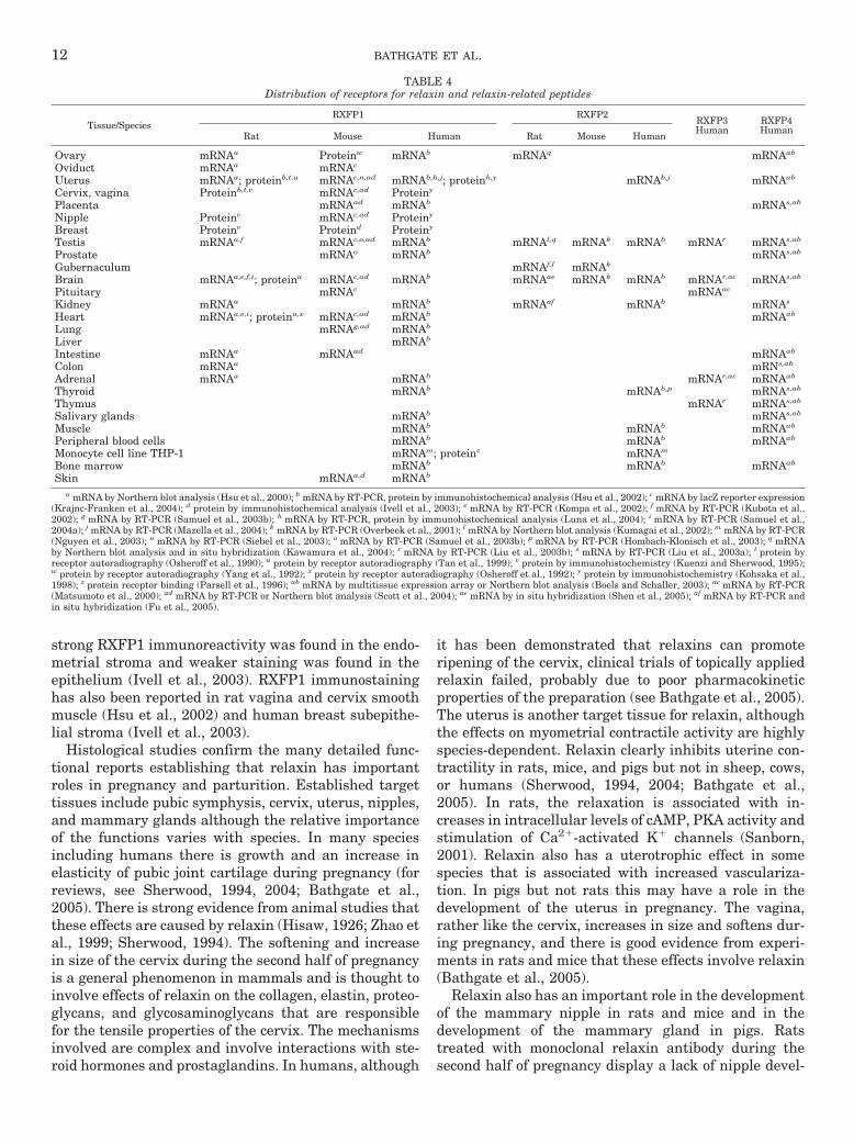
TABLE 4Distribution of receptors for relaxin and relaxin-related peptides
Tissue/SpeciesRXFP1 RXFP2
RXFP3Human
RXFP4Human
Rat Mouse Human Rat Mouse Human
Ovary mRNAa Proteinw mRNAb mRNAq mRNAab
Oviduct mRNAa mRNAc
Uterus mRNAa; proteinb,t,u mRNAc,n,ad mRNAb,h,j; proteinh,y mRNAb,j mRNAab
Cervix, vagina Proteinb,t,v mRNAc,ad Proteiny
Placenta mRNAad mRNAb mRNAs,ab
Nipple Proteinv mRNAc,ad Proteiny
Breast Proteinv Proteind Proteiny
Testis mRNAa,f mRNAc,o,ad mRNAb mRNAl,q mRNAk mRNAb mRNAr mRNAs,ab
Prostate mRNAo mRNAb mRNAs,ab
Gubernaculum mRNAf,l mRNAk
Brain mRNAa,e,f,i; proteinu mRNAc,ad mRNAb mRNAae mRNAk mRNAb mRNAr,ac mRNAs,ab
Pituitary mRNAc mRNAac
Kidney mRNAa mRNAb mRNAaf mRNAb mRNAs
Heart mRNAa,e,i; proteinu,x mRNAc,ad mRNAb mRNAab
Lung mRNAg,ad mRNAb
Liver mRNAb
Intestine mRNAa mRNAad mRNAab
Colon mRNAa mRNs,ab
Adrenal mRNAa mRNAb mRNAr,ac mRNAab
Thyroid mRNAb mRNAb,p mRNAs,ab
Thymus mRNAr mRNAs,ab
Salivary glands mRNAb mRNAs,ab
Muscle mRNAb mRNAb mRNAab
Peripheral blood cells mRNAb mRNAb mRNAab
Monocyte cell line THP-1 mRNAm; proteinz mRNAm
Bone marrow mRNAb mRNAb mRNAab
Skin mRNAa,d mRNAb
a mRNA by Northern blot analysis (Hsu et al., 2000); b mRNA by RT-PCR, protein by immunohistochemical analysis (Hsu et al., 2002); c mRNA by lacZ reporter expression(Krajnc-Franken et al., 2004); d protein by immunohistochemical analysis (Ivell et al., 2003); e mRNA by RT-PCR (Kompa et al., 2002); f mRNA by RT-PCR (Kubota et al.,2002); g mRNA by RT-PCR (Samuel et al., 2003b); h mRNA by RT-PCR, protein by immunohistochemical analysis (Luna et al., 2004); i mRNA by RT-PCR (Samuel et al.,2004a); j mRNA by RT-PCR (Mazella et al., 2004); k mRNA by RT-PCR (Overbeek et al., 2001); l mRNA by Northern blot analysis (Kumagai et al., 2002); m mRNA by RT-PCR(Nguyen et al., 2003); n mRNA by RT-PCR (Siebel et al., 2003); o mRNA by RT-PCR (Samuel et al., 2003b); p mRNA by RT-PCR (Hombach-Klonisch et al., 2003); q mRNAby Northern blot analysis and in situ hybridization (Kawamura et al., 2004); r mRNA by RT-PCR (Liu et al., 2003b); s mRNA by RT-PCR (Liu et al., 2003a); t protein byreceptor autoradiography (Osheroff et al., 1990); u protein by receptor autoradiography (Tan et al., 1999); v protein by immunohistochemistry (Kuenzi and Sherwood, 1995);w protein by receptor autoradiography (Yang et al., 1992); x protein by receptor autoradiography (Osheroff et al., 1992); y protein by immunohistochemistry (Kohsaka et al.,1998); z protein receptor binding (Parsell et al., 1996); ab mRNA by multitissue expression array or Northern blot analysis (Boels and Schaller, 2003); ac mRNA by RT-PCR(Matsumoto et al., 2000); ad mRNA by RT-PCR or Northern blot analysis (Scott et al., 2004); ae mRNA by in situ hybridization (Shen et al., 2005); af mRNA by RT-PCR andin situ hybridization (Fu et al., 2005).
12 BATHGATE ET AL.
opment and cannot suckle their young (Hwang et al.,1991). An identical phenotype was observed in relaxinknockout mice (Zhao et al., 1999). In relaxin-deficientpigs, however, nipple development is relatively normal.The situation is reversed with respect to the mammarygland. In mice and rats, relaxin is not essential formammary gland development although it may affect thedifferentiation of the tissue, whereas in pigs relaxin isimportant for the development of the mammary glandparenchyma. Relaxin-binding sites are present in themammary glands of pigs but also rats and humans. Inhumans and other primates, there is increasing evi-dence for a role for relaxin in preparing the endome-trium for implantation. A number of studies indicatedthat relaxin is associated with increased endometrialangiogenesis, thickening, and bleeding (Einspanier etal., 2001; Goldsmith et al., 2004). A link is also suggestedby the plasma levels of relaxin that are highest in thefirst trimester, the time corresponding to embryo im-plantation. However the role of relaxin is probably fa-cilitatory rather than mandatory for implantation be-cause this still occurs in humans and other primatesthat lack ovaries (Johnson et al., 1991). Relaxin is alsoproduced in the male reproductive tract, is present insemen, and increases sperm motility and penetrationinto oocytes (Weiss, 1989). As well as the potential func-tion in males, it is also tempting to speculate that re-laxin in semen targets the female reproductive tract toprepare the endometrium for implantation.
Studies of the distribution of RXFP2 are less exten-sive, but RXFP2 mRNA has been demonstrated in ratovary, testis, and gubernaculum by Northern analysis,RT-PCR, and in situ hybridization (Kubota et al., 2002;Kumagai et al., 2002; Kawamura et al., 2004; Scott etal., 2005a). In mouse, RXFP2 mRNA is present in testisand gubernaculum (Overbeek et al., 2001). In human,RXFP2 mRNA is found in uterus and testis (Hsu et al.,2002; Mazella et al., 2004). By immunohistochemistry,RXFP2 epitopes have been identified in spermatocytes,spermatids, and Leydig cells within the human testis,and in the epididymal epithelium (R. Ivell, unpublisheddata). The pattern of localization clearly indicates a rolefor INSL3/RXFP2 in reproductive physiology. This rolehas clearly been identified as testis descent because bothINSL3 and LGR8 male knockout mice display bilateralcryptorchidism (Nef and Parada, 1999; Zimmermann etal., 1999; Overbeek et al., 2001). The action of INSL3 ison RXFP2 receptors located on the gubernaculum(Kumagai et al., 2002).
Both RXFP3 and RXFP4 mRNA are found in humantestis (Liu et al., 2003a,b). RXFP4 is also found in pla-centa (Boels and Schaller, 2003) and prostate (Liu et al.,2003a,b).
B. Brain
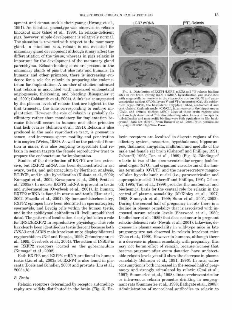
Relaxin receptors determined by receptor autoradiog-raphy are widely distributed in the brain (Fig. 3). Re-
laxin receptors are localized to discrete regions of theolfactory system, neocortex, hypothalamus, hippocam-pus, thalamus, amygdala, midbrain, and medulla of themale and female rat brain (Osheroff and Phillips, 1991;Osheroff, 1995; Tan et al., 1999) (Fig. 3). Binding ofrelaxin to two of the circumventricular organs [subfor-nical organ (SFO); and organum vasculosum of the lam-ina terminalis (OVLT)] and the neurosecretory magno-cellular hypothalamic nuclei (i.e., paraventricular andsupraoptic nuclei) (Osheroff and Phillips, 1991; Osher-off, 1995; Tan et al., 1999) provides the anatomical andbiochemical basis for the central role for relaxin in thecontrol of plasma osmolality (McKinley et al., 1997,1998; Sinnayah et al., 1999; Sunn et al., 2001, 2002).During the second half of pregnancy in rats there is adecline in plasma osmolality that is associated with in-creased serum relaxin levels (Sherwood et al., 1980;Lindheimer et al., 1989) that does not occur in pregnantrelaxin-deficient rats (Novak et al., 2001). Likewise, de-creases in plasma osmolality in wild-type mice in latepregnancy are not observed in relaxin knockout mice(Zhao et al., 1999). However in humans, although thereis a decrease in plasma osmolality with pregnancy, thismay not be an effect of relaxin, because women thatbecome pregnant after ovum donation have undetect-able relaxin levels yet still show the decrease in plasmaosmolality (Johnson et al., 1991, 1996). In rats, waterconsumption is both increased in the second half of preg-nancy and strongly stimulated by relaxin (Omi et al.,1997; Summerlee et al., 1998). Intracerebroventricularor intravenous relaxin promotes drinking in nonpreg-nant rats (Summerlee et al., 1998; Bathgate et al., 2005).Administration of monoclonal antibodies to relaxin to
FIG. 3. Distribution of RXFP1 (LGR7) mRNA and 33P-relaxin-bindingsites in rat brain. Strong RXFP1 mRNA hybridization was associatedwith magnocellular neurons in the supraoptic nucleus (SON) and para-ventricular nucleus (PVN), layers V and VI of neocortex (Cx), the subfor-nical organ (SFO), the basolateral amygdala (BLA), centromedial andcentrolateral thalamic nuclei (CM/CL), interneurons in the hippocampus(CA3), and arcuate nucleus (ARC). Most of these brain regions alsocontain high densities of 33P-relaxin-binding sites. Levels of nonspecifichybridization and nonspecific binding were both equivalent to film back-ground (data not shown). From Burazin et al. (2005), with permission;copyright © 2005 HighWire Press.
RECEPTORS FOR RELAXIN FAMILY PEPTIDES 13
rats in the second half of pregnancy reduces water con-sumption. These actions of relaxin are mediated byRXFP1 receptors located in the SFO and OVLT. Theintravenous administration of porcine or H2 relaxincauses increased expression of c-fos, the gene encodingproto-oncogene protein, in neurons located in the periph-eral and dorsal segments of the SFO and in the dorsalcap region of the OVLT, as well as the supraoptic andparaventricular nuclei of the hypothalamus (McKinleyet al., 1997, 1998; Sunn et al., 2001, 2002), all sites oflocalization of relaxin receptors. These regions of thebrain are accessible to circulating relaxin and may beresponsible for the plasma osmolality changes that occurduring pregnancy (Weisinger et al., 1993).
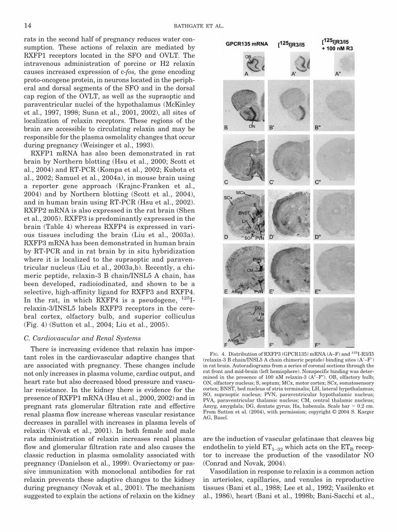
RXFP1 mRNA has also been demonstrated in ratbrain by Northern blotting (Hsu et al., 2000; Scott etal., 2004) and RT-PCR (Kompa et al., 2002; Kubota etal., 2002; Samuel et al., 2004a), in mouse brain usinga reporter gene approach (Krajnc-Franken et al.,2004) and by Northern blotting (Scott et al., 2004),and in human brain using RT-PCR (Hsu et al., 2002).RXFP2 mRNA is also expressed in the rat brain (Shenet al., 2005). RXFP3 is predominantly expressed in thebrain (Table 4) whereas RXFP4 is expressed in vari-ous tissues including the brain (Liu et al., 2003a).RXFP3 mRNA has been demonstrated in human brainby RT-PCR and in rat brain by in situ hybridizationwhere it is localized to the supraoptic and paraven-tricular nucleus (Liu et al., 2003a,b). Recently, a chi-meric peptide, relaxin-3 B chain/INSL5 A chain, hasbeen developed, radioiodinated, and shown to be aselective, high-affinity ligand for RXFP3 and RXFP4.In the rat, in which RXFP4 is a pseudogene, 125I-relaxin-3/INSL5 labels RXFP3 receptors in the cere-bral cortex, olfactory bulb, and superior colliculus(Fig. 4) (Sutton et al., 2004; Liu et al., 2005).
C. Cardiovascular and Renal Systems
There is increasing evidence that relaxin has impor-tant roles in the cardiovascular adaptive changes thatare associated with pregnancy. These changes includenot only increases in plasma volume, cardiac output, andheart rate but also decreased blood pressure and vascu-lar resistance. In the kidney there is evidence for thepresence of RXFP1 mRNA (Hsu et al., 2000, 2002) and inpregnant rats glomerular filtration rate and effectiverenal plasma flow increase whereas vascular resistancedecreases in parallel with increases in plasma levels ofrelaxin (Novak et al., 2001). In both female and malerats administration of relaxin increases renal plasmaflow and glomerular filtration rate and also causes theclassic reduction in plasma osmolality associated withpregnancy (Danielson et al., 1999). Ovariectomy or pas-sive immunization with monoclonal antibodies for ratrelaxin prevents these adaptive changes to the kidneyduring pregnancy (Novak et al., 2001). The mechanismsuggested to explain the actions of relaxin on the kidney
are the induction of vascular gelatinase that cleaves bigendothelin to yield ET1–32 which acts on the ETB recep-tor to increase the production of the vasodilator NO(Conrad and Novak, 2004).
Vasodilation in response to relaxin is a common actionin arterioles, capillaries, and venules in reproductivetissues (Bani et al., 1988; Lee et al., 1992; Vasilenko etal., 1986), heart (Bani et al., 1998b; Bani-Sacchi et al.,
FIG. 4. Distribution of RXFP3 (GPCR135) mRNA (A–F) and 125I-R3/I5(relaxin-3 B chain/INSL5 A chain chimeric peptide) binding sites (A�–F�)in rat brain. Autoradiograms from a series of coronal sections through therat front and mid-brain (left hemisphere). Nonspecific binding was deter-mined in the presence of 100 nM relaxin-3 (A�–F�). OB, olfactory bulb;ON, olfactory nucleus; S, septum; MCx, motor cortex; SCx, somatosensorycortex; BNST, bed nucleus of stria terminalis; LH, lateral hypothalamus;SO, supraoptic nucleus; PVN, paraventricular hypothalamic nucleus;PVA, paraventricular thalamic nucleus; CM, central thalamic nucleus;Amyg, amygdala; DG, dentate gyrus; Ha, habenula. Scale bar � 0.2 cm.From Sutton et al. (2004), with permission; copyright © 2004 S. KargerAG, Basel.
14 BATHGATE ET AL.
1995; Masini et al., 1997), liver (Bani et al., 2001) andcecum (Bigazzi et al., 1986). Relaxin acts as a physiolog-ical antagonist of vasoconstrictors in mesenteric arteries(St-Louis and Massicotte, 1985; Massicotte et al., 1989),primary bovine aortic smooth muscle cells (Bani et al.,1998a), and uterine artery (Longo et al., 2003). Thehormone also reduces the rise in intracellular Ca2� pro-duced by �-thrombin or angiotensin II (Bani et al.,1998a; Failli et al., 2002). The mechanisms suggested forthe vasodilator actions of relaxin are activation of nitric-oxide synthase (NOS) III via cAMP, induction of NOSII(Nistri and Bani, 2003) and modification of the extracel-lular matrix of the vessel walls (Conrad and Novak,2004).
The heart is clearly a target organ for relaxin in ro-dents. Atria of male and female rats possess high-affin-ity binding sites for relaxin (Osheroff et al., 1992; Os-heroff and Ho, 1993; Tan et al., 1999), and RXFP1mRNA has been detected in the rat (Hsu et al., 2000;Kompa et al., 2002), mouse (Krajnc-Franken et al.,2004), and human (Hsu et al., 2002) heart. Numerous invitro and in vivo studies demonstrate that relaxin haspotent, direct, and concentration-dependent chrono-tropic and inotropic effects on the rat heart. The positivechronotropic effects of relaxin have been reported inperfused intact hearts (Thomas and Vandlen, 1993;Bani-Sacchi et al., 1995; Coulson et al., 1996; Toth et al.,1996) and isolated right atria (Kakouris et al., 1992;Ward et al., 1992; Wade et al., 1994; Tan et al., 1998;Mathieu et al., 2001) and inotropic effects in left atria(Kakouris et al., 1992); (Ward et al., 1992; Wade et al.,1994; Tan et al., 1998; Mathieu et al., 2001). Little isknown about the mechanism or site of action of relaxinin the heart. The chronotropic effects are accompaniedby the secretion of atrial natriuretic peptide in isolatedperfused rat hearts (Toth et al., 1996). In rat atrialmyocytes, relaxin inhibits outward potassium currents,increases action potential duration, and enhances Ca2�
entry (Piedras-Renteria et al., 1997a,b). In rabbit sino-atrial node cells, relaxin increased the rate of actionpotentials and L-type Ca2� currents (Han et al., 1994)by a PKA-dependent mechanism. These actions of re-laxin on the heart are largely confined to rodents, andthe hormone has no positive inotropic effect in the atriaof sheep (Bathgate et al., 2001) or humans (R. J. Sum-mers and L. J. Castro, unpublished data). Radiolabeledrelaxin does not bind specifically to sheep or humanatria (Y. Y. Tan and R. J. Summers, unpublished data).mRNA for RXFP2 has been demonstrated in human(Hsu et al., 2002) and rat (Fu et al., 2005) kidney as hasRXFP4 (Liu et al., 2003a), but there are no known func-tional correlates for these expression patterns to date.
D. Other Sites of Action of Relaxin Family Peptides
In addition to the specific roles of relaxin alreadydescribed, it is becoming increasingly clear that it hasmore general physiological roles. The role of relaxin in
inhibiting collagen biosynthesis and promoting collagenbreakdown in reproductive tissues is well established,but it is also clear that it has similar effects in nonre-productive tissues (Gavino and Furst, 2001; Bathgate etal., 2003), which have led to the suggestion that relaxinwould be an effective treatment for fibrotic diseases.Relaxin acts directly on transforming growth factor-�-stimulated human dermal fibroblasts (Unemori andAmento, 1990), lung fibroblasts (Unemori et al., 1996),and cardiac fibroblasts (Samuel et al., 2004a) to promotea decrease in type I and type III collagen synthesis andan increase in matrix metalloproteinase expression andactivation. It has been used successfully to modify theextracellular matrix in the dermis (Kibblewhite et al.,1992; Unemori et al., 1993), lung (Unemori et al., 1996),liver (Williams et al., 2001), and kidney (Garber et al.,2001). In all of these experimental paradigms, relaxin iseffective only during artificially induced collagen depo-sition, induced by profibrotic stimuli, surgery, or chem-ical means.
Relaxin has been used in humans to treat scleroderma(systemic sclerosis), a connective tissue disease of un-known etiology in which tissue fibrosis is the predomi-nant clinical feature (Casten and Boucek, 1958). Clinicaltrials using recombinant H2 relaxin to treat sclerodermahave had mixed success. A phase II trial infusing recom-binant H2 relaxin subcutaneously for a 24-week period(Seibold et al., 2000) were encouraging and the peptidewas found to be safe and well tolerated. Twenty-fourweeks of treatment with a low but not a high dose ofrelaxin had significant beneficial effects on skin thick-ness and mobility. However, a larger study showed thatskin elasticity, hand extension, oral aperture, cutaneousulcers, and pulmonary function in recombinant H2-treated patients did not differ significantly from those inthe placebo control subjects (Erikson and Unemori,2001) even though relaxin did have beneficial actions insome patients. The positive results obtained in numer-ous in vivo and in vitro studies, in which relaxin hasbeen able to reverse fibrosis, suggest that it still hasenormous potential as an antifibrotic agent and thatmore research needs to be done to better understand itsmechanism of action and to identify those patients whoare likely to derive benefit from treatment.
Studies with the relaxin knockout mouse demonstratethat relaxin is an endogenous mediator of collagen turn-over in nonreproductive tissues. In these animals, in-creased interstitial collagen was detected in heart (Du etal., 2003), lung (Samuel et al., 2003b), kidney (Samuel etal., 2004a), and skin (Amento et al., 2001). The effectwas particularly prevalent in male mice and associatedwith abnormal function of these organs. In the heart,there is increased atrial hypertrophy and impaired leftventricular diastolic filling and venous return in oldermice, associated with increased left ventricular collagencontent and ventricular chamber stiffness (Du et al.,2003). In the lung, there was increased weight, collagen
RECEPTORS FOR RELAXIN FAMILY PEPTIDES 15
content, and collagen concentration with age, distortionof alveolar structure, and thickening of the bronchioles.The structural changes were associated with a signifi-cantly altered peak expiratory flow and lung recoil(Samuel et al., 2003b). Increased kidney collagen inmale relaxin knockout mice was associated with in-creased cortical thickening, focal increases in interstitialfibrosis, and a general increase in glomerulosclerosis,from 6 months of age and onward (Samuel et al., 2004b).This was associated with impaired renal function, dem-onstrated by increased serum creatinine and urinaryprotein, compared with wild-type mice (Samuel et al.,2004b). Collagen levels also increased in the subcutane-ous layer of the aging dermis (Amento et al., 2001).Treatment of relaxin knockout mice with recombinantH2 relaxin in the early and developed stages of fibrosisresulted in the reversal of collagen deposition in the lung(Samuel et al., 2003b), heart (Samuel et al., 2004a), andkidney (Samuel et al., 2004b) consistent with an antifi-brotic action.
E. Roles of Relaxin Family Peptides Determined fromStudies in Receptor Knockout Mice
The recent identification of RXFP1 and RXFP2 asreceptors for relaxin and INSL3, respectively, has en-abled the creation of mice that are devoid of functionalreceptors. The consequences of the relaxin receptorRXFP1 knockout on reproduction have been reported forboth females and males. Whereas studies with RXFP1knockout female mice showed normal fertility and littersize (Krajnc-Franken et al., 2004), a small portion of thefemales were incapable of delivering their litters andapproximately 15% of the pups were found dead soonafter birth. Moreover, all offspring died within 24 to48 h. These reproductive findings are consistent withthose observed with relaxin knockout female mice (Zhaoet al., 1999, 2000). The available evidence indicates thatrelaxin facilitates rapid and safe birth in mice, as it doesin rats and pigs, by promoting growth and softening ofthe cervix (Sherwood, 1994, 2004; Zhao et al., 2000).Those pups that survived delivery died within 2 days ofbirth because relaxin is also required for nipple devel-opment (Hwang et al., 1991; Kuenzi et al., 1995), andpups are unable to grasp the small nipples in micethat result from lack of relaxin or its receptor RXFP1(Krajnc-Franken et al., 2004; Zhao et al., 1999, 2000).Phenotypic consequences also occur in RXFP1 knockoutmales (Krajnc-Franken et al., 2004). Fertility was di-minished to a marked degree. Sexually mature maleRXFP1 knockout mice demonstrated disrupted spermat-ogenesis that was associated with an increased rate ofapoptosis of meiotic spermatocytes (Krajnc-Franken etal., 2004). The testis and epididymidis weights in RXFP1knockout mice were lower than those in wild-type con-trols. These reproductive findings are consistent withthose observed with relaxin knockout male mice (Sam-uel et al., 2003a). Importantly, the reproductive pheno-
type of the RXFP1 knockout mouse was lost in latergenerations of animals (Krajnc-Franken et al., 2004),and in another strain of RXFP1 knockout mice devel-oped independently (Kamat et al., 2004), there is nodeleterious male reproductive tract phenotype. It is pos-sible that the actions of relaxin on the male reproductivetract may be redundant and/or dependent on the geneticbackground of the animals. In tissues other than repro-ductive tissues, recent studies in the RXFP1 knockoutmouse have demonstrated an increase in interstitialcollagen in the lung, whereas other organs have not yetbeen examined (Kamat et al., 2004).
The consequences of RXFP2 receptor (LGR8/GREAT)knockout on reproduction are profound in the malemouse. The testes fail to descend into the scrotum andthe mouse is sterile (Overbeek et al., 2001; Gorlov et al.,2002; Tomiyama et al., 2003). The testes in adult RXFP2knockout mice are much smaller than those in wild-typecontrols, and spermatogenesis is arrested (Overbeek etal., 2001; Gorlov et al., 2002). These findings are in closeagreement with those obtained with INSL3 knockoutmice (Nef and Parada, 1999; Zimmermann et al., 1999).Testicular descent, which occurs during the latter stagesof embryonic development in mice, is dependent uponmodifications of two ligaments that attach the testis tothe abdominal wall. The cephalic cranial suspensoryligament, which attaches the upper gonadal tip of thetestis to the posterior abdominal wall, undergoes andro-gen-dependent regression. In contrast, the caudal ridge-shaped condensation of mesenchymal cells, whichprojects from the caudal pole of the testis to the futureintra-abdominal ring of the inguinal canal, is retainedand undergoes development (Ivell and Hartung, 2003;Klonisch et al., 2004). During development, the mesen-chymal cells within the caudal ligament, the gubernac-ulum, proliferate. The ligament thickens, and the testesare retained close to the region where the inguinal canalwill develop. Cryptorchidism in male RXFP2 knockoutmice and INSL3 mice is attributable to failure of thegubernacular ligament to develop (Nef and Parada,1999; Zimmermann et al., 1999; Overbeek et al., 2001;Gorlov et al., 2002). Available evidence indicates thatINSL3 acts directly on the gubernaculum because thistissue contains RXFP2 receptors (Kumagai et al., 2002)and in transgenic female mice overexpressing INSL3during embryonic development, the ovaries descend(Adham et al., 2002). The lack of fertility in male miceassociated with knockout of RXFP2 or INSL3, appears tobe attributable, at least in part, to the failure of thetestes to descend into the scrotum. Histological exami-nation of the seminiferous tubules in testes of RXFP2knockout mice that were surgically descended (orchio-pexy) appeared to demonstrate normal spermatogenesis(Overbeek et al., 2001), and a small proportion of RXFP2knockout mice demonstrated fertility following this pro-cedure (Nguyen et al., 2002). INSL3 may also play adirect role in promoting spermatogenesis. A recent study
16 BATHGATE ET AL.
in rats provides limited evidence that INSL3 producedby Leydig cells may act directly on RXFP2 receptors ingerm cells to suppress apoptosis (Kawamura et al.,2004). Leydig cell function does not appear to be influ-enced in RXFP2 knockout mice. These animals displaynormal testosterone levels, reproductive behavior, sem-inal vesicle weight, and prostate weight (Overbeek et al.,2001; Gorlov et al., 2002)
The influence of RXFP2 knockout in female mice hasreceived limited experimental attention. It is knownthat these animals are fertile (Overbeek et al., 2001).However, in the absence of INSL3 activity, female fer-tility may be impaired. Approximately 20% of INSL3-deficient female mice were reported to be infertile (Nefand Parada, 1999). In addition, there is evidence thatfollicular atresia and luteolysis are accelerated in INSL3knockout mice, probably because of increased apoptosis(Spanel-Borowski et al., 2001). Consistent with the find-ings in INSL3 knockout mice, a recent study with cul-tured rat cumulus-enclosed oocytes or preovulatory fol-licles provided evidence that INSL3 produced by thecalcells may act directly on oocytes to enhance meioticprogression of arrested oocytes in preovulatory follicles(Kawamura et al., 2004), and in cow ovaries INSL3expression correlates with protection of neighboring fol-licular cells from undergoing apoptosis (Irving-Rodgerset al., 2002).
The phenotypes of neither RXFP3 nor RXFP4 knock-out mice have been reported. It remains to be deter-mined if relaxin-3 acts through RXFP3 and/or RXFP4 tobring about a detectable physiological response.
III. Structure of Relaxin Family Peptides
A. Structural Features of Relaxin
The first information on the structure of the maturerelaxin peptide came from studies that determined theamino acid sequence of the A and B chains of variousnative relaxins by peptide sequence analysis (Sherwood,1994, 2004). Nucleotide sequence analysis not only con-firmed the amino acid sequences but also predicted theamino acid sequences of H1 relaxin and H2 relaxin. Theconformation of the disulfide linkages has been deter-mined for porcine relaxin (Schwabe and McDonald,1977b) and H2 relaxin (Canova et al., 1991), with anintrachain bond in the A chain between A10 and A15and two interchain bonds between A11 and B11 and A24and B23 (Fig. 2). In all species for which the amino acidsequences of relaxin are known, with the exception ofmouse relaxin (Evans et al., 1993), the half-cysteineresidues are found in positions comparable to those inporcine and human relaxin (Fig. 2). Although the struc-ture of relaxin has diverged considerably among speciesduring evolution and only 30 to 60% amino acid se-quence identity generally exists among species, thereare invariant positions in relaxin from all species thatare largely confined to the cysteine residues and adja-
cent glycine residues as well as the relaxin-binding motif(Arg-X-X-X-Arg-X-X-Ile/Val) (Fig. 2). Although the pres-ence of this relaxin receptor binding cassette generallymeans that relaxin peptides from different species arebioactive, there are marked differences in biological ac-tivities among species. Importantly, relaxin does notinteract with insulin receptors, show any insulin-likeactivity, or cross-react with any insulin immunoassays(Sherwood, 1994). This is consistent with the relaxinreceptor being a G-protein coupled receptor, completelydistinct from the insulin and insulin-like growth factorreceptors that are tyrosine kinases. A further differenceis that, unlike insulin, relaxin is able to interact withand activate its cognate receptor as the uncleaved pro-form retaining the long C domain (Zarreh-Hoshyari-Khah et al., 2001).
Direct analysis of the tertiary structure of H2 relaxinconfirmed the predicted structural homology of relaxinto insulin first deduced from molecular modeling tech-niques (Eigenbrot et al., 1991). H2 relaxin, like insulin,forms an asymmetric peptide dimer, although the orien-tation of the A and B chains in the relaxin dimer iscompletely different from those in the insulin dimer.Principal features of the relaxin molecule are a 24-resi-due A chain with two �-helices extending from A3 to A9and from A13 to A20 and a 29-residue B chain contain-ing one �-helix extending from B7 to B22. The relaxinmodel does not include the C terminus of the B chain,which appears to be disordered.
B. Structural Features of Other Relaxin FamilyPeptides
Unlike relaxin, the structure of all other relaxin fam-ily peptides was first determined from their cDNA se-quences. However, the derived amino acid sequences allshow the characteristic relaxin/insulin structure. Thenative peptide has only been isolated for bovine INSL3(Bullesbach and Schwabe, 2002) and porcine relaxin-3(Liu et al., 2003b). The disulfide linkages characteristicof relaxin and insulin were confirmed for the bovineINSL3 peptide (Fig. 2). The native peptides of INSL4through 6 have not been isolated. There is currently noother structural information on any other relaxin pep-tide, although circular dichroism analysis of INSL3 gen-erally indicates that it has a tertiary structure similar tothat of relaxin (Bullesbach and Schwabe, 1995; Dawsonet al., 1999; Smith et al., 2001).
IV. Structure-Activity Relationships
A. Relaxin
Comparison of relaxin sequences from many speciesas well as the testing of synthetic peptide analogs hasdefined many of the key structural features of relaxin.Relaxin A and B chains on their own have no biologicalactivity (Tan et al., 1998). In addition, the intrachainmolecular bond in the A chain is necessary for activity.
RECEPTORS FOR RELAXIN FAMILY PEPTIDES 17
The amino acids in the N-terminal regions of the A andB chain are not directly involved in activity; however,retaining the secondary structure in this region of thechains is essential (Tregear et al., 1981; Bullesbach andSchwabe, 1986, 1987). The carboxyl-terminal region ofthe B chain does not appear to be important and up toeight amino acids can be deleted from this region of pigrelaxin (Tregear et al., 1981) or H1 relaxin (Tan et al.,1999) without loss of activity. One of the invariantamino acids across all relaxin peptides, the glycine res-idue at A14 is absolutely essential for maintaining chainflexibility (Bullesbach and Schwabe, 1994). Hence theoverall structure of the A chain seems to be more impor-tant than the individual amino acids and the main roleof the A chain is to act as a scaffold to hold the B chainin the correct formation. In contrast, individual aminoacids in the B chain are vital for receptor binding. Theabsolutely conserved arginine B13 and B17 residues(Fig. 2) are essential for interaction with the relaxinreceptor (Bullesbach et al., 1992) and together with theB20 isoleucine residue are essential for relaxin binding(Bullesbach and Schwabe, 2000). The B20 isoleucine canbe substituted with valine without loss of activity, andvirtually all of the native relaxins have these aminoacids in this position. Hence these amino acid residues,Arg-X-X-X-Arg-X-X-Ile/Val (Fig. 2), constitute what isnow regarded as the “relaxin-binding cassette.” Theseresidues are also conserved in relaxin-3 (Fig. 2) and arethe basis for its interaction with relaxin receptors (Bath-gate et al., 2002a).
Although these residues are undoubtedly the key torelaxin-specific actions, other structural determinantsare essential for full relaxin activity. Substitution of therelaxin-binding cassette into a sheep INSL3 peptide pro-duced only weak relaxin-like activity (Claasz et al.,2002; Tan et al., 2002), and insertion of a partial cassetteinto a porcine insulin analog failed to produce full re-laxin activity (Bullesbach and Schwabe, 1996). Based onthe marked differences in relaxin structure between spe-cies it is unlikely that there are other key residues forreceptor interaction. As mentioned above, the structureof the B chain is especially important, with a full �-he-lical structure necessary for the correct positioning ofthe relaxin-binding cassette (Bullesbach and Schwabe,1991; Bullesbach et al., 1992). Importantly, the INSL3analogs with a full relaxin-binding cassette do not havethe full �-helix structure of H2 relaxin (Claasz et al.,2002). Interestingly, surface regions on the relaxinstructure involved in receptor binding that comprise therelaxin-binding cassette were accurately predicted frommolecular modeling studies more than 20 years ago(Dodson et al., 1982).
B. INSL3
Although all INSL3 peptides contain a partial “relax-in-like” binding motif with two arginine residues in apattern resembling relaxin (Fig. 2), these are displaced
toward the C terminus by four residues and show onlyminimal relaxin activity (Table 3) (Bullesbach andSchwabe, 1995; Dawson et al., 1999; Claasz et al., 2002).Furthermore, INSL3 peptides show only limited abilityto interact with the relaxin receptor (RXFP1) (Sudo etal., 2003) and are the cognate ligands for the relatedRXFP2 receptor (Table 3) (Kumagai et al., 2002). Thedeterminants for INSL3 binding to its receptor include atryptophan residue at B27 located in the N terminus ofthe B chain (Bullesbach and Schwabe, 1999a) (Fig. 2).More recently it has been shown that the N terminus ofthe A chain of INSL3 can be truncated by up to nineresidues and retain full binding activity (Bullesbach andSchwabe, 2005a). Truncation of up to six residues can beperformed with the peptides still able to induce cAMPsignaling although the loss of six residues is associatedwith lower potency and efficacy. Interestingly, furthertruncation produces peptides that bind with high affin-ity but are unable to induce cAMP signaling in RXFP2transfected cells and act as competitive antagonists(Bullesbach and Schwabe, 2005a). Mutation to Arg8Alaor Tyr9Ala in the A chain produces peptides that retainagonist properties indicating that the arginine and ty-rosine side chains are not essential for signal transduc-tion. Hence, regions of the N terminus of the A chain ofINSL3 appear to be essential for RXFP2 activation andpeptides lacking this region of the peptide are the firsthigh-affinity RXFP2 antagonists (Bullesbach andSchwabe, 2005a).
V. Binding of Relaxin and Relaxin FamilyPeptides
A. Relaxin Binding
Relaxin-binding sites have been identified in numer-ous reproductive tissues as well as in some nonreproduc-tive tissues. Early studies used purified porcine relaxin(Sherwood et al., 1975; McMurtry et al., 1978) that re-tained its bioactivity after iodination although the finalproduct was often a mix of multiply iodinated forms.These ligands were subsequently used to perform thefirst localization of relaxin receptors in multiple tissuesfrom various species. Specific binding was not displacedby insulin or insulin-like growth factor I or II. Relaxin-binding sites were identified in the uterus of rat(McMurtry et al., 1978; Cheah and Sherwood, 1980;Mercado-Simmen et al., 1980), mouse (McMurtry et al.,1978; Yang et al., 1992) and pig (Mercado et al., 1982);the pubic symphysis of mouse (McMurtry et al., 1978;Yang et al., 1992) and guinea pig (McMurtry et al.,1978); the cervix of pig (Mercado et al., 1982), rat (Weissand Bryant, 1982) and guinea pig (McMurtry et al.,1978); the rat mammary gland (McMurtry et al., 1978);human fetal membranes (Koay et al., 1986); and fibro-blasts from the mouse pubic symphysis and human skin(McMurtry et al., 1980).
18 BATHGATE ET AL.
H2 relaxin contains a tyrosine residue that can belabeled by conventional chloramine-T iodination (Tangand Chegini, 1995). 125I-H2 relaxin binds to epithelial,stromal, and smooth muscle cells in human fallopiantube, smooth muscle cells in the arterioles (Tang andChegini, 1995), and fibroblasts isolated from humanlower uterine segments (Palejwala et al., 1998). 125I-H2relaxin labeled by an alternate strategy has also beenused to characterize the binding activity of relaxin ana-logs (Bullesbach and Schwabe, 2000). H2 relaxin (B-33)can also be phosphorylated using 32P (Osheroff et al.,1990) to produce a specific high-activity radioligandmonolabeled at the B-32 serine residue (Osheroff et al.,1990; Tan et al., 1999). 32P-H2 relaxin has been shown tobind to rat uterus, cervix, and brain (Osheroff et al.,1990), rat heart atrium (Osheroff et al., 1992), rat atrialcardiomyocytes (Osheroff, 1995), and a number of re-gions of the rat brain (Osheroff and Ho, 1993; Osheroffand Phillips, 1991). In addition, binding sites have beenidentified in human uterine cells (Osheroff and King,1995), in human fetal membranes (Garibay-Tupas et al.,1995), and in the human monocytic cell line THP-1(Parsell et al., 1996). More recently 33P-H2 relaxin hasbeen used to localize relaxin binding in the rat uterus,atria, and brain (Tan et al., 1999) (Fig. 3) and for nu-merous structure function studies (Bathgate et al.,2002a,b; Sudo et al., 2003).
Cross-linking of 32P-H2 relaxin to human uterine cellsand rat atrial cardiomyocytes demonstrated binding to a�200-kDa protein that was postulated (Osheroff andKing, 1995) to be a tyrosine kinase. Later, a 220-kDaprotein was shown to be tyrosine phosphorylated follow-ing relaxin stimulation in human lower uterine segmentfibroblasts (Palejwala et al., 1998). These data wereinterpreted as evidence for the relaxin receptor being atyrosine kinase.
Biotinylated relaxin has been used to localize relaxinbinding in the cervix, mammary glands, and nipples ofrats (Kuenzi and Sherwood, 1995); the cervix, mammarygland, nipples, small intestine, skin (Min and Sherwood,1996), ovary, and testis (Min and Sherwood, 1998) ofpigs; the uterus, vagina, cervix, mammary gland, nip-ples, and placenta of pregnant women (Kohsaka et al.,1998), and the uterine endometrium and myometrium ofthe marmoset monkey (Einspanier et al., 2001). A mono-biotinylated rat relaxin peptide was produced by peptidesynthesis and shown to be biologically active (Mathieu etal., 2001).
B. Binding of Other Relaxin Family Peptides
Very little is known of specific binding sites for otherrelaxin family peptides. Binding studies with labeledINSL3 have not been carried out in the gubernaculumand ovary, the likely target tissues for the peptide. How-ever, 125I-INSL3 has been used to demonstrate specificbinding sites in mouse uterus and brain (Bullesbach andSchwabe, 1995), although the significance of these sites
has yet to be established. These binding sites were spe-cific for INSL3 and had a 1000-fold lower affinity for H2relaxin. 125I-INSL3 has been chemically cross-linked tomouse uterus and demonstrated a �200-kDa band(Bullesbach and Schwabe, 1999b) similar to that shownfor the relaxin receptor (Osheroff and King, 1995).
125I-H3 relaxin (Liu et al., 2003b) binds specifically toRXFP3 and RXFP4 receptors expressed in mammaliancells. As H3 relaxin also binds to the relaxin receptor(RXFP1) with high affinity (Bathgate et al., 2002a; Sudoet al., 2003), determination of specific receptor bindingsites for H3 relaxin distinct from relaxin-binding siteswas a challenge. The recent development of a chimericrelaxin-3/INSL5 peptide has provided a ligand that isselective for RXFP3 and RXFP4 and because the ratdoes not express RXFP4, the ligand can be used to mapRXFP3 in rat brain (Fig. 4) (Liu et al., 2005).
VI. Relaxin Family Peptide Receptors
A. RXFP1 and RXFP2
Because of their two-chain structure, the relaxin andINSL3 genes have traditionally been thought to belongto the insulin ligand family, and several of the relaxinparalogs have been named INSL3 to 7 based on theirorder of discovery. Based on the hypothesized coevolu-tion of peptide ligands and their receptors, it was origi-nally believed that the receptors for relaxin and INSL3were related to the known insulin receptors and likely tobe tyrosine kinases.
Recent advances in genome sequencing have facili-tated the identification of novel genes based on theirsequence relatedness to known genes in the hormonalsignaling pathway (Hsu et al., 2000). Searches for para-logs of the known gonadotropin and thyrotropin recep-tors led to the identification of a group of GPCRs calledLGRs. LGRs, similar to glycoprotein hormone receptors(Hsu et al., 1998, 2000), are mosaic proteins that containan extracellular domain with multiple leucine-rich re-peats (LRRs) that are important in ligand binding, anda typical GPCR seven-transmembrane (TM) domain.Studies of LGRs from different species suggest thatthree LGR subtypes (A, B, and C) evolved during theearly evolution of metazoans and that each subtype ofLGR shares a similar LRR domain and a unique hingeregion between the LRR and the transmembrane region(Hsu, 2003). The type A LGRs include the follicle-stim-ulating hormone receptor, the luteinizing hormone re-ceptor, and the thyroid-stimulating hormone receptor,important for signaling of the heterodimeric glycopro-tein hormones FSH, LH, and TSH, respectively. Inmammals, the type B LGR comprises three members,LGR4 through 6, which remain orphan GPCRs at thepresent time. By contrast, type C LGRs have only twomembers, RXFP1 and RXFP2 (Fig. 5). Because type ALGRs and the coevolved genes encoding glycoproteinhormone subunits could be traced to both nematodes
RECEPTORS FOR RELAXIN FAMILY PEPTIDES 19
and insects, it was concluded that the three LGR sub-types evolved before the emergence of vertebrates andnematodes (Hsu et al., 2000). Therefore, the type C LGRsignaling pathway represents one of the earliest forms ofGPCR signaling. The type C LGR ectodomain consists ofa low-density lipoprotein class A (LDLa) module, fol-lowed by an alternatively spliced flanking region, and
the LRRs. Cysteine-rich regions form “caps” at each endof the LRRs, and these caps have been demonstrated tobe an integral part of the LRR structure in many pro-teins (Kobe and Kajava, 2001). The ectodomain is con-nected to the seven-transmembrane-spanning regionsfollowed by the C-terminal tail (Fig. 5). Based on thedifferential splicing in the ectodomain, many different
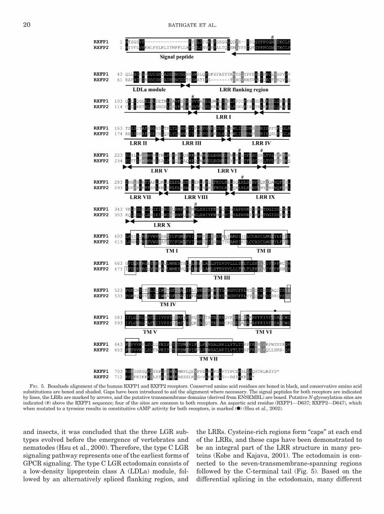
FIG. 5. Boxshade alignment of the human RXFP1 and RXFP2 receptors. Conserved amino acid residues are boxed in black, and conservative amino acidsubstitutions are boxed and shaded. Gaps have been introduced to aid the alignment where necessary. The signal peptides for both receptors are indicatedby lines, the LRRs are marked by arrows, and the putative transmembrane domains (derived from ENSEMBL) are boxed. Putative N-glycosylation sites areindicated (#) above the RXFP1 sequence; four of the sites are common to both receptors. An aspartic acid residue (RXFP1—D637; RXFP2—D647), whichwhen mutated to a tyrosine results in constituitive cAMP activity for both receptors, is marked (F) (Hsu et al., 2002).
20 BATHGATE ET AL.
isoforms of the receptors can be generated (Muda et al.,2005; Scott et al., 2005c). An exon 4-deleted RXFP1splice variant has been discovered in mouse, rat, and pig(Scott et al., 2005c). Removal of exon 4 causes a frame-shift, resulting in a premature stop codon and produc-tion of a truncated protein (RXFP1-truncate) that en-codes primarily the LDLa module without the LRRs ortransmembrane region. When cells were cotransfectedwith plasmids containing RXFP1 and the secretedRXFP1-truncate, relaxin-induced RXFP1 signaling wassignificantly reduced, suggesting that this protein actsas a functional antagonist in vitro and may be an endog-enous regulator of RXFP1 function.
A comparison of phenotypes of mice deficient inINSL3 (Nef and Parada, 1999; Zimmermann et al.,1999) and mice lacking a 550-kilobase region of chro-mosome 3, which contained the RXFP2 gene (Over-beek et al., 2001), led to the hypothesis that relaxinfamily peptides were cognate ligands for type C LGRs(Hsu et al., 2002). Functional studies established thefact that porcine relaxin (Hsu et al., 2002) and H2relaxin (Sudo et al., 2003) activate both RXFP1 andRXFP2 to increase cAMP. RXFP1 and RXFP2 havemore than 700 residues, share approximately 60%amino acid sequence identity, and contain 10 leucine-rich repeats in their extracellular domain. RXFP1transcripts are found in reproductive tissues, as wellas in brain, kidney, heart, and lung, in which actionsof relaxin have been reported. The rat and mouseorthologs of RXFP1 that have recently been cloned(Scott et al., 2004) bind H2 relaxin with high affinityand when activated increase cAMP accumulation.Both receptors had a higher affinity for rat relaxinthan the human receptor and also bind H3 relaxinwith high affinity (Scott et al., 2005b). Examination ofthe closely related relaxin-3 and INSL3 has demon-strated that the peptides act as selective agonists forRXFP1 and RXFP2, respectively (Kumagai et al.,2002; Sudo et al., 2003). The ectodomains of bothreceptors are important for ligand binding, as in thecase of type A LGRs (Osuga et al., 1997). When thesoluble ligand-binding region of RXFP1 (7BP) wasadministered subcutaneously to antagonize endoge-nous circulating relaxin during the last 4 days ofpregnancy, delivery was delayed by 27 h, and nippledevelopment was retarded (Hsu et al., 2002). It islikely that RXFP1-BP blocks the action of relaxin inlate pregnancy by acting as a relaxin-binding proteinand sequestering circulating relaxin. Although thephenotype is consistent with earlier work on relaxin(Zhao et al., 1999) and RXFP1 (Krajnc-Franken et al.,2004) null mice, the delay in parturition seems incon-sistent. However, delivery of pups is prolonged in ratstreated with a monoclonal antibody against relaxin(Lao-Guico et al., 1988), and some relaxin knockoutmice were observed to have difficulties in giving birth(Zhao et al., 1999).
Relaxin is therefore the cognate ligand for RXFP1and, although relaxin peptides from some species willactivate RXFP2 at high (supraphysiological) concentra-tions, rat relaxin does not activate human, rat, or mouseRXFP2, indicating that in rodents, relaxin is not anRXFP2 ligand (Scott et al., 2005a). INSL3 is the cognateligand for RXFP2, and the RXFP2 knockout mouse (Gor-lov et al., 2002) shows an identical phenotype to theINSL3 knockout (Nef and Parada, 1999; Zimmermannet al., 1999). Studies crossing INSL3 overexpressingmice and RXFP2 and RXFP1 knockout mice establishthat RXFP2 is the only receptor for INSL3, and there isno interaction between the INSL3/RXFP2 and relaxin/RXFP1 signaling systems in vivo (Bogatcheva et al.,2003; Kamat et al., 2004). However, as rodent relaxinwill not interact with RXFP2, it is still possible that inother species relaxin peptides will interact with RXFP2in vivo.
Thus, despite their structural similarity, relaxin andinsulin family peptides act through independent signal-ing pathways: the relaxin group activates the GPCRswhereas the insulin group activates tyrosine kinases.Phylogenetic analysis of LGRs and coevolved relaxinfamily peptides from different metazoans suggests that,whereas the number of relaxin receptors remained con-stant during vertebrate evolution, the ancestral gene forrelaxin duplicated multiple times in a vertebratebranch-specific manner (Hsu, 2003). Therefore, relaxinfamily peptides from different branches of vertebratesmay have adapted to distinct physiological roles via alimited number of receptor genes. Preliminary studieswith synthetic INSL4 (Lin et al., 2004) and INSL6 (Bo-gatcheva et al., 2003) indicate that they do not interactwith RXFP1 or RXFP2.
B. RXFP3 and RXFP4
In contrast to the high-affinity interactions betweenrelaxin and RXFP1 and INSL3 and RXFP2, relaxin-3has a lower affinity for RXFP1 than relaxin (Bathgateet al., 2002a; Sudo et al., 2003). A recent study indi-cates that relaxin-3 is a ligand for two orphan recep-tors, RXFP3 (GPCR135; somatostatin and angioten-sin-like peptide receptor) and RXFP4 (GPCR142;GPR100) (Liu et al., 2003a,b). These receptors (Fig. 6)differ structurally and functionally from RXFP1 andRXFP2 (Fig. 5). They have relatively short N-terminalextracellular domains and apparently couple exclu-sively to Gi. Studies using both native relaxin-3 puri-fied from brain extracts and recombinant humanrelaxin-3 indicated that this hormone potently stimu-lated guanosine 5�-[-thio]triphosphate binding and in-hibited cAMP accumulation in cells overexpressingRXFP3 and RXFP4. Endogenous expression of RXFP3in the rat brain is found in the paraventricular andsupraoptic nuclei of the hypothalamus (Liu et al.,2003b) and regions with connections to the nucleusincertus, where relaxin-3 is also expressed (Bathgate
RECEPTORS FOR RELAXIN FAMILY PEPTIDES 21
et al., 2002a; Burazin et al., 2002). Interestingly,these are also regions where relaxin-binding siteswere found (Osheroff and Phillips, 1991). RXFP4binds to relaxin-3 with slightly lower affinity thanRXFP3 and is less highly localized, with mRNA de-tected in the colon, thyroid, salivary gland, prostate,placenta, thymus, testis, kidney, and brain (Liu et al.,2003a). Although relaxin, INSL3, and INSL6 wereshown not to interact with RXFP3 and RXFP4, a re-cent study demonstrated that INSL5 is a specific li-gand for RFXP4 (Liu et al., 2005), and expressions ofINSL5 and RXFP4 overlap extensively, indicatingthat RXFP4 is probably the endogenous receptor forINSL5.
VII. Functional Domains of Receptors forRelaxin Family Peptides
A. General Features of Leucine-Rich Repeat-ContainingReceptors
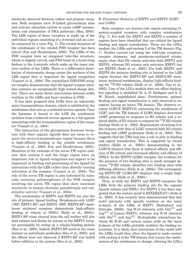
There are three LGR subgroups that all have multipleLRRs but have differences in their ectodomain featuresincluding the unique “hinge” region linking the LRRs tothe transmembrane domains (Fig. 7). Type A ectodo-mains contain 9 LRRs and include the receptors forFSH, LH, and TSH. Type B ectodomains contain 17 LRRand are currently orphan receptors. Type C ectodomains
contain 10 LRRs and an N-terminal LDLa module andare the RXFP1 (LGR7) and RXFP2 (LGR8) receptors(Hsu, 2003). RXFP1 and RXFP2 receptors are highlyconserved across species, with more than 90% sequence
FIG. 7. Schematic representation of the putative structure of theRXFP1 and RXFP2 receptors. The major structural features includeseven-transmembrane-spanning domains, hinge-like region, extracellu-lar LRRs, and LDLa module. The model was constructed based on thestructures of bovine rhodopsin (Okada and Palczewski, 2001) for thetransmembrane domains, FSH receptor (Fan and Hendrickson, 2005) forthe LRRs, and complement-like repeat CR8 from the low-density lipopro-tein receptor-related protein (Huang et al., 1999) for the LDLa module.The LDLa module structure contains a Ca2� molecule, which is necessaryfor the structure (Hopkins et al., 2005).
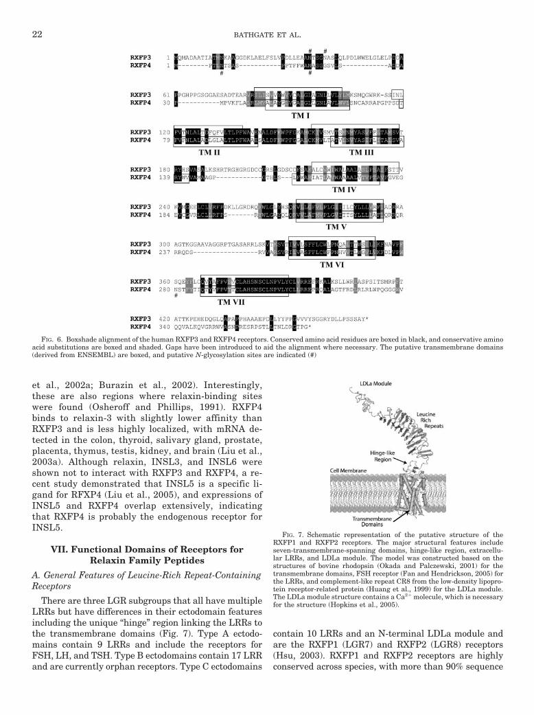
FIG. 6. Boxshade alignment of the human RXFP3 and RXFP4 receptors. Conserved amino acid residues are boxed in black, and conservative aminoacid substitutions are boxed and shaded. Gaps have been introduced to aid the alignment where necessary. The putative transmembrane domains(derived from ENSEMBL) are boxed, and putative N-glycosylation sites are indicated (#)
22 BATHGATE ET AL.
similarity observed between rodent and primate recep-tors. Both receptors have N-linked glycosylation sitesand activate adenylate cyclase to cause cAMP accumu-lation and stimulation of PKA pathways (Hsu, 2003).The LRR region of these receptors is made up of theindividual repeats consisting of a �-strand and an �-he-lix connected by a turn (Fig. 7). The crystal structure ofthe ectodomain of the related FSH receptor has beensolved (Fan and Hendrickson, 2005). The LRRs of theFSH receptor form an elongated tube-like structure,which is slightly curved, and FSH binds in a hand claspfashion to the �-strands, which make up the inner con-cave surface of the LRRs. There is a differential distri-bution of electrostatic charge across the surfaces of theLRR region that is important for ligand recognition(Vassart et al., 2004). The crystallized FSH-FSH recep-tor complex demonstrates that the ligand receptor inter-face contains an exceptionally high buried-charge den-sity. There are many direct interactions between acidicresidues in the LRRs and basic residues in FSH.
It has been proposed that LGRs have an inherentlynoisy transmembrane domain, which is inhibited by theectodomain that acts as a tethered inverse agonist. Oncethe hormone is bound to the LGR the ectodomainswitches from a tethered inverse agonist to a full agonistinteracting with the transmembrane region of the recep-tor (Vassart et al., 2004).
The interaction of the glycoprotein hormone recep-tors with their cognate ligands does not seem to re-quire the seven-transmembrane region because thereis high-affinity binding to the soluble ectodomain(Vassart et al., 2004; Fan and Hendrickson, 2005).Mutation of the exoloops of the seven TM region indi-cate that exoloop 2 and the hinge region have animportant role in ligand recognition but appear to beimportant in binding and positioning of the ligand forinteraction with the LRR rather than directly causingactivation of the receptor (Vassart et al., 2004). Therole of the seven TM region is also indicated by natu-rally occurring polymorphisms of the FSH receptorinvolving the seven TM region that show increasedsensitivity to human chorionic gonadotropin and con-stitutive activity (Vassart et al., 2004).
The ectodomains of RXFP1 and RXFP2 are also thesite of primary ligand binding. Ectodomain-only LGR7(7BP; RXFP1-BP) and RXFP2 (8BP; RXFP2-BP) mem-brane anchored receptors demonstrate high-affinitybinding of relaxin or INSL3 (Halls et al., 2005c).RXFP1-BP when cleaved from the cell surface will alsobind relaxin and blocks its actions at the RXFP1 recep-tor presumably by acting as a relaxin-binding protein(Hsu et al., 2002). Indeed, RXFP1-BP acted in the samemanner as antirelaxin antibodies (Hsu et al., 2002), andthe effects were not observed if RXFP1-BP was boiledbefore addition to the system (Hsu et al., 2002).
B. Functional Domains of RXFP1 and RXFP2 (LGR7and LGR8)
Both receptors are leucine-rich repeat-containing G-protein-coupled receptors with complex ectodomains(Fig. 7). For both the RXFP1 and RXFP2 a number ofregions have been identified that are critical for ligandbinding and signal transduction. These are the LDLamodule, the LRRs and exoloop 2 of the TM domain (Fig.7). Studies carried out using the wild-type receptors,receptor chimeras, and membrane-anchored ectodo-mains show that H2 relaxin activates both RXFP1 andRXFP2, whereas H3 relaxin only activates RXFP1 butnot RXFP2 (Sudo et al., 2003). For both RXFP1 andRXFP2 the primary binding site is located in the LRRregion because the RXFP1-BP and RXFP2-BP mem-brane anchored ectodomains, display high-affinity bind-ing for 33P-H2 relaxin (Halls et al., 2005c; Yan et al.,2005). Loss of the LDLa module does not affect binding,but signaling is abolished (R. A. D. Bathgate and A. J.W. Hsueh, unpublished data). Furthermore, optimalbinding and signal transduction is only observed in re-ceptors having an intact TM domain. The chimeric re-ceptor LGR7/8 (RXFP1/2) containing the ectodomain ofLGR7 and the TM domain of LGR8 displays decreasedcAMP production in response to H3 relaxin and a re-duced ability of H3 relaxin to compete for 33P-H2 relaxinbinding (Sudo et al., 2003). Replacement of exoloop 2 inthe chimera with that of LGR7 restored both H3 relaxinbinding and cAMP production (Sudo et al., 2003). Thissuggests that the TM2 loop has a role to play in bindingand signal transduction. This is supported by recentstudies (Halls et al., 2005c) demonstrating in theLGR7/8 chimera that there is reduced affinity and effi-cacy of H2 relaxin and increased affinity and efficacy ofINSL3. In the RXFP2 (LGR8) receptor, the evidence forthe presence of two binding sites is much stronger be-cause 33P-H2 relaxin identifies two binding sites withdiffering affinities (Halls et al., 2005c). The correspond-ing RXFP2-BP (LGR8-BP) displays only a single high-affinity site (Halls et al., 2005c).
Thus, in both the RXFP1 and RXFP2 receptors theLRRs form the primary binding site for the cognateligands relaxin and INSL3. For RXFP1 it has been sug-gested that the contact motif is RXXXRXX(I/V) (Sudo etal., 2003). It has recently been demonstrated that thismotif interacts with specific residues on the inner�-sheets of the LRRs of RXFP1 (Bullesbach andSchwabe, 2005b). Arg B-13 interacts with Glu277 andAsp279 of human RXFP1, whereas Arg B-16 interactswith Glu233 and Asp231. Hydrophobic interactions be-tween Ile B-20 and various amino acids on the inner�-sheets of the LRRs probably stabilize the binding in-teraction. It is likely that interaction of this motif withthe LRRs would then allow the ligand to make contactwith exoloop 2 of the TM domain that causes the confor-mation of the ectodomain to change, allowing the LDLa
RECEPTORS FOR RELAXIN FAMILY PEPTIDES 23
module to direct G-protein coupling and subsequentlyadenylate cyclase activation.
Examination of the amino acid sequence of both theRXFP1 and RXFP2 receptors reveals N-glycosylationsites in the ectodomains and the sizes of RXFP1, RXFP2,RXFP1-BP, and RXFP2-BP on Western blots are consis-tent with the ectodomain being highly glycosylated (Hsuet al., 2002; Kumagai et al., 2002; Yan et al., 2005). TheC-terminal regions of RXFP1 and RXFP2 have beenexamined for Ser, Thr, and Tyr residues that could rep-resent sites for protein kinase phosphorylation. The fol-lowing criteria were applied to determine the likelyphosphorylation sites: 1) a score �0.5 derived from theprogram NetPhos (http://www.cbs.dtu.dk/services/Net-Phos), 2) the vicinity of basic or acidic residues, prolineresidues, or other phosphorylated residues that are gen-erally required for protein kinase binding, and 3) theconservation of target sequences across the species or-thologs of each receptor. The RXFP1 receptor has twoconserved motifs in the C-terminal tail that fulfill thesecriteria, an RQRKSMDSK motif immediately distal tohelix 8 and a SQSTRLNSYS motif at the C terminus(Fig. 5). These are similar to predicted and/or consensussequences for protein kinase C (PKC) isozymes, includ-ing PKC�. In particular, the C-terminal motif has therequired Arg at the �3 position and a hydrophobic res-idue at �1 (Nishikawa et al., 1997).
The RXFP2 receptor has a motif KHQRKSIF distal tohelix 8 and similar to that in RXFP1 and an adjacentmotif KIKKKSLS (Fig. 5). Again these motifs may rep-resent target sites for particular PKC isozyme(s). Thesecond motif is unlikely to represent a PKA phosphory-lation site, as this kinase has high selectivity for Argover Lys residues at the �3 and �2 positions (Pinna andRuzzene, 1996). There is a third conserved motif (D/E)(D/E)SSSLK in the RXFP2 C-terminal tail (Fig. 5)that complies with the G-protein receptor kinase consen-sus sequence (Pinna and Ruzzene, 1996). G-protein re-ceptor kinase phosphorylation of activated RXFP2would represent an interesting point of divergence be-tween this receptor and RXFP1 and may contribute todifferences in activation of secondary signaling path-ways.
C. Functional Domains of RXFP3 and RXFP4(GPCR135 and GPCR142)
Much less is known about the functional domains ofthe RXFP3 and RXFP4 receptors. In contrast to RXFP1and RXFP2 that have large (�300 amino acid), complexN-terminal ectodomains, the RXFP3 and RXFP4 recep-tors are typical neuropeptide-type receptors withshorter (�100 amino acid) N-terminal domains (Liu etal., 2003a,b) (Fig. 6). In contrast to RXFP1 and RXFP2that signal through G�s to activate adenylate cyclaseand cause increases in intracellular levels of cAMP,RXFP3, and RXFP4 signal through G�i to inhibit cAMP(Liu et al., 2003a,b). There are also differences in the
way in which the ligands interact with the receptors.RXFP1 and RXFP2 are activated only by relaxin familypeptides that have A and B chains linked by the char-acteristic disulfide bonds and the separated A or Bchains are totally inactive (Tan et al., 1998). Howevercurrent evidence suggests that RXFP3 and RXFP4 donot interact with porcine relaxin, insulin, INSL3,INSL4, INSL6, or the relaxin-3 A chain but do interactwith relaxin-3 and the relaxin-3 B chain (Liu et al.,2003a,b).
Examination of the C-terminal region of RXFP3 andRXFP4 for putative protein kinase phosphorylation sitesshows that RXFP3 has conserved RKSSI and RR(L/R)SKV motifs at the distal ends of intracellular loops i1and i3, respectively. Although these motifs are very sim-ilar to those for PKA and PKC isozymes, they do notcomply completely with the consensus sequences. The i1motif may be a PKA phosphorylation site, although theLys at �2 is less favored than Arg or may be a substratefor PKC isozymes including PKC�, PKC�, and PKC�(Nishikawa et al., 1997). The i3 motif is less convincing,as the lack of a hydrophobic residue at �1 would behighly unusual for a PKA or PKC target site. LikeRXFP3, RXFP4 has an R(Q/R)(Q/R)(Q/R)(Q/R/W)QD-SRV motif at the distal end of intracellular loop i3, butagain this lacks the hydrophobic residue at the �1 po-sition.
The suggestions regarding phosphorylation of theRXFP receptors have one caveat, namely that there areknown kinase substrates that lack the typical consensussequences. As seen for other G-protein-coupled recep-tors, hypotheses about receptor phosphorylation willhave to be tested by experimental approaches includinguse of inhibitors, direct measurements of phosphoryla-tion in the presence or absence of dominant-negativekinase mutants, and study of receptor mutants lackingthe putative target residues.
VIII. Signaling Pathways Activated by RelaxinFamily Peptides
A. Signaling in Response to Relaxin
Early studies of the biochemical effects of relaxin dem-onstrated an association with increases in cAMP in tar-get tissues (Braddon, 1978; Cheah and Sherwood, 1980;Sanborn et al., 1980). Increased intracellular cAMP wasobserved in mouse pubic symphysis (Braddon, 1978), inrat uterus (Cheah and Sherwood, 1980; Judson et al.,1980; Sanborn et al., 1980), and in cultures of rat (Hsu etal., 1985) and rhesus monkey myometrium (Kramer etal., 1990), human endometrium (Chen et al., 1988; Fei etal., 1990), rat anterior pituitary cell (Cronin et al., 1987),and human THP-1 monocytes (Parsell et al., 1996). Theincreases in cAMP are important components in relaxin-induced myometrial inhibition in the rat (Dodge et al.,1999) and decidualization of human endometrial stro-mal cells (Huang et al., 1987; Tabanelli et al., 1992).
24 BATHGATE ET AL.
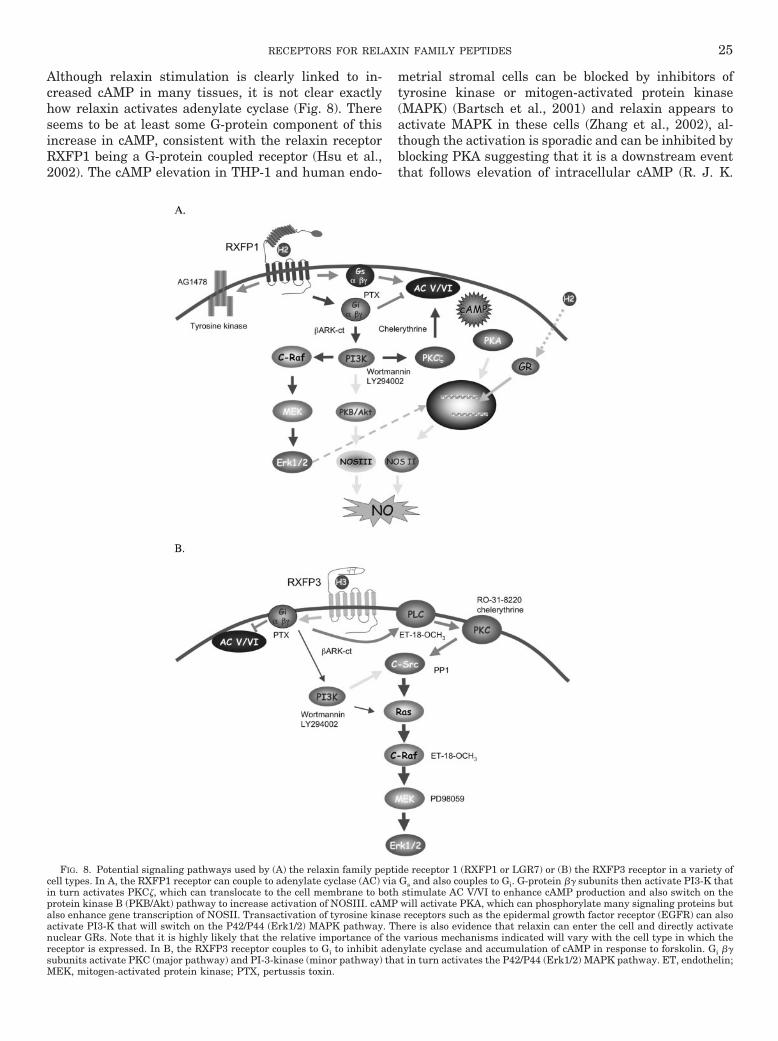
Although relaxin stimulation is clearly linked to in-creased cAMP in many tissues, it is not clear exactlyhow relaxin activates adenylate cyclase (Fig. 8). Thereseems to be at least some G-protein component of thisincrease in cAMP, consistent with the relaxin receptorRXFP1 being a G-protein coupled receptor (Hsu et al.,2002). The cAMP elevation in THP-1 and human endo-
metrial stromal cells can be blocked by inhibitors oftyrosine kinase or mitogen-activated protein kinase(MAPK) (Bartsch et al., 2001) and relaxin appears toactivate MAPK in these cells (Zhang et al., 2002), al-though the activation is sporadic and can be inhibited byblocking PKA suggesting that it is a downstream eventthat follows elevation of intracellular cAMP (R. J. K.
FIG. 8. Potential signaling pathways used by (A) the relaxin family peptide receptor 1 (RXFP1 or LGR7) or (B) the RXFP3 receptor in a variety ofcell types. In A, the RXFP1 receptor can couple to adenylate cyclase (AC) via Gs and also couples to Gi. G-protein �� subunits then activate PI3-K thatin turn activates PKC�, which can translocate to the cell membrane to both stimulate AC V/VI to enhance cAMP production and also switch on theprotein kinase B (PKB/Akt) pathway to increase activation of NOSIII. cAMP will activate PKA, which can phosphorylate many signaling proteins butalso enhance gene transcription of NOSII. Transactivation of tyrosine kinase receptors such as the epidermal growth factor receptor (EGFR) can alsoactivate PI3-K that will switch on the P42/P44 (Erk1/2) MAPK pathway. There is also evidence that relaxin can enter the cell and directly activatenuclear GRs. Note that it is highly likely that the relative importance of the various mechanisms indicated will vary with the cell type in which thereceptor is expressed. In B, the RXFP3 receptor couples to Gi to inhibit adenylate cyclase and accumulation of cAMP in response to forskolin. Gi ��subunits activate PKC (major pathway) and PI-3-kinase (minor pathway) that in turn activates the P42/P44 (Erk1/2) MAPK pathway. ET, endothelin;MEK, mitogen-activated protein kinase; PTX, pertussis toxin.
RECEPTORS FOR RELAXIN FAMILY PEPTIDES 25
Anand-Ivell and O. Bartsch, personal communication).One suggestion from experiments in human endothelialstromal cells is that relaxin stimulation leads to theinhibition of a cell-specific phosphodiesterase (PDE) toinduce sustained increases in cAMP (Bartsch et al.,2004). In THP-1 cells, the findings are equivocal withsome studies indicating that the increase in cAMP isassociated with PDE inhibition (Ivell et al., 2005) andothers showing that blockade of PDEs by the generalinhibitor isobutylmethylxanthine or the PDE8 inhibitordipyridamole fails to alter the cAMP response to relaxin(Nguyen et al., 2003). In HEK293 cells expressingRXFP1 and a secreted alkaline phosphatase reportergene containing a cAMP-response element, relaxincaused a marked response commensurate with an in-crease in intracellular cAMP (Halls et al., 2005a). InTHP-1 cells or HEK293T cells expressing RXFP1 recep-tors cAMP responses to relaxin during the first 5 min ofstimulation were not sensitive to PI3-K inhibitionwhereas responses to longer periods of stimulation wereinhibited by wortmannin or LY294002 (Nguyen et al.,2003; Halls et al., 2005b). In HEK293 cells it was ofinterest that the PI3-K inhibitor-sensitive component ofthe response was abolished by pretreatment of cells withpertussis toxin, suggesting that PI3-K activation inHEK293 cells expressing RXFP1 may be via �� subunitsderived from Gi (Halls et al., 2005b). Pertussis toxinsensitivity was not a feature of the responses in THP-1cells, suggesting that if activation of PI3-K is by ��subunits, then they may be derived from G-proteinsother than Gi in THP-1 cells (Nguyen et al., 2003). Thereis now (Nguyen and Dessauer, 2004) evidence that PKC�is the next step in PI3-K-mediated increases in cAMPproduction because inhibition of PKC with chelerythrinereduced and activation with sphingomyelinase in-creased the cAMP response to relaxin (Fig. 8). Relaxin-stimulated translocation of PKC� to the cell membranein a number of cell lines, an effect that was inhibited byPKC� antisense oligonucleotide treatment that reducedPKC� mRNA and protein levels. The antisense oligonu-cleotide also reduced the relaxin-induced increase incAMP levels, and the remaining response was insensi-tive to blockade by PI3-K inhibitors (Nguyen and Des-sauer, 2004). The cAMP response to activation of RXFP1receptors is clearly complex and can depend to someextent on the cell type and conditions of study.
This view is emphasized by the action of relaxin inother cell types where it is not associated with increasesin cAMP but may still involve some component of G-protein-coupled signaling. In rat ventricular fibroblastsrelaxin causes only a weak transient increase in cAMP(Samuel et al., 2004a) and studies in human lower uter-ine segment fibroblasts showed no increase in cAMP butdid show MAPK activation (Palejwala et al., 1998, 2001).Thus, in fibroblasts, cAMP does not appear to be thepredominant signaling mechanism but may link toMAPK signaling, which exerts the biological response.
Relaxin also activates Erk1/2 in a variety of cellsknown to express RXFP1 receptors (Zhang et al., 2002).In human endothelial stromal cells, THP-1 cells and inprimary cultures of human coronary artery and pulmo-nary artery smooth muscle cells addition of H2 relaxinproduced rapid (�5 min) phosphorylation of Erk1/2 withno change in levels of total Erk and the effect wasblocked by inhibition of mitogen-activated protein ki-nase (Zhang et al., 2002) (Fig. 8). No evidence was ob-tained for activation of Akt or c-Jun N-terminal kinasein these cells by relaxin. The transcription factor cAMP-response element-binding protein was also activated byrelaxin in endothelial stromal cells, but whether thisresults from the Erk1/2 cascade or the cAMP/PKA path-way is not clear at present (Zhang et al., 2002). Erk1/2activation by relaxin has also been demonstrated inHeLa cells and human umbilical vein endothelial cellsalthough the much longer time course of the effect(45–90 min) suggests that this may not be the result ofa direct interaction with relaxin receptors (Dschietzig etal., 2003).
The positive inotropic and chronotropic effects of re-laxin are also equivocal. Rabbit sinoatrial node cellsrespond to relaxin, and the effects appear to be mediatedby increases in intracellular cAMP and activation ofcAMP-dependent protein kinase (Han et al., 1994). Re-laxin also stimulates the secretion of atrial natriureticpeptide from isolated perfused rat hearts (Toth et al.,1996). These effects were mediated through protein ki-nase C, although treatment with a cAMP-dependentprotein kinase inhibitor also blocked atrial natriureticpeptide secretion as well as the chronotropic response,suggesting an involvement of cAMP. Measurement ofcAMP levels in atria, which are likely to reflect theinotropic response, demonstrates only weak coupling tocAMP (Kompa et al., 2002). It is feasible that the chro-notropic response of the few pacemaker cells in the si-noatrial node are mediated by cAMP, whereas the in-creased force of contraction of atrial myocytes usesanother mechanism.
There is also considerable evidence that relaxin canact on cells both acutely and chronically by increasingthe expression and or activity of NOS (Nistri and Bani,2003). Two mechanisms have been suggested, activationof NOSIII by a G-protein-coupled receptor mechanismsand induction of NOSII mediated by cAMP (Fig. 8).Activation of NOSIII by relaxin would involve stimula-tion of RXFP1, interaction of G-protein �� subunits withPI3-K followed by activation of Akt and subsequentlyendothelial NOS by phosphorylation at Ser1179. In-creased expression of NOSII would probably involve theincrease in cAMP levels, activation of PKA, phosphory-lation, and inactivation of I�B thus promoting increasedNOSII expression via nuclear factor �B (Nistri and Bani,2003). Recent studies also suggest that renal vasodila-tion in response to relaxin involves endothelin receptors.This mechanism involves up-regulation of matrix met-
26 BATHGATE ET AL.
alloproteinase activity by relaxin, followed by increasedprocessing of big endothelin to yield bioactive ET1–32,leading via ETB receptors to activation of NOSIII andincreased synthesis of NO (Conrad and Novak, 2004).
Relaxin has also been reported to interact with theglucocorticoid receptor (GR). Treatment with relaxin re-duces the production of inflammatory cytokines by hu-man macrophages in response to endotoxin (Dschietziget al., 2004), and the effect is blocked by the GR antag-onist RU486. Relaxin enters intact cells and is concen-trated in the nucleus where it induces GR activation,nuclear translocation, and DNA binding (Fig. 8). It ap-pears to act as a GR agonist and competes with GRagonists for binding to the GR. Because relaxins modi-fied to have no action at the RXFP1 receptor retainactivity at the GR, the effects appear to be mediatedindependently of an action on RXFP1.
B. Signaling in Response to Other Relaxin FamilyPeptides
Little is currently known of the mechanisms involvedin signaling of other relaxin family peptides. The mostdata have been gleaned from studies on cloned receptorsin transfected cell systems. When expressed in mamma-lian cells, LGR8 (RXFP2) responds to relaxin (Hsu et al.,2002) and INSL3 (Kumagai et al., 2002) with an in-crease in cAMP measured either directly (Hsu et al.,2002) or by an increase in secreted alkaline phosphatasein cells expressing RXFP2 and a cAMP-response ele-ment-responsive reporter gene (Halls et al., 2005a). Ratprimary gubernacular cells in culture respond to bothINSL3 and relaxin with an increase in cAMP (Kumagaiet al., 2002). It is likely that RXFP2 shares signalingpathways with RXFP1, with cAMP being an importantmediator of its cellular actions. However, recent studiesindicate that INSL3 produces effects in testicular germcells and oocytes by decreases in cAMP (Kawamura etal., 2004). These effects of INSL3 are blocked by pertus-sis toxin.
H3 relaxin will also increase cAMP in mammaliancells transfected with RXFP1 (Sudo et al., 2003) andTHP-1 cells (Bathgate et al., 2002a), suggesting thesame signaling pathway as relaxin. However, H3 relaxinacts at RXFP3 and RXFP4 receptors to inhibit cAMPaccumulation, and the effect is blocked by pertussistoxin, indicating that the receptor is coupled to Gi, Go, orGz (Liu et al., 2003a,b) (Fig. 8). Recent studies alsosuggest that stimulation of RXFP3 expressed in Chinesehamster ovary-K1 cells activates Erk1/2 signaling (Vander Westhuizen et al., 2005). The signaling mechanismsfor relaxin-3 via RXFP3/RXFP4 in vivo are currentlyunknown.
IX. Regulation of Relaxin Family PeptideReceptors
Very few studies have been conducted to examine theregulation of RXFP receptors. The most information is
available for RXFP1, and this information has primarilybeen obtained in studies not specifically designed toexamine receptor regulation. Thus, in a study of relaxinreceptor signaling in a rat model of cardiac failure, itwas shown that RXFP1 mRNA was decreased, and thisdecrease corresponded with a reduced response to re-laxin in animals in failure (Kompa et al., 2002). In malerats, treatment with 17�-estradiol decreases the num-ber of 33P-H2 relaxin binding sites in atria and cerebralcortex whereas the same treatment in female rats hadno effect in these tissues but increased the number ofbinding sites in uterine myometrium (Osheroff et al.,1992; Tan et al., 1999). More recently studies in humanendometrium have shown marked increases in 33P-H2relaxin binding and RXFP1 mRNA expression in thesecretory phase of the menstrual cycle although theagent responsible for the changes was not identified(Bond et al., 2004).
X. Nomenclature Issues for Relaxin FamilyPeptides and Their Receptors
A brief survey of the literature over the last 3 yearsprovided a dozen or more synonyms for relaxin includinghuman relaxin (Rln) H-2, human relaxin-2, relaxin-2,H2 relaxin, human gene 2 relaxin (hRLX2), human RLX,relaxin (RLX), rln-1, rhRLX (recombinant human re-laxin), human relaxin 2, and H2-relaxin. To preventfurther possible confusion among newcomers to the field(and even some of us in the field), we recommend thatthe human gene 1, gene 2, and gene 3 relaxin peptides beknown as H1, H2, and H3 relaxin, respectively. Becausemost other animals possess the equivalent of H3 relaxin,it is suggested that these be known as “species” relaxin-3(e.g., mouse relaxin-3). Only primates have the equiva-lent of H1 and H2 relaxins with most other speciesexpressing only a single relaxin equivalent to H2 re-laxin. It is suggested that these be known as “species”relaxin (e.g., mouse relaxin) to avoid the potential con-fusion caused by the attachment of numbers and com-parison with the human forms.
Fortunately, INSL3 has a shorter history than re-laxin, and there are fewer synonyms in the literature forthis peptide. However, INSL3 was formerly known asLey-I-L (Leydig insulin-like peptide) and RLF (relaxin-like factor) and has also been referred to as Leydig cellrelaxin. It is suggested that “species” INSL3 is the ap-propriate nomenclature.
With regard to the receptors for relaxin family it ispossible that RXFP1, RXFP2, RXFP3, and RXFP4 wereall originally receptors for relaxin-3, which is the mostancient of the relaxin peptides. However, to avoid thepotential confusion inherent in suggesting that all fourreceptors are relaxin receptors because the receptorshave now evolved to recognize a wider range of morespecialized ligands, it is suggested that the receptors beknown as relaxin family peptide receptors (RXFP recep-
RECEPTORS FOR RELAXIN FAMILY PEPTIDES 27
tors). The four receptors for relaxin family peptides,LGR7, LGR8, GPCR135, and GPCR142, would thus be-come “species” RXFP1, RXFP2, RXFP3, and RXFP4 re-ceptors.
In conclusion, although the hormone relaxin was dis-covered almost 80 years ago, the isolation of the receptorthat it activates and the elucidation of the signalingpathways involved in the cellular responses to the pep-tide have proved elusive. The past 5 years has seen theidentification, not only of the receptor for relaxin butalso three other receptors responding to relaxin familypeptides with all four receptors being G-protein-coupledreceptors. These discoveries have led very rapidly to thedetermination of the cognate ligands for the receptors,the tracing of their evolutionary history, and the deter-mination of their signaling pathways. Although muchstill needs to be learned about the way in which relaxinfamily peptides alter the activity of cells, the promisingprogress in the area of development of antagonists forthe receptors will provide valuable new tools to assist inthis process.
Acknowledgments. We thank Professor Geoffrey Tregear for ad-vice and encouragement, Dr. Bronwyn Evans, who analyzedRXFP1–4 receptors for putative protein kinase phosphorylationsites, and Daniel Scott and Tracey Wilkinson for assistance withfigures.
REFERENCES
Adham IM, Burkhardt E, Benahmed M, and Engel W (1993) Cloning of a cDNA fora novel insulin-like peptide of the testicular Leydig cells. J Biol Chem 268:26668–26672.
Adham IM, Steding G, Thamm T, Bullesbach EE, Schwabe C, Paprotta I, and EngelW (2002) The overexpression of the insl3 in female mice causes descent of theovaries. Mol Endocrinol 16:244–252.
Amento EP, Samuel CS, Hu M, and Tian H (2001) Deletion of the relaxin gene causesage-related progressive dermal fibrosis. Arthritis Rheum 44:A847.
Bani D, Failli P, Bello MG, Thiemermann C, Bani Sacchi T, Bigazzi M, and MasiniE (1998a) Relaxin activates the L-arginine-nitric oxide pathway in vascular smoothmuscle cells in culture. Hypertension 31:1240–1247.
Bani D, Masini E, Bello MG, Bigazzi M, and Sacchi TB (1998b) Relaxin protectsagainst myocardial injury caused by ischemia and reperfusion in rat heart. Am JPathol 152:1367–1376.
Bani D, Nistri S, Quattrone S, Bigazzi M, and Sacchi TB (2001) Relaxin causeschanges of the liver: in vivo studies in rats. Horm Metab Res 33:175–180.
Bani G, Bani ST, Bigazzi M, and Bianchi S (1988) Effects of relaxin on the micro-vasculature of mouse mammary gland. Histol Histopathol 3:337–343.
Bani-Sacchi T, Bigazzi M, Bani D, Mannaioni PF and Masini E (1995) Relaxin-induced increased coronary flow through stimulation of nitric oxide production.Br J Pharmacol 116:1589–1594.
Bartsch O, Bartlick B, and Ivell R (2001) Relaxin signalling links tyrosine phosphor-ylation to phosphodiesterase and adenylyl cyclase activity. Mol Hum Reprod7:799–809.
Bartsch O, Bartlick B, and Ivell R (2004) Phosphodiesterase 4 inhibition synergizeswith relaxin signaling to promote decidualization of human endometrial stromalcells. J Clin Endocrinol Metab 89:324–334.
Bathgate RAD, Balvers M, Hunt N, and Ivell R (1996) Relaxin-like factor gene ishighly expressed in the bovine ovary of the cycle and pregnancy: sequence andmessenger ribonucleic acid analysis. Biol Reprod 55:1452–1457.
Bathgate RAD, Hsueh AJW, and Sherwood OD (2006) Physiology and molecularbiology of the relaxin peptide family, in Knobil and Neill’s Physiology of Repro-duction (Neill JD ed) pp 679–768, Academic Press, San Diego.
Bathgate RAD, Moniac N, Bartlick B, Balvers M, Domagalski R, Dawson N, WadeJD, Tregear GW, and Ivell R (2001) The relaxin-like factor (RLF Insl3) is highlyexpressed in the ruminant ovary, in Relaxin 2000: Proceedings of the ThirdInternational Conference on Relaxin and Related Peptides (Tregear GW, Ivell R,Bathgate RA, and Wade JD eds) pp 349–356, Kluwer, Dordrecht, The Nether-lands.
Bathgate RAD, Samuel CS, Burazin TC, Gundlach AL, and Tregear GW (2003)Relaxin: new peptides, receptors and novel actions. Trends Endocrinol Metab14:207–213, 2003.
Bathgate RAD, Samuel CS, Burazin TC, Layfield S, Claasz AA, Reytomas IG,Dawson NF, Zhao C, Bond CP, Summers RJ, et al. (2002a) Human relaxin gene 3(H3) and the equivalent mouse relaxin (M3) gene: novel members of the relaxinpeptide family. J Biol Chem 277:1148–1157.
Bathgate RAD, Siebel AL, Tovote P, Claasz A, Macris M, Tregear GW, and Parry LJ(2002b) Purification and characterization of relaxin from the tammar wallaby(Macropus eugenii): bioactivity and expression in the corpus luteum. Biol Reprod67:293–300.
Bieche I, Laurent A, Laurendeau I, Duret L, Giovangrandi Y, Frendo JL, Olivi M,Fausser JL, Evain-Brion D, and Vidaud M (2003) Placenta-specific INSL4 expres-sion is mediated by a human endogenous retrovirus element. Biol Reprod 68:1422–1429.
Bigazzi M, Del MA, Petrucci F, Casali R, and Novelli GP (1986) The local adminis-tration of relaxin induces changes in the microcirculation of the rat mesocaecum.Acta Endocrinol Copenh 112:296–299.
Boels K and Schaller HC (2003) Identification and characterisation of GPR100 as anovel human G-protein-coupled bradykinin receptor. Br J Pharmacol 140:932–938.
Bogatcheva NV, Truong A, Feng S, Engel W, Adham IM, and Agoulnik AI (2003)GREAT/LGR8 is the only receptor for insulin-like 3 peptide. Mol Endocrinol17:2639–2646.
Bond CP, Parry LJ, Samuel CS, Gehring HM, Lederman FL, Rogers PA, andSummers RJ (2004) Increased expression of the relaxin receptor (LGR7) in humanendometrium during the secretory phase of the menstrual cycle. J Clin EndocrinolMetab 89:3477–3485.
Braddon SA (1978) Relaxin-dependent adenosine 6�5�-monophosphate concentrationchanges in the mouse pubic symphysis. Endocrinology 102:1292–1299.
Bryant-Greenwood G and Schwabe C (1994) Human relaxins: chemistry and biology.Endocr Rev 15:5–26.
Bullesbach EE and Schwabe C (1986) Preparation and properties of porcine relaxinderivatives shortened at the amino terminus of the A chain. Biochemistry 25:5998–6004.
Bullesbach EE and Schwabe C (1987) Relaxin structure: quasi allosteric effect of theNH2-terminal A-chain helix. J Biol Chem 262:12496–12501.
Bullesbach EE and Schwabe C (1991) Total synthesis of human relaxin and humanrelaxin derivatives by solid-phase peptide synthesis and site-directed chain com-bination. J Biol Chem 266:10754–10861.
Bullesbach EE and Schwabe C (1994) Functional importance of the A chain loop inrelaxin and insulin. J Biol Chem 269:13124–13128.
Bullesbach EE and Schwabe C (1995) A novel Leydig cell cDNA-derived protein is arelaxin-like factor. J Biol Chem 270:16011–16015.
Bullesbach EE and Schwabe C (1996) Introduction of relaxin properties into otherhormones of insulin-like structure. SAAS Bull Biochem Biotechnol 9:63–68.
Bullesbach EE and Schwabe C (1999a) Tryptophan B27 in the relaxin-like factor(RLF) is crucial for RLF receptor-binding. Biochemistry 38:3073–3078.
Bullesbach EE and Schwabe C (1999b) Specific high affinity relaxin-like factorreceptors. J Biol Chem 274:22354–22358.
Bullesbach EE and Schwabe C (2000) The relaxin receptor-binding site geometrysuggests a novel gripping mode of interaction. J Biol Chem 275:35276–35280.
Bullesbach EE and Schwabe C (2002) The primary structure and the disulfide linksof the bovine relaxin-like factor (RLF). Biochemistry 41:274–281.
Bullesbach EE and Schwabe C (2005a) LGR8 signal activation by the relaxin-likefactor. J Biol Chem 280:14586–14590.
Bullesbach EE and Schwabe C (2005b) The trap-like relaxin-binding site of LGR7.J Biol Chem 280:14051–14056.
Bullesbach EE, Yang S, and Schwabe C (1992) The receptor-binding site of humanrelaxin II A dual prong-binding mechanism. J Biol Chem 267:22957–22960.
Burazin TC, Bathgate RAD, Macris M, Layfield S, Gundlach AL, and Tregear GW(2002) Restricted but abundant expression of the novel rat gene-3 (R3) relaxin inthe dorsal tegmental region of brain. J Neurochem 82:1553–1557.
Burazin TCD, Johnson KJ, Ma S, Bathgate RAD, Tregear GW, and Gundlach AL(2005) Localization of LGR7 (relaxin receptor) mRNA and protein in rat forebrain:correlation with relaxin binding site distribution. Ann NY Acad Sci 1041 205–210.
Burkhardt E, Adham IM, Brosig B, Gastmann A, Mattei MG and Engel W (1994)Structural organization of the porcine and human genes coding for a Leydigcell-specific insulin-like peptide (LEY I-L) and chromosomal localization of thehuman gene (INSL3). Genomics 20:13–19.
Canova DE, Kessler TJ, Lee PJ, Fei DT, Griffin P, Stults JT, Wade JD, andRinderknecht E (1991) Use of recombinant DNA derived human relaxin to probethe structure of the native protein. Biochemistry 30:6006–6013.
Casten GG and Boucek RJ (1958) Use of relaxin in the treatment of scleroderma.J Am Med Assoc 166:319–324.
Chassin D, Laurent A, Janneau JL, Berger R, and Bellet D (1995) Cloning of a newmember of the insulin gene superfamily (INSL4) expressed in human placenta.Genomics 29:465–470.
Cheah SH and Sherwood OD (1980) Target tissues for relaxin in the rat: tissuedistribution of injected 125I-labeled relaxin and tissue changes in adenosine 3�5�-monophosphate levels after in vitro relaxin incubation. Endocrinology 106:1203–1209.
Chen GA, Huang JR, and Tseng L (1988) The effect of relaxin on cyclic adenosine3�5�-monophosphate concentrations in human endometrial glandular epithelialcells. Biol Reprod 39:519–525.
Claasz AA, Bond CP, Bathgate RA, Otvos L, Dawson NF, Summers RJ, Tregear GW,and Wade JD (2002) Relaxin-like bioactivity of ovine insulin 3 (INSL3) analogues.Eur J Biochem 269:6287–6293.
Conklin D, Lofton-Day CE, Haldeman BA, Ching A, Whitmore TE, Lok S, andJaspers S (1999) Identification of INSL5: a new member of the insulin superfamily.Genomics 60:50–56.
Conrad KP and Novak J (2004) Emerging role of relaxin in renal and cardiovascularfunction. Am J Physiol 287:R250–R261.
Coulson CC, Thorp JM Jr, Mayer DC and Cefalo RC (1996) Central hemodynamiceffects of recombinant human relaxin in the isolated perfused rat heart model.Obstet Gynecol 87:610–612.
Cronin MJ, Malaska T and Bakhit C (1987) Human relaxin increases cyclic AMP
28 BATHGATE ET AL.
levels in cultured anterior pituitary cells. Biochem Biophys Res Commun 148:1246–1251.
Danielson LA, Sherwood OD and Conrad KP (1999) Relaxin is a potent renalvasodilator in conscious rats. J Clin Investig 103:525–533.
Dawson NF, Tan YY, Macris M, Otvos L Jr, Summers RJ, Tregear GW, and Wade JD(1999) Solid-phase synthesis of ovine Leydig cell insulin-like peptide-a putativeovine relaxin? J Pept Res 53:542–547.
Del Borgo MP, Hughes RA, and Wade JD (2004) Design synthesis and analysis ofsingle chain mimetics of relaxin and relaxin-like factor (INSL3), in Peptides –Peptide Revolution: Genomics, Proteomics and Therapeutics (Chorev M and Saw-yer TK eds) pp 727–728, Springer, New York. Proceedings of the 18th AmericanPeptide Symposium; 2003 Jul 19–23; Boston, MA.
Dodge KL, Carr DW, and Sanborn BM (1999) Protein kinase A anchoring to themyometrial plasma membrane is required for cyclic adenosine 3�5�-monophos-phate regulation of phosphatidylinositide turnover. Endocrinology 140:5165–5170.
Dodson GG, Eliopoulos EE, Isaacs NW, McCall MJ, Niall HD, and North ACT (1982)Rat relaxin: insulin-like fold predicts a likely receptor binding region. Int J BiolMacromol 4:399–405.
Dschietzig T, Bartsch C, Richter C, Laule M, Baumann G, and Stangl K (2003)Relaxin, a pregnancy hormone, is a functional endothelin-1 antagonist: attenua-tion of endothelin-1-mediated vasoconstriction by stimulation of endothelin type-Breceptor expression via ERK-1/2 and nuclear factor-�B. Circ Res 92:32–40.
Dschietzig T, Bartsch C, Stangl V, Baumann G, and Stangl K (2004) Identification ofthe pregnancy hormone relaxin as glucocorticoid receptor agonist. FASEB J 18:1536–1538.
Du X-J, Samuel CS, Gao X-M, Zhao L, Parry LJ, and Tregear GW (2003) Increasedmyocardial collagen and ventricular diastolic dysfunction in relaxin deficient mice:a gender-specific phenotype. Cardiovasc Res 57:395–404.
Eigenbrot C, Randal M, Quan C, Burnier J, O’Connell L, Rinderknecht E, andKossiakoff AA (1991) X-ray structure of human relaxin at 1.5 A. Comparison toinsulin and implications for receptor binding determinants. J Mol Biol 221:15–21.
Einspanier A, Muller D, Lubberstedt J, Bartsch O, Jurdzinski A, Fuhrmann K, andIvell R (2001) Characterization of relaxin binding in the uterus of the marmosetmonkey. Mol Hum Reprod 7:963–970.
Erikson MS and Unemori EN (2001) Relaxin clinical trials in systemic sclerosis, inProceedings of the Third International Conference on Relaxin and Related Pep-tides, 22–27 October 2000, Broome, Australia (Tregear GW, Ivell R, Bathgate RA,and Wade JD eds) pp 373–381, Kluwer Publishers, Amsterdam, The Netherlands.
Evans BA, John M, Fowler KJ, Summers RJ, Cronk M, Shine J, and Tregear GW(1993) The mouse relaxin gene: nucleotide sequence and expression. J Mol Endo-crinol 10:15–23.
Failli P, Nistri S, Quattrone S, Mazzetti L, Bigazzi M, Sacchi TB, and Bani D (2002)Relaxin up-regulates inducible nitric oxide synthase expression and nitric oxidegeneration in rat coronary endothelial cells. FASEB J 16:252–254.
Fan QR and Hendrickson WA (2005) Structure of human follicle-stimulating hor-mone in complex with its receptor. Nature (Lond) 433:269–277.
Fei DT, Gross MC, Lofgren JL, Mora-Worms M, and Chen AB (1990) Cyclic AMPresponse to recombinant human relaxin by cultured human endometrial cells-aspecific and high throughput in vitro bioassay. Biochem Biophys Res Commun170:214–222.
Fu P, Shen P-J, Zhao C-X, Scott DJ, Samuel CS, Wade JD, Tregear GW, BathgateRAD, and Gundlach AL (2005) Detection localization and action of LGR8 receptorsin adult and developing rat kidney. Ann NY Acad Sci 1041:516–519.
Garber SL, Mirochnik Y, Brecklin CS, Unemori EN, Singh AK, Slobodskoy L, GroveBH, Arruda JA, and Dunea G (2001) Relaxin decreases renal interstitial fibrosisand slows progression of renal disease. Kidney Int 59:876–882.
Garibay-Tupas JL, Maaskant RA, Greenwood FC, and Bryant-Greenwood GD (1995)Characteristics of the binding of 32P-labelled human relaxins to the human fetalmembranes. J Endocrinol 145:441–448.
Gavino ES and Furst DE (2001) Recombinant relaxin: a review of pharmacology andpotential therapeutic use. BioDrugs 15:609–614.
Goldsmith LT, Weiss G, Palejwala S, Plant TM, Wojtczuk A, Lambert WC, AmmurN, Heller D, Skurnick JH, Edwards D, et al. (2004) Relaxin regulation of endo-metrial structure and function in the rhesus monkey Proc Natl Acad Sci USA101:4685–4689.
Gorlov IP, Kamat A, Bogatcheva NV, Jones E, Lamb DJ, Truong A, Bishop CE,McElreavey K, and Agoulnik AI (2002) Mutations of the GREAT gene causecryptorchidism. Hum Mol Genet 11:2309–2318.
Goto M, Swanson LW, and Canteras NS (2001) Connections of the nucleus incertus.J Comp Neurol 438:86–122.
Halls ML, Bathgate RAD, Roche PJ, and Summers RJ (2005a) Signaling pathwaysof the LGR7 and LGR8 receptors determined by reporter genes. Ann NY Acad Sci1041:292–295.
Halls ML, Bathgate RAD, and Summers RJ (2005b) Signal switching followingstimulation of LGR7 receptors by human relaxin 2. Ann NY Acad Sci 1041:288–291.
Halls ML, Bond CP, Sudo S, Kumagai J, Ferraro T, Layfield S, Bathgate RAD, andSummers RJ (2005c) Multiple binding sites revealed by interaction of relaxinfamily peptides with native and chimeric relaxin family peptide receptors 1 and 2(LGR7 & LGR8). J Pharmacol Exp Ther 313:677–687.
Han X, Habuchi Y, and Giles WR (1994) Relaxin increases heart rate by modulatingcalcium current in cardiac pacemaker cells. Circ Res 74:537–541.
Hisaw FL (1926) Experimental relaxation of the pubic ligament of the guinea pig.Proc Soc Exp Biol Med 23:661–663.
Hombach-Klonisch S, Buchmann J, Sarun S, Fischer B, and Klonisch T (2000)Relaxin-like factor (RLF) is differentially expressed in the normal and neoplastichuman mammary gland. Cancer 89:2161–2168.
Hombach-Klonisch S, Hoang-Vu C, Kehlen A, Hinze R, Holzhausen HJ, Weber E,Fischer B, Dralle H, and Klonisch T (2003) INSL-3 is expressed in human hyper-plastic and neoplastic thyrocytes. Int J Oncol 22:993–1001.
Hombach-Klonisch S, Seeger S, Tscheudschilsuren G, Buchmann J, Huppertz B,Seliger G, Fischer B, and Klonisch T (2001) Cellular localization of human relaxin-like factor in the cyclic endometrium and placenta. Mol Hum Reprod 7:349–356.
Hopkins EJ, Bathgate RAD, and Gooley PR (2005) The human LGR7 low-densitylipoprotein class A module requires calcium for structure. Ann NY Acad Sci1040:27–34.
Hsu CJ, McCormack SM, and Sanborn BM (1985) The effect of relaxin on cyclicadenosine 3�5�-monophosphate concentrations in rat myometrial cells in culture.Endocrinology 116:2029–2035.
Hsu S (1999) Cloning of two novel mammalian paralogs of relaxin/insulin familyproteins and their expression in testis and kidney Mol Endocrinol 13:2163–2174.
Hsu SY (2003) New insights into the evolution of the relaxin-LGR signaling system.Trends Endocrinol Metab 14:303–309.
Hsu SY, Kudo M, Chen T, Nakabayashi K, Bhalla A, van der Spek PJ, van Duin M,and Hsueh AJ (2000) The three subfamilies of leucine-rich repeat-containing Gprotein-coupled receptors (LGR): identification of LGR6 and LGR7 and the signal-ing mechanism for LGR7. Mol Endocrinol 14:1257–1271.
Hsu SY, Liang SG, and Hsueh AJ (1998) Characterization of two LGR genes homol-ogous to gonadotropin and thyrotropin receptors with extracellular leucine-richrepeats and a G protein-coupled seven-transmembrane region. Mol Endocrinol12:1830–1845.
Hsu SY, Nakabayashi K, Nishi S, Kumagai J, Kudo M, Sherwood OD, and Hsueh AJ(2002) Activation of orphan receptor by the hormone relaxin Science (Wash DC)25:637–638.
Huang JR, Tseng L, Bischof P, and Janne OA (1987) Regulation of prolactin produc-tion by progestin estrogen and relaxin in human endometrial stromal cells. Endo-crinology 121:2011–2017.
Huang W, Dolmer K, and Gettins PG (1999) NMR solution structure of complement-like repeat CR8 from the low density lipoprotein receptor-related protein. J BiolChem 274:14130–14136.
Hudson P, Haley J, John M, Cronk M, Crawford R, Haralambidis J, Tregear G, ShineJ, and Niall H (1983) Structure of a genomic clone encoding biologically activehuman relaxin. Nature (Lond) 301:628–631.
Hudson P, John M, Crawford R, Haralambidis J, Scanlon D, Gorman J, Tregear G,Shine J, and Niall H (1984) Relaxin gene expression in human ovaries and thepredicted structure of a human preprorelaxin by analysis of cDNA clones. EMBO(Eur Mol Biol Organ) J 3:2333–2339.
Hwang JJ, Lee AB, Fields PA, Haab LM, Mojonnier LE, and Sherwood OD (1991)Monoclonal antibodies specific for rat relaxin V: passive immunization with mono-clonal antibodies throughout the second half of pregnancy disrupts development ofthe mammary apparatus and hence lactational performance in rats. Endocrinol-ogy 129:3034–3042.
Irving-Rodgers HF, Bathgate RAD, Ivell R, Domagalski R, and Rodgers RJ (2002)Dynamic changes in the expression of relaxin-like factor (INSL3), cholesterolside-chain cleavage cytochrome p450, and 3�-hydroxysteroid dehydrogenase inbovine ovarian follicles during growth and atresia. Biol Reprod 66:934–943.
Ivell R, Anand-Ivell R, and Bartsch O (2005) Relaxin signaling from natural recep-tors. Ann NY Acad Sci 1041:280–287.
Ivell R, Balvers M, Pohnke Y, Telgmann R, Bartsch O, Milde-Langosch K, Bam-berger A-M, and Einspanier A (2003) Immunoexpression of the relaxin receptorLGR7 in breast and uterine tissues of humans and primates. Reprod Biol Endo-crinol 1:114.
Ivell R and Hartung S (2003) The molecular basis of cryptorchidism. Mol HumReprod 9:175–181.
John MJ, Borjesson BW, Walsh JR, and Niall HD (1981) Limited sequence homologybetween porcine and rat relaxins: implications for physiological studies. Endocri-nology 108:726–729.
Johnson MR, Abdalla H, Allman AC, Wren ME, Kirkland A, and Lightman SL (1991)Relaxin levels in ovum donation pregnancies. Fertil Steril 56:59–61.
Johnson MR, Brooks AA, and Steer PJ (1996) The role of relaxin in the pregnancyassociated reduction in plasma osmolality. Hum Reprod 11:1105–1108.
Judson DG, Pay S, and Bhoola KD (1980) Modulation of cyclic AMP in isolated ratuterine tissue slices by porcine relaxin. J Endocrinol 87:153–159.
Kakouris H, Eddie LW, and Summers RJ (1992) Cardiac effects of relaxin in rats.Lancet 339:1076–1078.
Kamat AA, Feng S, Bogatcheva NV, Truong A, Bishop CE, and Agoulnik AI (2004)Genetic targeting of relaxin and insulin-like factor 3 receptors in mice. Endocri-nology 145:4712–4720.
Kawamura K, Kumagai J, Sudo S, Chun SY, Pisarska M, Morita H, Toppari J, Fu P,Wade JD, Bathgate RAD, et al. (2004) Paracrine regulation of mammalian oocytematuration and male germ cell survival. Proc Natl Acad Sci USA 101:7323–7328.
Kibblewhite D, Larrabee WJ, and Sutton D (1992) The effect of relaxin on tissueexpansion. Arch Otolaryngol Head Neck Surg 118:153–156.
Kizawa H, Nishi K, Ishibashi Y, Harada M, Asano T, Ito Y, Suzuki N, Hinuma S,Fujisawa Y, Onda H, et al. (2003) Production of recombinant human relaxin 3 inAtT20 cells. Regul Pept 113:79–84.
Klonisch T, Fowler PA, and Hombach-Klonisch S (2004) Molecular and geneticregulation of testis descent and external genitalia development. Dev Biol 270:1–18.
Koay ES, Bryant GG, Yamamoto SY, and Greenwood FC (1986) The human fetalmembranes: a target tissue for relaxin. J Clin Endocrinol Metab 62:513–521.
Kobe B and Kajava AV (2001) The leucine-rich repeat as a protein recognition motif.Curr Opin Struct Biol 11:725–732.
Kohsaka T, Min G, Lukas G, Trupin S, Campbell ET, and Sherwood OD (1998)Identification of specific relaxin-binding cells in the human female. Biol Reprod59:991–999.
Koman A, Cazaubon S, Couraud PO, Ullrich A, and Strosberg AD (1996) Molecularcharacterization and in vitro biological activity of placentin, a new member of theinsulin gene family. J Biol Chem 271:20238–20241.
Kompa AR, Samuel CS, and Summers RJ (2002) Inotropic responses to human gene
RECEPTORS FOR RELAXIN FAMILY PEPTIDES 29
2 (B29) relaxin in a rat model of myocardial infarction (MI): effect of pertussistoxin. Br J Pharmacol 137:710–718.
Krajnc-Franken MA, van Disseldorp AJ, Koenders JE, Mosselman S, van Duin M,and Gossen JA (2004) Impaired nipple development and parturition in LGR7knockout mice. Mol Cell Biol 24:687–696.
Kramer SM, Gibson UE, Fendly BM, Mohler MA, Drolet DW, and Johnston PD(1990) Increase in cyclic AMP levels by relaxin in newborn rhesus monkey uteruscell culture. In Vitro Cell Dev Biol 26:647–656.
Kubota Y, Temelcos C, Bathgate RAD, Smith KJ, Scott D, Zhao C, and Hutson JM(2002) The role of insulin 3 testosterone Mullerian inhibiting substance andrelaxin in rat gubernacular growth. Mol Hum Reprod 8:900–905.
Kuenzi MJ, Connolly BA, and Sherwood OD (1995) Relaxin acts directly on ratmammary nipples to stimulate their growth. Endocrinology 136:2943–2947.
Kuenzi MJ and Sherwood OD (1995) Immunohistochemical localization of specificrelaxin-binding cells in the cervix, mammary glands, and nipples of pregnant rats.Endocrinology 136:1367–1373.
Kumagai J, Hsu SY, Matsumi H, Roh JS, Fu P, Wade JD, Bathgate RAD, and HsuehAJ (2002) INSL3/Leydig insulin-like peptide activates the LGR8 receptor impor-tant in testis descent. J Biol Chem 277:31283–31286.
Lao-Guico LM, Voss EWJ, and Sherwood OD (1988) Monoclonal antibodies specificfor rat relaxin. I. Production and characterization of monoclonal antibodies thatneutralize rat relaxin’s bioactivity in vivo. Endocrinology 123:2472–2478.
Lee AB, Hwang JJ, Haab LM, Fields PA, and Sherwood OD (1992) Monoclonalantibodies specific for rat relaxin. VI. Passive immunization with monoclonalantibodies throughout the second half of pregnancy disrupts histological changesassociated with cervical softening at parturition in rats. Endocrinology 130:2386–2391.
Lin F, Otvos L Jr, Kumagai J, Tregear GW, Bathgate RAD, and Wade JD (2004)Synthetic human insulin 4 does not activate the G-protein-coupled receptors LGR7or LGR8. J Pept Sci 10:257–264.
Lindheimer MD, Barron WM, and Davison JM (1989) Osmoregulation of thirst andvasopressin release in pregnancy. Am J Physiol 257 (Pt 2):F159–F169.
Liu C, Chen J, Sutton S, Roland B, Kuei C, Farmer N, Sillard R, and Lovenberg TW(2003a) Identification of relaxin-3/INSL7 as a ligand for GPCR142. J Biol Chem278:50765–50770.
Liu C, Eriste E, Sutton S, Chen J, Roland B, Kuei C, Farmer N, Jornvall H, SillardR, and Lovenberg TW (2003b) Identification of relaxin-3/INSL7 as an endogenousligand for the orphan G-protein-coupled receptor GPCR135. J Biol Chem 278:50754–50764.
Liu C, Kuei C, Sutton S, Chen J, Bonaventure P, Wu J, Nepomuceno D, Kamme F,Tran DT, Zhu J, et al. (2005) INSL5 is a high affinity specific agonist for GPCR142(GPR100). J Biol Chem 280:292–300.
Lok S, Johnston DS, Conklin D, Lofton-Day CE, Adams RL, Jelmberg AC, WhitmoreTE, Schrader S, Griswold MD, and Jaspers SR (2000) Identification of INSL6, anew member of the insulin family that is expressed in the testis of the human andrat. Biol Reprod 62:1593–1599.
Longo M, Jain V, Vedernikov YP, Garfield RE, and Saade GR (2003) Effects ofrecombinant human relaxin on pregnant rat uterine artery and myometrium invitro. Am J Obstet Gynecol 188:1468–1474.
Luna JJ, Riesewijk A, Horcajadas JA, de van Os R, Dominguez F, Mosselman S,Pellicer A, and Simon C (2004) Gene expression pattern and immunoreactiveprotein localisation of LGR7 receptor in human endometrium throughout themenstrual cycle. Mol Hum Reprod 10:85–90.
Masini E, Bani D, Bello MG, Bigazzi M, Mannaioni PF, and Sacchi TB (1997) Relaxincounteracts myocardial damage induced by ischemia-reperfusion in isolatedguinea pig hearts: evidence for an involvement of nitric oxide. Endocrinology138:4713–4720.
Massicotte G, Parent A, and St-Louis J (1989) Blunted responses to vasoconstrictorsin mesenteric vasculature but not in portal vein of spontaneously hypertensiverats treated with relaxin. Proc Soc Exp Biol Med 190:254–259.
Mathieu MN, Wade JD, Tregear GW, Bond CP, Summers RJ, Catimel B, Nice EC,and Otvos L (2001) Synthesis conformational studies and biological activity of��-mono-biotinylated rat relaxin. J Pept Res 57:374–382.
Matsumoto M, Kamohara M, Sugimoto T, Hidaka K, Takasaki J, Saito T, Okada M,Yamaguchi T, and Furuichi K (2000) The novel G-protein coupled receptor SALPRshares sequence similarity with somatostatin and angiotensin receptors. Gene248:183–189.
Mazella J, Tang M, and Tseng L (2004) Disparate effects of relaxin and TGF�1:relaxin increases but TGF�1 inhibits the relaxin receptor and the production ofIGFBP-1 in human endometrial stromal/decidual cells. Hum Reprod 19:1513–1518.
McKinley MJ, Allen AM, Burns P, Colvill LM, and Oldfield BJ (1998) Interaction ofcirculating hormones with the brain: the roles of the subfornical organ and theorganum vasculosum of the lamina terminalis. Clin Exp Pharm Physiol 25(Suppl):S61–S67.
McKinley MJ, Burns P, Colvill LM, Oldfield BJ, Wade JD, Weisinger RS, andTregear GW (1997) Distribution of Fos immunoreactivity in the lamina terminalisand hypothalamus induced by centrally administered relaxin in conscious rats.J Neuroendocrinol 9:431–437.
McMurtry JP, Floersheim GL, and Bryant GG (1980) Characterization of the bindingof 125I-labelled succinylated porcine relaxin to human and mouse fibroblasts. JReprod Fertil 58:43–49.
McMurtry JP, Kwok SC, and Bryant-Greenwood GD (1978) Target tissues for relaxinidentified in vitro with 125I-labelled porcine relaxin. J Reprod Fertil 53:209–216.
Mercado SR, Goodwin B, Ueno MS, Yamamoto SY, and Bryant GG (1982) Relaxinreceptors in the myometrium and cervix of the pig. Biol Reprod 26:120–128.
Mercado-Simmen RC, Bryant-Greenwood GD, and Greenwood FC (1980) Character-ization of the binding of 125I-relaxin to rat uterus. J Biol Chem 255:3617–3623.
Min G and Sherwood OD (1996) Identification of specific relaxin-binding cells in thecervix, mammary glands, nipples, small intestine and skin of pregnant pigs. BiolReprod 55:1243–1252.
Min G and Sherwood OD (1998) Localization of specific relaxin-binding cells in theovary and testis of pigs. Biol Reprod 59:401–408.
Muda M, He C, Martini PG, Ferraro T, Layfield S, Taylor D, Chevrier C, Schweick-hardt R, Kelton C, Ryan PL, et al. (2005) Splice variants of the relaxin and INSL3receptors reveal unanticipated molecular complexity. Mol Hum Reprod 11:591–600
Nef S and Parada LF (1999) Cryptorchidism in mice mutant for Insl3. Nat Genet22:295–299.
Nguyen BT and Dessauer CW (2004) Relaxin stimulates protein kinase C � trans-location: requirement for cyclic adenosine 3�,5�-monophosphate production. MolEndocrinol 19:1012–1023.
Nguyen BT, Yang L, Sanborn BM, and Dessauer CW (2003) Phosphoinositide 3-ki-nase activity is required for biphasic stimulation of cyclic adenosine 3�,5�-monophosphate by relaxin. Mol Endocrinol 17:1075–1084.
Nguyen MT, Showalter PR, Timmons CF, Nef S, Parada LF, and Baker LA (2002)Effects of orchiopexy on congenitally cryptorchid insulin-3 knockout mice. J Urol168:1779–1783.
Nishikawa K, Toker A, Johannes FJ, Songyang Z, and Cantley LC (1997) Determi-nation of the specific substrate sequence motifs of protein kinase C isozymes.J Biol Chem 272:952–960.
Nistri S and Bani D (2003) Relaxin receptors and nitric oxide synthases: search forthe missing link. Reprod Biol Endocrinol 1:5.
Novak J, Danielson LA, Kerchner LJ, Sherwood OD, Ramirez RJ, Moalli PA, andConrad KP (2001) Relaxin is essential for renal vasodilation during pregnancy inconscious rats. J Clin Investig 107:1469–1475.
Okada T and Palczewski K (2001) Crystal structure of rhodopsin: implications forvision and beyond. Curr Opin Struct Biol 11:420–426.
Omi EC, Zhao S, Shanks RD, and Sherwood OD (1997) Evidence that systemicrelaxin promotes moderate water consumption during late pregnancy in rats. JEndocrinol 153:33–40.
Osheroff PL (1995) Relaxin receptors in the rat uterus brain and heart, in Progressin Relaxin Research: The Proceedings of the Second International Congress on theHormone Relaxin (MacLennan AH, Tregear GW, and Bryant-Greenwood GD eds)pp 159–176, Global Publications, Adelaide, Australia.
Osheroff PL, Cronin MJ, and Lofgren JA (1992) Relaxin binding in the rat heartatrium. Proc Natl Acad Sci USA 89:2384–2388.
Osheroff PL and Ho WH (1993) Expression of relaxin mRNA and relaxin receptors inpostnatal and adult rat brains and hearts: localization and developmental pat-terns. J Biol Chem 268:15193–15199.
Osheroff PL and King KL (1995) Binding and cross-linking of 32P-labeled humanrelaxin to human uterine cells and primary rat atrial cardiomyocytes. Endocrinol-ogy 136:4377–4381.
Osheroff PL, Ling VT, Vandlen RL, Cronin MJ, and Lofgren JA (1990) Preparationof biologically active 32P-labeled human relaxin: displaceable binding to rat uteruscervix and brain. J Biol Chem 265:9396–9401.
Osheroff PL and Phillips HS (1991) Autoradiographic localization of relaxin bindingsites in rat brain. Proc Natl Acad Sci USA 88:6413–6417.
Osuga Y, Hayashi M, Kudo M, Conti M, Kobilka B, and Hsueh AJ (1997) Co-expression of defective luteinizing hormone receptor fragments partially reconsti-tutes ligand-induced signal generation. J Biol Chem 272:25006–25012.
Overbeek PA, Gorlov IP, Sutherland RW, Houston JB, Harrison WR, Boettger-TongHL, Bishop CE, and Agoulnik AI (2001) A transgenic insertion causing cryp-torchidism in mice. Genesis 30:26–35.
Palejwala S, Stein D, Wojtczuk A, Weiss G, and Goldsmith LT (1998) Demonstrationof a relaxin receptor and relaxin-stimulated tyrosine phosphorylation in humanlower uterine segment fibroblasts. Endocrinology 139:1208–1212.
Palejwala S, Stein DE, Weiss G, Monia BP, Tortoriello D, and Goldsmith LT (2001)Relaxin positively regulates matrix metalloproteinase expression in human loweruterine segment fibroblasts using a tyrosine kinase signaling pathway. Endocri-nology 142:3405–3413.
Parsell DA, Mak JY, Amento EP, and Unemori EN (1996) Relaxin binds to and elicitsa response from cells of the human monocytic cell line THP-1. J Biol Chem271:27936–27941.
Piedras-Renteria ES, Sherwood OD, and Best PM (1997a) Effects of relaxin on ratatrial myocytes. I. Inhibition of Ito via PKA-dependent phosphorylation. Am JPhysiol 272 (Pt 2):H1791–H1797.
Piedras-Renteria ES, Sherwood OD, and Best PM (1997b) Effects of relaxin on ratatrial myocytes. II. Increased calcium influx derived from action potential prolon-gation Am J Physiol 272 (Pt 2):H1798–H1803.
Pinna LA and Ruzzene M (1996) How do protein kinases recognize their substrates?Biochim Biophys Acta 1314:191–225.
Roche PJ, Butkus A, Wintour EM, and Tregear GW (1996) Structure and expressionof Leydig insulin-like peptide mRNA in the sheep. Mol Cell Endocrinol 121:171–177.
Samuel CS Tian H Zhao L, and Amento EP (2003a) Relaxin is a key mediator ofprostate growth and male reproductive tract development. Lab Invest 83:1055–1067.
Samuel CS, Unemori EN, Mookerjee I, Bathgate RAD, Layfield SL, Mak J, TregearGW, and Du X-J (2004a) Relaxin modulates cardiac fibroblast proliferation differ-entiation and collagen production and reverses cardiac fibrosis in vivo. Endocri-nology 145:4125–4133.
Samuel CS, Zhao C, Bathgate RAD, Bond CP, Burton MD, Parry LJ, Summers RJ,Tang ML, Amento EP, and Tregear GW (2003b) Relaxin deficiency in mice isassociated with an age-related progression of pulmonary fibrosis. FASEB J 17:121–123.
Samuel CS, Zhao C, Bond CP, Hewitson TD, Amento EP, and Summers RJ (2004b)Relaxin-1-deficient mice develop an age-related progression of renal fibrosis. Kid-ney Int 65:2054–2064.
Sanborn BM (2001) Hormones and calcium: mechanisms controlling uterine smoothmuscle contractile activity. The Litchfield Lecture. Exp Physiol 86:223–237.
Sanborn BM, Kuo HS, Weisbrodt NW, and Sherwood OD (1980) The interaction of
30 BATHGATE ET AL.
relaxin with the rat uterus. I. Effect on cyclic nucleotide levels and spontaneouscontractile activity. Endocrinology 106:1210–1215.
Schwabe C and McDonald JK (1977a) Primary structure of the B-chain of porcinerelaxin. Biochem Biophys Res Commun 75:503–510.
Schwabe C and McDonald JK (1977b) Relaxin: a disulfide homolog of insulin. Science(Wash DC) 197:914–915.
Schwabe C, McDonald JK, and Steinetz BG (1976) Primary structure of the A chainof porcine relaxin. Biochem Biophys Res Commun 70:397–405.
Scott DJ, Fu P, Shen P-J, Gundlach A, Layfield S, Riesewijk A, Tregear GW, andBathgate RAD (2005a) Characterisation of the rat INSL3 receptor. Ann NY AcadSci 1041:13–16.
Scott DJ, Layfield S, Riesewijk A, Morita H, Tregear GW, and Bathgate RAD (2004)Identification and characterization of the mouse and rat relaxin receptors as thenovel orthologues of human leucine-rich repeat-containing G-protein-coupled re-ceptor 7. Clin Exp Pharmacol Physiol 31:828–832.
Scott DJ, Layfield S, Riesewijk A, Morita H, Tregear GW, and Bathgate RAD (2005b)Characterisation of the mouse and rat relaxin receptors. Ann NY Acad Sci 1041:8–12.
Scott DJ, Tregear GW, and Bathgate RAD (2005c) LGR7-truncate is a splice variantof the relaxin receptor LGR7 and is a relaxin antagonist in vitro. Ann NY Acad Sci1041:22–26.
Seibold JR, Korn JH, Simms R, Clements PJ, Moreland LW, Mayes MD, Furst DE,Rothfield N, Steen V, Weisman M, et al. (2000) Recombinant human relaxin in thetreatment of scleroderma A randomized double-blind placebo-controlled trial. AnnIntern Med 132:871–879.
Shen P-J, Fu P, Phelan KD, Scott DJ, Layfield S, Tregear GW, Bathgate RAD, andGundlach AL (2005) Restricted expression of LGR8 in intralaminar thalamicnuclei of rat brain suggests a role in sensorimotor systems. Ann NY Acad Sci1041:510–515.
Sherwood OD (1994) Relaxin. in The Physiology of Reproduction (Knobil E and NeillJC eds) pp 861–1009, Raven Press, New York.
Sherwood OD (2004) Relaxin’s physiological roles and other diverse actions. EndocrRev 25:205–234.
Sherwood OD, Crnekovic VE, Gordon WL, and Rutherford JE (1980) Radioimmuno-assay of relaxin throughout pregnancy and during parturition in the rat. Endo-crinology 107:691–698.
Sherwood OD, Rosentreter KR, and Birkhimer ML (1975) Development of a radio-immunoassay for porcine relaxin using 125I-labeled polytyrosyl-relaxin. Endocri-nology 96:1106–1113.
Siebel AL, Gehring HM, Reytomas IG, and Parry LJ (2003) Inhibition of oxytocinreceptor and estrogen receptor-alpha expression but not relaxin receptors (LGR7)in the myometrium of late pregnant relaxin gene knockout mice. Endocrinology144:4272–4275.
Sinnayah P, Burns P, Wade JD, Weisinger RS, and McKinley MJ (1999) Waterdrinking in rats resulting from intravenous relaxin and its modification by otherdipsogenic factors. Endocrinology 140:5082–5086.
Smith KJ, Wade JD, Claasz AA, Otvos L Jr, Temelcos C, Kubota Y, Hutson JM,Tregear GW, and Bathgate RAD (2001) Chemical synthesis and biological activityof rat INSL3. J Pept Sci 7:495–501.
Spanel-Borowski K, Schafer I, Zimmermann S, Engel W, and Adham IM (2001)Increase in final stages of follicular atresia and premature decay of corpora luteain Insl3-deficient mice. Mol Reprod Dev 58:281–286.
St-Louis J and Massicotte G (1985) Chronic decrease of blood pressure by rat relaxinin spontaneously hypertensive rats. Life Sci 37:1351–1357.
Sudo S, Kumagai J, Nishi S, Layfield S, Ferraro T, Bathgate RA, and Hsueh AJ(2003) H3 relaxin is a specific ligand for LGR7 and activates the receptor byinteracting with both the ectodomain and the exoloop 2. J Biol Chem 278:7855–7862.
Summerlee AJ, Hornsby DJ, and Ramsey DG (1998) The dipsogenic effects of ratrelaxin: the effect of photoperiod and the potential role of relaxin on drinking inpregnancy. Endocrinology 139:2322–2328.
Sunn N, Egli M, Burazin TC, Burns P, Colvill L, Davern P, Denton DA, Oldfield BJ,Weisinger RS, Rauch M, et al. (2002) Circulating relaxin acts on subfornical organneurons to stimulate water drinking in the rat. Proc Natl Acad Sci USA 99:1701–1706.
Sunn N, McKinley MJ, and Oldfield BJ (2001) Identification of efferent neuralpathways from the lamina terminalis activated by blood-borne relaxin. J Neuroen-docrinol 13:432–437.
Tabanelli S, Tang B, and Gurpide E (1992) In vitro decidualization of humanendometrial stromal cells. J Steroid Biochem Mol Biol 42:337–344.
Tan YY, Dawson NF, Kompa AR, Bond CP, Claasz A ,Wade JD, Tregear GW, andSummers RJ (2002) Structural requirements for the interaction of sheep insulin-like factor 3 with relaxin receptors in rat atria. Eur J Pharmacol 457:153–160.
Tan YY, Wade JD, Tregear GW, and Summers RJ (1998) Comparison of relaxinreceptors in rat isolated atria and uterus by use of synthetic and native relaxinanalogues. Br J Pharmacol 123:762–770.
Tan YY, Wade JD, Tregear GW, and Summers RJ (1999) Quantitative autoradio-graphic studies of relaxin binding in rat atria uterus and cerebral cortex: charac-terization and effects of oestrogen treatment. Br J Pharmacol 127:91–98.
Tang X-M and Chegini N (1995) Human fallopian tube as an extraovarian source ofrelaxin: messenger ribonucleic acid expression and cellular localization of immu-noreactive protein and 125I-relaxin binding sites. Biol Reprod 52:1343–1349.
Thomas GR and Vandlen R (1993) The purely chronotropic effects of relaxin in therat isolated heart. J Pharm Pharmacol 45:927–928.
Tomiyama H, Hutson JM, Truong A, and Agoulnik AI (2003) Transabdominaltesticular descent is disrupted in mice with deletion of insulinlike factor 3 receptor.J Pediatr Surg 38:1793–1798.
Toth M, Taskinen P, and Ruskoaho H (1996) Relaxin stimulates atrial natriureticpeptide secretion in perfused rat heart. J Endocrinol 150:487–495.
Tregear GW, Fagan C, Reynolds H, Scanlon D, Jones P, Kemp B, and Niall HD(1981) Porcine relaxin: synthesis and structure activity relationships, in Peptides:Synthesis, Structure, and Function (Rich DH and Gross E eds) pp 249–252, PiercePublishing, Rockford, IL.
Unemori EN and Amento EP (1990) Relaxin modulates synthesis and secretion ofprocollagenase and collagen by human dermal fibroblasts. J Biol Chem 265:10681–11065.
Unemori EN, Beck LS, Lee WP, Xu Y, Siegel M, Keller G, Liggitt HD, Bauer EA, andAmento EP (1993) Human relaxin decreases collagen accumulation in vivo in tworodent models of fibrosis. J Invest Dermatol 101:280–285.
Unemori EN, Pickford LB, Salles AL, Piercy CE, Grove BH, Erikson ME, andAmento EP (1996) Relaxin induces an extracellular matrix-degrading phenotypein human lung fibroblasts in vitro and inhibits lung fibrosis in a murine model invivo. J Clin Invest 98:2739–2745.
Van der Westhuizen E, Sexton PM, Bathgate RAD, and Summers RJ (2005) Re-sponses of GPCR135 to human gene 3 (H3) relaxin in CHO-K1 cells determined bymicrophysiometry. Ann NY Acad Sci 1041:332–337.
Vasilenko P, Mead JP, and Weidmann JE (1986) Uterine growth-promoting effects ofrelaxin: a morphometric and histological analysis. Biol Reprod 35:987–995.
Vassart G, Pardo L, and Costagliola S (2004) A molecular dissection of the glyco-protein hormone receptors. Trends Biochem Sci 29:119–126.
Wade JD, Layden SS, Lambert PF, Kakouris H, and Tregear GW (1994) Primaterelaxin: synthesis of gorilla and rhesus monkey relaxins. J Protein Chem 13:315–321.
Ward DG, Thomas GR, and Cronin MJ (1992) Relaxin increases rat heart rate by adirect action on the cardiac atrium. Biochem Biophys Res Commun 186:999–1005.
Weisinger RS, Burns P, Eddie LW, and Wintour EM (1993) Relaxin alters the plasmaosmolality-arginine vasopressin relationship in the rat. J Endocrinol 137:505–510.
Weiss G (1989) Relaxin in the male. Biol Reprod 40:197–200.Weiss TJ and Bryant GG (1982) Localization of relaxin binding sites in the rat uterus
and cervix by autoradiography. Biol Reprod 27:673–679.Wilkinson TN, Speed TP, Tregear GW, and Bathgate RAD (2005) Evolution of the
relaxin-like peptide family. BMC Evol Biol 5:14.Williams EJ, Benyon RC, Trim N, Hadwin R, Grove BH, Arthur MJ, Unemori EN,
and Iredale JP (2001) Relaxin inhibits effective collagen deposition by culturedhepatic stellate cells and decreases rat liver fibrosis in vivo. Gut 49:577–583.
Yan Y, Cai J, Fu P, Layfield S, Ferraro T, Kumagai J, Sudo S, Giannakisa E, TregearG, Wade J, et al. (2005) Studies on soluble ectodomain proteins of relaxin (LGR7)and insulin 3 (LGR8) receptors Ann NY Acad Sci 1041:35–39.
Yang S, Rembiesa B, Bullesbach EE, and Schwabe C (1992) Relaxin receptors inmice: demonstration of ligand binding in symphyseal tissues and uterine mem-brane fragments. Endocrinology 130:179–185.
Zarreh-Hoshyari-Khah R, Bartsch O, Einspanier A, Pohnke Y, and Ivell R (2001)Bioactivity of recombinant prorelaxin from the marmoset monkey. Regul Pept97:139–146.
Zhang Q, Liu SH, Erikson M, Lewis M, and Unemori E (2002) Relaxin activates theMAP kinase pathway in human endometrial stromal cells. J Cell Biochem 85:536–544.
Zhao L, Roche PJ, Gunnersen JM, Hammond VE, Tregear GW, Wintour EM, andBeck F (1999) Mice without a functional relaxin gene are unable to deliver milk totheir pups. Endocrinology 140:445–453.
Zhao L, Samuel CS, Tregear GW, Beck F, and Wintour EM (2000) Collagen studiesin late pregnant relaxin null mice. Biol Reprod 63:697–703.
Zimmermann S, Steding G, Emmen JM, Brinkmann AO, Nayernia K, Holstein AF,Engel W, and Adham IM (1999) Targeted disruption of the Insl3 gene causesbilateral cryptorchidism. Mol Endocrinol 13:681–691.
RECEPTORS FOR RELAXIN FAMILY PEPTIDES 31