Embed Size (px)
Citation preview

International Union of Pharmacology. XXIII. TheAngiotensin II Receptors
M. DE GASPARO,1 K. J. CATT, T. INAGAMI, J. W. WRIGHT, AND TH. UNGER
Novartis Pharma AG, Metabolic & Cardiovascular Diseases, Basel, Switzerland (M.d.G.); Endocrinology and Reproduction ResearchBranch, National Institute of Child Health and Human Development, National Institutes of Health, Bethesda, Maryland (K.J.C.);
Department of Biochemistry, Vanderbilt University, School of Medicine, Nashville, Tennessee (T.I.); Department of Psychology,Washington State University, Pullman, Washington (J.W.W.); Institute of Pharmacology, Christian-Albrechts-University of Kiel
Hospitalstrasse 4, Kiel, Germany (Th.U.)
This paper is available online at http://www.pharmrev.org
I. Introduction . . . . . . . . . . . . . . . . . . . . . . . . . . . . . . . . . . . . . . . . . . . . . . . . . . . . . . . . . . . . . . . . . . . . . . . . . . . . 417A. Historical background . . . . . . . . . . . . . . . . . . . . . . . . . . . . . . . . . . . . . . . . . . . . . . . . . . . . . . . . . . . . . . . . 417B. International Union of Pharmacology Committee on Receptor Nomenclature and Drug
Classification criteria for classification . . . . . . . . . . . . . . . . . . . . . . . . . . . . . . . . . . . . . . . . . . . . . . . . . 418C. Current nomenclature . . . . . . . . . . . . . . . . . . . . . . . . . . . . . . . . . . . . . . . . . . . . . . . . . . . . . . . . . . . . . . . . 419D. Structural analysis . . . . . . . . . . . . . . . . . . . . . . . . . . . . . . . . . . . . . . . . . . . . . . . . . . . . . . . . . . . . . . . . . . . 419
II. The type 1 (AT1) angiotensin receptor . . . . . . . . . . . . . . . . . . . . . . . . . . . . . . . . . . . . . . . . . . . . . . . . . . . . . 420A. Angiotensin II receptors: early studies. . . . . . . . . . . . . . . . . . . . . . . . . . . . . . . . . . . . . . . . . . . . . . . . . . 420B. Cloned AT1 receptors . . . . . . . . . . . . . . . . . . . . . . . . . . . . . . . . . . . . . . . . . . . . . . . . . . . . . . . . . . . . . . . . . 421C. Genomic organization of rat AT1A and AT1B receptor genes. . . . . . . . . . . . . . . . . . . . . . . . . . . . . . . 421D. Expression and regulation of rat AT1A and AT1B receptor . . . . . . . . . . . . . . . . . . . . . . . . . . . . . . . . 422E. The human AT1 receptor . . . . . . . . . . . . . . . . . . . . . . . . . . . . . . . . . . . . . . . . . . . . . . . . . . . . . . . . . . . . . . 422
1. AT1 receptor gene polymorphisms and cardiovascular disease . . . . . . . . . . . . . . . . . . . . . . . . . 423F. The amphibian AT1 receptor . . . . . . . . . . . . . . . . . . . . . . . . . . . . . . . . . . . . . . . . . . . . . . . . . . . . . . . . . . 423G. The AT1 receptor null mouse . . . . . . . . . . . . . . . . . . . . . . . . . . . . . . . . . . . . . . . . . . . . . . . . . . . . . . . . . . 424H. Structural basis of ligand binding to the AT1 receptor. . . . . . . . . . . . . . . . . . . . . . . . . . . . . . . . . . . . 425
1. Determinants of Ang II bioactivity. . . . . . . . . . . . . . . . . . . . . . . . . . . . . . . . . . . . . . . . . . . . . . . . . . 4252. Agonist binding of the AT1 receptor. . . . . . . . . . . . . . . . . . . . . . . . . . . . . . . . . . . . . . . . . . . . . . . . . 4263. Antagonist binding of the AT1 receptor. . . . . . . . . . . . . . . . . . . . . . . . . . . . . . . . . . . . . . . . . . . . . . 427
I. AT1 receptor signaling mechanisms . . . . . . . . . . . . . . . . . . . . . . . . . . . . . . . . . . . . . . . . . . . . . . . . . . . . 4301. AT1 receptor activation and signal transduction. . . . . . . . . . . . . . . . . . . . . . . . . . . . . . . . . . . . . . 4302. AT1 receptor and tyrosine phosphorylation . . . . . . . . . . . . . . . . . . . . . . . . . . . . . . . . . . . . . . . . . . 4323. AT1 receptor-activated growth responses . . . . . . . . . . . . . . . . . . . . . . . . . . . . . . . . . . . . . . . . . . . . 4334. Transactivation of growth factor signaling by the AT1 receptor . . . . . . . . . . . . . . . . . . . . . . . . 4345. Other AT1 receptor-mediated signaling pathways . . . . . . . . . . . . . . . . . . . . . . . . . . . . . . . . . . . . 434
J. Receptor activation and endocytosis . . . . . . . . . . . . . . . . . . . . . . . . . . . . . . . . . . . . . . . . . . . . . . . . . . . . 435K. AT1 receptor function in selected tissues. . . . . . . . . . . . . . . . . . . . . . . . . . . . . . . . . . . . . . . . . . . . . . . . 436
1. The AT1 receptor and the brain . . . . . . . . . . . . . . . . . . . . . . . . . . . . . . . . . . . . . . . . . . . . . . . . . . . . 4362. Ang II-induced neuronal signaling pathways. . . . . . . . . . . . . . . . . . . . . . . . . . . . . . . . . . . . . . . . . 4373. Role of Ang III in the brain . . . . . . . . . . . . . . . . . . . . . . . . . . . . . . . . . . . . . . . . . . . . . . . . . . . . . . . . 4384. The AT1 receptor and the pituitary gland . . . . . . . . . . . . . . . . . . . . . . . . . . . . . . . . . . . . . . . . . . . 4385. The AT1 receptor and the heart . . . . . . . . . . . . . . . . . . . . . . . . . . . . . . . . . . . . . . . . . . . . . . . . . . . . 439
III. The type 2 (AT2) angiotensin receptor . . . . . . . . . . . . . . . . . . . . . . . . . . . . . . . . . . . . . . . . . . . . . . . . . . . . . 440A. Cloning, purification, and properties of the AT2 receptor . . . . . . . . . . . . . . . . . . . . . . . . . . . . . . . . . 440B. Regulation of the AT2 receptor. . . . . . . . . . . . . . . . . . . . . . . . . . . . . . . . . . . . . . . . . . . . . . . . . . . . . . . . . 442C. AT2 receptor diversity . . . . . . . . . . . . . . . . . . . . . . . . . . . . . . . . . . . . . . . . . . . . . . . . . . . . . . . . . . . . . . . . 442D. Targeted AT2 receptor gene overexpression and deletion . . . . . . . . . . . . . . . . . . . . . . . . . . . . . . . . . 443
1. Behavioral changes in AT2 receptor null mice. . . . . . . . . . . . . . . . . . . . . . . . . . . . . . . . . . . . . . . . 444E. Signaling mechanisms of the AT2 receptor . . . . . . . . . . . . . . . . . . . . . . . . . . . . . . . . . . . . . . . . . . . . . . 444
1 Address for correspondence: Marc de Gasparo, Novartis Pharma AG, Metabolic & Cardiovascular Diseases, WKL 121-210, P.O. Box4200, Basel, Switzerland. E-mail: [email protected] and [email protected] (after October 1, 2000)
0031-6997/00/5203-0415$03.00/0PHARMACOLOGICAL REVIEWS Vol. 52, No. 3U.S. Government work not protected by U.S. copyright 34/848966Pharmacol Rev 52:415–472, 2000 Printed in U.S.A
415
by guest on March 21, 2022
Dow
nloaded from

1. Dephosphorylation and inactivation of the mitogen-activated protein kinases ERK1 andERK2. . . . . . . . . . . . . . . . . . . . . . . . . . . . . . . . . . . . . . . . . . . . . . . . . . . . . . . . . . . . . . . . . . . . . . . . . . . . 445
2. Activation of phospholipase A2 and prostacyclin generation . . . . . . . . . . . . . . . . . . . . . . . . . . . 446F. Tissue distribution of the AT2 receptor . . . . . . . . . . . . . . . . . . . . . . . . . . . . . . . . . . . . . . . . . . . . . . . . . 446
1. Brain . . . . . . . . . . . . . . . . . . . . . . . . . . . . . . . . . . . . . . . . . . . . . . . . . . . . . . . . . . . . . . . . . . . . . . . . . . . . 4472. Heart . . . . . . . . . . . . . . . . . . . . . . . . . . . . . . . . . . . . . . . . . . . . . . . . . . . . . . . . . . . . . . . . . . . . . . . . . . . . 4483. Kidney. . . . . . . . . . . . . . . . . . . . . . . . . . . . . . . . . . . . . . . . . . . . . . . . . . . . . . . . . . . . . . . . . . . . . . . . . . . 4494. Vasculature . . . . . . . . . . . . . . . . . . . . . . . . . . . . . . . . . . . . . . . . . . . . . . . . . . . . . . . . . . . . . . . . . . . . . . 4505. Pancreas, lung, thymus, and other tissues . . . . . . . . . . . . . . . . . . . . . . . . . . . . . . . . . . . . . . . . . . . 4516. Cells in primary culture and cell lines expressing the AT2 receptor . . . . . . . . . . . . . . . . . . . . 451
G. Pathophysiological aspects of AT2 receptor activation . . . . . . . . . . . . . . . . . . . . . . . . . . . . . . . . . . . . 4521. The AT2 receptor can induce apoptosis . . . . . . . . . . . . . . . . . . . . . . . . . . . . . . . . . . . . . . . . . . . . . . 4522. Effects on vascular tone . . . . . . . . . . . . . . . . . . . . . . . . . . . . . . . . . . . . . . . . . . . . . . . . . . . . . . . . . . . 4533. Vascular hypertrophy and fibrosis and the AT2 receptor . . . . . . . . . . . . . . . . . . . . . . . . . . . . . . 4534. Renal tubular function . . . . . . . . . . . . . . . . . . . . . . . . . . . . . . . . . . . . . . . . . . . . . . . . . . . . . . . . . . . . 4535. Neuronal cell differentiation and nerve regeneration . . . . . . . . . . . . . . . . . . . . . . . . . . . . . . . . . 454
H. Summary. . . . . . . . . . . . . . . . . . . . . . . . . . . . . . . . . . . . . . . . . . . . . . . . . . . . . . . . . . . . . . . . . . . . . . . . . . . . 454IV. The AT4 receptor . . . . . . . . . . . . . . . . . . . . . . . . . . . . . . . . . . . . . . . . . . . . . . . . . . . . . . . . . . . . . . . . . . . . . . . . 455
A. Signaling mechanisms . . . . . . . . . . . . . . . . . . . . . . . . . . . . . . . . . . . . . . . . . . . . . . . . . . . . . . . . . . . . . . . . 455B. Tissue distribution of the AT4 receptor . . . . . . . . . . . . . . . . . . . . . . . . . . . . . . . . . . . . . . . . . . . . . . . . . 456
1. Brain . . . . . . . . . . . . . . . . . . . . . . . . . . . . . . . . . . . . . . . . . . . . . . . . . . . . . . . . . . . . . . . . . . . . . . . . . . . . 4562. Peripheral tissue. . . . . . . . . . . . . . . . . . . . . . . . . . . . . . . . . . . . . . . . . . . . . . . . . . . . . . . . . . . . . . . . . . 456
C. Development of agonists and antagonists . . . . . . . . . . . . . . . . . . . . . . . . . . . . . . . . . . . . . . . . . . . . . . . 4561. Binding requirements of the AT4 receptor . . . . . . . . . . . . . . . . . . . . . . . . . . . . . . . . . . . . . . . . . . . 4562. Antagonists of the AT4 receptor . . . . . . . . . . . . . . . . . . . . . . . . . . . . . . . . . . . . . . . . . . . . . . . . . . . . 457
D. Physiology associated with the AT4 receptor . . . . . . . . . . . . . . . . . . . . . . . . . . . . . . . . . . . . . . . . . . . . 4581. Regulation of blood flow . . . . . . . . . . . . . . . . . . . . . . . . . . . . . . . . . . . . . . . . . . . . . . . . . . . . . . . . . . . 4582. Cardiac hypertrophy . . . . . . . . . . . . . . . . . . . . . . . . . . . . . . . . . . . . . . . . . . . . . . . . . . . . . . . . . . . . . . 4583. Renal tubular reabsorption . . . . . . . . . . . . . . . . . . . . . . . . . . . . . . . . . . . . . . . . . . . . . . . . . . . . . . . . 4594. Electrophysiological analysis . . . . . . . . . . . . . . . . . . . . . . . . . . . . . . . . . . . . . . . . . . . . . . . . . . . . . . . 4595. Role of Ang IV in learning and memory . . . . . . . . . . . . . . . . . . . . . . . . . . . . . . . . . . . . . . . . . . . . . 459
E. Summary. . . . . . . . . . . . . . . . . . . . . . . . . . . . . . . . . . . . . . . . . . . . . . . . . . . . . . . . . . . . . . . . . . . . . . . . . . . . 460V. General conclusions. . . . . . . . . . . . . . . . . . . . . . . . . . . . . . . . . . . . . . . . . . . . . . . . . . . . . . . . . . . . . . . . . . . . . . 460
References . . . . . . . . . . . . . . . . . . . . . . . . . . . . . . . . . . . . . . . . . . . . . . . . . . . . . . . . . . . . . . . . . . . . . . . . . . . . . . 460
Abstract——The cardiovascular and other actions ofangiotensin II (Ang II) are mediated by AT1 and AT2receptors, which are seven transmembrane glycopro-teins with 30% sequence similarity. Most species ex-press a single autosomal AT1 gene, but two relatedAT1A and AT1B receptor genes are expressed in ro-dents. AT1 receptors are predominantly coupled to Gq/11, and signal through phospholipases A, C, D, inositolphosphates, calcium channels, and a variety of serine/threonine and tyrosine kinases. Many AT1-inducedgrowth responses are mediated by transactivation ofgrowth factor receptors. The receptor binding sites foragonist and nonpeptide antagonist ligands have beendefined. The latter compounds are as effective as an-giotensin converting enzyme inhibitors in cardiovas-cular diseases but are better tolerated. The AT2 recep-tor is expressed at high density during fetaldevelopment. It is much less abundant in adult tissues
and is up-regulated in pathological conditions. Its sig-naling pathways include serine and tyrosine phospha-tases, phospholipase A2, nitric oxide, and cyclicguanosine monophosphate. The AT2 receptor counter-acts several of the growth responses initiated by theAT1 and growth factor receptors. The AT4 receptorspecifically binds Ang IV (Ang 3–8), and is located inbrain and kidney. Its signaling mechanisms are un-known, but it influences local blood flow and is asso-ciated with cognitive processes and sensory and motorfunctions. Although AT1 receptors mediate most of theknown actions of Ang II, the AT2 receptor contributesto the regulation of blood pressure and renal function.The development of specific nonpeptide receptor an-tagonists has led to major advances in the physiology,pharmacology, and therapy of the renin-angiotensinsystem.
416 DE GASPARO ET AL.

I. Introduction
A. Historical Background
Blood pressure was measured for the first time in1733 by Stephen Hales, in a dramatic experiment on ahorse, by inserting a brass pipe into the carotid artery.The technique of modern blood pressure measurementwas introduced in 1905 by Nicolai Korotkov using thestethoscope invented by Laennec in 1815 and the rela-tively recently devised wraparound inflatable rubbercuff. The latter was first described by Riva-Rocci in 1896and was improved by von Recklinghausen in 1901(Freis, 1995).
The first insight into the regulation of blood pressurecame from the discovery of a pressor principle by Tiger-stedt and Bergman in 1897. They called this factor “re-nin” because it was extracted from the kidney. Thispioneering work led to the description of reno-vascularhypertension in animals and in humans (Goldblatt etal., 1934). However, it was not until 1940 (Braun-Me-nendez et al., 1940) that a vasoconstrictor substance wasisolated from renal venous blood from the ischemic kid-ney of a Goldblatt hypertensive dog. A similar findingwas made simultaneously and independently by Pageand Helmer (1940) after the injection of renin into anintact animal. This group also isolated a so-called “reninactivator” that later proved to be angiotensinogen. Thepressor substance was named “hypertensin” in Argen-tina and “angiotonin” in the United States and was laterisolated and shown to be an octapeptide (Skeggs et al.,1956; Bumpus et al., 1957; Elliott and Peart, 1957).There were differences between laboratories concerninginterpretations and nomenclature but in fact hyperten-sin and angiotonin were the same substance. In 1958,Braun-Menendez and Page agreed on the hybrid termangiotensin for the highly potent pressor octapeptide.This proved to be an appropriate choice, given the laterrecognition of angiotensin’s numerous actions in addi-tion to its hypertensive effects. The sequence of angio-tensin II is Asp-Arg-Val-Tyr-Ile-His-Pro-Phe in the hu-man, horse, and pig. In bovine angiotensin II, theisoleucine residue in position 5 is replaced by valine.
Following this major discovery, the various compo-nents of the cascade leading to the formation of angio-tensin II were characterized, including angiotensinogen,angiotensin converting enzyme (ACE),2 and angio-
tensins I, II, and III (Table 1). The synthesis of thepeptide angiotensin II by Bumpus et al. (1957) and byRittel et al. (1957) was followed by a continuing series ofinvestigations into the structure-activity relationship ofangiotensin analogs, mainly in the hope of finding apeptide antagonist.
In 1987, a committee of the International Society forHypertension, The American Heart Association, and theWorld Health Organization proposed abbreviating an-giotensin to Ang using the decapeptide angiotensin I asthe reference for numbering the amino acids of all an-giotensin peptides (Dzau et al., 1987).
Angiotensin II plays a key role in the regulation ofcardiovascular homeostasis. Acting on both the “con-tent” and the “container”, Ang II regulates blood volumeand vascular resistance. The wide spectrum of Ang IItarget tissues includes the adrenals, kidney, brain, pi-tuitary gland, vascular smooth muscle, and the sympa-thetic nervous system. Angiotensin is not only a blood-borne hormone that is produced and acts in thecirculation but is also formed in many tissues such asbrain, kidney, heart, and blood vessels. This has led tothe suggestion that Ang II may also function as a para-crine and autocrine hormone, which induces cell growthand proliferation and controls extracellular matrix for-mation (Dzau and Gibbons, 1987; Griffin et al., 1991;Weber et al., 1995a,b). Other angiotensin-derived me-tabolites such as angiotensin 2–8 (Ang III), angiotensin1–7, or angiotensin 3–8 (Ang IV) have all been shown tohave biological activities (Table 1) (Peach, 1977; Schia-vone et al., 1990; Ferrario et al., 1991; Ferrario and Iyer,1998; Wright et al., 1995).
As for other peptide hormones, Ang II was postulatedto act on a receptor located on the plasma membrane ofits target cells. This receptor should possess the dualfunctions of specific recognition of the ligand and stim-ulation of the characteristic cellular response. Compar-ison of changes in steroidogenesis in the adrenal cortex,adrenal catecholamine release, and developed tension inaortic strips in response to Ang I, Ang II, and Ang IIIclearly indicated different affinities of these target or-gans for the three peptides (Peach, 1977; Devynck andMeyer, 1978). These pharmacological experimentsshowed that effector organs responded to Ang I, II, and
2 Abbreviations: ACE, angiotensin converting enzyme; NC-IUPHAR, International Union of Pharmacology Committee on Re-ceptor Nomenclature and Drug Classification; GPCR, G protein-coupled receptor; GTPgS, guanosine 59-3-O-(thio)triphosphate; PKC,protein kinase C; kb, kilobase(s); bp, base pair(s); RT-PCR, reversetranscriptase-polymerase chain reaction; IGF-1, insulin-like growthfactor 1; NO, nitric oxide; NOS, NO synthase; VSMC, vascularsmooth muscle cell(s); MAPK, mitogen-activated protein kinase;EPO, erythropoietin; CHO, Chinese hamster ovary; TMD, trans-membrane domain; PKB, protein kinase B; EGF, epidermal growthfactor; GAP, GTPase-activating protein; PDGF, platelet-derivedgrowth factor; JNK, c-Jun N-terminal kinase; PAK, p21-activated
kinase; PLC, phospholipase; SFO, subfornical organ; OVLT, orga-num vasculosum lamina terminales; ACTH, adrenocorticotropin;APA, aminopeptidase A; APN, aminopeptidase N; GnRH, gonado-tropin-releasing hormone; DTT, dithiothreitol; CHAPS, 3-[(3-chol-amidopropyl)dimethylammonio]-1-propanesulfonic acid; MKP-1,MAPK-phosphatase-1; PTP, phosphotyrosine phosphatase; PGE2,prostaglandin E2; IL, interleukin; IRS, insulin response sequence;IRF, interferon regulatory factor; NGF, nerve growth factor; NBC,Na1/HCO2 symporter system; NHE, Na1/H1-exchanger; PAI, plas-minogen activator inhibitor; Ang, angiotensin; L-NAME, Nv-nitro-L-arginine; AP-1, activator protein-1; ERK, extracellular signal-regu-lated kinase; TMD, transmembrane domain; JAK, Janus cytosolicprotein kinase; STAT, signal transducers and activators of transcrip-tion.
ANGIOTENSIN RECEPTORS 417

III with 2 to 3 log differences in potency from tissue totissue. Based on these studies, Ang II receptor selectiv-ity for the agonists was proposed to be structure-activityrelated. Comparison of Ang II and a large number ofsynthetic agonists and antagonists formed by substitut-ing various amino acids of Ang II indicated markeddissimilarities between the analogs in each of the prep-arations, suggesting differences in the structure of thereceptor sites (Khosla et al., 1974; Papadimitriou andWorcel, 1974; Peach and Levens, 1980).
Early binding studies detected sites with bindingcharacteristics that differed between the various targettissues (Peach and Levens, 1980). Also, receptor densitywas up- or down-regulated in different tissues followingeither Ang II infusion or Na1 restriction (Aguilera andCatt, 1978). The characterization of receptor types in ratliver and kidney cortex (Gunther, 1984; Douglas, 1987;Bouscarel et al., 1988b) suggested further Ang II recep-tor heterogeneity. An early classification proposed forAng II receptor types was based on studies in only a fewtissues or species (Levens et al., 1980; Peach and Lev-ens, 1980; Ferrario et al., 1991). It was not until the endof the 1980s that tools became available to demonstratethe existence of at least two receptor types in manytissues for which the conventional peptide analogs suchas saralasin have high affinity but little or no selectivity.These included the nonpeptide antagonists losartan (orEx89 or DuP 753) and PD123177, and a new generationof peptide ligands such as CGP42112 and p-aminophe-nylalanine Ang II (Chiu et al., 1989a; Whitebread et al.,1989; Speth and Kim, 1990). This new development wasmade simultaneously and independently in three differ-ent laboratories, and the initial nomenclature was con-fusing: the receptor sensitive to losartan was called 1, B,or a, and that with no affinity for losartan was termed 2,A, or b. The High Blood Pressure Research Council in1990 and the International Union of PharmacologyCommittee on Receptor Nomenclature and Drug Classi-fication (NC-IUPHAR) in 1992 therefore appointed asubcommittee3 to address the problem, and a classifica-tion was proposed in 1991 and updated in 1995 (Bumpuset al., 1991; de Gasparo et al., 1995).
B. International Union of Pharmacology Committee onReceptor Nomenclature and Drug ClassificationCriteria for Classification
To obtain a “fingerprint” capable of identifying dis-tinct receptors, three main criteria have been proposed:operational, transductional, and structural (Humphreyet al., 1994). The operational criteria include the drug-related characteristics of the receptor, such as ligandbinding affinities, and selective agonists and antago-nists. The receptor-effector coupling events constitutethe transductional criteria, and the receptor sequenceand gene cloning represent the structural criteria. It isclear that all of these criteria are not necessarilyachieved simultaneously and at an early stage. The cou-pling mechanism may not have a major influence onreceptor pharmacology but it helps in differentiatingreceptor types. Also, receptors with diverse structuresmay respond to the same endogenous ligands. Finally,receptors may be cloned without having a known phar-macology. The combination of the three criteria shouldclearly help in defining true receptor types.
Any such classification will essentially evolve as ourknowledge increases. Nevertheless, there is a need foran official scheme that will help to avoid confusionamong investigators. Two Ang II receptor types fulfillthe three classification criteria, and are termed AT1 andAT2 receptors. According to the NC-IUPHAR recommen-dation, the AT1 and AT2 receptors have an IUPHARReceptor Code of 2.1.Ang.01.000.00.00 and 2.1Ang.02.000.00.00 (Humphrey and Barnard, 1998). Thefirst two numbers indicated the structural class: theyare seven transmembrane domain, G protein-coupledreceptor (GPCR) member of the rhodopsin subclass (2.1).The receptor family is abbreviated Ang. The types indi-cated as 01 and 02 for AT1 and AT2. The following seriesof null are reserved for splice variants chronologicallynumbered according to identification within species.
Two other receptors (AT3 and AT4) have been pro-posed, based on operational criteria, but their transduc-tion mechanisms are unknown and they have not yetbeen cloned. The name AT3 was initially given to abinding site described in the Neuro-2a mouse neuroblas-toma cell line that was not blocked by either AT1-specificlosartan, or AT2-specific PD123319 and was not affectedby GTP analogs (Chaki and Inagami, 1992). This AT3binding site, which has a low affinity for Ang III, should
3 Members of the NC-IUPHAR Subcommittee on Angiotensin Re-ceptors: R. Wayne Alexander, Kenneth E. Bernstein, Andrew T.Chiu, Theodore Goodfriend, Joseph W. Harding, Ahsan Hussain,Pieter B. M. W. M. Timmermans.
TABLE 1Amino acid sequences of Ang II precursors and metabolites
1 2 3 4 5 6 7 8 9 10 11 12 13 14
Angiotensinogen Asp-Arg-Val-Tyr-Ile-His-Pro-Phe-His-Leu-Leu-Val-Tyr-Ser
Ang I Asp-Arg-Val-Tyr-Ile-His-Pro-Phe-His-LeuAng II Asp-Arg-Val-Tyr-Ile-His-Pro-PheAng III Arg-Val-Tyr-Ile-His-Pro-PheAng IV Val-Tyr-Ile-His-Pro-PheAngiotensin 1–7 Asp-Arg-Val-Tyr-Ile-His-Pro
418 DE GASPARO ET AL.

be called a non-AT1-non-AT2 site until more informationabout its nature has been obtained. The endogenousligand for the AT4 receptor is Ang 3–8 or Ang IV. Itsbinding properties and physiological characteristics, de-scribed in more detail in another section, are sufficientlydifferent from those of the AT1 and AT2 receptor towarrant keeping the name AT4 for this putative AngIV-selective receptor until the binding protein is clonedand further characterized.
C. Current Nomenclature
The present angiotensin receptor identification isbased on six principles. 1) The receptor is abbreviated toAT followed by a numerical subscript. 2) Further subdi-visions are indicated by subscript letters that are inupper case for pharmacologically defined receptor sub-types (e.g., AT1B). 3) The species is identified by a low-ercase prefix preceding AT (e.g., r AT1, h AT2). There isa space between the species and the receptor name. 4)Mutant receptors should be designated with specifica-tion of the position of the amino acid substitution inbracket (e.g., [L112P]AT1A when leucine at position 112has been changed to proline. 5) The human gene iswritten in upper case and preferably but not essentiallyin italics (e.g., AGTR1 and AGTR2). In mouse and rats,it would be Agtr1a, Agtr1b and Agtr2 in lowercase.
D. Structural Analysis
The strategy of expression cloning was successfullyapplied to the AT1 receptors of rat smooth muscle andbovine adrenal gland, and subsequently the correspond-ing receptors of mouse, rabbit, human, pig, dog, turkey,and frog angiotensin receptors were cloned and se-quenced. The nonmammalian receptors have 60% iden-tity with the mammalian receptor and are pharmacolog-ically distinct in their ligand binding properties.Hydropathy analysis indicated that both AT1 and AT2
receptors contain seven hydrophobic transmembranesegments forming a helices in the lipid bilayer of the cellmembrane. The structural information for the AT1 re-ceptor is coded as follows: h 359 aa, P30556, chr.3. Thisindicates that the human AT1 receptor contains 359amino acids, with the sequence reported in theSwissProt file under the number 30556 and the genecoding for the receptor (abbreviated AGTR1) is locatedon chromosome 3 q. Similarly, the structural coding forrat and mouse AT1 receptor is r 359 aa, P29089, P25095and m 359 aa, P29754, P29755 as there are two subtypesA and B in rat and mouse located on chromosomes 17and 2 and 13 and 3, respectively. The AT2 receptor isonly 34% identical with its AT1 counterpart (Fig. 1). Thestructural information is coded h 363 aa, P50052, chr.X
FIG. 1. Secondary structure and consensus sequence of the mammalian angiotensin AT1 receptor. The amino acid sequence shown is based on thederived sequences of five individual cloned mammalian AT1 receptors. The amino acid residues that are highly conserved among G protein-coupledreceptors are indicated by bold letters. The positions of the three extracellular carbohydrate chains, and of the two extracellular disulfide bonds, arealso indicated.
ANGIOTENSIN RECEPTORS 419

q22-q23 as the gene AGTR2 is located on human chro-mosome X with the cytogenetic location q23-q24. For ratand mouse, the respective information is r 363 aa,P35351 and m 363 aa, P35374. As in human, the AT2receptor in rodents is also located on chromosome X.
An evolutionary analysis based on the alignment ofcloned AT1 receptor sequences, using the CLUSTAL al-gorithm of PC/gene, has suggested that rat and mouseAT1 receptors coevolved. (Sandberg, 1994). Amphibianand avian receptors diverged early during evolution. Sofar, gene duplication has been observed only in rats andmice (see following section). Two isoforms of the AT1receptor derived by alternative splicing of the same genehave been reported in man (Curnow et al., 1995). Theyhave similar binding and functional properties. A recep-tor with as much as 97% identity to the AT1 receptor hasbeen cloned from human placenta (Konishi et al., 1994).It differs in its C-terminal amino acid sequence, tissuedistribution, and pharmacological properties. The genehas not been cloned and it may well be a splice variantof the AT1 receptor.
II. The Type 1 (AT1) Angiotensin Receptor
The angiotensin AT1 receptor mediates virtually all ofthe known physiological actions of angiotensin II (AngII) in cardiovascular, renal, neuronal, endocrine, he-patic, and other target cells. These actions include theregulation of arterial blood pressure, electrolyte and wa-ter balance, thirst, hormone secretion, and renal func-tion. The AT1 receptor belongs to the G protein-coupledreceptor (GPCR) superfamily and is primarily coupledthrough pertussis toxin-insensitive G proteins to theactivation of phospholipase C and calcium signaling.The AT1 receptors of several species have been clonedand their amino acid sequences determined from therespective cDNAs. Ang II binding to the AT1 receptorinduces a conformational change in the receptor mole-cule that promotes its interaction with the G protein(s),which in turn mediate signal transduction via severalplasma membrane effector systems. These include en-zymes, such as phospholipase C, phospholipase D, phos-pholipase A2, and adenylyl cyclase, and ion channels,such as L-type and T-type voltage-sensitive calciumchannels. In addition to activating several intracellularsignaling pathways that mediate agonist-induced phe-notypic responses in a wide variety of Ang II target cells,the agonist-occupied AT1 receptor undergoes desensiti-zation and internalization in the same manner as manyother GPCRs.
The cellular responses to AT1 receptor signaling in-clude smooth muscle contraction, adrenal steroidogene-sis and aldosterone secretion, neuronal activation, neu-rosecretion, ion transport, and cell growth andproliferation. The AT1 receptor is coupled not only to thewell recognized Gq-mediated calcium and protein kinaseC signaling pathways, but also to intracellular signaling
cascades that extend into the nucleus. These pathwaysregulate gene transcription and the expression of pro-teins that control growth responses and cell proliferationin several Ang II target tissues. Some of the latter con-sequences of AT1 receptor activation are counteracted bythe structurally dissimilar AT2 receptor, which antago-nizes the effects of AT1-mediated growth responses inseveral cell types, in particular endothelial cells, cardi-omyocytes, and ovarian granulosa cells. These actions ofthe AT2 receptor are described in more detail below. Thisaccount of the AT1 receptor will address its gene expres-sion, ligand binding, activation and signal transductionpathways, and physiological roles in the regulation ofthe activity and growth of its major target cells in car-diovascular, neuronal, and endocrine tissues.
A. Angiotensin II Receptors: Early Studies
The angiotensin receptor was identified as a func-tional entity by Lin and Goodfriend (1970), who firstdescribed the binding of radioiodinated Ang II to itsreceptor sites in the adrenal gland. These sites weresubsequently shown to be located in the plasma mem-brane (Glossmann et al., 1974a), and the binding reac-tion was found to be influenced by the ambient Na1
concentration and guanyl nucleotides (Glossmann et al.,1974b,c). The G proteins had not been discovered at thattime, but this finding indicated that the binding activityof a noncyclic AMP-coupled receptor is regulated by gua-nine nucleotides. Subsequent studies showed that theAT1 receptor is coupled to both Gq and Gi proteins in theadrenal glomerulosa zone and several other tissues inthe rat.
Many of the properties of the angiotensin II receptorwere first identified in studies on the adrenal gland andliver, both of which are abundant sources of receptorsthat are coupled to well defined physiological responses(Saltman et al., 1975; Campanile et al., 1982). As in therat adrenal gland, guanine nucleotides reduced agonistbinding of 125I-Ang II to hepatic receptors, largely byincreasing its dissociation rate constant. Guanine nucle-otides also decreased the number of high-affinity bind-ing sites for Ang II, but not those for the peptide antag-onist, [Sar1,Ala8]Ang II. These changes were accompaniedby inhibition of adenylyl cyclase activity in hepatic mem-branes, and of cyclic AMP production in intact hepatocytes(Crane et al., 1982). The high-affinity Ang II receptors inthe liver were found to be inactivated by dithiothreitol,with a concomitant loss of Ang II-induced stimulation ofglycogen phosphorylase in isolated hepatocytes (Gunther,1984). These and related studies also presaged the exis-tence of angiotensin II receptor types with distinct bio-chemical properties and intracellular mechanisms of ac-tion. Differential effects of guanine nucleotides on receptorbinding of Ang II agonist and antagonist ligands were alsoobserved in the bovine adrenal gland. This effect was evi-dent for both membrane-bound and solubilized receptors.Concerning the latter, the association of the agonist-occu-
420 DE GASPARO ET AL.

pied receptor with a putative G protein was suggested byits larger size on steric exclusion HPLC (De Lean et al.,1984).
The ability of Ang II to inhibit glucagon-stimulatedcyclic AMP production in hepatocytes, and adenylyl cy-clase activity in hepatic membranes, was consistentwith its coupling to an inhibitory G protein, now termedGi. This was confirmed by the ability of pertussis toxin toprevent the inhibitory action of Ang II on adenylyl cy-clase. The ability of GTPgS to further reduce receptorbinding affinity when all Gi molecules were ADP-ribo-sylated by the toxin indicated that Ang II receptors arealso coupled to other G protein(s) that could mediateactions of Ang II on additional signaling pathways (Po-biner et al., 1985). Subsequent studies on cultured hepa-tocytes revealed a single population of Ang II bindingsites and demonstrated that agonist and antagonistanalogs had parallel actions on cytosolic calcium andphosphorylase activity, as did treatment with dithio-threitol to inactivate the receptors (Bouscarel et al.,1988a). Reconstitution studies in hepatocyte mem-branes showed that Gi3 is the major form of Gi in thesecells and is responsible for coupling the Ang II receptorto agonist-induced inhibition of adenylyl cyclase (Po-biner et al., 1991). One of the few physiological actions ofAng II that is mediated by Gi, rather than Gq/11, is theAT1 receptor-dependent stimulation of angiotensinogenproduction in the rat liver (Klett et al., 1993).
B. Cloned AT1 Receptors
The relatively low abundance of the AT1 receptor inmost Ang II target tissues, and the instability of thesolubilized receptor molecule, impeded efforts to isolateand sequence the receptor protein. For this reason, ex-pression cloning from bovine adrenal and rat smoothmuscle cells was necessary to isolate the cDNAs encod-ing the receptor proteins of these species (Sasaki et al.,1991; Murphy et al., 1991). Both AT1 receptors werefound to be typical seven transmembrane domain pro-teins, composed of 359 amino acids and with a molecularmass of about 41 kDa. The extracellular regions, com-posed of the N terminus and the three extracellularloops, contain three N-glycosylation sites and four cys-teine residues (Fig. 1). Each of the consensus sites isglycosylated in the native AT1 receptor (Jayadev et al.,1999), which has a molecular mass of about 65 kDa. Inaddition to the two conserved cysteines that form adisulfide bond between the first and second extracellularloops of all GPCRs, the AT1 receptor contains an addi-tional pair of extracellular cysteine residues. These arelocated in the N-terminal region and the third extracel-lular loop, and form a second disulfide bond that main-tains the conformation of the AT1 receptor protein(Ohyama et al., 1995). The latter disulfide bond, which isnot present in the AT2 receptor, renders the AT1 recep-tor susceptible to inactivation by dithiothreitol andother reducing agents. The cytoplasmic region of the
receptor, which is composed of the three intracellularloops and the C-terminal cytoplasmic tail, contains con-sensus sites for phosphorylation by several serine/thre-onine kinases, including protein kinase C (PKC) andGPCR kinases. Several of the specific residues that arephosphorylated during AT1 receptor activation havebeen identified, but there are no confirmed reports ofagonist-induced tyrosine phosphorylation of the AT1 re-ceptor or other GPCRs.
Similar structural features are present in severalother cloned mammalian and nonmammalian AT recep-tors. The rat and mouse AT1 receptors exist as twodistinct subtypes, termed AT1A and AT1B, that are 95%identical in their amino acid sequences. The two sub-types are also similar in terms of their ligand bindingand activation properties but differ in their tissue dis-tribution, chromosomal localization, genomic structure,and transcriptional regulation. None of the other clonedmammalian AT1 receptors, including those from cow(Sasaki et al., 1991), human (Bergsma et al., 1992;Curnow et al., 1992), pig (Itazaki et al., 1993), rabbit(Burns et al., 1993), and dog (Burns et al., 1994) appearto have subtypes. The two AT1 subtypes in the rodentgenome may be the consequence of a gene duplicationevent that occurred during evolution after the branchingof rodents from the mammalian phylogenetic tree (Aiyaret al., 1994b).
C. Genomic Organization of Rat AT1A and AT1B
Receptor Genes
The rat AT1A receptor gene is 84 kb in length andcontains three introns and four exons, the third of which(;2 kb) includes the entire 1077-base pair (bp) codingsequence of the receptor protein as well as 59 and 39untranslated sequences (Langford et al., 1992; Mura-sawa et al., 1993; Takeuchi et al., 1993). The first twosmall exons encode alternatively spliced 59 untranslatedsequences, and the fourth exon (1 kb) encodes an addi-tional 39 untranslated sequence. A 2.3-kb transcript isfound in all AT1A-expressing tissues and contains exons2 and 3. An additional 3.3-kb transcript containing ex-ons 2, 3, and 4 is present in vascular smooth muscle cellsand several other tissues but is not found in the brain.The transcription start site of the AT1A receptor gene islocated about 70 kb upstream from the exon that en-codes the receptor protein. The rat AT1B receptor gene isabout 15 kb in length and contains two introns and threeexons, the first two of which encode 59 untranslatedsequences. The third exon contains the entire codingregion of the receptor and the 39 untranslated sequence.The AT1B receptor has 92 and 95% homology with theAT1A at the nucleotide level and amino acid levels, re-spectively (Guo and Inagami, 1994) and is expressed inrelatively few tissues as a 2.4-kb transcript. The ratAT1A and AT1B receptor genes are located on chromo-somes 17q12 and 2q24, respectively (Tissir et al., 1995).
ANGIOTENSIN RECEPTORS 421

D. Expression and Regulation of Rat AT1A and AT1B
Receptor
AT1A and AT1B receptors exhibit similar ligand bind-ing and signal transduction properties but differ in theirtissue distribution and transcriptional regulation. In therat, AT1A and AT1B receptor mRNAs are expressed innumerous tissues, including adrenal, kidney, heart,aorta, lung, liver, testis, pituitary gland, and brain.AT1A transcripts are predominantly expressed in all tis-sues except the adrenal and pituitary glands, where theAT1B message is the major subtype. AT1A receptors areabundantly expressed in vascular smooth muscle cells,in which their properties and regulation have been ex-tensively investigated. In the adult mouse, AT1A recep-tors are expressed in the kidney, liver, adrenal gland,ovary, brain, testis, lung, heart, and adipose tissue. Incontrast, AT1B receptors are confined to the adrenalgland, brain, and testis (Burson et al., 1994).
Studies on the tissue-specific expression of AT1 recep-tor by in situ hybridization revealed that liver, heart,and lung contain solely AT1A receptors, whereas theanterior pituitary gland contains only AT1B receptors(Gasc et al., 1994). In the adrenal gland, the zona glo-merulosa contains both AT1A and AT1B transcripts, thezona fasciculata contains little of either subtype, andonly AT1A mRNA is present in the medulla. In the kid-ney, AT1A mRNA is present in mesangial and juxtaglo-merular cells, proximal tubules, vasa recta, and inter-stitial cells, whereas AT1B mRNA is found only inmesangial and juxtaglomerular cells, and in the renalpelvis. In male rats, quantitative reverse transcriptase-polymerase chain reaction (RT-PCR) showed that therelative abundance of AT1A transcripts is 100% in liver,85% in lung, 73% in kidney, 48% in adrenals, and 15% inthe pituitary gland (Llorens-Cortes et al., 1994). In con-trast to the adult animal, only AT1A receptors are ex-pressed in the pituitary gland during fetal and postnatallife.
The expression of the AT1A receptor is stimulated byglucocorticoids, which act via one of three putative glu-cocorticoid responsive elements located in its promoterregion (Guo et al., 1995). In the rat heart, where theAT1A receptor is expressed in 10-fold excess over theAT1B receptor, treatment with dexamethasone in-creased AT1A and AT1B mRNA levels by 100 and 300%,respectively (Della Bruna et al., 1995). Conversely, de-oxycorticosterone acetate suppressed AT1A mRNA levelsby 70%, indicating that glucocorticoids and mineralocor-ticoids exert reciprocal actions on AT1A receptor levels inthe heart. In the heart and aorta, transcripts for bothAT1 subtypes were reduced by treatment with an AT1
receptor antagonist. However, the AT1B subtype waspreferentially reduced, suggesting that the expression ofAT1B receptors in the adrenal is dependent on the activ-ity of the renin-angiotensin system (Kitami et al., 1992).
Estrogens also influence the expression of AT1 recep-tors, and exert divergent actions on subtype abundancein the pituitary gland and vascular smooth muscle. Es-trogen treatment suppresses the expression of AT1B butnot AT1A mRNA in the pituitary gland (Kakar et al.,1992). On the other hand, AT1A receptor expression invascular smooth muscle is elevated in ovariectomizedrats and restored to normal by estrogen replacement(Nickenig et al., 1996). In cultured vascular smooth mus-cle cells, a high concentration of estradiol (1 mM) re-duced AT1A mRNA by about 30%. Whether estrogendeficiency leads to increased vascular AT1 receptor ex-pression in the human has yet to be determined.
Other forms of hormonal regulation of AT1 receptorexpression include the insulin-induced up-regulation ofvascular AT1 receptor expression, which has been attrib-uted to a post-translational mechanism (Nickenig et al.,1998). In cultured vascular smooth muscle cells, insulincaused a doubling of AT1 receptor density and a concom-itant increase in the Ang II-induced intracellular Ca21
response. This increase in receptor content, which wasdependent on tyrosine phosphorylation and the intracel-lular Ca21 response, was due to an increase in receptormRNA stability rather than increased gene transcrip-tion. In rat astrocytes, growth hormone but not insulin-like growth factor 1 (IGF-1) also increased AT1A receptorexpression. This was associated with an increase in genetranscription and elevated mRNA levels. AT1B recep-tors, which were much less abundant than the AT1Asubtype, were not affected by growth hormone treat-ment (Wyse and Sernia, 1997). On the other hand, nitricoxide (NO) caused a marked decrease in AT1A geneexpression in vascular smooth muscle cell (VSMC) thatwas independent of changes in cyclic GMP. This wasaccompanied by an inhibitory action of NO on the ex-pression of a reporter gene containing 616 bp of the AT1receptor gene promoter, and reduced association with aDNA binding protein that interacts with this region(Ichiki et al., 1998).
E. The Human AT1 Receptor
The human AT1 receptor contains 359 amino acids,and its deduced amino acid sequence is 95% identicalwith those of the rat and bovine AT1 receptors (Curnowet al., 1992; Bergsma et al., 1992; Furuta et al., 1992).The receptor is derived from a single large gene thatcontains five exons ranging in size from 59 to 2014 bp(Guo et al., 1994). The open reading frame of the AT1receptor is located on exon 5. The other four exons par-ticipate to varying degrees in alternative splicing toproduce mature RNAs that encode two receptor isoformsthat are translated with different efficiencies (Curnow etal., 1995). The inclusion of exon 2 occurs in up to 50% ofAT1 mRNAs and inhibits the translation of the down-stream AT1 receptor sequence. In about one-third of AT1transcripts, the splicing of exon 3 to exon 5 yields areceptor with a 32 amino acid N-terminal extension. The
422 DE GASPARO ET AL.

ligand binding and signaling properties of this receptorare similar to those of the predominant shorter isoformof the AT1 receptor.
The human AT1 receptor gene is located on the q22band of chromosome 3 (MEM number 106165) (Curnowet al., 1992; Davies et al., 1994). An additional humanAT1 receptor gene was suggested by the report of ahuman cDNA clone that differed from the known se-quence in 10 of its 359 residues (Konishi et al., 1994), butsubsequent studies have not confirmed the existence of asecond gene (Curnow, 1996; Su et al., 1996). However,most human Ang II target tissues also express theslightly longer and functionally similar AT1 receptorthat results from alternative splicing of exons 3/5 asnoted above. The longer isoform appears to be betterexpressed at the plasma membrane in cell transfectionstudies, but there is no evidence to suggest that it has asignificant physiological role in AT1 receptor function(Curnow, 1995).
Expression of the human AT1 receptor is enhanced byepidermal growth factor in transfected COS-7 cells (Guoand Inagami, 1994b). Relatively little is known aboutthe control of expression of the AT1 receptor in most AngII target tissues in the human. In the reproductive sys-tem, both Ang II and its AT1 and AT2 receptor types arepresent in the endometrium and exhibit cyclic changesduring the menstrual cycle with a maximum in the earlysecretory phase (Ahmed et al., 1995). AT1 receptors areexpressed in the glands and the endometrial blood ves-sels and may participate in uterine vascular regulationand regeneration of the endometrium after menstrua-tion. The human placenta expresses the AT1 receptorand all other components of the renin-angiotensin sys-tem. The receptors are present throughout gestation inthe syncytiotrophoblast and cytotrophoblast, and in thefetal vascular endothelial cells (Cooper et al., 1999). AT1
receptor mRNA transcripts (2.4 kb) and receptor protein(83 kDa) increase progressively during pregnancy andreach their maximal level in the term placenta (Petit etal., 1996).
A local renin-angiotensin system is also present inhuman adipose tissue, with expression of angiotensino-gen, ACE, and AT1 receptor genes in omental and s.c. fatand cultured adipocytes (Engeli et al., 1999). The extentto which these components are related to the develop-ment of hypertension and obesity-related disorders hasyet to be established. In the human kidney, AT1 recep-tors are expressed in the renal vasculature, glomeruli,and the vasa recta bundles in the inner stripe of theouter medulla (Goldfarb et al., 1994). AT1 receptors arediminished in the glomeruli of patients with chronicrenal disease (Wagner et al., 1999). The AT1 receptorsexpressed in cultured human mesangial cells mediateAng II-induced hypertrophy and proliferative responses,implying that Ang II may be involved in the pathogen-esis of glomerulosclerosis (Orth et al., 1995).
Similar effects of Ang II are mediated by AT1 recep-tors in human pulmonary artery smooth muscle cells, inwhich Ang II stimulates DNA and protein synthesis.This response was associated with activation of mitogen-activated protein kinase (MAPK) and was prevented bylosartan and by the MAPK inhibitor, PD-98059. Thesefindings suggest that Ang II-induced activation of theAT1 receptor initiates signaling pathways that partici-pate in growth and remodeling of the human vascularsystem (Morrell et al., 1999).
In erythroid progenitor cells, which express both AT1and erythropoietin (EPO) receptors, Ang II enhancesEPO-stimulated erythroid proliferation in vitro (Mrug etal., 1997). In vivo, the b2-adrenergic receptor-inducedproduction of EPO in normal subjects was inhibited bylosartan treatment, implying that Ang II is a physiolog-ical regulator of EPO production in the human(Freudenthaler et al., 1999).
1. AT1 Receptor Gene Polymorphisms and Cardiovas-cular Disease. The discovery of several polymorphismsin the human AT1 receptor gene, one of which (A1166C)was more frequent in hypertensive subjects (Bon-nardeaux et al., 1994), initiated a series of studies on therole of such mutations in the genesis of hypertensionand other cardiovascular disorders. Subsequently, thispolymorphism was reported to act synergistically withthe angiotensin converting enzyme DD genotype on therisk of myocardial infarction (Tiret et al., 1994). How-ever, the results of subsequent reports on this topic havenot been consistent. In some studies, the A1166C poly-morphism had no effect on ambulatory blood pressure,left ventricular mass, or carotid arterial wall thickness(Castellano et al., 1996; Schmidt et al., 1997). In otherreports, the same AT1 receptor gene polymorphism wasassociated with increased coronary arterial vasocon-striction in response to methylergonovine maleate(Amant et al., 1997), essential hypertension (Szombathyet al., 1998; Kainulamen et al., 1999), and increased leftventricular mass but not hypertension (Takami et al.,1998). An analysis of the role of this polymorphism inrats overexpressing the mutant human AT1 receptor inthe myocardium suggested that it is associated withincreased responsiveness to Ang II. This may lead tocardiac hypertrophy under high-renin conditions or dur-ing pressure and volume overload (Van Geel et al.,1998).
F. The Amphibian AT1 Receptor
In the Xenopus laevis oocyte, endogenous Ang II re-ceptors were detected in the ovarian follicular cells thatsurround the oocyte. These receptors mediate Ang II-induced elevations of cytoplasmic Ca21 in the oocyte viagap junctions between follicular cells and oocyte (Sand-berg et al., 1990, 1992b) and are thus functionally iden-tifiable as AT1 receptors. However, the amphibian (xAT)receptor for Ang II did not recognize the nonpeptideantagonist, DuP753, that inhibits the binding and ac-
ANGIOTENSIN RECEPTORS 423

tions of Ang II at the mammalian AT1 receptor (Sand-berg et al., 1991). The xAT receptor cDNA was clonedfrom a Xenopus myocardial cDNA library to investigatethe structural basis of this functional distinction in li-gand binding. The xAT receptor is a 41-kDa proteincontaining 362 amino acids that has 60% amino acididentity and 65% nucleotide homology with the codingregions of known mammalian AT1 receptors (Ji et al.,1993; Aiyar et al., 1994a). When expressed in Xenopusoocytes, xAT receptors mediate Ang II-induced Ca21
mobilization and are pharmacologically distinct frommammalian AT1 receptors. Receptor transcripts arepresent in Xenopus lung, liver, kidney, spleen, andheart, but not in adrenal, intestine, and smooth muscle.Mutational analyses of xAT and rat AT1 receptors havelargely elucidated the structural basis of their individualligand binding properties, as described below.
G. The AT1 Receptor Null Mouse
Gene targeting experiments have provided several im-portant insights into the physiological role of the renin-angiotensin system in cardiovascular regulation, fluidand electrolyte balance, and development. Deletion ofthe genes encoding angiotensinogen (Tanimoto et al.,1994; Kim et al., 1995) and ACE (Krege et al., 1995;Esther et al., 1996) revealed that the lack of Ang II inAgt2/2 or Ace2/2 mice was associated with hypoten-sion, reduced survival, and marked abnormalities inrenal development. Most of the Agt null animals diedbefore weaning and most of the ACE null mice diedwithin 12 months. The Agt and ACE null animals thatsurvived until adult life had severe renal lesions. In bothcases, the kidneys showed focal areas of cortical inflam-mation, thickened arterial walls, and medullary hypo-plasia with a consequent deficit in urinary concentra-tion. An additional feature of interest in the Ace2/2animals was impaired fertility in the male animals, al-though histologically the sperm appeared to be normaland had normal motility (Krege et al., 1995; Esther etal., 1997). This was dependent on the loss of a testis-specific ACE isozyme that is expressed in round andelongating spermatids and was associated with defectsin sperm transport in the oviduct and binding to thezona pellucida of the oocyte (Hagaman et al., 1998). Inmice lacking both ACE isozymes, male fertility was se-lectively restored by sperm-specific expression of thetesticular isoenzyme (Ramaraj et al., 1998).
The effects of disruption of the mouse gene encodingthe AT1 receptor types were in part predictable from theresults of deleting the genes encoding angiotensinogenand ACE. Mice lacking a functional AT1A receptor had asignificant reduction of resting blood pressure (ca. 20mm Hg) and lacked the pressor/depressor responses toinfused Ang II that occur in normal animals. However,the Agtr1a(2/2) animals showed no marked impair-ment of development and survival and had no majorabnormalities in the heart, vascular system, and kidney
(Ito et al., 1995; Sugaya et al., 1995a,b; Chen et al.,1997). Closer examination of the Agtr1a(2/2) animalsrevealed a slight decrease in survival, marked hypertro-phy of the renal juxtaglomerular cells, and a moderatedegree of mesangial expansion (Oliviero et al., 1997).Also, the tubuloglomerular feedback loop that regulatessodium delivery to the distal tubule was not detectablein AT1A knockout mice, indicating a specific role of AT1receptor in the operation of this homeostatic mechanism(Schnermann et al., 1997). The absence of the severerenal lesions observed in animals lacking angiotensino-gen or ACE at first suggested that the AT1A receptor isnot a critical determinant of normal renal developmentand structure. However, the more severe effects of an-giotensinogen and ACE deficiency, and the prominentinhibitory action of losartan treatment on renal struc-ture and function in neonatal mice, indicated that Ang IIaction through the AT1 receptor is essential for normalrenal growth and development (Tufro-McReddie et al.,1995).
Subsequent studies revealed that Ang II infusions inAgtr1a(2/2) mice treated with enalapril to reduce en-dogenous Ang II production cause dose-related eleva-tions in blood pressure that were prevented by AT1receptor antagonists. These pressor responses weremuch smaller than those observed in wild-type mice, butnevertheless demonstrated that AT1B receptors partici-pate in blood pressure regulation in the absence of theAT1A receptor (Oliverio et al., 1997). This was confirmedby the finding that Ang II-induced calcium mobilizationwas similar in vascular smooth muscle cells from AT1A-deficient and wild-type mice, and was blocked by losar-tan (Zhu et al., 1998b). Thus, the AT1B receptor contrib-utes substantially to Ang II action in the cardiovascularsystem in the absence of the AT1A receptor, and presum-ably is subsidiary to the major AT1A subtype in normalanimals. This is supported by data obtained in mice witha double knockout of the AT1A and AT1B receptors,which have a more severe phenotype and lower bloodpressure than mice lacking only the AT1A receptor(Oliviero et al., 1998a; Tsuchida et al., 1998).
These conclusions were confirmed by the finding thatmice lacking both AT1A and AT1B receptors showed im-paired growth and marked abnormalities in renal struc-ture and function. The renal abnormalities in the doubleknockout animals were similar to those seen in Agtr2/2and Ace2/2 mice and were accompanied by comparabledecreases in blood pressure and the complete absence ofpressor responses to Ang II. In these animals, inhibitionof converting enzyme by enalapril caused a paradoxicalincrease in blood pressure that could result from impair-ment of AT2 receptor signaling and possibly an inhibi-tory effect on renal sodium excretion (Oliviero et al.,1998). These observations demonstrated that althoughthe AT1B receptor has a relatively minor role in normalanimals, its contributions to growth, renal development,and cardiovascular regulation can compensate for much
424 DE GASPARO ET AL.

of the loss of the major regulatory actions of the AT1Areceptors in Agtr1a2/2 animals.
Studies on the role of the AT1 receptors in sodiumhomeostasis revealed that Agtr1a2/2 mice have a fur-ther fall in blood pressure during sodium restrictionand, unlike wild-type mice, develop negative sodiumbalance. However, these animals showed normal in-creases in plasma aldosterone levels during sodium de-pletion, consistent with the abundance of AT1B receptorsin the adrenal zona glomerulosa. These findings suggestthat the hypotension observed in Agrt1a2/2 mice re-sults from sodium deficiency and blood volume depletionand are consistent with the major role of AT1A receptorsin renal sodium resorption. The mechanisms by whichAng II regulates water homeostasis through its actionsin the kidney and the brain were also clarified by obser-vations in AT1A receptor-deficient mice (Oliviero et al.,1998a,b). Agtr1a2/2 mice have a significant defect inurinary sodium concentration and develop marked se-rum hypotonicity during water deprivation. This doesnot result from impairment of vasopressin action onwater permeability in the distal tubule but from theinability to maintain a maximal sodium gradient in thekidney. This change is not increased by losartan treat-ment and appears to be solely dependent on AT1A recep-tor function. In contrast, the central action of Ang II ondrinking responses appears to be mediated by AT1B re-ceptors, since it is largely retained in Agtr1a2/2 micebut is almost abolished in the mice lacking AT1B recep-tor (Davisson et al., 1998).
In contrast to the hypertension and impaired vascularresponses observed in AT1-deficient mice, knockout ofthe AT2 receptor leads to elevation of blood pressure andincreased vascular sensitivity to Ang II (Hein et al.,1995a; Ichiki et al., 1995b). This has suggested that theAT2 receptor may exert a protective action in blood pres-sure regulation by counteracting AT1 receptor function.Such an action could be exerted in part by reducedexpression of the AT1 receptor, which is increased in thevascular smooth muscle of AT2-deficient mice (Tanakaet al., 1999). However, the sustained hypersensitivity toAng II in such animals is also attributable to loss of thecounter-regulatory action of renal bradykinin and cyclicGMP formation (an index of NO production) (Siragy etal., 1999). The relative contributions of these two factorsto AT2-dependent vascular regulation, and the extent towhich AT2 receptor deficiency could contribute to sus-tained blood pressure elevation, as in human hyperten-sion, have yet to be determined (see Section III, D).
H. Structural Basis of Ligand Binding to the AT1Receptor
The cloning of Ang II receptors from several specieswas followed by extensive studies on the structural fea-tures that are responsible for many of the specific func-tional properties of the AT1 receptor. Mutational analy-ses of AT1 receptors have identified many of the amino
acid sequences and residues that are involved in theprocesses of ligand binding, agonist activation, G proteincoupling, and internalization of agonist-receptor com-plexes.
1. Determinants of Ang II Bioactivity. The major fea-tures of the Ang II octapeptide (Asp1-Arg-Val-Tyr-Ile/Val-His-Pro-Phe8) that determine its biological activitywere identified in early studies on the in vivo and invitro actions of structurally modified Ang II peptides(Khosla et al., 1974). All of the biological responses thatwere analyzed in these early reports were mediated bywhat is now defined as the AT1 receptor. These findingswere extended by the development of radioligand-recep-tor binding assays that used radioiodinated Ang II or itspeptide analogs for in vitro analysis of the hormone-receptor interaction. More recent studies on the proper-ties of cloned and mutant AT1 receptors have led to thedevelopment of ligand-receptor models that incorporatethe major features currently believed to be important inagonist binding to the AT1 receptor. They have alsoprovided further insights into the nature of the peptidebinding site and the structural features that determinereceptor activation, G protein coupling, and agonist-induced desensitization and internalization of the recep-tor.
The biological activity of Ang II is highly dependent onthe aromaticity of its Phe8 C-terminal residue. The aro-matic side groups of Tyr4 and His6, the guanidine groupof Arg2, and the charged carboxyl terminus, are alsoessential for receptor activation (Khosla et al., 1974). Incontrast, the N-terminal residues are important for re-ceptor binding and the duration of action of Ang IIagonists but are not specifically required for biologicalactivity. The Ang II (2–8) heptapeptide (Ang III) formedby deletion of the Asp1 residue is almost as potent as thenative octapeptide. The Ang II (3–8) hexapeptide (AngIV) and the Ang II (4–8) pentapeptide also retain fullbiological efficacy but are weak agonists due to theirlow-binding affinity for the AT1 receptor. The des-Phe8
Ang II (1–7) heptapeptide also binds to the receptor withlow affinity, but has no agonist activity (Capponi andCatt, 1979) in most Ang II target cells, and is accord-ingly a weak Ang II antagonist in vitro (Mahon et al.,1994).
NMR analyses of Ang II and its peptide analogsyielded models for the spatial arrangement of the fourpharmacophore groups, Tyr4, His6, Phe8, and the C-terminal carboxyl group, that determine the biologicallyactive conformation of the peptide (Nikiforovich andMarshall, 1993; Matsoukas et al., 1994; Nikiforovich etal., 1994). These studies indicated that the three aro-matic rings cluster together and suggested that a bendin the Tyr-Ile-His region of the molecule is a prominentfeature of its agonistic conformation. The charged ami-no- and carboxyl-terminal regions of the Ang II moleculeare believed to form an ion pair that maintains thehairpin shape of the Ang II molecule and also stabilizes
ANGIOTENSIN RECEPTORS 425
the binding of Ang II to the receptor. Structural compar-isons of Ang II and its nonpeptide antagonist analogs byoverlapping the imidazole, phenyl ring, and acidic moi-eties confirmed the model of Ang II as a twisted hairpinshape with about 9 Å between the His6 imidazole andterminal carboxylate groups (Pierson and Freer, 1992).Also, significant interactions between the Asp1 and Arg2
side chains of Ang II were suggested by proton NMRstudies (Zhou et al., 1991).
Subsequent studies confirmed the compact foldedshape of the Ang II peptide backbone, and the relativepositions of the four pharmacophore groups. This shaperesulted from electrostatic interactions between the N-and C-terminal groups and was similar to that for anti-body-bound Ang II as determined by crystallography(Joseph et al., 1995a,b). The conformation of Ang II inphospholipid micelles also confirmed the hairpin struc-ture of the molecule and suggested the presence of astable hydrogen bond between the Phe8 NH and His6
carbonyl group, with an inverse gamma turn centered onPro7 (Carpenter et al., 1998).
The Phe8 residue of Ang II has long been recognized tobe crucial for its biological activity. This residue is im-portant for both the binding activity and the intrinsicactivity of Ang II, and even minor charges in its struc-ture have marked effects on biological activity. Its aro-maticity and steric influence determine the biologicallyactive conformation of the C terminus of Ang II, whichrequires an appropriate orientation of the position 8
carboxyl group relative to the aromatic group of thephenylalanine residue. Replacement of Phe8 by nonaro-matic residues endows antagonist properties that resultfrom distortion of the orientation of the C-terminal car-boxyl group (Aumelas et al., 1985). Consistent with itscentral role in receptor activation, replacement of Phe8
by D-Phe8 also caused antagonist activity, and replace-ment by Phe(Br5)8 causes prolonged pressor responsesattributed to slow dissociation of the more hydrophobicpeptide from the Ang II receptor (Bosse et al., 1990).
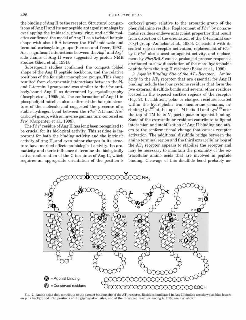
2. Agonist Binding Site of the AT1 Receptor. Aminoacids in the AT1 receptor that are essential for Ang IIbinding include the four cysteine residues that form thetwo external disulfide bonds and several other residueslocated in the exposed surface regions of the receptor(Fig. 2). In addition, polar or charged residues locatedwithin the hydrophobic transmembrane domains, in-cluding Lys102 at the top of TM helix III and Lys199 nearthe top of TM helix V, participate in agonist binding.Some of the extracellular residues contribute to ligandinteraction and stabilization of Ang II binding and oth-ers to the conformational change that causes receptoractivation. The additional disulfide bridge between theamino terminal region and the third extracellular loop ofthe AT1 receptor appears to stabilize the receptor andmay be necessary to maintain the proximity of the ex-tracellular amino acids that are involved in peptidebinding. Cleavage of this disulfide bond probably ac-
FIG. 2. Amino acids that contribute to the agonist binding site of the AT1 receptor. Residues implicated in Ang II binding are shown as blue letterson pink background. The positions of the glycosylation sites, and of the conserved residues among GPCRs, are also shown.
426 DE GASPARO ET AL.

counts for the impairment of AT1 receptor binding byreducing agents (Ohyama et al., 1995).
Ang II binds primarily to the extracellular region ofthe AT1 receptor (Hjorth et al., 1994) by interacting withresidues in its N terminus and its first and third extra-cellular loops. However the transmembrane helices alsoparticipate in Ang II binding, since its C-terminal car-boxyl group interacts with Lys199 in the upper part ofhelix 5 of the receptor (Underwood et al., 1994; Noda etal., 1995a; Yamano et al., 1995). This could involve theformation of a salt-linked triad between Lys199 of thereceptor and the carboxyl groups of Asp1 and Phe8 of theAng II peptide (Joseph et al., 1995a,b). The Trp253 resi-due has been proposed to stabilize the ionic bridgeformed between Lys199 and the carboxyl-terminal groupof the Phe8 residue. In addition, Phe259 and Asp263 intransmembrane helix VI could provide the docking sitefor His6 of the ligand (Yamano et al., 1995). Two otherresidues (Lys102 and Ser105) in the outer region of trans-membrane helix III of the receptor have also been im-plicated in Ang II binding (Groblewski et al., 1995; Nodaet al., 1995a). This region may participate in the forma-tion of the intramembrane binding pocket and possiblyin stabilization of the receptor’s conformation.
The Asp281 residue, located at the C-terminal end ofthe third extracellular loop of the AT1 receptor, serves asa major docking point for Ang II through its chargeinteraction with Arg2 of the ligand (Feng et al., 1995).The Asp278 residue could also be important in this re-gard, since its mutation causes an even greater loss ofreceptor binding affinity (Hjorth et al., 1994). The N-terminal Asp1 residue of Ang II has been proposed tointeract with His123 in the second extracellular loop ofthe AT1 receptor (Yamano et al., 1995). These findingssupport the view that Ang II attaches primarily via itscharged amino-terminal end to the extracellular bindingregion of the receptor. The major docking points for theamino- and carboxyl-terminal ends of Ang II, Asp281 andLys199, respectively, are located at the outer ends ofhelices 7 and 5, respectively. The intramembrane bind-ing pocket lies between these proposed contact points,and is adjacent to the cleft that contains the bindingsites of receptors for smaller ligands, as well as thenonpeptide binding site of the AT1 receptor. This regioncontains docking sites for the apolar/aromatic mid-por-tion of the Ang II molecule and for the carboxyl-terminalphenyl group that elicits the conformational change(s)leading to receptor activation.
The apolar nature of the essential aromatic ring of thePhe8 residue of Ang II binds to the AT1 receptor suggeststhat the phenyl group could interact with residues lo-cated within the membrane-spanning helices. This isconsistent with NMR-based predictions of the receptor-bound conformation of Ang II that position the phenylgroup in an appropriate location for such interactions(Nikiforovich et al., 1993, 1994). The phenyl group couldinteract with aromatic residues in the helices, some of
which have been suggested to form an aromatic floor forcharge interactions between ligand and receptor(Trumpp-Kallmeyer et al., 1992; Findlay et al., 1993).Modeling studies on the AT1 receptor have also sug-gested that conserved aromatic residues in helices IVand VI could form the base of the ligand binding site, asin other G protein-coupled receptors (Underwood et al.,1995). Mutations of two of these residues, Phe259 andTrp253, reduces Ang II binding to the receptor (Yamanoet al., 1995). Whether the aromatic group of Phe8 of theligand interacts with the binding pocket of the receptorvia these amino acids, or with aromatic residues in othersegments of the membrane domains, has yet to be de-termined. The Phe8 residue could also form a polar/aromatic interaction with the hydroxyl group of Ser105 inthe third transmembrane domain of the receptor (Jo-seph et al., 1995a,b).
The Tyr4 residue of Ang II is an important determi-nant of its binding and biological activities (Bumpus etal., 1977; Capponi and Catt, 1979; Nikiforivich et al.,1993, 1994). In fact, reversing the Tyr4 and Phe8 resi-dues of the Ang II molecule (Marshall et al., 1970)formed the first Ang II antagonist. The Tyr4 residue hasbeen proposed to interact with Arg167 at the top of thefourth transmembrane helix (Yamano et al., 1995), andcould also disrupt the hydrogen bonding between Asn111
and Tyr292 in transmembrane domains 3 and 7 of theunoccupied receptor by competing with Tyr292. Thiswould permit the latter to interact with Asp74 in thesecond transmembrane domain during receptor activa-tion. In this mechanism, the loss of a proton from theTyr4 phenolic hydroxyl group to the carboxyl group ofGlu91 could be part of a relay system initiated by theinteraction of His6 with Thr88 and Glu91 of the receptor(Joseph et al., 1995a,b).
3. Antagonist Binding of the AT1 Receptor. Since AngII is a major regulator of blood pressure, aldosteronesecretion, and fluid homeostasis, and is also an impor-tant etiological factor in hypertension and other cardio-vascular disorders, blockade of Ang II formation or ac-tion by ACE inhibitors or receptor antagonists is ofmajor therapeutic importance. Early attempt to developtherapeutic agents able to block the Ang II receptorimpeded by the peptidic nature of antagonists such assaralasin, which lacked oral activity and showed agonis-tic properties (Pals et al., 1979). More recently, based onimidazole derivatives first described by Furukawa et al.(1982), it became possible to develop specific nonpeptideAng II receptor antagonists that specifically and selec-tively block the angiotensin AT1 receptor (Timmermanset al., 1993; Goodfriend et al., 1996). The first of thisseries to reach the clinic, losartan, was followed by alarge number of orally active AT1 antagonists (Table2).These can be classified in two groups depending onthe presence of a biphenyltetrazole moiety, as in theprototype drug, losartan, in their structure. Receptorbinding of nonpeptide Ang II antagonists is saturable
ANGIOTENSIN RECEPTORS 427

and usually reversible and is independent of the path-way responsible for the synthesis of Ang II. This could berelevant to comparisons with ACE inhibitors, given thepossible role of alternative Ang II-generating enzymessuch as chymase, in human tissues (Urata et al., 1996).
In pharmacological studies on the properties of angio-tensin and its synthetic analogs, certain AT1 receptorantagonists not only cause a rightward shift in the AngII dose-response curve but also reduce the maximal re-sponse to agonist stimulation (Wienen et al., 1993; Mo-rimoto and Ogihara, 1994; Criscione et al., 1995; Goaand Wagstaff, 1996; Gillis and Markham, 1997; McClel-lan and Balfour, 1998). The latter compounds (candesar-tan, EXP 3174, valsartan, irbesartan) are termed insur-mountable antagonists, in contrast to surmountableantagonists such as losartan, eprosartan, and telmisar-tan, which do not impair the maximum response to AngII. One explanation for this difference is that nonpeptideantagonists can act by interfering with receptor activa-tion by occupying an intramembrane site that overlapswith the space occupied by the agonist (competitive orsurmountable antagonists) or by inducing conforma-tional changes that prevent agonist binding (noncompet-itive or insurmountable antagonists). Another proposalis that surmountable antagonists such as losartan dis-sociate rapidly from the receptor, whereas insurmount-able antagonists, exemplified by candesartan, bindtightly and dissociate so slowly as to cause functionalloss of the occluded receptors. Recent studies on the
properties of the human AT1 receptor expressed in Chi-nese hamster ovary (CHO) cells have shown that theagonist-receptor complexes are divisible into a rapidlyreversible, surmountable population, and a tightly bind-ing, insurmountable population (Fierens et al., 1999).
Although losartan is a potent antagonist in its ownright, about 10% of the dose is metabolized to EXP 3174,which has 10-fold higher affinity for the AT1 receptorand is responsible for the 24 h decrease in blood pres-sure. Candesartan cilexetil is an inactive ester prodrugand is completely cleaved during absorption in the gas-trointestinal tract. Twenty-five percent of candesartanis eliminated by metabolism to an inactive metabolite.Among the other antagonists, irbesartan, valsartan, andeprosartan do not require metabolism to be active. Irbe-sartan is mainly eliminated by the liver (75%) and lessthan 2% is excreted unchanged in the urine. Sixty per-cent of eprosartan is cleared unchanged via the bile.Valsartan is essentially eliminated by biliary excretionand 10% of the dosage appears intact in urine.
As expected, significant increases in renin activity,Ang I and Ang II are observed after blockade of the AT1receptor. It is conceivable that the increased circulatingAng II level could stimulate the AT2 receptor, whichappears to counterbalance the effect of the AT1 receptor(see below). Blockade of the AT1 receptor not only inhib-its smooth muscle contraction but also reduces the pro-duction of pressor agents including aldosterone, vaso-pressin, catecholamine, and endothelin. AT1 receptor
TABLE 2Structure of AT1 and AT2 receptor nonpeptidic antagonists
428 DE GASPARO ET AL.

antagonists have shown exceptionally good tolerabilityand their incidence of adverse effects is similar to that ofplacebos. The AT1 receptor antagonists are approved forthe treatment of hypertension, and in early clinical stud-ies also appear to be of use in the treatment of congestiveheart failure, postmyocardial infarction, and renal fail-ure. Several large clinical trials are in progress such asScope, Life and Value for hypertension, Regaal and Sil-ver for left ventricular hypertrophy, ValHeft and Charmfor congestive heart failure, Optimaal and Valiant forpostmyocardial infarction, and Irma, IDNT, Renaal andABCD 2V for diabetic nephropathy.
Identification of the losartan binding region of themammalian AT1 receptor was facilitated by the findingthat the amphibian Ang II receptor, which resembles themammalian AT1 receptor in its signal transductionmechanisms, does not recognize nonpeptide antagonistssuch as losartan. This enabled the amino acid residuesinvolved in the binding of losartan to the mammalianAT1 receptor to be determined by analysis of the ligandbinding properties of mutant rat AT1 receptors in whichnonconserved amino acids were replaced by the corre-sponding amphibian residues (Ji et al., 1994). Most ofthese mutant receptors showed only minor changes inbinding affinity for Ang II and its peptide antagonist[Sar1,Ile8]Ang II, indicating that the overall conforma-tion of the receptor was unaltered by such replacements.However, several residues located in the transmem-brane domains (TMDs) of the receptor were found to be
required for binding of the nonpeptide antagonist. Theseincluded Val108 in TMD III, Ala163 in TMD IV, Pro192
and Thr198 in TMD V, Ser252 in TMD VI, and Leu300 andPhe301 in TMD VII.
These findings demonstrated that the nonpeptide AT1antagonist binds to a site defined by amino acids locatedwithin the membrane-spanning regions of the receptor.Also, the nonpeptide binding site was largely distinctfrom the receptor domain that is involved in binding ofAng II and other peptide ligands. This conclusion isconsistent with the presence of a primordial binding sitefor small ligands between the transmembrane helices ofall GPCRs that can be used for the development ofnonpeptide analogs for a wide variety of peptide hor-mones. Other amino acid residues in the rat AT1B recep-tor that influence losartan binding include Ala73 in TMDII; Ser104, Ala114, and Ser115 in TMD III; Lys199 in TMDV; Phe248 in TMD VI; and Asn295 in TMD VII (Schambyeet al., 1994; Ji et al., 1994; Noda et al., 1995). These andother observations have implicated TMD III in losartanbinding to the mammalian AT1 receptor. The locations ofthe multiple amino acids that contribute to losartanbinding in the receptor are shown in Fig. 3.
A mutant amphibian receptor formed by exchangingthese residues for the corresponding amino acids in theXenopus AT receptor bound losartan with the same highaffinity as the rat AT1 receptor (IC50 values: rat AT2.2 6 0.2 nM; xAT . 50 :M: mutant xAT 2.0 6 0.1 nM)(Ji et al., 1993, 1995). This gain-of-function mutation, in
FIG. 3. Amino acids that contribute to the nonpeptide antagonist binding site of the AT1 receptor. Residues implicated in losartan binding areindicated by green letters on orange background. The positions of the glycosylation sites, and of the conserved residues among GPCRs, are also shown.
ANGIOTENSIN RECEPTORS 429

which the residues known to participate in the forma-tion of the ligand binding site in a mammalian hormonereceptor were transferred to an unresponsive amphibianreceptor, confirms the validity of the proposed losartanbinding site. The identification of residues involved inreceptor binding of nonpeptide Ang II analogs should aidin modeling studies on the structural basis of ligandrecognition and activation of the Ang II receptor.
Although there is little overlap between the in-tramembrane residues that influence Ang II and losar-tan binding, both ligands appear to interact with theregion located between helices III, V, VI, and VII of theAT1 receptor. This area also contains the binding sites ofGPCRs for small ligands such as catecholamines andacetyl choline, as well as other nonpeptide antagonistanalogs. In addition to sharing the binding pocketformed by the transmembrane helices of the receptor,Ang II and its nonpeptide analogs probably interact withcommon contact points at other locations in the receptor.In addition to the Lys102, Ser105, Arg167, and Lys199
residues, this could apply to Asn295, replacement ofwhich by serine impairs the binding of Ang II as well asits nonpeptide agonist and peptide antagonist analogs(Hunyady et al., 1998). It is noteworthy that peptideagonist binding is also influenced by Phe301, an aromaticresidue located near the end of helix VII (Hunyady et al.,1996a, 1999). Mutation of this residue to alanine im-pairs Ang II and losartan binding, as well as that of thepeptide antagonist, [Sar1,Ile8]Ang II. These findings in-dicate that multiple residues contribute to the integrityof the binding pocket and are required for optimal ago-nist and antagonist binding.
Although the extracellular loops of the receptor con-tribute significantly to the binding of Ang II and itspeptide antagonists, the intramembrane binding pockethas a major functional role in the AT1 and probably mostother peptide hormone receptors. In receptors for largeand/or bulky ligands, the intramembrane binding pocketper se may not permit efficient interaction without ad-ditional stabilization of the ligand through binding toresidues in the extracellular surface of the receptor.Nevertheless, the binding pocket is essential for theinitiation of receptor activation, and its interaction ofagonists triggers the change in receptor conformationthat leads to G protein coupling and second messengergeneration.
The importance of the transmembrane domains inreceptor activation also applies to glycoprotein hormonereceptors, which bind extremely large ligands. Thesedata suggest that a common mechanism for activation ofGPCRs is based on critical interactions within the in-tramembrane binding pocket that evoke specificchanges in receptor conformation. The extracellular do-mains of the receptor provide additional interactionswith the ligand that are necessary for high-affinity bind-ing. This is a prominent feature of receptors for largeragonists such as glycoprotein hormones, but also occurs
in those for small peptide hormones such as angiotensinII, bradykinin, and vasopressin.
I. AT1 Receptor Signaling Mechanisms
1. AT1 Receptor Activation and Signal Transduction.The AT1 receptor, like other GPCRs, has been proposedto undergo spontaneous isomerization between its inac-tive (R) and active (R*) states. The inactive R state isfavored in the absence of agonist ligands and is in equi-librium with a small proportion of the active R* state inits unliganded form. In the presence of Ang II, the activeR* form of the receptor is either selected or induced byagonist binding. The shift in equilibrium during agonistactivation favors the R* state, in which the altered con-formation of the receptor permits coupling to one ormore G proteins that mediate intracellular signaling viaphospholipase C and other pathways. Most receptorsappear to be kept in their inactive state by structuralrestraints that are removed by agonist binding, leadingto the formation of the activated R* state. Recent evi-dence suggests that the agonist-activated AT1 receptor,and possibly other GPCRs, undergoes transitions intomultiple conformational states that are associated withthe individual stages of receptor activation and regula-tion (Thomas et al., 2000).
The proportion of receptors in the activated state var-ies among individual GPCRs and is manifested by vary-ing degrees of basal signaling activity when receptorsare overexpressed in transfected cells. Significant de-grees of agonist-independent or constitutive activityhave been observed in wild-type thyroid-stimulatinghormone and muscarinic receptors (Burstein et al.,1997; Wang and Gershengorn, 1999) but are not evidentin the AT1 and most other GPCRs. Agonist-induced ac-tivation of the AT1 receptor appears to be triggered byan interaction between the Tyr4 residue of Ang II andthe Asn111 residue located in the third transmembranedomain of the receptor (Noda et al., 1996). This, and theinteraction of Phe8 of the angiotensin molecule withHis256 in the sixth transmembrane domain of the recep-tor, appear to drive the AT1 receptor into its fully activeR* state (Noda et al., 1995a).
The importance of Asn111 as a switch residue in ago-nist-induced AT1 receptor activation is further indicatedby the ability of its mutation to Gly111 or other smallresidues to induce constitutive activation of the receptor(Feng et al., 1998). This suggests that the ability ofAsn111 to act as a conformational switch is related to itsside chain size, rather than polarity or hydrogen bind-ing. On the other hand, it is the aromaticity rather thanthe size of the Tyr4 side chain that is important inreceptor activation, which probably depends on amino-aromatic binding between Tyr4 and Asn111 of the recep-tor (Miura et al., 1999a). Aromaticity is likewise impor-tant in the interaction between Phe6 and His256, whichis critical for full receptor activation by agonist ligands.
430 DE GASPARO ET AL.

Like other constitutively activated GPCRs, theAsn111Gly and Asn111Ala mutant receptors show in-creased affinity and efficacy for agonist ligands, andexhibit marked increases in the sensitivity of their bio-logical responses to agonist stimulation. They also dis-criminate between inverse agonists, which stabilize theinactive conformation of the receptor, and neutral an-tagonists. For these reasons, constitutively active AT1receptors are highly sensitive to Ang II derivatives thatare weak or partial agonists at the wild-type receptor(Miura et al., 1999b).
A proposed model of GPCR activation predicts thatrigid body movement of the third, sixth, and seventhtransmembrane domains induces conformationalchanges in the cytoplasmic loops that permit G proteininteraction with the agonist-activated receptor (Getherand Kobilka, 1998). However, the manner in which theactivating regions in the cytoplasmic domains of individ-ual GPCRs interact with specific subsets of G proteins isnot yet known. Although many amino acids in the trans-membrane and cytoplasmic regions of a wide range ofreceptors have been implicated in G protein coupling,there are very few cases in which individual residuescan be correlated with binding to specific types of Gproteins (Hunyady et al., 1996b). The limited degree ofselectivity of most such residues presumably reflects theprecise conformational requirements for specific inter-action of receptors with their cognate G proteins.
Although the AT1 receptor has been reported to inter-act with several G proteins, its major physiological func-tions are expressed through Gq-mediated activation ofphospholipase C followed by phosphoinositide hydroly-sis and Ca21 signaling. Although all GPCRs possess thebasic seven transmembrane structure, only a few aminoacids are highly conserved among the superfamily of Gprotein-coupled receptors (Fig. 1). One such conservedmotif is the characteristic NPX2–3Y sequence that islocated in the seventh transmembrane domain of mostreceptors, and in the rat AT1A receptor is Asn298-Pro299-Leu300-Phe301-Tyr302. A recent model of aminergicGPCRs suggests that the NPX2–3Y sequence is ideallyplaced to receive a signal from agonist-induced confor-mational changes in the ligand binding region (Donnellyet al., 1994). In the three-dimensional structure of thereceptor, this sequence is in close proximity to the as-paragine-aspartic acid pair located in transmembranesegments 1 and 2 and may form functionally importanthydrogen bonding interactions with this region. Suchmodels assume that the highly conserved proline resi-dues, which disrupt the a-helical structure of the trans-membrane domains, could serve as hinges during theconformational changes that occur in agonist-activatedreceptors. One such proline residue is located within theseventh transmembrane domain in the conservedNPX2–3Y sequence and has been found to be importantfor signal transduction by the m3 muscarinic receptor(Wess et al., 1993). However, this residue does not ap-
pear to be essential for signaling by Ang II and gonado-tropin-releasing hormone (GnRH) receptors.
Another interesting feature of the NPX2–3Y sequenceis its similarity to the NPXY internalization sequencethat is present in the cytoplasmic segment of receptorsfor low-density lipoprotein and several growth factors.The tyrosine residue in this sequence has been reportedto be essential for sequestration of the b-adrenergic re-ceptor (Barak et al., 1994). However, this does not applyto the gastrin-releasing peptide receptor (Slice et al.,1994) and the AT1 receptor, both of which contain anadditional aromatic amino acid (Phe321 and Phe301, re-spectively) in their NPX2–3Y sequences. The presence ofsuch residues might be important since phenylalaninecan substitute for the tyrosine residue in the NPXYinternalization sequence of nutrient receptors.
An analysis of the functional role of the Asn298-Pro299-Leu300-Phe301-Tyr302 sequence (Hunyady et al., 1995b)revealed that the ability of the receptor to interact withG proteins and to stimulate inositol phosphate re-sponses was markedly impaired by alanine replacementof Asn298 and was reduced by replacement of Pro299 orTyr302. The Phe301Ala mutant receptor exhibited normalG protein coupling and inositol phosphate responses,and the binding of the peptide antagonist, [Sar1,Ile8]AngII, was only slightly affected. However, its affinity forAng II and the nonpeptide antagonist losartan was re-duced by an order of magnitude, suggesting that Ang IIand losartan share an intramembrane binding site, pos-sibly through their aromatic moieties, in this region.None of the agonist-occupied mutant receptors, includ-ing Tyr302Ala and triple alanine replacement of Phe301,Tyr302, and Phe304 showed substantial changes in theirinternalization kinetics. These findings demonstratethat the NPLFY sequence of the AT1 receptor is not animportant determinant of agonist-induced internaliza-tion. However, the Phe301 residue contributes signifi-cantly to agonist binding, and Asn298 is required fornormal receptor activation and signal transduction.
Another highly conserved amino acid in the superfam-ily of G protein-coupled receptors is a tyrosine residuelocated in the fifth transmembrane helix, adjacent to theamino-terminal end of the third cytoplasmic loop. Thelocation of this amino acid, which is Tyr215 in the rat AT1receptor, suggests that it could be involved in receptoractivation. The amino-terminal region of the third cyto-plasmic loop adjacent to this residue has been shown tobe important in the signal generation and internaliza-tion of several G protein-coupled receptors. Further-more, modeling of G protein-coupled receptors based onthe crystal structure of bovine rhodopsin has suggestedthat this residue is in molecular proximity to regionsthat are important in receptor activation, including theconserved acidic-arginine-aromatic (DRY) triplet of thesecond intracellular loop (Baldwin et al., 1997). Al-though AT1 receptor internalization and signal trans-duction have different structural requirements, a
ANGIOTENSIN RECEPTORS 431

6-amino acid deletion of the amino-terminal end of thethird cytoplasmic loop, which included Tyr215, was foundto prevent both receptor internalization and signalingresponses. The role of the highly conserved Tyr215 resi-due in the activation of GPCRs was analyzed in a mu-tant AT1 receptor created by replacing this amino acidwith a phenylalanine residue (Hunyady et al., 1995b).
The resulting Tyr215Phe mutant receptor was nor-mally expressed in transfected COS-7 cells, and its bind-ing affinity for the peptide antagonist [Sar1, Ile8]Ang IIwas similar to that of the wild-type receptor. However,its affinity for Ang II was significantly reduced and itsability to mediate inositol phosphate responses to Ang IIstimulation was abolished. These changes were associ-ated with loss of coupling of the mutant receptor to Gproteins, as indicated by the lack of effect of GTPgS onagonist binding to the receptor. The agonist-inducedinternalization of the mutant receptor was also im-paired. The concomitant decreases in receptor internal-ization and G protein-mediated signaling of theTyr215Phe mutant receptor indicate that this residuehas a critical role in AT1 receptor activation. It is possi-ble that the Tyr215 residue takes part in propagation ofthe agonist-induced conformational change to the intra-cellular loops that participate in G protein coupling. Inview of its wide conservation among members of theseven transmembrane domain receptor superfamily,this residue is likely to be of general importance insignal transduction from G protein-coupled receptors.The AT1 receptor, like many other Ca21-mobilizingGPCRs, signals primarily through coupling to a Gq/11protein that activates phospholipase C-b (PLC-b), lead-ing to polyphosphoinositide hydrolysis, stimulation ofInsP3/calcium signaling, and activation of protein kinaseC. However, in cultured rat VSMC it is predominantlyPLC-g rather than PLC-b that is activated during Ang IIaction. This response, which is dependent on tyrosinephosphorylation of the PLC-g isozyme, is distinct fromthe usual process of activation of PLC-b1 and b2 byGPCRs through a and bg subunits derived from theheterotrimeric G proteins, Gq/11 and Gi/o, respectively.AT1 receptors have also been reported to couple to theG12/13 family of pertussis-insensitive G proteins, whichappear to mediate Ang II-induced L-channel activationin rat portal vein myocytes (Macrez et al., 1997). In therat, the AT1 receptor is also coupled to pertussive-sen-sitive Gi/o protein(s) that inhibit adenylyl cyclase in sev-eral Ang II target tissues, including the adrenal glo-merulosa zone, liver, kidney, and pituitary gland. Ang IIhas also been found to stimulate modest increases incyclic AMP production in some of its target tissues.However, this may result from activation of Ca21-sensi-tive adenylyl cyclases rather than coupling of the AT1receptor to Gs. Interestingly, although the cloned ratAT1A and AT1B receptors expressed in COS-7 and Y-1cells show the expected coupling to phosphoinositidehydrolysis and other Gq/11-mediated responses, they do
not exhibit coupling to Gi-mediated responses such asinhibition of adenylyl cyclase (Tian et al., 1996).
2. AT1 Receptor and Tyrosine Phosphorylation. Inaddition to its rapid actions on phosphoinositide/calciumsignaling, Ang II elicits many of the intracellular signal-ing responses that are typically associated with activa-tion of growth factors. These include tyrosine phosphor-ylation, most immediately of PLC-g and subsequently ofseveral other downstream proteins and effector en-zymes. These include pp60c-src, pp120, pp125FAK, JAK2,STATs, paxillin, TYK2, and MAPK (Bernstein and Mar-rero, 1996). Because GPCRs do not have intrinsic ty-rosine kinase activity, the ability of the AT1 receptor toinduce tyrosine phosphorylation and activation of PLC-gin rat VSMC, with consequent increases in InsP3 pro-duction and Ca21 signaling, must depend on other ty-rosine kinases. Such ligand-induced activation of PLC-gby tyrosine phosphorylation may occur during the ac-tions of growth factors on their target cells, and accountsfor the associated elevations in [Ca21]i. This aspect ofAT1 receptor function has been controversial, becausethe mechanisms by which the receptor couples to PLC inVSMC are poorly defined. In most cell types, calcium-mobilizing GPCRs are coupled through their cognate Gproteins to activation of the b isozymes of PLC. The b1and b2 enzymes are activated by Ga and Gbg subunits,respectively, the former derived from Gq/11 and the lat-ter largely from Gi/o proteins. In contrast, AT1 receptor-mediated activation of PLC-g is dependent on tyrosinephosphorylation and consequently on signaling from re-ceptor or nonreceptor tyrosine kinases.
The mechanism by which Ang II stimulates tyrosinephosphorylation and activation of PLC-g involves bind-ing of the enzyme to the C-terminal cytoplasmic domainof the AT1 receptor. This interaction occurs between theC-terminal cytoplasmic domain of PLC-g1 and a phos-phorylated YIPP motif located within the intracellulartail of the receptor (Venema et al., 1998a). The sameYIPP motif has been implicated in the binding of JAK2to the AT1 receptor, probably as a complex with thephosphotyrosine phosphatase, SHP-2 (Ali et al., 1997a).The way by which AT1 receptor activation regulatesligand-dependent binding of two distinct intracellularsignaling proteins to the same site in its cytoplasmic tailhas yet to be determined. The relative roles of PLC-band PLC-g in Ang II-induced phosphoinositide hydroly-sis in VSMC have been difficult to determine, and stud-ies on the relative abundance of the PLC isoforms inVSMC have given variable results. Relatively littlePLC-b1 was detected in rat and rabbit VSMC in somereports (Homma et al., 1993; Marrero et al., 1994), butothers have found that all three isozymes (a, b, and g)are abundant in rat VSMC (Ushio-Fukai et al., 1998). Inthe latter study, AT1 receptors were proposed to coupleinitially to PLC-b by heterotrimeric G proteins, andsubsequently to PLC-g. The G proteins responsible forthe early activation of PLC-b, and the initial maximum
432 DE GASPARO ET AL.

InsP3 response at 15 to 20 s, included both Gq/11 and G12.The subsequent activation of PLC-g and its contributionto the delayed InsP3 response are attributed to its ty-rosine phosphorylation by a downstream kinase. Thisstudy also suggested that bg subunits derived from G12
are involved in the activation of PLC in VSMC, andmediate Ang II-induced activation of PLD in these cells.
3. AT1 Receptor-Activated Growth Responses. In ad-dition to its contractile and secretory actions in smoothmuscle and adrenal cells, Ang II stimulates growthand/or proliferative responses in these and others of itstarget cells. This effect was first documented in ratadrenal glomerulosa cells, which respond to sodium de-ficiency with hypertrophy and hyperplasia, and undergoregression in animals on high sodium intake (Gross,1968). These changes are dependent on the circulatingAng II level, which is a major determinant of trophicchanges in the adrenal glomerulosa zone (Aguilera et al.,1978). Ang II also has trophic actions on renal mesangialcells and vascular smooth muscle cells, leading to cellu-lar hypertrophy and sometimes to cell division (Huckleand Earp, 1994). Cardiac myocytes and fibroblasts alsoexhibit hypertrophic responses to Ang II. However, car-diac fibroblasts undergo both hypertrophy and prolifer-ation during stimulation by Ang II, an action that con-tributes to the development of ventricular hypertrophy.Treatment with AT1 antagonists has been found to abol-ish the growth-promoting actions of Ang II in vitro andin vivo. As discussed below, many of the growth-relatedactions mediated by the AT1 receptor are inhibited byconcomitant binding of Ang II to the AT2 receptor.
The growth factor-like effects of Ang II include in-creases in tyrosine phosphorylation of numerous intra-cellular proteins, activation of MAPK and related path-ways, and increased expression of several early responsegenes including c-fos, c-jun, and c-myc (Clark et al.,1992; Berk and Corson, 1997). These changes are asso-ciated with cell growth, increased thymidine incorpora-tion, and cell proliferation (Tian et al., 1995). Such ac-tions of Ang II are consistent with its well definedgrowth effects in adrenal glomerulosa and VSMC, andits role in the vascular and cardiac lesions associatedwith endothelial damage, atherosclerosis, hypertension,and cardiac failure. One of the earliest Ang II-inducedevents in VSMC is the rapid tyrosine phosphorylationand activation of PLC-g, and the ensuing phosphoinosi-tide/Ca21 signaling responses that initiate many of theactions of Ang II in these cells (Marrero et al., 1994).This is accompanied by the rapid activation of Ras, andits dependent phosphorylation cascades that extendthrough the cytoplasm and into the nucleus, with con-sequent effects on cell growth and differentiation. Theseinclude the MAPK and related pathways, in which se-quential phosphorylations that are initiated at theplasma membrane lead to subsequent actions in thenucleus and other regions of the cell.
The consequences of Ang II-induced activation of Rasin cardiac myocytes resemble those elicited by agonistactivation of growth factor receptors, in which theMAPK cascade is initiated by the binding of adaptorproteins, such as Grb2 and Shc, to the autophosphory-lated receptors (Sadoshima et al., 1995). These adaptorsinteract with guanine nucleotide exchange factors thatenhance the activities of small GTP binding proteins,including Ras, Rac, and Rho. The small G proteins of theRas superfamily are activated by recruitment of guaninenucleotide exchange factors such as Sos, which promotesthe exchange of GTP for Ras-bound GDP to form theactive Ras-GTP complex. Ras-GTP then recruits andactivates serine/threonine kinases such Raf-1 or Mos,which in turn activate dual-specificity MAPK kinases,such as MEKs, by serine phosphorylation. MEKs in turnphosphorylate the MAPKs, p44MAPK and p42MAPK (alsoknown as extracellular signal-regulated kinases, orERK1 and ERK2) on threonine and tyrosine residuesthat are located within a TEY phosphorylation motif intheir activation loop. The activated MAPKs are translo-cated into the nucleus, where they phosphorylate otherkinases and transcription factors that regulate the co-ordinated expression of genetic programs that control awide variety of cellular functions. These include severalearly response genes and others such as c-myc, Elk1,Ets, RSK, and Mnk. MAPKs also exert regulatory ac-tions on other signaling pathways in the cytoplasm andat the cell membrane. The multiple actions of activatedMAPKs are terminated by a group of dual-specificityphosphatases that selectively dephosphorylate the indi-vidual MAPKs and related enzymes (Neel and Tonks,1997).
Ang II is but one of several calcium-mobilizing GPCRsthat mimic the effects of growth factors and other recep-tor kinases on the activation of MAPKs and early re-sponse genes. The way by which this response is linkedto the well defined phosphoinositide/calcium signalingsystem that is used by many such receptors has onlyrecently been established, and some aspects of this pro-cess still remain to be determined. In addition to theneed for Ca21 signaling (Sadoshima et al., 1995) a role ofPKC was suggested by reports of phorbol ester-inducedactivation of Raf and Ras in certain cell types. In addi-tion, the phorbol ester insensitive PKC isoform, PKC-z,was found to mediate Ang II-induced activation ofERK1/2 in VSMC and to associate with Ras during AngII stimulation (Liao et al., 1997). Such findings indicatedthat additional upstream factors must be of primaryimportance in MAPK activation by calcium-mobilizingGPCRs. In rat VSMC, Ang II also activates Ras (Schief-fer et al., 1996), an early and cardinal intermediate instimulation of the MAPK pathway by growth factors.The Ang II-induced activation of Ras is more rapid andless prominent than that elicited by epidermal growthfactor (EGF) and is substantially reduced by treatmentwith pertussis toxin. However, the concomitant in-
ANGIOTENSIN RECEPTORS 433

creases in MAPK activity and c-fos expression are notimpaired by pertussis toxin (Okuda et al., 1996), sug-gesting that these responses are not dependent on theGi-mediated activation of Ras that occurs in Ang II-stimulated VSMC.
Ang II not only increases Ras-GTP levels but alsostimulates the formation of Ras-Raf-1 complexes andtyrosine phosphorylation of GTPase-activating proteins(GAPs) such as p120 Ras-GAP and p190 Rho-GAP.These effects are prevented by the introduction of pp60c-
src antibodies into the cultured cells, as is the tyrosinephosphorylation of PLC-g1 and a substantial fraction ofthe Ang II-induced Ins(1,4,5)P3 response (Marrero et al.,1995; Schieffer et al., 1996). These and other findingshave shown that Ang II partially mimics the action ofgrowth factors by using the Ras pathway to activateMAPK. Furthermore, this appears to involve the nonre-ceptor tyrosine kinase, Src (pp60src), which probablyacts through tyrosine phosphorylation and activation ofa nucleotide exchange factor. Further evidence for theimportance of Src in Ang II-stimulated activation ofERK has come from studies with tyrosine kinase inhib-itors, and in Src-deficient and Src-overexpressing VSMC(Ishida et al., 1998).
4. Transactivation of Growth Factor Signaling by theAT1 Receptor, In addition to Src, a calcium-dependenttyrosine kinase has been implicated in the Ang II-in-duced stimulation of the Ras/MAPK cascade in VSMC(Eguchi et al., 1996). This observation led to the recog-nition that transactivation of the EGF receptor contrib-utes to the activation of MAPK by Ang II in these cells.This process involves the calcium-dependent phosphor-ylation of the EGF receptor to form docking sites for Srcand the adaptor protein, Shc, which then recruits Grb2and Sos to activate Ras (Eguchi et al., 1998).
These findings account for much of the similarity be-tween the actions of Ang II and growth factors on theactivation of PLC-g, tyrosine kinases, and MAPKs, andincreased expression of nuclear proto-oncogenes. Theyare also consistent with the observation that Ang andplatelet-derived growth factor (PDGF) signaling cas-cades converge in VSMC, with activation of the PDGF-breceptor (Linseman et al., 1995). Ang II also stimulatesthe tyrosine phosphorylation and activation of the IGF-1receptor, as well as insulin receptor substrate 1, inVSMC (Ali et al., 1997b). Recently, activation of the EGFreceptor by Ang II has been implicated in the Ang II-induced synthesis of TGF-b and fibronectin in cardiacfibroblasts. This effect was mediated by downstreamsignaling from the EGF receptor, which was transacti-vated by Ang II via a Ca21-dependent tyrosine kinase.The Ang II-induced expression of fibronectin mRNA wasregulated at the transcriptional level by binding of thefos/jun complex to an AP-1 site and also by stabilizationof the message through actions of TGF-b (Moriguchi etal., 1999).
These observations are relevant to the proposed role ofAT1 receptor activation in the remodeling of cardiacinterstitial tissue during ventricular hypertrophy. AngII has been shown to stimulate the expression of colla-gen and fibronectin in cardiac fibroblasts, and to pro-mote the release of newly formed collagen from the cells(Villareal et al., 1993; Crabos et al., 1994). At least one ofthe calcium-dependent tyrosine kinases that could me-diate the transactivation of the EGF receptor by Ang IIhas been identified as Pyk2 (also termed CAKb andRAFTK). However, it appears that Pyk2 does not fullyaccount for this process and that other mechanisms areinvolved in AT1 receptor-mediated transactivation of theEGF receptor (Murasawa et al., 1993). Another exampleof cross-talk from the AT1 receptor is its ability to influ-ence the activity of the LOX-1 receptor for oxidized low-density lipoprotein in human coronary artery endothe-lial cells (Li et al., 1999). Since oxidized low-densitylipoprotein has been implicated in the pathogenesis ofatherosclerosis, and attenuates nitric oxide-induced di-latation, and promotes leukocyte deposition in the vas-cular wall, the ability of Ang II to up-regulate LOX-1expression via AT1 receptors suggests another potentialrole for AT1 antagonists in the management of cardio-vascular disease.
5. Other AT1 Receptor-Mediated Signaling Path-ways. In addition to activating phosphoinositide/cal-cium signaling, tyrosine phosphorylation, and MAPKpathways, Ang II also acts through the AT1 receptor tostimulate the Jak/STAT signaling pathway. This wasfirst observed in neonatal cardiac fibroblasts and trans-fected CHO cells, in which the AT1A receptor activatesthe STAT signaling pathway and stimulates sis-induc-ing factor-like DNA binding activity (Bhat et al., 1994).The stimulatory effect of Ang II on SIF/STAT activationwas slower than that elicited by IL-6, and was precededby an initial inhibitory phase that was associated withconcomitant suppression of IL-6-induced STAT tyrosinephosphorylation and SIF responses (Bhat et al., 1995).The inhibitory action of Ang II on IL-6-induced Stat3signaling was attributed to activation of the MAPKpathway and could be attenuated by the MAPK inhibi-tor, PD98059 (Bhat et al., 1996). Ang II was subse-quently found to stimulate rapid serine phosphorylationof Stat3 via a MAPK1-dependent pathway (Bhat andBaker, 1997). In neonatal cardiac myocytes, the AT1receptor is coupled through Jak2 kinase to the activationof Stat1 and Stat3, and to the formation of SIF com-plexes (McWhinney et al., 1997).
Ang II also causes rapid and transient activation ofthe Jak/STAT pathway in rat aortic smooth muscle cells(Marrero et al., 1995). This is initiated by tyrosine phos-phorylation and increased catalytic activity of Jak2, andis dependent on its physical association with the ago-nist-activated AT1 receptor. This interaction occurs at aYIPP sequence that is located in the carboxy terminaltail of the receptor (Ali et al., 1997a,b), and is also
434 DE GASPARO ET AL.

present in the PDGF receptor. This motif has also beenimplicated in the binding of SH2 domains in PLC-g tothe AT1 receptor (Venema et al., 1998a). The binding ofJak2 to this motif has been attributed to the participa-tion of SHP-2 phosphotyrosine phosphatase, which alsocontains an SH2 domain and functions as an adaptorprotein in the association between JAK2 and the AT1receptor (Marrero et al., 1998).
In addition to ERK1 and ERK2, Ang II stimulates theactivity of c-Jun N-terminal kinase (JNK) members ofthe MAPK family. In vascular smooth muscle cells, p21-activated kinase (PAK), which mediates JNK activationby IL-1 and tumor necrosis factor, was rapidly stimu-lated by Ang II before the activation of JNK. Ang II-induced activation of both PAK and JNK was dependenton calcium and PKC, and partially on a tyrosine kinaseother than Src. These findings, and the ability of adominant negative PAK to attenuate the JNK responseto Ang II in transfected CHO and COS cells, indicatedthat PAK is a mediator of JNK activation by Ang II inVSMC (Schmitz and Berk, 1997). In this regard, Ang IIagain behaves like inflammatory cytokines such as IL-2and tumor necrosis factor-a, by acting through PAK asan upstream mediator of JNK.
Ang II also promotes hypertrophy of VSMC throughan oxidant stress-dependent mechanism. Reactive oxy-gen species such as hydrogen peroxide and O2
. appear tobe involved in the pathogenesis of hypertension andatherosclerosis, and are released from endothelial cellsand other vascular and circulating cells (Rajagopalan etal., 1996). These molecules have been implicated in vas-cular smooth muscle proliferation and in the develop-ment of hypertension (Laursen et al., 1997; Abe andBerk, 1998). The reactive oxygen species that are gen-erated by xanthine oxidase are potent stimuli of VSMCproliferation (Rao and Berk, 1992). Conversely, antioxi-dants reduce the cell response to growth factors, andinhibit the proliferation of VSMC (Boscoboinik et al.,1995). In cultured rat VSMC, Ang II causes rapid in-creases in intracellular H2O2 and phosphorylation ofp42/44 MAPK and p38 MAPK. Treatment with H2O2activates only p38 MAPK, and an NADH/NADPH oxi-dase inhibitor reduced only p38 MAPK phosphorylationin Ang II-treated cells. Also, blockade of Ang II-inducedp38 MAPK phosphorylation by transfected catalase in-hibited solely p38 MAPK phosphorylation, and reducedAng II-induced cell hypertrophy. These findings indicatethat both the p38 MAPK and the p42/44 MAPK path-ways are required for the hypertrophic response ofVSMC to Ang II (Ushio-Fukai et al., 1998).
In addition to their role in Ang II-induced activation ofp38 MAPK, reactive oxygen species mediate the activa-tion by Ang II of Akt/protein kinase B (Akt/PKB) inVSMC (Ushio-Fukai et al., 1999b). This serine-threo-nine kinase has been implicated in protein synthesis andalso appears to be an important component of a survivalpathway that protects cells from apoptosis. Akt/PKB is
activated by several growth factors via Ras and phos-phatidylinositol-3 kinase, and also by heat shock andother stresses. In rat VSMC, the activation of Akt/PKBby Ang II is dependent on tyrosine phosphorylation andthe activity of its upstream effector molecule, phospha-tidylinositol-3 kinase (Takahashi et al., 1999). Thesefindings suggest that Akt/PKB has an important role inthe intracellular actions of Ang II and in particular in itseffect on cell survival (Pollman et al., 1996).
NO inhibits several of the physiological actions of AngII, and prevents its activation of ERK, JNK, and p38MAPK (Wang and Murphy, 1998). In rat cardiac fibro-blasts, NO inhibits the activation of PYK2 and causes aconcomitant decrease in ERK phosphorylation in AngII-stimulated cells. Since PYK2 is essential for Ang II-induced activation of ERK in VSMC (Sabri et al., 1998),these findings suggest that it could be a locus at whichNO regulates calcium-dependent signaling by the AT1and other Gq-coupled receptors (Lev et al., 1995).
J. Receptor Activation and Endocytosis
Agonist activation of many plasma-membrane recep-tors is followed by clustering of the complexes in clath-rin-coated pits, and subsequent internalization in clath-rin-coated vesicles to undergo degradation or recyclingto the plasma membrane. Although nutrient receptors(e.g., for low-density lipoprotein and transferrin) fre-quently exhibit constitutive internalization, the hor-mone and growth factor receptors that evoke intracellu-lar signals usually undergo ligand-induced endocytosis.Growth factor receptors with intrinsic tyrosine kinaseactivity and G protein-coupled receptors are internal-ized by a similar clathrin-coated vesicular endocytic pro-cess. There is increasing evidence that the intracellularsignaling mechanisms that are activated by these recep-tors do not necessarily participate in the internalizationprocess. Such a dissociation between Ang II-induced Gprotein activation and receptor internalization was ob-served in studies on mutant and wild-type AT1A recep-tors expressed in COS-7 cells (Hunyady et al., 1994b).Mutations of the third cytoplasmic loop revealed thatthe N-terminal part of this region is important for bothreceptor endocytosis and intracellular signaling. Also,three point mutations of the conserved Asp74 residue inTMD II, which has been implicated in signal transduc-tion by the AT1A receptor and other GPCRs (Bihoreau etal., 1993), significantly impaired G protein coupling andphosphoinositide hydrolysis. However, the Asp74 (D74N,D74H, and D74Y) showed markedly different internal-ization kinetics. The D74Y receptor showed the greatestimpairment of internalization but retained the highestdegree of inositol phosphate stimulation. In contrast, theD74N mutant, which showed the most impaired G pro-tein coupling and inositol phosphate responses, had nor-mal internalization kinetics. The combined mutant re-ceptor containing the D74N substitution and deletion ofresidues 221–226 from the third cytoplasmic loop
ANGIOTENSIN RECEPTORS 435

showed no G protein coupling or inositol phosphate re-sponse but was internalized about 60% as rapidly as thewild-type receptor. These data demonstrate that endo-cytosis of the AT1 receptor is independent of agonist-activated signal transduction and indicate that receptorinternalization and activation phospholipase C have dif-ferent structural requirements.
The demonstration that coupling to heterotrimeric Gproteins and intracellular signaling responses is not re-quired for internalization of the AT1 receptor indicatedthat the conformational changes that result from ago-nist-induced receptor activation exert largely indepen-dent effects on signal transduction and receptor endocy-tosis. However, since the structural requirements forsignaling and internalization overlap, mutations thatcause changes in G protein coupling are frequently ac-companied by impairment of endocytosis. Thus, the N-terminal region of the third cytoplasmic loop is impor-tant for both receptor signaling and internalization.However, the ability of internalization-deficient mutantreceptors with specific changes in the cytoplasmic tail orthird loop to activate their respective G proteins andsignaling responses indicates that G protein activationalone is not sufficient to induce receptor endocytosis.The sequences in these regions that are important forreceptor internalization are frequently enriched inserine and threonine residues. For example, the rat AT1receptor contains 12 serine/threonine residues in the23-amino acid segment located between residues 326and 348 of the cytoplasmic tail (Fig. 4).
A mutational analysis of this region of receptor re-vealed that deletion of the carboxyl-terminal 22 aminoacids of the receptor did not affect agonist-induced en-docytosis (Hunyady et al., 1994a). However, internaliza-tion was markedly impaired by removal of one addi-tional residue (Leu337) and was reduced by 95% afterremoval of the adjacent Ser335 and Thr336. In addition,triple alanine replacement of the Ser-Thr-Leu residuesreduced internalization to almost the same extent as thecorresponding tail deletion mutant. The Ser-Thr-Leumotif is highly conserved in mammalian AT1 receptorsbut is not present in the noninternalizing AT2 receptor.These findings demonstrate that a serine/threonine-richregion including Leu337 in the cytoplasmic tail of the AT1receptor is a major requirement for endocytosis of the
hormone-receptor complex, and support the concept thatsimilar motifs in other G protein-coupled receptors aredeterminants of their agonist-induced internalization.An additional region in the cytoplasmic tail of the AT1receptor, involving hydrophobic and aromatic residueson a putative a-helix adjacent to the cell membrane, hasalso been found to be involved in agonist-stimulatedendocytosis (Thomas et al., 1995). It is possible thatthese two regions in the cytoplasmic tail function coop-eratively to trigger endocytosis, and that agonist-in-duced receptor activation leads to phosphorylation of theserine/threonine-rich region and causes a conforma-tional change that exposes the more proximal hydropho-bic residues for interaction with the endocytic mecha-nism.
K. AT1 Receptor Function in Selected Tissues
1. The AT1 Receptor and the Brain. Ang II regulatesnumerous physiological responses through its centralactions in the brain, where it functions as a neurotrans-mitter or neuromodulator to influence blood pressure,drinking behavior, salt appetite, and several neuroendo-crine processes. Some of these responses are induced bythe actions of circulating Ang II at the circumventricularorgans and other specialized regions, and others areinfluenced by locally formed Ang II generated within thebrain itself. Although circulating hormones are effec-tively excluded from most parts of the brain by theblood-brain barrier, neurons in the circumventricularorgans are accessible to many circulating ligands via thefenestrated endothelial cells of their dense capillary cir-culation. Due to the absence of the blood-brain barrier atthese sites, neurons within the subfornical organ (SFO),organum vasculosum lamina terminales (OVLT), andarea postrema are exposed to circulating hormones,ions, and other potential regulatory factors. These threestructures, sometimes called the sensory circumven-tricular organs (Johnson and Gross, 1993), are rich inAT1 receptors and have been implicated in numeroushomeostatic processes. Ang II is but one of several pep-tides that bind to high-affinity receptors located on cir-cumventricular neurons that project to hypothalamicnuclei and other brain regions. Other ligands with abun-dant circumventricular receptors include atrial natri-uretic peptide, vasopressin, cholecystokinin, GLP-1, cal-citonin, and relaxin (McKinley and Oldfield, 1998).
The pressor response to circulating Ang II is mediatedby the area postrema and the SFO, and its stimulatoryactions on water and salt ingestion, vasopressin secre-tion, and adrenocorticotropin (ACTH) release are medi-ated by the SFO. AT1-expressing neurons in the SFOsend projections to the supraoptic and paraventricularnuclei of the hypothalamus to influence vasopressin andACTH release (Oldfield et al., 1994), and to the medianoptic nucleus to stimulate water drinking (Cunninghamet al., 1991).
FIG. 4. Location of agonist-induced phosphorylation sites in the AT1receptor. The potential serine and threonine phosphorylation sites areshown in bold letters. The most highly phosphorylated region in theC-terminal cytoplasmic tail during Ang II action is indicated by theshaded box. Potential PKC sites are indicated by brackets, and thecorresponding phosphorylated setrines are shown by asterisks. Recentevidence suggests that low agonist concentrations selectively induce PKCphosphorylation by other kinases is predominant at higher agonist con-centrations (Quian et al., 1999).
436 DE GASPARO ET AL.
Like several other peptide hormones, Ang II exertseffects in the central nervous system that are comple-mentary to its primary physiological actions in periph-eral tissues. This is well exemplified by the presence ofbrain AT1 receptors at sites that influence cardiovascu-lar function, fluid and electrolyte homeostasis, and pi-tuitary hormone secretion (Allen et al., 1998, 1999). Theexistence of an endogenous brain renin-angiotensin sys-tem that is distinct from the systemic system comprisedof the kidneys, liver, and lungs has been demonstratedby numerous studies over the last 30 years. Early evi-dence for a central mechanism that contributes to thedevelopment of Ang II-induced hypertension (Bickertonand Buckley, 1961) was followed by the demonstration ofrenin and angiotensin II in the brain (Fisher-Ferraro etal., 1971). These and other studies led to the recognitionof an autonomous brain renin-angiotensin system thathas significant actions in cardiovascular regulation andseveral other brain functions (Ganten and Speck, 1978;Ganong, 1984; Phillips, 1987).
The ability of centrally administered Ang II to in-crease blood pressure, fluid intake, and vasopressin re-lease presaged the existence of specific receptors in thebrain regions controlling these functions. This was con-firmed by the selective effects of Ang II on brain struc-tures that influence blood pressure, sympathetic nerveactivity, drinking behavior, salt appetite, and pituitaryhormone secretion (Phillips, 1987; Ganong, 1993;Wright and Harding, 1994). Effects of angiotensin onlearning and memory, as well as sensory function, havealso been observed. The number and scope of these cen-tral effects of angiotensin indicate that the renin-angio-tensin system has a major role in the regulation ofneural components that control multiple physiologicalfunctions. Most of these actions are mediated by AT1receptors, which are expressed in brain regions knownto be involved in the control of cardiovascular functionand body fluid homeostasis.
At the cellular level, direct application of Ang II in-creases the firing rate of neurons in the supraoptic nu-cleus, subfornical organ, and paraventricular nucleus(Nicoll and Baker, 1971; Felix and Akert, 1974; Ambuhlet al., 1992a,b) as well as in the nucleus tractus soli-tarius, dorsal motor nucleus of the vagus, and thalamus(Wayner et al., 1973; Felix et al., 1988). In several ofthese studies, the angiotensin II (2–7) heptapeptide(Ang III) was found to be as or more effective than AngII, raising the possibility that Ang III may be the cen-trally active form of Ang II (Harding et al., 1986). Theapplication of topical autoradiography to receptor map-ping in tissue sections revealed that angiotensin II re-ceptors are present in many of the brain nuclei and otherregions that are known to be involved in the centralregulation of cardiovascular regulation and fluid ho-meostasis (Mendelsohn et al., 1984). The distributions ofAng II receptors in the brains of several mammalianspecies have been mapped by topical autoradiography
with radioiodinated ligands, usually 125I-[Sar1,IIe8]AngII. Specific AT1 and AT2 receptors were identified byinhibition of radioligand binding by selective AT1 (losar-tan) and AT2 (PD123177) receptor antagonists. The dis-tribution of AT1 receptor messenger RNA in the rodentbrain has also been analyzed by in situ hybridizationusing specific riboprobes to detect AT1A and AT1B recep-tor subtypes (Lenkei et al., 1998). Such studies haveshown that all of the well defined physiological actions ofAng II in the brain are mediated by AT1 receptors.
In all species studied, AT1 receptors are most abun-dant in the hypothalamus and the circumventricularorgans. In the hypothalamus, AT1 receptors are local-ized to the parvocellular region of the paraventricularnucleus, which has been implicated in the control ofanterior pituitary hormone secretion, ingestive behav-ior, and autonomic regulation of the cardiovascular sys-tem. In contrast, AT1 receptor expression is low or ab-sent in the magnocellular neurons of theparaventricular nucleus that control the release of vaso-pressin and oxytocin from the neurohypophysis. Thecircumventricular organs, which include the median em-inence, arcuate nucleus, SFO, and OVLT, contain highdensities of AT1 receptors. At each of these sites, thelocal deficiency in the blood-brain barrier renders themaccessible to Ang II and other regulatory ligands in thecirculation. Together with the interposed median preop-tic nucleus, which also contains abundant AT1 receptors,the SFO and OVLT comprise the lamina terminalis ofthe forebrain. This structure has neural connectionswith the parvocellular neurons of the hypothalamus, aswell as efferent projections to the supraoptic nucleusthat subserve vasopressin secretion and other osmoreg-ulatory responses including sodium excretion and thirst(McKinley et al., 1992).
In contrast to the relatively wide distribution of AT1Areceptor expression in the CNS, the AT1B receptor is notdetectable in most brain areas. However, AT1B receptorsare the predominant subtype expressed in the anteriorpituitary gland, where they mediate the direct actions ofAng II on pituitary function. Both Ang II and Ang III canelicit a variety of physiological changes, including pres-sor and dipsogenic responses, and increased salt appe-tite, when injected into the cerebral ventricles (Wrightand Harding, 1992). This is accompanied by increasedfiring of magnocellular neurons in the supraoptic nu-cleus (Okuya et al., 1987; Yang et al., 1992). Injection ofAng II or Ang III into the supraoptic and paraventricu-lar nuclei also stimulates magnocellular neurons, lead-ing to the release of vasopressin from the posterior pi-tuitary gland (Phillips, 1987).
2. Ang II-Induced Neuronal Signaling Pathways.AT1 receptor-mediated signaling pathways in neuronsare generally similar to those observed in other Ang IItarget cells. In addition to phosphoinositide hydrolysis,with formation of InsP3 and diacylglcerol, elevation of[Ca21]i, and activation PKC, Ang II stimulates the ras/
ANGIOTENSIN RECEPTORS 437

raf/MAPK cascade and translocation of MAPK into thenucleus (Lu et al., 1996). It also increases the expressionof c-fos, c-jun, and other early response genes. TheMAPK pathway mediates Ang II-stimulated norepi-nephrine synthesis and reuptake mechanisms in cul-tured neurons (Yang et al., 1996). In addition, Ang II-induced increases in [Ca21]i activate calcium/calmodulin protein kinase II, which acts together withPKC to mediate the rapid actions of Ang II on neuronalmembrane currents. These include a PKC-mediated in-crease in total neuronal Ca21 current (ICa), as well asPKC- and calcium/calmodulin protein kinase II-medi-ated inhibition of delayed rectifier K1 current (Kv) andtransient K1 current (IA) (Zhu et al., 1997a, 1998a). Inrat supraoptic neurons, AT1 receptors mediate Ang II-induced activation of a nonselective Na1/Ca21 channelthat promotes neuronal depolarization (Yang et al.,1992). Some of these effects of AT1 receptor activation inneurons, including MAPK activation, are under the in-hibitory control of AT2 receptors via activation of serine/threonine type 2A (PP-2A) (Huang et al., 1996) andpossibly tyrosine phosphatases such as MAPK phospha-tase.
3. Role of Ang III in the Brain. Ang II is rapidlycleared from the circulation with a half-life of 13 s and isextensively metabolized in its target tissues to form AngIII, Ang IV, and Ang (4–8) (Harding et al., 1986; DeSilva et al., 1988). Although the Ang II octapeptide is themajor active species acting at AT1 receptors in periph-eral Ang II target tissues, the possibility that Ang IIIcould be the predominant effector peptide in the brainhas been recognized for several decades. This was firstsuggested by the observation that Ang III was twice aseffective as Ang II in stimulating the firing rat of SFOneurons (Felix and Schlegel, 1978). This finding, andsimilar observations in paraventricular nucleus neu-rons, led to the suggestion that Ang III could be thecentrally active form of Ang II (Harding et al., 1986).This proposal gained support from studies using nonse-lective aminopeptidase inhibitors such as bestatin,which increased the half-lives of Ang II and Ang III inthe brain and potentiated the stimulatory action of AngII and Ang III on paraventricular neurons (Harding andFelix, 1987).
The recent development of specific inhibitors of ami-nopeptidase A (APA) and aminopeptidase N (APN),which hydrolyze the N-terminal residues of Ang II andAng III, respectively, has facilitated the investigation ofthe roles of Ang II and Ang III in the brain. The effectsof i.c.v. administration of these compounds indicatedthat the central action of Ang II on vasopressin releasein mice depends on its prior conversion to Ang III, con-firming that Ang III acts as a major effector peptide ofthe renin-angiotensin system in the brain (Zini et al.,1996). Furthermore, the ability of centrally adminis-tered Ang II to stimulate the firing rate of magnocellularvasopressinergic neurons of the supraoptic nucleus
(SON) of the rat was prevented by central administra-tion of the selective APA inhibitor, EC33. Since the SONdoes not contain AT1 receptors, this effect of EC33 pre-sumably results from its inhibition of APA in the sub-fornical organ, from which Ang II-sensitive neuronsproject to the SON (Zini et al., 1998).
Recent studies on the effects of selective APA andAPN inhibitors on blood pressure in the rat have indi-cated that Ang III is also the main effector peptide of thebrain RAS in the regulation of blood pressure (Reaux etal., 1999). Central inhibition of APA by EC33 not onlyblocked the pressor action of Ang II but also caused a fallin basal blood pressure comparable to that caused byi.c.v. administration of losartan, indicating a require-ment for Ang III in the tonic regulation of blood pres-sure. Furthermore, the inhibition of APN activity byPC18 caused an increase in blood pressure that wasblocked by losartan. This suggests that inhibition of AngIII degradation by PC18 causes an increase in Ang IIIlevels that elevates blood pressure through an action onAT1 receptors. These observations indicate the potentialapplications of APA inhibitors as centrally acting com-pounds in the management of hypertension.
These observations raise the question of whether thecentral actions of Ang III are mediated by an as yetunidentified AT1-type receptor with higher affinity forAng III over Ang II. This seems unlikely in view of theprobability that such a receptor with similarity to theAT1 site would have been detected during isolation andcloning of the AT1 and AT2 subtypes. The alternativeproposal, that blockade of the conversion of Ang II toAng III by APA favors the activation of other Ang II-degrading pathways, appears to be a more likely expla-nation.
4. The AT1 Receptor and the Pituitary Gland. Thepresence of Ang II receptors in the anterior pituitarygland was suggested by early reports of actions of theoctapeptide on pituitary hormone secretion (Steele et al.,1981). This was later confirmed by binding studies with125I-Ang II, which demonstrated specific, high-affinitybinding sites in the anterior pituitary glands of rat,rabbit, and dogs. No receptors were found in the poste-rior pituitary gland or in GH3 pituitary tumor cells(Hauger et al., 1982; Mukherjee et al., 1982). The siteswere nanomolar affinity, and bound the heptapeptidedes-Asp1-Ang II with about one-tenth the affinity of theoctapeptide. Ang II and the heptapeptide showed a sim-ilar potency ratio in their ability to stimulate ACTHrelease from pituitary cells (Capponi et al., 1982). Frac-tionation of rat pituitary cells revealed that Ang II re-ceptors were associated with lactotrophs and stimulatedprolactin as well as ACTH release. These findings sug-gested that Ang II contributes to the physiological con-trol of prolactin secretion (Aguilera et al., 1982).
The stimulatory action of Ang II on prolactin produc-tion by pituitary lactotrophs is mediated by the AT1receptor, which is coupled to phosphoinositide hydroly-
438 DE GASPARO ET AL.

sis and PKC activation, and also affects cyclic AMPproduction (Audinot et al., 1991). In addition to thesestimulatory actions, Ang II also exerts AT1-mediatedinhibitory actions on adenylyl cyclase activity in pitu-itary cell membranes. In intact cells, blockade of AT1
receptors by losartan prevented AT1-induced prolactinrelease, as well as the phospholipase C and PKC-depen-dent increase in cyclic AMP production (Moreau et al.,1994). During development the responsiveness of ratpituitary lactotrophs to Ang II increases with age, andby 28 days was more prominent in female than in malerats (Diaz-Torga et al., 1994). The ability of the hypo-thalamic GnRH to stimulate prolactin release in peri-fused pituitary cells, but not gonadotropin release, wasabolished by losartan, consistent with a paracrine role ofAng II in GnRH-stimulated prolactin release (Becu-Vil-lalobos et al., 1994). Interestingly, the ability of Ang II tostimulate prolactin release from cultured pituitary cellsfrom animals fed on an essential fatty acid-free diet wasmarkedly decreased, although the stimulatory action ofthyrotropin-releasing hormone, and the inhibitory ac-tion of dopamine were not affected (Alessio et al., 1994).
Ang II also increases the proliferation of culturedmammatrophs, and this effect is suppressed by tamox-ifen and restored by estradiol-17b. Treatment with thepeptide Ang II antagonist, saralasin, inhibited not onlythe Ang II-stimulated proliferative response but alsothat of GnRH, again indicating that GnRH-stimulatedrelease of Ang II, together with estradiol, regulates lac-totroph proliferation in the rat pituitary gland (Shinkaiand Ooka, 1995). The stimulatory action of Ang II onprolactin release was associated with a rapid increase inintracellular calcium concentration that was abolishedby losartan. However, in the same report no increase in[3H]thymidine incorporation was observed (Diaz-Torgaet al., 1998). An analysis of the distribution of the AT1
receptor subtypes in the rat anterior pituitary gland,which expresses predominantly (80%) AT1B receptors,revealed that the majority of the AT1B receptor-express-ing cells are lactotrophs, and the remainder are cortico-trophs. In adult male rats, AT1B receptors are located inabout 50% of the pituitary lactotrophs and about 25% ofthe corticotrophs. These findings suggest that Ang IIsynthesized in gonadotrophs can directly stimulate pro-lactin and ACTH release from lactotrophs or cortico-trophs through activation of AT1B receptors (Lenkei etal., 1999). This AT1B-mediated paracrine mechanism inthe pituitary gland is subsidiary to its endocrine regu-lation by AT1A-mediated changes in the hypothalamicproduction of releasing hormones and neurotransmit-ters including dopamine and catecholamines (Denef,1986; Ganong, 1993).
5. The AT1 Receptor and the Heart. The role of therenin-angiotensin system in cardiovascular control andbody water homeostasis is well documented (reviewed inSaavedra, 1992; Wright and Harding, 1994, 1997; Hohle
et al., 1995; Mosimann et al., 1996; Unger et al., 1996).Ang II acts primarily at the AT1 receptor type in avariety of tissues including vascular smooth muscle, kid-ney, and adrenal gland to influence vasoconstriction andsodium reabsorption. Furthermore, it is known that AngII acts locally within cardiac tissues to influence proteinsynthesis and cellular growth (Baker and Aceto, 1990;Lindpainter and Ganten, 1991; Baker et al., 1992;Dostal and Baker, 1992; Dostal et al., 1992, 1996). Thereis accumulating evidence that the AT1 receptor may beinvolved in the development of cardiac hypertrophy viathe regulation of extracellular matrix accumulation(Weber and Brilla, 1991; Weber et al., 1993, 1995a,b;Brilla et al., 1993, 1995b). Specifically, cardiac fibroblaststimulation appears to facilitate the accumulation ofcollagen in the extracellular matrix of the heart (Brillaet al., 1995a). Furthermore, Ang II appears to blockMMP-1 activity, an enzyme directly involved in the deg-radation of fibrillar collagen. Thus, interference withMMP-1 results in a proliferation of extracellular matrix.Ang II-induced hypertrophy in cardiac myocytes andvascular smooth muscle cells follows the pattern of aninitial increase in protein synthesis and cell size withoutan elevation in numbers of cells (Geisterfer et al., 1988;Berk et al., 1989). After prolonged exposure to Ang II,mitogenic changes were also observed (Weber et al.,1994). The ability of ACE inhibitors and AT1 receptorantagonists to prevent the onset of cardiac hypertrophyin animal models (Zhu et al., 1997b), and to promote itsregression in hypertensive patients (Thurmann et al.,1998), has also demonstrated the role of AT1 receptors inventricular hypertrophy. These agents also prevent theonset of ventricular dilatation in rats after myocardialinfarction by suppressing the expression of gene expres-sion (ANF, b-myosin heavy chain, collagen) associatedwith cardiac remodeling (Yoshiyama et al., 1999). Todetermine whether this effect results from inhibition ofthe direct action of angiotensin on the heart, or reduc-tion of pressure overload, Paradis et al. (2000) createdtransgenic mice over-expressing the AT1 receptor in car-diac myocytes. These animals spontaneously developedcardiac hypertrophy and remodeling, with increased ex-pression of cardiac ANF and interstitial collagen depo-sition. They showed no change in heart rate or bloodpressure, but died of heart failure at an early age. Thesefindings indicate that the AT1 receptor mediates thedirect actions of Ang II in the development of ventricularhypertrophy and heart failure. However, it should benoted that some of the beneficial effects of ACE inhibi-tors and AT1 antagonists on cardiac function could re-sult from activation of B2 kinin and AT2 receptor-depen-dent pathways that modulate myocyte contractility andgrowth (Wollert and Drexler, 1999). The role of the bra-dykinin B2 receptor remains, however, disputed (Mad-eddu et al., 2000).
ANGIOTENSIN RECEPTORS 439
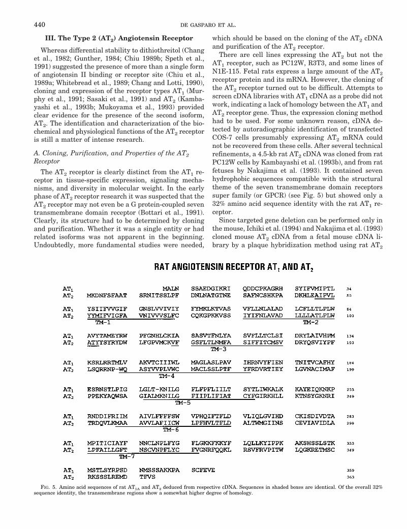
III. The Type 2 (AT2) Angiotensin Receptor
Whereas differential stability to dithiothreitol (Changet al., 1982; Gunther, 1984; Chiu 1989b; Speth et al.,1991) suggested the presence of more than a single formof angiotensin II binding or receptor site (Chiu et al.,1989a; Whitebread et al., 1989; Chang and Lotti, 1990),cloning and expression of the receptor types AT1 (Mur-phy et al., 1991; Sasaki et al., 1991) and AT2 (Kamba-yashi et al., 1993b; Mukoyama et al., 1993) providedclear evidence for the presence of the second isoform,AT2. The identification and characterization of the bio-chemical and physiological functions of the AT2 receptoris still a matter of intense research.
A. Cloning, Purification, and Properties of the AT2Receptor
The AT2 receptor is clearly distinct from the AT1 re-ceptor in tissue-specific expression, signaling mecha-nisms, and diversity in molecular weight. In the earlyphase of AT2 receptor research it was suspected that theAT2 receptor may not even be a G protein-coupled seventransmembrane domain receptor (Bottari et al., 1991).Clearly, its structure had to be determined by cloningand purification. Whether it was a single entity or hadrelated isoforms was not apparent in the beginning.Undoubtedly, more fundamental studies were needed,
which should be based on the cloning of the AT2 cDNAand purification of the AT2 receptor.
There are cell lines expressing the AT2 but not theAT1 receptor, such as PC12W, R3T3, and some lines ofN1E-115. Fetal rats express a large amount of the AT2
receptor protein and its mRNA. However, the cloning ofthe AT2 receptor turned out to be difficult. Attempts toscreen cDNA libraries with AT1 cDNA as a probe did notwork, indicating a lack of homology between the AT1 andAT2 receptor gene. Thus, the expression cloning methodhad to be used. For some unknown reason, cDNA de-tected by autoradiographic identification of transfectedCOS-7 cells presumably expressing AT2 mRNA couldnot be recovered from these cells. After several technicalrefinements, a 4.5-kb rat AT2 cDNA was cloned from ratPC12W cells by Kambayashi et al. (1993b), and from ratfetuses by Nakajima et al. (1993). It contained sevenhydrophobic sequences compatible with the structuraltheme of the seven transmembrane domain receptorssuper family (or GPCR) (see Fig. 5) but showed only a32% amino acid sequence identity with the rat AT1 re-ceptor.
Since targeted gene deletion can be performed only inthe mouse, Ichiki et al. (1994) and Nakajima et al. (1993)cloned mouse AT2 cDNA from a fetal mouse cDNA li-brary by a plaque hybridization method using rat AT2
FIG. 5. Amino acid sequences of rat AT1A and AT2 deduced from respective cDNA. Sequences in shaded boxes are identical. Of the overall 32%sequence identity, the transmembrane regions show a somewhat higher degree of homology.
440 DE GASPARO ET AL.

cDNA as a probe. A mouse AT2 genomic 4.4-kb DNAfragment was cloned by Ichiki et al. (1994) and Koike etal. (1994). Since these studies show that the entire cod-ing sequence is contained in one exon, the human AT2coding sequence (Tsuzuki et al., 1994) or fragments con-taining more extensive sequences could be cloned (Mar-tin et al., 1994; Koike et al., 1994). The genomic DNA ofall three species consist of three exons with an uninter-rupted coding region being confined to the third exon.The genes encoding the AT2 receptor are localized inhuman chromosome Xq22-q2, rat chromosome Xq3, andmouse chromosome X (Koike et al., 1994; Hein et al.,1995b; Tissir et al., 1995). The localization in the longarm of the X chromosome throughout species may haveimportant implications in targeted gene deletion as wellas in the genetics of the AT2 receptor gene.
The open reading frame of AT2 cDNA encodes 363amino acid residues in all three species with 99% se-quence identity between rat and mouse and 72% identitybetween rat and human. The divergence between rodentand human AT2 occurs mainly in the N-terminal region.AT1 and AT2 receptors have only 33 to 24% amino acidsequence identity (Kambayashi et al., 1993b; Nakajimaet al., 1993). The homology was mainly localized in thetransmembrane hydrophobic domains, which are be-lieved to form seven transmembrane helical columns.Residues located in these helical column domains andconsidered to be essential for Ang II binding to the AT1receptor are also preserved in the AT2 receptor. Theseinclude Lys118 (Lys102 of AT1) at the top of TM3, Arg183
(Arg167 of AT1) at the top of TM4 and Lys216 (Lys199 ofAT1) and more.
Almost complete divergence between AT1 and AT2receptors is seen in the third intracellular loop and moreextensive differences in the carboxyl terminal tail (C-tail). The amino acid sequence of the rat AT2 receptorindicates that it has five potential N-glycosylation siteswith the consensus amino acid sequences -Asn-X-Ser/Thr. It is possible that carbohydrate chains would giverise to increased and diverse molecular weights of theAT2 receptor. Southern hybridization analysis of restric-tion enzyme fragments of rat genomic DNA using ratAT2 cDNA as a probe did not produce evidence for asubtype of the AT2 receptor. Thus, the molecular weightdivergence for the AT2 receptor ranging from 60 to 140kDa may be due to a difference in the extent of glycosyl-ation, as shown by Servant et al. (1994).
An earlier discriminator of the AT1 and AT2 receptors,before the isoform-specific antagonist became available,was the reducing agent dithiothreitol (DTT). The DTTsensitive Ang II receptor turned out to be the AT1 recep-tor, whereas DTT increased the ligand binding capabil-ity of the AT2 receptor and the AT2 receptor remainedstable for hours (Chang, Lotti, Keegan, 1982; White-bread et al., 1989; Chang and Lotti, 1990; Speth et al.,1991). This property was exploited for the direct purifi-cation of the AT2 receptor protein (see below). Another
discriminator is Sar-p-benzoylphenylalanine8-angioten-sin II, which can be used for selective labeling of AT2 bya photoaffinity technique. Although it binds to the bo-vine adrenal AT1 receptor with a Kd value of 6.5 nM andhuman myometrium with a Kd value of 0.39 nM, itscovalent binding upon illumination takes place onlywith the AT2 receptor (Bosse et al., 1990). The selectivelabeling of the AT2 receptor is in contrast to anotherphotoaffinity labeling peptide, Sar1-D-azido Phe8-Ang II,which is specific for the AT1 receptor (Guillemette et al.,1986). The 125I-radiolabeled peptide was used to visual-ize AT2 receptors in various cells to determine theirmolecular sizes by SDS gel electrophoresis. Cells knownto contain the AT2 receptor produced radiolabeled AT2receptor bands of 68 kDa (human uterine myometrium),91 kDa (R3T3 cells), and 113 kDa (PC12 cells). However,endoglycosydase treatment reduced the myometrial 68-kDa receptor to 40- and 31-kDa proteins, the 91-kDaR3T3 receptor was reduced to 46- and 31-kDa and the113-kDa PC12 AT2 receptor to 55 and 31 kDa. Theseresults may involve artifacts of proteolytic digestion.More decisive evidence for glycosylation diversity camefrom the inhibition of N-glycosylation by treatment ofrat PC12 cells with tunicamycin (Servant et al., 1996).The 140-kDa AT2 receptor in untreated PC12 cells wasreduced to 63- and 47-kDa glycosylated and 32-kDa un-glycosylated receptors. An interesting byproduct of thesestudies is the observation of nonglycosylated receptorexpression on the cell surface.
The structural diversity of the AT2 receptor is still acomplex and unresolved issue. Attempts have beenmade to purify the AT2 receptor protein (Ciuffo et al.,1993a) by combining affinity chromatography onCGP42112-Sepharose and size separation by gel filtra-tion and SDS gel electrophoresis from AT2 solubilized byCHAPS from neonatal rat kidney which had a 71-kDamolecular mass. The murine neuroblastoma cell lineN1E-115 cell line contains both AT1 and AT2 receptors.The latter increases upon cellular differentiation. Ang IIreceptors were solubilized from membranes by the de-tergent CHAPS (1%). The labile AT1 receptor could beremoved by denaturation and the AT2 receptor was af-finity purified on an Ang II-Sepharose affinity column bySiemens et al. (1991). They found that the AT2 receptorfrom N1E 115 can be separated to two different forms,peak I and peak III, by a Sepharose column chromatog-raphy. Peak III seemed to be an AT2 receptor whoseligand binding activity is enhanced by the reducingagent DTT, but the stable GTP analog, GTPgS, did notsuppress the binding. Ligand binding to a receptor un-der peak I was clearly reduced by GTPgS, but treatmentwith DTT reduced the ligand binding function (Siemenset al., 1994b). The peak I components consisted of twoproteins 110 and 60 kDa. It has been proposed that the110 kDa is a dimer of 60 kDa that is stabilized by theagonist (Yee et al., 1994). Interestingly, stabilization ofthe AT2 receptor on the cell membrane of R3T3 cells by
ANGIOTENSIN RECEPTORS 441

an as yet undefined endogenous ligand, which is notidentical with Ang II, has recently been described byCsikos et al. (1998).
B. Regulation of the AT2 Receptor
The AT2 receptor gene is expressed ubiquitously at avery high level in the fetus and it declines precipitouslyafter birth in many but not all tissues. In the skin, forinstance, it decreases to undetectable levels, but in cer-tain tissues such as the adrenal and heart, the declinestops at certain levels, and AT2 receptor expression per-sists for the remainder of life. In uterine myometrium,the AT2 receptor is expressed under nonpregnant condi-tions and declines during pregnancy, but returns to non-pregnant levels after parturition (de Gasparo et al.,1994). These few examples demonstrate clearly that theAT2 receptor expression is regulated. Theoretically, suchregulatory changes may be controlled by transcriptionand/or by changing the stability of mRNA.
Post-transcriptional (translational) regulation alsoseems to take place. After confluency, the AT2 bindingsites increase in R3T3 cells without an increase inmRNA (Dudley et al., 1991), whereas mRNA is low inthe growth phase and increases in the preconfluentstate. The AT2 receptor in these cells may signal toactivate protein phosphotyrosine phosphatases (PTP),which inactivates mitotic or hypertrophic growth. Tele-ologically, its activity should then be expressed more orless constantly in normal nongrowing cells. That thisreceptor lacks an internalization and desensitizationmechanism is compatible with its role to maintain dif-ferentiated cells in a quiescent state (Unger, 1999).
Attempts to investigate the transcriptional regulationhave begun. AT2 genomic DNA have been cloned frommouse (Nakajima et al., 1993; Ichiki et al., 1994) andhumans (Koike et al., 1994). The behavior of the AT2receptor gene expression in rat vascular smooth musclecells from the spontaneously hypertensive (SHR) andWistar Kyoto or Sprague-Dawley rats are markedly dif-ferent. This finding is in line with the observation thatAT2 receptor binding in endothelial cells from SHR ismarkedly higher than in those from Wistar Kyoto rats(Unger, 1999). The 59-flanking region of rat genomicDNA was also cloned to determine its functional ele-ments (Ichiki and Inagami, 1995a).
The expression of the AT2 receptor was shown to bedependent on growth factors or growth states. In mouseR3T3 cells, AT2 mRNA is expressed when growth isarrested in the confluent state and is decreased in thevigorous growth phase (Dudley et al., 1991; Dudley andSummerfeldt, 1993). Prolonged serum depletion in thepresence of insulin or IGF-1 stimulates the expression ofAT2 receptor in vascular smooth muscle cells, whereasserum growth factors, like PDGF, lysophospatidic acid,and phorbol ester rapidly abolished AT2 receptor expres-sion (Kambayashi et al., 1993a, 1996). Similar observa-tions with respect to IGF-1 as well as interleukin-1b
(IL-1b) were made in R3T3 fibroblast cells (Ichiki et al.,1995; Kambayashi et al., 1996). In contrast, growth fac-tors (phorbol ester, lysophosphatidic acid, and basic fi-broblast growth factor) markedly suppressed mouse AT2mRNA expression in the fibroblast cells but did notcompletely eliminate it as they did in vascular smoothmuscle cells (Ichiki et al., 1995a,b, 1996).
The transcriptional activity of the 59-flanking regionof the mouse AT2 receptor gene was investigated furtherby using constructs consisting of truncated 59-flankingsequences (up to 1.2 kb) fused to the luciferase gene inR3T3 cells. An AP-1 site may transmit the suppressiveeffects of noninsulin growth factors. Thus, PKC-activat-ing growth factors and phorbol esters rapidly down-regulate the AT2 receptor expression (Kambayashi etal., 1993a; Kizima et al., 1996). The C/EBP site maymediate the positive transcription effect of IL-1b as itstimulates the expression of C/EBPb and g (Ichiki andInagami, 1995a,b, 1996).
AT2 receptor expression by insulin or IGF-1 is inter-esting in that the process has a very slow onset, takingseveral days. A putative insulin response sequence (IRS)is located at 2126 to 2117 of the 59-flanking region ofthe rat AT2 receptor gene. This sequence TAATGTTTTGis 80% homologous to the IRS in the phosphoenolpyru-vate carboxykinase gene TGGTGTTTTG (O’Brien andGranner, 1991). An interesting aspect of the effect ofinsulin is that the aorta of obese (fa/fa) Zucker rats,which have about 10 times higher plasma insulin con-centrations than Sprague-Dawley rats, expresses AT2mRNA at a level clearly detectable by Northern blotting,whereas it was not detectable by the Northern blottingin the aorta of Sprague-Dawley rats (Kambayashi et al.,1996).
In these studies, an enhancer activity of the AP-1,C/EBP, NF/IL-6, IRS was not examined rigorously. Ho-riuchi et al. (1995) demonstrated by elegant methodsthat interferon regulatory factor IRF-2 attenuated AT2receptor expression, both in the confluent and growingcells, whereas IRF-1 enhanced AT2 expression in conflu-ent cells only. Several IRF binding motifs were found inthe promoter in positions between 2453 and 2225.Whether these elements alone account for up-regulationis not yet known. Tissue-specific expression and wide-spread low level expression of the AT2 receptor ratherpoint to complex mechanisms of AT2 receptor regulation.
C. AT2 Receptor Diversity
Although the AT2 receptor was cloned and purified,there remains a nagging question as to whether the AT2receptor is a single entity, because there is evidence fordivergence in the molecular and functional properties(Siemens et al., 1994a). As discussed above, the largedisparity of molecular size was attributed to a differencein the size of carbohydrate structure (Servant et al.,1994) but it could be also due to dimerization or oli-gomerization (Yee et al., 1994). Although DTT enhances
442 DE GASPARO ET AL.

ligand binding of the AT2 receptor in most tissues inves-tigated, in some this is not the case (Tsutsumi andSaavedra, 1992b). Most intriguing is the observationthat agonist binding of typical AT2 receptors, includingthose that had been cloned and expressed, is not sup-pressed by GTPgS, or pertussis toxin. Some AT2 recep-tors in rat locus ceruleus and thalamic or geniculatenuclei are markedly affected by these reagents, whichaffect the heterotrimeric G proteins, Gi and Go. On theother hand, GTPgS has no effect on the AT2 receptor inR3T3 cells or the inferior olive (Table 3). The lack ofeffect has also been seen in other cells expressing theAT2 receptor (Tsutsumi and Saavedra, 1991). Moreover,AT2 mRNA was not detected by in situ hybridizationusing a sequence of the cloned AT2 receptor as an anti-sense oligonucleotide probe (Johren et al., 1996). Thequestion as to whether diversity in the functional prop-erties of the AT2 receptor in different types of cells andtissues is due to another molecular species, or to addi-tional factors associated with it, may be addressed byinvestigating various modes of the signal transductionmechanism of the AT2 receptor associated with the mo-lecular diversity.
D. Targeted AT2 Receptor Gene Overexpression andDeletion
The use of AT2 receptor antagonists has provided ev-idence for a variety of functions of the AT2 receptor inseveral different types of cells and tissues includingcardiovascular, renal, adrenal, central nervous, as wellas dermal mesenchymal systems. Gene manipulation toincrease, decrease, or abolish the expression of a gene inquestion are effective approaches to assess the patho-physiological roles of the gene or its product. Transgenictransfection of AT2 receptor expression vectors intoCOS-7 cells (Kambayashi et al., 1993b; Mukoyama et al.,1993), and vascular smooth muscle in vivo (Nakajima etal., 1995) in “gain of function” approach have proven tobe efficient methods. Whereas cardiac targeted overex-pression of the AT2 receptor in mice did not cause obvi-ous morphological or functional changes, Ang II infusionparadoxically decreased blood pressure and produced anegative chronotropic effect (Masaki et al., 1998). More-over, Tsutsumi et al. (1999) reported stimulation of bra-dykinin activity and nitric oxide production in AT2transgenic mice following inhibition of the Na1/H1 ex-changer.
The targeted gene deletion (AT2 knockout mouse orAT2 null mouse) method produced a very interestingstrain of mice (Hein et al., 1995a; Ichiki et al., 1995a). Incontrast to the hypertension and impaired vascular re-sponses observed in AT1-deficient mice, knockout of theAT2 receptor leads to elevation of blood pressure andincreased vascular sensitivity to Ang II (Hein et al.,1995a; Ichiki et al., 1995). This has suggested that theAT2 receptor may exert a protective action in blood pres-sure regulation by counteracting AT1 receptor function.Such an action could be exerted in part by reducedexpression of the AT1 receptor, which is increased in thevascular smooth muscle of AT2-deficient mice (Tanakaet al., 1999). However, the sustained hypersensitivity toAng II in such animals is also attributable to loss of thecounter-regulatory action of renal bradykinin and cyclicGMP formation (an index of NO production) (Siragy etal., 1999). The relative contributions of these two factorsto AT2-dependent vascular regulation, and the extent towhich AT2 receptor deficiency could contribute to sus-tained blood pressure elevation, as in human hyperten-sion, have yet to be determined.
The AT2 receptor null mouse Agtr2(2/2) also showedbehavioral changes and proclivity to have renal dis-eases. However, despite its massive expression in themesenchymal tissues of skin, kidney, as well as in thebrain and heart in fetal stages of development, the AT2receptor null mouse does not show discernible grossanatomical defects or deformity to date. The early fetalform of the kidney mesonephros is programmed to dis-appear during the fetal development. Its tubular tissueis densely surrounded by mesenchymal cells expressingAT2 mRNA (Kakuchi et al., 1995). However, its disap-pearance also takes place in the fetus of the AT2 receptornull mouse. The medulla of the metanephros is alsofilled with AT2 receptor-expressing mesenchymal cells,which undergo apoptosis as tubulogenesis proceeds(Kakuchi et al., 1995). Again, kidney formation in theAT2 receptor null mice takes place normally and new-born mice grew to adult mice with a normal pair ofkidneys (Ichiki et al., 1995b). These studies with AT2receptor gene deleted mice indicate that the AT2 recep-tor is not essential for apoptosis in nephrogenesis.
Functionally, the AT2 receptor seems to exert a hypo-tensive effect—although this function is still discussedcontroversially—and to blunt the sensitivity of AT1 re-ceptors to Ang II. Its role in the regulation of cardiac,
TABLE 3Binding characteristics of the AT2 receptora
TreatmentAtypical AT2 AT2A
Ventral Thalamic Nuclei Medial Geniculate Nucleus Locus Ceruleus Inferior Olive
GTPgS effect Kd: 100% increase Slight increase 120% increase No increasePertussis toxin
treatmentAgonist binding reduced to 45% Agonist binding reduced to 63% Agonist binding reduced to 45%
a Taken from Tsutsumi and Saavedra (1992a).
ANGIOTENSIN RECEPTORS 443

renal, and adrenal function is still under investigation.As for the renal function, the role of the AT2 receptor asa regulator (or modulator) of pressure natriuresis is stillcontroversial (Keiser et al., 1992; Lo et al., 1995; Siragyand Carey, 1996). Whereas studies with AT2 receptoragonists or antagonists in intact rats suggested an an-tidiuretic and antinatriuretic effect of the AT2 receptor,results in AT2 receptor knockout mice showed the oppo-site effects (Siragy et al., 1999).
Although AT2 receptor null mice did not produce grossmorphological abnormalities, vascular differentiationdriving the expression of the constituents of the contrac-tile apparatus was altered. The expression of h-caldes-mon and calponin are delayed in the aorta, suggestingthat the AT2 receptor enhances the differentiation ofvascular smooth muscle cells and is involved in vascu-logenesis (Yamada et al., 1999). In addition, more de-tailed studies with a larger population of AT2 receptornull mice are producing exciting evidence that a loss ofthe AT2 receptor in the AT2 null mice results in thedevelopment of congenital urinary tract anomalies witha 23% penetrance.
1. Behavioral Changes in AT2 Receptor Null Mice. Inthe brain, the AT2 receptor is expressed in distinct areas(Obermuller et al., 1991), prominently in the locus cer-uleus (Rowe et al., 1990a,b; Saavedra, 1992). The locusceruleus contains a high concentration of noradrenergicnerve endings that release norepinephrine upon treat-ment with Ang II (Sumners and Phillips, 1983) andsends out long noradrenergic projections to the cerebralcortex, hypothalamus, and hippocampus. The AT2 recep-tor is also expressed in the central amygdaloid nucleus(Song et al., 1992), and/or medial amygdaloid nucleus(Saavedra, 1992). Loss of the AT2 receptor in these areascould entail behavioral effects.
AT2 receptor null mice showed a markedly reducedexploratory behavior when placed in a new environmentwith ambulation reduced by 40% (Hein et al., 1995a;Ichiki et al., 1995). After water deprivation, AT2 nullmice show a greater stimulation of dipsogenesis (Hein etal., 1995a).
E. Signaling Mechanisms of the AT2 Receptor
Studies on hormone receptor signaling mechanismsare aimed at explaining cellular and physiologic re-sponses by intracellular biochemical processes. As Nah-mias and Strosberg (1995) point out, the search for ma-jor physiologic functions of the AT2 receptor isprogressing and several interesting clues are at hand.The AT2 receptor seems to open a delayed rectifier po-tassium channel at least in hypothalamic neuronal tis-sues (Kang et al., 1994, 1995), to close a T-type Ca21
channel (Buisson et al., 1992, 1995), to suppress tissueand cellular growth (Nakajima et al., 1995; Meffert etal., 1996; Munzenmaier and Green, 1996; Stoll et al.,1995; Tsuzuki et al., 1996a,b), to induce (neuronal) celldifferentiation (Laflamme et al., 1996; Meffert et al.,
1996; Gallinat et al., 1997; Stroth et al., 1998; Cote et al.,1999; Gendron et al., 1999) and to support apoptosis(Yamada et al., 1996; Chamoux et al., 1999; Gallinat etal., 1999). In addition, the AT2 receptor may exert hy-potensive effects (Scheuer and Perrone, 1993; Ichiki etal., 1995) and appears to influence behavior (Hein et al.,1995a; Ichiki et al., 1995).
The consensus among investigators working with cellsexpressing the AT2 receptor exclusively without the AT1
receptor, such as R3T3 (Dudley et al., 1990, 1991),PC12W (Bottari et al., 1991, 1992; Leung et al., 1992;Webb et al., 1992), ovarian granulosa cells (Pucell et al.,1991), and COS-7 cells expressing cloned AT2 cDNA(Kambayashi et al., 1993b; Mukoyama et al., 1993) isthat the AT2 receptor does not modulate cytosolic Ca21
or cyclic AMP, which are sensitive indicators of theheterotrimeric Gq protein-coupled phospholipase Cb ac-tivation and Gs or Gi-coupled activation or inhibition ofadenylyl cyclase, respectively. Furthermore, agonistbinding did not induce receptor internalization (Dudleyet al., 1991; Csikos et al., 1998), nor did treatment ofplasma membrane with stable GTP analogs (e.g.,GTPgS or GDP-NH-P) result in a decrease of ligandbinding. Heterotrimeric G proteins show a binding of astable analog of GTP upon stimulation by agonist totheir respective receptor. Stimulation of the AT2 recep-tor in membranes from human myometrium, bovine cer-ebellar cortex, and rat adrenal glomerulosa did not re-sult in an increase in the binding of [35S]GTPgS (Bottariet al., 1991). These findings, obtained before the molec-ular structure of the AT2 receptor had been disclosed,led the authors to propose that the AT2 receptor was notcoupled to a heterotrimeric G protein since the above aregeneral criteria for the family of G protein-coupled re-ceptors. Thus, even though the AT2 receptor, as we knowtoday, has basic structural features commonly shared byGPCR (seven transmembrane domain receptors), it didnot reveal any functional features ascribable to thisclass of receptors.
An elaborate study on the binding of the AT2 labeledwith 125I-Sar1-Ang II indicated that the AT2 receptorbinds to Gia2 or Gia3 (Zhang and Pratt, 1996). However,the mode of interaction may be somewhat different fromthose seen with other Gi-coupling receptors.
There is evidence that the AT2 receptor inhibits theGq-coupled phospholipase C activation by the AT1 recep-tor. Wounded rat skin expresses both AT1 and AT2 re-ceptors (Kimura et al., 1992; Viswanathan and Saave-dra, 1992). When rat skin slices are incubated with AngII (1027–1024 M), the production of inositol-monophos-phate is stimulated and is abolished by losartan. TheAT1 receptor-mediated inositol monophosphate produc-tion is markedly potentiated by the AT2 antagonistPD123319 at 1023 M (Gyurko et al., 1992). Although theresults were obtained at high concentrations of Ang IIand AT2 antagonist, these observations indicate that
444 DE GASPARO ET AL.

AT2 receptor could negatively modulate the AT1 recep-tor-mediated phospholipase C activation.
The effect of Ang II on cGMP in AT2 receptor-express-ing cells has been controversial (Dudley et al., 1991;Sumners et al., 1991; Sumners and Myers, 1991; Bottariet al., 1992; Leung et al., 1992; Webb et al., 1992; Bre-chler et al., 1993; Kambayashi et al., 1993b; Reagan etal., 1993b; Israel et al., 1995). The reason for this dis-crepancy is not yet clear. More recent in vivo studies onthe effect of the AT2 receptor in the kidney indicate thatthe stimulation of the AT2 receptor increases renal med-ullary and cortical interstitial cGMP by a bradykinin-dependent mechanism (Siragy and Carey, 1996,1997a,b; Siragy et al., 2000). Similar observations weremade by Golke et al. (1998) in the aorta of stroke proneSHR infused with Ang II, and some therapeutic effects ofAT2 receptor blockade are clearly mediated by the AT2receptor and kinin stimulation (Liu et al., 1997). Miceoverexpressing the AT2 receptor have confirmed thesefindings (Tsutsumi et al., 1999). Thus, the signalingmechanisms of the AT2 receptor have been less clearlydefined as yet compared with those of the AT1 receptor.In addition to the above-mentioned signaling pathwaysincluding Gi protein coupling and cGMP (NO) genera-tion, activation of protein tyrosine phosphatase, serine/threonine phosphatase 2A (PP2A), protein kinase phos-phatase (MKP-1) acting on both phosphotyrosine andphosphothreonine as well as SHP-1 tyrosine phospha-tase have been reported (for details see Blume et al.,1999; Horiuchi et al., 1999; Millat et al., 1999).
1. Dephosphorylation and Inactivation of the Mitogen-Activated Protein Kinases ERK1 and ERK2. MAPKplays a key role in cellular proliferation, and its activa-tion can be detected by highly sensitive methods. VSMCtransfected with an AT2-expressing vector responded toAng II by a very slight increase in MAPK activity,whereas untransfected VSMC clearly increases MAPK.The AT2 antagonist markedly increased the MAPK ac-tivity (both ERK1 and ERK2 isoenzymes) close to thelevel attained in untransfected VSMC when stimulatedwith 21027 M Ang II. These results indicate that stim-ulation of the AT2 receptor by Ang II suppresses the AT1receptor-mediated activation of MAPK (Nakajima et al.,1995). Although adult aortic media do not seem to ex-presses AT2 at measurable levels, the carotid artery mayexpress it to a limited degree (Viswanathan et al., 1991).AT2 receptor expressed, even in a limited quantity, atthe edge of neointima, (or, for that matter, in a healingwound) may suppress MAPK in the growing edge. Theserine/threonine phosphatase, PP2A, which is activatedby Ang II stimulation of the AT2 receptor in the neonatalhypothalamic neurons, was recently shown to inactivateMAPK following the stimulation of the AT1 receptor. Inthe presence of the AT2 receptor antagonist, PD123319,MAPK activity was markedly enhanced. Thus, the AT2receptor is able to block MAPK activation either by thedephosphorylation of serine/threonine phosphate by
PP2A or tyrosine phosphate by PTPs, most likely byMAPK phosphatase, MKP-1 (Huang et al., 1996).
The influence of the AT2 receptor on MAPK activitymay also depend on the biological program engaged bythe receptor under defined experimental conditions.Thus in PC12W cells expressing AT2, but not AT1, re-ceptors, treatment with Ang II under conditions allow-ing for cell differentiation induced a short-lasting in-crease in ERK1 and ERK2 activity as was seen withNGF, a classical differentiation factor. Costimulationwith NGF and Ang II led to initial MAPK stimulationbut the subsequent NGF-induced plateau of ERK1 andERK2 stimulation was suppressed by the AT2 receptor(Stroht et al., 2000). Altered ERK activity was also ob-served in the heart of AT2 transgenic mice suggestingthat the ERK inactivated by the AT2 receptor has aphysiological role in vivo (Masaki et al., 1998).
Further evidence for a Gi-coupled activation of PTPwas obtained in vascular smooth muscle cells in culture,even if the AT2 receptor was not expressed in these cells.Instead of using Ang II to stimulate Gi, it was activatedby a 22 amino acid residue peptide with the sequence ofthe third intracellular loop of the AT2 receptor protein,which was transferred into the cells expressing the ATreceptor by LipofectAMINE liposome. Cells which re-ceived the AT2-related peptide showed small but signif-icant attenuation of serum-stimulated MAPK and thy-midine incorporation. The suppression of thymidineincorporation was reversed by pretreatment of cells withpertussis toxin, which indicates the role of a Gi or Goprotein. The inhibition of MAPK activation by serumwas also reversed by sodium orthovanadate, a tyrosinephosphatase inhibitor but not by okadaic acid, a serine/threonine phosphatase inhibitor, which indicates thatthe third intracellular loop of AT2 receptor activates aPTP via a Gi or Go protein (Hayashida et al.,1996). In theneuroblastoma cell line, N1E-115, the AT2 receptor hasbeen found to activate the catalytic activity of SH-PTP1,a soluble PTP implicated in termination of signaling bycytokine and growth factor receptors, resulting in ERKinactivation (Bedecs et al., 1997).
Recently, ceramide, which is linked to phosphataseactivation, was proposed to be the second messenger inAT2 receptor-mediated apoptosis, (Gallinat et al., 1999;Lehtonen et al., 1999), and pertussis toxin and or-thovanadate blocked its production. A negative cross-talk appears to exist between AT2 and AT1 receptors notonly on a functional level as has been observed by vari-ous authors (see Unger, 1999) but also on the level ofintracellular signaling. For instance, stimulation by AngII, interferon, EGF, or PDGF of rat adult vascularsmooth muscle cells transfected with AT2 receptor cDNAinhibits AT1 receptor-mediated ERK activation and ty-rosine phosphorylation of STAT without influence onJanus kinase (Horiuchi et al., 1999). However, theseresults also indicate that the Gi-coupled activation ofprotein tyrosine phosphatase(s) or protein serine/threo-
ANGIOTENSIN RECEPTORS 445

nine phosphatase is not targeted narrowly to specificchannel proteins and that many other proteins can betheir substrates.
2. Activation of Phospholipase A2 and ProstacyclinGeneration. In contrast to cells in which the AT2 recep-tor is the predominant or exclusive Ang II receptor,cardiac ventricular myocytes are interesting as they ex-press both AT1 and AT2 receptors (Busche et al., 2000).In the hypertrophic ventricles of SHR and two-kidneyone-clip hypertensive rats, AT1 receptor expression isincreased along with AT2 receptor density (Suzuki et al.,1993). Thus, the heart offers unique materials for stud-ies on interactive regulation of AT1 and AT2 receptors.The AT2 receptor mediates sustained arachidonic acidrelease in isolated pure cardiac myocytes, and this effectis completely blocked by the AT2-specific antagonistPD123317. Losartan suppresses it by about 50%. On theother hand, the AT1 receptor supports largely the re-lease of inositol phosphates. The fact that 1 mM DTTdoes not completely abolish arachidonic acid release, butcompletely eliminates inositol phosphate, supports therole of the AT2 receptor in the activation of phospho-lipase A2 but not phospholipase C (Lokuta et al., 1994).
Kohout and Rogers (1995) found evidence indicatingthat the AT2 receptor-mediated release of arachidonicacid may contribute to the activation of Na1/HCO2 sym-porter system (NBC), which increases the pH value ofmyocytes by 0.08 pH unit. This stimulation of the sym-porter system by Ang II can be blocked by the AT2blocker PD123319, but not by the AT1 blocker losartan.Furthermore, superfusion of myocytes with exogenousarachidonic acid (5 mM) mimicked the Ang II-mediatedalkalinization. Thus, the arachidonate release and sym-porter activation is a unique function of the AT2 receptorin the heart. These observations were extended by Sand-mann et al. (1998) in an ex vivo study in rats aftermyocardial infarction. Whereas pretreatment with anACE inhibitor prevented the postinfarct up-regulation ofboth ion transporter systems, the Na1/H1-exchanger(NHE-1) and NBC, in cardiac tissue, the AT1 receptorantagonist, valsartan, selectively blocked the increase inNHE-1 and the AT2 antagonist, PD123319, selectivelyblocked the increase of the NBC.
AT2 receptor-dependent production of prostacyclin(PGI2) was reported in differentiated adipocytes Ob1771in culture. The PGI2 formation is seen only in differen-tiated Ob1771 cells, and PGI2 thus formed induces dif-ferentiation of undifferentiated Ob1771 cells in cocul-ture by a paracrine mechanism. This action is blocked byPD123177, but not by losartan, indicating that the pros-tacyclin formation is mediated by the AT2 receptor (Da-rimont et al., 1994). The mechanism involved in thissignaling pathway remains to be clarified.
In summary, diverse AT2 receptor signaling pathwayswere unveiled, which include activation of serine/threo-nine phosphatase PP2A and subsequent opening of thedelayed rectifier K1-channel, activation of cytosolic
PTPs, which may lead to closing of the T-type Ca21
channel, inactivation, but in some cases also transientactivation, of MAPK, the inhibitory effect presumablythrough PP2A, MKP-1, or other PTP, and the activationof phospholipase A2 (Nouet and Nahmias, 2000). Withrespect to the specific pathways or substrates involvedin AT2 receptor-mediated intracellular signaling, thereare still numerous unresolved problems such as GTPgSsensitivity, PTP activation, or cGMP production.
F. Tissue Distribution of the AT2 Receptor
To identify the physiologic functions of the AT2 recep-tor, investigations were initiated on tissue-specific ex-pression, changes in AT2 expression in relation to onto-genic stages and tissue development, and identificationof cell types in vivo and in cell lines carrying the AT2receptor. In these studies, the use of isoform selectiveantagonists for the AT1 and AT2 receptors enabled in-vestigators to detect biphasic binding of Ang II, whichindicated the coexistence of AT1 and AT2 receptors.Later, cloning of AT2 cDNA allowed for specific identifi-cation of AT2 mRNA expressed in various tissues (Kaku-chi et al., 1995; Shanmugam et al., 1995; Johren et al.,1996). Distribution of the AT2 receptor appears to betissue- and species-specific. Both AT1 and AT2 receptorsare expressed in the adrenal gland at varying ratios indifferent regions. In the rat adrenal medulla, the ratio ofAT1 to AT2 receptors was approximately 20:80 (Changand Lotti, 1990), whereas in rat, rabbit, monkey andhuman adrenal cortex, the AT2 receptor comprised ;10to 40% of the total angiotensin binding sites (Chiu et al.,1989a; Whitebread et al., 1989; Chang and Lotti, 1990).Other tissues that expressed the AT2 receptor at a highproportion, as compared to the AT1 receptor, were thenonpregnant human uterus (Whitebread et al., 1989;Criscione et al., 1990; Bottari et al., 1991; de Gasparoand Levens, 1994); sheep uterine myometrium (Cox etal., 1993); bovine cerebellar cortex (Bottari et al., 1991);and rat ovarian follicular granulosa cells (Pucell et al.,1991). Species dependence exists, as only 40% and 60%of Ang II binding sites are AT2 in rat and rabbit uterus,respectively (Dudley et al., 1990; Bottari et al., 1991).The proportion of the AT2 receptor in the kidney cortexis less than 10% of the AT1 receptor in the rat andrabbit, and ;55% in monkey (Chang and Lotti, 1991).On the other hand, the heart in the rat, rabbit, andmonkey contains the AT2 receptor in small but finite andmeasurable amounts, which comprises about 30% of thetotal Ang II binding sites (Chang and Lotti, 1991). Therat and primate pancreas were found to contain AT1 andAT2 receptors (Chappell et al., 1992, 1994). Expressionof the AT2 receptor was particularly high in pancreaticacinar cells (Chappell et al., 1995). Table 4 summarizesadult and fetal tissues that express AT2 receptors.
Some fetal tissues express the AT2 receptor at highlevels. As summarized in Table 4, in many of thesetissues the AT2 receptor emerges on embryonic days 11
446 DE GASPARO ET AL.

to 13 (E11–E13) and reaches a maximal level on E19.The AT2 receptor then rapidly declines in newborn ani-mals to lower levels or to undetectable levels. As exam-ined by autoradiographic techniques and/or in situ hy-bridization, expression of the AT2 receptor wasparticularly dense in differentiated mesenchymes, suchas mesenchymal tissues in the tongue, subdermal, ands.c. regions of the skin and the diaphragm. In thesetissues, the preponderant fetal Ang II binding sites areAT2 (.97%) (Grady et al., 1991; Feuillan et al., 1993;Grady and Kalinyak, 1993; Shanmugam et al., 1995).The skin and tongue are the tissues in which fetal ex-pression of AT2 receptor is particularly intense, how-ever, rapid postpartum disappearance is seen. In manyother tissues where fetal AT2 receptor expression level isdetectable, a precipitous lowering of the AT2 receptor isobserved. The very dense expression of Ang II receptorsin rat fetal skin cells noted by Millan et al. (1989) seemsto be AT2 receptor (Feuillan et al.,1993). These cellswere isolated and cultured. Earlier passage cells expressmore AT2 than AT1 receptor. Switching of AT2 to AT1expression, or AT2-expressing cells to AT1-expressingcells seems to take place (Johnson and Aguilera, 1991).In some tissues, AT2 receptors reach undetectable levels(submucosal cells of the stomach and intestine or tra-chea). On the other hand, in certain tissues the processof rapid postpartum decrease is arrested, and the AT2receptor remains at detectable levels in the adrenal me-dulla, zona glomerulosa, (Shanmugam et al., 1995), pan-creas (Chappell et al., 1994), uterus (Cox et al., 1993),and heart (see below).
In the fetal kidney, AT2 receptor expression is seenmostly in the mesenchymal cells of differentiating cortexand medulla, which are surrounding glomeruli in thecortex and tubular tissues in the medulla (Kakuchi etal., 1995). The mesenchymal cells will undergo apoptosisand will be replaced by tubular tissues. The role of the
AT2 receptor in fetal development is not yet clearlyestablished. Although the apoptosis of the AT2 receptor-expressing cells in fetal tissue development is an attrac-tive concept, hemizygotic male AT2 receptor gene de-leted mice (the AT2 receptor gene is localized to theX-chromosome) and homozygotic female AT2 receptorgene null mice undergo normal fetal development, andnewborns and adults do not show any abnormality in thegross morphology of the skin, tongue, kidney, or adrenal.A delay in vasculogenesis was observed, however(Yamada et al., 1998).
Although in many tissues the AT2 receptor undergoesa unidirectional decrease in its expression, this receptoralso undergoes reversible changes. The myometria of thehuman, sheep, and rat express measurable AT2 recep-tors at a high level (Criscione et al., 1990; Bottari et al.,1991; Cox et al., 1993; de Gasparo et al., 1994). In sheeputerus, for instance, the ratio of AT1:AT2 receptor is15:85. In the pregnant ewe, the AT2 receptor is reducedby more than 90% and AT1 is reduced by 60% with theAT1 to AT2 ratio being reversed to 80:20. However, uponparturition, the AT2 receptor rapidly returns to the highprepregnancy levels (Cox et al., 1993). Similar observa-tion was made in human myometrium during pregnancy(de Gasparo et al., 1994). The AT2 receptor also emergesduring wound healing of the skin (Kimura et al., 1992;Viswanathan and Saavedra, 1992). This increase occursparticularly in the superficial dermis. A low level of AT2receptor expression was also seen in the neointimal tis-sue of rat carotid artery following balloon catheteriza-tion (Janiak et al., 1992; Viswanathan et al., 1994b).These changes in the uterus during pregnancy or inwound healing in the skin and brain indicate that theAT2 receptor has a definitive but not yet identified reg-ulatory function in these tissues.
1. Brain. Although circumventricular organs of thebrain respond to circulating Ang II, an endogenous brain
TABLE 4Tissue distribution of the AT2 receptor and ontogenic change
Tissues Fetus Newborn 3 wk 8 wk Reference
AdrenalCortex 1 1 1 1 Shanmugam et al., 1995Medulla 2 1 1 1 Feuillan et al., 1993
OvaryFollicular granulosa 1 1 1 Pucell et al., 1991
KidneyCortex 111 1 Shanmugam et al., 1995Medulla (outer strip) 1 11 Shanmugam et al., 1995; Kakuchi et al., 1995Heart 6 Shanmugam et al., 1995
UterusMyometrium 1Blood vessels
Heart 6 Shanmugam et al., 1995Aorta 1 1 Feuillan et al., 1997; Shanmugam et al., 1995Pancreas 1 Chappell et al., 1992Trachea 1 Shanmugam et al., 1995Stomach 1 Shanmugam et al., 1995Mesenchyme 1Skin 111 6 Grady et al., 1991; Shanmugam et al., 1995; Feuillan et al., 1993Tongue 111 6 Grady et al., 1991; Feuillan et al., 1993Skeletal muscle 1 Feuillan et al., 1993
ANGIOTENSIN RECEPTORS 447

renin-angiotensin system can generate Ang II in tissuesprotected from circulating Ang II by the blood-brainbarrier. Ang II generated by this system reacts withangiotensin receptors within the brain. The brain angio-tensin receptors have been studied and reviewed exten-sively by Gehlert et al. (1991a,b), Rowe et al. (1992),Saavedra (1992), Song et al. (1992), and Hohle et al.(1995). An expression of the AT2 receptor in fetal (E18)brain tissues was reported in several areas: inferiorolive, paratrigeminal nucleus, and hypoglossal nucleus.Saavedra (1992) determined AT1 and AT2 receptor ex-pression in various regions of the brain, and its agedependence in 2- and 8-week-old rat brains. AlthoughAT1 receptor expression did not show marked age de-pendence, the AT2 receptor showed a significant de-crease from 2 to 8 weeks of age (see Table 5).
Obermuller et al. (1991) investigated the distributionof Ang II receptor types in adult rat brain nuclei usingcompetitive radioligand binding. Whereas midbrain andbrain stem contained AT1 and AT2 receptors in compa-rable concentrations, AT1 receptors were by far predom-inant in several hypothalamic nuclei, although a limitedamount of AT2 receptors was detectable in most of them.Although minor differences may exist among these stud-ies, they share the following observations: AT2 receptorexpression is consistently high in the cerebellar nuclei,inferior olive, and locus ceruleus in the brain stem,
which is rich in noradrenergic neurons. In contrast tothe AT1 receptor, which participates in various centralcardiovascular functions and is expressed in the hypo-thalamus and in brain stem nuclei (nucleus of the soli-tary tract, dorsal motor nucleus of vagus at a low level),the AT2 receptor is much less present in distinct hypo-thalamic and brain stem nuclei associated with the reg-ulation of cardiovascular functions. The presence of theAT2 receptor in the dorsal motor nucleus of vagus is notcompletely settled.
Although the distribution of AT2 receptors in thebrain is well known, their effects are still not clear. Asoutlined above, brain AT2 receptors may play a role incognitive functions and certain types of behavior such asexploration or drinking (Hein et al., 1995a). On the otherhand, they may also antagonize the central effects ofangiotensin peptides in osmoregulation mediated viaAT1 receptors (Hohle et al., 1995, 1996). A possibly im-portant role of AT2 receptors in neuroregeneration andneuroprotection will be dealt with below.
2. Heart. Earlier studies suggested that myocytes ofrabbit and rat hearts contain AT1 and AT2 receptors incomparable quantities as shown in Table 6 (Rogg et al.,1990; Scott et al., 1992; Sechi et al., 1992b). However,questions remained as to the cell-specific localization ofthese receptors in situ. A recent study using single cellRT-PCR has demonstrated that in the adult rat about50% of cardiomyocytes contain the AT1 receptor,whereas the AT2 receptor is much more scarce with onlyabout 10% carrying this receptor type (Busche et al.,2000).
Autoradiographic studies of the rat heart show abouta 2-fold increase in AT1 and AT2 receptors after birth,and their receptor numbers are about equal from E16through 10 to 16 weeks of age (Sechi et al., 1992b). Theyare expressed in the myocardium of all four chambers,as well as in the vascular smooth muscles of the aortaand pulmonary arteries. However, very little ligandbinding was seen in the coronary artery. The conductionsystem (the atrioventricular and sino-aortic nodes) wasreported to contain both AT1 and AT2 receptors by Sechiet al. (1992b), whereas Saavedra et al. (1993), Brink etal. (1996), and Wharton et al. (1998), showed only AT1receptors in these tissues.
Primary culture of cardiomyocytes from neonatal ratleft ventricles showed about a 50% decrease in the AT2
TABLE 5Distribution of the AT2 receptor in the brain—effect of age
2-wk-old 8-wk-old
fmol/mg protein(mean 6 S.E.M.)
Regions containing only the AT2receptor
Persistent AT2 receptor with ageLateral septal nucleus 58 6 6 18 6 3Ventral thalamic nuclei 101 6 8 24 6 3Mediodorsal thalamic nucleus 165 6 11 38 6 13Locus ceruleus 289 6 19 98 6 13Principal sensory trigeminal
nucleus75 6 6 15 6 4
Parasolitary nucleus 220 6 15 57 6 14Inferior olive 1328 6 61 181 6 32Medial amygdaloid nucleus 159 6 8 94 6 9Medial geniculate nucleus 338 6 24 71 6 6
Transient expression of the AT2receptor
Anterior pretectal nucleus 53 6 8 N.D.Nucleus of the optic tract 101 6 13 N.D.Ventral tegmental area 101 6 11 N.D.Posteodorsal tegmental nucleus 110 6 21 N.D.Hypoglossal nucleus 141 6 11 N.D.Central medial and paracentral
thalamic nucleus202 6 14 N.D.
Laterodorsal thalamic nucleus 110 6 10 N.D.Oculomotor nucleus 98 6 13 N.D.
Regions containing both AT1 andAT2 receptors
Persistent AT2 receptor with ageSuperior collicus 145 6 5 65 6 8Cingulate cortex 19 6 4 8 6 4
Transient expression of the AT2receptor
Cerebellar cortex 59 6 6 N.D.
N.D., not determined.
TABLE 6Coexistence of AT1 and AT2 in cardiac tissues
% of AT1 1 AT2References
AT1 AT2
Rabbit ventricle 60 40 Rogg et al., 1990Rat (10-wk-old) 50 50 Sechi et al., 1992bMyocytes 65 35 Matsubara et al., 1994Fibroblasts .95 ,5 Villareal et al., 1993Fibroblasts (adult) 100
Bovine 67 33 Nozaki et al., 1994Human 33 69 Regitz-Zagrosek, 1995
448 DE GASPARO ET AL.

receptor from those of E19 fetuses, whereas the AT1
receptor did not decrease (Matsubara et al., 1994). Incontrast, the AT2 receptor in cultured fibroblasts col-lected from 1-day-old rat heart was less than 10% of theAT2 receptor in fibroblasts cultured from E19 fetalheart, whereas there was no change in AT1 receptornumber. In cardiac fibroblasts cultured from 7-day-oldrats, AT2 receptor density was negligible and the major-ity of the Ang II receptor population was reported to beAT1, whereas in cultured cardiomyocytes AT2 receptornumbers are maintained at a finite and measurablelevel (about 50% of AT1). Bovine and human ventricularand atrial myocardium also contain both AT2 and AT1
receptors (Rogg et al., 1991; Nozawa et al., 1994; Regitz-Zagrosek et al., 1995). Although the AT2 receptor isusually expressed at low density in adult, it is up-regu-lated to different extents in pathological circumstancessuch as cardiac hypertrophy, myocardial infarction, car-diomyopathy, and congestive heart failure (Matsubara,1998; Unger, 1999). Interestingly, both in nonfailing andexplanted end-stage human heart the AT2 receptor pop-ulation measured in a binding assay (65% of total Ang IIreceptor) is greater than AT1, and there may be a corre-lation between the density of the AT2 receptors and theseverity of heart failure (Rogg et al., 1996). Wharton etal. (1998) also observed a significant increased density ofhigh-affinity binding sites in endocardial, interstitial,perivascular, and infarcted regions of the ventricle ofpatients with end-stage ischemic heart disease or di-lated cardiomyopathy, greater than in adjacent nonin-farcted myocardium. The border zone between nonin-farcted and infarcted myocardium was rich inmicrovessels with perivascular AT2 receptors. Ohkuboet al. (1997) reported that in the heart of cardiomyo-pathic hamster (Bio 146), both AT1 and AT2 receptorswere increased during heart failure (153% and 72%,respectively). In human, the expression of the AT2 re-ceptor was markedly (3-fold) increased in patients withdilated cardiomyopathy at both protein and mRNA lev-els compared with patients with acute or well organizedold myocardial infarction (Tsutsumi et al., 1998). Incontrast, the AT1 receptor expression was significantlydown-regulated. The AT2 receptor sites were highly lo-calized in the interstitial region in the fibrotic areaswhere fibroblasts are present. Collagen and fibronectinformation from fibroblasts were suppressed by the AT1
antagonist TCV116, and increased by the inhibition ofAT2 receptor with PD123319 in the cardiomyopathichamster. These results are intriguing as they suggestthe possibility that the AT1 and AT2 receptors modulateextracellular matrix formation in an opposite way as hasalready been suggested from experiments using cardiacmicrovascular enothelial cells (Fischer et al., 1996).
Although much remains to be learned about the var-ious functions of cardiac AT2 receptors, their distribu-tion and time- and event-driven regulation suggest roles
in cardiac development, repair and remodeling with ac-tions opposing those mediated by cardiac AT1 receptors.
3. Kidney. The kidney is a major target organ of AngII. Ang II exerts its regulatory function on both hemo-dynamic and tubular functions, and most of its actionsseem to be explained by AT1 receptor functions. The AT1receptors are indeed abundantly expressed in small cor-tical arteries, glomeruli, proximal tubules, and intersti-tium. The AT2 receptor is, however, also expressed inthe kidney. Ontogenic studies on rat and humans byautoradiography, in the presence of losartan (Sechi etal., 1992a; Ciuffo et al., 1993b), on rat (Shanmugan etal., 1995), and on mouse (Kakuchi et al., 1995) showedmarked development-dependent changes in AT2 recep-tor expression in the kidney.
In the mouse (Kakuchi et al., 1995), on embryonicdays 12 to 16 (E12–E16) AT2 mRNA was densely ex-pressed in the mesenchymal cells of the mesonephrossurrounding the mesonephric tubules. In this develop-mental period, the mesonephros begins regression, anda possible function of the AT2 receptor in the apoptoticloss of mesonephros can be envisioned. On E14, the AT2receptor emerges in the interstitial mesenchymes of thekidney, but not in the glomeruli or the S-body, whereasthe AT1 receptor is richly expressed in the preglomeruliand S-bodies. On E16, AT2 mRNA is seen in the renalcapsule and inner medulla where it is prominent alongthe papillary duct and between the collecting ducts. AT2receptor on the medullary ray extend into the cortex,whereas the detection signals are reduced to undetect-able levels after birth (Kakuchi et al., 1995; Shanmugamet al., 1995).
About 10 to 15% of the renal Ang II binding sites wereblocked by CGP42112 (de Gasparo et al., 1990; Zhuo etal., 1992, 1993, 1998). The outermost layer of the cortexoutside of the glomeruli and inner layer of medulla seemto show signaling for the AT2 receptor, the proximalconvoluted tubule is a candidate site for AT2 receptorlocalization, which is functional in the renal tubularsystem, as its existence has been reported by Dulin et al.(1994). The same group also reported the presence of theAT2 receptor in cultured rat mesangial cells by bindingstudies (Ernsberger et al., 1992). However, quantitativeautoradiographic studies of rat renal glomeruli by Ciuffoet al. (1993b) did not detect the AT2 receptor in rat renalglomeruli. Reviewing many of these studies, it appearsthat the competitive ligand binding autoradiography orin situ hybridization does not provide sufficient sensitiv-ity for detecting a low-level expression of AT2, compris-ing only 10 to 20% of total binding. Along this line,earlier studies by a similar technique by Gibson et al.(1991) did not detect AT2 receptor in the rat renal cortex.By contrast, in rhesus monkey kidneys, AT2 receptors(PD12198 sensitive sites) were seen in the juxtaglomer-ular apparatus and in arterial smooth muscle cells.
Ozono et al. (1997) examined the expression of the AT2receptor in the fetal, newborn, and adult kidney by im-
ANGIOTENSIN RECEPTORS 449

munohistochemistry using antibodies raised against thepeptide epitope with the amino acid sequence of the Nterminus of the rat AT2 receptor. Positive immunohisto-chemical staining of the AT2 receptor was observed inthe mesenchymal cells and ureteral bands of the 14-day-old fetal kidney and in the glomeruli, tubules, and ves-sels in the 19-day-old fetal and newborn kidney. Glomer-uli expressing the AT2 receptor were localized mainly inthe outer layers of the renal cortex. In the young andadult rat, AT2 receptors were present in glomeruli at asubstantially diminished level. Lowering dietary sodiumintake increased glomerular and interstitial AT2 recep-tors. In human kidney, the AT2 receptor is clearlypresent in large preglomerular vessels of the renal cor-tex (Grone et al., 1992) and in the tubular interstitium(Chansel et al., 1992; Goldfarb et al., 1994). Possiblerenal functions of the AT2 receptor have recently beenreviewed (Carey et al., 2000).
4. Vasculature. Although it is well known that theAT1 receptor, present in vascular smooth muscle, medi-ates the contractile and hypertrophic effects of Ang II,the presence of the AT2 receptor in the vasculature invivo was seen only by autoradiography. Its exact site oflocalization and functional roles are yet to be clarified.In coronary endothelial cells derived from SHR, AT2
receptor mRNA as well as AT2 binding were presentwith a ratio AT1/AT2 of about 80%/20% (Stoll et al.,1995). In ontogenic studies on AT2 receptor expressionin fetal (E18), 2-week-, and 8-week-old rat aorta autora-diographic determination of specific binding of 125I-Sar1-Ang II in the presence of losartan and PD123177 wasperformed (Viswanathan et al., 1991). Interestingly, theAT2 receptor was the major or almost exclusive Ang IIreceptor in the fetal aorta (E18), accounting for 85% ofthe total binding. Even at 2 weeks of age the AT2 wasstill the dominant form but it became a minor Ang IIreceptor at 8 weeks of age (25%). However, the total AngII receptor sites decrease after birth and throughout theprocess of maturation from a very high level (300fmol/mg of protein) in the fetus down to less than 10fmol/mg of protein in 8 weeks. Autoradiography showedthe AT2 receptor in the medial layer even at 8 weeks ofage. Similar results were obtained by competitive bind-ing of the vascular membrane fraction (Chang and Lotti,1991). This small but finite population of AT2 receptorsin the media of the vasculature may explain a rapidpressor and subsequent depressor effect of Ang III in-fused into rats pretreated with losartan. The AT2
blocker almost completely eliminated the depressorphase (Scheuer and Perrone, 1993). The presence of AT2
receptors even at a low level may be compatible with theincreased sensitivity to the pressor action of Ang II at alow concentration (10-12-10-10M) under AT2 blockade.Thus, it has been speculated that the AT2 blockadeunmasks the sensitivity of the vascular AT1 receptor toa low concentration of Ang II (Hong et al., 1994).
Formation of neointima following balloon catheteriza-tion of the rat carotid artery can be suppressed by an-giotensin converting enzyme inhibitors or AT1 antago-nists (see above). However, there was a report that anAT2 agonist was efficient in preventing neointima for-mation, but not an AT1 antagonist (Janiak et al., 1992).Autoradiographic studies of Ang II receptor expressionindicated a marked increase in AT1 and a decrease inAT2 receptors in the balloon catheter-treated aorta, andequally elevated AT1 but no expression of AT2 receptorsin the normal and injured carotid artery (Viswanathanet al., 1994b). However, the neointimal area of the ves-sels transfected with and expressing the AT2 receptortransgene was decreased by 70% compared to untrans-fected or control-vector transfected vessels. This inhibi-tory effect on the development of the neointima wasblocked by an AT2 receptor antagonist, PD123319 (Na-kajima et al., 1995). The in vivo localization of the AT2receptor in the aorta of rats may provide a basis for apossible interpretation of the interesting report thatchronic blockade of AT2 rather than AT1 receptor antag-onizes Ang II-induced aortic hypertrophy and fibrosis(Levy et al., 1996). In a model of chronic Ang II infusion,Cao et al. (1999) reported on an increased mesentericweight and wall-lumen ratio of the vessels as well asproliferation of smooth muscle cells. These vascularchanges were attenuated by both AT1 and AT2 receptorantagonists. Moreover, AT1 receptor blockade was asso-ciated with down-regulation of both receptors, whereasAT1 receptor blockade was associated with reduced AngII binding to only the AT2 receptor. Although theseobservations suggest that vasotrophic effects of Ang IIcould, at least partially and under certain experimentalconditions, be mediated by the AT2 receptor type, thistopic is still controversially discussed. The marked AT2-mediated increase of aortic cGMP under Ang II infusionin SHR as observed by Gohlke et al. (1998) is just oneexample speaking against AT2 as a vasotrophic factor.
Despite the fact that the media of rat thoracic aortaexpress the AT2 receptor, smooth muscle cells derivedfrom the aorta and cultured in vitro do not (Kambayashiet al., 1996; Ichiki et al., 1996). This is possibly due todown-regulation of the AT2 receptor gene by growthfactors in serum-containing culture medium since AT2receptor expression can be regained upon prolonged se-rum depletion in the presence of insulin (Kambayashi etal., 1996).
Coronary endothelial cells cultured from adult SHRcontain AT1 and AT2 receptors in a ratio of about 80/20%as determined by the demonstration of AT2 mRNA, AT2binding and functional studies (Stoll et al., 1995). Thesame authors showed that in these cells, serum- or basicfibroblast growth factor-induced proliferation is dose-dependently inhibited by Ang II (Metsarinne et al.,1992; Stoll et al., 1995) and that this antiproliferativeeffect is sensitive to AT2 receptor blockade. Further-more, that under nonstimulated conditions, the mito-
450 DE GASPARO ET AL.

genic activity of Ang II is suppressed by the AT1 antag-onist losartan but is markedly enhanced by the AT2
antagonist PD123177 (Stoll et al., 1995). This is presum-ably because the mitogenic action of AT1 receptor ispartially suppressed by the AT2 receptor. The antipro-liferative effect of the AT2 receptor has subsequentlybeen confirmed in several laboratories and also extendedto nonvascular cell systems (Meffert et al., 1996; Mun-zenmaier et al., 1999; Goto et al., 1997; Van Kesteren etal., 1997; Maric et al., 1998).
5. Pancreas, Lung, Thymus, and Other Tissues. Ca-nine and primate pancreas were found to contain theAT2 receptor at a relatively high level (Chappell et al.,1992, 1994). This receptor was characterized by highbinding affinity (Kd ; 0.48 nM) for Ang II, a 2-foldenhancement of binding by DTT and inhibition of radio-active Ang II binding by AT2-specific antagonists. Of thetotal Ang II binding sites approximately 70% seems to beAT2. Autoradiographic studies showed a predominanceof AT2 over AT1 receptors throughout the pancreas in-cluding islet cells, acinar and duct cells, as well as vas-culature cells (Chappell et al., 1991, 1992). Subse-quently, the rat pancreatic acinar cell line AR42J wasshown to contain a high level of AT2 receptors (250fmol/mg of protein) with concomitant AT1 receptors at;10 to 15% of the level of the AT2 receptor (Chappell etal., 1995). The pancreas also contains a large amount ofangiotensinogen and its mRNA studied (Chappell et al.,1991).
The distribution of the AT1 and AT2 receptor in hu-man lung was studied using immunohistochemistrywith specific polyclonal antibodies and with in situ hy-bridization. The AT1 receptor mRNA and protein werelocalized on vascular smooth muscle cells, macrophages,and, in particular, in the stroma underlying the airwayepithelium. In contrast, the AT2 receptor mRNA andprotein was observed in the epithelium, with strongstaining on the bronchial epithelial cell brush borderand also on many of the underlying mucous glands. TheAT2 receptor was also present on some endothelial cells(Bullock et al., 1999). The thymus was shown to containsites able to bind Sar1-Ang II at a high affinity. Thisbinding is inhibited by an AT2 antagonist (Correa et al.,1994).
There are tissues or cells that are devoid of AT2 re-ceptor when using either 125I-Ang II in the presence ofAT1 selective antagonists or the AT2-specific bindingagent 125I-CGP 42112. These tissues are the liver(Whitebread et al., 1989; Dudley et al., 1990) and thepituitary (Leung et al., 1991).
6. Cells in Primary Culture and Cell Lines Expressingthe AT2 Receptor. To clarify the signaling mechanismand pathophysiological significance of the AT2 receptor,numerous types of cells derived from tissues known tocontain the AT2 receptor were prepared or identified.They provided useful materials for studies on AT2 recep-
tor signaling, pathophysiologic roles, and regulation forcloning and purification.
Among primary cultured cells were neuronal cells dis-persed from rat neonatal hypothalamus (Sumners andMyers, 1991), neonatal rat cardiomyocytes (Lokuta etal., 1994; Suzuki et al., 1993), adult rat aortic endothe-lial cells (Stoll et al., 1995), rat fetal skin cells (Tsutsumiet al., 1991), and rat fetal fibroblasts (Johnson and Agu-ilera, 1991).
Rat adrenomedullary pheochromocytoma PC12 cellsexpress both AT1 and AT2 receptors reflecting their ad-renal medullar origin where both AT1 and AT2 receptorsare coexpressed. A subline, PC12W, which expresses alarge amount of AT2 but not AT1 receptors (Speth andKim, 1990), were extensively used for studies of AT2signaling mechanisms and for AT2 cloning. A subline ofthe Swiss mouse 3T3 fibroblast cell line, R3T3, alsoexpresses AT2 without AT1 receptor (Dudley et al., 1991;Csikos et al., 1998). Although in both of these cell linesexpression of the AT2 receptor is suppressed in the cel-lular growth phase, AT2 mRNA increases as they ap-proach the subconfluent or confluent phase. When R3T3cells are shifted to a quiescent condition by serum de-privation, the AT2 receptor protein production increaseswithout proportional increase in mRNA (Dudley andSummerfeldt, 1993).
An undifferentiated mouse neuroblastoma cell line,NG108-15, was shown to express the AT2 but not theAT1 receptor (Buisson et al., 1992) and was used instudies of the signaling mechanism: AT2 receptor acti-vation by Ang II resulted in phosphotyrosine phospha-tase activation and closing of calcium T-channel (Buis-son et al., 1995).
Another mouse neuroblastoma cell, N1E-115, ex-presses both AT1 and AT2 receptors (Reagan et al.,1990). Since AT1 receptors can be rapidly destroyed bydithiothreitol, AT2 receptors could be preserved in frac-tions solubilized by the detergent CHAPS and were usedas a source for purification of the AT2 receptor. Thetransformed rat pancreatic acinar cell line, AR42J, con-tains both AT1 and AT2 receptors and shows a markedrise in Ca21 characteristic of the AT1 receptor response(Chappell et al., 1995). Thus, this cell line may not bevery convenient for the study of the role of AT2 receptor.However, AR42J cells have an exocrine function thatmay or may not be regulated by AT2. Ovarian granulosacells isolated to a high degree of purity and maintainedin primary culture were also very useful to study AT2receptor-dependent differentiation and apoptosis (Pucellet al., 1991; Tanaka et al., 1995). The cloned mousepreadipocyte cell line, Ob1771, expresses the AT2 recep-tor upon differentiation in a serum-free medium andresponds to Ang II by producing prostacyclin, whichpromotes the differentiation of the preadipocytes by aparacrine mechanism (Darimont et al., 1994). RatVSMC in culture do not express the AT2 receptor underregular culture conditions in the presence of fetal calf
ANGIOTENSIN RECEPTORS 451

serum (Stoll et al., 1995; Ichiki et al., 1996; Kambayashiet al., 1996), but, as outlined above, the AT2 receptor canemerge in rat thoracic VSMC when cultured in serum-depleted medium supplemented with insulin or otherinsulin-like growth factors such as IGF (Ichiki et al.,1996; Kambayashi et al., 1996). Thus rat VSMC grownand maintained under ordinary culture conditions donot seem to provide convenient materials for the study ofthe role of the AT2 receptor. However, VSMC can betransfected with an artificial AT2 receptor gene consist-ing of an AT2 receptor coding region with a myosinheavy chain promoter (Nakajima et al., 1995).
G. Pathophysiological Aspects of AT2 ReceptorActivation
The widespread distribution of AT2 receptors in vari-ous brain nuclei, heart, vascular tissues, adrenal, kid-ney, skin, and during wound healing suggests a physio-logical role for the AT2 receptor. The identification ofseveral AT2 receptor signaling pathways as reviewed inpreceding sections implies diverse pathophysiologic con-sequences. Studies to identify the role of the AT2 recep-tor in pathophysiology have been conducted using di-verse approaches, which include the use of culturedcells, in vivo vascular tissues injured by balloon cathe-ter, transgenic expression of AT2 receptor, targeted AT2receptor gene deletion (gene knockout), and chronic ad-ministration of AT2 antagonists and agonists (Horiuchiet al.,1998; Matsubara, 1998; Unger, 1999; Carey et al.,2000).
1. The AT2 Receptor Can Induce Apoptosis. The ef-fect of the AT2 receptor does not stop at the inhibition ofcellular proliferation but is associated with cellular pro-grams such as differentiation and regeneration (see be-low) and, under appropriate conditions, also with apo-ptosis.
In PC12W cells, expressing only AT2 but not AT1receptors, nuclear condensation, fragmentation, andmarginalization were observed in serum-free mediumcontaining NGF (1 ng/ml) after treatment with 1027 MAng II for 2 days (Yamada et al., 1996; Gallinat et al.,1999). Characteristic internucleosomal DNA fragmenta-tion was also observed. In R3T3 cells, serum depletionseemed to be the major contributor to apoptotic DNAfragmentation although activation of AT2 receptor hasan additional effect.
Skin fibroblasts collected from mice embryos with ge-netic deletion of the AT2 receptor did not show any DNAfragmentation characteristic of apoptosis after stimula-tion with Ang II, whereas fibroblasts from wild mice did(Li et al., 1998b). Ovarian granulosa cells undergo apo-ptosis during follicular atresia. These cells contain theAT2 but not the AT1 receptor (Pucell et al., 1991).Tanaka et al. (1995) showed that these cells in cultureunderwent apoptotic DNA fragmentation when depletedof follicle-stimulating hormone. Although no direct effectof added Ang II on the apoptosis was observed, the AT2
receptor level in these cells was increased by the re-moval of follicle-stimulating hormone and the additionof Ang II, indirectly suggesting a possible role of the AT2receptor in apoptosis and even perhaps in ovulation.
“Programmed cell death” is an important concept indevelopmental morphogenesis. The fetal kidney ex-presses the AT1 receptor in the preglomerular S-bodies,whereas the AT2 receptor is expressed in fetal renalmesenchymal cells, which are replaced by tubular tis-sues in the later stages of renal development. Mesone-phros also express the AT2 receptor in the mesenchymesurrounding its tubular system. Mesonephros areknown to disappear by apoptosis (Kakuchi et al., 1995).However, the nephrogenesis occurs even in AT2 receptornull mice. Thus, triggering of the AT2 receptor-mediatedactivation of MKP-1 and apoptosis may be contingent onadditional growth suppressive measures such as serumdepletion.
The mechanism behind AT2-mediated apoptosis ap-pear complex as both AT1 and AT2 receptors are in-volved in the process (de Gasparo and Siragy, 1999). Thecalcium flow between endoplasmic reticulum and mito-chondria and the ratio Bcl2/Bax, which is modulated byboth AT1 and AT2 receptors play a key role in homeosta-sis between cell growth and cell death (Kajstura et al.,1997; Berridge et al., 1998; Fortuno et al., 1998; Tea etal., 1998). Horiuchi et al. (1997b) reported that AT2receptor stimulation in PC12W cells dephosphorylatesBcl2 by activation of MKP-1. In PC12W cells, AT2 stim-ulation markedly lowered MAPK. This effect was sup-pressed by pretreatment with vanadate and pertussistoxin, indicating that a G protein-driven PTP decreasedMAPK activity. The PTP was shown to be MKP-1, sincethe elimination of MKP-1 by an antisense oligonucleo-tide transfection abolished DNA fragmentation(Yamada et al., 1996). Activation of caspase may beinvolved in AT2 receptor-induced apoptosis in umbilicalvenous endothelial cells (Dimmeler et al., 1997). A stim-ulation of ceramide, which replaces the JNK-stress-ac-tivated protein kinase pathway leads to caspase stimu-lation (Hayashida et al., 1996).
Gallinat et al. (1999) observed that in PC12W cells,kept under conditions allowing for apoptosis, the AT2receptor-induced apoptosis was associated with an AngII concentration-dependent ceramide production. Be-cause sphingomyelin concentrations were unaltered byAng II these findings suggested a de novo synthesis ofceramide by AT2 receptor stimulation. This report wassubsequently confirmed by Lehtonen et al. (1999).
Stretch also activates apoptosis following increasedexpression of the transcriptional factor p53 and up-reg-ulation of the local renin-angiotensin system, whereasBcl2 expression is decreased. p53 indeed binds to thepromoter of the angiotensinogen and the AT1 receptorgenes and stimulates production of Ang II and expres-sion of the AT1 receptor (Pierzchalski et al., 1997; Leri etal., 1998).
452 DE GASPARO ET AL.

Together, these reports clearly indicate that the AT2receptor is able to induce apoptosis, although the signal-ing pathways await further investigation. It should benoted, however, that the proapoptotic features of theAT2 receptor can only be unveiled under specific exper-imental conditions, for instance serum deprivation/NGFdependence, which generally prepare the grounds forapoptosis.
2. Effects on Vascular Tone. The presence of AT2receptors in the vasculature has been discussed above.Whereas the vascular antiproliferative and neointima-reducing effects of AT2 receptor stimulation are now wellestablished (Horiuchi et al., 1998; Unger, 1999), theparticipation of the AT2 receptor in the regulation ofvascular tone is still controversial. When a bolus injec-tion of Ang III was given i.v. into anesthetized rats, bothpressor and depressor effects were observed. Under AT1receptor blockade, Ang III exerted a dose-dependent de-pressor effect, which was abolished when both AT1 andAT2 receptors were blocked (Scheuer and Perrone,1993). In rabbit abdominal aorta, the AT2 blockerPD123177 unmasked losartan-sensitive pressor effectsof Ang II at a low-concentration range of 10212 to 10210
M where the peptide alone was not pressoric. These invivo results suggested that a pressor action of the AT1receptor was masked by the AT2 receptor (Hong et al.,1994). AT2 receptor gene null mice prepared by Ichiki etal. (1995) show an elevated systolic and diastolic pres-sure, indicating that the AT2 receptor has a depressoreffect. In a renal wrap Grollman hypertension model,AT1 receptor blockade normalized blood pressure,whereas AT2 receptor antagonism significantly in-creased blood pressure but decreased renal interstitialconcentration of bradykinin and nitric oxide (Siragy andCarey, 1999). These results suggest that the AT2 recep-tor mediates counterregulatory vasodilation. Similarly,AT2 receptor activation with CGP42112 facilitates inSHR the depressor response caused by an AT1 receptorantagonist (Barber et al., 1999). On the other hand, inadult conscious SHR a concomitant infusion of Ang IIand the AT2 antagonist, PD123319, did not result inblood pressure elevation, whereas the AT1 antagonist,losartan, consistently lowered blood pressure in thepresence or absence of Ang II (Gohlke et al., 1998).
Thus, although the results of several studies suggestthat the AT2 receptor exerts depressor effects upon stim-ulation by Ang II or III, there is also evidence againstthe idea of AT2 as a vasodilator. It appears that, undernormal conditions, the pressor effect of the AT1 receptorexerts a dominant effect, and overall effect of Ang II isdirected to blood pressure elevation. A defect of the AT2receptor, either genetic or otherwise, could lead to anelevation in blood pressure or increased sensitivity toAng II or III. On the other hand, blockade of AT1 recep-tor in juxtaglomerular cells by selective AT1 receptorantagonists increases circulating Ang II, which will theninteract with the unopposed AT2 receptor. Stimulation
of AT2 receptors could thus contribute to a further de-crease of blood pressure. However, the exact mechanismof AT2 receptor-mediated depressor effect is still notclear.
3. Vascular Hypertrophy and Fibrosis and the AT2Receptor. Arterial hypertrophy, remodeling, and fi-brotic changes induced by chronic administration of AngII have been believed to be due to diverse effects medi-ated by the AT1 receptor. However, when normotensiverats received a chronic (3 weeks) s.c. infusion of Ang II(120 ng/kg/min), Ang II 1 losartan (10 mg/kg/day) orAng 1 PD123319, it was the AT1 receptor blocked (withlosartan) rat that showed a marked increase in collagenand elastin in the thoracic aorta, despite normal bloodpressure maintained by losartan. By contrast, collagenand elastin in the Ang II-PD123319-treated rats were atlow control levels despite markedly elevated blood pres-sure. Media thickness was also increased in the AT2-blocked rats rather than AT1-blocked animals (Levy etal., 1996; Cao et al., 1999). A persuasive explanation forthese intriguing observations, which were not confirmedby Li et al. (1998a) and are in disagreement with a hostof reports on the antigrowth effects of the AT2 receptorin many tissues including the vasculature (Horiuchi etal., 1999; Unger, 1999) is not yet available.
4. Renal Tubular Function. The presence of anAT2-like (termed AT1B) receptor in renal tubules partic-ularly in the proximal tubules have been reported byDouglas and associates (Douglas, 1987; Ernsberger etal., 1992; Dulin et al., 1994). However, AT2 mRNA is notreadily detected in adult rat kidney. On the other hand,the AT1 receptor is clearly expressed in the glomeruliand in the inner stripe of the outer medulla. GlomerularAT1 receptors may control the hemodynamic function ofthe kidney.
The AT2 receptor seems to have some detectable effecton renal tubular function. The AT2 selective antagonist,PD123319, infused i.v. at 300 mg/kg/min into anesthe-tized dogs increased free water clearance 4-fold andsodium excretion 3-fold, whereas the AT1 selective losar-tan had only insignificant effects on the tubular func-tions (Keiser et al., 1992). In these experiments, renalblood flow was decreased only by 10%. On the otherhand, salt replete dogs infused with PD123177 into therenal artery did not show appreciable changes in freewater clearance and natriuresis (Clark et al., 1993).When the hemodynamic function and tubular functionare dissociated by maintaining the renal blood flow con-stant using inflatable aortic cuffs above and below therenal artery of an unilaterally nephrectomized rat (Ro-man et al., 1984), infusion of the AT2 blocker, PD123319,rapidly induces an increase in diuresis and natriuresis.As the extent of diuresis was perfusion pressure-depen-dent, it is considered to represent “pressure natriuresis”.CGP42112, an AT2 agonist, suppresses the diuretic re-sponse. The glomerular filtration rate remained remark-ably constant (Lo et al., 1995). These results can be
ANGIOTENSIN RECEPTORS 453

interpreted to mean that AT2 receptor stimulation byAng II induces Na1 retention when kidney perfusionpressure is increased. In contrast, in conscious rats witha chronic microdialysis cannula in the renal medullaryinterstitium, AT1 receptor blockade showed a markeddiuretic effect, whereas AT2 receptor blockade byPD123319 did not exert any actions on water clearanceor natriuresis. When animals were placed on a low-saltdiet, cGMP and PGE2 in the microdialysate from thekidney interstitial fluid were generally increased 2- to3-fold over a 5-day period. Upon infusion of losartan,PGE2 was reduced to the basal level of rats on regulardiet, whereas cGMP was not affected. PD123319 mark-edly increased (;4-fold) PGE2 and reduced cGMP tobasal levels. Losartan plus PD123319 reduced PGE2 tobasal levels overriding the effect of PD123319, whereascGMP was essentially regulated by PD123319. Theseresults indicate that the AT2 blocker, PD123319, rapidlyinduces an increase in diuresis and production of cGMP,which is regulated primarily by the AT2 receptor,whereas the AT1 receptor has no effect. On the otherhand, PGE2 production was abolished by losartan andwas markedly increased by PD123319 (Siragy andCarey, 1996, 1997a,b). Similar microdialysis has indi-cated that tissue bradykinin, nitric oxide, and PGF2aformation were released upon stimulation of the AT2receptor (Siragy et al., 2000).
In AT2 receptor knockout mice, a subpressor dose ofAng II inhibits natriuresis and diuresis suggesting thatthe AT2 receptor stimulation physiologically increasepressure natriuresis and that this effect is sustainedover a prolonged period (Siragy et al., 1999). An expla-nation for the discrepancy with the data of Lo et al.(1995) is not obvious as yet.
5. Neuronal Cell Differentiation and Nerve Regenera-tion. Cultured Schwann cells express both AT1 andAT2 receptors, and Ang II decreases the expression ofthe neurite-promoting protease nexin-1. Blockade of theAT1 receptor or stimulation of the AT2 receptor leads toa severalfold increase of nexin-1 favoring nerve regen-eration (Bleuel et al., 1995). Similarly, treatment of non-differentiated NG108-15 or PC12W cells with Ang II ornerve growth factor induces growth arrest and morpho-logical differentiation of neuronal cells including neuriteoutgrowth and up-regulation of polymerized tubulin aswell as microtubule-associated protein MAP2c expres-sion and differential regulation of neurofilament M(Laflamme et al., 1996; Meffert et al., 1996; Gallinat etal., 1997; Stroth et al., 1998). These effects were mim-icked by the AT2 receptor agonist, CGP42112A, andabolished by coincubation with the antagonist,PD123319. The underlying signaling mechanisms mayinclude inhibition of p21ras as well as activation ofMAPK (Gendron et al., 1999; Stroth et al., 2000). Re-pression of c-fos and c-jun expression does not seem to beinvolved in AT2 receptor-mediated growth arrest andcell differentiation in PC12W cells (Steckelings et al.,
1998). Moreover, there is a marked up-regulation of AT2receptor mRNA in regenerating neurons in response tonerve crush coinciding, as an AT2 expression wave fromproximal to distal, with the regeneration of nerve fibers(Gallinat et al., 1998). Both in vitro and in vivo, Ang IIpromotes axonal elongation of postnatal retinal explantsand dorsal root ganglia neurons and, in adult rats, ax-onal regeneration of retinal ganglion cells after opticnerve crush. This effect is completely abolished by anAT2 receptor antagonist but not affected by an AT1 re-ceptor antagonist (Lucius et al., 1998). Thus, the AT2receptor plays a role in neuronal cell differentiation andnerve regeneration via regulation of the cytoskeleton,whereas the AT1 receptor may inhibit this process (Un-ger, 1999).
H. Summary
AT2 receptor expression is widespread. However, inseveral tissues it disappears after birth or when cells aretransferred in culture in the presence of serum andgrowth factor. On the other hand, a sometimes dramaticup-regulation of the AT2 receptor can occur after tissueinjury. Research on the AT2 receptor has revealed ac-tions differing greatly from those of the AT1 receptorsuch as antiproliferative effects in several tissues, cellu-lar differentiation, nerve regeneration, and apoptosis. Itappears that the AT2 receptor often plays the role of amodulator of biological programs in tissue developmentor repair.
The signaling mechanisms of the AT2 receptor arediverse, and only a few of them have as yet been char-acterized reasonably well. In some cases they are cou-pled to Gi proteins. One pathway in neurons (and per-haps other tissues) involves activation of protein serine/threonine phosphatase PP2A, which leads to theactivation of the delayed rectifier K1-channel (outwardK1 current in neuronal cells). This will result in hyper-polarization of plasma membranes that suppress cellu-lar activities stimulated by depolarization. A second sig-naling pathway involves the activation of proteinphosphotyrosine phosphatases (PTPases), which are ofpivotal importance to prevent or rapidly shut off unde-sired and uncontrolled growth of normal tissues.
The AT2 receptor lacks the mechanism of rapid desen-sitization, internalization, and low-affinity shift by GTP.Molecular mechanisms underlying such a unique signal-ing mechanism have not been clarified and warrantfurther intensive investigation.
Arachidonic acid release from cardiac myocytes lead-ing to the activation of Na1/HCO3 symporter may be aunique regulatory mechanism of cardiac myocytes. Re-lease of nitric oxide with subsequent formation of cGMPappears to be another important intracellular effect ofAT2 stimulation occurring in vascular and renal tissues.In many adult tissues, the modulatory or suppressiveactions of the AT2 receptor, often counteracting the AT1receptor-mediated stimulatory actions, require measure
454 DE GASPARO ET AL.

of reduction in activities, which is technically more dif-ficult than measurement of increased activity from avery low level. This may have been the reason for whichthe AT2 receptor has sometimes escaped the attention ofinvestigators. Chronic application of AT2 antagonistsand AT2 receptor gene deletion are beginning to revealthe role of AT2 receptor in the maintenance of, or restor-ing normality not only in the cardiovascular system butalso in other tissues e.g., the central nervous system. Apossible role of AT2 receptor in apoptosis of mesenchy-mal cells in conjunction with other factors is beginningto be clarified as an important basis of organogenesis,failure of which may lead to some childhood diseases.
IV. The AT4 Receptor
In 1990, during the process of purifying and sequenc-ing the AT1 receptor type, it was noticed that heat-denatured purified receptor from the bovine adrenalgland lost binding with 125I-Sar1,Ile8Ang II, whereas125I-Ang III binding persisted. At first it was suspectedthat an angiotensin receptor specific to Ang III wasisolated. However, with sufficient peptidase inhibitorsadded to prevent the conversion of Ang III to shorterfragments, this binding activity was lost. This was puz-zling given that the two known receptor types at thattime, AT1 and AT2, each accepted Ang II and Ang III asligands, albeit with different affinities. A fragment ofAng III was suspected to be acting at this new sitebecause Sar1,Ang II, Sar1,Ile8Ang II (Sarile), Sar1,Ala8AngII (Saralasin), DuP753 (losartan), PD123177, CGP42112A,Ang II(1–7) and Ang III did not serve as ligands (Hardinget al., 1992; Swanson et al., 1992). In fact, 125I-Ang II(3–8)[Ang IV] did bind at this site reversibly, saturably, andwith high affinity (Kd 5 1 nM) (Harding et al., 1992;Swanson et al., 1992; Sardinia et al., 1993). Having iden-tified a ligand that bound at this site it was possible todetermine its brain distribution using in vitro autoradio-graphic techniques. The brain distribution of this receptorsite was unlike that of either the AT1 or AT2 receptors. Thegreatest concentrations of what has now been termed theAT4 site (IUPHAR Nomenclature Committee, de Gasparoet al., 1995) were in structures classically associated withcognitive processes and sensory and motor functions, notin structures associated with the functions of body waterbalance, cardiovascular regulation, and control of repro-ductive hormones and behaviors where the AT1 receptor ispredominant. The following sections summarize what ispresently known about this new Ang IV/AT4 system withemphasis on signaling mechanisms, tissue distributions,the development of analogs that act as agonists and antag-onists, and the physiologies and behaviors associated withthis AT4 site.
A. Signaling Mechanisms
Given that small peptides are able to activate the AT4receptor and that the vast majority of small peptide
receptors are G protein-linked, it would seem logical topredict that the AT4 receptor might be a serpentine, Gprotein-linked receptor as well. This, however, does notappear to be the case since the AT4 receptor exhibits amolecular weight between 160 and 190 kDa as deter-mined by reduced SDS-polyacrylamide gel electrophore-sis. Of particular interest is the observation that theadrenal AT4 receptor may be multimeric. This sugges-tion comes as a result of nonreducing gels that indicatea second specifically labeled band at 225 kDa. A similarmolecular weight has been observed for other bovinetissues including heart, thymus, kidney, bladder, aorta,and hippocampus (Zhang et al., 1999). Bernier et al.(1995) have reported a similar molecular weight for thebinding subunit of the AT4 receptor in bovine aorticendothelial cells. Together these data all but precludethe linkage of AT4 receptors to G proteins. The lack oflinkage to G proteins is further supported by the obser-vation that GTPgS fails to alter 125I-Ang IV binding inrabbit heart (Hanesworth et al., 1993), guinea pig brain(Miller-Wing et al., 1993), and rat vascular smooth mus-cle (Hall et al., 1993). However, a single report by Dulinet al. (1995) indicates that GTPgS can inhibit binding inopossum kidney cells. As experienced with the AT2 re-ceptor, a definite conclusion has to wait the cloning ofthe AT4 receptor.
The initial events that characterize AT4 receptor in-tracellular signaling mechanisms are presently un-known. Nevertheless, downstream targets appear to in-clude immediate early genes. Intracerebroventricularinfusion of Ang IV in rats induces c-Fos expression inbrain regions associated with cognition (Roberts et al.,1995). Angiotensin IV agonists can also stimulate c-Fos,c-Jun, and egr-1 in isolated, nonstimulated rabbit hearts(Slinker, personal communication). Studies carried outin the laboratory of Vaughan (Kerins et al., 1995) indi-cate that AT4 activation increases the expression of plas-minogen activator inhibitor, PAI-1, and is blocked bycoapplication of an AT4 receptor antagonist. However, apossible involvement of the AT1 receptor has been pos-tulated both in vitro and in vivo (Brown et al., 1999;Chabielska et al., 1999; Goodfield et al., 1999; Sironi etal., 1999).
A final area of investigation indirectly associated withAT4 signaling is the identification of endogenous ligandsfor the AT4 receptor. The first putative ligand identifiedfor the AT4 receptor was the hexapeptide, Ang IV. Al-though Ang IV is known to be present in the circulation(Semple et al., 1976) and is generated from Ang II or AngIII (Abhold and Harding, 1988), recent studies (Møelleret al., 1997) indicate that other peptides like LVV-he-morphin-7 are also capable of binding and activatingAT4 receptors. This is not surprising given that thestructural requirements for AT4 ligands are fairly min-imal. Presumably, other putative ligands will be foundin the future.
ANGIOTENSIN RECEPTORS 455

B. Tissue Distribution of the AT4 Receptor
1. Brain. To date, brain distributions of the AT4
binding site using in vitro autoradiography have beencompleted in rat (Roberts et al., 1995), guinea pig (Mill-er-Wing et al., 1993), macaca fascicularis (Møeller et al.,1996a), rhesus monkey (Wright et al., 1995), and human(hippocampus only; Harding, unpublished observa-tions), and there is cross-species consistency. The pre-dominant brain distribution of AT4 receptor is presentedin Table 6 by comparison with the AT1 and AT2 recep-tors. The highest densities of the AT4 site are located inregions involved in cognitive processing, and motor andsensory functions. Specifically, the AT4 receptor site isprominent in structures associated with the cholinergicsystem (Møeller et al., 1996a). This system is composedof two major pathways: one from the basal forebrainwith cell bodies located in the nucleus basalis magnocel-lularis, which project primarily to the neocortex,whereas the other originates in the medial septum-di-agonal band of Broca complex and projects primarily tothe hippocampus (Wenk et al., 1980; Mesulam et al.,1983; Dutar et al., 1995). There are also additional pro-jections to the amygdala and thalamus presumed to beinvolved in the integration of subcortical contributionsto this system (Goldman and Cote, 1991), and the piri-form cortex.
Equally impressive binding has been reported instructures associated with motor function including theventral horn of the spinal cord (i.e., spinal motor nuclei)(Møeller et al., 1995, 1996a; Wright and Harding, 1995)inferior olivary nucleus, motor trigeminal nucleus, ves-tibular and reticular nuclei of the hindbrain, red nu-cleus, oculomotor nucleus, substantia nigra, and ventraltegmentum of the midbrain. In the forebrain, consider-able binding is present in the globus pallidus, caudate-putamen, and nucleus accumbens (Miller-Wing et al.,1993; Møeller et al., 1995, 1996a). There are also highdensities of AT4 receptors in the granular cell layer ofthe cerebellum and deep cerebellar nuclei (Miller-Winget al., 1993), as well as Betz cells of the primary motorneocortex (Møeller et al., 1996a). AT4 receptors have alsobeen identified in brain autonomic nuclei such as thedorsal motor nucleus of the vagus, nucleus ambiguus,rostral ventral lateral medulla, and paraventricular nu-cleus of the hypothalamus (Møeller et al., 1995, 1996a).
Finally, less dense distributions of AT4 sites havebeen identified in sensory associated structures includ-ing spinal trigeminal nuclei, the colliculi, gracile andcuneate nuclei, and lateral geniculate nuclei, thalamicnuclei (anterior, lateral, and ventral), lateral olfactorytract, and primary sensory neocortex (Miller-Wing et al.,1993; Møeller et al., 1995, 1996a).
A comparison of the adult brain structures mostdensely distributed with AT1, AT2, and AT4 receptorsreveal some overlap (Table 7). Most notably in the dorsalmotor nucleus of the vagus, inferior olivary nucleus,
cerebellum, superior colliculus, lateral geniculate nu-cleus, and paraventricular nucleus (Hohle et al., 1995;Wright and Harding, 1995, 1997; Møeller et al., 1996a).However, there are structures rather uniquely distrib-uted with AT4 receptors. These include: reticular forma-tion (motor areas), motor trigeminal and vestibular nu-clei, cuneate and gracile nuclei, ventral tegmental area,periaqueductal gray, caudate-putamen, medial habe-nula, nucleus basalis of Meynert, hippocampus, piriformcortex, Betz cells in neocortex, and granular layer of thecerebellum.
2. Peripheral Tissue. Table 8 provides the bindingconstants for AT4 receptors located in several peripheraltissues. Bovine adrenal cortex revealed a mean (6 S.D.)Kd value of 0.7 (6 0.14) nM and a Bmax value of 3.82 (61.12) pmol/mg of protein for 125I-Ang IV (Harding et al.,1994). The binding of 125I-Ang IV at this AT4 site couldnot be inhibited with Ang II, Sar1,Ile8Ang II, Ang II(1–7), DuP753, CGP42112A, PD123177. The metabolicallyresistant form of Ang III, [D-Arg1],Ang III had a signif-icantly lower apparent binding affinity for this site.
Monkey kidney displayed a Kd value of 1.5 (6 0.31)nM and a Bmax value of approximately 1.0 (6 0.21)pmol/mg of protein (Harding et al., 1994). Autoradio-graphic analyses of rat kidney indicated heavy concen-trations of AT4 receptors in the outer stripe of the me-dulla and a few receptors in glomeruli and the core of themedulla (Harding et al., 1994; Coleman et al., 1998). Thereverse pattern was observed for 125I-Sar1,Ile8Ang IIbinding to the AT1 and AT2 receptor sites.
Rat, guinea pig, and rabbit hearts revealed heavyconcentrations of AT4 receptors with Kd values of 3.3(61.1), 1.33(6 0.02), and 1.75(6 0.5) nM, respectively. Cor-responding Bmax values were: 0.32(6 0.03), 0.14(6 0.02),and 0.73(6 0.16) pmol/mg of protein (Wright et al.,1995). Autoradiographic analyses indicated heavy[125I]Ang IV binding throughout the heart muscle.Guinea pig and bovine vascular smooth muscle evi-denced considerable binding with Kd values of 0.40(60.09) and 1.85(6 0.45) nM, respectively. The Bmax valueswere: 1.04(6 0.24) and 0.96(6 0.1) pmol/mg of protein,respectively. Binding constants for other tissues of in-terest, such as guinea pig colon and spleen, human blad-der and prostate, are also included in Table 8.
C. Development of Agonists and Antagonists
1. Binding Requirements of AT4 Receptor. The char-acteristics of the Ang IV molecule that result in high-affinity binding at the AT4 type are much different thanthose described above for the AT1 receptor. Specifically,they include the following. 1) Removal of the N-terminalvaline significantly reduces binding affinity as comparedwith Ang IV (Kd ' 2.6 z 1029 M) (reviewed in Wright etal., 1995). 2) Glycine substitution in positions 1, 2, or 3 ofthe Ang IV molecule or the use of D-isomers at thesepositions, greatly reduces affinity (Sardinia et al., 1993).3) On the other hand, positions 4, 5, and 6 can accom-
456 DE GASPARO ET AL.

modate several amino acid constituents without signifi-cantly impacting affinity. A detailed study of position 1of Ang IV revealed the following specific characteristics.1) High affinity binding necessitates the presence of aprimary amine in position 1. Methylation, cyclization, orremoval of the N-terminal amine yields a dramatic lossof affinity. 2) Hydrophobic residues in position 1 producethe greatest binding affinity. Thus, norleucine1-Ang IVwith a 4-carbon straight chain aliphatic group in posi-tion 1 results in a very high affinity (Kd ' 3,zmd.10212
M). The addition of the amino group in position 1 signif-icantly lowered affinity as compared with analogousstraight chain analogs. 3) The amino acids substitutedin position 1 must be in the L-configuration. For exam-ple, replacing L-norleucine with D-norleucine in position1 significantly reduces affinity (Kd ' 6 z 1027 M). Rea-sonably high affinity can be achieved with a CH2-NHisostere substituted for the 1–2 bond. These isosterespermit increased rotation around the peptide bond (Sar-
dinia et al., 1994). They also introduce a formal positivecharge with a bond length similar to the normal peptidebond accompanied by increased metabolic stability. Po-sition 2 of the Ang IV molecule requires an aromaticamino acid in the L-configuration to achieve high-affin-ity binding. Position 3 requires a hydrophobic aminoacid also in the L-configuration. Thus, the requirementsfor significant occupancy and activation of the AT4 re-ceptor type include a cluster group of Val-Tyr-Ile-R1-R2-R3. Removal of phenylalanine from the C terminus ofAng IV yields a binding affinity, which is nearly equiv-alent with that of native angiotensin IV, but is physio-logically inactive as measured by its influence on bloodflow. With further successive removal of amino acids,binding affinity declines such that the Kd value for AngIV(1–4) ' 5.7 z 1028 M, and for Ang IV(1–3) ' 4.8 z 1027
M (reviewed in Wright et al., 1995). In sum, the minimalligand requirements for an Ang IV-like peptide to bindwith reasonably high affinity at the AT4 receptor typeappears to be Val-Tyr-Ile-R1-R2, although an agonistappears to require the addition of phenylalanine at the Cterminus yielding a required structure of Val-Tyr-Ile-R1-R2-Phe.
2. Antagonists of the AT4 Receptor. Antagonists ofthe AT4 receptor type have been reasonably difficult todesign and synthesize. Harding’s laboratory has hadsuccess with the substitution of Val for Ile in the thirdposition of Ang IV accompanied by reduced peptidebonds between Val-Tyr and Tyr-Val (i.e., Val-Tyr-Val-His-Pro-Phe). This compound called divalinal-Ang IV(Krebs et al., 1996) binds at the AT4 receptor type withhigh affinity to a similar number of binding sites as125I-Ang IV. It does not bind at the AT1 and AT2 receptor
TABLE 7Predominant brain distributions of the three angiotensin receptor types identified in the mammalian braina
AT1 AT2 AT4
Structures: Amygdala AmygdalaAnterior pituitary gland Anterior pituitary glandArea postrema Amygdala Caudate-putamenDorsal motor nucleus of the vagus Caudate putamen CerebellumHabenula Globus pallidus Cerebral neocortexInferior olivary nucleus Hypoglossal nucleus Dorsal motor nucleus of the vagusInterpeduncular nucleus Inferior colliculusLateral geniculate Inferior olivary nucleus
Globus pallidus
Lateral olfactory tract Lateral habenulaHabenula
Locus ceruleus Locus ceruleusHippocampus
Medial preoptic area Medial geniculate ponsInferior colliculus
Median eminence Reticular formationInferior olivary nucleus
Nucleus of the solitary tract SeptumLateral geniculate
Olfactory bulbs organum ThalamusLateral olfactory tract
Vasculosum of the lateral terminalis Ventral tegmental areaLocus ceruleus
Nucleus accumbensParaventricular nucleusNucleus basalis of magnocellularis
Septum Nucleus cuneatusSubforical organ Nucleus gracilusSuperior colliculusSupraoptic nucleusVentromedial hypothalamic nucleusRed nucleus
Paraventricular nucleus of hypothalamusPeriaqueductal gray piriform cortexSeptumSpinal cord-ventral hornSubstantia nigraSuperior colliculusThalamusVentral tegmental area
a Adapted from Wright and Harding (1997).
TABLE 8Binding constants for AT4 receptors in various species and tissues
Species Tissue Kd Bmax
nM fmol/mg proteinRabbit Heart 1.75 6 a5a 731 6 163Rat Heart 3.3 6 1.1 320 6 66Bovine Adrenals 0.74 6 0.14 3820 6 1120Guinea pig Heart 1.33 6 0.02 144 6 19Guinea pig Hippocampus 1.29 6 0.18 499 6 62Bovine Vascular smooth muscle 1.85 6 0.45 960 6 100Bovine Vascular smooth muscle 0.70 6 1.0 476 6 57Guinea pig Vascular smooth muscle 0.40 6 0.09 1040 6 239Monkey Kidney 1.5 6 0.31 1000 6 212Guinea pig Colon 0.84 6 0.08 659 6 75Human Prostate 0.60 6 0.15 1399 6 152
a Mean 6 S.D., n 5 2–4. Study performed with 125I-Ang IV.
ANGIOTENSIN RECEPTORS 457

types and has been shown to prevent i.c.v. Ang IV-induced c-Fos expression in rats (Roberts et al., 1995),Ang IV-induced elevation in cerebral (Kramar et al.,1997) and kidney cortical blood flow (Coleman et al.,1998), and Ang IV-induced inhibition of proximal tubuleNa1 transport (Handa et al., 1998).
D. Physiology Associated with the AT4 Receptor
1. Regulation of Blood Flow. Haberl et al. (1991)have reported dilation of rabbit pial arterioles with top-ical application of Ang IV using the closed cranial win-dow technique (Haberl, 1994). Ang IV-induced dilationwas dependent upon pretreatment with exogenous L-arginine. Application of L-arginine alone resulted in onlyminimal vasodilation (14% at 1025 M), whereas coap-plication of L-arginine and Ang IV produced a 201%vasodilation. Pretreatment with methylene blue blocksvasodilation suggesting that it is dependent upon anendothelial-dependent factor, probably nitric oxide(Riedel et al., 1995). A degradation product of Ang IIcould therefore induce this endothelium-dependent dila-tion (Haberl et al., 1991).
Laser Doppler flowmetry was used to measure AngIV-induced increase in blood flow within the cerebralcortex (Kramar et al., 1997) and kidney cortex (Swansonet al., 1992; Harding et al., 1994; Coleman et al., 1998).Angiotensin IV was intra-arterially administered viathe internal carotid or renal arteries, respectively. An-giotensin II induced decreases in blood flow in thesepreparations. Pretreatment with DuP753 blocked sub-sequent Ang II-induced reductions in cerebral or renalcortical blood flow, whereas both PD123177 and divali-nal-Ang IV failed to inhibit these responses. In contrast,pretreatment with the antagonist divalinal-Ang IVblocked the Ang IV-induced elevation in cerebral or re-nal cortical blood flow, whereas pretreatment withDuP753 or PD123177 failed to influence this response.In a related study, Kramar et al. (1998) have noted thatpretreatment with the NO synthase inhibitor, Nv-nitro-L-arginine (L-NAME) blocked the vasodilatory effect ofAng IV suggesting that this Ang IV-induced elevation incerebral blood flow is dependent upon the synthesis andrelease of NO from vascular endothelial cells. Similarresults have been obtained by Coleman et al. (1998)concerning kidney cortical blood flow.
Finally, Naveri et al. (1994a,b) have noted that i.v.administration of Ang IV reversed the decreases in ce-rebral blood flow that accompanies experimentally in-duced subarachnoid hemorrhage. Pretreatment with thenonspecific AT1 and AT2 receptor antagonist, Sar1-Ile8
Ang II, failed to influence this Ang IV-induced effectsuggesting that the elevation in blood flow was mediatedby the AT4 receptor type. Taken together these resultsargue in favor of important roles for angiotensins II, III,and IV in the control of blood flow. In contrast with theabove findings, Gardiner et al. (1993) have noted AngIV-induced reductions in renal and mesenteric blood
flows and vascular conductances in conscious rats usingimplanted pulsed Doppler flow probes. Pretreatmentwith DuP753 blocked these responses suggesting medi-ation by the AT1 receptor type. The authors concludedthat Ang IV acts as a weak agonist at the AT1 receptortype. In agreement with these findings, Fitzgerald et al.(1999) infused Ang IV via the left renal artery and noteddose-dependent reductions in renal artery blood flow viatransit-time flow probes placed around the artery. Onceagain these responses could be blocked by pretreatmentwith DuP753. Infusion of LVV-hemorphin-7 failed toalter blood flow. Presently it is difficult to resolve theseconflicting results. Those reports noting Ang IV-medi-ated vasoconstriction determined that it was mediatedby the AT1 receptor type. Those reports noting AngIV-induced vasodilation attributed it to the AT4 receptortype. These Ang IV responses have been further sepa-rated into direct and indirect effects. Specifically, pre-treatment with L-NAME prevented subsequent Ang IV-induced elevations in cerebral (Kramar et al., 1998) andrenal cortical blood flow (Coleman et al., 1998). Nossa-man et al. (1995) have shown that L-NAME and thecyclooxygenase inhibitor, meclofenamate, shift the AngIV-induced vasoconstrictor dose-response curve in thepulmonary circulation. These researchers concludedthat Ang IV could be promoting the release of vasodila-tors such as prostaglandins and nitric oxide. Consistentwith these results, Yoshida et al. (1996) found the vaso-constriction induced by Ang IV to be initially facilitatedby L-NAME, and by L-NAME and indomethacin (anothercyclooxygenase inhibitor). In contrast, Li et al. (1997)have reported that indomethacin failed to impact theAng IV dose-response curve in isolated human saphe-nous veins. Recently, Patel et al. (1998) and Hill-Kap-turczak et al. (1999) determined that blockade of AT1and AT2 receptor types failed to influence Ang II-in-duced NO release, or the release of NO synthase (NOS).Ang IV stimulated significantly greater NO release andgreater endothelial-type constitutive NOS activity thanthe equivalent dose of Ang II. Divalinal-Ang IV blockedthese Ang II and Ang IV-mediated NO effects and NOSactivation. The authors concluded that the Ang IV/AT4system is primarily responsible for the Ang II facilita-tion of NO release and endothelium-dependent vasore-laxation. This endothelial cell NOS activation appears tobe regulated by intracellular Ca21 release and increasedexpression of calreticulin (Patel et al., 1999). Resolutionof these issues awaits further investigation.
2. Cardiac Hypertrophy. Autoradiographic analysesutilizing 125I-Ang IV reveals considerable binding inguinea pig and rabbit heart membranes that is distinctfrom AT1 or AT2 receptor types (Hanesworth et al.,1993). Angiotensin IV binding has also been shown us-ing cultured vascular smooth muscle cells (Hall et al.,1993), coronary microvascular endothelial cells (Jarviset al., 1992; Hall et al., 1995), and cultured rabbit car-diac fibroblasts (Wang et al., 1995b). Baker et al. (1990,
458 DE GASPARO ET AL.

1992) have reported that Ang IV blocks increases inRNA and protein synthesis initiated by Ang II in chickcardiocytes. Rats, predisposed to left ventricular hyper-trophy via abdominal aortic coarctation, revealed signif-icant accumulations of collagen that is facilitated withthe AT4 receptor antagonist, divalinal-Ang IV, and sig-nificantly reduced when treated with the AT4 receptoragonist norleucine1-Ang IV. These results point to apotentially important role for the Ang IV/AT4 system incollagen accumulation in hypertrophied hearts.
3. Renal Tubular Reabsorption. Autoradiograms uti-lizing 125I-Ang IV demonstrate heavy distributions ofAT4 sites in the outer stripe of the outer medulla withdiffuse labeling of the cortex (Coleman et al., 1998). Thisbinding of Ang IV was not influenced by the specific AT1receptor antagonist losartan, or the AT2 receptor antag-onist PD123177. However, the AT4 receptor antagonist,divalinal-Ang IV, or Ang IV, completely displaced thisbinding. Emulsion autoradiography determined thatthese AT4 receptors are localized on cell bodies andapical membranes of convoluted and straight proximaltubules (Handa et al., 1998). Activation of these recep-tors by Ang IV produced a concentration-dependent de-cline in sodium transport as measured by rate of tissueoxygen consumption (Handa et al., 1998). Thus, theseresults provide strong evidence that Ang IV acts ontubular epithelium to inhibit sodium reabsorption. Thisnatriuretic effect of Ang IV has been confirmed using invivo infusion via the renal artery in anesthetized rats.Urine sodium concentration was found to significantlyincrease, whereas urine volume and glomerular filtra-tion rate were not affected. This Ang IV-induced natri-uresis was unaffected by pretreatment with DuP753 butwas blocked with divalinal-Ang IV. These results sup-port an important role for Ang IV in the control ofsodium transport by the kidney (Ardaillou and Chansel,1996).
4. Electrophysiological Analysis. Albrecht and col-leagues (1997a) have investigated the ability of micro-iontophoretic administration of Ang IV to influence neu-rons in the hippocampus and in the dorsal lateralgeniculate nucleus (Albrecht et al., 1997b). Of 43 hip-pocampal neurons tested, Ang IV produced a greaterthan 40% increase in firing frequency in 16 of these cells(37%) with a decrease in three neurons. Of the 72 hip-pocampal neurons tested with Ang II, 21 evidenced agreater than 40% elevation in firing rate (29%), with adecrease in eight cells. With both Ang II and Ang IV theexcitation effects were seen primarily in neurons thatevidenced slower spontaneous discharge rates, whereasdecreases in firing rate were usually seen in cells withhigher spontaneous discharge rates. These changes infiring rates induced by Ang II could be blocked with thespecific AT1 receptor antagonist DuP753 (eight of tenneurons examined), however the specific AT4 receptorantagonist divalinal-Ang IV was ineffective. On theother hand divalinal-Ang IV was effective at blocking
the Ang IV-induced increases in discharge rate in six ofnine neurons tested, while DuP753 was ineffective.These results suggest that Ang II and Ang IV are actingat different receptors, i.e., AT1 and AT4, respectively.Along these lines, the authors saw no evidence of cross-desensitization between Ang II and Ang IV. The authorssuggest that the presumed colocalization of AT1 and AT4receptor types on the same hippocampal neurons sup-ports the notion that both Ang II and Ang IV are capableof influencing hippocampal neurons.
Similar findings were obtained with angiotensin-sen-sitive geniculate neurons (Albrecht et al.,1997b). Theseinvestigators further reported that Ang II applicationproduced a potent inhibition of N-methyl-D-aspartate-and kainate-induced facilitation of firing rates in someneurons, whereas increased discharge frequencies wereobserved in others. Ang IV was also found to block suchglutamate receptor excitation in three neurons, whereasthis excitation was facilitated in two neurons. Finally,these investigators found the iontophoretic applicationof Ang II onto these geniculate neurons suppressedlight-evoked excitation (nine neurons) in some neurons,although other units also revealed facilitation (eightneurons). A similar evaluation of Ang IV indicated afacilitation of this light-evoked excitation in ten neuronsand a suppression in seven neurons. These angiotensineffects could be blocked by the appropriate AT1 or AT4receptor antagonists. Furthermore, the suppression ef-fect of Ang II and Ang IV on light-evoked activity coulditself be blocked by g-aminobutyric acid receptor antag-onists. Albrecht et al. (1976) suggested that Ang II andAng IV appear to act as neuromodulators in the dorsallateral geniculate nucleus. Determination of the preciserole of each ligand will require further investigation.
5. Role of Ang IV in Learning and Memory. Intrac-erebroventricular injection of Ang IV leads to c-Fos ex-pression in the hippocampus and piriform cortex,whereas similar injection of Ang II failed to induce c-Fos-like immunoreactivity in these structures but didactivate c-Fos expression in circumventricular organs(Roberts et al., 1995) as well as paraventricular nucleus,SON, and the medial preoptic nucleus (Zhu and Herbert,1996, 1997). Pretreatment with losartan prevented thisAng II-induced c-Fos-like immunoreactivity, whereaspretreatment with divalinal-Ang IV blocked the AngIV-induced c-Fos expression (Roberts et al., 1995). Therewere no crossover effects demonstrated by these antag-onists. Along these lines, Braszko and colleagues (1988,1991) established that i.c.v. injected Ang II and Ang IV(1 nmol) were equivalent at facilitating exploratory be-havior in rats tested in an open field, and furthermore,improved recall of passive avoidance conditioning andacquisition of active avoidance conditioning. Intracere-broventricular treatment with divalinal-Ang IV (10nmol), disrupted recall of this response (Wright et al.,1995). Metabolically resistant analogs of Ang IV injectedi.c.v. can be used to facilitate acquisition of successful
ANGIOTENSIN RECEPTORS 459
search patterns in a circular water maze task in scopol-amine-treated rats (Pederson et al., 1998). Taken to-gether, these results suggest an important role for theAng IV/AT4 system in learning and memory processes.Recently Møeller et al. (1997) have isolated the globinfragment LVV-hemorphin-7, a decapeptide (Leu-Val-Val-Tyr-Pro-Trp-Thr-Gln-Arg-Phe) that binds at thebrain AT4 receptor type, which could be the endogenousligand that acts at this receptor.
Finally, Delorenzi and colleagues (1997) have re-ported Ang IV facilitation of long-term memory of anescape response in the crab Chasmagnathus. Angioten-sin II was also shown to facilitate long-term memory inthis species, although not as robustly; however, this AngII effect could not be blocked by losartan or PD123177.These results suggest that the Ang IV/AT4 system mayhave been involved in invertebrate memory processeslong before the emergence of mammals.
E. Summary
Recently a new angiotensin receptor type, AT4, hasbeen discovered and characterized that preferentiallybinds Ang II(3–8), a fragment of angiotensin II, nowreferred to as Ang IV. This receptor site is prominentamong brain structures concerned with cognitive pro-cessing, motor and sensory functions. Specifically, highdensities of AT4 sites have been localized in neocortex,piriform cortex, hippocampus, amygdala, and nucleusbasalis of Meynert. Major motor structures with highlevels of AT4 receptors include basal ganglia, red nu-cleus, substantia nigra, ventral tegmentum, vestibularand reticular nuclei of the hindbrain, motor trigeminalnucleus, Betz cells of primary motor neocortex, cerebel-lum, and ventral horn of the spinal cord. Significantsensory structures include thalamus, the colliculi, grac-ile and cuneate nuclei, lateral geniculate, lateral olfac-tory tract, and primary sensory neocortex. Peripheraltissues that reveal heavy distributions of AT4 sites arekidney, bladder, heart, spleen, prostate, adrenals, andcolon.
The primary functions thus far associated with thisAng IV/AT4 system include memory acquisition and re-call, the regulation of blood flow, inhibition of renaltubular sodium reabsorption, and cardiac hypertrophy.There are preliminary indications that this system mayalso be involved in neurite outgrowth (Møeller et al.,1996b), angiogenesis, and stimulation of endothelial cellexpression of PAI-1 (Kerins et al., 1995). The identifica-tion of additional functions awaits further elucidation ofthis new receptor system. Cloning of the AT4 receptorwill certainly substantiate its potential role in patho-physiology of the renin-angiotensin system.
V. General Conclusions
Most of the known effects of Ang II are mediatedthrough the AT1 receptor, e.g., vasoconstriction, aldoste-
rone and vasopressin release, salt and water retention,and sympathetic activation without neglecting the im-portant autocrine and paracrine effects of Ang II on cellproliferation and migration and on extracellular matrixformation. The function of the AT2 receptor has becomeunraveled over the last few years owing to various so-phisticated approaches including gene transfection anddeletion. Accumulated published data suggests that theAT2 receptor counterbalances the effect of the AT1 re-ceptor in vitro as well as in vivo. There is an inactivationof MAPK, antiproliferation, promotion of apoptosis, dif-ferentiation and regeneration, opening of delayed-recti-fier K1 channels and closing of T-type Ca21 channels.The re-expression of the AT2 receptor in various diseasessuggests a role of this receptor in pathophysiology.
The AT4 receptor appears to be involved in memoryacquisition and recall. Like the AT2 receptor, it may alsooppose the effect of the AT1 receptor as it regulates renalblood flow, inhibits tubular sodium reabsorption andaffects cardiac hypertrophy.
Cloning of the described angiotensin receptors and theability to express these clones in mammalian cells willallow exhaustive structure/function studies. Furtherpharmacological and molecular studies will allow for abetter and more complete understanding of the role ofthe renin-angiotensin system in pathology.
REFERENCES
Abe J-I and Berk BC (1998) Reactive oxygen species as mediators of signal trans-duction in cardiovascular disease. Trends Cardiovasc Med 8:59–64.
Abhold RH and Harding JW (1988) Metabolism of angiotensins II and III by mem-brane-bound peptidases from rat brain. J Pharmacol Exp Ther 245:171–177.
Adams B, Obertone TS, Wang X and Murphy TJ (1999) Relationship betweeninternalization and mRNA decay in down-regulation of recombinant type 1 angio-tensin II receptor (AT1) expression in smooth muscle cells. Mol Pharmacol 55:1028–1036.
Aguilera G, Capponi A, Baukal A, Fujita K, Hauger R and Catt KJ (1979) Metabo-lism and biological actions of angiotensin II and des-Asp1-angiotensin II in iso-lated adrenal glomerulosa cells. Endocrinology 104:1279–1285.
Aguilera G and Catt KJ (1978) Regulation of aldosterone secretion by the renin-angiotensin system during sodium restriction in rats. Proc Natl Acad Sci USA75:4057–4061.
Aguilera G, Hauger RL and Catt KJ (1978) Control of a aldosterone secretion duringsodium restriction: adrenal receptor regulation and increased adrenal sensitivityto angiotensin II. Proc Natl Acad Sci USA 75:975–979.
Aguilera G, Hyde CL and Catt KJ (1982) Angiotensin II receptors and prolactinrelease in pituitary lactotrophs. Endocrinology 111:1045–1050.
Aguilera G, Young WS, Kiss A and Bathia A (1995) Direct regulation of hypothalamiccorticotropin-releasing-hormone neurons by angiotensin II. Neuroendocrinology61:437–444.
Ahmed A, Li XF, Shams M, Gregory J, Rollason T, Barnes NM and Newton JR (1995)Localization of the angiotensin II and its receptor subtype expression in humanendometrium and identification of a novel high-affinity angiotensin II binding site.J Clin Invest 96:848–857.
Aiyar N, Baker E, Pullen M, Nuthulaganti P, Bergsma DJ, Kumar C and Nambi P(1994a) Characterization of a functional angiotensin II receptor in Xenopus laevisheart. Pharmacology 48:242–249.
Aiyar N, Baker E, Wu HL, Nambi P, Edwards RM, Trill JJ, Ellis C and Bergsma DJ(1994b) Human AT1 receptor is a single copy gene: Characterization in a stable cellline. Mol Cell Biochem 131:75–86.
Albrecht D, Broser M and Kruger H (1997a) Excitatory action of angiotensins II andIV on hippocampal neuronal activity in urethane anesthetized rats. Regul Pept70:105–109.
Albrecht D, Broser MK, Kruger H and Bader M (1997b) Effects of angiotensin II andIV on geniculate activity in nontransgenic and transgenic rats. Eur J Pharmacol332:53–63.
Alessio ML, Leger CL, Rasolonjanahary R, Wandscheer DE, Clauser H, Enjalbert Aand Kardon C (1994) Selective effect of a diet-induced decrease in the arachidonicacid membrane-phospholipid content on in vitro phospholipase C and adenylatecyclase-mediated pituitary response to angiotensin II. Neuroendocrinology 60:400–409.
Ali MS, Sayeski PP, Dirksen LB, Hayzer DJ, Marrero MB and Bernstein KE (1997a)Dependence on the motif YIPP for the physical association of Jak2 kinase with theintracellular carboxyl tail of the angiotensin II AT1 receptor. J Biol Chem 272:23382–23388.
460 DE GASPARO ET AL.
Ali MS, Schieffer B, Delafontaine P, Bernstein KE, Ling BN and Marrero MB (1997b)Angiotensin II stimulates tyrosine phosphorylation and activation of insulin re-ceptor substrate 1 and protein-tyrosine phosphatase 1D in vascular smooth musclecells. J Biol Chem 272:12373–12379.
Allen AM, MacGregor DP, McKinley MJ and Mendelsohn FAO (1999a) AngiotensinII receptors in the human brain. Regul Pept 79:1–7.
Allen AM, Moeller I, Jenkins TA, Zhuo J, Aldred GP, Chai S-Y and Mendelsohn FAO(1998) Angiotensin receptors in the nervous system. Brain Res Bull 47:17–28.
Allen AM, Oldfield BJ, Giles ME, Paxinos GG, McKinley MJ and Mendelsohn FAO(1999b) Localization of angiotensin receptors in the nervous system. Handbook ofChemical Neuroanatomy, in press.
Amant C, Hamon M, Bauters C, Richard F, Helbecque N, McFadden EP, Escudero X,Lablanche JM, Amouyel P and Bertrand ME (1997) The angiotensin II type 1receptor gene polymorphism is associated with coronary artery vasoconstriction.J Am Coll Cardiol 29:486–490.
Ambroz C and Catt KJ (1992) Angiotensin II receptor-mediated calcium influx inbovine adrenal glomerulosa cells. Endocrinology 131:408–414.
Ambroz C, Clark AJL and Catt KJ (1991) The mas oncogene enhances angiotensin-induced [Ca21] responses in cells with pre-existing angiotensin II receptors. Bio-chim Biophys Acta 1133:107–111.
Ambuhl P, Felix D, Imboden H, Khosla H and Ferrario CM (1992a) Effects ofangiotensin II and its selective antagonist on inferior olivary neurons. Regul Pept41:19–26.
Ambuhl P, Felix D, Imboden H, Khosla M and Ferrario CM (1992b) Effects ofangiotensin analogues and angiotensin receptor antagonists on paraventricularneurons. Regul Pept 38:111–120.
Anderson KM, Murahashi T, Dostal DE and Peach MJ (1993) Morphological andbiochemical analysis of angiotensin II internalization in cultured rat aortic smoothmuscle cells. Am J Physiol 264:C179–C188.
Ardaillou R and Chansel D (1996) Angiotensin IV, a new component of the renin-angiotensin system, which acts on kidney cells. Bull Acad Natl Med 180:475–486.
Armstrong DL, Garcia EA, Ma T, Quinones B and Wayner MJ (1996) Angiotensin IIblockade of long-term potentiation at the perforant path-granule cell synapse invitro. Peptides 17:689–693.
Audinot V, Rasolonjanahary R, Bertrand P, Priam M, Kordon C and Enjalbert A(1991) Involvement of protein kinase-C in the effect of angiotensin-II on adenosine39,59-monophosphate production in lactotroph cells. Endocrinology 129:2231–2239.
Aumelas A, Sakarellos C, Lintner K, Fermandjian S, Khosla MC, Smeby RR andBumpus FM (1985) Studies on angiotensin II and analogs: Impact of substitutionin position 8 on conformation and activity. Proc Natl Acad Sci USA 82:1881–1885.
Baker KM and Aceto JF (1990) Angiotensin II stimulation of protein synthesis andcell growth in chick heart cells. Am J Physiol 259:H610–H618.
Baker KM, Booz GW and Dostal DE (1992) Cardiac actions of angiotensin II: Role ofan intracardiac renin-angiotensin system. Annu Rev Physiol 54:227–241.
Baldwin JM, Schertler GFX and Unger VM (1997) An alpha-carbon template for thetransmembrane helices in the rhodopsin family of G-protein-coupled receptors. JMol Biol 272:144–164.
Barak LS, Tiberi M, Freedman NJ, Kwatara MM, Lefkowitz RJ and Caron MG(1994) A highly conserved residue in G protein-coupled receptors is required foragonist-mediated beta 2 adrenergic receptor sequestration. J Biol Chem 269:2790–2795.
Barber MN, Sampey DB and Widdop RE (1999) AT2 receptor stimulation enhancesantihypertensive effects of AT1 receptor antagonist in hypertensive rats. Hyper-tension 34:1117–1122.
Baukal AJ, Hunyady L, Catt KJ and Balla T (1994) Evidence for participation ofcalcineurin in potentiation of agonist-stimulated cyclic AMP formation by thecalcium-mobilizing hormone, angiotensin II. J Biol Chem 269:24546–24549.
Becu-Villalobos D, Lacau-Mengido IM, Thyssen SM, Diaz-Torga GS and Libertun C(1994) Effects of LHRH and ANG II on prolactin stimulation are mediated byhypophysial AT1 receptor subtype. Am J Physiol 266:E274–E278.
Bedecs K, Elbaz N, Sutren M, Masson M, Susini C, Strosberg AD and Nahmias C(1997) Angiotensin II type 2 receptors mediate inhibition of mitogen-activatedprotein kinase cascade and functional activation of SHP-1 tyrosine phosphatase.Biochem J 325:449–454.
Begeot M, Langlois D, Vilgrain I and Saez JM (1987) Angiotensin II (A-II) steroido-genic refractoriness in Y-1 cells in the presence of A-II receptors negatively coupledto adenylate cyclase. Endocr Res 13:301–316.
Berecek KH, Olpe HR, Jones RSG and Hofbauer KG (1984) Microinjection of vaso-pressin into the locus coeruleus of conscious rats. Am J Physiol 247:H675–H681.
Bergsma DJ, Ellis C, Kumar C, Nuthulaganti P, Kerstein H, Elshourbagy N, GriffinE, Stadel JM and Aiyar N (1992) Cloning and characterization of a humanangiotensin II type 1 receptor. Biochem Biophys Res Commun 183:989–995.
Berk BC and Corson MA (1997) Angiotensin II signal transduction in vascularsmooth muscle: Role of tyrosine kinases. Circ Res 80:607–616.
Berk BC, Vekshtein V, Gordon HM and Tsuda T (1989) Antiotensin II-stimulatedprotein synthesis in cultured vascular smooth muscle cells. Hypertension 13:305–314.
Bernier SG, Servant G, Boudreau M, Fournier A and Guillemette G (1995) Charac-terization of binding site for antiotensin IV on bovine aortic endothelial cells. EurJ Pharmacol 291:191–200.
Bernstein KE and Marrero MB (1996) The importance of tyrosine phosphorylation inangiotensin II signaling. Trends Cardiovasc Med 6:179–187.
Berridge MJ, Bootman MD and Lipp P (1998) Calcium – a life and death signal.Nature (Lond) 395:645–648.
Bhat GJ, Abraham ST and Baker KM (1996) Angiotensin II interferes with inter-leukin 6-induced Stat3 signaling by a pathway involving mitogen-activated pro-tein kinase kinase 1. J Biol Chem 271:22447–22452.
Bhat GJ and Baker KM (1997) Angiotensin II stimulates rapid serine phosphoryla-tion of transcription factor Stat3. Mol Cell Biochem 170:171–176.
Bhat GJ, Thekkumkara TJ, Thomas WG, Conrad KM and Baker KM (1994) Angio-
tensin II stimulates sis-including factor-like DNA binding activity. Evidence thatthe AT1A receptor activates transcription factor-Stat91 and/or a related protein.J Biol Chem 269:31443–31449.
Bhat GJ, Thekkumkara TJ, Thomas WG, Conrad KM and Baker KM (1995) Activa-tion of the STAT pathway by angiotensin II in T3CHO/AT1A cells. Cross-talkbetween angiotensin II and interleukin-6 nuclear signaling. J Biol Chem 270:19059–19065.
Bickerton RK and Buckley JP (1961) Evidence for a central mechanism of agonist-induced hypertension. Proc Soc Exp Biol Med 106:834–837.
Bihoreau C, Monnot C, Davies E, Teursch B, Bernstein KE, Corvol P and Clauser E(1993) Mutation of Asp74 of the rat angiotensin II receptor confers changes inantagonist affinities and abolishes G-protein coupling. Proc Natl Acad Sci USA90:5133–5137.
Birabeau MA, Capponi AM and Vallotton MB (1984) Solubilized adrenal angiotensinII receptors: Studies on the site of action of sodium and calcium ions, and on therole of disulfide bridges. Mol Cell Endocrinol 37:181–189.
Bleuel A, de Gasparo M, Whitebread S, Puttner I and Monard D (1995) Regulationof protease nexin-1 expression in cultured Schwann cells is mediated by angioten-sin II receptors. J Neurosci 15:750–761.
Blume A, Herdegen T and Unger T (1999) Angiotensin peptides and inducibletranscription factors. J Mol Med 77:339–357.
Bonnardeaux A, Davies E, Jeunemaitre X, Fery I, Charru A, Clauser E, Tiret L,Cambien F, Corvol P and Soubrier F (1994) Angiotensin II type 1 receptor genepolymorphisms in human essential hypertension. Hypertension 24:63–69.
Booz GW, Conrad KM, Hess AL, Singer HA and Baker KM (1992) Angiotensin-II-binding sites on hepatocyte nuclei. Endocrinology 130:3641–3649.
Boscoboinik D, Ozer NK, Moser U and Azzi A (1995) Tocopherols and 6-hydroxy-chroman-2-carbonitrile derivatives inhibit vascular smooth muscle cell prolifera-tion by a nonantioxidant mechanism. Arch Biochem Biophys 318:241–246.
Bosse R, Gerold M, Fischli W, Holck M and Escher E (1990) An angiotensin withprolonged action and blood pressure-lowering properties. J Cardiovasc Pharmacol16 (Suppl 4):50–55.
Bottari SP, King IN, Reichlin S, Dahlstroem I, Lydon N and de Gasparo M (1992)The angiotensin AT2 receptor stimulates protein tyrosine phosphatase activity andmediates inhibition of particulate guanylate cyclase. Biochem Biophys Res Com-mun 183:206–211.
Bottari SP, Taylor V, King IN, Bogdal Y, Whitebread S and de Gasparo M (1991)Angiotensin II AT2 receptors do not interact with guanine nucleotide bindingproteins. Eur J Pharmacol 207:157–163.
Boulay G, Chretien L, Richard DE and Guillemette G (1994) Short-term desensiti-zation of the angiotensin II receptor of bovine adrenal glomerulosa cells corre-sponds to a shift from a high to a low affinity state. Endocrinology 135:2130–2136.
Bouscarel B, Blackmore PF and Exton JH (1988) Characterization of the angiotensinII receptor in primary cultures of rat hepatocytes. Evidence that a single popula-tion is coupled to two different responses. J Biol Chem 263:14913–14919.
Braszko JJ, Kupryszewski G, Witczuk B and Wisniewski K (1988) Angiotensin II(3–8)-hexapeptide affects motor activity, performance of passive avoidance and aconditioned avoidance response in rats. Neuroscience 27:777–783.
Braszko JJ, Wlasienko J, Koziolkiewicz W, Janecka A and Wisniewski K (1991) The3–7 fragment of angiotensin II is probably responsible for its psychoactive prop-erties. Brain Res 542:49–54.
Braun-Menendez E, Fasciolo JC, Leloir LF and Munoz JM (1940) The substancecausing renal hypertension. J Physiol 98:283–298.
Brechler V, Jones PW, Levens NR, de Gasparo M and Bottari SP (1993) Agonisticand antagonistic properties of angiotensin analogs at the AT2 receptor in PC12Wcells. Regul Pept 44:207–213.
Brilla CG, Reams GP, Maisch B and Weber KT (1993) Renin-angiotensin system andmyocardial fibrosis in hypertension: Regulation of the myocardial collagen matrix.Eur Heart J 14 (Suppl J):57–61.
Brilla CG, Rupp H, Funck R and Maisch B (1995a) The renin-angiotensin-aldosterone system and myocardial collagen matrix remodeling in congestive heartfailure. Eur Heart J 16:107–109.
Brilla CG, Zhou G, Rupp H, Maisch B and Weber KT (1995b) Role of angiotensin IIand prostaglandin E2 in regulating cardiac fibroblast collagen turnover. Am JCardiol 76:80–130.
Brink M, Erne P, de Gasparo M, Rogg H, Schmid A, Stulz P and Bullock G (1996)Localization of the angiotensin II receptor subtypes in the human atrium. J MolCell Cardiol 28:1789–1799.
Brown NJ, Agirbasli M and Vaughan DE (1999) Comparative effect of angiotensin-converting enzyme inhibition and angiotensin II type 1 receptor antagonism onplasma fibrinolytic balance in humans. Hypertension 34:285–290.
Buisson B, Bottari SP, de Gasparo M, Gallet-Payet N and Payet MD (1992) Theangiotensin AT2 receptor modulates T-type calcium current in non-differentiatedNG108 cells. FEBS Lett 309:161–164.
Buisson B, Laflamme L, Bottari SP, de Gasparo M, Gallo-Payet N and Payet MD(1995) A G-protein is involved in the angiotensin AT2 receptor inhibition of theT-type calcium current in non-differential NG108-15 cells. J Biol Chem 270:1670–1674.
Bullock GR, Steyaert I, Carey R, Siragy H, Bilbe G, Praet M and de Gasparo M(1999) Distribution of angiotensin II type 1 and type 2 receptors in the humanlung. Eur Respir J Suppl 30:229.
Bumpus FM (1997) Mechanisms and sites of action of newer angiotensin agonistsand antagonists in terms of activity and receptor. Fed Proc 36:2128–2132.
Bumpus FM, Catt KJ, Chiu AT, de Gasparo M, Goodfriend T, Husain A, Peach MJ,Taylor DG and Timmermans PbmwM (1991) Nomenclature for angiotensin recep-tors. Hypertension 17:720–721.
Bumpus FM, Schwartz H and Page IH (1957) Synthesis and pharmacology of theoctapeptide angiotonin. Science (Wash DC) 125:886–887.
Burns KD, Inagami T and Harris RC (1993) Cloning of a rabbit kidney cortex AT1angiotensin II receptor that is present in proximal tubule epithelium. Am J Physiol264:F645–F654.
ANGIOTENSIN RECEPTORS 461

Burns L, Clark KL, Bradley J, Robertson MJ and Clarke AJ (1994) Molecular cloningof the canine angiotensin II receptor. An AT1-like receptor with reduced affinity forDuP753. FEBS Lett 343:146–150.
Burson JM, Aguilera G, Gross KW and Sigmund CD (1994) Differential expressionof angiotensin receptor 1A and 1B in mouse. Am J Physiol 276:E260–E267.
Burstein ES, Spalding TA and Brann MR (1997) Pharmacology of muscarinic recep-tor subtypes constitutively activated by G proteins. Mol Pharmacol 51:312–319.
Busche S, Gallinat S, Bohle RM, Reinecke A, Seebeck J, Franke F, Fink L, Zhu M,Sumners C and Unger T (2000) Expression of angiotensin AT1- and AT2-receptorsin adult rat cardiomyocytes after myocardial infarction: a single-cell RT-PCRstudy. Am J Pathol, in press.
Busse R, Servant LM, Zhou LM, Bouley G, Guillemette G and Escher E (1993)Sar1-p-benzoyl-phenfalanine8-angiotensin, a new photo affinity probe for selectivelabeling of the type 2 angiotensin receptor. Regul Pept 44:215–223.
Campanile CP, Crane JK, Peach MJ and Garrison JC (1982) The hepatic angiotensinII receptor. I. Characterization of the membrane-binding site and correlation withphysiological response in hepatocytes. J Biol Chem 257:4951–4958.
Cao Z, Dean R, Wu L, Casley D and Cooper ME (1999) Role of angiotensin receptorsubtypes in mesenteric vascular proliferation and hypertrophy. Hypertension 34:404–414.
Capponi AM and Catt KJ (1979) Angiotensin II receptors in adrenal cortex anduterus. J Biol Chem 254:5120–5127.
Capponi AM, Favrod-Coune CA, Gaillard RC and Muller AF (1982) Binding andactivation properties of angiotensin II in dispersed rat anterior pituitary cells.Endocrinology 110:1043–1045.
Carey RM, Wang Z-Q and Siragy HM (2000) Role of the angiotensin type 2 receptorin the regulation of blood pressure and renal function. Hypertension 35:155–163.
Carpenter KA, Wilkes BC and Schiller PW (1998) The octapeptide angiotensin IIadopts a well-defined structure in a phospholipid environment. Eur J Biochem251:448–453.
Carsia RV, McIlroy PJ, Kowalski KI and Tilly JL (1993) Isolation of turkey adreno-cortical cell angiotensin II (AII) receptor partial cDNA: evidence for a single-copygene expressed predominantly in the adrenal gland. Biochem Biophys Res Com-mun 31:1073–1080.
Carson MC, Leach-Harper CM, Baukal AJ, Aguilera G and Catt KJ (1987) Physico-chemical characterization of photoaffinity-labeled angiotensin II receptors. MolEndocrinol 1:147–153.
Castellano M, Muiesan ML, Beschi M, Rizzoni D, Cinelli A, Salvetti M, Pasini G,Porteri E, Bettoni G, Zulli R and Agabiti-Rosei E (1996) The angiotensin II type 1receptor A/C 1166 polymorphism. Relationships with blood pressure and cardio-vascular structure. Hypertension 28:1076–1080.
Catt KJ, Mendelsohn FA, Millan MA and Aguilera G (1984) The role of angiotensinII receptors in vascular regulation. J Cardiovasc Pharmacol 6 (Suppl 4):575–586.
Chabielska E, Pawlak R, Wollny T, Rolkowski R and Buczko W (1999) Antithrom-botic activity of losartan in two kidney, one clip hypertensive rats. A study on themechanism of action. J Physiol Pharmacol 50:99–109.
Chaki S, Guo DF, Yamano Y, Ohyama K, Tani M, Mizukoshi M, Shirai H andInagami T (1994) Role of carboxyl tail of the rat angiotensin II type 1A receptor inagonist-induced internalization of the receptor. Kidney Int 46:1492–1495.
Chaki S and Inagami T (1992) Identification and characterization of a new bindingsite for angiotensin II in mouse neuroblastoma Neuro-2A cells. Biochem BiophysRes Commun 182:388–394.
Chamoux E, Breault L, Lehoux J-G and Gallo-Payet N (1999) Involvement of theangiotensin II type 2 receptor in apoptosis during human fetal adrenal glanddevelopment. J Clin Endocrinol Soc 84:4722–4730.
Chang RS, Lotti VJ and Keegan ME (1982) Inactivation of angiotensin II receptorsin bovine adrenal cortex by dithiothreitol: Further evidence for the essentialnature of disulfide bonds. Biochem Pharmacol 31:1903–1906.
Chang RSL and Lotti VJ (1990) Two distinct angiotensin II receptor binding sites inadrenal revealed by new selective nonpeptide ligands. Mol Pharmacol 29:347–351.
Chang RSL and Lotti VJ (1991) Angiotensin receptor subtypes in rat, rabbit andmonkey tissues: Relative distribution and species dependency. Life Sci 49:1485–1490.
Chansel D, Czekalski S, Pham P and Ardaillou R (1992) Characterization of angio-tensin II receptor subtypes in human glomeruli and mesangial cells. Am J Physiol262:F432–F441.
Chappell MC, Millsted A, Diz DI, Brosnihan KB and Ferrario CM (1991) Evidencefor an intrinsic angiotensin system in the canine pancreas. J Hypertens 9:751–759.
Chappell MC, Bosch SM, Hansen RC, Ferrario CM and Diz DI (1994) Angiotensin IIreceptor subtype expression in the primate pancreas. Am J Hypertens 4:92A.
Chappell MC, Diz DI and Jacobsen DW (1992) Pharmacological characterization ofangiotensin II binding sites in the pancreas. Peptides 13:313–318.
Chappell MD, Jacobsen DW and Tallant EA (1995) Characterization of angiotensinII receptor subtypes in pancreatic acinar AR42J cells. Peptides 16:741–747.
Chen X, Li W, Yoshida H, Tsuchida S, Nishimura H, Takemoto F, Okubo S, Fogo A,Matsusaka T and Ichikawa I (1997) Targeting deletion of angiotensin type 1Breceptor gene in the mouse. Am J Physiol 272:F299–F304.
Chiu AT, Herblin WF, Ardecky RJ, McCall DE, Carini DJ, Duncia JV, Pease LJ,Wexler RR, Wong P, Johnson AL and Timmermans PBMWM (1989a) Identifica-tion of angiotensin II receptor subtypes. Biochem Biophys Res Commun 165:196–203.
Chiu AT, McCall DE, Nguyen TT, Carini DJ, Duncia JV, Wexler RR, Yoo SE,Johnson AL and Timmermans PBMWM (1989b) Discrimination of angiotensin IIreceptor subtypes by dithiothreitol. Eur J Pharmacol 170:117–118.
Ciuffo GM, Heemskerk FMJ and Saavedra JM (1993a) Purification and character-ization of angiotensin II AT2 receptor from neonatal rat kidney. Proc Natl Acad SciUSA 90:11009–11013.
Ciuffo GM, Viswanathan M, Seltzer AM, Tsutsumi K and Saavedra JM (1993b)Glomerular angiotensin II receptor subtypes during development of rat kidney.Am J Physiol 265:F264–F271.
Clark AJL, Balla T, Jones MR and Catt KJ (1992) Stimulation of early gene expres-
sion angiotensin II in glomerulosa cells: Roles of calcium and protein kinase C. MolEndocrinol 6:1889–1898.
Clark K, Robertson MJ and Drew GM (1993) Role of angiotensin AT1 and AT2receptors in mediating the renal effect of angiotensin II in the anesthetized dog.Br J Pharmacol 109:148–156.
Clauser E, Curnow KM, Davies E, Conchon S, Teutsch B, Vianello B and Corvol P(1996) Angiotensin II receptors: protein and gene structures, expression andpotential pathological involvements. Eur J Endocrinol 134:403–411.
Coffman TM (1997) A genetic approach for studying the physiology of the type 1A(AT1A) angiotensin receptor. Semin Nephrol 17:404–411.
Coffman TM (1998) Gene targeting in physiological investigations: Studies on therenin-angiotensin system. Am J Physiol 274:F999–F1005.
Coleman JKM, Krebs LT, Hamilton TA, Ong B, Lawrence KA, Sardinia MF, HardingJW and Wright JW (1998) Autoradiographic identification of kidney angiotensinIV binding sites and angiotensin IV-induced renal cortical blood flow changes inrats. Peptides 19:269–277.
Colley PA, Sheu FS and Routtenberg A (1990) Inhibition of proteinkinase C blockstwo components of LTP persistence, leaving initial potentiation intact. J Neurosci10:3353–3360.
Conchon S, Barrault M-B, Miserey S, Corvol P and Clauser E (1997) The C-terminalthird intracellular loop of the rat AT1A angiotensin receptor plays a key role in Gprotein coupling specificity and transduction of the mitogenic signal. J Biol Chem272:25566–25572.
Conchon S, Miserey S, Parnot C, Monnot C, Corvol P and Clauser E (1999) Severalinteresting phenotypes of the AT1 receptor produced by site-directed mutagenesis.J Am Soc Nephrol 10 (Suppl 11):8–14.
Conchon S, Peltier N, Corvol P and Clauser E (1998) A noninternalized nondesen-sitized truncated AT1A receptor transduces an amplified ANG II signal. Am JPhysiol 274:E336–E345.
Cooper AC, Robinson G, Vinson GP, Cheung WT and Broughton Pipkin F (1999) Thelocalization and expression of the renin-angiotensin system in the human placentathroughout pregnancy. Placenta 20:467–474.
Correa FMA, de Oliveria AM, Viswanathan M and Saavedra JM (1994) Autoradio-graphic localization and characterization of angiotensin II receptor subtypes in ratthymus. Peptides 15:821–824.
Cote F, Do TH, Laflamme L, Gallo J-M and Gallo-Payet N (1999) Activation of theAT2 receptor of angiotensin II induces neurite outgrowth and cell migration inmicroexplant cultures of the cerebellum. J Biol Chem 274:31686–31692.
Cox BE, Ipson MA, Shaul PW, Kamm KE and Rosenfeld CR (1993) Myometrialangiotensin II receptor subtypes change during ovine pregnancy. J Clin Invest92:2240–2248.
Crabos M, Roth M, Hahn AW and Erne P (1994) Characterization of angiotensin IIreceptors in cultured adult rat cardiac fibroblasts. Coupling to signaling systemsand gene expression. J Clin Invest 93:2372–2378.
Crane JK, Campanile CP and Garrison JC (1982) The hepatic angiotensin II recep-tor. II. Effect of guanine nucleotides and interaction with cyclic AMP production.J Biol Chem 257:4959–4965.
Crawford KW, Frey EA and Cote TE (1992) Angiotensin II receptor recognized byDuP753 regulates two distinct guanine nucleotide-binding protein signaling path-ways. Mol Pharmacol 41:154–162.
Criscione L, Bradley WA, Buehlmayer P, Whitebread S, Glazer R, Lloyd P, MuellerP and de Gasparo M (1995) Valsartan: Preclinical and clinical profile of anantihypertensive angiotensin II antagonist. Cardiovasc Drug Rev 13:230–250.
Criscione L, Thomann H and Whitebread S (1990) Binding characteristics andvascular effects of various angiotensin II antagonists. J Cardiovasc Pharmacol 16(Suppl 4):56–59.
Csikos T, Balmforth A, Grojec M, Gohlke P, Culman J and Unger T (1998) Angio-tensin AT2 degradation is prevented by ligand occupation. Biochem Biophys ResCommun 243:142–147.
Cunningham JT, Sullivan MJ, Edwards GL, Farinpour R, Betz TG and Johnson AK(1999) Dissociation of experimentally induced drinking behavior by ibotenateinjection into the median preoptic nucleus. Brain Res 554:153–158.
Curnow KM, Pascoe L and White PC (1992) Genetic analysis of the human type-1angiotensin II receptor. Mol Endocrinol 6:1113–1118.
Curnow KM (1996) Human type-1 angiotensin II (AT1) receptor gene structure andfunction. Clin Exp Pharmacol Physiol (Suppl 3):67–73.
Curnow KM, Pascoe L, Davies E, White PC, Corvol P and Clauser E (1995) Alter-natively spliced human type 1 angiotensin II receptor mRNAs are translated atdifferent efficiencies and encode two receptor isoforms. Mol Endocrinol 9:1250–1262.
Damron DS, Nadim HS, Hong SJ, Darvish A and Murray PA (1998) Intracellulartranslocation of PKC isoforms in canine pulmonary artery smooth muscle cells byAng II. Am J Physiol 274:L278–L288.
Darimont C, Vassaux G, Alhand G and Negrel R (1994) Differentiation of preadiposecells: Paracrine role of prostacyclin upon stimulation of adipose cells by angioten-sin II. Endocrinology 135:2030–2036.
Davies E, Bonnardeaux A, Lathrop GM, Corvol P, Clauser E and Soubrier F (1994)Angiotensin II (type 1) receptor locus: CA repeat polymorphism and genetic map-ping. Hum Mol Genet 3:83–84.
Daviett L, Lehtonen JYA, Tamura K, Griese DP, Horiuchi M and Dzau VJ (1999)Cloning and characterization of ATRAP, a novel protein that interacts with theangiotensin II type 1 receptor. J Biol Chem 274:17058–17062.
Davisson RL, Yang G, Beltz TG, Cassell MD, Johnson AK and Sigmund CD (1998)The brain renin-angiotensin system contributes to the hypertension in mice con-taining both the human renin and human angiotensinogen transgenes. Circ Res83:1047–1058.
de Gasparo M, Husain A, Alexander W, Cat KJ, Chiu AT, Drew M, Goodfriend T,Harding JW, Inagami T and Timmermans PBMWM (1995) Proposed update ofangiotensin receptor nomenclature. Hypertension 25:924–939.
de Gasparo M and Siragy HM (1999) The AT2 receptor: Fact, fancy and fantasy.Regul Pept 81:11–24.
462 DE GASPARO ET AL.

de Gasparo M, Whitebread S, Kalenga MK, de Hertogh R, Crevoisier P and ThomasK (1994) Down regulation of the angiotensin II receptor subtype AT2 in humanmyometrium during pregancy. Regul Pept 53:39–45.
de Gasparo M, Whitebread S, Mele M, Motani AS, Whitecombe PJ, Ramjoue HP andKamber B (1990) Biochemical characterization of two angiotensin II receptorsubtypes in the rat. J Cardiovasc Pharmacol 16 (Suppl 4):31–35.
De Lean A, Ong H, Gutkowska J, Schiller PW and McNicol N (1984) Evidence foragonist-induced interaction of angiotensin receptor with a guanine nucleotide-binding protein in bovine adrenal zona glomerulosa. Mol Pharmacol 26:498–508.
Denef C (1986) Paracrine interactions in the anterior pituitary. Clin EndocrinolMetab 15:1–32.
Della Bruna R, Ries S, Himmelstoss C and Kurtz A (1995) Expression of cardiacangiotensin II AT1 receptor genes in rat hearts is regulated by steroids but not byangiotensin II. J Hypertens 13:763–769.
Delorenzi A, Locatelli F, Romano A, Nahmod V and Maldonado H (1997) AngiotensinII (3–8) induces long-term memory improvement in the crab Chasmagnathus.Neurosci Lett 226:143–146.
de Oliveria AM, Viswanathan M, Heemskerk FMJ, Correa FMA and Saavedra JM(1994) Specific non-angiotensin, [125I]-CGP42112 binding sites in rat spleen mac-rophages. Biochem Biophys Res Commun 200:1049–1058.
DeSilva PE, Husain A, Smeby RR and Kairallah PA (1988) Measurements of immu-noreactive angiotensin peptides in rat tissues: Some pitfalls in angiotensin IIanalysis. Anal Biochem 174:80–87.
Devynck MA and Meyer P (1978) Angiotensin receptors. Biochem Pharmacol 27:1–5.Diaz-Torga G, Gonzalez Iglesias A, Achaval-Zaia R, Libertun C and Becu-Villalobos
D (1998) Angiotensin II-induced Ca21 mobilization and prolactin release in normaland hyperplastic pituitary cells. Am J Physiol 274:E534–E540.
Diaz-Torga GS, Becu-Villalobos D and Libertun C (1994) Ontogeny of angiotensin-II-induced prolactin release in vivo and in vitro in female and male rats. Neuroen-docrinology 59:57–62.
Dimmeler S, Rippmann V, Weiland U, Haendeler J and Zeiher AM (1997) Angio-tensin II induces apoptosis of human endothelial cells: Protective effect of nitricoxide. Circ Res 81:970–976.
Dohlman HG, Caron MG, DeBlasi A, Frielle T and Lefkowitz RJ (1990) Role ofextracellular disulfide-binded cysteines in the ligand binding function of the beta2-adrenergic receptor. Biochemistry 29:2335–2342.
Donnelly D, Findlay JB and Blundell TL (1994) The evolution and structure ofaminergic G protein coupled receptors. Receptors Channels 2:61–78.
Dostal DE and Baker KM (1992a) Angiotensin II stimulation of left ventricularhypertrophy in adult rat heart. Am J Hypertens 5:276–280.
Dostal DE, Booz GW and Baker KM (1996) Angiotensin II signalling pathways incardiac fibroblasts: Conventional versus novel mechanisms in mediating cardiacgrowth and function. Mol Cell Biochem 157:15–21.
Dostal DE, Rothblum KN, Chernin MI, Cooper GR and Baker KM (1992b) Intracar-diac detection of angiotensinogen and renin: A localized renin-angiotensin systemin neonatal rat heart. Am J Physiol 263:C838–C850.
Douglas J and Catt KJ (1976) Regulation of angiotensin II receptors in the ratadrenal cortex by dietary electrolytes. J Clin Invest 58:834–843.
Douglas JG (1987) Angiotensin receptor subtypes of the kidney cortex. Am J Physiol253:F1–F7.
Dudley DT, Panek RI and Major TC (1990) Subclasses of angiotensin II binding sitesand their functional significance. Mol Pharmacol 38:370–377.
Dudley DT, Hubbell SE and Summerfelt RM (1991) Characterization of angiotensinII (AT2) binding sites in R3T3 cells. Mol Pharmacol 40:360–367.
Dudley DT and Summerfeldt RM (1993) Regulated expression of angiotensin II (AT2)binding sites in R3T3 cells. Regul Pept 44:199–206.
Dulin NO, Ernsberger P, Suciu DJ and Douglas JC (1994) Rabbit renal epithelialangiotensin II receptors. Am J Physiol 267:F776–F782.
Dulin N, Madhun ZT, Chang CH, Berti-Mattera L, Dickens D and Douglas JG (1995)Angiotensin IV receptors and signaling in opossum kidney cells. Am J Physiol269:F644–F652.
Dutar P, Bassant MH, Senut MC and Lamour Y (1995) The septohippocampalpathway: Structure and function of a central cholinergic system. Physiol Rev75:393–427.
Dzau VJ, Baxter JA, Cantin M, de Bold A, Ganten D, Gross K, Husain A, Inagami T,Menard J, Poole S, Robertson JI, Tang J and Yamamoto K (1987) Report of theJoint Nomenclature and Standardization Committee of the International Societyof Hypertension, American Heart Association and the World Health Organization.Hypertension 5:507–511.
Dzau VJ and Gibbons GH (1987) Autocrine and paracrine mechanisms of vascularmyocytes in systemic hypertension. Amer J Cardiol 60:991–1031.
Eguchi S, Matsumoto T, Motley ED, Utsunomiya H and Inagami T (1996) Identifi-cation of an essential signaling cascade for mitogen-activated protein kinaseactivation by angiotensin II in cultured rat vascular smooth muscle cells. J BiolChem 271:14169–14175.
Eguchi S, Numaguchi K, Iwasaki H, Matsumoto T, Yamakawa T, Utsunomiya H,Motley ED, Kawakatsu H, Owada KM, Hirata Y, Marumo F and Inagami T (1998)Calcium-dependent epidermal growth factor receptor transactivation mediates theangiotensin II-induced mitogen-activated protein kinase activation in vascularsmooth muscle cells. J Biol Chem 273:8890–8896.
Elliott DF and Peart WS (1956) Amino acid sequence in a hypertensin. Nature (Lond)177:527–528.
Engeli S, Gorzelniak K, Kreutz R, Runkel N, Distler A and Sharma AM (1999)Co-expression of renin-angiotensin system genes in human adipose tissue. J Hy-pertens 17:555–560.
Ernsberger P, Zhuo J, Damon T and Douglas JG (1992) Angiotensin II receptorsubtypes in cultured rat mesangial cells. Am J Physiol 263:F411–F416.
Esther CR, Howard TE, Marino EM, Goddard JM, Capecchi MR and Bernstein KE(1996) Mice lacking angiotensin-converting enzyme have loww blood pressure,renal pathology, and reduced male fertility. Lab Invest 74:953–965.
Esther CR, Marino EM, Howard TE, Machaud A, Corvol P, Capecchi MR and
Bernstein KE (1997) The critical role of tissue angiotensin-converting enzyme asrevealed by gene targeting in mice. J Clin Invest 15:2375–2385.
Felix D and Akert K (1974) The effect of angiotensin II on neurons of the catsubfornical organ. Brain Res 76:350–353.
Felix D and Harding JW (1986) Manipulation of aminopeptidase activities: differ-ential effects of iontophoretically applied angiotensins in rat brain. J Hypertens4:S398–S401.
Felix D, Harding JW and Imboden H (1988) The hypothalamic-angiotensin system:location and functional considerations. Clin Exp Hypertens [A] 10 (Suppl 1):45–62.
Felix D and Schlegel W (1978) Angiotensin receptive neurons in the subfornicalorgan-structure-activity relations. Brain Res 149:107–116.
Feng Y-H, Miura S-I, Husain A and Karnik SS (1998) Mechanism of constitutiveactivation of the AT1 receptor: Influence of the size of the agonist switch bindingresidue Asn111. Biochemistry 57:15791–13798.
Feng Y-H, Noda K, Saad Y, Liu XP, Husain A and Karnik SS (1995) The docking ofArg2 of angiotensin II with Asp281 of AT1 receptor is essential for full agonism.J Biol Chem 270:12846–12850.
Ferguson AV and Bains JS (1997) Actions of angiotensin in the subfornical organ andarea postrema: Implications for long term control of autonomic output. Clin ExpPharmacol Physiol 24:96–101.
Ferrario CM, Brosnihan KB, Diz DI, Jaiswal N, Khosla MC, Milsted A and TallantEA (1991) Angiotensin-(1–7): A new hormone of the angiotensin system. Hyper-tension 18 (Suppl 3):123–133.
Ferrario CM and Iyer SN (1998) Angiotensin-(1–7): A bioactive fragment of therenin-angiotensin system. Regul Pept 78:13–18.
Feuillan PP, Millan MA and Aguilera G (1993) Angiotensin II binding sites in the ratfetus characterization of receptor subtypes and interaction with guanyl nucleo-tides. Regul Pept 44:159–169.
Fierens FL, Vanderheyden PM, De Backer JP and Vauquelin G (1999) Insurmount-able angiotensin AT1 receptor antagonists: The role of tight antagonist binding.Eur J Pharmacol 372:199–206.
Findlay JB, Donnelly D, Bhogal N, Hurrell C and Attwood TK (1993) Structure ofG-protein-linked receptor. Biochem Soc Trans 21:869–873.
Fischer J, Stoll M and Unger T (1996) Thrombospondin mRNA expression is in-creased in endothelial cells following stimulation of AT2-receptors. J Vasc Res 33(Suppl 1):26.
Fisher-Ferraro C, Nahmod VE, Goldstein DJ and Finkielman S (1971) Angiotensinand renin in the dog brain. J Exp Med 133:353–361.
Fitzgerald SM, Evans RG, Bergstrom G and Anderson WP (1999) Renal hemody-namic response to intrarenal infusion of ligands for the putative angiotensin IVreceptor in anesthetized rats. J Cardiovasc Pharmacol 34:206–211.
Florio T, Pan MG, Newman B, Hershberger RE, Civelli O and Stork PJC (1992)Dopaminergic inhibition of DNA synthesis in pituitary tumor cells is associatedwith phosphotyrosine phosphatase activity. J Biol Chem 267:24169–24172.
Fortuno MA, Ravassa S, Etayo JC and Diez J (1998) Overexpression of Bax proteinand enhanced apoptosis in the left ventricle of spontaneously hypertensive rats–effects of AT1 blockade with losartan. Hypertension 32:280–286.
Freis ED (1995) Historical development of antihypertensive treatment, in Hyperten-sion: Pathophysiology, Diagnosis, and Management (Laragh JH and Brenner BMeds) pp 2741–2751, Raven Press Ltd, New York.
Freudenthaler SM, Schenck T, Lucht I and Gleiter CH (1999) Fenoterol stimulateshuman erythropoietin production via activation of the renin angiotensin system.Br J Clin Pharmacol 48:631–634.
Friberg P, Sundelin B, Bohman S-O, Bobik A, Nilson H, Wickman A, Gustafsson H,Petersen J and Adams M (1994) Renin-angiotensin system in neonatal rats:Induction of a renal abnormality in response to ACE inhibition or angiotensin IIantagonism. Kidney Int 45:485–492.
Fu ML, Schulze W, Wallukat G, Elies R, Eftekhari P, Hjalmarson A and Hoebeke J(1998) Immunohistochemical localization of angiotensin II receptors (AT1) in theheart with anti-peptide antibodies showing a positive chronotropic effect. ReceptChannels 6:99–111.
Furukawa Y, Kishimoto S and Nishikawa T (1982) Hypotensive imidazole deriva-tives. US Patent 4,340,598 issued to Takeda Chenical Industries, Ltd, Osaka,Japan.
Furuta H, Guo DF and Inagami T (1992) Molecular cloning and sequencing of thegene encoding human angiotensin II type 1 receptor. Biochem Biophys Res Com-mun 28:8–13.
Gaborik Z, Mihalik B, Jayadev S, Jagadeesh G, Catt KJ and Hunyady L (1998)Requirement of membrane-proximal amino acids in the carboxyl-terminal tail forexpression of the rat AT1A angiotensin receptor. FEBS Letters 428:147–151.
Gallinat S, Busche S, Schutze S, Kronke M and Unger T (1999) AT2 receptorstimulation induces generation of ceramides in PC12W cells. FEBS Lett 443:75–79.
Gallinat S, Csikos T, Meffert S, Herdegen T, Stoll M and Unger T (1997) Theangiotensin AT2 receptor down-regulates neurofilament M in PC12W cells. Neu-rosci Lett 227:29–32.
Gallinat S, Yu M, Dorst A, Unger T and Herdegen T (1998) Sciatic nerve transectionevokes lasting up-regulation of angiotensin AT2 and AT1 receptor mRNA in adultrat dorsal root ganglia and sciatic nerves. Brain Res 57:111–122.
Ganong WF (1984) The brain renin-angiotensin system. Annu Rev Physiol 46:17–31.Ganong WF (1993) Blood, pituitary, and brain renin-angiotensin systems and reg-
ulation of secretion of anterior pituitary gland. Front Neuroendocrinol 14:233–249.Ganten D, Hutchinson S and Schelling P (1975) The intrinsic brain iso-renin angio-
tensin system: Its possible role in central mechanisms of blood pressure. Clin MolMed 48:2655–2685.
Ganten D and Speck G (1978) The brain renin-angiotensin system: A model for thesynthesis of peptides in the brain. Biochem Pharmacol 27:2379–2389.
Gardiner SM, Kemp PA, March JE and Bennett T (1993) Regional haemodynamiceffects of angiotensin II (3–8) in conscious rats. Br J Pharmacol 110:159–162.
Gasc JM, Shanmugam S, Sibony M and Corvol P (1994) Tissue-specific expression of
ANGIOTENSIN RECEPTORS 463

type 1 angiotensin II receptor subtypes. An in situ hybridization study. Hyperten-sion 24:531–537.
Gehlert DR, Gackenheimer SL and Schober DA (1991a) Angiotensin II receptorsubtypes in rat brain: Dithiotherital inhibits ligand binding to AII-1 and enhancesbinding to AII-2. Brain Res 546:161–165.
Gehlert DR, Gackenheimer SL and Schober DA (1991b) Autoradiographic localiza-tion of subtypes of angiotensin II antagonist binding in the rat brain. Neuroscience44:501–514.
Gehlert DR, Speth RC and Wamsley JK (1986) Quantitative autoradiography ofangiotensin II receptors in SHR brain. Peptides 7:1021–1027.
Geisterfer AA, Peach MJ and Owens GK (1988) Angiotensin II induces hypertrophy,not hyperplasia, of cultured rat aortic smooth muscle cells. Circ Res 62:749–856.
Gendron L, Laflamme L, Rivard N, Asselin C, Payet MD and Gallo-Payet N (1999)Signals from the AT2 (angiotensin type 2) receptor of angiotensin II inhibit p21ras
and activate MAPK (mitogen-activated protein kinase) to induce morphologicalneuronal differentiation in NG108-15 cells. Mol Endocrinol 13:1615–1626.
Gether U and Kobilka BK (1998) G protein-coupled receptors. II. Mechanism ofagonist activation. J Biol Chem 273:17979–17982.
Gibson RE, Thorpe HH, Cartwrite ME, Frank, JD, Shoru TW, Bunting PB and SieglPKS (1991) Angiotensin II receptor subtypes in renal cortex of rats and rhesusmonkeys. Am J Physiol 261:F512–F518.
Gillis JC and Markham A (1997) Irbesartan, a review of its pharmacodynamic andpharmacokinetic properties and therapeutic use in the management of hyperten-sion. Drugs 54:885–902.
Glossmann H, Baukal A and Catt KJ (1974a) Angiotensin II receptors in bovineadrenal cortex. Modification of angiotensin II binding by guanyl nucleotides. J BiolChem 249:664–666.
Glossmann H, Baukal A and Catt KJ (1974b) Cation dependence of high-affinityangiotensin II binding to adrenal cortex receptors. Science (Wash DC) 185:281–283.
Glossmann H, Baukal A and Catt KJ (1974c) Properties of angiotensin II receptorsin the bovine and rat adrenal cortex. J Biol Chem 249:825–831.
Goa KL and Wagstaff AJ (1996) Losartan potassium. A review of its pharmacology,clinical efficacy and tolerability in the management of hypertension. Drugs 51:820–845.
Gohlke P, Pees C and Unger T (1998) AT2 receptor stimulation increases aortic cyclicGMP in SHRSP by a kinin-dependent mechanism. Hypertension 81:349–355.
Goldblatt H, Lynch J, Hanzal RF and Summerville WW (1934) Studies on experi-mental hypertension: I. The production of persistent elevation of systolic bloodpressure by means of renal ischemia. J Exp Med 59:347–349.
Goldfarb DA, Diz DI, Tubbs RR, Ferrario CM and Novock AC (1994) Angiotensin-IIreceptor subtypes in the human renal cortex and renal cell carcinoma. J Urol151:208–213.
Goldman J and Cote L (1991) Aging of the brain: Dementia of the Alzheimer’s type,in Principles of Neural Science, (Kandel ER, Schwartz JH and Jessell TM eds) pp974–983, Elsevier Science Publishing, New York.
Goodfield NE, Newby DE, Ludlam CA and Flapan AD (1999) Effects of acuteangiotensin II type 1 receptor antagonism and angiotensin converting enzymeinhibition of plasma fibrinolytic parameters in patients with heart failure. Circu-lation 99:2983–2985.
Goodfriend TL, Elliott ME and Catt KJ (1996) Angiotensin receptors and theirantagonists. New Engl J Med 344:1649–1654.
Goodfriend TL and Lin SY (1970) Receptors for angiotensin I and II. Circ Res27:163–174.
Goto M, Mukoyama M, Suga S, Matsumoto T, Nakagawa M, Ishibashi R, KasaharaM, Sugawara I, Tanaka I and Nakao K (1997) Growth-dependent induction ofangiotensin type 2 receptor in rat mesangial cells. Hypertension 30:358–362.
Govantes C and Marin J (1996) Effect of angiotensin converting enzyme inhibitors onquality of life in hypertensive patients. Pharmacodynamic basis. Fundam ClinPharmacol 10:400–405.
Grady EF and Kalinyak JE (1993) Expression of AT2 receptors in rat fetal subdermalcells. Regul Pept 44:171–180.
Grady EF, Sechi LA, Griffin CA, Schambelan M and Kalinyak JE (1991) Expressionof AT2 receptors in the developing rat fetus. J Clin Invest 88:921–933.
Griendling KK, Ushio-Fukai M, Lassegue B and Alexander RW (1997) AngiotensinII signaling in vascular smooth muscle. New concepts. Hypertension 29:366–373.
Griffin SA, Brown WC, MacPherson F, McGrath JC, Wilson VG, Korsgaard N,Mulvany MJ and Lever AF (1991) Angiotensin II causes vascular hypertrophy inpart by a non-pressor mechanism. Hypertension 17:626–635.
Groblewski T, Maigret B, Nouet S, Larguier R, Lombard C, Bonnafous JC and MarieJ (1995) Amino acids of the third transmembrane domain of the AT1A angiotensinII receptor are involved in the differential recognition of peptide and nonpeptideligands. Biochem Biophys Res Commun 209:153–160.
Grone HJ, Simon M and Fuchs E (1992) Autoradiographic characterization of an-giotensin receptor subtypes in fetal and adult human kidney. Am J Physiol262:F326–F331.
Gross F (1968) The regulation of aldosterone secretion by the renin-angiotensinsystem under various conditions. Acta Endocrinol (Copenh) (Suppl 124):141.
Guillemette G, Guillon G, Marie J, Balestre MN, Escher E and Jard S (1986) Highyield photoaffinity labeling of angiotensin II receptors. Mol Pharmacol 30:544–551.
Gunther S (1984) Characterization of angiotensin II receptor subtypes in rat liver.J Biol Chem 259:7622–7629.
Guo DF and Inagami T (1994a) The genomic organization of the rat angiotensin IIreceptor AT1B. Biochim Biophys Acta 1218:91–94.
Guo DF, Furuta H, Mizukoshi M and Inagami T (1994) The genomic organization ofhuman angiotensin II type 1 receptor. Biochem Biophys Res Commun 200:313–319.
Guo DF and Inagami T (1994b) Epidermal growth factor-enhanced human angio-tensin II type 1 receptor. Hypertension 23:1032–1035.
Guo DF, Uno S and Inagami T (1995) Steroid hormones upregulate rat angiotensin
II type 1A receptor gene: Role of glucocorticoid responsive elements in rat angio-tensin II type 1A promoter. J Steroid Biochem Mol Biol 53:69–73.
Gyurko R, Kimura B, Kurian P, Crews FT and Phillips MI (1992) Angiotensin IIreceptor subtypes play opposite roles in regulating phosphatidylinositol hydrolysisin rat skin slices. Biochem Biophys Res Commun 188:285–292.
Haberl RL (1994) Role of angiotensin receptor subtypes in the response of rabbitbrain arterioles to angiotensin. Stroke 25:1476–1480.
Haberl RL, Anneser F, Villringer A and Einhaupl KM (1990) Angiotensin II inducesendothelium-dependent vasodilation of rat cerebral arterioles. Am J Physiol 258:H1840–H1846.
Haberl RL, Decker PJ and Einhaupl KM (1991) Angiotensin degradation productsmediate endothelium-dependent dilation of rabbit brain arterioles. Circ Res 68:1621–1627.
Hagaman JR, Moyer JS, Bachman ES, Sibony M, Magyar PL, Welch JE, Smithies O,Krege JH and O’Brien DA (1998) Angiotensin-converting enzyme and male fertil-ity. Proc Natl Acad Sci USA 95:2552–2557.
Hall KL, Hanesworth JM, Ball AE, Felgenhauer GP, Hosick HL and Harding JW(1993) Identification and characterization of a novel angiotensin binding site incultured vascular smooth muscle cells that is specific for the hexapeptide (3–8)fragment of angiotensin II, angiotensin IV. Regul Pept 44:225–232.
Hall KL, Ventkateswaran S, Hanesworth JM, Schelling ME and Harding JW (1995)Characterization of a functional antiotensin IV receptor on coronary microvascularendothelial cells. Regul Pept 58:107–115.
Hamawaki M, Coffman TM, Lashus A, Koide M, Zile MR, Oliverio MI, DeReyte G,Cooper G and Carabello BA (1998) Pressure-overload hypertrophy is unabated inmice devoid of AT1A receptors. Am J Physiol 274:H868–H873.
Han HM, Shimuta SI, Kanashiro CA, Oliveira L, Han SW and Paiva AC (1998)Residues Val254, His256, and Phe259 of the angiotensin II AT1 receptor are notinvolved in ligand binding but participate in signal transduction. Mol Endocrinol12:810–814.
Handa RK, Krebs LT, Harding JW and Handa SE (1998) The angiotensin IV-AT4receptor system in the rat kidney. Am J Physiol 274:F290–F299.
Hanesworth JM, Sardinia MF, Krebs LT, Hall KL and Harding JW (1993) Elucida-tion of a specific binding site for angiotensin II (3–8), angiotensin IV, in mamma-lian heart membranes. J Pharmacol Exp Ther 266:1036–1042.
Harding JW, Cook VI, Miller-Wing AV, Hanesworth JM, Sardinia MF, Hall KL,Stobb JW, Swanson GN, Coleman JK, Wright JW and Harding EC (1992) Identi-fication of an AII (3–8) [AIV] binding site in guinea pig hippocampus. Brain Res583:340–343.
Harding JW and Felix D (1987) The effects of the aminopeptidase inhibitors amasta-tin and bestatin on angiotensin-evoked neuronal activity in brain. Brain Research424:299–304.
Harding JW, Imboden H and Felix D (1986) Is angiotensin III the centrally activeform of angiotensin? Experientia 42:706–709.
Harding JW, Wright JW, Swanson GN, Hanesworth JM and Krebs LT (1994) AT4receptors: Specificity and distribution. Kidney Int 46:1510–1512.
Hauger RL, Aguilera G, Baukal AJ and Catt KJ (1982) Characterization of angio-tensin II receptors in the anterior pituitary gland. Mol Cell Endocrinol 25:203–212.
Hayashida W, Horiuchi M and Dzau VJ (1996) Intracellular third loop domain ofangiotensin II type-2 receptor: Role in mediating signal transduction and cellularfunction. J Biol Chem 271:21985–21992.
Healy DP, Ye MQ and Troyanovskaya M (1995) Localization of angiotensin II type 1receptor subtype mRNA in rat kidney. Am J Physiol 268:F220–F226.
Heemskerk FMJ and Saavedra JM (1995) Quantitative autoradiography of angio-tensin II AT2 receptors with [125I]CGP 42112. Brain Res 671:29–38.
Hein L (1998) Genetic deletion and overexpression of angiotensin II receptors. J MolMed 76:756–763.
Hein L, Barsk GS, Pratt RE, Dzau VJ and Kobilka BK (1995a) Behavioral andcardiovascular effects of disruption the angiotensin II type-2 receptor gene in mice.Nature (Lond) 377:744–747.
Hein L, Dzau VJ and Barsh GS (1995b) Linkage mapping of the angiotensin AT2receptor gene (Agtr2) to the mouse X chromosome. Genomics 30:369–371.
Hill-Kapturczak N, Kapturczak MH, Block ER, Patel JM, Malinski T, Madsen KMand Tisher CC (1999) Angiotensin II-stimulated nitric oxide release from porcinepulmonary endothelium is mediated by angiotensin IV. J Am Soc Nephrol 10:481–491.
Hirai K, Song K, Kanehara H, Shiota N, Ueda H, Kim S, Miyazaki H, Katsuoka Yand Miyazaki M (1998) Pituitary-dependent expression of the testicular angioten-sin II receptor and its subtypes in rats. Int J Androl 21:177–185.
Hiruma Y, Inoue A, Hirose S and Hagiwara H (1997) Angiotensin II stimulates theproliferation of osteoblast-rich populations of cells from rat calvariae. BiochemBiophys Res Commun 230:176–178.
Hjorth SA, Schambye HT, Greenlee WJ and Schwartz TW (1994) Identification ofpeptide binding residues in the extracellular domains of the AT1 receptor. J BiolChem 269:30953–30959.
Hohle S, Blume A, Lebrun C, Culman J and Unger T (1995) Angiotensin receptors inthe brain. Pharmacol Toxicol 77:306–315.
Hohle S, Culman J, Boser M, Qadri F and Unger T (1996) Effect of angiotensin AT2and muscarinic receptor blockade on osmotically induced vasopressin release. EurJ Pharmacol 300:119–123.
Holubarsch C, Schmidt-Schwedn SL, Knorr A, Duis J, Pieske B, Ruf T, Fasol R,Hasenfus G and Just H (1994) Functional significance of angiotensin receptors inhuman myocardium: Sufficient difference between atrial and ventricular myocar-dium. Eur Heart J 15 (Suppl D):88–91.
Holzmeister J, Graf K, Warnecke C and Fleck Eand Regitz-Zagrosek V (1997)Protein kinase C-dependent regulation of the human AT1 promoter in vascularsmooth muscle cells. Am J Physiol 273:H655–H664.
Homma Y, Sakamoto H, Tsunoda M, Aoki M, Takenawa T and Ooyama T (1993)Evidence for involvement of phospholipase C-gamma 2 in signal transduction of
464 DE GASPARO ET AL.

platelet-derived growth factor in vascular smooth-muscle cells. Biochem J 290:649–653.
Hong KW, Rhim BY, Shin YW and Yoo SE (1994) Characterization of PD12198 andCGP421120 induced unmasking of low concentration effects of angiotensin II inrabbit abdominal aorta. J Pharmacol Exp Ther 271:1591–1596.
Horiuchi M, Aksishita M and Dzau VJ (1999) Recent progress in angiotensin II type2 receptor research in the cardiovascular system. Hypertension 33:613–621.
Horiuchi M, Hayashida W, Akishiba M, Tamura K, Daviet J, Lehtonen JY and DzauVJ (1998) Stimulation of different subtypes of angiotensin II receptors, AT1 andAT2 receptors, regulates STAT activation by negative cross-talk. Circ Res 84:876–882.
Horiuchi M, Hayashida W, Kambe T, Yamada T and Dzau VJ (1997a) Angiotensintype 2 receptor dephosphorylates Bcl2 by activating mitogen-activated proteinkinase phosphatase-1 and induces apoptosis. J Biol Chem 272:19022–19026.
Horiuchi M, Koike G, Yamada T, Mukoyama M, Nakajima M and Dzau VJ (1995)The growth dependent expression of angiotensin II type 2 receptor is regulated bytranscription factor interferon regulatory factor-1 and -2. J Biol Chem 270:20225–20230.
Horiuchi M, Yamada T, Hayashida W and Dzau VJ (1997b) Interferon regulatatoryfactor –1 up-regulates angiotensin II type 2 receptor and induces apoptosis. J BiolChem 272:11952–11958.
Huang XC, Richards EM and Sumners C (1996) Mitogen activated protein kinases inrat brain neuronal cultures are activated by angiotensin II type 1 receptors andinhibited by angiotensin II type 2 receptors. J Biol Chem 271:15635–15641.
Huckle WR and Earp HS (1994) Regulation of cell proliferation and growth byangiotensin II. Prog Growth Factor Res 5:177–194.
Humphrey PPA and Barnard EA (1998) International Union of Pharmacology. XIX.The IUPHAR receptor code: A proposal for an alphanumeric classification system.Pharmacol Rev 50:271–277.
Humphrey PPA, Spedding M and Vanhoutte P (1994) Receptor classification andnomenclature: The revolution and the resolution. Trends Pharmaco Sci 15:203–204.
Hunyady L (1999) Molecular mechanisms of angiotensin II receptor internalization.J Am Soc Nephrol 10:S47–S56.
Hunyady L, Balla T and Catt KJ (1996a) The ligand binding site of the AT1 receptor.Trends Pharmacol Sci 17:135–140.
Hunyady L, Baukal AJ, Balla T and Catt KJ (1994a) Independence of type Iangiotensin II receptor endocytosis from G protein coupling and signal transduc-tion. J Biol Chem 269:24798–24804.
Hunyady L, Bor M, Balla T and Catt KJ (1994b) Identification of a cytoplasmicser-thr-leu motif that determines-agonist-induced internalization of the AT1 an-giotensin receptor. J Biol Chem 269:31378–31382.
Hunyady L, Bor M, Balla T and Catt KJ (1995a) A conserved NPLFY sequencecontributes to agonist binding and signal transduction but is not an internalizationsignal for the AT1 angiotensin receptor. J Biol Chem 270:16602–16616.
Hunyady L, Bor M, Balla T and Catt KJ (1995b) Critical role of a conservedintramembrane tyrosine residue in angiotensin II receptor activation. J Biol Chem270:9702–9705.
Hunyady L, Ji H, Jagadeesh G, Zhang M, Gaborik Z, Mihalik B and Catt KJ (1998)Dependence of AT1 angiotensin receptor function on adjacent asparagine residuesin the seventh transmembrane helix. Mol Pharmacol 54:427–434.
Hunyady L, Zhang M, Jagadeesh G, Bor M, Balla T and Catt KJ (1996b) Dependenceof agonist activation on a conserved apolar residue in the third intracellular loopof the AT1 angiotensin receptor. Proc Natl Acad Sci USA 93:10040–10045.
Huwiler A, van Rossum G, Wartmann M and Pfeilschifter J (1998) Angiotensin IIstimulation of the stress-activated protein kinases in renal messangial cells ismediated by the angiotensin AT1 receptor subtype. Eur J Pharmacol 343:297–302.
Ichiki T, Herold CL, Kambayashi Y, Bardhan S and Inagami T (1994) Cloning of thecDNA and the genomic DNA of the mouse angiotensin II type 2 receptor. BiochimBiophys Acta 1218:91–94.
Ichiki T and Inagami T (1995a) Expression genomic organization and transcriptionof the mouse angiotensin II type 2 receptor gene. Circ Res 76:693–700.
Ichiki T and Inagami T (1995b) Transcriptional regulation of the mouse angiotensinII type 2 receptor gene. Hypertension 25:720–725.
Ichiki T, Kambayashi Y and Inagami T (1996) Differential inducibility of angiotensinII AT2-receptor between SHR and WKY vascular smooth muscle cells. Kidney Int49 (Suppl 55):14–17.
Ichiki T, Kambayashi Y and Inagami T (1995a) Multiple growth factors modulatemRNA expression of angiotensin II type-2 receptor in R3T3 cells. Circ Res 77:1070–1076.
Ichiki T, Labosky PA, Shiota C, Okuyama S, Imagawa Y, Fogo A, Niimura F,Ichikawa I, Hogan BLM and Inagami T (1995b) Effects on blood pressure andreduced exploratory behavior in mice lacking angiotensin II type 2 receptor.Nature (Lond) 377:748–750.
Ichiki T, Usui M, Kato M, Funakoshi Y, Ito K, Egashira K and Takeshita A (1998)Downregulation of angiotensin II type 1 receptor gene transcription by nitricoxide. Hypertension 31:342–348.
Inagami T, Guo DF and Kitami Y (1994) Molecular biology of angiotensin II recep-tors: Overview. J Hypertens 12 (Suppl 10):83–94.
Inoue Y, Nakamura N and Inagami T (1997) A review of mutagenesis studies ofangiotensin II type 1 receptor, the three-dimensional receptor model in search ofthe agonist and antagonist binding site and the hypothesis of a receptor activationmechanism. J Hypertension 15:703–714.
Ishida M, Ishida T, Thomas SM and Berk BC (1998) Activation of extracellularsignal-regulated kinases (ERK1/2) by angiotensin II is dependent on c-Src invascular smooth muscle cells. Circ Res 82:7–12.
Ishizaka N, Griendling KK, Lassegue B and Alexander RW (1998) Angiotensin IItype 1 receptor: Relationship with caveolae and caveolin after initial agoniststimulation. Hypertension 32:459–466.
Israel A, Stromberg C, Tsutsumi K, Garrido MDR, Torres M and Saavedra JM (1995)
Angiotensin II receptor subtypes and phosphoinositide hydrolysis in rat adrenalmedulla. Brain Res 38:441–446,.
Itazaki K, Shigeri Y and Fujimoto M (1993) Molecular cloning and characterizationof the angiotensin receptor subtype in porcine aortic smooth muscle. Eur J Phar-macol 245:147–156.
Ito M, Oliverio MI, Mannon PJ, Best CF, Maeda N, Smithies O and Coffman TM(1995) Regulation of blood pressure by the type 1A angiotensin II receptor gene.Proc Natl Acad Sci USA 92:3521–3525.
Janiak P, Pillan A, Prost JT and Vilaine JP (1992) Role of angiotensin subtype 2receptor in neointima formation after vascular injury. Hypertension 20:737–745.
Jarvis MF, Gessner GW and Lyn CQ (1992) The angiotensin hexapeptide 3–8fragment potently inhibits [1251] angiotensin II binding to non-AT1 or -AT2 recog-nition sites in bovine adrenal cortex. Eur J Pharmacol 219:319–322.
Jayadev S, Smith RD, Jagadeesh G, Baukal AJ, Hunyady L and Catt KJ (1999)N-linked glycosylation is required for optimal AT1a angiotensin receptor expres-sion in COS-7 cells. Endocrinology 140:2010–2017.
Jezova D, Ochedalski T, Kiss A and Aguilera G (1998) Brain angiotensin II modu-lates sympathoadrenal and hypothalamic pituitary adrenocortical activation dur-ing stress. J Neuroendocrinol 10:67–72.
Ji H, Leung M, Zhang YL, Catt KJ and Sandberg K (1994) Differential structuralrequirements for specific binding of nonpeptide and peptide antagonists to the AT1angiotensin receptor. J Biol Chem 269:16533–16536.
Ji H, Sandberg K, Zhang Y and Catt KJ (1993) Molecular cloning, sequencing andfunctional expression of an amphibian angiotensin II receptor. Biochem BiophysRes Commun 194:756–762.
Ji H, Zheng W, Zhang Y, Catt KJ and Sandberg K (1995) Genetic transfer of anonpeptide binding site to a previously unresponsive angiotensin receptor. ProcNatl Acad Sci USA 92:9240–9244.
Johnson AK and Gross PM (1993) Sensory circumventricular organs and brainhomeostatic pathways. FASEB J 7:678–686.
Johnson MC and Aguilera G (1991) Angiotensin-II receptor subtypes and coupling tosignaling systems in cultured fetal fibroblasts. Endocrinology 129:1266–1274.
Johren O, Imboden H, Hauser W, Maye I, Sanvitto GL and Saavedra JM (1997a)Localization of angiotensin-converting enzyme, angiotensin II, angiotensin II re-ceptor subtypes, and vasopressin in the mouse hypothalamus. Brain Res 757:218–227.
Johren O, Inagami T and Saavedra JM (1995) AT1a, ATb and AT2 angiotensin IIreceptor subtype gene expression in rat brain. Mol Neurosci 6:2549–2552.
Johren O, Inagami T and Saavedra JM (1996) Localization of AT2 angiotensin IIreceptor gene expression in rat brain by in situ hybridization histochemistry. MolBrain Res 37:192–200.
Johren O, Sanvitto GL, Egidy G and Saavedra JM (1997b) Angiotensin II AT1Areceptor mRNA expression is induced by estrogen-progesterone in dopaminergicneurons of the female rat arcuate nucleus. J Neurosci 17:8283–8292.
Joseph MP, Maigret B, Bonnafous JC, Marie J and Scheraga HA (1995a) A computermodeling postulated mechanism for angiotensin II receptor activation. J ProteinChem 14:381–398.
Joseph MP, Maigret B and Scheraga HA (1995b) Proposals for the angiotensin IIreceptor-bound conformation by comparative computer modeling of AII and cyclicanalogs. Int J Pept Protein Res 46:515–526.
Kai H, Alexander RW, Ushio-Fukai M, Lyons PR, Akers M and Griendling KK (1998)G-Protein binding domains of the angiotensin II AT1A receptors mapped withsynthetic peptides selected from the receptor sequence. Biochem J 332:781–787.
Kainulainen K, Perola M, Terwilliger J, Kaprio J, Koskenvuo M, Syvanen AC,Vartianen E, Peltonen L and Kontula K (1999) Evidence for involvement of thetype 1 angiotensin II receptor locus in essential hypertension. Hypertension 33:844–849.
Kajstura J, Cigola E, Malhotra A, Li P, Cheng W, Meggs LG and Anversa P (1997)Angiotensin II induces apoptosis of adult ventricular myocytes in vitro. J Mol CellCardiol 29:859–870.
Kakar SS, Sellers JC, Devor DC, Musgrove LC and Neill JD (1992) Angiotensin IItype-1 receptor subtype cDNAs: Differential tissue expression and hormonal reg-ulation. Biochem Biophys Res Commun 31:1090–1096.
Kakuchi J, Ichiki T, Kiyama S, Hogan BLM, Fogo A, Inagami T and Ichikawa I(1995) Developmental expression of renal angiotensin receptor gene in the mouse.Kidney Int 47:140–147.
Kambayashi Y, Bardhan S and Inagami T (1993a) Peptide growth factors markedlydecrease the ligand binding of angiotensin II type 2 receptor in rat culturedvascular smooth muscle cells. Biochem Biophys Res Commun 194:478–482.
Kambayashi Y, Bardhan S, Takahashi K, Tsuzuki S, Inui H, Hamakubo T andInagami T (1993b) Molecular cloning of a novel angiotensin II receptor isoforminvolved in phosphotyrosine phosphatase inhibition. J Biol Chem 268:24543–24546.
Kambayashi Y, Nagata K, Ichiki T and Inagami T (1996) Insulin and insulin-likegrowth factor-induced expression of angiotensin type 2 receptor in vascularsmooth muscle cells. Eur J Biochem 239:558–565.
Kang J, Posner P and Sumners C (1994) Angiotensin II type 2 receptor stimulationof neuronal K1 currents involves an inhibitory GTP binding protein. Am J Physiol267:C1389–C1397.
Kang J, Richards EM, Posner P and Sumners C (1995) Modulation of the delayedrectifier K1 current in neurons by an angiotensin II type 2 receptor fragment.Am J Physiol 268:C278–C282.
Kang J, Sumners C and Posner P (1992) Modulation of net outward ionic current incultured neurons by angiotensin II: Involvement of AT1 and AT2 receptors. BrainRes 580:317–324.
Kang J, Sumners C and Posner P (1993) Angiotensin II type 2 receptor-modulatedchanges in potassium current in cultured neurons. Am J Physiol 265:C607–C616.
Karnik SS, Husain A and Graham RM (1996) Molecular determinants of peptide andnon-peptide binding to the AT1 receptor. Clin Exp Pharmacol Physiol (Suppl3):58–66.
Keiser JA, Bjork FA, Hodges JC and Taylor DG Jr (1992) Renal hemodynamic and
ANGIOTENSIN RECEPTORS 465

excretory responses to PD123319 and losartan, nonpeptide AT1 and AT2 subtype-specific angiotensin II ligands. J Pharmacol Exp Ther 263:1154–1160.
Kerins DM, Hao Q and Vaughn DE (1995) Angiotensin induction of PAI-1 expressionin endothelial cells is mediated by the hexapeptide angiotensin IV. J Clin Invest96:2515–2520.
Khosla MC, Smeby RR and Bumpus FM (1974) Structure activity relationship inangiotensin II analogs, in Handbook of Experimental Pharmacology, (Page IH andBumpus RM eds) XXXVII, pp 126–156, Springer-Verlag, New York.
Kim H, Krege J, Kluckman K, Hagaman J, Hodgin J, Best C, Jennette J, Coffman T,Maeda N and Smithies O (1995) Genetic control of blood pressure and the angio-tensinogen locus. Proc Natl Acad Sci USA 92:2735–2739.
Kimura B, Sumners C and Phillips MI (1992) Changes in skin angiotensin IIreceptors in rats during wound healing. Biochem Biophys Res Commun 187:1083–1090.
Kiron MA and Soffer RL (1989) Purification and properties of a soluble angiotensinII-binding protein from rabbit liver. J Biol Chem 264:4138–4142.
Kitami Y, Okura T, Marumoto K, Wakamiya R and Hiwada K (1992) Differentialgene expression and regulation of type-1 angiotensin II receptor subtypes in therat. Biochem Biophys Res Commun 188:446–452.
Kizima K, Matsubara H, Murasawa S, Ohkubo N, Mori Y and Inada M (1996)Regulation of angiotensin II type 2 receptor gene by the protein kinase C-calciumpathway. Hypertension 27:529–534.
Klett C, Nobiling R, Gierschik P and Hackenthal E (1993) Angiotensin II stimulatesthe synthesis of angiotensinogen in hepatocytes by inhibiting adenylyl cyclaseactivity and stabilizing angiotensinogen mRNA. J Biol Chem 268:25095–25107.
Kohout TA and Rogers TB (1995) Angiotensin II activates the Na1/NCO3 symportthrough a phosphoinositide-independent mechanism in cardiac cells. J Biol Chem269:20432–20438.
Koike G, Horiuchi M, Yamada T, Szpirere C, Jacob HJ and Dzau VJ (1994) Humantype 2 angiotensin receptor gene cloned, mapped to the X chromosome, and itsmRNA is expressed in human lung. Biochem Biophys Res Commun 203:1842–1850.
Konishi H, Kuroda S, Inada Y and Fujisawa Y (1994) Novel subtype of humanangiotensin II type 1 receptor: cDNA cloning and expression. Biochem Biophys ResComm 199:467–474.
Konvicka K, Guarnieri F, Ballesteros JA and Weinstein H (1998) A proposed struc-ture for transmembrane segment 7 of G protein-coupled receptors incorporating anAsn-Pro/Asp-Pro motif. Biophys J 75:601–611.
Kramar EA, Harding JW and Wright JW (1997) Angiotensin II- and IV-inducedchanges in cerebral blood flow: Roles of AT1, AT2, and AT4 receptor subtypes.Regul Pept 68:131–138.
Kramar EA, Krishnan R, Harding JW and Wright JW (1998) Role of nitric oxide inangiotensin IV-induced increases in cerebral blood flow. Regul Pept 74:185–192.
Krebs LT, Kramar EA, Hanesworth JM, Sardinia MF, Ball AE, Wright JW andHarding JW (1996) Characterization of the binding properties and physiologicalaction of divalinal-angiotensin IV, a putative AT4 receptor antagonist. Regul Pept67:123–130.
Krege J, John S, Langenbach L, Hodgin J, Hagaman J, Bachman E, Jennette J,O’Brien D and Smithies O (1995) Male-female differences in fertility and bloodpressure in ACE-deficient mice. Nature (Lond) 375:146–149.
Krege J, Kim H, Moyer J, Jenette J, Peng L, Hiller S and Smithies O (1997)Angiotensin converting enzyme gene mutations, blood pressures, and cardiovas-cular homeostasis. Hypertension 29:150–157.
Kudoh S, Komuro I, Hiroi Y, Zou Y, Harada K, Sugaya T, Takekoshi N, MurakamiK, Kaowaki T and Yazaki Y (1998) Mechanical stretch induces hypertrophicresponses in cardiac myocytes of angiotensin II type 1a receptor knockout mice.J Biol Chem 273:24037–24043.
Kulakowska A, Karwowska W, Wisniewski K and Braszko JJ (1996) Losartaninfluences behavioural effects of angiotensin II in rats. Pharmacol Res 34:109–115.
Laflamme L, de Gasparo M, Gallo JM, Payet MD and Gallopayet N (1996) Angio-tensin II induction of neurite outgrowth by AT2 receptors in NG108-15 cells - effectcounteracted by the AT1 receptors. J Biol Chem 271:22729–22735.
Langford K, Frenzel K, Martin BM and Bernstein KE (1992) The genomic organi-zation of the rat AT1 angiotensin receptor. Biochem Biophys Res Commun 183:1025–1032.
Laporte SA, Boucard AA, Servant G, Guillemette G, Leduc R and Escher E (1999)Determination of peptide contact points in the human angiotensin II type Ireceptor (AT1) with photosensitive analogs of angiotensin II. Mol Endocrinol13:578–586.
Laursen DA, Benjamin CW and Jones DA (1997) Role of superoxide in angiotensin-induced but not catecholamine-induced hypertension. Circulation 95:588–593.
Lazard D, Briend-Sutren MM, Villageois P, Mattei MG, Strosberg AD and NahmiasC (1994) Molecular characterization and chromosome localization of a humanangiotensin II AT2 receptor gene highly expressed in fetal tissues. ReceptorsChannels 2:71–80.
Lehtonen JY, Horiuchi M, Daviet L, Akishita M and Dzau VJ (1999) Activation of thede novo biosynthesis of sphyngolipids mediates angiotensin II type 2 receptor-induced apoptosis. J Biol Chem 274:16901–16906.
Lenkei Z, Corvol P and Llorens-Cortes C (1995) The angiotensin receptor subtypeAT1A predominates in rat forebrain areas involved in blood pressure, body fluidhomeostasis and neuroendocrine control. Brain Res Mol Brain Res 30:53–60.
Lenkei Z, Nuyt AM, Grouselle D, Corvol P and Llorens-Cortes C (1999) Identificationof endocrine cell populations expressing the AT1B subtype of angiotensin II recep-tors in the anterior pituitary. Endocrinology 140:472–477.
Lenkei Z, Palkovits M, Corvol P and Llorens-Cortes C (1988) Distribution of angio-tensin type-1 receptor messenger RNA in the adult rat brain. Neuroscience 82:827–841.
Lenkei Z, Palkovits M, Corvol P and Llorens-Cortes C (1997) Expression of angio-tensin type-1 (AT1) and type-2 (AT2) receptor mRNAs in the adult rat brain: Afunctional neuroanatomical review. Front Neuroendocrinol 18:383–439.
Leri A, Claudio PP, Li Q, Wang XW, Reiss K, Wang SG, Malhotra A, Kajstura J andAnversa P (1998) Stretch-mediated release of angiotensin II induces myocyteapoptosis by activating p53 that enhances the local renin-angiotensin system anddecreases the Bcl-2-to-Bax protein ratio in the cell. J Clin Invest 101:1326–1342.
Leung KH, Roscoe WA, Smith RD, Timmermans PBMWM and Chiu AT (1992)Characterization of biochemical responses of angiotensin II (AT2) binding sites inthe rat pheochromocytoma PC12W cells. Eur J Pharmacol 227:63–70.
Leung KH, Smith RD, Timmermans BMWM and Chiu AT (1991) Regional distribu-tion of the two types of angiotensin II receptor in rat brain using selective non-peptide antagonists. Neurosci Lett 123:95–98.
Lev S, Moreno H, Martinez R, Canoll P, Peles E, Musacchio JM, Plowman GD, RudyB and Schlessinger J (1995) Protein tyrosine kinase PYK2 involved in Ca(21)-induced regulation of ion channel and MAP kinase functions. Nature (Lond)376:737–745.
Levens NR, Peach MJ, Carey RM, Poat JA and Munday KA (1980) Stimulation ofintestinal sodium and water transport in vivo ny angiotensin II and analogs.Endocrinology 107:1946–1953.
Levy BI, Benessiano J, Henrion D, Caputo L, Heymes C, Durier M, Poitevin P andSamuel JL (1996) Chronic blockage of AT2-subtype receptors prevents the effect ofangiotensin II on the rat ventricular structure. J Clin Invest 98:418–425.
Li DY, Zhang YC, Philips MI, Sawamura T and Mehta JL (1999) Upregulation ofendothelial receptor for oxidized low-density lipoprotein (LOX-1) in cultured hu-man coronary artery endothelial cells by angiotensin II type 1 receptor activation.Circ Res 84:1043–1049.
Li JS, Touyz RM and Schiffrin EL (1998a) Effect of AT1 and AT2 angiotensin receptorantagonists in angiotensin II-infused rats. Hypertension 31:487–492.
Li Q, Feenstra M, Pfaffendorf M, Eijsman L and van Zwieten PA (1997) Comparativevasoconstrictor effects of angiotensin II, III, IV in human isolated saphenous vein.J Cardiovasc Pharmacol 29:451–456.
Li WG, Ye YH, Fu B, Wang JZ, Yu LF, Ichiki T, Inagami T, Ichikawa I and Chen XM(1998b) Genetic deletion of AST2 receptor antagonizes angiotensin II-inducedapoptosis in fibroblasts of the mouse embryo. Biochem Biophys Res Commun250:72–76.
Liang H, Venema VJ, Wang X, Ju H, Venema RC and Marrero MB (1999) Regulationof angiotensin II-induced phosphorylation of STAT3 in vascular smoth musclecells. J Biol Chem 274:19846–19851.
Liao DF, Duff JL, Daum G, Pelech SL and Berk BC (1996) Angiotensin II stimulatesMAP kinase kinase kinase activity in vascular smooth muscle cells –Role of Raf.Circ Res 79:1007–1014.
Liao DF, Monia B, Dean N and Berk BC (1997) Protein kinase C-zeta mediatesangiotensin II activation of ERK1/2 in vascular smooth muscle cells. J Biol Chem272:6146–6150.
Lin SY and Goodfriend TL (1970) Angiotensin receptors. Am J Physiol 218:1319–1328.
Lindpainter K and Ganten D (1991) The cardiac renin-angiotensin system; anappraisal of present experimental and clinical evidence. Circ Res 68:905–921.
Linseman DA, Benjamin CW and Jones DA (1995) Convergence of angiotensin II andplatelet-derived growth factor receptor signaling cascades in vascular smoothmuscle cells. J Biol Chem 270:12563–12568.
Liu YH, Yang XP, Sharov VG, Nass O, Sabbah HN, Peterson E and Carretero OA(1997) Effects of angiotensin-converting enzyme inhibitors and angiotensin II type1 receptor antagonists in rats with heart failure—Role of kinins and angiotensin IItype 2 receptors. J Clin Invest 99:1926–1935.
Liyou N, Davis D, James K, Simons L, Friedlander Y, Simons J, McCallum J andJohnson A (1999) The A1166C mutation in the angiotensin II type I receptor andhypertension in the elderly. Clin Exp Pharmacol Physiol 26:525–526.
Llorens-Cortes C, Greenberg B, Huang H and Corvol P (1994) Tissular expressionand regulation of type 1 angiotensin II receptor subtypes by quantitative reversetranscriptase-polymerase chain reaction analysis. Hypertension 24:538–548.
Lo M, Liu KL, Lanteime P and Sassard J (1995) Subtype 2 of angiotensin II receptorscontrols pressure natriuresis in rats. J Clin Invest 95:1394–1397.
Lokuta AJ, Cooper C, Gaa ST, Wang HE and Rogers TB (1994) Angiotensin IIstimulates the release of phospholipid derived second messengers through multi-ple subtypes in heart cells. J Biol Chem 269:4832–4838.
Lu D, Yang H and Raizada MK (1996) Angiotensin II regulation of neuromodulation:Downstream signaling mechanism from activation of mitogen-activated proteinkinase. J Cell Biol 135:1609–1617.
Lucius R, Gallinat S, Rosenstiel P, Herdegen T, Sievers J and Unger T (1998) Theangiotensin II type 2 (AT2) receptor promotes axonal regeneration in the opticnerve of adult rats. J Exp Med 188:661–670.
Macari D, Whitebread S, Cumin F, de Gasparo M and Levens N (1994) Renal actionsof the angiotensin AT2 receptor ligands CGP 42112 and PD123319 after blockadeof the renin-angiotensin system. Eur J Pharmacol 259:27–36.
Macrez N, Morel JL, Kalkbrenner F, Viard P, Schultz G and Mironneau J (1997) Abetagamma dimer derived from G13 transduces the angiotensin AT1 receptorsignal to stimulation of Ca21 channels in rat portal vein myocytes. J Biol Chem272:23180–23185.
Macrez-Lepretre N, Kalkbrenner F, Morel JL, Schultz G and Mironneau J (1997) Gprotein heterotrimer Galphal13beta1 gamma3 couples the angiotensin AT1A re-ceptor to increases in cytoplasmic Ca21 in rat portal vein myocytes. J Biol Chem272:10095–100102.
Madeddu P, Emanueli C, Maestri R, Salis MB, Minasi A, Capogrossi MC and OlivettiG (2000) Angiotensin II type 1: Receptor blockade prevents cardiac remodeling inBradykinin B2 receptor knockout mice. Hypertension 35:391–396.
Mahon JM, Carr RD, Nicol AK and Henderson IW (1994) Angiotensin (1–7) is anantagonist at the type 1 angiotensin II receptor. J Hypertens 12:1377–1381.
Maloney JA, Tsygankova O, Szot A, Yang L, Li Q and Williamson JR (1998) Differ-ential translocation of protein kinase C isozymes by phorbol esters, EGF, and AngII in rat liver WB cells. Am J Physiol 274:C974–C982.
Maric C, Aldred GP, Harris PJ and Alcorn D (1998) Angiotensin II inhibits growth
466 DE GASPARO ET AL.
of cultured embryonic renomedullary interstitial cells through the AT2 receptor.Kidney Int 53:92–99.
Marie J and Jard S (1983) Angiotensin II inhibits adenylate cyclase from adrenalcortex glomerulosa zone. FEBS Lett 159:97–101.
Marie J, Maigret B, Joseph MP, Larguier R, Nouet S, Lombard C and Bonnafous JC(1994) Tyr292 in the seventh transmembrane domain of the AT1A angiotensin IIreceptor is essential for its coupling to phospholipase C. J Biol Chem 269:20815–20818.
Marrero MB, Paxton WG, Duff JL, Berk BC and Bernstein KE (1994) Angiotensin IIstimulates tyrosine phosphorylation of phospholipase C-g1 in vascular smoothmuscle cells. J Biol Chem 269:10935–10939.
Marrero MB, Schieffer B, Li B, Sun J, Harp JB and Ling BN (1997) Role of Januskinase/signal transducer and activator of transcription and mitogen-activatedprotein kinase cascades in angiotensin II- and platelet-derived growth factor-induced vascular smooth muscle cell proliferation. J Biol Chem 272:24684–24690.
Marrero MB, Schieffer B and Paxton WG (1995) Direct stimulation of JAK/STATpathway by the angiotensin II AT1 receptor. Nature (Lond) 375:247–250.
Marrero MB, Venema VJ, Ju H, Eaton DC and Venema RC (1998) Regulation ofangiotensin II-induced JAK2 tyrosine phosphorylation: Roles of SHP-1 and SHP-1.Am J Physiol 275:C1216–C1223.
Marshall GR, Vine W and Needlemann P (1970) A specific competitive inhibitor ofangiotensin II. Proc Natl Acad Sci USA 67:1624–1630.
Martin J and Govantes C (1995) Angiotensin converting enzyme inhibitors and therole of nitric oxide and excitatory amino acids in improvement of cognition andmemory. J Auton Pharmacol 15:129–149.
Martin MM, Su B and Elton TS (1994) Molecular cloning of the human angiotensinII type 2-receptor. Biochem Biophys Res Commun 205:645–651.
Masaki H, Kurihara T, Yamaki A, Inimata N, Nazawa Y, Mori Y, Murasawa S,Kizima K, Maruyama K, Horiuchi M, Dzau VJ, Takahashi H, Iwasaka T, Inada Mand Marsubara H (1998) Cardiac-specific overexpression of angiotensin II AT2receptor causes attenuated response to AT1 receptor-mediated pressor and chro-notropic effects. J Clin Invest 101:527–535.
Matsoukas JM, Bigam G, Zhou N and Moore GJ (1990) 1H-NMR studies of [Sar1]angiotensin II conformation by nuclear Overhauser effect spectroscopy in therotating frame (Roesy) clustering of the aromatic rings in dimethylsulfoxide.Peptides 11:359–366.
Matsoukas JM, Hanrelis J, Keramida M, Marromoustados T, Makrixannis A, Yam-dagni R, Wu Q and Moore GJ (1994) Role of the NH2-terminal domain of angio-tensin II (AngII) and [Sar1]angiotensin II on conformation and activity. NMRevidence for aromatic virus clustering and peptide backbone folding comparedwith [des-1,2,3] angiotensin II. J Biol Chem 269:5303–5312.
Matsubara H (1998) Pathophysiological role of angiotensin II type 2 receptor incardiovascular and renal diseases. Circ Res 83:1182–1191.
Matsubara H, Kanasaki M, Murasawa S, Tsukaguchi Y, Nio Y and Inada M (1994)Differential gene expression and regulation of angiotensin II receptor subtypes inrat cardiac fibroblasts and cardiomyocytes in culture. J Clin Invest 93:1592–1601.
Matsusaka T, Nishimura H, Utsunomiya H, Kakuchi J, Niimura F, Inagami T, FogoA and Ichikawa I (1996) Chimeric mice carrying ‘regional targeted’ deletion of theangiotensin type 1A receptor gene. Evidence against the role for local angiotensinin the in vivo feedback regulation of renin synthesis in juxtaglomerular cells.J Clin Invest 98:1867–1877.
Matsusaka T and Ichikawa I (1997) Biological functions of angiotensin and itsreceptors. Annu Rev Physiol 59:395–412.
Maturana AD, Burnay MM, Capponi AM, Vallotton MB and Rossier MF (1999a)Angiotensin II type 1 receptor activation modulates L- and T-type calcium channelactivity through distinct mechanisms in bovine adrenal glomerulosa cells. J ReceptSignal Transduct Res 19:509–520.
Maturana AD, Casal AJ, Demaurex N, Vallotton MB, Capponi AM and Rossier MF(1999b) Angiotensin II negatively modulates L-type calcium channels through apertussis toxin-sensitive G protein in adrenal glomerulosa cells. J Biol Chem274:19943–19948.
Mauzy CA, Hwang O, Egloff AM, Wu LH and Chung FZ (1992) Cloning, expression,and characterization of a gene encoding the human angiotensin II type 1A recep-tor. Biochem Biophys Res Commun 186:277–284.
McClellan KJ and Balfour JA (1998) Eprosartan. Drugs 55:713–718.McKinley MJ, Bicknell RJ, Hards D, McAllen RM, Vivas L, Weisinger RS and
Oldfield BJ (1992) Efferent neural pathways of the lamina terminalis subservingosmoregulation. Prog Brain Res 91:395–402.
McKinley MJ and Oldfield BJ (1998) The brain as an endocrine target for peptidehormones. Trends Endocrinol Metab 9:349–353.
McWhinney CD, Hunt RA, Conrad KM, Dostal DE and Baker KM (1997) The type Iangiotensin II receptor couples to Stat1 and Stat3 activation through Jak2 kinasein neonatal rat cardiac myocytes. J Mol Cell Cardiol 29:2513–2524.
Meffert S, Stoll M, Steckelings UM, Bottari SP and Unger T (1996) The angiotensinAT2 receptor inhibits proliferation and promotes differentiation in PC12W cells.Mol Cell Endocrinol 122:59–67.
Mendelsohn FA, Quirion R, Saavedra JM, Aguilera G and Catt KJ (1984) Autora-diographic localization of angiotensin II receptors in rat brain. Proc Natl Acad SciUSA 81:1575–1579.
Mesulam MM, Mufson EJ, Wainer BH and Levey AI (1983) Central cholinergicpathways in the rat: An overview based on an alternative nomenclature (Ch1-Ch6). Neuroscience 10:1185–1201.
Metsarinne KP, Stoll M, Gohlke P, Paul M and Unger T (1992) Angiotensin II isantiproliferative for coronary endothelial cells in vitro. Pharm Pharmacol Lett2:150–152.
Millan MA, Carvallo P, Izumi S, Zemel S, Catt KJ and Aguilera G (1989) Novel sitesof expression of functional angiotensin II receptors in the late gestation fetus.Science (Wash DC) 244:1340–1342.
Millan M, Jacobowitz DM, Aguilera G and Catt KJ (1991) Differential distribution ofAT1 and AT2 angiotensin II receptor subtypes in the rat brain during development.Proc Natl Acad Sci USA 88:11440–11444.
Millan MA, Jacobowitz DM, Catt KJ and Aguilera G (1990) Distribution of angio-tensin II receptors in the brain of subhuman primates. Peptides 11:243–253.
Millatt LJ, Abdel-Rahman EM and Siragy HM (1999) Angiotensin II and nitric oxide:A question of balance. Regul Pept 81:1–10.
Miller-Wing AV, Hanesworth JM, Sardinia MF, Wright JW, Speth RC, Grove KLand Harding JW (1993) Central angiotensin IV receptors: Distribution and spec-ificity in guinea pig brain. J Pharmacol Exp Ther 266:1718–1726.
Miura S, Feng YH, Husain A and Karnik SS (1999a) Role of aromaticity of agonistswitches of angiotensin II in the activation of the AT1 receptor. J Biol Chem274:7103–7110.
Miura SI and Karnik SS (1999b) Angiotensin II type 1 and type 2 receptors bindangiotensin II through different types of epitope recognition. J Hypertens 17:397–401.
Mlinar B, Biagi BA and Enyeart JJ (1995) Losartan-sensitive AII receptors linked todepolarization-dependent cortisol secretion through a novel signaling pathway.J Biol Chem 270:20942–20951.
Moeller I, Allen AM, Chai SY, Zhuo J and Mendelsohn FAO (1998) Bioactiveangiotensin peptides. J Hum Hypertens 12:289–293.
Moeller I, Chai S, Oldfield BJ, McKinley MJ, Casley D and Mendelsohn FAO (1995)Localization of angiotensin IV binding sites to motor and sensory neurons in thesheep spinal cord and hindbrain. Brain Res 701:301–306.
Moeller I, Lew RA, Mendelsohn FA, Smith AI, Brennan ME, Tetaz TJ and Chai SY(1997) The globin fragment LVV-hemorphin-7 is an endogenous ligand for the AT4receptor in the brain. J Neurochem 68:2530–2537.
Moeller I, Paxinos G, Mendelsohn FA, Aldred GP, Casley D and Chai SY (1996a)Distribution of AT4 receptors in the Macaca fascicularis brain. Brain Res 712:307–324.
Moeller I, Small DH, Reed G, Harding JW, Mendelsohn FA and Chai SY (1996b)Angiotensin IV inhibits neurite outgrowth in cultured embryonic chicken sympa-thetic neurones. Brain Res 726:61–66.
Monck JR, Williamson RE, Rogulia WI, Fluharty SJ and Williamson JR (1990)Angiotensin II effects on the cytosolic free Ca21 concentration in N1E-115 neuro-blastoma cells: Kinetic properties of the Ca21 transient measured in single Fura-2-Loaded cells. J Neurochem 54:278–287.
Monnot C, Bihoreau C, Conchon S, Curnow KM, Corvol P and Clauser E (1996) Polarresidues in the transmembrane domains of the type 1 angiotensin II receptor arerequired for binding and coupling. Reconstitution of the binding site by co-expression of two deficient mutants. J Biol Chem 271:1507–1513.
Monnot C, Weber V, Stinnakre J, Bihoreau C, Teutsch B, Corvol P and Clauser E(1991) Cloning and functional characterization of a novel mas-related gene, mod-ulating intracellular angiotensin II actions. Mol Endocrinol 5:1477–1487.
Moreau C, Rasolonjanahary R, Audinot V, Kordon C and Enjalbert A (1994) Angio-tensin II effects on second messengers involved in prolactin secretion are mediatedby AT1 receptor in anterior pituitary cels. Mol Cell Neurosci 5:597–603.
Moriguchi Y, Matsubara H, Mori Y, Murasawa S, Masaki H, Maruyama K, TsutsumiY, Shibasaki Y, Tanaka Y, Nakajima T, Oda K and Iwasaka T (1999) AngiotensinII-induced transactivation of epidermal growth factor receptor regulates fibronec-tin and transforming growth factor-b synthesis via transcriptional and posttran-scriptional mechanisms. Circ Res 84:1073–1084.
Morimoto S and Ogihara T (1994) TCV-116 A new angiotensin II type-1 receptorantagonist. Cardiovasc Drug Rev 12:153–164.
Moriuchi R, Shibata S, Himeno A, Johren O, Hoe KL and Saavedra JM (1998)Molecular cloning and pharmacological characterization of an atypical gerbil an-giotensin II type-1 receptor and its mRNA expression in brain and peripheraltissues. Brain Res Mol Brain Res 60:234–246.
Morrell NW, Upton PD, Kotecha S, Huntley A, Yacoub MH, Polak JM and WhartonJ (1999) Angiotensin II activates MAPK and stimulates growth of human pulmo-nary artery smooth muscle via AT1 receptors. Am J Physiol 277:L440–L448.
Morris M, Li P, Callahan MF, Oliverio MI, Coffman TM, Bosch SM and Diz DI (1999)Neuroendocrine effects of dehydration in mice lacking the angiotensin AT1A re-ceptor. Hypertension 33:482–486.
Mosimann R, Imboden H and Felix D (1996) The neuronal role of angiotensin II inthirst, sodium appetite, cognition and memory. Biol Rev Camb Philos Soc 71:545–559.
Mrug M, Stopka T, Julian BA, Prchal JF and Prchal JT (1997) Angiotensin IIstimulates proliferation of normal early erythroid progenitors. J Clin Invest 100:2310–2314.
Mukherjee A, Kulkarni P, McCann SM and Negro-Vilar A (1982) Evidence for thepresence and characterizaton of angiotensin II receptors in rat anterior pituitarymembranes. Endocrinology 110:665–667.
Mukoyama M, Kakajima M, Horiuchi M, Sasamura H, Pratt RE and Dzau VJ (1993)Expression cloning of type 2 angiotensin II receptor reveals a unique class of seventransmembrane receptors. J Biol Chem 268:24539–24542.
Munzenmaier DH and Green AS (1999) Opposing actions of angiotensin II onmicrovascular growth and arterial blood pressure. Hypertension 27:760–765.
Murasawa S, Matsubara H, Urakami M and Inada M (1993) Regulatory elementsthat mediate expression of the gene for the angiotensin II type 1a receptor for therat. J Biol Chem 268:26996–27003.
Murphy TJ, Alexander RW, Griendling KK, Runge MSand Bernstein KE (1991)Isolation of a cDNA encoding the vascular type-1 angiotensin II receptor. Nature(Lond) 16:233–236.
Murphy TJ, Nakamura Y, Takeuchi K and Alexander RW (1993) A cloned angioten-sin receptor isoform from the turkey adrenal gland is pharmacologically distinctfrom mammalian angiotensin receptors. Mol Pharmacol 44:1–7.
Nahmias C, Cazaubon SM, Brion-Sutren M, Lazard D, Villageois P and StrosbergAD (1995) Angiotensin II AT2 receptors are functionally coupled to protein ty-rosine dephosphorylation in N1E-115 neuroblastoma cells. Biochem J 306:87–92.
Nahmias C and Strosberg AD (1995) The angiotensin AT2 receptor: Searching forsignal-transduction pathway and physiological function. Trends Pharmacol Sci16:223–225.
Najmoutin G, Abdulaev NG and Ridge KD (1998) Light-induced exposure of the
ANGIOTENSIN RECEPTORS 467

cytoplasmic end of transmembrane helix seven in rhodopsin. Biochemistry 95:12854–12859.
Nakajima M, Hutchinson HG, Fujinaga M, Hayashida W, Morishita R, Zhang L,Horichi M, Pratt R and Dzau VJ (1995) The angiotensin II (AT2) receptor antag-onizes the growth effects of the AT1 receptor: Gain-of-function study using genetransfer. Proc Natl Acad Sci USA 92:10663–10667.
Nakajima M, Mukoyama M, Pratt RE, Horiuchi M and Dzau VJ (1993) Cloning ofcDNA and analysis of the gene for mouse angiotensin II type 2 receptor. BiochemBiophys Res Commun 197:393–399.
Natarajan R, Lanting L, Xu L and Nadler J (1994) Role of specific isoforms of proteinkinase C in angiotensin II and lipoxygenase action in rat adrenal glomerulosacells. Mol Cell Endocrinol 101:59–66.
Naveri L, Stromberg C and Saavedra JM (1994a) Angiotensin II AT2 receptorstimulation extends the upper limit of cerebral blood flow autoregulation: Agonisteffects of CGP 42112 and PD123119. J Cereb Blood Flow Metab 14:38–44.
Naveri L, Stromberg C and Saavedra JM (1994b) Angiotensin II AT2 receptorstimulation increases cerebrovascular resistance during hemorrhagic hypotensionin rats. Regul Pept 52:21–29.
Neel BG and Tonks NK (1997) Protein tyrosine phosphatases in signal transduction.Curr Opin Cell Biol 9:193–204.
Nickenig G, Baumer AT, Grohe C, Kahlert S, Strehlow K, Rosenkranz S, Stablein A,Beckers F, Smits JF, Daemen MJ, Vetter H and Bohm M (1996) Estrogen modu-lates AT1 receptor gene expression in vitro and in vivo. Circulation 97:2197–2201.
Nickenig G, Laufs U, Schnabel P, Knorr, Paul M and Bohm MP (1997) Down-regulation of aortic and cardiac AT1 receptor gene expression in transgenic(mRen-2) 27 rats. Br J Pharmacol 121:134–140.
Nickenig G, Roling J, Strehlow K, Schnabel P and Bohm M (1998) Insulin inducesupregulation of vascular AT1 receptor gene expression by posttranscriptionalmechanisms. Circulation 98:2453–2460.
Nicoll RA and Baker JL (1971) Excitation of supraoptic neuroscretory cells byangiotensin II. Nature New Biol 233:172–174.
Niimura F, Labosky P, Kakuchi J, Okubo S, Yoshida T, Oikawa T, Ichiki A, NaftilanA, Fogo A, Inagami T, Hogan B and Ichikawa I (1995) Gene targeting in micereveals a requirement for angiotensin in the development and maintenance ofkidney morphology and growth factor regulation. J Clin Invest 66:2947–2954.
Nikiforovich GV and Marshall GR (1993) Three-dimensional recognition require-ments for angiotensin agonists: A novel solution for an old problem. BiochemBiophys Res Commun 195:222–228.
Nikiforovich GV, Kuo JL, Plucinska K, Zhang WJ and Marshall GR (1994) Confor-mational analysis of two cyclic analogs of angiotensin: Implications for the biolog-ically active conformation. Biochemistry 33:3591–3598.
Nio Y, Matsubara H, Murasawa S, Kanasaki M and Inada M (1995) Regulation ofgene transcription of angiotensin II receptor subtypes in myocardial infarction.J Clin Invest 95:46–54.
Nishimura H, Yerkes E, Hohenfellner K, Miyazaki Y, Ma J, Hunley TE, Yoshida H,Ichiki T, Threadgill D, Phillips JA 3rd, Hogan BM, Fogo A, Brock JW 3rd, InagamiT and Ichikawa I (1999) Role of the angiotensin type 2 receptor gene in congenitalanomalies of the kidney and urinary tract, CAKUT, of mice and men. Mol Cell3:1–10.
Noda K, Feng YH, Liu XP, Saad Y, Husain A and Karnik SS (1996) The active stateof the AT1 angiotensin receptor is generated by angiotensin II induction. Biochem-istry 35:16435–16442.
Noda K, Saad Y and Karnik SS (1995a) Interaction of Phe8 of angiotensin II withLys199 and His256 of AT1 receptor in agonist activation. J Biol Chem 270:28511–28514.
Noda K, Saad Y, Kinoshita A, Boyle TP, Graham RM, Husain A and Karnik SK(1995b) Tetrazole and carboxylate groups of angiotensin receptor antagonists bindto the same subsite by different mechanisms. J Biol Chem 270:2284–2289.
Nossaman BD, Feng CJ, Kaye AD and Kadowitz PJ (1995) Analysis of responses toAng IV: Effects of PD-123319 and Dup-753 in the pulmonary circulation of the rat.Am J Physiol 268:L302–L308.
Nouet S and Nahmias C (2000) Signal transduction from the angiotensin AT2receptor. Trends Endocrinol Metab 11:1–6.
Nozawa Y, Haruno A, Oda N, Yamasaki Y, Matsuura N, Yamada S, Inabe K, KimuraR, Suzuki H and Hoshino T (1994) Angiotensin II receptor subtypes in bovine andhuman ventricular myocardium. J Pharmacol Exp Ther 270:566–571.
Obermuller N, Unger T, Culman J, Gohlke P, de Gasparo M and Bottari SP (1991)Distribution of angiotensin II receptor subtypes in rat brain nuclei. Neuro Sci Lett132:11–15.
O’Brien RM and Granner DK (1991) Regulation of gene expression by insulin.Biochem J 278:609–619.
Ohkubo N, Matsubara H, Nozawa Y, Mori Y, Murasawa S, Kijima K, Maruyama K,Masaki H, Tsutumi Y, Shibazaki Y, Iwasaka T and Inada M (1997) Angiotensintype 2 receptors are reexpressed by cardiac fibroblasts from failing myopathichamster hearts and inhibit cell growth and fibrillar collagen metabolism. Circu-lation 96:3954–3962.
Ohnishi J, Ishido M, Shibata T, Inagami T, Murakami K and Miyazaki H (1992) Therat angiotensin II AT1a receptor couples with three different signal transductionpathways. Biochem Biophys Res Commun 186:1094–1101.
Ohyama K, Yamano Y, Sano T, Nakagomi Y, Hamakubo T, Morishima I and InagamiT (1995) Disulfide bridges in extracellular domains of angiotensin II receptor typeIA. Regul Pept 57:141–147.
Okuda M, Kawahara Y and Yokoyama M (1996) Angiotensin II type 1 receptor-mediated activation of Ras in cultured rat vascular smooth muscle cells. Am JPhysiol 271:H595–H601.
Okuya S, Inenago K, Kaneko T and Yamashita H (1987) Angiotensin II sensitiveneurons in the supraoptic nucleus, subfornical organ and anteroventral thirdventricle of rats in vitro. Brain Res 402:58–67.
Oldfield BJ, Badoer E, Hards DK and McKinley MD (1994) Fos production inretrogradely labeled neurons of the lamina terminalis following intravenous infu-sion of either hypertonic saline or angiotensin II. Neuroscience 60:255–262.
Oliverio MI, Best CF, Kim HS, Arendshorst WJ, Smithies O and Coffman TM (1997)Angiotensin II responses in AT1A receptor-deficient mice: A role for AT1B receptorsin blood pressure regulation. Am J Physiol 272:F515–F520.
Oliverio MI, Kim H-S, Ito M, Le T, Audoly L, Best CF, Hiller S, Kluckman K, MaedaN, Smithies O and Coffman TM (1998a) Reduced growth, abnormal kidney struc-ture, and type 2 (AT2) angiotensin receptor-mediated blood pressure regulation inmice lacking both AT1A and AT1B receptors for angiotensin II. Proc Natl Acad SciUSA 95:15496–15501.
Oliverio MI, Madsen K, Best CF, Ito M, Maeda N, Smithies O and Coffman TM(1998b) Renal growth and development in mice lacking AT1A receptors for angio-tensin II. Am J Physiol 274:F43–F50.
Orth SR, Weinreich T, Bonisch S, Weih M and Ritz E (1995) Angiotensin II induceshypertrophy and hyperplasia in adult human mesangial cells. Exp Nephrol 3:23–33.
Ozono R, Wang ZQ, Moore AF, Inagami T, Siragy HM and Carey RM (1997)Expression of the Subtype 2 Angiotensin (AT2) Receptor Protein in Rat Kidney.Hypertension 30:1238–1246.
Page IH (1990) Hypertension research. A memoir 1920–1960. Hypertension 16:199–200.
Page IH and Helmer OM (1940) A crystalline pressor substance (angiotonin) result-ing from the reaction between renin and renin activator. J Exp Med 71:29–42.
Pals DT, Denning GS Jr and Keenan RE (1979) Historical development of saralasin.Kidney Int 15:S7–S10.
Pan MG, Florio T and Stork PJ (1992) G-protein activation of a hormone-stimulatedphosphatase in human tumor cells. Science (Wash DC) 256:1215–1217.
Papadimitriou A and Worcel M (1974) Dose-response curves for angiotensin II andsynthetic analogues in three types of smooth muscle: Existence of different formsof receptor sites for angiotensin II. Br J Pharmacol 50:292–297.
Paradis P, Dali-Youcef N, Paradis FW, Thibault G and Nemer M (2000) Overexpres-sion of angiotensin II type 1 receptor in cardiomyocytes induces cardiac hypertro-phy and remodelling. Proc Natl Acad Sci USA 97:931–936.
Parnot C, Bardin S, Misery-Lenkei S, Guedin D, Corvol P and Clauser E (2000)Systematic identification of mutations that constitutively activate the angiotensinII type 1A receptor by screening a randomly mutated cDNA library with anoriginal pharmacological bioassay. Proc Natl Acad Sci USA 97:7615–7620.
Patel JM, Li YD, Zhang J, Gelband CH, Raizada MK and Block ER (1999) Increasedexpression of calreticulin is linked to Ang IV-mediated activation of lung endothe-lial NOS. Am J Physiol 277:L794–L801.
Patel JM, Martens JR, Li YD, Gelband CH, Raizada MK and Block ER (1998)Angiotensin IV receptor-mediated activation of lung endothelial NOS is associatedwith vasorelaxation. Am J Physiol 275:L1061–L1068.
Payet MD, Bilodeau L, Drolet P, Ibarrondo J, Guillon G and Gallo-Payet N (1995)Modulation of a Ca21-activated K1 channel by angiotensin II in rat adrenalglomerulosa cells: Involvement of a G protein. Mol Endocrinol 9:935–947.
Peach MJ (1977) Renin-angiotensin system: Biochemistry and mechanisms of action.Physiol Rev 57:313–370.
Peach MJ and Levens RN (1980) Molecular approaches to the study of angiotensinreceptors. Adv Exp Med Biol 130:171–191.
Pederson ES, Harding JW and Wright JW (1998) Attenuation of scopolamine-induced spatial learning impairments by an Angiotensin IV analog. Regul Pept74:97–103.
Petit A, Geoffroy P and Belisle S (1996) Expression of angiotensin II type-I receptorand phospholipase C-linked G alpha q/11 protein in the human placenta. J SocGynecol Investig 3:316–321.
Phillips MI (1987) Functions of angiotensin II in the central nervous system. AnnuRev Physiol 49:413–435.
Phillips MI, Wang H, Kimura B, Speth RC and Ghazi N (1997) Brain angiotensinand the female reproductive cycle. Adv Exp Med Biol 377:357–370.
Phillips MI and Sumners C (1998) Angiotensin II in central nervous system physi-ology. Regul Pept 78:1–11.
Pierson ME and Freer RJ (1992) Analysis of the active conformation of angiotensinII: A comparison of AII and non-peptide AII antagonists. Pept Res 5:102–125.
Pierzchalski P, Reiss K, Cheng W, Cirielli C, Kajstura J, Nitahara JA, Rizk M,Capogrossi MC and Anversa P (1997) p53 induces myocyte apoptosis via theactivation of the renin-angiotensin system. Exp Cell Res 234:57–65.
Pobiner BF, Hewlett EL and Garrison JC (1985) Role of Ni in coupling angiotensinreceptors to inhibition of adenylate cyclase in hepatocytes. J Biol Chem 260:16200–16209.
Pobiner BF, Northup JK, Bauer PH, Fraser ED and Garrison JC (1991) InhibitoryGTP-binding regulatory protein Gi3 can couple angiotensin II receptors to inhibi-tion of adenylyl cyclase in hepatocytes. Mol Pharmacol 40:156–167.
Poitras M, Sidibe A, Richard DE, Chretien L and Guillemette G (1998) Effect ofuncoupling agents on AT1 receptor affinity for antagonist analogs of angiotensinII. Recept Channels 6:65–72.
Pollman MJ, Yamada T, Horiuchi M and Gibbons GH (1996) Vasoactive substancesregulate vascular smooth muscle cell apoptosis. Countervailing influences of nitricoxide and angiotensin II. Circ Res 79:748–756.
Pucell AG, Hodge JC, Sen I, Bumpus FM and Husain A (1991) Biochemical proper-ties of the ovarian granulosa cell type 2 angiotensin II receptor. Endocrinology128:1291–1296.
Quian H, Pipolo L and Thomas WG (1999) Identification of protein kinase phosphor-ylation sites in the angiotensin II (AT1A) receptor. Biochem J 343:637–644.
Rajagopalan S, Kurz S, Munzel T, Tarpey M, Freeman BA, Griendling KK andHarrison DG (1996) Angiotensin II-mediated hypertension in the rat increasesvascular superoxide production via membrane NADH/NADPH oxidase activation.Contribution to alterations of vasomotor tone. J Clin Invest 97:1916–1923.
Ramaraj P, Kessler SP, Colmenares C and Sen GC (1998) Selective restoration ofmale fertility in mice lacking angiotensin converting enzymes by sperm-specificexpression of the testicular isozyme. J Clin Invest 102:371–378.
Rangel LB, Caruso-Neves C, Lara LS, Brasil FL and Lopes AG (1999) Angiotensin II
468 DE GASPARO ET AL.

activates the ouabain-insensitive Na1-ATPase from renal proximal tubulesthrough a G-protein. Biochim Biophys Acta 1416:309–319.
Rao GN and Berk BC (1992) Active oxygen species stimulate vascular smooth musclecell growth and proto-oncogene expression. Circ Res 70:593–599.
Re RN, Vizard DL, Brown J, LeGros L and Bryan SE (1984) Angiotensin II receptorsin chromatin. J Hypertens (Suppl 2):S271–S273.
Reagan LP, Flanagan LM, Yee DK, Ma LY, Sakai RR and Fluherty SJ (1994)Immunohistochemical mapping of angiotensin type 2 (AT2) receptors in rat brain.Brain Res 662:45–59.
Reagan LP, Thevenian M, Yang XD, Siemens IR, Yee DK, Reisine T and Fluharty SJ(1993a) Development of polyclonal antibodies against angiotensin type 2 receptors.Proc Natl Acad Sci USA 90:7956–7960.
Reagan LP, Ye X, Maretzski CH and Fluharty SJ (1993b) Down-regulation ofangiotensin II receptor subtypes and desensitization of cyclic GMP production inneuroblastoma N1E-115 cells. J Neurochem 60:24–31.
Reagan LP, Ye XH, Mir R, DePalo LR and Fluherty SJ (1990) Up-regulation ofangiotensin II receptors by in vitro differentiation of murine N1E-115 neuroblas-toma cells. Mol Pharmacol 38:878–886.
Reaux A, Fournie-Zaluski MC, David C, Zini S, Roques BP, Corvol P and Llorens-Cortes C (1999) Aminopeptidase A inhibitors as potential central antihypertensiveagents. Proc Natl Acad Sci USA 96:13415–13420.
Regitz-Zagrosek V, Friedel N, Heymann A, Bauer P, Neuss M, Rolfs A, Steffen C,Hildebandt A, Hetzer R and Fleck E (1995) Regulation chamber localization andsubtype distribution of angiotensin receptors in human hearts. Circulation 91:1461–1471.
Regitz-Zagrosek V, Neuss M, Holzmeister J, Warnecke C and Fleck E (1996) Molec-ular biology of angiotensin receptors and their role in human cardiovasculardisease. J Mol Med 74:233–251.
Richard DE, Chretien L, Caron M and Guillemette G (1997a) Stimulation of theangiotensin II type I receptor on bovine adrenal glomerulosa cells activates atemperature-sensitive internalization-recycling pathway. Mol Cell Endocrinol129:209–218.
Richard DE, Laporte SA, Bernier SG, Leduc R and Guillemette G (1997b) Desensi-tization of AT1 receptor-mediated cellular responses requires long term receptordown-regulation in bovine adrenal glomerulosa cells. Endocrinology 138:3828–3835.
Riedel MW, Anneser F and Haberl RL (1995) Different mechanisms of L-arginineinduced dilation of brain arterioles in normotensive and hypertensive rats. BrainRes 671:21–26.
Rittel W, Iselin B, Kappeler H, Riniker B and Schwyzer R (1957) Synthesis of ahighly effective angiotensin II amide (L-asparagynyl-L-arginyl-L-valyl-L-tyrosyl-L-isoleucyl-L-histidyl-L-prolyl-L-phenylalanine [in German]. Helv Chim Acta 40:614–624.
Roberts KA, Krebs LT, Kramar EA, Shaffer MJ, Harding JW and Wright JW (1995)Autoradiographic identification of brain angiotensin IV binding sites and differ-ential c-Fos expression following intracerebroventricular injection of angiotensinII and IV in rats. Brain Res 682:13–21.
Robinson GS, Crooks GB, Shinkman PG and Gallagher M (1989) Behavioral effectsof MK-801 mimic deficits associated with hippocampal damage. Psychobiology17:156–164.
Rogg H, de Gasparo M, Graedel E, Stulz P, Burkart F, Eberhard M and Erne P (1996)Angiotensin II-receptor subtypes in human atria and evidence for alterations inpatients with cardiac dysfunction. Eur Heart J 17:1112–1120.
Rogg H, de Gasparo M, Graedel E, Stulz P and Erne P (1991) Identifikation vonAngiotensin II receptor subtype am human Vorhofgewebe. Schweiz Med Wochen-schr 121:23.
Rogg H, Schmid A and de Gasparo M (1990) Identification and characterization ofangiotensin II receptor subtypes in rabbit ventricular myocardium. Biochem Bio-phys Res Commun 173:416–422.
Rolfs A, Weber-Rolfs I, Regitz-Zagrosek V, Kallisch H, Riedel K and Fleck E (1994)Genetic polymorphisms of the angiotensin II type 1 (AT1) receptor gene. EurHeart J 15 (Suppl D):108–112.
Roman RJ and Cowley AW Jr (1985) Characterization of a new model for the studyof pressure natriuresis in the rat. Am J Physiol 248:F190–F198.
Rondeau JJ, McNicoll N, Escher E, Meloche S, Ong H and DeLean A (1990) Hydro-dynamic properties of the angiotensin II receptor from bovine adrenal zona glo-merulosa. Biochem J 268:443–448.
Roper D, Kruger P, Grotzinger J, Wollmer A and Strassburger W (1995) Models ofG-protein coupled receptors revised for family-wide compliance with experimentaldata. A new sequence accommodation suggested for helix G. Recept Channels3:97–106.
Rowe BP, Grovwe DL, Saylor DL and Speth RC (1990a) Angiotensin II receptorsubtypes in the rat brain. Eur J Pharmacol 186:339–342.
Rowe BP, Kalivas PW and Speth RC (1990b) Autoradiographic localization of angio-tensin II receptor 2 binding sites on noradrenergic neurons of the locus coeruleusof the rat. J Neurochem 55:535–540.
Rowe BP, Saylor DL and Speth RC (1992) Analysis of angiotensin II receptorsubtypes in individual rat brain nuclei. Neuroendocrinology 55:563–573.
Ruan X, Wagner C, Chatziantoniou C, Kurtz A and Arendshorst W (1997) Regulationof angiotensin II receptor AT1 subtypes in renal afferent arterioles during chronicchanges in sodium diet. J Clin Invest 99:1072–1081.
Saavedra JM (1992) Brain and pituitary angiotensin. Endocr Rev 13:324–380.Saavedra JM, Viswanathan M and Shigematsu K (1993) Localization of angiotensin
AT1 receptors in the rat heart conduction system. Eur J Pharmacol 235:301–303.Sabri A, Govindarajan G, Griffin TM, Byron KL, Samarel AM and Lucchesi PA
(1998) Calcium- and protein kinase C-dependent activation of the tyrosine kinasePYK2 by angiotensin II in vascular smooth muscle. Circ Res 83:841–851.
Sadoshima J, Qiu Z, Morgan JP and Izumo S (1995) Angiotensin II and otherhypertrophic stimuli mediated by G protein-coupled receptors activate tyrosinekinase, mitogen-activated protein kinase, and 90-kD S6 kinase in cardiac myo-cytes. The critical role of Ca21-dependent signaling. Circ Res 76:1–15.
Saltman S, Baukal A, Waters S, Bumpus FM and Catt KJ (1975) Competitivebinding activity of angiotensin II analogues in an adrenal cortex radioligand-receptor assay. Endocrinology 97:275–282.
Sandberg K (1994) Structural analysis and regulation of angiotensin II receptors.Trends Endocrinol Metab 5:28–35.
Sandberg K, Bor M, Ji H, Markwick A, Millan MA and Catt KJ (1990) AngiotensinII-induced calcium mobilization in oocytes by signal transfer through gap junc-tions. Science (Wash DC) 249:298–301.
Sandberg K, Ji H and Catt KJ (1994) Regulation of angiotensin II receptors in ratbrain during dietary sodium changes. Hypertension 23:I-137-I-141.
Sandberg K, Ji H, Clark AJ, Shapira H and Catt KJ (1992a) Cloning and expressionof a novel rat angiotensin II receptor. J Biol Chem 267:9455–9458.
Sandberg K, Ji H, Iida T and Catt KJ (1992b) Intercellular communication betweenfollicular AII receptors and Xenopus laevis oocytes: Mediation by an inositol1,4,5-trisphosphate-dependent mechanism. Cell Biol 4:157–167.
Sandberg K, Ji H, Millan MA and Catt KJ (1991) Amphibian myocardial angiotensinII receptors are distinct from mammalian AT1 and AT2 receptor subtypes. FEBSLett 284:281–284.
Sandmann St, Yu MH, Kaschina E and Unger T (1999) Differential roles of AT1 andAT2 receptor subtypes in the expression of Na1-HCO3
--symporter and Na1-H1-exchanger in the rat heart after myocardial infarction. Dtsch Med Wochschr 124(Suppl 3):S97 (P71).
Sano T, Ohyama K, Yamano Y, Nakagomi Y, Nakazawa S, Kikyo M, Shirai H, BlankJS, Exton JH and Inagami T (1997) A domain for G protein coupling in carboxyl-terminal tail of rat angiotensin II receptor type 1A. J Biol Chem 272:23631–23636.
Sardinia MF, Hanesworth JM, Krebs LT and Harding JW (1993) AT4 receptorbinding characteristics: D-amino acid- and glycine-substituted peptides. Peptides14:949–954.
Sardinia MF, Hanesworth JM, Krishnan R and Harding JW (1994) AT4 receptorstructure-binding relationship: N-terminal modified angiotensin IV analogs. Pep-tides 15:1399–1406.
Sasaki K, Yamano Y, Bardhan S, Iwai N, Murray JJ, Hasegawa M, Matsuda Y andInagami T (1991) Cloning and expression of a complementary DNA encoding abovine adrenal angiotensin II type-1 receptor. Nature (Lond) 351:230–233.
Sayeski PP, Ali MS, Semeniuk DJ, Doan TN and Bernstein KE (1998) AngiotensinII signal transduction pathways. Regul Pept 78:19–29.
Schambye HT, Hjorth SA, Bergsma DJ, Sathe G and Schwartz TW (1994) Differen-tiation between binding sites for angiotensin II and nonpeptide antagonists on theangiotensin II type 1 receptor. Proc Natl Acad Sci USA 91:7046–7050.
Scheuer DA and Perrone MH (1993) Angiotensin type 2 receptors mediate depressorphase of biphasic pressure response to angiotensin. Am J Physiol 264:R917–R923.
Schiavone MT, Khosla MC and Ferrario CM (1990) Angiotensin-(1–7): Evidence fornovel actions in the brain. J Cardiovasc Pharmacol 16 (Suppl 4):19–24.
Schieffer B, Paxton WG, Chai Q, Marrero MB and Bernstein KE (1996) AngiotensinII controls p21ras activity via pp60c-src. J Biol Chem 271:10329–10333.
Schmidt S, Beige J, Walla-Friedel M, Michel MC, Sharma AM and Ritz E (1997) Apolymorphism in the gene for the angiotensin II type 1 receptor is not associatedwith hypertension. J Hypertens 15:1385–1388.
Schmitz U and Berk BC (1997) Angiotensin II signal transduction. Stimulation ofmultiple mitogen-activated protein kinase pathways. Trends Endocrinol Metab8:261–266.
Schnermann JB, Traynor T, Yang T, Hhuang YG, Oliverio MI, Coffman T and BriggsJP (1997) Absence of tubuloglomerular feedback responses in AT1A receptor-deficient mice. Am J Physiol 273:F315–F320.
Schorb W, Booz GW, Dostal DE, Conrad KM, Chang KC and Baker KM (1993)Angiotensin II is mitogenic in neonatal rat cardiac fibroblasts. Circ Res 72:1245–1254.
Schunkert H (1997) Polymorphism of the angiotensin-converting enzyme gene andcardiovascular disease. J Mol Med 75:867–875.
Scott AL, Chang RS, Lotti VJ and Siegel PK (1992) Cardiac angiotensin receptors:Effects of selective angiotensin II receptor antagonists Dup 753 and PD123177 inrabbit heart. J Pharmacol Exp Ther 261:931–935.
Sechi LA, Griffin CA, Giacchetti G, Valentin JP, Llorens-Cortes C, Corvol P andSchambelan M (1996) Tissue-specific regulation of type 1 angiotensin II receptormRNA levels in the rat. Hypertension 28:403–408.
Sechi LA, Grady EF, Griffin CA, Kalinyak JE and Schambelan M (1992a) Distribu-tion of angiotensin II receptor subtypes in rat and human kidney. Am J Physiol262:F236–F240.
Sechi LA, Griffin CA, Grady EF, Kalinyak JE and Schambelan M (1992b) Charac-terization of angiotensin II receptor subtypes in rat heart. Circ Res 71:1482–1489.
Semple PF, Boyd AS, Dawes PM and Morton JJ (1976) Angiotensin II and itsheptapeptide (2–8) hexapeptide (3–8) and pentapeptide (4–8). Metabolites inarterial and venous blood of man. Circ Res 39:671–678.
Sen I, Bull HG and Soffer RL (1984) Isolation of an angiotensin II-binding proteinfrom liver. Proc Natl Acad Sci USA 81:1679–1683.
Servant G, Dudley DT, Escher E and Guillemette G (1994) The marked disparitybetween the sizes of angiotensin type 2 receptors from different tissues is relatedto different degree of N-glycosylation. Mol Pharmacol 45:1112–1118.
Servant G, Dudley DT, Escher E and Guillemette G (1996) Analysis of the role ofN-glycosylation in cell surface expression and binding properties of angiotensin IItype-2 of rat pheochromocytoma cells. Biochem J 313:297–304.
Shanmugam S, Lenkel ZG, Gase JMR, Corvol PL and Llorent-Cortes CM (1995)Ontogeny of angiotensin II type 2 (AT2) receptor mRNA in the rat. Kidney Int47:1095–1100.
Shanmugam S, Monnot C, Corvol P and Gasc JM (1994) Distribution of type 1angiotensin II receptor subtype messenger RNAs in the rat fetus. Hypertension23:137–141.
Shapiro MS, Wollmuth LP and Hille B (1994) Angiotensin II inhibits calcium and Mcurrent channels in rat sympathetic neurons via G proteins. Neuron 12:1319–1329.
ANGIOTENSIN RECEPTORS 469

Sharp M, Fettes D, Brooker G, Clark A, Peters J, Fleming S and Mullins J (1996)Targeted inactivation of the Ren-2 gene in mice. Hypertension 28:1126–1131.
Shibata T, Suzuki C, Ohnishi J, Murakami K and Miyazaki H (1996) Identificationof regions in the human angiotensin II receptor type 1 responsible for Gi and Gqcoupling by mutagenesis study. Biochem Biophys Res Commun 218:383–389.
Shinkai T and Ooka H (1995) Effect of angiotensin II on the proliferation of mam-motrophs from the adult rat anterior pituitary in culture. Peptides 16:25–29.
Shirai H, Takahashi K, Katada T and Inagami T (1995) Mapping of G proteincoupling sites of the angiotensin II type 1 receptor. Hypertension 25(4 Pt 2):726–730.
Siemens IR, Adler HJ, Addya Mah SJ and Fluharty SJ (1991) Biochemical analysisof solubilized angiotensin II receptors from murine neuroblastoma N1E-115 cellsby covalent cross-linking and affinity purification. Mol Pharmacol 40:717–726.
Siemens IR, Reagan LP, Yee DK and Fluharty SJ (1994a) Biochemical character-ization of two distinct angiotensin AT2 receptor population in murine neuroblas-toma N1E-115 cells. J Neurochem 62:2106–2115.
Siemens IR, Yee DK, Reagan LP and Fluharty SJ (1994b) Affinity purification ofangiotensin type 2 receptors from N1E-115 cells: Evidence for agonist-inducedformation of multimeric complexes. J Neurochem 62:257–264.
Sims C, Ashby K and Douglas JG (1992) Angiotensin II-induced changes in guaninenucleotide binding and regulatory proteins. Hypertension 19:146–152.
Siragy HM and Carey RM (1996) The subtype-2 (AT2) angiotensin receptor regulatesrenal cyclic guanosine 39, 59-mono-phosphate and AT1 receptor mediated prosta-glandin E2 production in conscious rats. J Clin Invest 97:1978–1982.
Siragy HM and Carey RM (1997a) The subtype 2 (AT2) angiotensin receptor medi-ates renal production of nitric oxide in conscious rats. J Clin Invest 100:264–269.
Siragy HM and Carey RM (1997b) The subtype 2 angiotensin receptor regulatesrenal prostaglandin F-2-alpha formation in conscious rats. Am J Physiol 42:R1103–R1107.
Siragy HM and Carey RM (1999) Protective role of the angiotensin AT2 receptor ina renal wrap hypertension model. Hypertension 33:1237–1242.
Siragy HM, de Gasparo M and Carey RM (2000) Angiotensin type 2 receptor medi-ates valsartan-induced hypotension in conscious rats. Hypertension 35:1074–1077.
Siragy HM, Inagami T, Ichiki T and Carey RM (1999) Sustained hypersensitivity toangiotensin II and its mechanism in mice lacking the subytype-2 (AT2) angioten-sin receptor. Proc Natl Acad Sci USA 25:6506–6510.
Sironi L, Mussoni L, Calvio AM, Arnaboldi L, Corsini A and Tremoli E (1999) Effectof valsartan, a type-1 angiotensin II receptor (AT1) antagonist, on PAI-1 accumu-lation in smooth muscle cells. Thromb Haemost Suppl A:2485.
Skeggs LT, Lentz KE, Kahn Jr, Shumway NP and Woods KR (1956) The amino acidsequence of hypertensin II. J Exp Med 104:193–197.
Slice LW, Wong HC, Sternini C, Grady EF, Bunnett NW and Walsh JH (1994) Theconserved NPXnY motif present in the gastrin-releasing peptide receptor is not ageneral sequestration sequence. J Biol Chem 269:21755–21762.
Smith RD (1999) Marked ion dependence of 125I-Angiotensin I binding to atypicalsites on Mycoplasma hyorhinis. Peptides 20:165–169.
Smith RD, Baukal AJ, Dent P and Catt KJ (1999) Raf-1 kinase activation byangiotensin II in adrenal glomerulosa cells: Roles of Gi, phosphatidylinositol3-kinase, and Ca21 influx. Endocrinology 140:1385–1391.
Smith RD, Hunyady L, Olivares-Reyes A, Mihalik B, Jayadev S and Catt KJ (1998)Agonist-induced phosphorylation of the angiotensin AT1a receptor is localized to aserine/threonine-rich region of its cytoplasmic tail. Mol Pharmacol 54:935–941.
Snyder SH and Bredt DS (1991) Nitric oxide as a neuronal messenger. TrendsPharmacol Sci 12:125–128.
Song K, Allen AM, Paxinos G and Mendelsohn Faa (1992) Mapping of angiotensin IIreceptor subtype heterogeneity in rat brain. J Comp Neurol 316:467–484.
Speth RC and Kim KH (1990) Discrimination of two angiotensin II receptor subtypeswith a selective agonist analogue of angiotensin II p-aminophenylalanine8 angio-tensin II. Biochem Biophys Res Commun 169:997–1006.
Speth RC, Rowe BP, Grove KL, Carter MR and Saylor D (1991) Sulfhydryl reducingagents distinguish two subtypes of angiotensin II receptors in the rat brain. BrainRes 548:1–8.
Stanhope KJ, Choules M, Yudko E and Dourish CT (1994) Re-evaluation of theeffects of putative cognitive disrupters in the reinforced-alternation T-maze task inthe rat. Br J Pharmacol 112:15P.
Steckelings UM, Bottari SP, Stoll M, Wagner J and Unger T (1998) Repression ofc-fos and c-jun gene expression is not part of AT2 receptor coupled signal trans-duction. J Mol Med 76:202–207.
Steele MK, Negro-Vilar A and McCann SM (1981) Effect of angiotensin II on in vivoand in vitro release of anterior pituitary hormones in the female rat. Endocrinol-ogy 109:893–899.
Stoll M, Steckelings UM, Paul M, Bottari SP, Metzger R and Unger T (1995) Theangiotensin AT2-receptor mediates inhibition of cell proliferation in coronary en-dothelial cells. J Clin Invest 95:651–657.
Streuli M, Krueger NX, Thai T, Tang M and Saito H (1990) Distinct functional rolesof the two intracellular phosphatase like domains of the receptor-linked proteintyrosine phosphatase LCA and LAR. EMBO J 9:2399–2407.
Stroth U, Meffert S, Gallinat S and Unger T (1998) Angiotensin II and NGFdifferentially influence microtubule proteins in PC12W cells –Role of the AT2receptor. Mol Brain Res 53:187–195.
Stroth U, Blume A, Mielke K and Unger T (2000) Angiotensin AT2 receptor stimu-lates Erk1 and Erk2 in quiscent but inhibits ERK in NGF-stimulated PC12W cells.Mol Brain Res, in press.
Su B, Martin MM, Beason KB, Miller PJ and Elton TS (1994) The genomic organi-zation and functional analysis of the promoter for the human angiotensin II type1 receptor. Biochem Biophys Res Commun 204:1039–1046.
Sugaya T, Nishimatsu S, Tanimoto K, Takimoto E, Yamagishi T, Imamura K, GotoS, Imaizumi K, Hisada Y, Otsuka A, Uchida H, Sugiura M, Fukuta K, FukamizuA and Murakami K (1995) Angiotensin II type 1a receptor-deficient mice withhypotension and hyperreninemia. J Biol Chem 270:18719–18722.
Sugiura N, Hagiwara H and Hirose S (1992) Molecular cloning of porcine solubleangiotensin-binding protein. J Biol Chem 267:18067–18072.
Sumners C and Myers LM (1991) Angiotensin II decreases cGMP levels in neuronalcultures from rat brain. Am J Physiol 260:C79–C87.
Sumners C and Phillips MI (1983) Central injection of angiotensin II alters cate-cholamine activity in rat brain. Am J Physiol 244:R257–R263.
Sumners C, Tang W, Zelezna B and Raizada MK (1991) Angiotensin II receptorsubtypes and coupled with distinct signal-transduction mechanisms in neuronsand astrocytes from rat brain. Proc Natl Acad Sci USA 88:7567–7571.
Suzuki J, Matsubara H, Urakami M and Inada M (1993) Rat angiotensin II (Type1A) receptor mRNA regulation and subtype expression in myocardial growth andhypertrophy. Circ Res 73:439–447.
Swanson GN, Hanesworth JM, Sardinia MF, Coleman JK, Wright JW, Hall KL,Miller-Wing AV, Stobb JW, Cook VI, Harding EC and Harding JW (1992) Discov-ery of a distinct binding site for angiotensin II (3–8), a putative angiotensin IVreceptor. Regul Pept 40:409–419.
Szombathy T, Szalai C, Katalin B, Palicz T, Romics L and Csaszar A (1998) Associ-ation of angiotensin II type 1 receptor polymorphism with resistant essentialhypertension. Clin Chim Acta 269:91–100.
Takahashi T, Taniguchi T, Konishi H, Kikkawa U, Ishikawa Y and Yokoyama M(1999) Activation of Akt/protein kinase B after stimulation with angiotensin II invascular smooth muscle cells. Am J Physiol 276:H1927–H1934.
Takami S, Katsuya T, Rakugi H, Sato N, Nakata Y, Kamitani A, Miki T, Higaki Jand Ogihara T (1998) Angiotensin II type 1 receptor gene polymorphism is asso-ciated with increase of left ventricular mass but not with hypertension. Am JHypertens 11:316–321.
Takayanagi R, Ohnaka K, Sakai Y, Ikuyama S and Nawata H (1994) Molecularcloning and characterization of the promoter for human type-1 angiotensin IIreceptor gene. Biochem Biophys Res Commun 200:1264–1270.
Takeuchi K, Alexander RW, Nakamura Y, Tsujino T and Murphy TJ (1993) Molec-ular structure and transcriptional function of the rat vascular AT1a angiotensinreceptor gene. Circ Res 73:612–621.
Tamaki K, Saku Y and Ogata J (1992) Effects of angiotensin and atrial natriureticpeptide on the cerebral circulation. J Cereb Blood Flow Metab 12:318–325.
Tanaka M, Ohnishi J, Ozawa Y, Sugimoto M, Usuki S, Naruse M, Murakami K andMiyazake H (1995) Characterization of angiotensin II receptor type 2 duringdifferentiation and apoptosis of rat ovarian cultured granulosa cells. BiochemBiophys Res Commun 207:593–598.
Tanaka M, Tsuchida S, Imai T, Fujii N, Miyazaki H, Ichiki T, Naruse M and InagamiT (1999) Vascular response to angiotensin II is exaggerated through an upregu-lation of AT1 receptor in AT2 knockout mice. Biochem Biophys Res Commun258:194–198.
Tang H, Guo DF, Porter JP, Wanaka Y and Inagami T (1998) Role of cytoplasmic tailof the type 1A angiotensin II receptor in agonist- and phorbol ester-induceddesensitization. Circ Res 82:523–531.
Tang SS, Rogg H, Schumacher RM and Dzau VJ (1992) Characterization of nuclearangiotensin-II-binding sites in rat liver and comparison with plasma membranereceptors. Endocrinology 131:374–380.
Tanimoto K, Sugiyama F, Goto Y, Ishida J, Takimoto E, Yagami K, Fukamizu A andMurakami K (1994) Angiotensinogen-deficient mice with hypotension. J BiolChem 269:31334–31337.
Tazawa S, Nakane T and Chiba S (1999) Angiotensin II type 1 receptor blockadeprevents up-regulation of angiotensin II type 1A receptors in rat injured artery.J Pharmacol Exp Ther 288:898–904.
Tea BS, Der Sarkissian S, Hamet P and Debois D (1998) Apoptotic and antiprolif-erative role of angiotensin II receptor subtype 2 in the heart of spontaneoushypertensive rats in vivo. Hypertension 232:63.
Thekkumkara TJ, Thomas WG, Motel TJ and Baker KM (1998) Functional role forthe angiotensin II receptor (AT1A) 3N-untranslated region in determining cellularresponses to agonist: Evidence for recognition by RNA binding proteins. BiochemJ 15:255–264.
Thomas WG (1999) Regulation of angiotensin II type 1 (AT1) receptor function. RegulPept 79:9–23.
Thomas WG, Baker KM, Booz GW and Thekkumkara TJ (1996) Evidence against arole for protein kinase C in the regulation of the angiotensin II (AT1A) receptor.Eur J Pharmacol 295:119–122.
Thomas WG, Baker KM, Motel TJ and Thekkumkara TJ (1995) Angiotensin IIreceptor endocytosis involves two distinct regions of the cytoplasmic tail. A role forresidues on the hydrophobic face of a putative amphipathic helix. J Biol Chem270:22153–22159.
Thomas WG, Motel TJ, Kule CE, Karoor V and Baker KM (1998) Phosphorylation ofthe angiotensin II (AT1A) receptor carboxyl terminus: A role in receptor endocyto-sis. Mol Endocrinol 12:1513–1524.
Thomas WG, Qian H, Chang CS and Karnik S (2000) Agonist-induced phosphory-lation of the angiotensin II (AT(1A)) receptor requires generation of a conformationthat is distinct from the inositol phosphate-signaling state. J Biol Chem 275:2893–2900.
Thompson JB, Wade SM, Harrison JK, Salafranca MN and Neubig RR (1998)Cotransfection of second and third intracellular loop fragments inhibit angiotensinAT1a receptor activation of phospholipase C in HEK-293 cells. J Pharmacol ExpTher 285:216–222.
Thurmann PA, Kenedi P, Schmidt A, Harder S and Rietbrock N (1998) Influence ofthe angiotensin antagonist valsartan on left ventricular hypertrophy in patientswith essential hypertension. Circulation 98:2037–2042.
Tian Y, Balla T, Baukal AJ and Catt KJ (1995) Growth responses to angiotensin IIin bovine adrenal glomerulosa cells. Am J Physiol 268:E135–E144.
Tian Y, Baukal AJ, Sandberg K, Bernstein KE, Balla T and Catt KJ (1996) Proper-ties of AT1a and AT1b angiotensin receptors expressed in adrenocortical Y-1 cells.Am J Physiol 270:E831–E839.
Tigerstedt R and Bergman PG (1898) Niere und Kreislauf. Scand Arch Physiol8:223–271.
470 DE GASPARO ET AL.

Timmermans PBMWM, Wong PC, Chiu AT, Herblin WF, Carini DJ, Lee RJ, WexlerRR, Saye JAM and Smith RD (1993) Angiotensin II receptor and angiotensin IIreceptor antagonists. Pharmacol Rev 45:205–251.
Tiret L, Bonnardeaux A, Poirier O, Ricard S, Marques-Vidal P, Evans A, Arveiler D,Luc G, Kee F, Ducimetiere P, Soubrier F and Cambien F (1994) Synergistic effectsof angiotensin-converting enzyme and angiotensin-II type 1 receptor gene poly-morphisms on risk of myocardial infarction. Lancet 344:910–913.
Tissir F, Riviere M, DF, Tsuzuki S, Inagami T, Levan G, Szpirer J and Szpirer C(1995) Localization of the genes encoding the three rat angiotensin II receptors,Agtr1a, Agtr1b, Agtr2, and the human AGTR2 receptor respectively to rat chro-mosomes 17q12, 2q24 and Xq34, and the human Xq22. Cytogenet Cell Genet71:77–80.
Trumpp-Kallmeyer S, Hoflack J, Bruinvels A and Hibert M (1992) Modeling ofG-protein-coupled receptors: Application to dopamine, adrenaline, serotonin, ace-tylcholine, and mammalian opsin receptors. J Med Chem 35:3448–3462.
Tsuchida S, Matsusaka T, Chen X, Okubo S, Niimura F, Nishimura H, Fogo A,Utsunomiya H, Inagami T and Ichikawa I (1998) Murine double nullizygotes of theangiotensin type 1A and 1B receptor genes duplicate severe abnormal phenotypesof angiotensinogen nullizygotes. J Clin Invest 101:755–760.
Tsutsumi K and Saavedra JM (1992) Heterogeneity of angiotensin II AT2 receptorsin the rat brain. Mol Pharmacol 41:290–297.
Tsutsumi K, Stromberg C, Viswanathan M and Saavedra JM (1991) Angiotensin-IIreceptor subtypes in fetal tissues of rat-autoradiography, guanine nucleotide sen-sitivity and association with phosphoinositide hydrolysis. Endocrinology 129:1075–1082.
Tsutsumi K, Zorad S and Saavedra JM (1992b) The AT2 subtype of the angiotensinII receptors has differential sensitivity to dithiothreitol in specific brain nuclei ofyoung rats. Eur J Pharmacol Mol Pharmacol 226:169–173.
Tsutsumi Y, Matsubara H, Masaki H, Kurihara H, Murasawa S, Takai S, MiyazakiM, Nozawa Y, Ozono R, Nakagawa K, Miwa T, Kawada N, Mori Y, Shibasaki Y,Tanaka Y, Fujiyama S, Koyama Y, Fujiyama A, Takahashi H and Iwasaka T(1999) Angiotensin II type 2 receptor overexpression activates the vascular kininsystem and causes vasodilation. J Clin Invest 104:925–935.
Tsutsumi Y, Matsubara H, Ohkubo N, Mori Y, Nozawa Y, Murasawa S, Kijima K,Maruyama K, Masaki H, Moriguchi Y, Shibasaki Y, Kamihata H, Inada M andIwasaka T (1998) Angiotensin II type 2 receptor is upregulated in human heartwith interstitial fibrosis, and cardiac fibroblasts are the major cell type for itsexpression. Circ Res 83:1035–1046.
Tsuzuki S, Eguchi S and Inagami T (1996a) Inhibition of cell proliferation andactivation of protein tyrosine phosphatase mediated by angiotensin II type 2 (AT2)receptor in R3T3 cells. Biochem Biophys Res Commun 228:825–830.
Tsuzuki S, Ichiki T, Nakakubo H, Kitami Y, Guo DF, Shirai H and Inagami T (1994)Molecular cloning and expression of the gene encoding human angiotensin II type2 receptor. Biochem Biophys Res Commun 200:1449–1454.
Tsuzuki S, Matoba T, Eguchi S and Inagami T (1996b) Angiotensin II type 2 receptorinhibits cell proliferation and activates tyrosine phosphatase. Hypertension 28:916–918.
Tufro-McReddie A, Harrison AL, Everett A and Gomez R (1993) Ontogeny of type 1angiotensin II receptor gene expression in the rat. J Clin Invest 91:530–537.
Tufro-McReddie AL, Romano J, Harris L, Ferder L and Gomez R (1995) AngiotensinII regulates nephrogenesis and renal vascular development. Am J Physiol 269:F110–F115.
Underwood DJ, Strader CD, Rivero R, Patchett AA and Prendergast K (1994)Structural model of antagonist and agonist binding to the angiotensin II, AT1subtype, G protein coupled receptor. Chem Biol 1:211–221.
Unger T (1999) The angiotensin type 2 receptor: Variations on an enigmatic theme.J Hypertens 17:1775–1786.
Unger T, Chung O, Csikos T, Culman J, Gallinat S, Gohlke P, Hohle S, Meffert S,Stoll M, Stroth U and Zhu Y (1996) Angiotensin receptors. J Hypertens 14 (Suppl):95–103.
Urata H, Healy B, Stewart RW, Bumpus FM and Hussain A (1989) Angiotensin IIreceptors in normal and failing human hearts. J Clin Endocrinol Metab 69:54–66.
Urata H, Nishimura H and Ganter D (1996) Chymase-dependent angiotensin IIforming system in humans. Am J Hypertens 9:277–284.
Ushio-Fukai M, Alexander RW, Akers M and Griendling KK (1998) p38 mitogen-activated protein kinase is a critical component of the redox-sensitive signalingpathways activated by angiotensin II. J Biol Chem 273:15022–15029.
Ushio-Fukai M, Alexander RW, Akers M, Lyons PR, Lassegue B and Griendling KK(1999a) Angiotensin receptor coupling to phospholipase D is mediated by thebetagamma subunits of heterotrimeric G proteins in vascular smooth muscle cells.Mol Pharmacol 55:142–149.
Ushio-Fukai M, Alexander RW, Akers M, Yin Q, Fujio Y, Walsh K and GriendlingKK (1999b) Reactive oxygen species mediate the activation of Akt/Protein kinaseB by angiotensin II in vascular smooth muscle cells. J Biol Chem 274:22699–22704.
Van Bilsen M (1997) Signal transduction revisited: Recent developments in angio-tensin II signaling in the cardiovascular system. Cardiovasc Res 36:310–322.
Van Geel PP, Pinto YM, Buikema H and van Gilst WH (1998) Is the A1166Cpolymorphism of the angiotensin II type 1 receptor involved in cardiovasculardisease? Eur Heart J 19:G13–G17.
Van Kesteren CAM, Vanheugten HAA, Lamers JMJ, Saxena PR, SchalekampMADH and Danser AHJ (1997) Angiotensin II mediated growth and antigrowtheffects in cultured neonatal rat cardiac myocytes and fibroblasts. J Mol CellCardiol 29:2147–2157.
Venema RC, Ju H, Venema VJ, Schieffer B, Harp JB, Ling BN, Eaton DC andMarrero MB (1998a) Angiotensin II-induced association of phospholipase Cg1 withthe G-protein-coupled AT1 receptor. J Biol Chem 273:7703–7708.
Venema RC, Venema VJ, Eaton DC and Marrero MB (1998b) Angiotensin II-inducedtyrosine phosphorylation of signal transducers and activators of transcription 1 isregulated by Janus-activated kinase 2 and Fyn kinases and mitogen-activatedprotein kinase phosphatase 1. J Biol Chem 273:30795–30800.
Vianello B, Clauser E, Corvol P and Monnot C (1998) Functional interactions ofL-162,313 with angiotensin II receptor subtypes and mutants. Eur J Pharmacol347:113–118.
Villareal FJ, Kim NN, Ungab GD, Printz MP and Dillman WH (1993) Identificationof functional angiotensin II receptors on rat cardiac fibroblast. Circulation 88:2849–2861.
Viswanathan M, de Oliveria AM, Correa FMA and Saavedra JM (1994a) Expressionof novel-non-angiotensin II [125I-]CGP 42112 binding site in healing wounds of therat brain. Brain Res 658:265–270.
Viswanathan M and Saavedra JM (1992) Expression of angiotensin II AT2 receptorsin the rat skin during experimental wound healing. Peptides 13:783–786.
Viswanathan M, Seltzer A and Saavedra JM (1994b) Heterogeneous expression ofangiotensin II AT2 receptor in neointima of rat carotid artery and aorta afterballoon catheter injury. Peptides 15:1205–1212.
Viswanathan M, Tsutsumi K, Correa FMA and Saavedra JM (1991) Changes inexpression of angiotensin receptor subtypes in the rat aorta during development.Biochem Biophys Res Commun 179:1361–1367.
Voisin L, Larose L and Meloche S (1999) Angiotensin II stimulates serine phosphor-ylation of the adapter protein Nck: Physical association with the serine/threoninekinases Pak1 and casein kinase I. Biochem J 341:217–223.
Wagner J, Gehlen F, Ciechanowicz A and Ritz E (1999) Angiotensin II receptor type1 gene expression in human glomerulonephritis and diabetes mellitus. J Am SocNephrol 10:545–551.
Wallakat G, Homuth V, Fischer T, Lindschau C, Horstkamp B, Jupner A, Baur E,Nissen E, Vitter K, Neichel D, Dudenhausen JW, Haller H and Luft FC (1999)Patients with preeclampsia develop agonistic autoantibodies against the angio-tensin AT1 receptor. J Clin Invest 103:945–952.
Wang C, Jayadev S and Escobedo JA (1995a) Identification of a domain in theangiotensin II type 1 receptor determining Gq coupling by the use of receptorchimeras. J Biol Chem 270:16677–16682.
Wang D, Yu X and Brecher P (1999) Nitric oxide inhibits angiotensin II-inducedactivation of the calcium-sensitive tyrosine kinase proline-rich tyrosine kinase 2without affecting epidermal growth factor receptor transactivation. J Biol Chem274:24342–24348.
Wang L, Eberhard M and Erne P (1995b) Stimulation of DNA and RNA synthesis incultured rabbit cardiac fibroblasts by antgiotensin IV. Clin Sci (Colch) 88:557–562.
Wang W and Gershengorn MC (1999) Rat TRH receptor type 2 exhibits higher basalsignaling activity than TRH receptor type 1. Endocrinology 140:4916–4919.
Wang X and Murphy TJ (1998) Inhibition of cyclic AMP-dependent kinase by ex-pression of a protein kinase inhibitor/enhanced green fluorescent fusion proteinattenuates angiotensin II-induced type 1 AT1 receptor mRNA down-regulation invascular smooth muscle cells. Mol Pharmacol 54:514–524.
Wang X, Nickenig G and Murphy TJ (1997) The vascular smooth muscle type Iangiotensin II receptor mRNA is destabilized by cyclic AMP-elevating agents. MolPharmacol 52:781–787.
Wayner MJ, Armstrong DL, Polan-Curtain JL and Denny JB (1993) Ethanol anddiazepam inhibition of hippocampal LTP is mediated by angiotensin II and AT1receptors. Peptides 14:441–444.
Wayner MJ, Ono T and Nolley D (1973) Effects of angiotensin II on central neurons.Pharmacol Biochem Behav 1:679–691.
Webb ML, Liu ECK, Cohen RB, Hedberg A, Bogosian EA, Monshizadegan H, MolloyC, Serafino R, Moreland S, Murphy TJ and Dickinson Kej (1992) Molecularcharacterization of angiotensin II type II receptors in rat pheochromocytoma cells.Peptides 13:499–508.
Weber H, Taylor DS and Molloy CJ (1994) Angiotensin II induces delayed mitogen-esis and cellular proliferation in rat aortic smooth muscle cells. Correlation withexpression of specific growth factors and reversal by suramin. J Clin Invest93:788–798.
Weber KT and Brilla CG (1991) Pathological hypertrophy and cardiac interstitium.Fibrosis and renin-angiotensin-aldosterone system. Circulation 83:1849–1865.
Weber KT, Brilla CG, Campbell SE, Guarda E, Zhou G and Srivam K (1993)Myocardial fibrosis: Role of angiotensin II and aldosterone. Basic Res Cardiol 88(Suppl 1):107–124.
Weber KT, Sun Y and Campbell SE (1995a) Structural remodelling of the heart byfibrous tissue: Role of circulating hormones and locall produced peptides. EurHeart J 16 (Suppl N):12–18.
Weber KT, Sun Y, Katwa LC and Cleutjens JPM (1995b) Connective tissue - ametabolic entity [Review]. J Mol Cell Cardiol 27:107–120.
Weber KT, Sun Y, Tyagi SC and Cleutjens JPM (1994) Collagen network of themyocardium: Function, structural remodeling and regulatory mechanisms. J MolCell Cardiol 26:279–292.
Weerackody RP, Chatterjee PK, Mistry SK, McLaren J, Hawksworth GM and McLayJS (1997) Selective antagonism of the AT1 receptor inhibits the effect of angioten-sin II on DNA and protein synthesis of rat proximal tubular cells. Exp Nephrol5:253–262.
Wenk H, Bigl V and Meyer U (1980) Cholinergic projects from magnocellular nucleiof the basal forebrain to cortical areas in rats. Brain Res Rev 2:295–316.
Wess J (1993) Molecular basis of muscarinic acetylcholine receptor function. TrendsPharmacol Sci 14:308–313.
Wharton J, Morgan M, Rutherford RAD, Catravas JD, Chester A, Whitehead BF, DeLeval MR, Yacoub MH and Polak JM (1998) Differential distribution of angioten-sin AT2 receptors in the normal and failing human heart. J Pharmacol Exp Ther284:323–336.
Whitebread S, Mele M, Kamber B and de Gasparo M (1989) Preliminary biochemicalcharacterization of two angiotensin II receptor subtypes. Biochem Biophys ResCommun 163:284–291.
Whitebread SE, Taylor V, Bottari SP, Kamber B and de Gasparo M (1992) Radioio-dinated CGP 42 112A: A novel high affinity and highly selective ligand for thecharacterization of angiotensin AT2 receptors. Biochem Biophys Res Comm 181:1365–1371.
Wienen W, Hauel N, vanMeel JC, Narr B, Ries U and Entzeroth M (1993) Pharma-
ANGIOTENSIN RECEPTORS 471

cological characterization of the novel nonpeptide angiotensin II receptor antago-nist BIBR 277. Br J Pharmacol 110:245–252.
Wollert KC and Drexler H (1999) The renin-angiotensin system and experimentalheart failure. Cardiovasc Res 43:838–849.
Woodcock EA and Johnston CI (1982) Inhibition of adenylate cyclase by angiotensinII in rat renal cortex. Endocrinology 111:1687–1691.
Wright JW and Harding JW (1992) Regulatory role of brain angiotensins in thecontrol of physiological and behavioral responses. Brain Res Rev 17:227–262.
Wright JW and Harding JW (1994) Brain angiotensin receptor subtypes in thecontrol of physiological and behavioral responses. Neurosci Biobehav Rev 18:21–53.
Wright JW and Harding JW (1995) Brain angiotensin receptor subtypes AT1, AT2,and AT4 and their functions. Regul Pept 59:269–295.
Wright JW and Harding JW (1997) Important roles for angiotensin III and IV in thebrain renin-angiotensin system. Brain Res Rev 25:96–124.
Wright JW, Krebs LT, Stobb JW and Harding JW (1995) The angiotensin IV system:Functional implications. Front Neuroendocrinol 16:23–52.
Wright JW, Mizutani S, Murray CE, Amir HZ and Harding JW (1990) Aminopepti-dase-induced elevations and reductions in blood pressure in the spontaneouslyhypertensive rat. J. Hypertens 8:969–974.
Wright JW, Sullivan MJ, Bredl CR, Hanesworth JM, Cushing LL and Harding JW(1987) Delayed cerebroventricular metabolism of [125I] angiotensins in the spon-taneously hypertensive rat. J Neurochem 49:651–654.
Wright JW, Sullivan MJ and Harding JW (1985) Dysfunction of central angiotensin-ergic aminopeptidase activity in spontaneously hypertensive rats. Neurosci Lett61:351–356.
Wyse B and Sernia C (1997) Growth hormone regulates AT-1a angiotensin receptorsin astrocytes. Endocrinology 138:4176–4180.
Xi X-P, Graf K, Goetze S, Fleck E, Hsueh WA and Law RE (1999) Central role of theMAPK pathway in Ang II-mediated DNA synthesis and migration in rat vascularsmooth muscle cells. Arterioscler Throm Vasc Biol 19:73–82.
Xiong H and Marshall KC (1994) Angiotensin II depresses glutamate depolarizationsand excitatory postsynaptic potentials in locus coeruleus through angiotensin IIsubtype 2 receptors. Neuroscience 62:163–175.
Yamada H, Akishita M, Lto M, Tamura K, David L, Lehtonen JYA, Dzau VJ andHoriuchi M (1999) AT2 receptor and vascular smooth muscle cell differentiation invascular development. Hypertension 33:1414–1419.
Yamada T, Horiuchi M and Dzau VJ (1996) Angiotensin II type 2 receptor mediatesprogrammed cell death. Proc Acad Sci USA 93:156–160.
Yamano Y, Ohyama K, Kikyo M, Sano T, Nakagomi Y, Inoue Y, Nakamura N,Morishima I, Guo D-F, Hamakubo T and Inagami T (1995) Mutagenesis and themolecular modeling of the rat angiotensin II receptor (AT1). J Biol Chem 270:14024–14030.
Yang CR, Phillips MI and Renaud LP (1992) Angiotensin II receptor activationdepolarizes rat supraoptic neurons in vitro. Am J Physiol Reg Integ Comp Physiol321:R1333–R1338.
Yang H, Lu D, Yu K and Raizada MK (1996) Regulation of the neuromodulatoryactions of angiotensin II in brain neurons by the RAS-dependent mitogen-activated protein kinase pathway. J Neurosci 16:4047–4058.
Yee DK, Reagan LP, Moga CN, Siemens IR and Fluharty SJ (1994) Angiotensin IIstabilizes a multimeric type 2 (AT2) receptor complex in murine neuroblastomaN1E-115 cells. Regul Pept 54:355–366.
Yoshida M, Kikukawa M, Hisa H and Satoh S (1996) Modulation of nitric oxide andprostaglandin of the renal vascular response to angiotensin II (3–8). Br J Phar-macol 117:885–890.
Yoshiyama M, Takeuchi K, Omura T, Kim S, Yamagishi H, Toda I, Teragaki M,Akioka K, Iwao H and Yoshikawa J (1999) Effects of candesartan and cilazapril onrats with myocardial infarction assessed by echocardiography. Hypertension 33:961–968.
Yu M, Sandmann S and Unger T (1998) Angiotensin AT2 receptor mediates expres-sion of Na1-HCO3 symporter in rat myocardium after experimental myocardialinfarction. Hypertension 32:807.
Yu SS, Lefkowitz RJ and Hausdorff WP (1993) Beta-adrenergic receptor sequestra-tion. A potential mechanism of receptor resensitization. J Biol Chem 268:337–341.
Zarahn ED, Ye X, Ades AM, Reagan LP and Fluharty SJ (1992) Angiotensin-inducedcyclic GMP production is mediated by multiple receptor subtypes and nitric oxidein N1E-115 Neuroblastoma cells. J Neurochem 58:1960–1963.
Zhang J and Pratt RE (1996) The AT2 receptor selectively associates with Gia2 andGia3 in the rat fetus. J Biol Chem 271:15026–15033.
Zhang JH, Hanesworth JM, Sardinia M, Alt JA, Wright JW and Harding JW (1999)Structural analysis of angiotensin IV receptor (AT4) from selected bovine tissues.J Pharmacol Exp Ther 289:1075–1083.
Zhou H, Moore GJ and Vogel HJ (1991) Proton NMR studies of angiotensin II and itsanalogs in aqueous solution. J Protein Chem 10:333–343.
Zhu B and Herbert J (1996) Central antagonism of atrial natriuretic peptides onbehavioral and hormonal responses to angiotensin II: Mapping with c-Fos. BrainRes 734:55–60.
Zhu B and Herbert J (1997) Angiotensin II interacts with nitric oxide-cyclic GmPpathway in the central control of drinking behaviour: Mapping with c-fos andNADPH-diaphorase. Neuroscience 79:543–553.
Zhu M, Gelband CH, Posner P and Sumners C (1998a) Angiotensin II type 1receptor-mediated inhibition of voltage-dependent K1 current in cultured neurons:Role of calcium/calmodulin-dependent protein kinase II. FASEB J 12:A54.
Zhu M, Neubig RR, Wade SM, Posner P, Gelband CH and Sumners C (1997a)Modulation of K1 and Ca21 currents in cultured neurons by an angiotensin II type1a receptor peptide. Am J Physiol 273:C1040–C1048.
Zhu YC, Zhu YZ, Gohlke P, Strauss HN and Unger T (1997b) Effects of angiotensin-converting enzyme inhibition and angiotensin II receptor antagonism on cardiacparameters in left ventricular hypertrophy (Review). Am J Cardiol 80:110A–117A.
Zhu Z, Zhang SH, Wagner C, Kurtz A, Maeda N, Coffman T and Arendshorst WJ(1998b) Angiotensin AT1B receptor mediates calcium signaling in vascular smoothmuscle cells of AT1A receptor-deficient mice. Hypertension 31:1171–1177.
Zhuo J, Alcorn D, Harris PJ and Mendelsohn Fao (1993) Localization and propertiesof angiotensin II receptors in rat kidney. Kidney Int 44 (Suppl 42):40–46.
Zhuo J, Maric C, Harris PJ, Alcorn D and Mendelsohn Fao (1997) Localization andfunctional properties of angiotensin II AT1 receptors in the kidney: Focus onrenomedullary interstitial cells. Hypertens Res 20:233–250.
Zhuo J, Moeller I, Jenkins T, Chai SY, Allen AM, Ohishi M and Mendelsohn FAO(1998) Mapping tissue angiotensin-converting enzyme and angiotensin AT1, AT2and AT4 receptors. J Hypertens 16:2027–2037.
Zhuo J, Song K, Harris PJ and Mendelsohn Fao (1992) In vitro autoradiographyreveals predominantly AT1 angiotensin II receptor subtypes in rat kidney. RenalPhysiol Biochem 15:231–239.
Zini S, Fournie-Zaluski M-C, Chauvel E, Roques BP, Corvol P and Lorens-Cortes C(1996) Identification of metabolic pathways of brain angiotensin II and III usingspecific aminopeptidase inhibitors: Predominant role of angiotensin III in thecontrol of vasopressin release. Proc Natl Acad Sci USA 93:11968–11973.
Zini S, Demassey Y, Fournie-Zaluski M-C, Bischoff L, Corvol P, Llorens-Cortes C andSanderson P (1998) Inhibition of vasopressinergic neurons by central injection ofa specific aminopeptidase A inhibitor. NeuroReport 9:825–828.
472 DE GASPARO ET AL.
![Chukwuma and Chiamaka iochem Pharmacol (os Angel) · PDF fileChukwuma and Chiamaka iochem Pharmacol ... stimulation of internal antioxidant enzymes [9]. Monodora myristica ... in human](https://img.pdfslide.us/doc/110x75/5aa137fa7f8b9a436d8b51a7/chukwuma-and-chiamaka-iochem-pharmacol-os-angel-and-chiamaka-iochem-pharmacol.jpg)