Embed Size (px)
Citation preview

Neurotoxicity and Metabolism of the Catecholamine-Derived 3,4-Dihydroxyphenylacetaldehyde and
3,4-Dihydroxyphenylglycolaldehyde:The Role of Aldehyde Dehydrogenase
SATORI A. MARCHITTI, RICHARD A. DEITRICH, AND VASILIS VASILIOU
Molecular Toxicology and Environmental Health Sciences Program, Department of Pharmaceutical Sciences, University of ColoradoHealth Sciences Center, Denver, Colorado (S.A.M., V.V.); and Alcohol Research Center and Department of Pharmacology, University of
Colorado Health Sciences Center at Fitzsimmons, School of Medicine, Aurora, Colorado (R.A.D.)
Abstract . . . . . . . . . . . . . . . . . . . . . . . . . . . . . . . . . . . . . . . . . . . . . . . . . . . . . . . . . . . . . . . . . . . . . . . . . . . . . . . 126I. Introduction. . . . . . . . . . . . . . . . . . . . . . . . . . . . . . . . . . . . . . . . . . . . . . . . . . . . . . . . . . . . . . . . . . . . . . . . . . . . 126
II. Aldehydes. . . . . . . . . . . . . . . . . . . . . . . . . . . . . . . . . . . . . . . . . . . . . . . . . . . . . . . . . . . . . . . . . . . . . . . . . . . . . . 126A. Biological significance and reactivity . . . . . . . . . . . . . . . . . . . . . . . . . . . . . . . . . . . . . . . . . . . . . . . . . . 126B. Biogenic aldehydes . . . . . . . . . . . . . . . . . . . . . . . . . . . . . . . . . . . . . . . . . . . . . . . . . . . . . . . . . . . . . . . . . . 128
III. 3,4-Dihydroxyphenylacetaldehyde and 3,4-dihydroxyphenylglycolaldehyde in the centralnervous system . . . . . . . . . . . . . . . . . . . . . . . . . . . . . . . . . . . . . . . . . . . . . . . . . . . . . . . . . . . . . . . . . . . . . . . . . 129A. Intraneuronal formation . . . . . . . . . . . . . . . . . . . . . . . . . . . . . . . . . . . . . . . . . . . . . . . . . . . . . . . . . . . . . 129B. Transport mechanisms . . . . . . . . . . . . . . . . . . . . . . . . . . . . . . . . . . . . . . . . . . . . . . . . . . . . . . . . . . . . . . . 130C. Identification and quantification in biological samples. . . . . . . . . . . . . . . . . . . . . . . . . . . . . . . . . . . 130
IV. Toxicity of 3,4-dihydroxyphenylacetaldehyde and 3,4-dihydroxyphenylglycolaldehyde in thecentral nervous system . . . . . . . . . . . . . . . . . . . . . . . . . . . . . . . . . . . . . . . . . . . . . . . . . . . . . . . . . . . . . . . . . . 131A. Cytotoxicity . . . . . . . . . . . . . . . . . . . . . . . . . . . . . . . . . . . . . . . . . . . . . . . . . . . . . . . . . . . . . . . . . . . . . . . . . 131B. Protein adduction . . . . . . . . . . . . . . . . . . . . . . . . . . . . . . . . . . . . . . . . . . . . . . . . . . . . . . . . . . . . . . . . . . . 132C. Isoquinoline formation . . . . . . . . . . . . . . . . . . . . . . . . . . . . . . . . . . . . . . . . . . . . . . . . . . . . . . . . . . . . . . . 132D. Free radical generation . . . . . . . . . . . . . . . . . . . . . . . . . . . . . . . . . . . . . . . . . . . . . . . . . . . . . . . . . . . . . . 133E. Mechanisms of apoptosis . . . . . . . . . . . . . . . . . . . . . . . . . . . . . . . . . . . . . . . . . . . . . . . . . . . . . . . . . . . . . 133F. Potential role in neurodegeneration . . . . . . . . . . . . . . . . . . . . . . . . . . . . . . . . . . . . . . . . . . . . . . . . . . . 134
V. Metabolism of 3,4-dihydroxyphenylacetaldehyde and 3,4-dihydroxyphenylglycolaldehyde in thecentral nervous system . . . . . . . . . . . . . . . . . . . . . . . . . . . . . . . . . . . . . . . . . . . . . . . . . . . . . . . . . . . . . . . . . . 137A. Overview . . . . . . . . . . . . . . . . . . . . . . . . . . . . . . . . . . . . . . . . . . . . . . . . . . . . . . . . . . . . . . . . . . . . . . . . . . . 137B. Aldehyde dehydrogenase . . . . . . . . . . . . . . . . . . . . . . . . . . . . . . . . . . . . . . . . . . . . . . . . . . . . . . . . . . . . . 138
1. Human aldehyde dehydrogenases . . . . . . . . . . . . . . . . . . . . . . . . . . . . . . . . . . . . . . . . . . . . . . . . . . 1382. Aldehyde dehydrogenases involved in 3,4-dihydroxyphenylacetaldehyde and 3,4-
dihydroxyphenylglycolaldehyde metabolism . . . . . . . . . . . . . . . . . . . . . . . . . . . . . . . . . . . . . . . . . 1403. Role of aldehyde dehydrogenase dysfunction. . . . . . . . . . . . . . . . . . . . . . . . . . . . . . . . . . . . . . . . . 142
C. Alcohol dehydrogenase . . . . . . . . . . . . . . . . . . . . . . . . . . . . . . . . . . . . . . . . . . . . . . . . . . . . . . . . . . . . . . . 144D. Aldehyde and aldose reductase. . . . . . . . . . . . . . . . . . . . . . . . . . . . . . . . . . . . . . . . . . . . . . . . . . . . . . . . 144E. Downstream metabolic pathways. . . . . . . . . . . . . . . . . . . . . . . . . . . . . . . . . . . . . . . . . . . . . . . . . . . . . . 144
1. Catechol-O-methyltransferase. . . . . . . . . . . . . . . . . . . . . . . . . . . . . . . . . . . . . . . . . . . . . . . . . . . . . . 1442. Phenolsulfotransferase . . . . . . . . . . . . . . . . . . . . . . . . . . . . . . . . . . . . . . . . . . . . . . . . . . . . . . . . . . . . 1453. UDP-glucuronosyltransferase . . . . . . . . . . . . . . . . . . . . . . . . . . . . . . . . . . . . . . . . . . . . . . . . . . . . . . 145
VI. Concluding remarks. . . . . . . . . . . . . . . . . . . . . . . . . . . . . . . . . . . . . . . . . . . . . . . . . . . . . . . . . . . . . . . . . . . . . 145Acknowledgments. . . . . . . . . . . . . . . . . . . . . . . . . . . . . . . . . . . . . . . . . . . . . . . . . . . . . . . . . . . . . . . . . . . . . . . 145References . . . . . . . . . . . . . . . . . . . . . . . . . . . . . . . . . . . . . . . . . . . . . . . . . . . . . . . . . . . . . . . . . . . . . . . . . . . . . 145
Address correspondence to: Dr. Vasilis Vasiliou, Molecular Toxicology and Environmental Health Sciences Program, Department ofPharmaceutical Sciences, School of Pharmacy, University of Colorado Health Sciences Center, 4200 E. 9th Ave., C238, Denver, CO 80262.E-mail: [email protected]
The work described in this review was supported by National Institutes of Health Grant EY11490.This article is available online at http://pharmrev.aspetjournals.org.doi:10.1124/pr.59.2.1.
0031-6997/07/5902-125–150$20.00PHARMACOLOGICAL REVIEWS Vol. 59, No. 2Copyright © 2007 by The American Society for Pharmacology and Experimental Therapeutics 50439/3195807Pharmacol Rev 59:125–150, 2007 Printed in U.S.A
125
by guest on May 31, 2018
Dow
nloaded from

Abstract——Aldehydes are highly reactive moleculesformed during the biotransformation of numerous en-dogenous and exogenous compounds, including bio-genic amines. 3,4-Dihydroxyphenylacetaldehyde is thealdehyde metabolite of dopamine, and 3,4-dihydroxy-phenylglycolaldehyde is the aldehyde metabolite of bothnorepinephrine and epinephrine. There is an increasingbody of evidence suggesting that these compounds areneurotoxic, and it has been recently hypothesized thatneurodegenerative disorders may be associated with in-creased levels of these biogenic aldehydes. Aldehyde de-hydrogenases are a group of NAD(P)�-dependent en-zymes that catalyze the oxidation of aldehydes, such asthose derived from catecholamines, to their correspond-ing carboxylic acids. To date, 19 aldehyde dehydroge-nase genes have been identified in the human genome.Mutations in these genes and subsequent inborn errorsin aldehyde metabolism are the molecular basis of sev-
eral diseases, including Sjogren-Larsson syndrome, typeII hyperprolinemia, �-hydroxybutyric aciduria, and pyr-idoxine-dependent seizures, most of which are charac-terized by neurological abnormalities. Several pharma-ceutical agents and environmental toxins are alsoknown to disrupt or inhibit aldehyde dehydrogenasefunction. It is, therefore, possible to speculate that re-duced detoxification of 3,4-dihydroxyphenylacetalde-hyde and 3,4-dihydroxyphenylglycolaldehyde from im-paired or deficient aldehyde dehydrogenase functionmay be a contributing factor in the suggested neurotox-icity of these compounds. This article presents a compre-hensive review of what is currently known of both theneurotoxicity and respective metabolism pathways of3,4-dihydroxyphenylacetaldehyde and 3,4-dihydroxy-phenylglycolaldehyde with an emphasis on the role thataldehyde dehydrogenase enzymes play in the detoxifica-tion of these two aldehydes.
I. Introduction
Aldehyde species are generated during numerousphysiological processes from a wide variety of endoge-nous and exogenous precursors. They are known to behighly reactive and cytotoxic and are involved in pro-cesses such as enzyme inactivation, protein modifica-tion, and DNA damage (Lindahl, 1992; O’Brien et al.,2005). In the central nervous system (CNS1), the cat-echolamines, dopamine, norepinephrine, and epineph-rine, are intraneuronally metabolized to their respect-ive aldehyde metabolite by monoamine oxidase (MAO).Dopamine is deaminated to 3,4-dihydroxyphenylacetal-dehyde (DOPAL), and both norepinephrine and epineph-rine are deaminated to form 3,4-dihydroxyphenylglycol-aldehyde (DOPEGAL). Increasing evidence suggeststhat these catecholamine-derived aldehydes may in factbe neurotoxins, and their intraneuronal accumulationhas been theorized as one mechanism that may beinvolved in cell death associated with neurodegenerativeconditions, including Parkinson’s disease (PD) andAlzheimer’s disease (AD) (Mattammal et al., 1995;Burke et al., 2003). Aldehydes, including DOPAL and
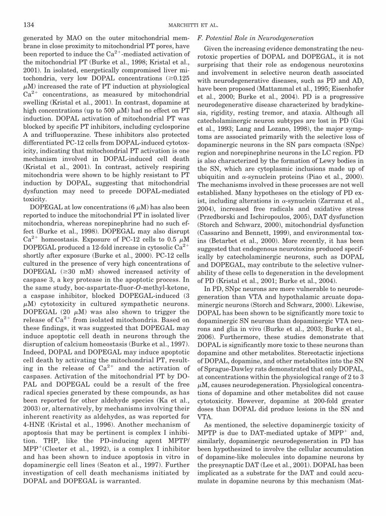
DOPEGAL, are detoxified by various enzyme systemsincluding aldehyde dehydrogenase (ALDH), which is ex-clusively responsible for their oxidative metabolism(Fig. 1). This article presents a comprehensive review ofthe role of DOPAL and DOPEGAL in the CNS. First, ageneral overview of the biological significance and reac-tivity of aldehydic compounds is given, followed by anintroduction to biogenic aldehydes. Second, the intra-neuronal formation of DOPAL and DOPEGAL, theirpossible transport systems, and their identification andquantification in biological samples will be reviewed indetail. Third, the neurotoxicity of DOPAL and DOP-EGAL will be examined (including possible mechanismsof toxicity), including their potential role in cell deathand neurodegeneration. Finally, the metabolism ofDOPAL and DOPEGAL by various enzyme systems willbe comprehensively examined with an emphasis on therole of the human ALDH isozymes and the impact ofALDH dysfunction.
II. Aldehydes
A. Biological Significance and Reactivity
Aldehydes are organic compounds containing termi-nal carbonyl groups. They can be divided into four gen-eral carbonyl classes: 1) saturated alkanals, such asformaldehyde, acetaldehyde, and hexanal; 2) unsatur-ated alkenals, such as acrolein, 4-hydroxy-2-nonenal (4-HNE), and crotonaldehyde; 3) aromatic aldehydes, suchas benzaldehyde, DOPAL and DOPEGAL; and 4) dicar-bonyls such as glyoxal and malondialdehyde (MDA).
1 Abbreviations: CNS, central nervous system; MAO, monoamineoxidase; DOPAL, 3,4-dihydroxyphenylacetaldehyde; DOPEGAL, 3,4-dihydroxyphenylglycolaldehyde; PD, Parkinson’s disease; AD, Alzhei-mer’s disease; ALDH, aldehyde dehydrogenase; 4-HNE, 4-hydroxy-2-nonenal; MDA, malondialdehyde; GSH, glutathione; SN, substantianigra; VTA, ventral tegmental area; LC, locus ceruleus; RVLM, rostralventral lateral medulla; DAT, dopamine transporter; MPP�, 1-methyl-4-phenylpyridine; MPTP, 1-methyl-4-phenyl-1,2,3,6-tetrahydropyri-dine; THP, tetrahydropapaveroline; L-dopa, L-3,4-dihydroxyphenylala-nine; MHPG, 3-methoxy-4-hydroxyphenylglycol; HVA, homovanillicacid; LDH, lactic acid dehydrogenase; TH, tyrosine hydroxylase; ROS,reactive oxygen species; H2O2, hydrogen peroxide; PT, permeabilitytransition; SNpc, substantia nigra pars compacta; AR, aldose reductase;ADH, alcohol dehydrogenase; ALR, aldehyde reductase; COMT, cate-chol-O-methyltransferase; PST, phenolsulfotransferase; UGT, UDP-glucuronosyltransferase; DOPAC, 3,4-dihydroxyphenylacetic acid; DO-PET, 3,4-dihydroxyphenylethanol; DOPEG, 3,4-dihydroxyphenylglycol;DOMA, 3,4-dihydroxymandelic acid; VMA, vanillylmandelic acid;MOPEGAL, 3-methoxy-4-hydroxyphenylglycolaldehyde; AASA, �-ami-noadipic semialdehyde; AKR, aldo-keto reductase.
FIG. 1. Consequences of aldehyde toxicity and the general detoxifica-tion reaction catalyzed by ALDH.
126 MARCHITTI ET AL.

These compounds are widespread in nature and ubiqui-tous in the environment. Various aldehydes, includingformaldehyde, acetaldehyde, and acrolein, are producedduring combustion and are present in smog and ciga-rette smoke (Rickert et al., 1980; Destaillats et al.,2002). Motor vehicle exhaust represents a major sourceof environmental aldehydes in air both through directemission of aldehydes and through the emission of hy-drocarbons, which can give rise to aldehydes. Cigarettesmoke is also an important source of aldehydes; inter-estingly, second-hand smoke can contain significantlyhigher levels of aldehydes than first-hand smoke. Alde-hydes are also used or generated in a wide variety ofindustrial applications (O’Brien et al., 2005). Formalde-hyde is used in the production of resins, polyurethane,and polyester plastics and as a fumigant and a preser-vative in animal feed. Acetaldehyde also has many in-dustrial uses including use in alkyd resin production.Drugs and environmental agents are also important al-dehyde precursors. The hepatotoxins allyl alcohol andethanol are directly metabolized to their correspondingaldehydes, acrolein, and acetaldehyde, respectively.Moreover, acrolein, ethanol, and other agents, such ascarbon tetrachloride, also can induce the formation oflipid peroxidation-derived aldehydes (O’Brien et al.,2005). Many drugs, including the anticancer drugs cy-clophosphamide and ifosfamide, are also metabolized toaldehyde intermediates (Maki and Sladek, 1993).
A range of aliphatic and aromatic dietary alde-hydes, including citral, benzaldehyde, acetaldehyde,and formaldehyde, exist naturally in various foods,particularly in fruits and vegetables, to which theyimpart flavor and odor (Lindahl, 1992). Cooking fumesalso contain a variety of aldehydes. Similarly, alde-hydes including hexenal and cinnamaldehyde are ap-proved by the U.S. Food and Drug Administration foruse as flavoring additives and spices. In animals, al-dehydes, including acrolein, benzaldehyde, and hex-anal, act as communication molecules, having roles inattraction or defense (Schauenstein et al., 1977). Like-wise, plant species produce aldehydes, including var-ious hexenals, as part of a natural pesticide systemagainst animals and insects. Interestingly, some in-sects have evolved to feed on these toxic plants and,therefore, can exploit plant-derived aldehydes fortheir own use in the chemical defense against preda-tors (Williams et al., 2001).
Aldehydes are also generated as physiologically de-rived intermediates during the biotransformation ofmany endogenous compounds, including lipids, aminoacids, neurotransmitters, and carbohydrates. For exam-ple, more than 200 aldehyde species arise from the oxi-dative degradation of cellular membrane lipids (lipidperoxidation), including 4-HNE and MDA (Esterbauer,1993). Amino acid catabolism generates several alde-hyde intermediates including glutamate �-semialde-hyde, produced during proline and arginine metabolism,
and malonate semialdehyde, produced during valine ca-tabolism (Vasiliou et al., 2004). Neurotransmitters, suchas �-aminobutyric acid (GABA), serotonin, norepineph-rine, epinephrine, and dopamine, also give rise to alde-hyde metabolites (Duncan and Sourkes, 1974; Gibson etal., 1998). Carbohydrate metabolism and ascorbate au-to-oxidation generate glycolaldehyde and the dicarbonyl,glyoxal (O’Brien et al., 2005).
Aldehydes play vital roles in normal physiologicaland therapeutic processes. For example, the aldehyderetinal is essential for vision and its ALDH-dependentoxidation product, retinoic acid, is critical for embry-onic development (Siegenthaler et al., 1990). Betaine,the ALDH-oxidation product of betaine aldehyde, is anosmolyte and methyl donor that has been shown toprotect cells and organs from osmotic stress-inducedtoxicity (Horio et al., 2001). Other critical processesinvolving aldehydes include the biosynthesis of neu-rotransmitters. The inhibitory neurotransmitterGABA can be formed through the ALDH-dependentoxidation of its aldehyde precursor, �-aminobutyral-dehyde (Ambroziak and Pietruszko, 1987). In addi-tion, the excitatory neurotransmitter, glutamate, isformed by the ALDH-induced oxidation of glutamate�-semialdehyde. In terms of a therapeutic role, alde-hyde intermediates can mediate the efficacy of certaindrugs. The antineoplastic agent cyclophosphamide,through its aldehyde intermediate aldophosphamide,gives rise to phosphoramide mustard and acrolein,which are responsible for its tumor-cell killing effects(Sladek et al., 1989).
Although some aldehydes are essential for normalbiological processes, many are cytotoxic and even car-cinogenic (Yokoyama et al., 1996; Feng et al., 2004)(Fig. 1). Aldehydes are strong electrophilic compoundswith terminal carbonyl groups, making them highlyreactive. In fact, the aldehyde group is the most reac-tive among the functional groups of biomolecules. Inaddition to the electrophilic carbonyl carbon, � and�-unsaturated aldehydes, considered bifunctional al-dehydes, such as 4-HNE and acrolein, contain a sec-ond electrophile at the �-carbon. Furthermore, 4-hy-droxylalkenel aldehydes, including 4-HNE, contain ahydroxyl group that can also participate in reactions.Unlike free radicals, aldehydes are relatively long-lived and, therefore, they not only react with targetsin the same vicinity of their formation but also candiffuse or be transported to reach sites that are somedistance away (Esterbauer et al., 1991).
Because of their electrophilic nature, aldehydes formadducts with various cellular nucleophiles, resulting inimpaired cellular homeostasis, dramatically reduced en-zyme activity, and even DNA damage (Sayre et al., 2001;Schaur, 2003). Aldehydes readily form adducts with glu-tathione (GSH) (Esterbauer et al., 1975), nucleic acids(Basu et al., 1988), and protein amino acids (Nadkarniand Sayre, 1995). Furthermore, the ability of aldehydes
TOXICITY AND METABOLISM OF DOPAL AND DOPEGAL: ROLE OF ALDH 127

to cross-link proteins and DNA and to even form DNA-protein cross-links (through various adduction mecha-nisms) has been reported (Nair et al., 1986; Brooks andTheruvathu, 2005). Aldehyde-protein adducts typicallyinvolve sulfhydryl groups of cysteine residues and aminogroups of lysine residues but can also involve otheramino acid side chains including histidine and arginine(Sayre et al., 2001). Aldehydes adduct proteins by vari-ous mechanisms including Michael addition-type reac-tions and Schiff base-type condensation reactions. Alde-hyde-nucleic acid adducts primarily involve aminogroups of both purines and pyrimidines (Brooks andTheruvathu, 2005). Aldehydes have also been shown tobe involved in the adduction of coenzymes, leading totheir inactivation and depletion by mechanisms such asKnoevenagel condensation (Farrant et al., 2001). Ac-cordingly, the adduction of aldehydes with various cel-lular components is believed to be the primary mecha-nism underlying their toxicity. Subsequent biologicaleffects of aldehyde adduction can be cytotoxic, muta-genic, and even carcinogenic (Krokan et al., 1985; Ester-bauer et al., 1991), involving the rapid depletion of GSHand protein thiols and the inactivation of enzymes (Chioand Tappel, 1969) with subsequent alterations in signaltransduction pathways (Leonarduzzi et al., 2004), geneexpression (Kumagai et al., 2000) and DNA repair (Fenget al., 2004).
Although many enzyme systems exist to detoxify al-dehydes, perturbations in aldehyde metabolism do occurand contribute to a variety of disease states. Indeed, theaccumulation of aldehydes from inborn errors of alde-hyde metabolism has been associated with many patho-logical conditions (Vasiliou and Pappa, 2000). For exam-ple, the impaired metabolism of various endogenousaldehydes is causally associated with many diseases,including Sjogren-Larsson syndrome (Rizzo and Carney,2005), type II hyperprolinemia (Valle et al., 1976), �-hy-droxybutyric aciduria (Pearl et al., 2003), pyridoxine-dependent seizures (Mills et al., 2006), and hyperam-monemia and hypoprolinemia (Baumgartner et al.,2000). In addition, lipid-derived aldehydes, such as4-HNE, acrolein, and MDA, have been implicated inalcohol-related diseases, including alcoholic liver dis-ease, fibrosis, and atherosclerosis (Poli, 2000; Sun et al.,2001), and neurological diseases, such as PD and AD(Yoritaka et al., 1996; Lovell et al., 2001). Similarly,impaired metabolism of the ethanol metabolite acetal-dehyde has been implicated in many alcohol-related dis-eases, including cirrhosis (Enomoto et al., 1991; Chao etal., 1994) and numerous head and neck cancers (Muto etal., 2000; Yokoyama et al., 2001), and late onset AD(Kamino et al., 2000).
B. Biogenic Aldehydes
Oxidative deamination of various biogenic amines (in-cluding indoleamines and catecholamines) results in theformation of “biogenic aldehydes.” The indoleamines se-
rotonin and tryptamine generate the biogenic aldehydes5-hydroxyindole-3-acetaldehyde and indole-3-acetalde-hyde, respectively. The catecholamines, dopamine, nor-epinephrine, and epinephrine, also give rise to biogenicaldehydes upon deamination (Fig. 2). Dopamine gener-ates DOPAL, whereas both norepinephrine and epi-nephrine are deaminated to form DOPEGAL. DespiteBlaschko’s hypothesis in the early 1950s that indoleam-ine- and catecholamine-derived aldehydes may be toxic(Blaschko, 1952), biogenic aldehydes were originally be-lieved to be innocuous intermediates in biogenic aminemetabolism. However, early studies investigating theirpossible role in the pharmacological actions of ethanol(levels of biogenic aldehydes may increase during etha-nol metabolism) and their ability to form isoquinoline-derived condensation products with their parent amine(Deitrich and Erwin, 1975; Tipton et al., 1977) led to thediscovery that biogenic aldehydes actually have distinctphysiological properties of their own. In fact, the in-doleamine-derived aldehydes 5-hydroxyindole-3-acetal-dehyde and indole-3-acetaldehyde have been shown toillicit many biological effects including neurotransmit-ter-like actions in the CNS (Sabelli et al., 1969; Palmeret al., 1986) and the inhibition of various enzymes(Tabakoff, 1974; Erwin et al., 1975).
As mentioned, DOPAL and DOPEGAL represent cat-echolamine-derived biogenic aldehydes. The major cate-cholamine neurotransmitters found in the human brainare dopamine, norepinephrine, and epinephrine. Dopa-mine is synthesized by neurons in the substantia nigra(SN), ventral tegmental area (VTA) and hypothalamus,whereas synthesis of norepinephrine and epinephrinetakes place primarily in neurons of the locus ceruleus(LC) and rostral ventral lateral medulla (RVLM), re-spectively (Nestler et al., 2001). Many important func-tions of the brain including memory, learning, move-ment, and behavior are thought to be mediated bycatecholamines. Accordingly, the loss of specific popula-tions of catecholaminergic neurons and subsequent def-icits in related brain function are the basis of variousneurodegenerative pathological conditions, includingPD and AD. The specific vulnerability and death of theseneurons have been hypothesized to involve toxic cate-cholamine metabolites or compounds that are selectivelyproduced by, or accumulated in, catecholamine neuronssuch as DOPAL and DOPEGAL (Li et al., 2001; Burke etal., 2004). Indeed, there is an increasing body of evidencedemonstrating the neurotoxic properties of the catechol-amine-derived aldehydes DOPAL and DOPEGAL byvarious cytotoxic mechanisms including the generationof free radicals and initiation of apoptosis (Burke et al.,1998; Li et al., 2001). Along those lines, it has beensuggested that DOPAL and DOPEGAL represent endog-enous neurotoxins that may play a significant role in celldeath associated with neurodegenerative diseases (Kri-stal et al., 2001; Eisenhofer et al., 2004).
128 MARCHITTI ET AL.

III. 3,4-Dihydroxyphenylacetaldehyde and3,4-Dihydroxyphenylglycolaldehyde in the
Central Nervous System
A. Intraneuronal Formation
Oxidative deamination of catecholamines to form al-dehyde metabolites was first described in the mid-1930s(Richter, 1937). Dopamine is deaminated to formDOPAL, whereas norepinephrine and epinephrine areboth deaminated to form DOPEGAL (Fig. 2). A commonmisconception is that catecholamines are metabolized atsites distant from those of their synthesis and release(for a review of common fallacies about catecholaminemetabolism, see Eisenhofer et al., 2004). Rather, mostcatecholamine metabolism has been shown to take placein the same cells in which they are produced and withoutprior release (Kopin, 1964; Maas et al., 1970). Accord-ingly, the formation of DOPAL and DOPEGAL is be-lieved to occur primarily in the cytoplasm of the neuronsthat synthesize their parent catecholamines (Eisenhoferet al., 1992). This process is thought to occur primarilyafter the passive leakage of catecholamines into the
cytoplasm from storage vesicles or, as a minor pathway,following their reuptake into the nerve terminal (Kopin,1964; Eisenhofer et al., 1992). Monoamine transportersare responsible for sequestering catecholamines intostorage vesicles within neurons, but it is estimated that�10% of catecholamines escape into the neuronal cyto-plasm and are subsequently metabolized (Eisenhofer etal., 1992). Indeed, 70 to 75% of norepinephrine turnoverseems to occur from intraneuronal metabolism of nor-epinephrine leaking from storage vesicles, with the re-mainder made up by intraneuronal metabolism afterreuptake, extraneuronal uptake and metabolism or lossof norepinephrine to the circulation (Eisenhofer et al.,1996b; Eisenhofer et al., 1998). Numerous in vitro and invivo studies in animals and humans have demonstratedthat leakage of catecholamines from storage vesicles isthe primary pathway leading to catecholamine catabo-lism (Goldstein et al., 1988; Halbrugge et al., 1989; Tyceet al., 1995). The drug reserpine, which blocks the se-questration of catecholamines into storage vesicles, hasbeen shown to cause depletion of catecholamine stores
FIG. 2. Formation and metabolism of DOPAL and DOPEGAL. DOPAL is formed from dopamine, whereas DOPEGAL is derived from bothnorepinephrine and epinephrine. They are metabolized by various enzymes including ALDH, ADH, and ALR/AR. DOPAL is predominantlymetabolized by ALDH in the oxidative pathway, whereas DOPEGAL is predominantly metabolized by ADH or ALR/AR in the reductive pathway.Predominant pathways of DOPAL and DOPEGAL metabolism pathways are shown in bold. Free HVA is the major brain metabolite of dopamine. FreeMHPG is the major brain metabolite of both norepinephrine and epinephrine and is also the major precursor to VMA formation in the liver. DA,dopamine; NE, norepinephrine; EPI, epinephrine; DBH, dopamine-�-hydroxylase; MHPE, 3-methoxy-4-hydroxyphenylethanol.
TOXICITY AND METABOLISM OF DOPAL AND DOPEGAL: ROLE OF ALDH 129

due to the rapid metabolism of unsequestered cat-echolamines (Kopin and Gordon, 1962). Studies with ratbrain synaptic vesicles have shown dopamine turnoverto be more rapid than that of norepinephrine (Floor etal., 1995), suggesting that leakage of dopamine fromstorage vesicles into the neuronal cytoplasm may beeven more significant than it is for norepinephrine.
Intraneuronal formation of DOPAL and DOPEGAL iscatalyzed by the actions of MAO (Fig. 2). Whereas otherenzymes are involved in the degradation of cat-echolamines, MAO metabolism seems to be the principalintraneuronal pathway (Rivett et al., 1982). MAO is aflavin-containing, particle-bound enzyme localized pri-marily in the outer mitochondrial membrane (Schnait-man et al., 1967). In the brain, the enzyme is almostexclusively localized in nerve terminals (Westlund et al.,1985, 1993) where it catalyzes the oxidative deamina-tion of dopamine, norepinephrine, and epinephrine totheir respective aldehydes. The MAO reaction first in-volves the formation of an imine, which subsequentlyundergoes a nonenzymatic conversion to the correspond-ing aldehyde. MAO exists as two distinct genetic iso-forms, MAO-A and MAO-B, both of which are catalyti-cally active with catecholamines (Youdim et al., 2006).However, studies using preferential inhibitors ofMAO-A and MAO-B have indicated that the productionof DOPAL and DOPEGAL from dopamine, norepineph-rine, and epinephrine can be attributed primarily toMAO-A (Waldmeier et al., 1976; Fowler and Benedetti,1983; Fornai et al., 2000). The CNS distribution of thetwo MAO isozymes is consistent with this contention inthat catecholaminergic neurons in the LC, RVLM, andSN primarily contain MAO-A whereas MAO-B is local-ized within serotonergic neurons in the dorsal raphenucleus and superior central nucleus (Westlund et al.,1985).
B. Transport Mechanisms
After intraneuronal formation, DOPAL and DOPE-GAL may leave nerve terminals by simple diffusionand reenter cells through various transport processes.DOPAL is taken up by rat neostriatal synaptosomes,and studies using mazindol, a selective dopamine up-take inhibitor, have suggested that the aldehyde reen-ters dopaminergic nerve terminals via the dopaminetransporter (DAT) (Mattammal et al., 1995). The DAT isexpressed in presynaptic terminals of SN neurons whereit primarily mediates the reuptake of neuronally re-leased dopamine (Reith et al., 1997; Jones et al., 1998).The DAT is also responsible for the uptake of 1-methyl-4-phenylpyridine (MPP�), the active metabolite of1-methyl-4-phenyl-1,2,3,6-tetrahydropyridine (MPTP),an exogenous dopaminergic neurotoxin that inducesparkinsonian symptoms in affected individuals and isused as a model of PD in animal studies (Javitch et al.,1985). DOPEGAL is taken up by catecholaminergicPC-12 cells and studies using desipramine, which blocks
the neuronal catecholamine uptake transporter, havesuggested that DOPEGAL is actively transported by thismechanism (Burke et al., 2001).
C. Identification and Quantification in BiologicalSamples
The presence and activity of biogenic aldehydes inbiological tissues was first supported by the finding thattetrahydropapaveroline (THP), the tetrahydroisoquino-line alkaloid condensation product of a Pictet-Spenglercondensation reaction between dopamine and DOPAL(Fig. 3), was present in the urine of L-3,4-dihydroxyphe-nylalanine (L-dopa)-treated PD patients (Sandler et al.,1973). Since then, other studies have confirmed thepresence of both DOPAL and DOPEGAL in various tis-sues, including human brain. In normal human tissues,these aldehydes have been quantitated using chemicallysynthesized and purified standards (Burke et al.,1999a). In this study, DOPAL and DOPEGAL were sep-arated from 12 catecholamines (and other metabolites),and their levels were analyzed in normal plasma, urine,and postmortem human brain regions. The concentra-tion of DOPEGAL in normal postmortem LC was esti-mated to be 1.4 �M, a level �50% of that of 3-methoxy-4-hydroxyphenylglycol (MHPG), a major metabolite ofnorepinephrine and epinephrine (Burke et al., 1999a,2004). Normal postmortem human brain SN levels ofDOPAL were estimated to be 2.3 �M, a level �25%higher than that of homovanillic acid (HVA), a majordopamine metabolite. Nominal levels were seen inurine for both DOPAL and DOPEGAL. Plasma levelsof DOPAL were minor, whereas those of DOPEGALwere similar to that of epinephrine. Other studieshave examined the presence of DOPAL and DOPE-GAL in animal tissues. DOPAL has been identified invivo in rat striatum using trans-striatal microdialysisin freely moving rats (Colzi et al., 1996), and DOPE-GAL has been detected in pulverized rat adrenalglands (Burke et al., 1995). These studies demonstratephysiological production of DOPAL and DOPEGAL atlevels approaching or exceeding those observed formore well-established catecholamine metabolites.
FIG. 3. Formation of tetrahydropapaveroline (THP), the Pictet-Spen-gler condensation product of DOPAL and dopamine.
130 MARCHITTI ET AL.

IV. Toxicity of 3,4-Dihydroxyphenylacetaldehydeand 3,4-Dihydroxyphenylglycolaldehyde in the
Central Nervous System
Although their toxicity was predicted in the 1950s,researchers initially believed that aldehyde metabolitesderived from biogenic amines were innocuous interme-diates (Renson et al., 1964). Since then, these aldehydeshave been shown to be active compounds with physio-logical properties distinct from those of their parentamines (Palmer et al., 1986). The body of research re-porting on investigations of the biogenic aldehydesDOPAL and DOPEGAL that began in the 1950s is not aslarge as one would expect considering the many molec-ular and genomic advances that have occurred in theensuing period. This is due, in part, to the unstable andreactive properties of these catecholamine-derived com-pounds, making them difficult to synthesize (eitherchemically or enzymatically) in pure form and/or inlarge enough quantities to be useful for experimentalpurposes. Recent advances, however, in the chemicalsynthesis of DOPAL and DOPEGAL (Narayanan et al.,2003) and the growing number of studies indicating thatthey may be important neurotoxins will no doubt lead toincreased interest in and focus on these compounds infuture investigations. Nonetheless, a substantial andconvincing body of relatively recent evidence does exist,which indicates the toxicity and reactivity of these com-pounds in the CNS. On the basis of these studies, a rolefor DOPAL and DOPEGAL in neurodegenerative dis-eases has been proposed (Mattammal et al., 1995; La-mensdorf et al., 2000b; Burke et al., 2004).
A. Cytotoxicity
Concentrations of DOPAL and DOPEGAL �6 �M in-duce dose-dependent toxicity in various cell lines, in-cluding differentiated PC-12 cells (Mattammal et al.,1995; Burke et al., 1996; Kristal et al., 2001). Levels ofthe aldehydes identified as being toxic in vitro have beenreported to be close to physiological levels found in nor-mal human postmortem brain (Burke et al., 1999a; Kri-stal et al., 2001). For example, DOPAL at 6.6 �M, aconcentration close to the physiological levels reportedin normal human autopsy specimens of the SN (Burke etal., 1999a), induced significant cytotoxicity in PC-12cells and reduced viability by �30% (Kristal et al., 2001).DOPAL at 66 �M caused cell death in �67% of neuro-nally differentiated PC-12 cells after 72 h, whereas cul-tures incubated with equivalent concentrations of dopa-mine, HVA, or THP were indistinguishable fromcontrols (Kristal et al., 2001). These results are consis-tent with previous findings demonstrating that PC-12cells are resistant to dopamine concentrations �1 mM(Cantuti-Castelvetri and Joseph, 1999). On the basis ofthese findings, it has been suggested that DOPAL is�100-fold more toxic to PC-12 cells than is dopamine(Kristal et al., 2001).
DOPAL-induced PC-12 cell damage elicits concentra-tion-dependent lactic acid dehydrogenase (LDH) release,a measure of cytotoxicity. PC-12 cells incubated with 33�M DOPAL for 8 h produced a 6-fold increase in LDH(Mattammal et al., 1995). In the same study, incubationfor 24 h with 6.5 �M DOPAL induced both significantdegeneration of the neuritic processes and a decrease inthe number of viable cells. Incubation for 24 h with 33�M DOPAL resulted in almost no cell survival. Shortterm (5 min) incubation of PC-12 cells with DOPAL (100�M) also results in significant LDH release (Hashimotoand Yabe-Nishimura, 2002).
DOPAL is also toxic to neuroblastoma SK-N-SH andSH-SY5Y cells, fetal rat mesencephalic cultures and ratneostriatal synaptosomal preparations. In SK-N-SHcells, exposure to DOPAL (1–500 �M) for 24 h produceda concentration-dependent increase in LDH leakage intothe cell medium with toxicity being pronounced at 100�M (Lamensdorf et al., 2000a). In catecholaminergicSH-SY5Y cells, early increased levels of DOPAL inducedby dopamine treatment and ALDH inhibition produceddelayed cell toxicity and cell losses that increased withtime (Legros et al., 2004a). Dopaminergic cultures, pre-pared from the ventral mesencephalon of rat embryos,exposed to 1 to 5 �M DOPAL for 24 h showed no toxicityas measured by the disappearance of tyrosine hydroxy-lase (TH) immunoreactivity (Mattammal et al., 1995).Between 7.5 and 20 �M DOPAL, a gradual reduction indopamine uptake was seen without a reduction in thenumber of TH-immunoreactive cells. However, treat-ment of cultures with 33 �M DOPAL resulted in thedisappearance of TH immunoreactivity, with the surviv-ing TH-immunoreactive cells showing rounded cell bod-ies and highly fragmented fiber networks. These mor-phological changes were specific to dopaminergicneurons and were not evident in other CNS cells. In ratneostriatal synaptosomes, treatment with DOPAL for 30min (10–100 �M), produced a concentration-dependentdecrease in the number of living cells and a concomitantincrease in the release of LDH (Mattammal et al., 1995).
DOPEGAL is also toxic in vitro. DOPEGAL concen-trations of 5.9 and 59.5 �M decreased PC-12 cell viabil-ity (by 23 and 61%, respectively), with most of the cellloss occurring after 2 days of exposure (Burke et al.,1996). Epinephrine itself was also slightly toxic to thesecells, reducing viability by 17%, but no other oxidative ormethylated metabolite of epinephrine (aside fromDOPEGAL) was toxic to PC-12 cells. The results of thisstudy also underscored the importance of the terminalcarbonyl group in the toxicity of DOPEGAL. Convertingthe terminal carbonyl moiety of DOPEGAL to a hydroxylgroup to form its tautomer, 2�,3,4-trihydroxyacetophe-none, diminished toxicity significantly.
In vivo cytotoxicity of DOPAL has been reported inneurons and glia in the SN and VTA (Burke et al., 2003).DOPAL, dopamine, and oxidative, reduced, and methyl-ated metabolites of dopamine were injected into rat SN
TOXICITY AND METABOLISM OF DOPAL AND DOPEGAL: ROLE OF ALDH 131

and VTA. Five days after treatment, these regions wereevaluated by Nissl preparation and cell-specific immu-noreactivities. At doses of 100 ng, DOPAL was mosttoxic to SN neurons, followed by VTA neurons and, fi-nally, glial cells. Neurons of the SN were consistentlymore affected, indicating the selective toxicity of DOPALtoward dopaminergic nerves. Neither dopamine nor itsother metabolites elicited evidence of neurotoxicity, evenwhen injected at 5-fold higher doses than that shown forDOPAL to cause toxicity. In contrast, rats treated sys-temically for 30 days with L-dopa, which is enzymati-cally converted to dopamine (by L-dopa decarboxylase),exhibited a 3-fold increase in the levels of DOPAL in thebrain but showed no evidence of nigrostriatal dopami-nergic cytotoxicity as measured by striatal dopaminecontent (Legros et al., 2004b).
In vivo toxicity of DOPEGAL has also been demon-strated—in rat RVLM (Burke et al., 2001). DOPEGALwas injected into adrenergic neurons in the RVLM andapoptosis of these adrenergic neurons (identified immu-nohistochemically by their content of the epinephrine-synthesizing enzyme phenylethanolamine N-methyl-transferase) was evaluated. Fifty nanograms ofDOPEGAL caused apoptotic loss of epinephrine neuronsafter 18 h, as evaluated by in situ terminal deoxynucle-otidyl-transferase mediated dUTP nick-end label stain-ing. The degree of neurotoxicity was both dose- andtime-dependent. Ten-fold higher doses of DOPEGALproduced necrosis of these neurons. Neither epinephrinenor MHPG was shown to be cytotoxic in this study.
Various mechanisms have been suggested to explainthe observed cytotoxicity of DOPAL and DOPEGAL.These include protein adduction, isoquinoline formation,and free radical generation.
B. Protein Adduction
It is well known that aldehydes react with proteins toform various adducts that can disrupt protein functionand cause cellular damage. It has been suggested thatbiogenic aldehydes react with proteins to form both un-stable and stable adducts (Esterbauer et al., 1991). Ev-idence of this formation has been illustrated in reactionsbetween DOPAL and the protein hemoglobin (Helanderand Tottmar, 1989). DOPAL initially reacts with hemo-globin to form Schiff bases involving its aldehyde groupand the free amino groups of lysine, tyrosine, or valineresidues (Fig. 4). Schiff bases are inherently unstableand the binding that involves them can be reversed orfurther converted into more stable products by physio-
logical reducing agents, such as glutathione or ascorbate(Tuma et al., 1984). Stable adducts formed betweenDOPAL and hemoglobin rendered the aldehyde unableto serve as a substrate for ALDH, whereas the DOPALthat was more loosely bound (i.e., Schiff base) could.Interestingly, at concentrations exceeding 5 �M, DO-PAL actually inactivated ALDH by interacting with thesurface of the protein through irreversible covalent mod-ification in areas deemed important for enzyme activity(MacKerell and Pietruszko, 1987). Exhaustive dialysisand reducing agents did not reverse the inactivation.The substrate analog, chloral, protected ALDH againstDOPAL-mediated inactivation. The authors suggestedthat DOPAL inactivates ALDH by formation of a cova-lent bond involving its catechol ring rather than itsaldehyde group. A recent study supports this observa-tion of substrate-mediated inhibition of ALDH by DO-PAL, although the mechanism was not investigated(Florang et al., 2006).
C. Isoquinoline Formation
Carbonyl compounds can react with �-arylethyl-amines, such as dopamine, in a rapid, nonenzymaticPictet-Spengler condensation reaction to form isoquino-line alkaloid derivatives (Yamanaka et al., 1970; San-dler et al., 1973). For example, the reaction betweenacetaldehyde and dopamine results in the formation ofsalsolinol (Fig. 5) (Sandler et al., 1973), whereas that ofDOPAL and dopamine generates THP (Davis andWalsh, 1970; Cohen, 1976) (Fig. 3). THP, salsolinol, andother isoquinoline derivatives are structurally related tothe selective dopaminergic neurotoxin and PD-inducingagent MPTP/MPP� and have been suggested to play arole in dopaminergic cell death characterized by PD(McNaught et al., 1998; Storch et al., 2002). Indeed, THPand salsolinol, have been demonstrated to be selectivedopaminergic neurotoxins (Goto et al., 1997; Storch etal., 2002). Salsolinol and THP are found in high concen-trations in the urine of PD patients on L-dopa treatment(Sandler et al., 1973) and salsolinol is found in the cere-brospinal fluid of untreated PD patients (Maruyama etal., 1996). The primary mechanism underlying MPTP/
FIG. 4. Proposed Schiff base formation between DOPAL and aminoacid residues of proteins, such as lysine.
FIG. 5. Formation of salsolinol, the Pictet-Spengler condensationproduct of acetaldehyde and dopamine.
132 MARCHITTI ET AL.

MPP� dopaminergic toxicity (after MPP� uptake by theDAT into dopamine neurons) is MPP�-induced inhibi-tion of complex I of the mitochondrial respiratory chain,leading to depleted ATP, enhanced ROS production andultimately dopaminergic cell death (Brooks et al., 1989;Cleeter et al., 1992). Similarly, THP is also a complex Iinhibitor (Suzuki et al., 1990; McNaught et al., 1995).Inhibition of complex I also leads to the decreased avail-ability of NAD�, the required cofactor in ALDH-medi-ated DOPAL metabolism. This could result in the accu-mulation of DOPAL and increased generation of THP.As mentioned, the dopaminergic specificity of MPTP/MPP� relies on the uptake of MPP� by the DAT, whichtransports this toxin selectively into dopaminergic cells.In vitro, THP has been shown to inhibit dopamine up-take through the DAT (Okada et al., 1998), indicatingthat it may also be a DAT substrate. THP also inhibitsdopamine biosynthesis through mechanisms involvingincreased oxidative stress and the inhibition of TH ac-tivity (Kim et al., 2005). Indeed, many isoquinolinesexhibit pharmacological actions and can act as falseneurotransmitters or inhibitors of physiological mecha-nisms that regulate the actions of catecholamines (Co-hen, 1976). For example, THP has been suggested to beresponsible for some of the hypotensive and �-adrener-gic actions of L-dopa (Sandler et al., 1973). Accordingly,it is possible that some of the neurotoxic effects reportedfor DOPAL may be due, in part, to the generation ofTHP.
D. Free Radical Generation
Like aldehydes, free radicals and reactive oxygen spe-cies (ROS) are known to damage cellular componentsand interrupt physiological functions, which can lead tovarious disease states, including cell death and cancer.The reactions catalyzed by MAO to form DOPAL andDOPEGAL produce hydrogen peroxide (H2O2), whichcan generate other ROS and free radicals. Recently,DOPAL itself has been reported to generate free radi-cals, specifically the hydroxyl radical, in the presence ofH2O2 (Li et al., 2001). This free hydroxyl radical produc-tion was not observed with DOPEGAL, dopamine, orother dopamine metabolites. The authors speculatedthat DOPAL may act as a cofactor in the generation ofhydroxyl radicals. In support of this hypothesis, DOPALdid not produce the hydroxyl radical in the absence ofH2O2 or in the presence of Fe2� alone. However, H2O2 inthe presence of either Fe2� or DOPAL did result inhydroxyl radical formation. Thus, DOPAL may have arole similar to that of Fe2� in the formation of hydroxylradicals from H2O2 wherein DOPAL functions as a re-ducing agent and is oxidized in the process. In additionto its electrophilic carbonyl carbon, DOPAL contains twoeasily oxidizable phenolic groups. Auto-oxidation of DO-PAL to the DOPAL-o-quinone, a process reported fordopamine (Hasegawa et al., 2006), may also produce free
hydroxyl radicals in the presence of H2O2, similar tothat reported for the dopamine derivative, 6-hydroxydo-pamine (Cohen and Heikkila, 1974). Furthermore, itwas suggested that the ability of DOPAL to produce thehydroxyl radical may be due to its lower redox potentialin relation to DOPEGAL or other dopamine metabolites(Cohen and Heikkila, 1974; Liu and Mori, 1993).
DOPEGAL has also been reported to generate a freeradical in the presence of H2O2. In this case, the freeradical seems to be a DOPEGAL radical that may in-volve the side-chain �-hydroxyl group (Burke et al.,1998). According to electron paramagnetic resonanceanalyses, this radical elicited a signal different from thatfor the hydroxyl radical, leading the authors to suggestthat DOPEGAL itself has a free radical form (Li et al.,2001). This observation was not seen with norepineph-rine. It is intriguing that DOPAL and DOPEGAL havethe capacity to generate free radicals, and they seem todo so by different pathways. Generation of ROS by DO-PAL and DOPEGAL could have serious functional sig-nificance arising from GSH depletion and increased cel-lular oxidative stress. Further study of these processesis necessary to elucidate the exact mechanisms involved.
E. Mechanisms of Apoptosis
Apoptosis is a form of programmed cell death associ-ated with a disruption in Ca2� homeostasis (Orreniusand Nicotera, 1994) and the activation of caspase pro-tease proteins (Scarlett and Murphy, 1997). Mitochon-dria are known to play a role in apoptotic neuron death(Wallace, 1999), and recent evidence has associated ap-optosis with the induction of the mitochondrial mem-brane permeability transition (PT). Indeed, mitochon-drial PT activation is believed to be a critical factor inthe development of neurotoxicity and neurodegenera-tion (Bachurin et al., 2003), and it has been linked to theapoptotic type of catecholaminergic neuron death seenin PD and other neurodegenerative diseases (Lassmannet al., 1995; Anglade et al., 1997). Induction of the PT ischaracterized by matrix swelling, outer membrane rup-ture, release of apoptotic signaling molecules from theintermembrane space, and a collapse of the mitochon-drial membrane potential due to increased permeabilityof the inner membrane (Bachurin et al., 2003; Fiskum etal., 2003). PT activation is thought to be caused by theopening of PT pores on the inner mitochondrial mem-brane. These pores control the transport of Ca2� ionsand small compounds (up to 1.5 kDa) in and out of themitochondria and thereby function to maintain Ca2�
homeostasis in the cell. PT induction also seems to beinvolved in releasing the apoptotic initiation factors,cytochrome c and Apaf-1, which activate downstreamcaspases, leading to apoptosis (Kluck et al., 1997; Scar-lett and Murphy, 1997). Various reactive species, suchas free radicals, ROS, and aldehydes, are known to ac-tivate the mitochondrial PT (Kristal et al., 1996; Packeret al., 1997). DOPAL and DOPEGAL, both of which are
TOXICITY AND METABOLISM OF DOPAL AND DOPEGAL: ROLE OF ALDH 133
generated by MAO on the outer mitochondrial mem-brane in close proximity to mitochondrial PT pores, havebeen reported to induce the Ca2�-mediated activation ofthe mitochondrial PT (Burke et al., 1998; Kristal et al.,2001). In isolated, energetically compromised liver mi-tochondria, very low DOPAL concentrations (�0.125�M) increased the rate of PT induction at physiologicalCa2� concentrations, as measured by mitochondrialswelling (Kristal et al., 2001). In contrast, dopamine athigh concentrations (up to 500 �M) had no effect on PTinduction. DOPAL activation of mitochondrial PT wasblocked by specific PT inhibitors, including cyclosporineA and trifluoperazine. These inhibitors also protecteddifferentiated PC-12 cells from DOPAL-induced cytotox-icity, indicating that mitochondrial PT activation is onemechanism involved in DOPAL-induced cell death(Kristal et al., 2001). In contrast, actively respiringmitochondria were shown to be highly resistant to PTinduction by DOPAL, suggesting that mitochondrialdysfunction may need to precede DOPAL-mediatedtoxicity.
DOPEGAL at low concentrations (6 �M) has also beenreported to induce the mitochondrial PT in isolated livermitochondria, whereas norepinephrine had no such ef-fect (Burke et al., 1998). DOPEGAL may also disruptCa2� homeostasis. Exposure of PC-12 cells to 0.5 �MDOPEGAL produced a 12-fold increase in cytosolic Ca2�
shortly after exposure (Burke et al., 2000). PC-12 cellscultured in the presence of very high concentrations ofDOPEGAL (�30 mM) showed increased activity ofcaspase 3, a key protease in the apoptotic process. Inthe same study, boc-aspartate-fluor-O-methyl-ketone,a caspase inhibitor, blocked DOPEGAL-induced (3�M) cytotoxicity in cultured sympathetic neurons.DOPEGAL (20 �M) was also shown to trigger therelease of Ca2� from isolated mitochondria. Based onthese findings, it was suggested that DOPEGAL mayinduce apoptotic cell death in neurons through thedisruption of calcium homeostasis (Burke et al., 1997).Indeed, DOPAL and DOPEGAL may induce apoptoticcell death by activating the mitochondrial PT, result-ing in the release of Ca2� and the activation ofcaspases. Activation of the mitochondrial PT by DO-PAL and DOPEGAL could be a result of the freeradical species generated by these compounds, as hasbeen reported for other aldehyde species (Ka et al.,2003) or, alternatively, by mechanisms involving theirinherent reactivity as aldehydes, as was reported for4-HNE (Kristal et al., 1996). Another mechanism ofapoptosis that may be pertinent is complex I inhibi-tion. THP, like the PD-inducing agent MPTP/MPP�(Cleeter et al., 1992), is a complex I inhibitorand has been shown to induce apoptosis in vitro indopaminergic cell lines (Seaton et al., 1997). Furtherinvestigation of cell death mechanisms initiated byDOPAL and DOPEGAL is warranted.
F. Potential Role in Neurodegeneration
Given the increasing evidence demonstrating the neu-rotoxic properties of DOPAL and DOPEGAL, it is notsurprising that their role as endogenous neurotoxinsand involvement in selective neuron death associatedwith neurodegenerative diseases, such as PD and AD,have been proposed (Mattammal et al., 1995; Eisenhoferet al., 2000; Burke et al., 2004). PD is a progressiveneurodegenerative disease characterized by bradykine-sia, rigidity, resting tremor, and ataxia. Although allcatecholaminergic neuron subtypes are lost in PD (Gaiet al., 1993; Lang and Lozano, 1998), the major symp-toms are associated primarily with the selective loss ofdopaminergic neurons in the SN pars compacta (SNpc)region and norepinephrine neurons in the LC region. PDis also characterized by the formation of Lewy bodies inthe SN, which are cytoplasmic inclusions made up ofubiquitin and �-synuclein proteins (Piao et al., 2000).The mechanisms involved in these processes are not wellestablished. Many hypotheses on the etiology of PD ex-ist, including alterations in �-synuclein (Zarranz et al.,2004), increased free radicals and oxidative stress(Przedborski and Ischiropoulos, 2005), DAT dysfunction(Storch and Schwarz, 2000), mitochondrial dysfunction(Cassarino and Bennett, 1999), and environmental tox-ins (Betarbet et al., 2000). More recently, it has beensuggested that endogenous neurotoxins produced specif-ically by catecholaminergic neurons, such as DOPALand DOPEGAL, may contribute to the selective vulner-ability of these cells to degeneration in the developmentof PD (Kristal et al., 2001; Burke et al., 2004).
In PD, SNpc neurons are more vulnerable to neurode-generation than VTA and hypothalamic arcuate dopa-minergic neurons (Storch and Schwarz, 2000). Likewise,DOPAL has been shown to be significantly more toxic todopaminergic SN neurons than dopaminergic VTA neu-rons and glia in vivo (Burke et al., 2003; Burke et al.,2006). Furthermore, these studies demonstrate thatDOPAL is significantly more toxic to these neurons thandopamine and other metabolites. Stereotactic injectionsof DOPAL, dopamine, and other metabolites into the SNof Sprague-Dawley rats demonstrated that only DOPAL,at concentrations within the physiological range of 2 to 3�M, causes neurodegeneration. Physiological concentra-tions of dopamine and other metabolites did not causecytotoxicity. However, dopamine at 200-fold greaterdoses than DOPAL did produce lesions in the SN andVTA.
As mentioned, the selective dopaminergic toxicity ofMPTP is due to DAT-mediated uptake of MPP� and,similarly, dopaminergic neurodegeneration in PD hasbeen hypothesized to involve the cellular accumulationof dopamine-like molecules into dopamine neurons bythe presynaptic DAT (Lee et al., 2001). DOPAL has beenimplicated as a substrate for the DAT and could accu-mulate in dopamine neurons by this mechanism (Mat-
134 MARCHITTI ET AL.

tammal et al., 1995). Moreover, the DAT is expressed inhigher concentrations in SNpc neurons than in otherdopaminergic neurons, and this mechanism may explainthe increased sensitivity of SNpc neurons to DOPAL(Storch and Schwarz, 2000). As mentioned, THP, mayalso serve as a DAT substrate. The preferential uptakeof DOPAL and/or THP into dopaminergic neurons by theDAT may enhance dopaminergic neurotoxicity.
Compared with other neuronal subpopulations, SNpcneurons may have increased cytosolic Ca2� concentra-tions and be more susceptible to free radical damage(Hirsch et al., 1997). As mentioned, both DOPAL andDOPEGAL have been suggested to initiate apoptosisthrough Ca2�-mediated processes (Burke et al., 1998;Kristal et al., 2001) and to generated free radical species(Burke et al., 1998; Li et al., 2001). The specific vulner-ability of SNpc neurons coupled with these mechanismscould also lead to enhanced cytotoxicity.
Excess dopamine release from dopaminergic neuronsis another mechanism thought to be involved in PDneurodegeneration. Indeed, the PD-inducing agentMPTP/MPP� is a potent dopamine-releasing agent(Obata, 2002). DOPAL is also reported to increase dopa-mine release in PC-12 cells and rat striatal synapto-somes. In PC-12 cells, DOPAL (10 �M) caused a �2-foldincrease in the release of dopamine (Hashimoto andYabe-Nishimura, 2002). This effect was shown to beCa2�-independent. Other reactive aldehydes tested (i.e.,4-HNE) did not initiate dopamine release, suggestingthat the effect was specific to DOPAL. In rat striatalsynaptosomes, 33 �M DOPAL caused a significant in-crease in dopamine release (�55%) (Mattammal et al.,1995). This effect was specific to dopamine-containingterminals as opposed to those containing GABA. ExcessDOPAL-induced dopamine release may be one mecha-nism underlying the neurotoxicity of DOPAL and maybe a factor in dopaminergic neurodegeneration charac-terized by PD.
Central to the hypothesis that endogenous neurotox-ins, such as DOPAL and DOPEGAL, are involved inneurodegenerative diseases is the requirement that theyaccumulate to levels that become toxic to catecholamineneurons. Many mechanisms could lead to increased in-traneuronal concentrations of these aldehydes includ-ing, as mentioned, the preferential uptake of DOPAL bythe DAT. In addition, the increased synthesis of DOPALand DOPEGAL could influence their levels. Accordingly,age-related increases in MAO have been reported (Ore-land and Gottfries, 1986), which could lead to increasedformation of these aldehydes. Furthermore, pharmaco-logical treatment with the dopamine precursor, L-dopa,used to treat PD, has been shown to elevate rat brainDOPAL levels by as much as 18-fold (Fornai et al., 2000;Legros et al., 2004b).
Impaired metabolism of DOPAL may also affect itsaccumulation. Indeed, in catecholaminergic neuroblas-toma SH-SY5Y cells, significant accumulation of DO-
PAL was achieved by treatment with dopamine (1 mM)along with the inhibition of DOPAL metabolism by theALDH inhibitor disulfiram (10 �M) (Bonnet et al., 2004;Legros et al., 2004a). Under these conditions, increasedcytotoxicity was also demonstrated. Similarly, mito-chondrial dysfunction of complex I and the resultingdecreased availability of NAD�, the required cofactor forALDH-mediated oxidation of aldehydes, may lead toincreased levels of both DOPAL and DOPEGAL. Indeed,complex I inhibition by rotenone leads to significantDOPAL accumulation in PC-12 cells (Lamensdorf et al.,2000a). Under these conditions, the further inhibition ofDOPAL metabolism by specific inhibitors of ALDH andaldose reductase (AR) increased DOPAL levels evenmore (12-fold that of control) (Lamensdorf et al., 2000b).DOPAL-mediated substrate inhibition of ALDH (Mac-Kerell and Pietruszko, 1987; Florang et al., 2006) andage-related ALDH deficiencies (Chen and Yu, 1996) mayalso play a role in DOPAL accumulation in the CNS.
Mitochondrial dysfunctions, including complex I inhi-bition, are associated with neurodegenerative diseases(Robinson, 1998; Olanow and Tatton, 1999). Complex Iinhibitors, including rotenone, MPP�, isoquinoline, andTHP, induce apoptosis in dopaminergic cell lines (Seatonet al., 1997). A mitochondrial complex I deficit has beenidentified in the SN of PD patients (Schapira et al.,1990) and hypothesized to result from genetic mutationsand/or environmental toxins (Bachurin et al., 2003; Fis-kum et al., 2003). Chronic complex I inhibition by rote-none is used to create a model of PD in rats that involvesa selective loss of SN and LC neurons, as well as Lewybodies in the SN (Betarbet et al., 2000). Accumulation ofDOPAL along with preexisting mitochondrial dysfunc-tions may act synergistically, leading to enhancedneurotoxicity. Indeed, at minimally toxic concentra-tions, rotenone significantly increased DOPAL-in-duced cytotoxicity and death in nerve growth factor-differentiated PC-12 cells (Kristal et al., 2001).Likewise, accumulation of DOPAL by both complex Iinhibition (rotenone) and ALDH/AR inhibition poten-tiated rotenone-induced toxicity in PC-12 cells (La-mensdorf et al., 2000b). Both the accumulation of DO-PAL and the enhancement of rotenone-inducedtoxicity were abrogated by inhibiting the formation ofDOPAL with the MAO inhibitor, clorgyline. Theseobservations suggest that the MAO-catalyzed forma-tion of DOPAL and its accumulation by various mech-anisms may be important processes that aggravatethe neurotoxicity associated with mitochondrial dys-function. Accordingly, there is substantial documen-tation of a neuroprotective effect of irreversible MAOinhibitors in vitro and in animal models of PD (Tabak-man et al., 2004). Furthermore, the MAO inhibitorsselegiline and the newly available, rasagiline, areboth currently approved by the U.S Food and DrugAdministration to treat PD and, used alone or as anadjunct to L-dopa therapy, seem to be promising treat-
TOXICITY AND METABOLISM OF DOPAL AND DOPEGAL: ROLE OF ALDH 135

ments for patients with both early and advanced PD(Henchcliffe et al., 2005). Central to the activity ofMAO inhibitors is their capacity to boost dopaminelevels by blocking dopamine metabolism. It is, there-fore, conceivable that the concomitant blockade of theproduction of DOPAL and DOPEGAL may also con-tribute to their therapeutic effects. In support of thiscontention, it has been postulated that increased do-pamine metabolism, occurring after an initial loss ofdopamine neurons, plays a role in the progression ofnigrostriatal degeneration in PD (Graham, 1978; Co-hen et al., 1997).
Oxidative stress is believed to be a critical factor inneurodegenerative diseases (Sayre et al., 2001). SNpcneurons have been reported to be particularly sensitiveto oxidative stress and may have increased levels ofH2O2 (Hirsch et al., 1997). H2O2 is generated during theformation of DOPAL and DOPEGAL and has beenshown to enhance formation of DOPAL- and DOPEGAL-mediated free radicals (Burke et al., 1998; Li et al.,2001). These processes could exacerbate oxidative stressin the SNpc and be a factor in neurodegeneration ofthese neurons in disease states. Similarly, the formationof Lewy bodies in PD has been suggested to involveincreased oxidative stress. Aggregation of �-synuclein isenhanced in the presence of free hydroxyl radicals(Hashimoto et al., 1999). Free hydroxyl radicals gener-ated by DOPAL have also been hypothesized to be in-volved in the oxidative modification of �-synuclein andthe formation of Lewy bodies in dopaminergic SN neu-rons (Li et al., 2001). Physiological concentrations ofDOPAL (1.5–3 �M) cause aggregation of �-synuclein incatecholaminergic SH-SY5Y and dopaminergic MN9Dcells (Burke et al., 2006). Moreover, it has been postu-lated that �-synuclein may actually contribute to theneurotoxicity of DOPAL (Burke et al., 2004) in a mech-anism similarly described for dopamine-induced neuro-toxicity. Accordingly, �-synuclein has been shown tobind to the DAT, enhancing both dopamine uptake anddopamine-induced apoptosis (Lee et al., 2001) and theseprocesses may affect DOPAL neurotoxicity. �-Synucleinalso catalyzes the formation of H2O2 (Turnbull et al.,2001), which could contribute to the production ofDOPAL-generated free hydroxyl radicals and subse-quent aggregation of �-synuclein into Lewy bodies (Liet al., 2001). Recently, it has been demonstrated that�-synuclein and oxidized catechol metabolites maywork synergistically to potentiate each other’s toxiceffects (Hasegawa et al., 2006). �-Synuclein wasshown to exacerbate apoptotic cell death induced byo-quinone metabolites of dopamine and L-dopa, and itwas suggested that oxidized catechol metabolites mayform adducts with �-synuclein, leading to enhancedaggregation. Furthermore, it was suggested that themitochondrial membrane may represent an initial tar-get for these compounds in dopaminergic neurodegen-eration. As mentioned, DOPAL may also be oxidized to
its o-quinone form, and this process may facilitate theformation of free radicals and the activation of themitochondrial PT. Therefore, it is possible that DO-PAL-o-quinone may participate in reactions with�-synuclein similar to those described for oxidizedcatechol metabolites.
AD is primarily a late-onset, progressive, age-depen-dent neurodegenerative disorder characterized clinicallyby the impairment of cognitive functions and changes inbehavior and personality (Robert et al., 2005). The dis-ease is associated with the presence of intracellular neu-rofibrillary tangles and extracellular � amyloid plaquesand the apoptotic degeneration of neuronal subpopula-tions, specifically norepinephrine neurons of the LC andepinephrine neurons of the C-1 area of the RVLM (Bond-areff et al., 1982; Lassmann et al., 1995). As for PD, themolecular mechanisms of AD are not fully understood,and many hypotheses have been advanced to explain theneuron loss underlying its pathological changes. Mito-chondrial dysfunction and increased oxidative stress(Hirai et al., 2001), defective axonal transport (Younkinet al., 1986; Burke et al., 1999b), and free radicals,especially those generated by amine oxidation (Sano etal., 1997), have all been implicated in AD. The neurofi-brillary tangles associated with AD consist of a phos-phorylated form of tau protein (Lee et al., 1991). Hyper-phosphorylated tau protein and/or neurofibrillarytangles can cause mitochondrial dysfunction and inhibi-tion of axonal transport in AD, which can lead to theaccumulation of neurotoxins (Younkin et al., 1986; Man-delkow et al., 2003). Accordingly, accumulation ofDOPEGAL, along with enzymes involved in its synthe-sis, including dopamine-�-hydroxylase and MAO-A, hasbeen demonstrated in norepinephrine cell bodies inthe LC in AD (Burke et al., 1999b). Accumulation ofDOPEGAL in noradrenergic cells may also proceedfrom many of the same processes described above forDOPAL, including increased uptake and synthesisand/or decreased metabolism. Relatively limited stud-ies of the role of DOPEGAL in AD exist. However, ithas been hypothesized that DOPEGAL may play arole in AD by mediating apoptotic neuron death(Burke et al., 1997, 2001) through mechanisms involv-ing the activation of the mitochondrial PT through itsreactivity as an aldehyde or its reported free radicalform (Burke et al., 1998). Adrenergic neurons in ADbecome atrophic (Burke et al., 1994) and demonstratemorphological characteristics similar to those seen inadrenergic neurons exposed to DOPEGAL in vivo(Burke et al., 2000). Further investigation is neces-sary to determine the possible role, if any, of DOPE-GAL in neurodegenerative processes of AD.
In summary, experimental evidence suggests thatboth DOPAL and DOPEGAL can be neurotoxic and mayact as neuronal death messengers in the CNS. However,although an attractive theory, the significance of therole, if any, of DOPAL and DOPEGAL in neurodegen-
136 MARCHITTI ET AL.

eration and associated diseases has yet to be elucidated.To date, evidence of their possible role in these neuro-pathological conditions is compelling but remains to befurther substantiated. The fact that they are synthe-sized intraneuronally and seem to be selectively neuro-toxic to the distinct populations of catecholamine neu-rons that are most affected in these disease states issuggestive. Likewise, their accumulation in the CNS bymechanisms associated with these disease states hasbeen demonstrated. They also have been reported togenerate free radical species and to activate the mito-chondrial PT, processes believed to be involved in theapoptotic neurodegeneration characterized by PD andAD. In the case of PD, mitochondrial impairment result-ing from genetic or environmental sources may serve tolower the threshold for DOPAL-mediated toxicity in cat-echolaminergic neurons. Likewise, high local levels ofDOPAL and DOPEGAL at the mitochondrial level(where they are formed) may be a causal or contributingfactor in the mitochondrial dysfunction associated withneurodegenerative diseases. Whether the suggestedneurotoxicity of DOPAL and DOPEGAL reflects a pri-mary causal event or merely a secondary contribution orpotentiation of disease conditions that are intrinsicallytoxic for neurons is unknown. To date, the neurotoxicityof DOPAL and DOPEGAL and, specifically, their poten-tial role in neurodegenerative diseases have been inves-tigated in a relatively limited number of studies. Moreinvestigation is needed to show whether any relation-ship exists between these aldehyde species and neuro-degeneration, including more study to elucidate theirprecise mechanisms and actions of neurotoxicity.
V. Metabolism of 3,4-Dihydroxyphenylacetaldehyde and 3,4-
Dihydroxyphenylglycolaldehyde in the CentralNervous System
A. Overview
Several enzymes are known to be involved in themetabolism of both DOPAL and DOPEGAL, includingprimarily ALDH, alcohol dehydrogenase (ADH), andaldehyde/AR reductases (ALR/AR) (Fig. 2). Down-stream pathways also involve catechol-O-methyl-transferase (COMT), phenolsulfotransferase (PST),and UDP-glucuronosyltransferase (UGT). Given thepotential toxicity of these compounds, it is not surpris-ing that this level of metabolic redundancy exists.These enzyme systems are discussed in detail in des-ignated subsections below. Herein will be found anoverview of DOPAL and DOPEGAL metabolism.
As mentioned, misconceptions of catecholamine me-tabolism exist, specifically, that involving norepineph-rine and epinephrine. It is generally agreed that DOPALand DOPEGAL are formed from the MAO-catalyzed ox-idative deamination of their parent catecholamines,which represents the primary pathway of catecholamine
metabolism (Fig. 2). However, two different respectivepathways exist for the direct metabolism of DOPAL andDOPEGAL, namely an oxidative pathway catalyzed byALDH or a reductive pathway catalyzed by either ADHor ALR/AR. The presence of these two divergent path-ways has led to erroneous reports on the metabolism ofthese aldehydes. Although both pathways are repre-sented to some degree under normal biological condi-tions, DOPEGAL metabolism predominantly proceedsby the reductive pathway, whereas DOPAL metabolismproceeds mainly by the oxidative pathway. It is nowaccepted that the presence or absence of the �-hydroxylgroup on the respective aldehyde metabolite determineswhich pathway will predominate (Breese et al., 1969;Tabakoff et al., 1973; Duncan and Sourkes, 1974). Thus,DOPAL, lacking the �-hydroxyl group, is preferentiallymetabolized by the ALDH-catalyzed oxidative pathwayto 3,4-dihydroxyphenylacetic acid (DOPAC) (Tank et al.,1981; Lamensdorf et al., 2000a). DOPAC is then pre-dominantly O-methylated by COMT to HVA, the majorCNS metabolite of dopamine (Dedek et al., 1979). For-mation of the sulfate conjugates of DOPAC and HVArepresent minor metabolic pathways. The reduction ofDOPAL to the corresponding alcohol, 3,4-dihydroxyphe-nylethanol (DOPET), by ADH or ALR/AR, followed byO-methylation of DOPET to 3-methoxy-4-hydroxyphe-nylethanol, is considered a minor pathway of DOPALmetabolism (Kopin, 1985). Whereas DOPAL metabolismis relatively simple and the predominant pathways in-volved are widely accepted, DOPEGAL metabolism hasproven to be more complex, leading to the confusion thatexists in earlier reports in the literature. This confusionpersists to some degree today despite the fact that asignificant body of definitive evidence from the past fourdecades has conclusively demonstrated the following:
1. Because of the presence of the �-hydroxyl groupon DOPEGAL, reduction of DOPEGAL to thecorresponding alcohol, 3,4-dihydroxyphenylgly-col (DOPEG), by ADH or ALR/AR is favored overthe oxidation pathway (Duncan and Sourkes,1974) (Fig. 2). Accordingly, the ALDH oxidationof DOPEGAL to the corresponding carboxylicacid, 3,4-dihydroxymandelic acid (DOMA) repre-sents a minor pathway (Kopin, 1985) that onlybecomes significant upon inhibition of ALR/AR(Kawamura et al., 1997).
2. MHPG (free, unconjugated) produced by the O-methylation of DOPEG represents the major CNSmetabolite of norepinephrine and epinephrine(Karoum et al., 1977).
3. MHPG enters the systemic circulation and is themajor intermediate in the systemic production of va-nillylmandelic acid (VMA) (Mardh et al., 1983;Mardh and Anggard, 1984; Eisenhofer et al., 1996a).In this process, MHPG is oxidized by hepatic class IADH isozymes to the aldehyde, 3-methoxy-4-hy-
TOXICITY AND METABOLISM OF DOPAL AND DOPEGAL: ROLE OF ALDH 137

droxyphenylglycolaldehyde (MOPEGAL) (Mardh etal., 1985), which is then oxidized by hepatic ALDH toVMA (Messiha, 1978). As such, the production ofVMA from the O-methylation of DOMA represents aminor pathway. Much confusion in the literature ex-ists regarding VMA formation from norepinephrineand epinephrine, and it was erroneously reported inearlier studies that VMA is principally produced bythe O-methylation of DOMA. Despite substantial ev-idence to the contrary, including the fact that DOMAis only a minor metabolite of DOPEGAL, this viewpersists �40 years later.
4. VMA is the major systemic end-product of norepi-nephrine and epinephrine metabolism, followed bythe sulfate and glucuronide conjugates of MHPG(Mardh et al., 1983; Mardh and Anggard, 1984;Eisenhofer et al., 1996a).
Impaired metabolism of DOPAL and DOPEGAL by adysfunction in any of the pathways described abovecould potentially contribute to their accumulation andenhanced toxicity in disease states. Many polymorphicALDH alleles that display phenotypes of decreased cat-alytic activity and reduced metabolism of aldehyde sub-strates exist. In support of this notion, mutations inALDH genes and subsequent errors in aldehyde metab-olism are the molecular basis of several known diseasestates, most of which are characterized by neurologicalabnormalities. Furthermore, several pharmaceuticalagents and environmental toxins are also known to dis-rupt or inhibit ALDH function, and these processes playa role in impaired metabolism and enhanced toxicity ofaldehydes, which could also be a factor in DOPAL andDOPEGAL toxicity.
B. Aldehyde Dehydrogenase
In the 1960s, the involvement of ALDH in the oxi-dation of catecholamine-derived aldehydes in mam-malian brain was postulated (Erwin and Deitrich,1966). Since then, biogenic aldehydes, includingDOPAL and DOPEGAL, have been shown to be spe-cific physiological substrates for ALDH in the humanbrain (Tabakoff and Gelpke, 1975; Ryzlak and Pi-etruszko, 1989; Ambroziak and Pietruszko, 1991). Inthe CNS, ALDH catalyzes the irreversible oxidation ofDOPAL to DOPAC, which can then be excreted as itssulfate conjugate or further metabolized by COMT toHVA (Dedek et al., 1979; Lamensdorf et al., 2000a)(Fig. 2). In a minor metabolic pathway of VMA forma-tion, ALDH oxidizes DOPEGAL to its carboxylic acid,DOMA, which is further metabolized by COMT (Ko-pin, 1985). Hepatic ALDH is involved in the majorpathway of VMA formation as it oxidizes MOPEGAL,the aldehyde metabolite of MHPG, to VMA (Messiha,1978).
1. Human Aldehyde Dehydrogenases. The humanALDH gene superfamily consists of 19 putatively func-
tional genes with distinct chromosomal locations (Fig. 6)(Vasiliou and Nebert, 2005). The ALDH enzymes cata-lyze the NAD(P)�-dependent irreversible oxidation of awide spectrum of aliphatic and aromatic aldehydes gen-erated during the metabolism of endogenous and exog-enous compounds (Table 1). The ALDH proteins arefound in all subcellular regions including cytosol, mito-chondria, endoplasmic reticulum, and nucleus withsome found in more than one compartment (e.g.,ALDH3A1 and ALDH7A1). In addition, most of theALDHs have a wide tissue distribution, and some dis-play distinct substrate specificity (Vasiliou et al., 2004).Generally regarded as detoxification enzymes, ALDHsserve to protect cells from the effects of aldehydes byconverting them to their respective carboxylic acids (Fig.1). This is evident from several experimental settings inwhich an ALDH protects against aldehyde-induced cy-totoxicity or apoptosis (Pappa et al., 2005). However, themost compelling evidence relies on the observation thatmutations in ALDH genes (leading to loss of function)are associated with distinct phenotypes in humans androdents (Table 1) (Vasiliou and Pappa, 2000; Vasiliou etal., 2004). A genetic polymorphism in the ALDH2 gene,frequent in and confined to individuals of Asian origin(Goedde et al., 1992), results in the catalytic inactivationof ALDH2, leading to the accumulation of acetaldehyde(Yoshida et al., 1984). Although this ALDH2 polymor-phism confers protection against the development ofalcoholism, it seems to be associated with an increasedrisk for esophageal, stomach, colon, lung, and head can-cers (Yokoyama et al., 1996, 2001) and is identified as arisk factor for late-onset AD (Kamino et al., 2000). Mu-tations in ALDH3A2 result in impaired metabolism ofmedium- and long-chain fatty aldehydes derived from
FIG. 6. Clustering dendrogram showing the evolutionary relationshipof the nineteen human ALDH genes and their chromosomal (Chr.) loca-tions.
138 MARCHITTI ET AL.

fatty alcohols, phytanic acid, ether glycerolipids, andleukotriene B4 (Kelson et al., 1997) and are the molecu-lar basis of Sjogren-Larsson syndrome (Rizzo and Car-ney, 2005). This is a rare autosomal recessive neurocu-taneous disorder characterized by mental retardation,diplegia or tetraplegia, and congenital ichthyosis (Wil-lemsen et al., 2001). ALDH4A1 oxidizes glutamate�-semialdehyde, a proline metabolite that exists in equi-librium with �1-pyrroline-5-carboxylate. Mutations inALDH4A1 cause type II hyperprolinemia, an autosomalrecessive disorder characterized by neurological mani-festations, such as seizures and mental retardation, andplasma accumulation of proline and �1-pyrroline-5-car-boxylate (Valle et al., 1976). The latter seems to be avitamin B6 antagonist (Mills et al., 2006). ALDH5A1metabolizes succinic semialdehyde, a GABA metabolite(Chambliss and Gibson, 1992). Mutations in ALDH5A1cause �-hydroxybutyric aciduria, a rare autosomal re-cessive disorder in GABA metabolism that is character-ized by neurodevelopmental effects, including mentalretardation and epilepsy (Gibson et al., 1998) and asso-ciated with GABA and 4-hydroxybutyrate accumulationin blood serum and cerebrospinal fluid (Pearl et al.,2003). ALDH6A1 catalyzes the oxidative decarboxyl-ation of malonate and methylmalonate semialdehyde toacetyl- and propionyl-CoA, respectively (Vasiliou andPappa, 2000). Loss of ALDH6A1 function is associatedwith an inborn metabolic disorder that results in devel-opmental delay (Roe et al., 1998). Mutations in
ALDH7A1, which encodes �-aminoadipic semialdehyde(AASA) dehydrogenase (also known as antiquitin), haverecently been identified as the cause of pyridoxine-dependent seizures through a defect in the cerebral ly-sine degradation pathway (Mills et al., 2006). As in thecase of ALDH4A1, lack of ALDH7A1 leads to accumula-tion of piperideine-6-carboxylic acid, which exists inequilibrium with AASA and has been shown to inacti-vate pyridoxalphosphate by a Knoevenagel condensa-tion (Farrant et al., 2001). Pipecolic acid and AASA aremarkedly elevated in urine, plasma, and cerebrospinalfluid in patients with pyridoxine-dependent seizuresand thus can be used as biomarkers of the disease (Millset al., 2006; Plecko et al., 2006). Recently, three poly-morphisms in the human ALDH1A2 gene have beenassociated with increased risk for neural tube defectssuch as spina bifida (Deak et al., 2005). ALDH1A2 isimportant in the conversion of retinal to retinoic acidand seems vital in normal spinal cord development(Niederreither et al., 2002). In addition to clinical phe-notypes associated with mutations in ALDH genes,transgenic knockout mice suggest the pivotal role ofALDHs in physiological functions and processes. Forexample, Aldh1a2 (also known as Raldh2)-null mice areembryonic-lethal (Niederreither et al., 1999), Aldh1a3(also known as Raldh3)-null mice die shortly after birth(Dupe et al., 2003), and Aldh3a1- and Aldh1a1-null micedevelop cataracts (Lassen et al., 2006).
TABLE 1Human ALDH proteins
ALDH Subcellular Location Present in CNS? Preferred Substrate Mutational Phenotypea
ALDH1A1 Cytosol Yes Retinal Alcohol sensitivity; alcoholismALDH1A2 Cytosol Yes Retinal Increased risk for neural tube
defectsALDH1A3 Cytosol Yes Retinal (?) Aldh1a3(�/�) mice die
after birthALDH1B1 Mitochondria Yes Aliphatic aldehydes UnknownALDH1L1 Cytosol Yes 10-Formyltetrahydrofolate (?) Aldh1l1(�/�) mice have
low fertilityALDH1L2 Unknown Unknown Unknown UnknownALDH2 Mitochondria Yes Acetaldehyde Decreased risk for alcoholism;
increased risk for variouscancers; increased risk forlate onset AD
ALDH3A1 Cytosol, nucleusb No Aromatic and medium chainaliphatic
(?) Aldh3a1(�/�) micedevelop cataracts
ALDH3A2 Microsomes Yes Fatty aldehydes Sjögren-Larsson syndromeALDH3B1 Cytosolc Yesc Unknown UnknownALDH3B2 Unknown Unknown Unknown UnknownALDH4A1 Mitochondria Yes Glutamate �-semialdehyde Type II hyperprolinemiaALDH5A1 Mitochondria Yes Succinate semialdehyde �-Hydroxybutyric aciduriaALDH6A1 Mitochondria Yes Malonate semialdehyde Developmental delayALDH7A1 Cytosol, nucleusd Yes �-Aminoadipic semialdehyde Pyridoxine-dependent
seizuresALDH8A1 Cytosol Yes Retinal UnknownALDH9A1 Cytosol Yes �-Aminobutyraldehyde UnknownALDH16A1 Unknown Unknown Unknown UnknownALDH18A1 Mitochondria Yes Glutamic �-semialdehyde Hyperammonemia,
hypoprolinemiaa (?) denotes evidence of phenotype in animals, but not yet demonstrated in human studies.b Although predominantly cytosolic, ALDH3A1 is also found in the nucleus.c S. Marchitti and V. Vasiliou, unpublished data.d N. Lassen, M. Cantore, P. A. Weston, T. Estey, and V. Vasiliou, unpublished data.
TOXICITY AND METABOLISM OF DOPAL AND DOPEGAL: ROLE OF ALDH 139

2. Aldehyde Dehydrogenases Involved in 3,4-Dihy-droxyphenylacetaldehyde and 3,4-Dihydroxyphenylgly-colaldehyde Metabolism. Although ALDH activity isfound in virtually all neural tissues (Raskin andSokoloff, 1972), the identification and properties of thespecific ALDH isozyme(s) in the brain involved in themetabolism of DOPAL and DOPEGAL remain to beresolved with certainty. There have been a number ofinvestigations on the ALDH oxidation of these alde-hydes, specifically DOPAL (Table 2). However, some, ifnot most, early attempts to characterize specific ALDHsinvolved in the oxidation of these aldehydes were per-formed using crude homogenates, subcellular fractionsand semipurified (rather than purified) ALDH enzymes.In light of the present knowledge of the ALDH family(Table 1) and the multiplicity of ALDH isozymes presentin various subcellular regions, the findings of these stud-ies, particularly those concerning ALDH isozyme iden-tities, must be viewed as informative but not conclusive.Likewise, it is not surprising that early reports describethe presence of multiple ALDH isozymes capable of ox-idizing DOPAL that were detectable in various brain
regions and subcellular fractions in mammalian brain(Harada et al., 1978; Agarwal et al., 1982; Pietruszkoet al., 1984; Maring et al., 1985). In post-mortem humanbrain, the highest ALDH activities for DOPAL havebeen found in corpus striatum, cerebellum, medulla ob-longata, and pons (Agarwal et al., 1982; Harada et al.,1982). ALDH DOPAL-oxidizing activities have been de-tected in cytosolic, mitochondrial, microsomal, and nu-clear fractions of human brain (Agarwal et al., 1982).However, the highest activity was localized primarily inthe mitochondrial and cytosolic fractions (Agarwal et al.,1982; Hafer et al., 1987). Ryzlak and Pietruszko (1987)described two distinct mitochondrial ALDH isozymes inthe human brain (Ryzlak and Pietruszko, 1987). Theseisozymes resembled mitochondrial ALDH2 and dis-played Km values for DOPAL of �1 �M. Both “high Km”(human brain, 197 �M; rat brain, 31 �M, 60 �M) and“low Km” (human brain, 4.2 �M; rat brain, 0.3 �M, 0.5�M) ALDH isozyme activities for DOPAL have beenreported in the mitochondrial fraction of human (Maringet al., 1985) and rat brain (Pettersson and Tottmar,1982; Nilsson, 1988). In human brain, the high Km
TABLE 2ALDH isozymes involved in the metabolism of DOPAL
ALDH Isozyme Reported IsozymeDescription Sourcea Subcellular Location of Activityb Km for DOPAL References
ALDH1A1 ALDH-E1 H liver Cytosol 0.4 �M MacKerell et al. (1986); Ambroziakand Pietruszko (1991)
ALDH2 ALDH2 H recombinantc Mitochondria N.D. Florang et al. (2006)ALDH2 ALDH-E2 H Liver Mitochondria 1.0 �M MacKerell et al. (1986); Ambroziak
and Pietruszko (1991)ALDH2 Low Km MT ALDH H brain Mitochondria (MX) 4.2 �M Maring et al. (1985)ALDH3A1 ALDH3A1 H recombinantc Cytosol 15 �M S. Marchitti and V. Vasiliou,
unpublished dataALDH9A1 ALDH-E3 H liver Cytosol 2.6 �M Ambroziak and Pietruszko (1991)ALDH1A1 ALDH-II H liver Cytosol 17 �M Harada et al. (1982)Unknown Similar to liver
ALDH-E1H brain Cytosol 3 �M Ryzlak and Pietruszko (1987)
Unknown Low Km cytosolicALDH
H brain Cytosol 1.5 �M Maring et al. (1985)
Unknown Cytosolic ALDHs (5isozymes)
R liver Cytosol Range:5 �M–3 mM
Tank et al. (1981)
Unknown Similar to liverALDH-E2
H brain Mitochondria �1 �M Ryzlak and Pietruszko (1987)
Unknown Similar to liverALDH-E2
H Brain Mitochondria 0.5 �M Ryzlak and Pietruszko, 1987
Unknown Low Km MT ALDH R Brain Mitochondria (MX) 0.3 �M Pettersson and Tottmar (1982)Unknown High Km MT ALDH R Brain Mitochondria (MX) 31 �M Pettersson and Tottmar (1982)Unknown High Km MT ALDH H Brain Mitochondria (MB) 197 �M Maring et al. (1985)Unknown MT ALDH B Brain MT Mitochondria 10 �M Erwin and Deitrich (1966)Unknown MT ALDHs
(3 isozymes)R Liver Mitochondria Range:
�1–180 �MTank et al. (1981)
Unknown MC ALDH R Liver Microsomes 450 �M Tank et al. (1981)Unknown ALDH-II H Brain Cytosol, mitochondria MT 2 mM;
C 2 mMHafer et al. (1987)
Unknown High-affinity ALDH T Brain Cytosol, mitochondria 3.7 �Md Nilsson (1988)Unknown Low-affinity ALDH T Brain Cytosol, mitochondria 150 �Mc Nilsson (1988)Unknown ALDH-I and ALDH-II H brain SN C, MC, MT, N N.D. Agarwal et al. (1982)Unknown ALDH-I H brain C, MC, MT MT 0.3 mM;
MC 35 mMC 0.6 mM
Hafer et al. (1987)
Unknown Low-affinity ALDH R Brain N.D. 60 �M Nilsson (1988)Unknown High-affinity ALDH R Brain N.D. 0.5 �M Nilsson (1988)
a The source of ALDH protein is abbreviated as follows: human (H), rat (R), or rainbow trout (T).b The subcellular location of DOPAL-oxidizing activity is abbreviated as follows: N.D., not detected; MT, mitochondrial; MC, microsomal; C, cytosolic; MX, mitochondrial
matrix; MB, mitochondrial, membrane-bound; N, nucleus.c Recombinant purified protein.d A Km was given, but it was unclear as to which subcellular compartment it referred to.
140 MARCHITTI ET AL.

activity was localized to the mitochondrial membrane,whereas the low Km activity was found in mitochondrialmatrix. In rat brain, however, both high Km and low Kmactivities were found in mitochondrial matrix (Petters-son and Tottmar, 1982), a finding in agreement withinvestigations of the subcellular location of ALDH DO-PAL-oxidizing activity in rat liver (Tank et al., 1981).Total ALDH activity in bovine brain mitochondria, wasshown to have a Km of 10 �M for DOPAL and 3 �M forDOPEGAL (Erwin and Deitrich, 1966). Furthermore,efficient DOPAL metabolism by rat brain mitochondriallysate and total rat brain mitochondrial ALDH activityhas been described recently (Florang et al., 2006). Sim-ilarly, rainbow trout brain exhibited low Km ALDH ac-tivity (3.7 �M) and high Km ALDH activity (150 �M) forDOPAL with the majority of activity being localized inthe mitochondrial fraction (60–70%), whereas the re-mainder seemed to be cytosolic (30–40%) (Nilsson,1988). This finding is in agreement with studies ofALDH activity with other biogenic aldehyde substratesin rat brain (Erwin and Deitrich, 1966). These studiessuggested that DOPAL as well as, possibly, DOPEGALoxidation by ALDH occurs primarily in mitochondria(Pettersson and Tottmar, 1982). However, other studieshave suggested that the cytosol represents the primarylocation of ALDH DOPAL-oxidizing activity. Ryzlak andPietruszko (1987) and Maring et al. (1985) described thepresence of a cytosolic ALDH isozyme in human brainwith a low Km for DOPAL (3 and 1.5 �M, respectively)(Maring et al., 1985; Ryzlak and Pietruszko, 1987).Moreover, Maring et al. (1985) found a higher percent-age of total ALDH activity for DOPAL in the cytosol, andthe authors suggested that ALDH-mediated oxidation ofbiogenic aldehydes may take place primarily in thissubcellular compartment., 1985). The presence of a lowKm cytosolic ALDH isozyme has also been demonstratedin rat brain, and a similar hypothesis was suggested(Weiner and Ardelt, 1984). It is important to be awarethat the subcellular location of DOPAL-oxidizing ALDHactivity could be affected by postmortem decay of thehuman and animal tissues being studied. Such decaycould result in the breakdown of membranes and theincreased release of ALDH proteins into the cytosoliccompartment, leading to spurious results. Further in-vestigation of the subcellular location of DOPAL-oxidiz-ing ALDH activity in the brain is warranted.
Many studies point to a role for either the cytosol ormitochondria as the site of DOPAL oxidation. However,multiple ALDH isozymes are believed to have cytosolic(eight) and mitochondrial (six) localizations in brain (Ta-ble 1). The cytosolic and mitochondrial ALDH isozymesreferred to in many early studies may be, but not con-clusively, cytosolic ALDH1A1 and mitochondrialALDH2, respectively (Harada et al., 1982; Ryzlak andPietruszko, 1987). Both isozymes have been reported tobe present in various human brain areas, including thecerebellum, medulla, pons, striatum, substantia nigra,
and cortex (Pietruszko et al., 1984; Hafer et al., 1987),with ALDH1A1 usually present in markedly lower con-centrations than ALDH2 (Pietruszko et al., 1984).ALDH2 has also been found in numerous rat brainstructures, including the LC, SN, and regions of thebasal ganglia (Zimatkin and Karpuk, 1996). ALDH1A1is highly and specifically expressed in human dopami-nergic cells of the SN and VTA (Galter et al., 2003a;Grimm et al., 2004). High levels of both ALDH1A1 andALDH2 have also been demonstrated in cerebral mi-crovessels and postulated to function as an enzymaticblood-brain barrier to biogenic aldehydes (Petersen,1985). ALDH1A1, purified from human liver, has Kmvalues of 0.4 and 8 �M for DOPAL and DOPEGAL,respectively (MacKerell et al., 1986; Ambroziak and Pi-etruszko, 1991). The same study showed that ALDH2(purified from human liver) has similar affinity for thesealdehydes with Km values of 1.0 and 18 �M for DOPALand DOPEGAL, respectively. A recent study has showneffective metabolism of DOPAL by human recombinantALDH2, although no Km value was determined (Floranget al., 2006). Daidzein, reportedly a selective inhibitor ofALDH2 (Keung, 2001), leads to a significant increase inDOPAL levels in PC-12 cells (Lamensdorf et al., 2000b).In the presence of acetaldehyde, the metabolism ofDOPAL and DOPEGAL by ALDH2 has been reportedto be significantly impaired, suggesting that thesealdehydes compete with acetaldehyde for metabolismby this isozyme (MacKerell et al., 1986). Other studiesreport that the ALDH-induced oxidation of DOPAL isnot dramatically impaired by relatively high concen-trations of acetaldehyde (50 �M–1 mM) and suggestthat two distinct isozymes of ALDH are responsiblefor the oxidation of DOPAL and acetaldehyde (Tankand Weiner, 1979; Tank et al., 1981). Metabolism ofDOPAL and DOPEGAL by ALDH1A1 was not nearlyas affected by acetaldehyde as was ALDH2 and me-tabolism only began to be significantly impaired in thepresence of 100 �M acetaldehyde (MacKerell et al.,1986). These results are in agreement with the well-established observation that ALDH1A1 has a loweraffinity for acetaldehyde than ALDH2 and suggestthat the opposite may be true in the case of biogenicaldehydes such as DOPAL and DOPEGAL. Furtherstudy of ALDH2 and ALDH1A1 isozymes is needed todetermine their respective roles in the metabolism ofDOPAL and DOPEGAL. Yet to be investigated iswhether DOPAL and/or DOPEGAL levels are elevatedin individuals lacking functional ALDH2 or who havelow ALDH1A1 expression. Aldh1a1- and Aldh2-nullmutant mice have been created and would also beuseful to study (Isse et al., 2002; Fan et al., 2003). It ispossible that both isozymes participate in the metab-olism of these aldehydes in a complementary manner.
Other ALDH isozymes may also be involved in DO-PAL and DOPEGAL metabolism. ALDH9A1 partici-pates in an alternate biosynthetic pathway for GABA
TOXICITY AND METABOLISM OF DOPAL AND DOPEGAL: ROLE OF ALDH 141

in the brain, and this isozyme, purified from humanliver, has been shown to efficiently oxidize DOPAL(Km � 2.6 �M) (Ambroziak and Pietruszko, 1991).Recent association studies of single nucleotide poly-morphism combination patterns have linked bothALDH3B1 and COMT with the development of para-noid schizophrenia and have led to the hypothesis thatALDH3B1 may be involved in dopamine metabolismpathways (Xu et al., 2004; Sun et al., 2005). Manyother ALDH isozymes have been shown to be presentin the CNS and cannot be ruled out as candidates inDOPAL and DOPEGAL oxidation (Table 1). A compre-hensive and systematic investigation of the ALDHsuperfamily is necessary to definitively identify theisozyme(s) responsible for DOPAL and DOPEGAL me-tabolism in the human brain. This study would serveto clarify the past literature and lead to more preciseknowledge of DOPAL and DOPEGAL metabolism.
3. Role of Aldehyde Dehydrogenase Dysfunction.Neuronal accumulation of DOPAL and DOPEGAL has
been postulated to be a critical factor in the neurotoxic-ity of these aldehydes. A potential mechanism thatwould influence the concentrations of these aldehydes,particularly DOPAL, is a defect or impairment of theirmetabolism by ALDH. Under normal circumstances,ALDH represents the predominant DOPAL metabolismpathway but is considered a minor enzymatic pathwayin DOPEGAL metabolism. However, both oxidative andreductive pathways have been shown to contribute, atleast to some extent, to DOPAL and DOPEGAL metab-olism (Kawamura et al., 1997) and impairment of minormetabolic pathways is believed to contribute to theiraccumulation (Lamensdorf et al., 2000b). Moreover, assuch, it is also possible that during disease states, thebalance between the reductive and oxidative pathwaysof DOPAL and DOPEGAL metabolism may be dis-rupted. Mechanisms potentially affecting ALDH func-tion that could influence neuronal concentrations and
neurotoxicity of DOPAL and DOPEGAL include geneticand age-related deficiencies, substrate and xenobioticinhibition, metabolic stress conditions, mitochondrialdysfunctions, and oxidative stress (Fig. 7). As mentionedabove, mutations in several ALDH genes resulting incatalytic deficiency have been causally linked to severaldiseases with neurological abnormalities includingSjogren-Larsson syndrome. In addition, ALDH2 defi-ciency in humans is a risk factor for late-onset AD (Ka-mino et al., 2000), and transgenic mice with low activityof ALDH2 exhibit an age-dependent neurodegenerationand memory loss similar to the pathogenesis of AD(Ohta and Ohsawa, 2006). ALDH18A1 deficiency in hu-mans is associated with progressive neurodegeneration(Baumgartner et al., 2005). Age-related decreases inALDH have also been described in the CNS (Chen andYu, 1996) and associated with reduced CNS metabo-lism of aldehydes (Meyer et al., 2004), which couldalso potentially affect neuronal levels of DOPAL andDOPEGAL and be a factor in age-related neurodegen-eration such as idiopathic AD and PD. Indeed,ALDH1A1 mRNA expression is significantly de-creased in the SNpc of patients with sporadic PD(Galter et al., 2003a; Mandel et al., 2005). Further-more, ALDH1A1 protein expression is extremely lowwithin the TH-positive dopaminergic neurons of theSNpc in patients with PD (Mandel et al., 2005).
Substrate inhibition of ALDH has been reported forseveral aldehydes including DOPAL. As mentioned,DOPAL (�5 �M) has been reported to impair its ownmetabolism by directly inhibiting the catalytic func-tion of ALDH (MacKerell and Pietruszko, 1987). Sup-porting this earlier finding, substrate inhibition ofALDH (specifically ALDH2) by DOPAL (�19 �M) wasrecently confirmed and shown to proceed in a dose-dependent manner (Florang et al., 2006). The abilityof DOPAL to participate in substrate inhibition ofALDH suggests that this aldehyde can influence its
FIG. 7. Potential mechanisms of ALDH dysfunction leading to increased accumulation and neurotoxicity of DOPAL. DA, dopamine; ETS, electrontransport system.
142 MARCHITTI ET AL.

own cellular concentration, possibly leading to in-creased accumulation within neurons.
A striking feature of ALDH isozymes is their sensitiv-ity to in vitro and in vivo inhibition by a number ofdiverse drugs and environmental agents. Antidipsotro-pic agents, such as daidzein and disulfiram, are potentinhibitors of ALDH that lead to the accumulation ofDOPAL in vitro and in vivo (Lamensdorf et al., 2000b;Legros et al., 2004b). The actions of these inhibitors arenot clear, but it seems that in the case of disulfiram themechanism involves adduction with ALDH (Shen et al.,2001). Disulfiram treatment in humans can be neuro-toxic and, in some cases, can lead to basal ganglia dam-age and even parkinsonism (Laplane et al., 1992), andthese effects may be due to ALDH inhibition. Further-more, it has been suggested that the antidipsotropiceffects of daidzein, a selective inhibitor of ALDH2, maybe mediated by the accumulation of DOPAL and otherbiogenic aldehydes (Keung, 2001). Other xenobiotics, in-cluding ethanol and several pharmaceuticals can alsoinhibit the function of ALDH, and it is possible thatthese agents may influence DOPAL and DOPEGAL lev-els in the brain. For example, ethanol, through its activemetabolite acetaldehyde, has been shown to interferewith the metabolism of biogenic aldehydes includingDOPAL and DOPEGAL through the competitive inhibi-tion of ALDH (Davis et al., 1967; Deitrich and Erwin,1975; MacKerell et al., 1986). A number of ALDHisozymes thought to play a role in acetaldehyde metab-olism, namely ALDH2, ALDH1A1, and ALDH1B1, maybe affected by this process (Ueshima et al., 1993; Stew-art et al., 1995). Acetaldehyde-mediated competitive in-hibition of ALDH-DOPAL oxidation has also been shownto result in DOPAL accumulation and increase the gen-eration of THP (Davis et al., 1970). Chlorpropamide, ahypoglycemic drug, undergoes bioactivation to variousreactive derivatives that inhibit ALDH (Lee et al., 1992).Haloperidol and pimozide, two medications forTourette’s syndrome, have both been shown to signifi-cantly inhibit ALDH activity and may have effects onbiogenic amine metabolism (Messiha, 1988). Otherdrugs including various antineoplastic, antipsychotic,and antibiotic agents also inhibit ALDH activity (Mes-siha et al., 1983; Vasiliou et al., 1986; Ren et al., 1999).It is therefore highly likely that ALDH inhibition alsooccurs from various other drugs, dietary substances,and/or environmental agents. For example, toxicologicaleffects associated with the flavoring and fragranceagent, citral, include ALDH inhibition (Boyer and Pe-tersen, 1991).
Metabolic stress, such as glycolysis inhibition, andmitochondrial dysfunction have been shown to adverselyaffect the activity of ALDH, resulting in a shift in themetabolism of DOPAL that favors its accumulation (La-mensdorf et al., 2000a). Mitochondrial dysfunction, spe-cifically complex I defects, as reported to occur in PD,decrease the availability of the ALDH cofactor NAD�
and have been shown to affect aldehyde metabolism byALDH in the CNS (Meyer et al., 2004). Treatment withrotenone, a complex I inhibitor, leads to significantDOPAL accumulation in PC-12 cells, and combined glu-cose deprivation markedly potentiates this effect (La-mensdorf et al., 2000a,b). Under these conditions, theaddition of the ALDH inhibitor daidzein significantlyenhanced levels of DOPAL (Lamensdorf et al., 2000b).As mentioned above, the condensation product of DO-PAL and dopamine, THP, has also been shown to be acomplex I inhibitor, and its formation could further ex-acerbate ALDH inhibition under conditions of DOPALaccumulation.
Oxidative stress, implicated as a major factor in neu-rodegenerative diseases (Sayre et al., 2001), may alsoaffect ALDH function. The formation of DOPAL andDOPEGAL involves the generation of H2O2, which hasbeen shown to inhibit ALDH activity (Towell and Wang,1987) and, in the presence of ferrous iron, can give riseto hydroxyl radicals that can dramatically affect cellviability by initiating lipid peroxidation. Levels of4-HNE and acrolein, highly cytotoxic lipid peroxidationmarkers, are elevated in AD brain (Lovell et al., 1997,2001) and increased 4-HNE-protein adducts are found innigral neurons of PD brain (Yoritaka et al., 1996; Dalfoet al., 2005). The identity of these proteins adducted by4-HNE has not been elucidated but evidence suggeststhat ALDH may represent a target. 4-HNE has beenshown to be a potent mixed-type inhibitor of ALDH2with a measured Ki � 0.5 �M (Mitchell and Petersen,1991). Moreover, 4-HNE, at concentrations within thepathophysiological range (1.5–30 �M) (Lovell et al.,1997), significantly inhibited ALDH2-dependent metab-olism of DOPAL in a dose-dependent manner (Florang etal., 2006). 4-HNE (2–50 �M) was also shown to inhibitboth rat brain mitochondrial lysate and total rat brainmitochondrial ALDH activity directed toward DOPAL.These findings are especially interesting because thesemitochondrial preparations are known to contain a num-ber of other mitochondrial ALDH isozymes, in additionto ALDH2 (Ryzlak and Pietruszko, 1989). Acrolein (30�M), a lipid peroxidation marker and environmentalagent, has been shown to inhibit both mitochondrial andcytosolic ALDH isozymes by a mixed-type inhibition(Mitchell and Petersen, 1988). Therefore, the lipidperoxidation markers, 4-HNE and acrolein, have thepotential to significantly affect ALDH function in mul-tiple subcellular regions, providing information aboutthe potential significance of oxidative stress on DOPALmetabolism.
In summary, impaired metabolism of DOPAL andDOPEGAL by a defect in ALDH could lead to the accu-mulation of these reported neurotoxins and enhancetheir toxicity by various mechanisms (Fig. 7). Furtherstudy of the ALDH proteins involved in the detoxifica-tion of these compounds and possible mechanisms dis-rupting these pathways will further elucidate the role of
TOXICITY AND METABOLISM OF DOPAL AND DOPEGAL: ROLE OF ALDH 143

this enzyme family in the metabolism and neurotoxicityof DOPAL and DOPEGAL.
C. Alcohol Dehydrogenase
ADHs catalyze the reversible oxidation of alcohols toaldehydes or ketones using NAD�/NADH as electronacceptor and donor, respectively. These enzymes actu-ally favor (and have higher Kcat values for) aldehydereduction over alcohol oxidation, an observation madefor most alcohol/aldehyde pairs (Mardh and Vallee,1986; Hoog et al., 2001). There are five classes of humanADH enzymes (corresponding to at least seven individ-ual human genes) that differ significantly in substratespecificities and inhibition characteristics (Hoog et al.,2001). Class I ADH is composed of three isozymes,namely ADH1A, ADH1B, and ADH1C, whereas classesII through V each have one isozyme corresponding to theclass number (Duester et al., 1999). In the metabolism ofDOPAL and DOPEGAL, the corresponding alcohols gen-erated by ADH are DOPET and DOPEG, respectively(Kopin, 1985) (Fig. 2). As mentioned, the reductive path-way by ADH or ALR/AR is favored in DOPEGAL metab-olism whereas the oxidative pathway by ALDH is fa-vored in DOPAL metabolism (Kawamura et al., 1997).In DOPEGAL metabolism, hepatic class I ADH isozymescatalyze the conversion of MHPG to the aldehyde,MOPEGAL (Mardh et al., 1985), which is then convertedby hepatic ALDH to VMA (Messiha, 1978). Liver class IADH enzymes have also been reported to efficientlycatalyze the reduction of DOPAL (Km � 8.9 �M), and ithas been reported that class I ADH is involved in themetabolism of catecholamines (Mardh and Vallee, 1986;Svensson et al., 1999). The presence of ADH class 1mRNA has been reported in adult rat brain (Martinez etal., 2001). However, most studies indicate that class IADH is not present in human brain and, in fact, identifyclass III ADH as the only brain isozyme (Beisswenger etal., 1985; Galter et al., 2003b). Further investigationinto ADH isozyme distribution and function is needed toelucidate the role of the ADH enzyme family in the CNSmetabolism of DOPAL and DOPEGAL.
D. Aldehyde and Aldose Reductase
Aldehyde reductase (ALR; AKR1A1) and AR(AKR1B1) are members of the aldo-keto reductase(AKR) superfamily. The AKR family comprises �40known enzymes and proteins with unique, but broad,substrate specificity. On the basis of their wide sub-strate specificity, AKRs are thought to be involved in thegeneral detoxification of reactive aldehydes. HumanALR is involved in the reduction of biogenic and xeno-biotic aldehydes and is present in virtually every tissue,with the largest amounts being found in kidney cortexand liver (Gabbay and Cathcart, 1974; Wirth and Wer-muth, 1985). ALR has been shown to be specificallyinvolved in the metabolism of MAO products of cat-echolamines, including DOPAL and DOPEGAL (Taba-
koff et al., 1973; Tipton et al., 1977), and it is commonlycited as the primary enzyme responsible for the reduc-tion of DOPEGAL. However, it has been proposed on thebasis of Km, which is 1 order of magnitude lower for ARthan ALR, and other enzymatic assays, that AR may bethe predominant enzyme (Wermuth, 1985; Kawamuraet al., 1999). Nonetheless, in the CNS, ALR/AR catalyzethe NADPH-dependent reduction of aldehydes to theircorresponding alcohols (Tabakoff and Erwin, 1970). Inthe reduction of DOPAL and DOPEGAL, the corre-sponding alcohols produced are the same as those pro-duced by ADH, namely DOPET and DOPEG, respec-tively (Fig. 2). Despite DOPAL being preferentiallymetabolized by the oxidative pathway, inhibition ofALR/AR has been shown to contribute to the accumula-tion of DOPAL (Lamensdorf et al., 2000b), indicatingthat the reductive pathway of DOPAL catabolism isrepresented to some extent. Similarly, in DOPEGALmetabolism, ALR/AR inhibition has been shown to leadto increased levels of DOMA and decreased levels ofDOPEG (Kawamura et al., 1997).
E. Downstream Metabolic Pathways
1. Catechol-O-Methyltransferase. COMT catalyzesthe magnesium-dependent transfer of methyl groupsfrom S-adenosylmethionine to the m-hydroxyl group ofcatechol compounds, including catecholamines and theirmetabolites (Axelrod and Tomchick, 1958). The COMTgene is located on chromosome 22q11 and encodes twotranscriptional variants encoding soluble and mem-brane-bound isoforms of COMT (Craddock et al., 2006).Although these isoforms are present in the brain andshare many similarities including reaction mechanismsand structure, they are not identical (Jeffery and Roth,1984). Membrane-bound COMT is the major brain spe-cies and exhibits a higher affinity for catecholaminesthan does the soluble form (Rivett et al., 1982). In thebrain, COMT seems to be predominantly localized innon-neuronal cellular elements, although the presenceof small quantities in neurons cannot be excluded. In-deed, one study suggested that membrane-bound COMTis localized predominantly to neurons, whereas solubleCOMT is found in glial cells (Rivett et al., 1983). In theDOPAL metabolism pathway, COMT methylatesDOPAC to HVA, which, in its free form, is the majorhuman brain metabolite of dopamine released into theblood (Dedek et al., 1979) (Fig. 2). In DOPEGAL metab-olism, the O-methylation of DOMA to VMA represents aminor pathway, whereas the O-methylation of DOPEGto MHPG, followed by sequential hepatic oxidation byADH and ALDH to produce VMA, predominates (Mardhand Anggard, 1984; Eisenhofer et al., 1996a). Geneticvariations in COMT have been associated with a varietyof diverse clinical phenotypes, most notably with psychi-atric conditions including schizophrenia (Craddock etal., 2006). L-dopa is a substrate for COMT, and highCOMT activity has been associated with poor therapeu-
144 MARCHITTI ET AL.

tic management of PD and an increased incidence ofdrug-induced toxicity (Muller et al., 2006).
2. Phenolsulfotransferase. PSTs are members of alarge multigene family of cytosolic enzymes found pri-marily in the liver, kidney, lung, and brain. They cata-lyze the sulfate conjugation of many xenobiotics andendogenous substrates using 3�-phosphoadenosine-5�-phosphosulfate as the cofactor. Sulfation is considered tobe a detoxification reaction generating highly water-soluble sulfuric acid esters that can be excreted in theurine. PSTs are present in human brain (Rivett et al.,1982), and in the metabolism pathway of DOPAL andDOPEGAL they catalyze the sulfation of DOPAC, HVA,and MHPG (Fig. 2). However, sulfate conjugation ofthese metabolites occurs primarily in mesenteric organs(Eisenhofer et al., 1996a). There are two human PSTsthat can be distinguished by their thermal stability, viz.,thermally stable and thermally labile. Thermally labilePST preferentially catalyzes the sulfation of dopamine,epinephrine, and L-dopa, whereas thermally stable PSTprefers simple phenols, such as acetaminophen, as sub-strates (Veronese et al., 1994).
3. UDP-Glucuronosyltransferase. UGTs are micro-somal enzymes that catalyze the glucuronidation of avariety of exogenous and endogenous compounds, manyof which play important roles in carcinogenesis. Glucu-ronidation represents a major detoxification pathwaythat facilitates the elimination of many lipophilic reac-tive species. To date, 22 human UGT isozymes havebeen identified (Mackenzie et al., 2005). A wide varietyof xenobiotics are known to be glucuronidated and UGTisozymes show broad overlapping substrate specificitytoward these chemicals (Miners and Mackenzie, 1991).In contrast, UGT isozymes display distinct substratespecificity toward endogenous compounds such as bili-rubin, which is glucuronidated almost exclusively byUGT1A1 (Bosma et al., 1994). Mutations in this genelead to unconjugated hyperbilirubinemias (Tukey andStrassburg, 2000). Genetic polymorphisms have beenidentified for almost all UGT isozymes, and many havebeen associated with various types of cancer (Guil-lemette et al., 2000; Nagar and Remmel, 2006). Glucu-ronidation occurs mainly in the liver but can also takeplace in other organs such as the kidney and gastroin-testinal tract. In the metabolism of catecholamines,UGT catalyzes the glucuronidation of MHPG, the majorbrain metabolite of norepinephrine and epinephrine(Karoum et al., 1977; Boobis et al., 1980) (Fig. 2).
VI. Concluding Remarks
This article is a comprehensive review of the forma-tion, toxicity, and metabolism of DOPAL and DOPE-GAL, with particular emphasis on the role ALDH en-zymes play in these processes. The presence and activityof DOPAL and DOPEGAL in the human brain have beendemonstrated, and their role as neurotoxins in the CNS
has been suggested. DOPAL and DOPEGAL seem toinduce selective apoptotic cell death in dopaminergicand noradrenergic neurons (respectively) by variousmechanisms including free radical generation and acti-vation of the mitochondrial PT. THP, formed from thereaction of DOPAL and dopamine, has been shown itselfto be a selective dopaminergic neurotoxin and may proveto be significant in the pathological processes associatedwith DOPAL. The accumulation of DOPAL and DOPE-GAL is believed to be one mechanism underlying theirsuggested neurotoxicity and may result from their im-paired metabolism by various enzyme systems includingALDH. The physiological significance of the ALDH en-zyme family is underscored by the fact that impairedmetabolism of many endogenous aldehydes, caused bymutations in ALDH genes, is associated with a numberof known disease states, many characterized by neuro-logical abnormalities. In addition to genetic deficiencies,many other mechanisms exist that can result in im-paired ALDH function and potentially lead to the accu-mulation and enhanced toxicity of DOPAL and DOPE-GAL. The function of the ALDH family may be toprovide an initial defense against the toxicity of thesealdehydes, particularly DOPAL. However, if this path-way is compromised, elevated levels of DOPAL and/orDOPEGAL may overpower cellular defense mechanismsand eventually lead to neuron death. In this regard,ALDH dysfunction may contribute to the reported neu-rotoxicity of DOPAL and DOPEGAL and be a factor inthe neuropathological conditions with which these com-pounds may be associated. Further investigation of themechanisms of neurotoxicity of DOPAL and DOPEGALand their detoxification by ALDHs is necessary to deter-mine whether any relationship exists between these al-dehyde species, the ALDH family, and neurodegenera-tive diseases.
Acknowledgments. We thank Dr. David Thompson for valuablediscussions and critical reading of this manuscript.
REFERENCES
Agarwal DP, Hafer G, Harada S, and Goedde HW (1982) Studies on aldehydedehydrogenase and aldehyde reductase in human brain. Prog Clin Biol Res 114:319–327.
Ambroziak W and Pietruszko R (1987) Human aldehyde dehydrogenase: metabolismof putrescine and histamine. Alcohol Clin Exp Res 11:528–532.
Ambroziak W and Pietruszko R (1991) Human aldehyde dehydrogenase: activitywith aldehyde metabolites of monoamines, diamines, and polyamines. J Biol Chem266:13011–13018.
Anglade P, Vyas S, Javoy-Agid F, Herrero MT, Michel PP, Marquez J, Mouatt-Prigent A, Ruberg M, Hirsch EC, and Agid Y (1997) Apoptosis and autophagy innigral neurons of patients with Parkinson’s disease. Histol Histopathol 12:25–31.
Axelrod J and Tomchick R (1958) Enzymatic O-methylation of epinephrine and othercatechols. J Biol Chem 233:702–705.
Bachurin SO, Shevtsova EP, Kireeva EG, Oxenkrug GF, and Sablin SO (2003)Mitochondria as a target for neurotoxins and neuroprotective agents. Ann NYAcad Sci 993:334–344.
Basu AK, O’Hara SM, Valladier P, Stone K, Mols O, and Marnett LJ (1988) Identi-fication of adducts formed by reaction of guanine nucleosides with malondialde-hyde and structurally related aldehydes. Chem Res Toxicol 1:53–59.
Baumgartner MR, Hu CA, Almashanu S, Steel G, Obie C, Aral B, Rabier D, KamounP, Saudubray JM, and Valle D (2000) Hyperammonemia with reduced ornithine,citrulline, arginine and proline: a new inborn error caused by a mutation in thegene encoding �1-pyrroline-5-carboxylate synthase. Hum Mol Genet 9:2853–2858.
Baumgartner MR, Rabier D, Nassogne MC, Dufier JL, Padovani JP, Kamoun P,Valle D, and Saudubray JM (2005) �1-Pyrroline-5-carboxylate synthase deficiency:neurodegeneration, cataracts and connective tissue manifestations combined with
TOXICITY AND METABOLISM OF DOPAL AND DOPEGAL: ROLE OF ALDH 145

hyperammonaemia and reduced ornithine, citrulline, arginine and proline. EurJ Pediatr 164:31–36.
Beisswenger TB, Holmquist B, and Vallee BL (1985) �-ADH is the sole alcoholdehydrogenase isozyme of mammalian brains: implications and inferences. ProcNatl Acad Sci USA 82:8369–8373.
Betarbet R, Sherer TB, MacKenzie G, Garcia-Osuna M, Panov AV, and GreenamyreJT (2000) Chronic systemic pesticide exposure reproduces features of Parkinson’sdisease. Nat Neurosci 3:1301–1306.
Blaschko H (1952) Amine oxidase and amine metabolism. Pharmacol Rev 4:415–458.Bondareff W, Mountjoy CQ, and Roth M (1982) Loss of neurons of origin of the
adrenergic projection to cerebral cortex (nucleus locus ceruleus) in senile demen-tia. Neurology 32:164–168.
Bonnet JJ, Legros H, Janin F, Dourmap N, and Costentin J (2004) Assessment of thein vitro and in vivo toxicity of 3,4-dihydroxyphenylacetaldehyde (DOPAL). AnnPharm Fr 62:323–331.
Boobis AR, Murray S, Jones DH, Reid JL, and Davies DS (1980) Urinary conjugatesof 4-hydroxy-3-methoxyphenylethylene glycol do not provide an index of brainamine turnover in man. Clin Sci (Lond) 58:311–316.
Bosma PJ, Seppen J, Goldhoorn B, Bakker C, Oude Elferink RP, Chowdhury JR,Chowdhury NR, and Jansen PL (1994) Bilirubin UDP-glucuronosyltransferase 1 isthe only relevant bilirubin glucuronidating isoform in man. J Biol Chem 269:17960–17964.
Boyer CS and Petersen DR (1991) The metabolism of 3,7-dimethyl-2,6-Octadienal(citral) in rat hepatic mitochondrial and cytosolic fractions: interactions withaldehyde and alcohol dehydrogenases. Drug Metab Dispos 19:81–86.
Breese GR, Chase TN, and Kopin IJ (1969) Metabolism of some phenylethylaminesand their �-hydroxylated analogs in brain. J Pharmacol Exp Ther 165:9–13.
Brooks PJ and Theruvathu JA (2005) DNA adducts from acetaldehyde: implicationsfor alcohol-related carcinogenesis. Alcohol 35:187–193.
Brooks WJ, Jarvis MF, and Wagner GC (1989) Astrocytes as a primary locus for theconversion MPTP into MPP�. J Neural Transm 76:1–12.
Burke WJ, Chung HD, and Li SW (1999a) Quantitation of 3,4-dihydroxyphenylac-etaldehyde and 3, 4-dihydroxyphenylglycolaldehyde, the monoamine oxidase me-tabolites of dopamine and noradrenaline, in human tissues by microcolumn high-performance liquid chromatography. Anal Biochem 273:111–116.
Burke WJ, Franko MW, Chung HD, Kumar VB, and Galvin JE (2006) DOPAL, theMAO metabolite of dopamine, triggers aggregation of �-synuclein in vitro: mech-anism in PD pathogenesis. In press.
Burke WJ, Galvin NJ, Chung HD, Stoff SA, Gillespie KN, Cataldo AM, and Nixon RA(1994) Degenerative changes in epinephrine tonic vasomotor neurons in Alzhei-mer’s disease. Brain Res 661:35–42.
Burke WJ, Kristal BS, Yu BP, Li SW, and Lin TS (1998) Norepinephrine transmittermetabolite generates free radicals and activates mitochondrial permeability tran-sition: a mechanism for DOPEGAL-induced apoptosis. Brain Res 787:328–332.
Burke WJ, Li SW, Chung HD, Ruggiero DA, Kristal BS, Johnson EM, Lampe P,Kumar VB, Franko M, Williams EA, et al. (2004) Neurotoxicity of MAO metabo-lites of catecholamine neurotransmitters: role in neurodegenerative diseases. Neu-rotoxicology 25:101–115.
Burke WJ, Li SW, Schmitt CA, Xia P, Chung HD, and Gillespie KN (1999b) Accu-mulation of 3,4-dihydroxyphenylglycolaldehyde, the neurotoxic monoamine oxi-dase A metabolite of norepinephrine, in locus ceruleus cell bodies in Alzheimer’sdisease: mechanism of neuron death. Brain Res 816:633–637.
Burke WJ, Li SW, Schmitt CA, Zahm DS, Chung HD, Conway AD, Conway AD,Lampe P, Johnson EM, Lin TS, et al. (2000) Catecholamine-derived aldehydeneurotoxins, in Neurotoxic Factors in Parkinson’s Disease and Related Disorders(Storch A and Collins MA eds) pp 167–180, Kluwer Academic Publishers, NewYork.
Burke WJ, Li SW, Williams EA, Nonneman R, and Zahm DS (2003) 3,4-Dihydroxyphenylacetaldehyde is the toxic dopamine metabolite in vivo: implica-tions for Parkinson’s disease pathogenesis. Brain Res 989:205–213.
Burke WJ, Li SW, Zahm DS, Macarthur H, Kolo LL, Westfall TC, Anwar M,Glickstein SB, and Ruggiero DA (2001) Catecholamine monoamine oxidase ametabolite in adrenergic neurons is cytotoxic in vivo. Brain Res 891:218–227.
Burke WJ, Schmitt CA, Gillespie KN, and Li SW (1996) Norepinephrine transmittermetabolite is a selective cell death messenger in differentiated rat pheochromocy-toma cells. Brain Res 722:232–235.
Burke WJ, Schmitt CA, Li SW, Gillespie KN, Park DH, and Joh TH (1995) Simul-taneous measurement of enzyme activity, protein, mRNA, and metabolites insmall samples. Anal Biochem 230:345–348.
Burke WJ, Schmitt CA, Miller C, and Li SW (1997) Norepinephrine transmittermetabolite induces apoptosis in differentiated rat pheochromocytoma cells. BrainRes 760:290–293.
Cantuti-Castelvetri I and Joseph JA (1999) Differential effect of dopamine catabo-lism and uptake inhibition on dopamine-induced calcium dysregulation and via-bility loss. Free Radic Biol Med 27:1393–1404.
Cassarino DS and Bennett JP Jr (1999) An evaluation of the role of mitochondria inneurodegenerative diseases: mitochondrial mutations and oxidative pathology,protective nuclear responses, and cell death in neurodegeneration. Brain ResBrain Res Rev 29:1–25.
Chambliss KL and Gibson KM (1992) Succinic semialdehyde dehydrogenase frommammalian brain: subunit analysis using polyclonal antiserum. Int J Biochem24:1493–1499.
Chao YC, Liou SR, Chung YY, Tang HS, Hsu CT, Li TK, and Yin SJ (1994)Polymorphism of alcohol and aldehyde dehydrogenase genes and alcoholic cirrho-sis in Chinese patients. Hepatology 19:360–366.
Chen JJ and Yu BP (1996) Detoxification of reactive aldehydes in mitochondria:effects of age and dietary restriction. Aging (Milano) 8:334–340.
Chio KS and Tappel AL (1969) Inactivation of ribonuclease and other enzymes byperoxidizing lipids and by malonaldehyde. Biochemistry 8:2827–2832.
Cleeter MW, Cooper JM, and Schapira AH (1992) Irreversible inhibition of mito-
chondrial complex I by 1-methyl-4-phenylpyridinium: evidence for free radicalinvolvement. J Neurochem 58:786–789.
Cohen G (1976) Alkaloid products in the metabolism of alcohol and biogenic amines.Biochem Pharmacol 25:1123–1128.
Cohen G, Farooqui R, and Kesler N (1997) Parkinson disease: a new link betweenmonoamine oxidase and mitochondrial electron flow. Proc Natl Acad Sci USA94:4890–4894.
Cohen G and Heikkila RE (1974) The generation of hydrogen peroxide, superoxideradical, and hydroxyl radical by 6-hydroxydopamine, dialuric acid, and relatedcytotoxic agents. J Biol Chem 249:2447–2452.
Colzi A, Musolino A, Iuliano A, Fornai F, Bonuccelli U, and Corsini GU (1996)Identification and determination of 3,4-dihydroxyphenylacetaldehyde, the dopa-mine metabolite in in vivo dialysate from rat striatum. J Neurochem 66:1510–1517.
Craddock N, Owen MJ, and O’Donovan MC (2006) The catechol-O-methyl trans-ferase (COMT) gene as a candidate for psychiatric phenotypes: evidence andlessons. Mol Psychiatry 11:446–458.
Dalfo E, Portero-Otin M, Ayala V, Martinez A, Pamplona R, and Ferrer I (2005)Evidence of oxidative stress in the neocortex in incidental Lewy body disease.J Neuropathol Exp Neurol 64:816–830.
Davis VE, Brown H, Huff JA, and Cashaw JL (1967) Ethanol-induced alterations ofnorepinephrine metabolism in man. J Lab Clin Med 69:787–799.
Davis VE and Walsh MJ (1970) Alcohol, amines, and alkaloids: a possible biochem-ical basis for alcohol addiction. Science (Wash DC) 167:1005–1007.
Davis VE, Walsh MJ, and Yamanaka Y (1970) Augmentation of alkaloid formationfrom dopamine by alcohol and acetaldehyde in vitro. J Pharmacol Exp Ther174:401–412.
Deak KL, Dickerson ME, Linney E, Enterline DS, George TM, Melvin EC, GrahamFL, Siegel DG, Hammock P, Mehltretter L, et al. (2005) Analysis of ALDH1A2,CYP26A1, CYP26B1, CRABP1, and CRABP2 in human neural tube defects sug-gests a possible association with alleles in ALDH1A2. Birth Defects Res A Clin MolTeratol 73:868–875.
Dedek J, Baumes R, Tien-Duc N, Gomeni R, and Korf J (1979) Turnover of free andconjugated (sulphonyloxy) dihydroxyphenylacetic acid and homovanillic acid in ratstriatum. J Neurochem 33:687–695.
Deitrich RA and Erwin VG (1975) Involvement of biogenic amine metabolism inethanol addiction. Fed Proc 34:1962–1968.
Destaillats H, Spaulding RS, and Charles MJ (2002) Ambient air measurement ofacrolein and other carbonyls at the Oakland-San Francisco Bay Bridge toll plaza.Environ Sci Technol 36:2227–2235.
Duester G, Farres J, Felder MR, Holmes RS, Hoog JO, Pares X, Plapp BV, Yin SJ,and Jornvall H (1999) Recommended nomenclature for the vertebrate alcoholdehydrogenase gene family. Biochem Pharmacol 58:389–395.
Duncan RJ and Sourkes TL (1974) Some enzymic aspects of the production ofoxidized or reduced metabolites of catecholamines and 5-hydroxytryptamine bybrain tissues. J Neurochem 22:663–669.
Dupe V, Matt N, Garnier JM, Chambon P, Mark M, and Ghyselinck NB (2003) Anewborn lethal defect due to inactivation of retinaldehyde dehydrogenase type 3 isprevented by maternal retinoic acid treatment. Proc Natl Acad Sci USA 100:14036–14041.
Eisenhofer G, Aneman A, Hooper D, Rundqvist B, and Friberg P (1996a) Mesentericorgan production, hepatic metabolism, and renal elimination of norepinephrineand its metabolites in humans. J Neurochem 66:1565–1573.
Eisenhofer G, Esler MD, Meredith IT, Dart A, Cannon RO 3rd, Quyyumi AA,Lambert G, Chin J, Jennings GL, and Goldstein DS (1992) Sympathetic nervousfunction in human heart as assessed by cardiac spillovers of dihydroxyphenylgly-col and norepinephrine. Circulation 85:1775–1785.
Eisenhofer G, Friberg P, Rundqvist B, Quyyumi AA, Lambert G, Kaye DM, Kopin IJ,Goldstein DS, and Esler MD (1996b) Cardiac sympathetic nerve function in con-gestive heart failure. Circulation 93:1667–1676.
Eisenhofer G, Kopin IJ, and Goldstein DS (2004) Catecholamine metabolism: acontemporary view with implications for physiology and medicine. Pharmacol Rev56:331–349.
Eisenhofer G, Lamensdorf I, Kirk K, Kawamura M, and Sato S (2000) Oxidativedeamination of monoamines and biogenic aldehydes in neurodegeneration, in Roleof Quinone Species in Cellular Toxicity (Creveling CR ed) pp 103–145, F. P.Graham, Johnson City, TN.
Eisenhofer G, Rundqvist B, and Friberg P (1998) Determinants of cardiac tyrosinehydroxylase activity during exercise-induced sympathetic activation in humans.Am J Physiol 274:R626–R634.
Enomoto N, Takase S, Takada N, and Takada A (1991) Alcoholic liver disease inheterozygotes of mutant and normal aldehyde dehydrogenase-2 genes. Hepatology13:1071–1075.
Erwin VG and Deitrich RA (1966) Brain aldehyde dehydrogenase. localization,purification, and properties. J Biol Chem 241:3533–3539.
Erwin VG, Kim J, and Anderson AD (1975) Effects of aldehydes on sodium pluspotassium ion-stimulated adenosine triphosphatase of mouse brain. BiochemPharmacol 24:2089–2095.
Esterbauer H (1993) Cytotoxicity and genotoxicity of lipid-oxidation products. Am JClin Nutr 57:779S–785S.
Esterbauer H, Schaur RJ, and Zollner H (1991) Chemistry and biochemistry of4-hydroxynonenal, malonaldehyde and related aldehydes. Free Radic Biol Med11:81–128.
Esterbauer H, Zollner H, and Scholz N (1975) Reaction of glutathione with conju-gated carbonyls. Z Naturforsch [C] 30:466–473.
Fan X, Molotkov A, Manabe S, Donmoyer CM, Deltour L, Foglio MH, Cuenca AE,Blaner WS, Lipton SA, and Duester G (2003) Targeted disruption of Aldh1a1(Raldh1) provides evidence for a complex mechanism of retinoic acid synthesis inthe developing retina. Mol Cell Biol 23:4637–4648.
Farrant RD, Walker V, Mills GA, Mellor JM, and Langley GJ (2001) Pyridoxalphosphate de-activation by pyrroline-5-carboxylic acid: increased risk of vitamin
146 MARCHITTI ET AL.

B6 deficiency and seizures in hyperprolinemia type II. J Biol Chem 276:15107–15116.
Feng Z, Hu W, and Tang MS (2004) Trans-4-hydroxy-2-nonenal inhibits nucleotideexcision repair in human cells: a possible mechanism for lipid peroxidation-induced carcinogenesis. Proc Natl Acad Sci USA 101:8598–8602.
Fiskum G, Starkov A, Polster BM, and Chinopoulos C (2003) Mitochondrial mecha-nisms of neural cell death and neuroprotective interventions in Parkinson’s dis-ease. Ann NY Acad Sci 991:111–119.
Floor E, Leventhal PS, Wang Y, Meng L, and Chen W (1995) Dynamic storage ofdopamine in rat brain synaptic vesicles in vitro. J Neurochem 64:689–699.
Florang VR, Rees JN, Brogden NK, Anderson DG, Hurley TD, and Doorn JA (2007)Inhibition of the oxidative metabolism of 3,4-dihydroxyphenylacetaldehyde, a re-active intermediate of dopamine metabolism, by 4-hydroxy-2-nonenal. Neurotoxi-cology 28:76–82.
Fornai F, Giorgi FS, Bassi L, Ferrucci M, Alessandri MG, and Corsini GU (2000)Modulation of dihydroxyphenylacetaldehyde extracellular levels in vivo in the ratstriatum after different kinds of pharmacological treatment. Brain Res 861:126–134.
Fowler CJ and Benedetti MS (1983) The metabolism of dopamine by both forms ofmonoamine oxidase in the rat brain and its inhibition by cimoxatone. J Neurochem40:1534–1541.
Gabbay KH and Cathcart ES (1974) Purification and immunologic identification ofaldose reductases. Diabetes 23:460–468.
Gai WP, Geffen LB, Denoroy L, and Blessing WW (1993) Loss of C1 and C3 epineph-rine-synthesizing neurons in the medulla oblongata in Parkinson’s disease. AnnNeurol 33:357–367.
Galter D, Buervenich S, Carmine A, Anvret M, and Olson L (2003a) ALDH1 MRNA:presence in human dopamine neurons and decreases in substantia nigra in Par-kinson’s disease and in the ventral tegmental area in schizophrenia. Neurobiol Dis14:637–647.
Galter D, Carmine A, Buervenich S, Duester G, and Olson L (2003b) Distribution ofclass I, III and IV alcohol dehydrogenase mRNAs in the adult rat, mouse andhuman brain. Eur J Biochem 270:1316–1326.
Gibson KM, Hoffmann GF, Hodson AK, Bottiglieri T, and Jakobs C (1998) 4-Hy-droxybutyric acid and the clinical phenotype of succinic semialdehyde dehydroge-nase deficiency, an inborn error of GABA metabolism. Neuropediatrics 29:14–22.
Goedde HW, Agarwal DP, Fritze G, Meier-Tackmann D, Singh S, Beckmann G,Bhatia K, Chen LZ, Fang B, Lisker R, et al. (1992) Distribution of ADH2 andALDH2 genotypes in different populations. Hum Genet 88:344–346.
Goldstein DS, Eisenhofer G, Stull R, Folio CJ, Keiser HR, and Kopin IJ (1988)Plasma dihydroxyphenylglycol and the intraneuronal disposition of norepineph-rine in humans. J Clin Investig 81:213–220.
Goto K, Mochizuki H, Hattori T, Nakamura N, and Mizuno Y (1997) Neurotoxiceffects of papaverine, tetrahydropapaverine and dimethoxyphenylethylamine ondopaminergic neurons in ventral mesencephalic-striatal co-culture. Brain Res754:260–268.
Graham DG (1978) Oxidative pathways for catecholamines in the genesis of neu-romelanin and cytotoxic quinones. Mol Pharmacol 14:633–643.
Grimm J, Mueller A, Hefti F, and Rosenthal A (2004) Molecular basis for cat-echolaminergic neuron diversity. Proc Natl Acad Sci USA 101:13891–13896.
Guillemette C, Ritter JK, Auyeung DJ, Kessler FK, and Housman DE (2000) Struc-tural heterogeneity at the UDP-glucuronosyltransferase 1 locus: functional conse-quences of three novel missense mutations in the human UGT1A7 gene. Pharma-cogenetics 10:629–644.
Hafer G, Agarwal DP, and Goedde HW (1987) Human brain aldehyde dehydroge-nase: activity with DOPAL and isozyme distribution. Alcohol 4:413–418.
Halbrugge T, Wolfel R, and Graefe KH (1989) Plasma 3,4-dihydroxyphenylglycol asa tool to assess the role of neuronal uptake in the anaesthetized rabbit. Naunyn-Schmiedeberg’s Arch Pharmacol 340:726–732.
Harada S, Agarwal DP, and Goedde HW (1978) Isozyme variations in acetaldehydedehydrogenase (E.C. 1.2.1.3) in human tissues. Hum Genet 44:181–185.
Harada S, Agarwal DP, and Goedde HW (1982) Human aldehyde dehydrogenase:3,4-dihydroxyphenylaldehyde metabolizing isozymes. Prog Clin Biol Res 114:147–153.
Hasegawa T, Matsuzaki-Kobayashi M, Takeda A, Sugeno N, Kikuchi A, FurukawaK, Perry G, Smith MA, and Itoyama Y (2006) �-Synuclein facilitates the toxicity ofoxidized catechol metabolites: implications for selective neurodegeneration in Par-kinson’s disease. FEBS Lett 580:2147–2152.
Hashimoto M, Hsu LJ, Xia Y, Takeda A, Sisk A, Sundsmo M, and Masliah E (1999)Oxidative stress induces amyloid-like aggregate formation of NACP/�-synuclein invitro. Neuroreport 10:717–721.
Hashimoto T and Yabe-Nishimura C (2002) Oxidative metabolite of dopamine,3,4-dihydroxyphenylacetaldehyde, induces dopamine release from PC12 cells by aCa2�-independent mechanism. Brain Res 931:96–99.
Helander A and Tottmar O (1989) Reactions of biogenic aldehydes with hemoglobin.Alcohol 6:71–75.
Henchcliffe C, Schumacher HC, and Burgut FT (2005) Recent advances in Parkin-son’s disease therapy: use of monoamine oxidase inhibitors. Expert Rev Neurother5:811–821.
Hirai K, Aliev G, Nunomura A, Fujioka H, Russell RL, Atwood CS, Johnson AB,Kress Y, Vinters HV, Tabaton M, et al. (2001) Mitochondrial abnormalities inAlzheimer’s disease. J Neurosci 21:3017–3023.
Hirsch EC, Faucheux B, Damier P, Mouatt-Prigent A, and Agid Y (1997) Neuronalvulnerability in Parkinson’s disease. J Neural Transm Suppl 50:79–88.
Hoog JO, Hedberg JJ, Stromberg P, and Svensson S (2001) Mammalian alcoholdehydrogenase—functional and structural implications. J Biomed Sci 8:71–76.
Horio M, Ito A, Matsuoka Y, Moriyama T, Orita Y, Takenaka M, and Imai E (2001)Apoptosis induced by hypertonicity in Madin Darley canine kidney cells: protectiveeffect of betaine. Nephrol Dial Transplant 16:483–490.
Isse T, Oyama T, Kitagawa K, Matsuno K, Matsumoto A, Yoshida A, Nakayama K,Nakayama K, and Kawamoto T (2002) Diminished alcohol preference in trans-
genic mice lacking aldehyde dehydrogenase activity. Pharmacogenetics 12:621–626.
Javitch JA, D’Amato RJ, Strittmatter SM, and Snyder SH (1985) Parkinsonism-inducing neurotoxin, N-methyl-4-phenyl-1,2,3,6-tetrahydropyridine: uptake of themetabolite N-methyl-4-phenylpyridine by dopamine neurons explains selectivetoxicity. Proc Natl Acad Sci USA 82:2173–2177.
Jeffery DR and Roth JA (1984) Characterization of membrane-bound and solublecatechol-o-methyltransferase from human frontal cortex. J Neurochem 42:826–832.
Jones SR, Gainetdinov RR, Jaber M, Giros B, Wightman RM, and Caron MG (1998)Profound neuronal plasticity in response to inactivation of the dopamine trans-porter. Proc Natl Acad Sci USA 95:4029–4034.
Ka H, Park HJ, Jung HJ, Choi JW, Cho KS, Ha J, and Lee KT (2003) Cinnamalde-hyde induces apoptosis by ROS-mediated mitochondrial permeability transition inhuman promyelocytic leukemia HL-60 cells. Cancer Lett 196:143–152.
Kamino K, Nagasaka K, Imagawa M, Yamamoto H, Yoneda H, Ueki A, Kitamura S,Namekata K, Miki T, and Ohta S (2000) Deficiency in mitochondrial aldehydedehydrogenase increases the risk for late-onset Alzheimer’s disease in the Japa-nese population. Biochem Biophys Res Commun 273:192–196.
Karoum F, Moyer-Schwing J, Potkin SG, and Wyatt RJ (1977) Presence of free,sulfate and glucuronide conjugated 3-methoxy-4-Hydroxyphenylglycol (MHPG) inhuman brain, cerebrospinal fluid and plasma. Brain Res 125:333–339.
Kawamura M, Eisenhofer G, Kopin IJ, Kador PF, Lee YS, Tsai JY, Fujisawa S, LizakMJ, Sinz A, and Sato S (1999) Aldose reductase, a key enzyme in the oxidativedeamination of norepinephrine in rats. Biochem Pharmacol 58:517–524.
Kawamura M, Kopin IJ, Kador PF, Sato S, Tjurmina O, and Eisenhofer G (1997)Effects of aldehyde/aldose reductase inhibition on neuronal metabolism of norepi-nephrine. J Auton Nerv Syst 66:145–148.
Kelson TL, Secor M Jr, and Rizzo WB (1997) Human liver fatty aldehyde dehydro-genase: microsomal localization, purification, and biochemical characterization.Biochim Biophys Acta 1335:99–110.
Keung WM (2001) Biogenic aldehyde(s) derived from the action of monoamineoxidase may mediate the antidipsotropic effect of daidzin. Chem-Biol Interact130–132:919–930.
Kim YM, Kim MN, Lee JJ, and Lee MK (2005) Inhibition of dopamine biosynthesisby tetrahydropapaveroline. Neurosci Lett 386:1–4.
Kluck RM, Bossy-Wetzel E, Green DR, and Newmeyer DD (1997) The release ofcytochrome c from mitochondria: a primary site for Bcl-2 regulation of apoptosis.Science (Wash DC) 275:1132–1136.
Kopin IJ (1964) Storage and metabolism of catecholamines: the role of monoamineoxidase. Pharmacol Rev 16:179–191.
Kopin IJ (1985) Catecholamine metabolism: basic aspects and clinical significance.Pharmacol Rev 37:333–364.
Kopin IJ and Gordon EK (1962) Metabolism of norepinephrine-H3 released bytyramine and reserpine. J Pharmacol Exp Ther 138:351–359.
Kristal BS, Conway AD, Brown AM, Jain JC, Ulluci PA, Li SW, and Burke WJ (2001)Selective dopaminergic vulnerability: 3,4-dihydroxyphenylacetaldehyde targetsmitochondria. Free Radic Biol Med 30:924–931.
Kristal BS, Park BK, and Yu BP (1996) 4-Hydroxyhexenal is a potent inducer of themitochondrial permeability transition. J Biol Chem 271:6033–6038.
Krokan H, Grafstrom RC, Sundqvist K, Esterbauer H, and Harris CC (1985) Cyto-toxicity, thiol depletion and inhibition of O6-methylguanine-DNA methyltrans-ferase by various aldehydes in cultured human bronchial fibroblasts. Carcinogen-esis 6:1755–1759.
Kumagai T, Kawamoto Y, Nakamura Y, Hatayama I, Satoh K, Osawa T, and UchidaK (2000) 4-Hydroxy-2-nonenal, the end product of lipid peroxidation, is a specificinducer of cyclooxygenase-2 gene expression. Biochem Biophys Res Commun 273:437–441.
Lamensdorf I, Eisenhofer G, Harvey-White J, Hayakawa Y, Kirk K, and Kopin IJ(2000a) Metabolic stress in PC12 cells induces the formation of the endogenousdopaminergic neurotoxin, 3,4-dihydroxyphenylacetaldehyde. J Neurosci Res 60:552–558.
Lamensdorf I, Eisenhofer G, Harvey-White J, Nechustan A, Kirk K, and Kopin IJ(2000b) 3,4-Dihydroxyphenylacetaldehyde potentiates the toxic effects of meta-bolic stress in PC12 cells. Brain Res 868:191–201.
Lang AE and Lozano AM (1998) Parkinson’s disease: first of two parts. N Engl J Med339:1044–1053.
Laplane D, Attal N, Sauron B, de BA, and Dubois B (1992) Lesions of basal gangliadue to disulfiram neurotoxicity. J Neurol Neurosurg Psychiatry 55:925–929.
Lassen N, Bateman JB, Estey T, Kuszak JR, Piatigorsky J, Duester G, and VasiliouV (2006) Cataract phenotype in Aldh1a1�/�/Aldh3a1�/� double knockout mice.The Association for Research in Vision and Ophthalmology (ARVO) 2006 AnnualMeeting; 2006 Apr 30–May 4; Ft. Lauderdale, FL. Invest Ophthalmol Vis SciE-Abstract 4105.
Lassmann H, Bancher C, Breitschopf H, Wegiel J, Bobinski M, Jellinger K, andWisniewski HM (1995) Cell death in Alzheimer’s disease evaluated by DNAfragmentation in situ. Acta Neuropathol (Berl) 89:35–41.
Lee FJ, Liu F, Pristupa ZB, and Niznik HB (2001) Direct binding and functionalcoupling of �-synuclein to the dopamine transporters accelerate dopamine-inducedapoptosis. FASEB J 15:916–926.
Lee MJ, Elberling JA, and Nagasawa HT (1992) N1-hydroxylated derivatives ofchlorpropamide and its analogs as inhibitors of aldehyde dehydrogenase in vivo.J Med Chem 35:3641–3647.
Lee VM, Balin BJ, Otvos L Jr, and Trojanowski JQ (1991) A68: a major subunit ofpaired helical filaments and derivatized forms of normal tau. Science (Wash DC)251:675–678.
Legros H, Dingeval MG, Janin F, Costentin J, and Bonnet JJ (2004a) Toxicity of atreatment associating dopamine and disulfiram for catecholaminergic neuroblas-toma SH-SY5Y cells: relationships with 3,4-dihydroxyphenylacetaldehyde forma-tion. Neurotoxicology 25:365–375.
Legros H, Janin F, Dourmap N, Bonnet JJ, and Costentin J (2004b) Semi-chronic
TOXICITY AND METABOLISM OF DOPAL AND DOPEGAL: ROLE OF ALDH 147

increase in striatal level of 3,4-dihydroxyphenylacetaldehyde does not result inalteration of nigrostriatal dopaminergic neurones. J Neurosci Res 75:429–435.
Leonarduzzi G, Robbesyn F, and Poli G (2004) Signaling kinases modulated by4-hydroxynonenal. Free Radic Biol Med 37:1694–1702.
Li SW, Lin TS, Minteer S, and Burke WJ (2001) 3,4-Dihydroxyphenylacetaldehydeand hydrogen peroxide generate a hydroxyl radical: possible role in Parkinson’sdisease pathogenesis. Brain Res Mol Brain Res 93:1–7.
Lindahl R (1992) Aldehyde dehydrogenases and their role in carcinogenesis. Crit RevBiochem Mol Biol 27:283–335.
Liu J and Mori A (1993) Monoamine metabolism provides an antioxidant defense inthe brain against oxidant- and free radical-induced damage. Arch Biochem Bio-phys 302:118–127.
Lovell MA, Ehmann WD, Mattson MP, and Markesbery WR (1997) Elevated 4-hy-droxynonenal in ventricular fluid in Alzheimer’s disease. Neurobiol Aging 18:457–461.
Lovell MA, Xie C, and Markesbery WR (2001) Acrolein is increased in Alzheimer’sdisease brain and is toxic to primary hippocampal cultures. Neurobiol Aging22:187–194.
Maas JW, Benensohn H, and Landis DH (1970) A kinetic study of the disposition ofcirculating norepinephrine in normal male subjects. J Pharmacol Exp Ther 174:381–387.
Mackenzie PI, Walter BK, Burchell B, Guillemette C, Ikushiro S, Iyanagi T, MinersJO, Owens IS, and Nebert DW (2005) Nomenclature update for the mammalianUDP glycosyltransferase (UGT) gene superfamily. Pharmacogenet Genomics 15:677–685.
MacKerell AD Jr, Blatter EE, and Pietruszko R (1986) Human aldehyde dehydro-genase: kinetic identification of the isozyme for which biogenic aldehydes andacetaldehyde compete. Alcohol Clin Exp Res 10:266–270.
MacKerell AD Jr and Pietruszko R (1987) Chemical modification of human aldehydedehydrogenase by physiological substrate. Biochim Biophys Acta 911:306–317.
Maki PA and Sladek NE (1993) Sensitivity of aldehyde dehydrogenases in murinetumor and hematopoietic progenitor cells to inhibition by chloral hydrate asdetermined by the ability of chloral hydrate to potentiate the cytotoxic action ofmafosfamide. Biochem Pharmacol 45:231–239.
Mandel S, Grunblatt E, Riederer P, Amariglio N, Jacob-Hirsch J, Rechavi G, andYoudim MB (2005) Gene expression profiling of sporadic Parkinson’s diseasesubstantia nigra pars compacta reveals impairment of ubiquitin-proteasome sub-units, SKP1A, aldehyde dehydrogenase, and chaperone HSC-70. Ann NY Acad Sci1053:356–375.
Mandelkow EM, Stamer K, Vogel R, Thies E, and Mandelkow E (2003) Clogging ofaxons by tau, inhibition of axonal traffic and starvation of synapses. NeurobiolAging 24:1079–1085.
Mardh G and Anggard E (1984) Norepinephrine metabolism in man using deuteriumlabelling: origin of 4-hydroxy-3-methoxymandelic acid. J Neurochem 42:43–46.
Mardh G, Luehr CA, and Vallee BL (1985) Human class I alcohol dehydrogenasescatalyze the oxidation of glycols in the metabolism of norepinephrine. Proc NatlAcad Sci USA 82:4979–4982.
Mardh G, Sjoquist B, and Anggard E (1983) Norepinephrine metabolism in humansstudied by deuterium labelling: turnover of 4-hydroxy-3-methoxyphenylglycol.J Neurochem 41:246–250.
Mardh G and Vallee BL (1986) Human class I alcohol dehydrogenases catalyze theinterconversion of alcohols and aldehydes in the metabolism of dopamine. Bio-chemistry 25:7279–7282.
Maring JA, Deitrich RA, and Little R (1985) Partial purification and properties ofhuman brain aldehyde dehydrogenases. J Neurochem 45:1903–1910.
Martinez SE, Vaglenova J, Sabria J, Martinez MC, Farres J, and Pares X (2001)Distribution of alcohol dehydrogenase mRNA in the rat central nervous system:consequences for brain ethanol and retinoid metabolism. Eur J Biochem 268:5045–5056.
Maruyama W, Narabayashi H, Dostert P, and Naoi M (1996) Stereospecific occur-rence of a parkinsonism-inducing catechol isoquinoline, n-methyl(R)salsolinol, inthe human intraventricular fluid. J Neural Transm 103:1069–1076.
Mattammal MB, Haring JH, Chung HD, Raghu G, and Strong R (1995) An endog-enous dopaminergic neurotoxin: implication for Parkinson’s disease. Neurodegen-eration 4:271–281.
McNaught KS, Carrupt PA, Altomare C, Cellamare S, Carotti A, Testa B, Jenner P,and Marsden CD (1998) Isoquinoline derivatives as endogenous neurotoxins in theaetiology of Parkinson’s disease. Biochem Pharmacol 56:921–933.
McNaught KS, Thull U, Carrupt PA, Altomare C, Cellamare S, Carotti A, Testa B,Jenner P, and Marsden CD (1995) Inhibition of complex I by isoquinoline deriva-tives structurally related to 1-methyl-4-phenyl-1,2,3,6-tetrahydropyridine(MPTP). Biochem Pharmacol 50:1903–1911.
Messiha FS (1978) Voluntary drinking of ethanol by the rat: biogenic amines andpossible underlying mechanism. Pharmacol Biochem Behav 9:379–384.
Messiha FS (1988) Tourette’s medications: effect on minor oxidative and reductivepathways of biogenic amines. Neurosci Biobehav Rev 12:215–218.
Messiha FS, Striegler RL, and Sproat HF (1983) Modification of mouse liver alcoholand aldehyde-dehydrogenase by chlorpromazine. Drug Chem Toxicol 6:409–419.
Meyer MJ, Mosely DE, Amarnath V and Picklo MJ Sr (2004) Metabolism of 4-hy-droxy-trans-2-nonenal by central nervous system mitochondria is dependent onage and NAD� availability. Chem Res Toxicol 17:1272–1279.
Mills PB, Struys E, Jakobs C, Plecko B, Baxter P, Baumgartner M, Willemsen MA,Omran H, Tacke U, Uhlenberg B, et al. (2006) Mutations in antiquitin in individ-uals with pyridoxine-dependent seizures. Nat Med 12:307–309.
Miners JO and Mackenzie PI (1991) Drug glucuronidation in humans. PharmacolTher 51:347–369.
Mitchell DY and Petersen DR (1988) Inhibition of rat liver aldehyde dehydrogenasesby acrolein. Drug Metab Dispos 16:37–42.
Mitchell DY and Petersen DR (1991) Inhibition of rat hepatic mitochondrial alde-hyde dehydrogenase-mediated acetaldehyde oxidation by trans-4-hydroxy-2-nonenal. Hepatology 13:728–734.
Muller T, Erdmann C, Muhlack S, Bremen D, Przuntek H, and Woitalla D (2006)Inhibition of catechol-O-methyltransferase contributes to more stable levodopaplasma levels. Mov Disord 21:332–336.
Muto M, Hitomi Y, Ohtsu A, Ebihara S, Yoshida S, and Esumi H (2000) Associationof aldehyde dehydrogenase 2 gene polymorphism with multiple oesophageal dys-plasia in head and neck cancer patients. Gut 47:256–261.
Nadkarni DV and Sayre LM (1995) structural definition of early lysine and histidineadduction chemistry of 4-hydroxynonenal. Chem Res Toxicol 8:284–291.
Nagar S and Remmel RP (2006) Uridine diphosphoglucuronosyltransferase pharma-cogenetics and cancer. Oncogene 25:1659–1672.
Nair V, Cooper CS, Vietti DE, and Turner GA (1986) The chemistry of lipid peroxi-dation metabolites: crosslinking reactions of malondialdehyde. Lipids 21:6–10.
Narayanan J, Hayakawa Y, Fan J, and Kirk KL (2003) Convenient syntheses ofbiogenic aldehydes, 3,4-dihydroxyphenylacetaldehyde and 3,4-dihydroxyphenyl-glycolaldehyde. Bioorg Chem 31:191–197.
Nestler EJ, Hyman SE, and Malenka RC (2001) Molecular Neuropharmacology: aFoundation for Clinical Neuroscience. McGraw Hill, New York.
Niederreither K, Fraulob V, Garnier JM, Chambon P, and Dolle P (2002) Differentialexpression of retinoic acid-synthesizing (RALDH) enzymes during fetal develop-ment and organ differentiation in the mouse. Mech Dev 110:165–171.
Niederreither K, Subbarayan V, Dolle P, and Chambon P (1999) Embryonic retinoicacid synthesis is essential for early mouse post-implantation development. NatGenet 21:444–448.
Nilsson GE (1988) Aldehyde dehydrogenase activity in brain and liver of the rainbowtrout (Salmo gairdneri Richardson). J Exp Zool 247:189–197.
O’Brien PJ, Siraki AG, and Shangari N (2005) Aldehyde sources, metabolism, mo-lecular toxicity mechanisms, and possible effects on human health. Crit RevToxicol 35:609–662.
Obata T (2002) Dopamine efflux by MPTP and hydroxyl radical generation. J NeuralTransm 109:1159–1180.
Ohta S and Ohsawa I (2006) Dysfunction of mitochondria and oxidative stress in thepathogenesis of Alzheimer’s disease: on defects in the cytochrome c oxidase com-plex and aldehyde detoxification. J Alzheimers Dis 9:155–166.
Okada T, Shimada S, Sato K, Kotake Y, Kawai H, Ohta S, Tohyama M, andNishimura T (1998) Tetrahydropapaveroline and its derivatives inhibit dopamineuptake through dopamine transporter expressed in HEK293 cells. Neurosci Res30:87–90.
Olanow CW and Tatton WG (1999) Etiology and pathogenesis of Parkinson’s disease.Annu Rev Neurosci 22:123–144.
Oreland L and Gottfries CG (1986) Brain and brain monoamine oxidase in aging andin dementia of Alzheimer’s type. Prog Neuropsychopharmacol Biol Psychiatry10:533–540.
Orrenius S and Nicotera P (1994) The calcium ion and cell death. J Neural TransmSuppl 43:1–11.
Packer MA, Scarlett JL, Martin SW, and Murphy MP (1997) Induction of themitochondrial permeability transition by peroxynitrite. Biochem Soc Trans 25:909–914.
Palmer MR, Tottmar O, and Deitrich RA (1986) Electrophysiological effects ofmonoamine-derived aldehydes on single neurons in neocortex and cerebellum inrats. Alcohol Clin Exp Res 10:682–685.
Pappa A, Brown D, Koutalos Y, DeGregori J, White C, and Vasiliou V (2005) Humanaldehyde dehydrogenase 3A1 inhibits proliferation and promotes survival of hu-man corneal epithelial cells. J Biol Chem 280:27998–28006.
Pearl PL, Gibson KM, Acosta MT, Vezina LG, Theodore WH, Rogawski MA, NovotnyEJ, Gropman A, Conry JA, Berry GT, et al. (2003) Clinical spectrum of succinicsemialdehyde dehydrogenase deficiency. Neurology 60:1413–1417.
Petersen DR (1985) Aldehyde dehydrogenase and aldehyde reductase in isolatedbovine brain microvessels. Alcohol 2:79–83.
Pettersson H and Tottmar O (1982) Aldehyde dehydrogenases in rat brain. subcel-lular distribution and properties. J Neurochem 38:477–487.
Piao YS, Wakabayashi K, Hayashi S, Yoshimoto M, and Takahashi H (2000) Aggre-gation of �-synuclein/NACP in the neuronal and glial cells in diffuse Lewy bodydisease: a survey of six patients. Clin Neuropathol 19:163–169.
Pietruszko R, Meier J, Major LF, Saini N, Manz H, and Hawley RJ (1984) Humanbrain: aldehyde dehydrogenase activity and isozyme distribution in different ar-eas. Alcohol 1:363–367.
Plecko B, Paul K, Paschke E, Stoeckler-Ipsiroglu S, Struys E, Jakobs C, HartmannH, Luecke T, di CM, Korenke C, et al. (2006) Biochemical and molecular charac-terization of 18 patients with pyridoxine-dependent epilepsy and mutations of theantiquitin (ALDH7A1) Gene. Hum Mutat 28:10–26
Poli G (2000) Pathogenesis of liver fibrosis: role of oxidative stress. Mol Aspects Med21:49–98.
Przedborski S and Ischiropoulos H (2005) Reactive oxygen and nitrogen species:weapons of neuronal destruction in models of Parkinson’s disease. Antioxid RedoxSignal 7:685–693.
Raskin NH and Sokoloff L (1972) Enzymes catalysing ethanol metabolism in neuraland somatic tissues of the rat. J Neurochem 19:273–282.
Reith ME, Xu C, and Chen NH (1997) Pharmacology and regulation of the neuronaldopamine transporter. Eur J Pharmacol 324:1–10.
Ren S, Kalhorn TF, and Slattery JT (1999) Inhibition of human aldehyde dehydro-genase 1 by the 4-hydroxycyclophosphamide degradation product acrolein. DrugMetab Dispos 27:133–137.
Renson J, Weissbach H, and Udenfriend S (1964) Studies on the biological activitiesof the aldehydes derived from norepinephrine, serotonin, tryptamine and hista-mine. J Pharmacol Exp Ther 143:326–331.
Richter D (1937) Adrenaline and amine oxidase. Biochem J 31:2022–2028.Rickert WS, Robinson JC, and Young JC (1980) Estimating the hazards of “less
hazardous” cigarettes. i. tar, nicotine, carbon monoxide, acrolein, hydrogen cya-nide, and total aldehyde deliveries of Canadian cigarettes. J Toxicol EnvironHealth 6:351–365.
Rivett AJ, Eddy BJ, and Roth JA (1982) Contribution of sulfate conjugation, deami-
148 MARCHITTI ET AL.

nation, and O-methylation to metabolism of dopamine and norepinephrine inhuman brain. J Neurochem 39:1009–1016.
Rivett AJ, Francis A, and Roth JA (1983) Distinct cellular localization of membrane-bound and soluble forms of catechol-O-methyltransferase in brain. J Neurochem40:215–219.
Rizzo WB and Carney G (2005) Sjogren-Larsson syndrome: diversity of mutationsand polymorphisms in the fatty aldehyde dehydrogenase gene (ALDH3A2). HumMutat 26:1–10.
Robert PH, Verhey FR, Byrne EJ, Hurt C, De Deyn PP, Nobili F, Riello R, RodriguezG, Frisoni GB, Tsolaki M, et al. (2005) Grouping for behavioral and psychologicalsymptoms in dementia: clinical and biological aspects: consensus paper of theEuropean Alzheimer Disease Consortium. Eur Psychiatry 20:490–496.
Robinson BH (1998) Human complex I deficiency: clinical spectrum and involvementof oxygen free radicals in the pathogenicity of the defect. Biochim Biophys Acta1364:271–286.
Roe CR, Struys E, Kok RM, Roe DS, Harris RA, and Jakobs C (1998) Methylmalonicsemialdehyde dehydrogenase deficiency: psychomotor delay and methylmalonicaciduria without metabolic decompensation. Mol Genet Metab 65:35–43.
Ryzlak MT and Pietruszko R (1987) Purification and characterization of aldehydedehydrogenase from human brain. Arch Biochem Biophys 255:409–418.
Ryzlak MT and Pietruszko R (1989) Human brain glyceraldehyde-3-phosphate de-hydrogenase, succinic semialdehyde dehydrogenase and aldehyde dehydrogenaseisozymes: substrate specificity and sensitivity to disulfiram. Alcohol Clin Exp Res13:755–761.
Sabelli HC, Giardina WJ, Alivisatos SG, Seth PK, and Ungar F (1969) Indoleacet-aldehydes: serotonin-like effects on the central nervous system. Nature (Lond)223:73–74.
Sandler M, Carter SB, Hunter KR, and Stern GM (1973) Tetrahydroisoquinolinealkaloids: in vivo metabolites of L-dopa in man. Nature (Lond) 241:439–443.
Sano M, Ernesto C, Thomas RG, Klauber MR, Schafer K, Grundman M, WoodburyP, Growdon J, Cotman CW, Pfeiffer E, et al. (1997) A controlled trial of selegiline,alpha-tocopherol, or both as treatment for Alzheimer’s disease: the Alzheimer’sDisease Cooperative Study. N Engl J Med 336:1216–1222.
Sayre LM, Smith MA, and Perry G (2001) Chemistry and biochemistry of oxidativestress in neurodegenerative disease. Curr Med Chem 8:721–738.
Scarlett JL and Murphy MP (1997) Release of apoptogenic proteins from the mito-chondrial intermembrane space during the mitochondrial permeability transition.FEBS Lett 418:282–286.
Schapira AH, Cooper JM, Dexter D, Clark JB, Jenner P, and Marsden CD (1990)Mitochondrial complex I deficiency in Parkinson’s disease. J Neurochem 54:823–827.
Schauenstein E, Esterbauer H, and Zollner H (1977) Aldehydes in Biological Sys-tems: Their Natural Occurrence and Biological Activities. Pion, London.
Schaur RJ (2003) Basic aspects of the biochemical reactivity of 4-hydroxynonenal.Mol Aspects Med 24:149–159.
Schnaitman C, Erwin VG, and Greenawalt JW (1967) The submitochondrial local-ization of monoamine oxidase: an enzymatic marker for the outer membrane of ratliver mitochondria. J Cell Biol 32:719–735.
Seaton TA, Cooper JM, and Schapira AH (1997) Free radical scavengers protectdopaminergic cell lines from apoptosis induced by complex I inhibitors. Brain Res777:110–118.
Shen ML, Johnson KL, Mays DC, Lipsky JJ, and Naylor S (2001) Determination ofin vivo adducts of disulfiram with mitochondrial aldehyde dehydrogenase. Bio-chem Pharmacol 61:537–545.
Siegenthaler G, Saurat JH, and Ponec M (1990) Retinol and retinal metabolism.relationship to the state of differentiation of cultured human keratinocytes. Bio-chem J 268:371–378.
Sladek NE, Manthey CL, Maki PA, Zhang Z, and Landkamer GJ (1989) Xenobioticoxidation catalyzed by aldehyde dehydrogenases. Drug Metab Rev 20:697–720.
Stewart MJ, Malek K, Xiao Q, Dipple KM, and Crabb DW (1995) The novel aldehydedehydrogenase gene, ALDH5, encodes an active aldehyde dehydrogenase enzyme.Biochem Biophys Res Commun 211:144–151.
Storch A, Ott S, Hwang YI, Ortmann R, Hein A, Frenzel S, Matsubara K, Ohta S,Wolf HU, and Schwarz J (2002) Selective dopaminergic neurotoxicity of isoquino-line derivatives related to Parkinson’s disease: studies using heterologous expres-sion systems of the dopamine transporter. Biochem Pharmacol 63:909–920.
Storch A and Schwarz J (2000) The dopamine transporter: involvement in selectivedopaminergic neurodegeneration, in Neurotoxic Factors in Parkinson’s Diseaseand Related Disorders (Storch A and Collins MA eds) pp 17–40, Kluwer AcademicPublishers, New York.
Sun AY, Ingelman-Sundberg M, Neve E, Matsumoto H, Nishitani Y, Minowa Y,Fukui Y, Bailey SM, Patel VB, Cunningham CC, et al. (2001) Ethanol and oxida-tive stress. Alcohol Clin Exp Res 25:237S–243S.
Sun X, Jia Y, Zhang X, Xu Q, Shen Y, and Li Y (2005) Multi-locus association studyof schizophrenia susceptibility genes with a posterior probability method. SciChina C Life Sci 48:263–269.
Suzuki K, Mizuno Y, and Yoshida M (1990) Inhibition of mitochondrial respirationby 1,2,3,4-tetrahydroisoquinoline-like endogenous alkaloids in mouse brain. Neu-rochem Res 15:705–710.
Svensson S, Some M, Lundsjo A, Helander A, Cronholm T, and Hoog JO (1999)Activities of human alcohol dehydrogenases in the metabolic pathways of ethanoland serotonin. Eur J Biochem 262:324–329.
Tabakman R, Lecht S, and Lazarovici P (2004) Neuroprotection by monoamineoxidase B inhibitors: a therapeutic strategy for Parkinson’s disease? Bioessays26:80–90.
Tabakoff B (1974) Inhibition of sodium, potassium, and magnesium activated AT-Pases by acetaldehyde and “biogenic” aldehydes. Res Commun Chem Pathol Phar-macol 7:621–624.
Tabakoff B, Anderson R, and Alivisatos SG (1973) Enzymatic reduction of “biogenic”aldehydes in brain. Mol Pharmacol 9:428–437.
Tabakoff B and Erwin VG (1970) Purification and characterization of a reducednicotinamide adenine dinucleotide phosphate-linked aldehyde reductase frombrain. J Biol Chem 245:3263–3268.
Tabakoff B and Gelpke CC (1975) Alcohol and aldehyde metabolism in brain. AdvExp Med Biol 56:141–164.
Tank AW and Weiner H (1979) Ethanol-induced alteration of dopamine metabolismin rat liver. Biochem Pharmacol 28:3139–3147.
Tank AW, Weiner H, and Thurman JA (1981) Enzymology and subcellular localiza-tion of aldehyde oxidation in rat liver: oxidation of 3,4-dihydroxyphenylacetalde-hyde derived from dopamine to 3,4-dihydroxyphenylacetic acid. Biochem Pharma-col 30:3265–3275.
Tipton KF, Houslay MD, and Turner AJ (1977) Metabolism of aldehydes in brain.Essays Neurochem Neuropharmacol 1:103–138.
Towell JF III and Wang RI (1987) Hydrogen peroxide-induced glutathione depletionand aldehyde dehydrogenase inhibition in erythrocytes. Biochem Pharmacol 36:2087–2093.
Tukey RH and Strassburg CP (2000) Human UDP-glucuronosyltransferases: metab-olism, expression, and disease. Annu Rev Pharmacol Toxicol 40:581–616.
Tuma DJ, Donohue TM Jr, Medina VA, and Sorrell MF (1984) Enhancement ofacetaldehyde-protein adduct formation by L-ascorbate. Arch Biochem Biophys234:377–381.
Turnbull S, Tabner BJ, El-Agnaf OM, Moore S, Davies Y, and Allsop D (2001)�-Synuclein implicated in Parkinson’s disease catalyses the formation of hydrogenperoxide in vitro. Free Radic Biol Med 30:1163–1170.
Tyce GM, Hunter LW, Ward LE, and Rorie DK (1995) Effluxes of 3,4-dihydroxyphe-nylalanine, 3,4-dihydroxyphenylglycol, and norepinephrine from four bloo-imulation. J Neurochem 64:833–841.
Ueshima Y, Matsuda Y, Tsutsumi M, and Takada A (1993) Role of the aldehydedehydrogenase-1 isozyme in the metabolism of acetaldehyde. Alcohol AlcoholSuppl 1B:15–19.
Valle D, Goodman SI, Applegarth DA, Shih VE, and Phang JM (1976) Type IIhyperprolinemia: �1-pyrroline-5-carboxylic acid dehydrogenase deficiency in cul-tured skin fibroblasts and circulating lymphocytes. J Clin Investig 58:598–603.
Vasiliou V, Malamas M, and Marselos M (1986) The mechanism of alcohol intoler-ance produced by various therapeutic agents. Acta Pharmacol Toxicol (Copenh)58:305–310.
Vasiliou V and Nebert DW (2005) Analysis and Update of the Human AldehydeDehydrogenase (ALDH) gene family. Hum Genomics 2:138–143.
Vasiliou V, Pappa A, and Estey T (2004) Role of human aldehyde dehydrogenases inendobiotic and xenobiotic metabolism. Drug Metab Rev 36:279–299.
Vasiliou V and Pappa A (2000) Polymorphisms of human aldehyde dehydrogenases:consequences for drug metabolism and disease. Pharmacology 61:192–198.
Veronese ME, Burgess W, Zhu X, and McManus ME (1994) Functional Character-ization of Two Human Sulphotransferase cDNAs that encode monoamine- andphenol-sulphating forms of phenol sulphotransferase: substrate kinetics, thermal-stability and inhibitor-sensitivity studies. Biochem J 302 (Pt 2):497–502.
Waldmeier PC, Delini-Stula A, and Maitre L (1976) Preferential deamination ofdopamine by an a type monoamine oxidase in rat brain. Naunyn-Schmiedeberg’sArch Pharmacol 292:9–14.
Wallace DC (1999) Mitochondrial diseases in man and mouse. Science (Wash DC)283:1482–1488.
Weiner H and Ardelt B (1984) Distribution and properties of aldehyde dehydroge-nase in regions of rat brain. J Neurochem 42:109–115.
Wermuth B (1985) Aldo-keto reductases, in Enzymology of Carbonyl Metabolism 2:Aldehyde Dehydrogenase, Aldo Keto Reductase and Alcohol Dehydrogenase(Weiner H and Wermuth B eds) pp 209–230, Alan R. Liss, New York.
Westlund KN, Denney RM, Kochersperger LM, Rose RM, and Abell CW (1985)Distinct monoamine oxidase A and B populations in primate brain. Science (WashDC) 230:181–183.
Westlund KN, Krakower TJ, Kwan SW, and Abell CW (1993) Intracellular distribu-tion of monoamine oxidase A in selected regions of rat and monkey brain andspinal cord. Brain Res 612:221–230.
Willemsen MA, IJlst L, Steijlen PM, Rotteveel JJ, de Jong JG, van Domburg PH,Mayatepek E, Gabreels FJ, and Wanders RJ (2001) Clinical, biochemical andmolecular genetic characteristics of 19 patients with the Sjogren-Larsson syn-drome. Brain 124:1426–1437.
Williams L III, Evans PE, and Bowers WS (2001) Defensive chemistry of an apose-matic bug, Pachycoris stallii Uhler and volatile compounds of its host plant Crotoncalifornicus Muell.-Arg. J Chem Ecol 27:203–216.
Wirth HP and Wermuth B (1985) Immunohistochemical localisation of aldehyde andaldose reductase in human tissues. Prog Clin Biol Res 174:231–239.
Xu Q, Jia YB, Zhang BY, Zou K, Tao YB, Wang YP, Qiang BQ, Wu GY, Shen Y, JiHK, et al. (2004) Association study of an SNP combination pattern in the dopa-minergic pathway in paranoid schizophrenia: a novel strategy for complex disor-ders. Mol Psychiatry 9:510–521.
Yamanaka Y, Walsh MJ, and Davis VE (1970) Salsolinol, an alkaloid derivative ofdopamine formed in vitro during alcohol metabolism. Nature (Lond) 227:1143–1144.
Yokoyama A, Muramatsu T, Ohmori T, Makuuchi H, Higuchi S, Matsushita S,Yoshino K, Maruyama K, Nakano M, and Ishii H (1996) Multiple primary esoph-ageal and concurrent upper aerodigestive tract cancer and the aldehyde dehydro-genase-2 genotype of Japanese alcoholics. Cancer 77:1986–1990.
Yokoyama A, Muramatsu T, Omori T, Yokoyama T, Matsushita S, Higuchi S,Maruyama K, and Ishii H (2001) Alcohol and aldehyde dehydrogenase gene poly-morphisms and oropharyngolaryngeal, esophageal and stomach cancers in Japa-nese alcoholics. Carcinogenesis 22:433–439.
Yoritaka A, Hattori N, Uchida K, Tanaka M, Stadtman ER, and Mizuno Y (1996)Immunohistochemical detection of 4-hydroxynonenal protein adducts in Parkin-son disease. Proc Natl Acad Sci USA 93:2696–2701.
TOXICITY AND METABOLISM OF DOPAL AND DOPEGAL: ROLE OF ALDH 149

Yoshida A, Huang IY, and Ikawa M (1984) Molecular abnormality of an inactivealdehyde dehydrogenase variant commonly found in Orientals. Proc Natl Acad SciUSA 81:258–261.
Youdim MB, Edmondson D, and Tipton KF (2006) The therapeutic potential ofmonoamine oxidase inhibitors. Nat Rev Neurosci 7:295–309.
Younkin SG, Goodridge B, Katz J, Lockett G, Nafziger D, Usiak MF, and YounkinLH (1986) Molecular forms of acetylcholinesterases in Alzheimer’s disease. Fed
Proc 45:2982–2988.Zarranz JJ, Alegre J, Gomez-Esteban JC, Lezcano E, Ros R, Ampuero I, Vidal L,
Hoenicka J, Rodriguez O, Atares B, et al. (2004) The new mutation, E46K, of�-synuclein causes Parkinson and Lewy body dementia. Ann Neurol 55:164–173.
Zimatkin SM and Karpuk Y (1996) Regional and cellular distribution of mitochon-drial high-affinity aldehyde dehydrogenase in the rat brain (immunocytochemicalinvestigation). Neurosci Behav Physiol 26:225–230.
150 MARCHITTI ET AL.