Embed Size (px)
Citation preview

www.PRSJournal.com818e
As the human face ages, soft-tissue descent is seen and manifested in the form of deep folds and wrinkles, prominent jowling,
and loss of malar projection. In recent decades, clinical scientists in our field have redefined how we perceive and understand midfacial anatomy.
Evidence has emerged discussing the anatomical changes that occur in compartmentalized facial fat and facial retaining ligaments. As a result, a para-digm shift has occurred implicating descent and deflation of fat compartments along with ligamen-tous attenuation as components to facial aging. These findings have provoked both cadaveric and clinical studies exploring the cause, prevention, and treatment of these aesthetic changes.1–28 To date, there is universal agreement that certain
Disclosure: The authors have no conflicts of interest to disclose.
Copyright © 2015 by the American Society of Plastic Surgeons
DOI: 10.1097/PRS.0000000000001226
Christopher C. Surek, D.O.Javier Beut, M.D.
Robert Stephens, Ph.D.Glenn Jelks, M.D.
Jerome Lamb, M.D.
Kansas City, Kan.; New York, N.Y.; Independence, Mo.; and Palma de
Mallorca, Spain
Background: The study was conducted to construct an anatomically inspired mid-facial analysis facilitating safe, accurate, and dynamic nonsurgical rejuvenation. Emphasis is placed on determining injection target areas and adverse event zones.Methods: Twelve hemifacial fresh cadavers were dissected in a layered fashion. Dimensional measurements between the midfacial fat compartments, prezygo-matic space, mimetic muscles, and neurovascular bundles were used to develop a topographic analysis for clinical injections.Results: A longitudinal line from the base of the alar crease to the medial edge of the levator anguli oris muscle (1.9 cm), lateral edge of the levator anguli oris muscle (2.6 cm), and zygomaticus major muscle (4.6 cm) partitions the cheek into two aesthetic regions. A six-step facial analysis outlines three target zones and two adverse event zones and triangulates the point of maximum cheek projection. The lower adverse event zone yields an anatomical explanation to inadvertent jowling during anterior cheek injection. The upper adverse event zone localizes the palpebral branch of the infraorbital artery. The medial malar target area isolates quadrants for anterior cheek projection and tear trough effacement. The middle malar target area addresses lid-cheek blending and superficial compartment turgor. The lateral malar target area highlights lateral cheek projection and locates the prezygomatic space.Conclusions: This stepwise analysis illustrates target areas and adverse event zones to achieve midfacial support, contour, and profile in the repose position and simultaneous molding of a natural shape during animation. This reproduc-ible method can be used both procedurally and in record-keeping for midface volumizing procedures. (Plast. Reconstr. Surg. 135: 818e, 2015.)
From the Department of Plastic Surgery, University of Kan-sas Medical Center; the Department of Anatomy, Kansas City University of Medicine and Biosciences; the Department of Plastic and Reconstructive Surgery, New York University; private practice; and Instituto Dr. Beut.Received for publication April 20, 2014; accepted August 28, 2015.Presented at Plastic Surgery The Meeting 2013, the 82nd Annual Meeting of the American Society of Plastic Surgeons, in San Diego, California, October 11 through 15, 2013; the ASAPS Las Vegas 2014 Aesthetic Symposium, in Las Vegas, Nevada, January 23 through 25, 2014; and Plastic Surgery The Meeting 2014, the 83rd Annual Meeting of the Ameri-can Society of Plastic Surgeons, in Chicago, Illinois, October 10 through 14, 2014.
Pertinent Anatomy and Analysis for Midface Volumizing Procedures
Supplemental digital content is available for this article. Direct URL citations appear in the text; simply type the URL address into any Web browser to access this content. Clickable links to the material are provided in the HTML text of this article on the Journal’s Web site (www.PRSJournal.com).
SUPPLEMENTAL DIGITAL CONTENT IS AVAIL-ABLE IN THE TEXT.
COSMETIC

Volume 135, Number 5 • Midface Volumization Analysis
819e
changes do exist; however, the spatial relationship of these compartments with surrounding struc-tures in dynamic facial movement has not been fully established. A greater in-depth understand-ing of the fat compartment synergy with surround-ing structures and topographic impact of facial fat in the aging patient is essential. Understand-ing these relationships will facilitate more pre-cise treatment modalities, providing an effective and durable result for our patients, and facilitate record-keeping in volumizing procedures.
Clinically, we have seen three particular adverse events following midface injections. The first is superficial volumizing of the infraorbital “malar” fat compartment following percutane-ous injection targeted at improving lateral cheek projection. This results in an iatrogenic malar mound (Figs. 1 and 2). The second is intraarterial needle injection following percutaneous injection targeted at tear-trough effacement. The last is significant jowling following percutaneous injec-tions targeted at the deep medial cheek fat com-partment for increased anterior cheek projection (Fig. 3). The objective of this study was to develop a three-dimensional understanding of anatomi-cal relationships existing in the midface and to translate this understanding into a functional analysis for procedural planning and safety. We hope to examine the anatomical sequence that occurs between the fat compartment layers and potential spaces during facial animation, to better
understand the relationship between the orbito-malar ligament, orbicularis oculi muscle, and the clinical malar mound. Based on our findings, we intend to outline target areas and adverse event zones to be used for dynamic nonsurgical and sur-gical (fat grafting) rejuvenation of the midface. With knowledge of the membranous property of the posterior surface of the orbicularis oculi, blunt cannulas should be able to penetrate the prezygomatic space laterally and inferiorly, glid-ing freely within the space.
MATERIALS AND METHODSTwelve hemifacial fresh cadaver specimens
were injected with methylene blue using the tech-nique described previously by Rohrich et al.1,8 The superficial and deep fat compartment layers were injected in an alternating fashion to delineate sep-tal partitions of each compartment. Each speci-men was dissected under loupe magnification in a layered fashion. The first layer consisted of a skin-only flap elevated medial to lateral from the alar base along the lateral border of the nasolabial fat compartment and superiorly along the cutaneous
Fig. 1. Photographic illustration of an iatrogenic malar mound resulting from superficial injection in the infraorbital “malar” fat compartment. Note the cutaneous insertions of the orbitomalar ligament superiorly and the zygomaticocutaneous ligaments inferiorly. (Printed with permission from Dr. Levent Efe.)
Fig. 2. Illustration of the anatomical depth relationships involved in iatrogenic malar mounds. The prezygomatic space is dem-onstrated in the deep suborbicularis plane (blue capsule). The orbitomalar ligament is demonstrated arborizing through the orbicularis oculi muscle inserting into the skin forming the tear-trough crease. The zygomaticocutaneous ligaments arborize through the orbicularis, forming a partition between the infra-orbital “malar” fat compartment superiorly and superficial cheek compartment inferiorly. The cutaneous insertion of the liga-ments forms the characteristic skin crease demonstrated in clini-cal malar mounds. (Printed with permission from Dr. Levent Efe.)

820e
Plastic and Reconstructive Surgery • May 2015
insertion of the zygomaticocutaneous ligaments (Fig. 1). The second layer consisted of the superfi-cial midface fat compartments (nasolabial, medial superficial, middle superficial, and infraorbital “malar” compartments). A separate dissection on the same specimen raised a classic skin and muscle flap at the ciliary margin and exposed the arcus marginalis. The arcus marginalis was released and the space anterior to the preperiosteal fat was entered. A vertical incision was made in the skin-muscle flap at the level of the pupil. This was extended downward to the surface anatomy of the zygomaticocutaneous ligaments. Upward dis-traction of the two sides of the split lower eyelid exposed an areolar space posteriorly bounded by the dense capsule of the preperiosteal fat. Cau-dally, dense fibrous attachments are present com-posed of the zygomaticocutaneous ligaments and maxillary insertions of the orbicularis oculi. Lat-erally, this space arborizes with the lateral orbital
thickening. After removal of the second layer, the remaining in situ layer consisted of the mimetic muscles and underlying deep midface fat compart-ments (i.e., medial sub–orbicularis oculi fat, lateral sub–orbicularis oculi fat, and deep medial cheek). On elevation of the orbicularis oculi and sub–orbi-cularis oculi fat, the preperiosteal fat was identi-fied. The facial artery, zygomaticofacial vascular bundle, and infraorbital neurovascular bundle were identified (Figs. 4 and 5).
The locations of the zygomaticus major, leva-tor anguli oris, and levator labii superioris muscles were measured from the alar crease (Table 1). Dimensions of the midface fat compartments were measured. Spatial relationships between the mimetic muscles and fat compartments were documented. The collected measurements and dissection observations were used to create a step-wise facial analysis (Figs. 4 and 6). (See Video, Supplemental Digital Content 1, which shows the
Fig. 3. Preprocedure and postprocedure photographic series of deep medial cheek fat compartment injection. (Above) Preprocedure photographs. (Below) Postprocedure photographs taken 2 weeks after attempted injection of the deep medial cheek fat compartment demonstrating jowling.
Volume 135, Number 5 • Midface Volumization Analysis
821e
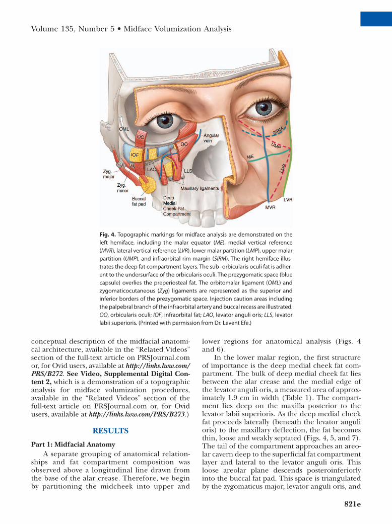
conceptual description of the midfacial anatomi-cal architecture, available in the “Related Videos” section of the full-text article on PRSJournal.com or, for Ovid users, available at http://links.lww.com/PRS/B272. See Video, Supplemental Digital Con-tent 2, which is a demonstration of a topographic analysis for midface volumization procedures, available in the “Related Videos” section of the full-text article on PRSJournal.com or, for Ovid users, available at http://links.lww.com/PRS/B273.)
RESULTS
Part 1: Midfacial AnatomyA separate grouping of anatomical relation-
ships and fat compartment composition was observed above a longitudinal line drawn from the base of the alar crease. Therefore, we begin by partitioning the midcheek into upper and
lower regions for anatomical analysis (Figs. 4 and 6).
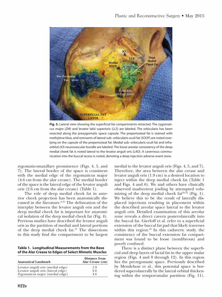
In the lower malar region, the first structure of importance is the deep medial cheek fat com-partment. The bulk of deep medial cheek fat lies between the alar crease and the medial edge of the levator anguli oris, a measured area of approx-imately 1.9 cm in width (Table 1). The compart-ment lies deep on the maxilla posterior to the levator labii superioris. As the deep medial cheek fat proceeds laterally (beneath the levator anguli oris) to the maxillary deflection, the fat becomes thin, loose and weakly septated (Figs. 4, 5, and 7). The tail of the compartment approaches an areo-lar cavern deep to the superficial fat compartment layer and lateral to the levator anguli oris. This loose areolar plane descends posteroinferiorly into the buccal fat pad. This space is triangulated by the zygomaticus major, levator anguli oris, and
Fig. 4. Topographic markings for midface analysis are demonstrated on the left hemiface, including the malar equator (ME), medial vertical reference (MVR), lateral vertical reference (LVR), lower malar partition (LMP), upper malar partition (UMP), and infraorbital rim margin (SIRM). The right hemiface illus-trates the deep fat compartment layers. The sub–orbicularis oculi fat is adher-ent to the undersurface of the orbicularis oculi. The prezygomatic space (blue capsule) overlies the preperiosteal fat. The orbitomalar ligament (OML) and zygomaticocutaneous (Zyg) ligaments are represented as the superior and inferior borders of the prezygomatic space. Injection caution areas including the palpebral branch of the infraorbital artery and buccal recess are illustrated. OO, orbicularis oculi; IOF, infraorbital fat; LAO, levator anguli oris; LLS, levator labii superioris. (Printed with permission from Dr. Levent Efe.)
822e
Plastic and Reconstructive Surgery • May 2015
zygomaticomaxillary prominence (Figs. 4, 5, and 7). The lateral border of the space is consistent with the medial edge of the zygomaticus major (4.6 cm from the alar crease). The medial border of the space is the lateral edge of the levator anguli oris (2.6 cm from the alar crease) (Table 1).
The role of deep medial cheek fat in ante-rior cheek projection has been anatomically dis-cussed in the literature.8,25 The delineation of the interplay between the levator anguli oris and the deep medial cheek fat is important for anatomi-cal isolation of the deep medial cheek fat (Fig. 4). Previous studies have described the levator anguli oris as the partition of medial and lateral portions of the deep medial cheek fat.18 The dissections in this study find the compartment to be largest
medial to the levator anguli oris (Figs. 4, 5, and 7). Therefore, the area between the alar crease and levator anguli oris (1.9 cm) is a desired location to inject within the deep medial cheek fat (Table 1 and Figs. 4 and 6). We and others have clinically observed inadvertent jowling by attempted volu-mizing of the deep medial cheek fat29,32 (Fig. 1). We believe this to be the result of laterally dis-placed injections resulting in placement within the described areolar space lateral to the levator anguli oris. Detailed examination of this areolar zone reveals a direct cavern posterolaterally into the buccal fat. Gierloff et al. refer to a superficial extension of the buccal fat pad that likely traverses within this region.18 In this cadaveric study, the consistency of the buccal extension fat compart-ment was found to be loose (nonfibrous) and poorly confined.
There is a distinct plane between the superfi-cial and deep layers of facial fat in the upper malar region (Figs. 4 and 8 through 12). In this region lies the prezygomatic space. Previously described by Mendelson et al., this potential space is bor-dered superolaterally by the lateral orbital thicken-ing within the temporomalar partition (Fig. 11).
Fig. 5. Lateral view showing the superficial fat compartments retracted. The zygomati-cus major (ZM) and levator labii superioris (LLS) are labeled. The orbicularis has been resected along the prezygomatic space capsule. The preperiosteal fat is stained with methylene blue, and remnants of lateral sub–orbicularis oculi fat (SOOF) are noted over-lying on the capsule of the preperiosteal fat. Medial sub–orbicularis oculi fat and infra-orbital (IO) neurovascular bundle are labeled. The loose areolar consistency of the deep medial cheek fat is noted lateral to the levator anguli oris (LAO). A cavernous commu-nication into the buccal recess is noted, denoting a deep injection adverse event zone.
Table 1. Longitudinal Measurements from the Base of the Alar Crease to Edges of Select Mimetic Muscles
Anatomical LandmarkDistance from
Alar Crease (cm)
Levator anguli oris (medial edge) 1.9 Levator anguli oris (lateral edge) 2.6 Zygomaticus major (medial edge) 4.6

Volume 135, Number 5 • Midface Volumization Analysis
823e
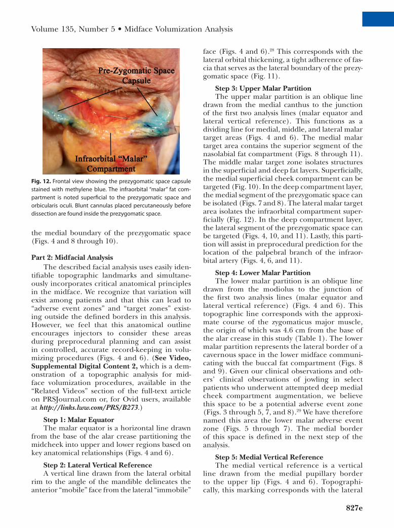
The roof is the obliquely oriented orbitomalar liga-ment. The space is bordered inferiorly by a fibrous network of zygomaticocutaneous ligaments and maxillary insertions of the orbicularis oculi form-ing a discernible capsule as previously reported by Mendelson30 (Figs. 4 and 10 through 12). This fascial encasement coalesces with the thick and well-demarcated capsule of the preperiosteal fat
to form a uniform prezygomatic space capsule. Adherent to the undersurface of the orbicularis oculi and superficial to the capsule is the loose and areolar sub–orbicularis oculi fat (Fig. 10). The prezygomatic space is a trapezoidal space, which becomes crescentoid during facial animation. The medial and middle superficial fat compart-ments were found to be fibrofatty in composition (Fig. 10). Both superficial compartments become thicker and denser laterally in the midface. Manual superolateral traction on the superficial compart-ment layer demonstrated ease of movement of the medial superficial cheek compartment over the suborbicularis contents. This gliding movement simulates the sequence occurring during facial animation. (See Video, Supplemental Digital Con-tent 1, which shows the conceptual description of the midfacial anatomical architecture, available in the “Related Videos” section of the full-text article on PRSJournal.com or, for Ovid users, available at http://links.lww.com/PRS/B272.)
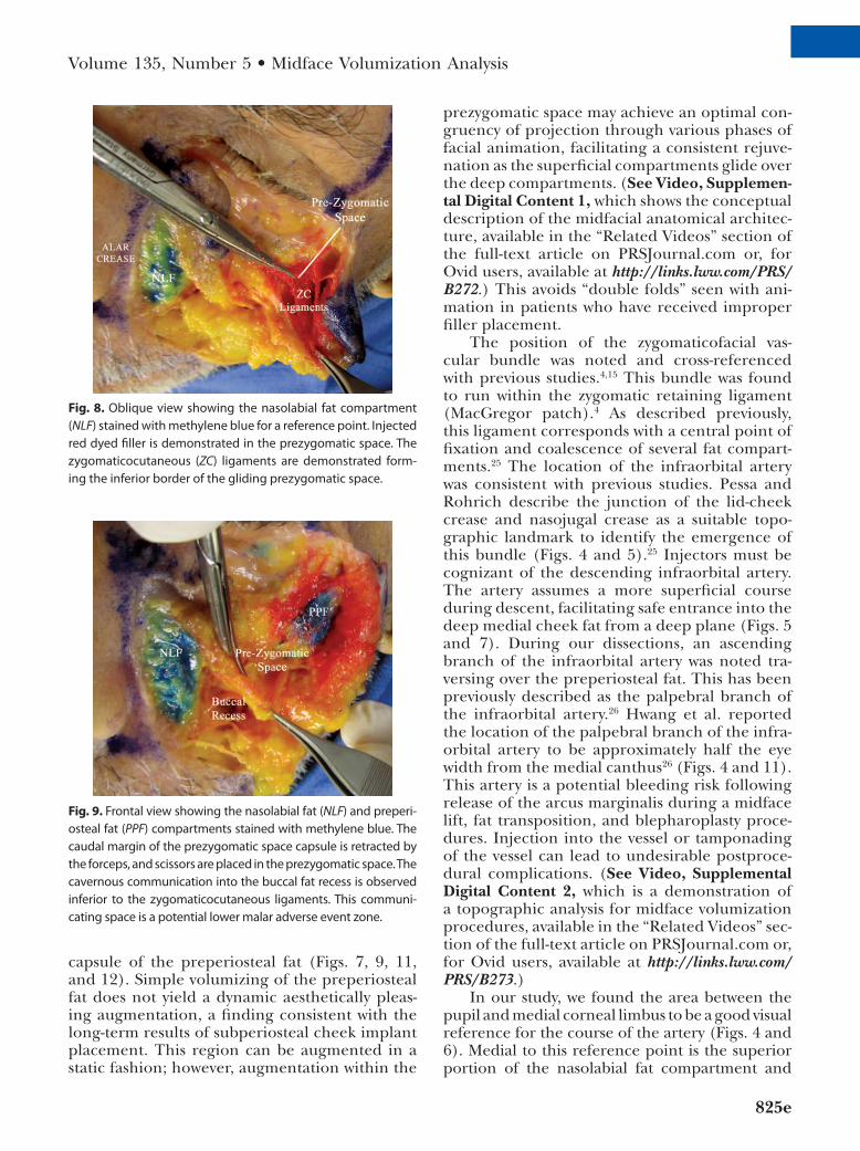
In an attempt to demonstrate the accessibility of this pocket through percutaneous injection, red-dyed hyaluronic acid filler was injected into this region before dissection in selected speci-mens (Figs. 8 and 12). To target this space, the cutaneous insertion of the zygomaticocutaneous ligaments was used as a guide for finding a point inferolateral to this surface anatomy (Fig. 1). The zygomaticocutaneous ligaments arborize through the orbicularis inserting onto the skin. Cephalic to this skin insertion and superficial to the orbicularis lies the infraorbital “malar” fat compartment (Fig. 2). Understanding this depth relationship is important because injec-tions placed in the infraorbital compartment are
Fig. 6. Topographic facial analysis markings are displayed. Malar equator (blue lines); LVR, lateral vertical reference (pink lines); MVR, medial vertical reference (green lines); UMP, upper malar partition (orange lines); LMP, lower malar partition (yellow lines); black rectangle, upper malar adverse event zone; AEZ, lower malar adverse event zone; MED, medial malar target area; MID, middle malar target area; LAT, lateral malar target area.
Video 1. Supplemental Digital Content 1 shows the conceptual description of the midfacial anatomical architecture, available in the “Related Videos” section of the full-text article on PRSJournal.com or, for Ovid users, available at http://links.lww.com/PRS/B272.

824e
Plastic and Reconstructive Surgery • May 2015
prone to poor aesthetic outcomes secondary to diminished lymphatic drainage in this region.25 This can result in iatrogenic malar mounds (Fig. 1). In this study, blunt cannulas consis-tently arrived at the deep prezygomatic space, positioned posterior to the orbicularis and ante-rior to the dense capsule of the preperiosteal fat (Figs. 11 and 12).
We postulate that the loss of volume pro-posed by Lambros occurs in the medial sub–orbi-cularis oculi fat and lateral sub–orbicularis oculi fat, contributing to the formation of the malar mound. Given the position and loose consistency of the sub–orbicularis oculi fat, targeted augmen-tations can be challenging. The specimens in this study had well-volumized fat within the tight
Video 2. Supplemental Digital Content 2 is a demonstration of a topographic analysis for midface volumization procedures, available in the “Related Videos” section of the full-text article on PRSJournal.com or, for Ovid users, available at http://links.lww.com/PRS/B273.
Fig. 7. Lateral view showing that the superficial fat compartment layer has been reflected. Demonstration of the deep medial cheek fat and medial sub–orbicularis oculi fat (SOOF) stained with methylene blue. Zygomaticus major (ZM), levator anguli oris (LAO), levator labii superioris (LLS), and levator labii superioris alaeque nasi (LLSAN) are labeled. Hyaluronic acid filler homogenized with red dye has been injected into the lat-eral sub–orbicularis oculi fat overlying the preperiosteal fat compartment.
Volume 135, Number 5 • Midface Volumization Analysis
825e
capsule of the preperiosteal fat (Figs. 7, 9, 11, and 12). Simple volumizing of the preperiosteal fat does not yield a dynamic aesthetically pleas-ing augmentation, a finding consistent with the long-term results of subperiosteal cheek implant placement. This region can be augmented in a static fashion; however, augmentation within the
prezygomatic space may achieve an optimal con-gruency of projection through various phases of facial animation, facilitating a consistent rejuve-nation as the superficial compartments glide over the deep compartments. (See Video, Supplemen-tal Digital Content 1, which shows the conceptual description of the midfacial anatomical architec-ture, available in the “Related Videos” section of the full-text article on PRSJournal.com or, for Ovid users, available at http://links.lww.com/PRS/B272.) This avoids “double folds” seen with ani-mation in patients who have received improper filler placement.
The position of the zygomaticofacial vas-cular bundle was noted and cross-referenced with previous studies.4,15 This bundle was found to run within the zygomatic retaining ligament (MacGregor patch).4 As described previously, this ligament corresponds with a central point of fixation and coalescence of several fat compart-ments.25 The location of the infraorbital artery was consistent with previous studies. Pessa and Rohrich describe the junction of the lid-cheek crease and nasojugal crease as a suitable topo-graphic landmark to identify the emergence of this bundle (Figs. 4 and 5).25 Injectors must be cognizant of the descending infraorbital artery. The artery assumes a more superficial course during descent, facilitating safe entrance into the deep medial cheek fat from a deep plane (Figs. 5 and 7). During our dissections, an ascending branch of the infraorbital artery was noted tra-versing over the preperiosteal fat. This has been previously described as the palpebral branch of the infraorbital artery.26 Hwang et al. reported the location of the palpebral branch of the infra-orbital artery to be approximately half the eye width from the medial canthus26 (Figs. 4 and 11). This artery is a potential bleeding risk following release of the arcus marginalis during a midface lift, fat transposition, and blepharoplasty proce-dures. Injection into the vessel or tamponading of the vessel can lead to undesirable postproce-dural complications. (See Video, Supplemental Digital Content 2, which is a demonstration of a topographic analysis for midface volumization procedures, available in the “Related Videos” sec-tion of the full-text article on PRSJournal.com or, for Ovid users, available at http://links.lww.com/PRS/B273.)
In our study, we found the area between the pupil and medial corneal limbus to be a good visual reference for the course of the artery (Figs. 4 and 6). Medial to this reference point is the superior portion of the nasolabial fat compartment and
Fig. 8. Oblique view showing the nasolabial fat compartment (NLF) stained with methylene blue for a reference point. Injected red dyed filler is demonstrated in the prezygomatic space. The zygomaticocutaneous (ZC) ligaments are demonstrated form-ing the inferior border of the gliding prezygomatic space.
Fig. 9. Frontal view showing the nasolabial fat (NLF) and preperi-osteal fat (PPF) compartments stained with methylene blue. The caudal margin of the prezygomatic space capsule is retracted by the forceps, and scissors are placed in the prezygomatic space. The cavernous communication into the buccal fat recess is observed inferior to the zygomaticocutaneous ligaments. This communi-cating space is a potential lower malar adverse event zone.

826e
Plastic and Reconstructive Surgery • May 2015
Fig. 10. Oblique view showing the nasolabial fat compartment stained with methy-lene blue for anatomical reference. The medial superficial fat compartment is seen as a separate layer from the deeper compartments. The instrument is inserted into the prezygomatic space. The inferior boundary of the space consists of a fibrous network of zygomaticocutaneous ligaments. The sub–orbicularis oculi fat (SOOF) lies on the under-surface of the orbicularis oculi, superficial to the prezygomatic space capsule. The pre-periosteal fat is visualized on the bone.
Fig. 11. Frontal view showing retroseptal fat and orbicularis oculi muscle labeled for anatomical reference. The lateral orbital thickening is shown as the adherent lateral border of the prezygomatic space. The preperiosteal fat has been stained with methy-lene blue and is noted in the floor of the prezygomatic space. The palpebral branch of the infraorbital artery is noted coursing through the medial sub–orbicularis oculi fat compartment.
Volume 135, Number 5 • Midface Volumization Analysis
827e
the medial boundary of the prezygomatic space (Figs. 4 and 8 through 10).
Part 2: Midfacial AnalysisThe described facial analysis uses easily iden-
tifiable topographic landmarks and simultane-ously incorporates critical anatomical principles in the midface. We recognize that variation will exist among patients and that this can lead to “adverse event zones” and “target zones” exist-ing outside the defined borders in this analysis. However, we feel that this anatomical outline encourages injectors to consider these areas during preprocedural planning and can assist in controlled, accurate record-keeping in volu-mizing procedures (Figs. 4 and 6). (See Video, Supplemental Digital Content 2, which is a dem-onstration of a topographic analysis for mid-face volumization procedures, available in the “Related Videos” section of the full-text article on PRSJournal.com or, for Ovid users, available at http://links.lww.com/PRS/B273.)
Step 1: Malar EquatorThe malar equator is a horizontal line drawn
from the base of the alar crease partitioning the midcheek into upper and lower regions based on key anatomical relationships (Figs. 4 and 6).
Step 2: Lateral Vertical ReferenceA vertical line drawn from the lateral orbital
rim to the angle of the mandible delineates the anterior “mobile” face from the lateral “immobile”
face (Figs. 4 and 6).28 This corresponds with the lateral orbital thickening, a tight adherence of fas-cia that serves as the lateral boundary of the prezy-gomatic space (Fig. 11).
Step 3: Upper Malar PartitionThe upper malar partition is an oblique line
drawn from the medial canthus to the junction of the first two analysis lines (malar equator and lateral vertical reference). This functions as a dividing line for medial, middle, and lateral malar target areas (Figs. 4 and 6). The medial malar target area contains the superior segment of the nasolabial fat compartment (Figs. 8 through 11). The middle malar target zone isolates structures in the superficial and deep fat layers. Superficially, the medial superficial cheek compartment can be targeted (Fig. 10). In the deep compartment layer, the medial segment of the prezygomatic space can be isolated (Figs. 7 and 8). The lateral malar target area isolates the infraorbital compartment super-ficially (Fig. 12). In the deep compartment layer, the lateral segment of the prezygomatic space can be targeted (Figs. 4, 10, and 11). Lastly, this parti-tion will assist in preprocedural prediction for the location of the palpebral branch of the infraor-bital artery (Figs. 4, 6, and 11).
Step 4: Lower Malar PartitionThe lower malar partition is an oblique line
drawn from the modiolus to the junction of the first two analysis lines (malar equator and lateral vertical reference) (Figs. 4 and 6). This topographic line corresponds with the approxi-mate course of the zygomaticus major muscle, the origin of which was 4.6 cm from the base of the alar crease in this study (Table 1). The lower malar partition represents the lateral border of a cavernous space in the lower midface communi-cating with the buccal fat compartment (Figs. 8 and 9). Given our clinical observations and oth-ers’ clinical observations of jowling in select patients who underwent attempted deep medial cheek compartment augmentation, we believe this space to be a potential adverse event zone (Figs. 3 through 5, 7, and 8).29 We have therefore named this area the lower malar adverse event zone (Figs. 5 through 7). The medial border of this space is defined in the next step of the analysis.
Step 5: Medial Vertical ReferenceThe medial vertical reference is a vertical
line drawn from the medial pupillary border to the upper lip (Figs. 4 and 6). Topographi-cally, this marking corresponds with the lateral
Fig. 12. Frontal view showing the prezygomatic space capsule stained with methylene blue. The infraorbital “malar” fat com-partment is noted superficial to the prezygomatic space and orbicularis oculi. Blunt cannulas placed percutaneously before dissection are found inside the prezygomatic space.
828e
Plastic and Reconstructive Surgery • May 2015
border of the levator anguli oris muscle, which lies approximately 2.6 cm from the base of the alar crease (Table 1). The medial vertical ref-erence is critical for multiple reasons. First, it functions as the medial border of the lower malar adverse event zone (Figs. 4 through 6). The region medial to this line is where we rec-ommend injectors target the deep medial cheek fat compartment, as this is where the compart-ment is most compact and may decrease the chance of filler migration laterally into the lower malar adverse event zone (Figs. 5 and 7). As the line ascends into the upper malar region it will intersect with the upper malar partition (Figs. 4 and 6). Topographically, this inter-secting region represents the location of the ascending palpebral branch of the infraorbital artery. We have clinically observed intraarterial injections of this vessel following a volumizing procedure and therefore have delineated this intersection a potential upper malar adverse event zone (Figs. 5, 6, and 11).
CONCLUSIONSA sound understanding of the midfacial anat-
omy is essential to achieve optimal filler or fat augmentation and minimize adverse outcomes. This cadaveric study yields a detailed visual rep-resentation of critical anatomical relationships in the midface. We define the clinical relevance of the prezygomatic space, palpebral branch of the infraorbital artery, and facial fat compartments as they relate to facial injections and fat grafting. We address the anatomical relationships involved in iatrogenic malar mounds. The described stepwise analysis provides surgeons with a scientific system to identify adverse event zones and safe harbors in midface augmentation. These zones are not absolute, as each patient is unique. This informa-tion offers the aesthetic surgeon an anatomically inspired outline to assist in the delivery of safe, accurate, and reproducible dynamic rejuvenation through volumization.
Christopher C. Surek, D.O.Department of Plastic Surgery
University of Kansas Medical Center3901 Rainbow Boulevard, Mailstop 3015
Kansas City, Kan. [email protected]
(Requests for Reprints)
Jerome Lamb, M.D.Private Practice
4820 South Arrowhead DriveIndependence, Mo. 64055
ACkNOwLEDgMENTThe supplemental digital content videos were funded
by the University of Kansas Department of Plastic Sur-gery. The medical illustrations were funded by Jerome Lamb, M.D.
PATIENT CONSENTPatients provided written consent for the use of their
images.
REfERENCES 1. Rohrich RJ, Pessa JE. The fat compartments of the face:
Anatomy and clinical implications for cosmetic surgery. Plast Reconstr Surg. 2007;119:2219–2227; discussion 2228.
2. Aiache AE, Ramirez OH. The suborbicularis oculi fat pads: An anatomic and clinical study. Plast Reconstr Surg. 1995;95:37–42.
3. Stuzin JM, Baker TJ, Gordon HL. The relationship of the superficial and deep facial fascias: Relevance to rhytidectomy and aging. Plast Reconstr Surg. 1992;89:441–449; discussion 450.
4. Furnas DW. The retaining ligaments of the cheek. Plast Reconstr Surg. 1989;83:11–16.
5. Stuzin JM, Wagstrom L, Kawamoto HK, Baker TJ, Wolfe SA. The anatomy and clinical applications of the buccal fat pad. Plast Reconstr Surg. 1990;85:29–37.
6. Jackson I. Anatomy of the buccal fat pad and its clinical sig-nificance. Plast Reconstr Surg. 1999;103:2059–2060.
7. Furnas DW. Festoons, mounds, and bags of the eyelids and cheek. Clin Plast Surg. 1993;20:367–385.
8. Rohrich RJ, Pessa JE, Ristow B. The youthful cheek and the deep medial fat compartment. Plast Reconstr Surg. 2008;121:2107–2112.
9. Raskin E, LaTrenta G. Why do we age our face? Aesthetic Plast Surg. 2007;1:19–28.
10. Rohrich RJ, Pessa JE. The retaining system of the face: Histo-logic evaluation of the septal boundaries of the subcutaneous fat compartments. Plast Reconstr Surg. 2008;121:1804–1809.
11. Little J. Volumetric perceptions in midfacial aging with altered priorities for rejuvenation. Plast Reconstr Surg. 2000;105:252–266.
12. Schaverien MV, Pessa JE, Rohrich RJ. Vascularized membranes determine the anatomical boundaries of the subcutaneous fat compartments. Plast Reconstr Surg. 2009;123:695–700.
13. Rohrich RJ, Arbique GM, Wong C, Brown S, Pessa JE. The anatomy of suborbicularis fat: Implications for periorbital rejuvenation. Plast Reconstr Surg. 2009;124:946–951.
14. Lambros V. Models of facial aging and implications for treat-ment. Clin Plast Surg. 2008;35:319–327; discussion 317.
15. Mendelson BC, Muzaffar AR, Adams WP Jr. Surgical anat-omy of the midcheek and malar mounds. Plast Reconstr Surg. 2002;110:885–896; discussion 897.
16. Mendelson BC, Jacobson S. Surgical anatomy of the mid-cheek: Facial layers, spaces, and the midcheek segments. Clin Plast Surg. 2008;35:395–404; discussion 393.
17. Ghavami A, Pessa JE, Janis J, Khosla R, Reece EM, Rohrich RJ. The orbicularis retaining ligament of the medial orbit: Closing the circle. Plast Reconstr Surg. 2008;121:994–1001.
18. Gierloff M, Stöhring C, Buder T, Gassling V, Açil Y, Wiltfang J. Aging changes of the midfacial fat compart-ments: A computed tomographic study. Plast Reconstr Surg. 2012;129:263–273.
19. Matarasso A. Anatomy of the buccal fat pad and its clinical significance. Plast Reconstr Surg. 1999;103:2061–2063.
Volume 135, Number 5 • Midface Volumization Analysis
829e
20. Zhang HM, Yan YP, Qi KM, Wang JQ, Liu ZF. Anatomical structure of the buccal fat pad and its clinical application. Plast Reconstr Surg. 2002;109:2509–2518.
21. Fitzgerald R, Graivier MH, Kane M, et al. Update on facial aging. Aesthet Surg J. 2010;30(Suppl):11S–24S.
22. Coleman SR, Grover R. The anatomy of the aging face: Volume loss and changes in 3-dimensional topography. Aesthet Surg J. 2006;26(Suppl):S4–S9.
23. Guisantes E. Autologous Adipose Tissue Grafts in HIV Facial Lipoatrophy: Comparison of Two Strategies for Treatment and Usefulness of Computerized Tomography in the Evaluation of Its Durability (dissertation, directed by Joan Fontdevila). Barcelona, Spain: University of Barcelona; .
24. Fontdevila J. Treatment of HIV-related lipodystrophy. In: Coleman S, Mazzola R, eds. Fat Injection: From Filling to Regeneration. St. Louis: Quality Medical Publishing; 2009.
25. Pessa J, Rohrich R. Facial Topography: Clinical Anatomy of the Face. St. Louis: Quality Medical Publishing; 2012.
26. Hwang K, Kim DH, Huan F, Nam YS, Han SH. The anatomy of the palpebral branch of the infraorbital artery relating to midface lift. J Craniofac Surg. 2011;22:1489–1490.
27. Wong CH, Hsieh MK, Mendelson B. The tear trough liga-ment: Anatomical basis of the tear trough deformity. Plast Reconstr Surg. 2012;129:1392–1402.
28. Mendelson B, Wong C. Anatomy of the aging face. In: Plastic Surgery. Vol. 2. 3rd ed. Philadelphia: Elsevier, 2012; 78–92.
29. Lambros V. Personal communication, 2013. 30. Mendelson B. Personal communication, 2014. 31. Marten T. Personal communication, 2013. 32. American Society for Aesthetic Plastic Surgery. Anatomy of
the facial fat compartments: A new interpretation. RADAR resource. Available at: Accessed March 4, 2014.


![Palpebral Involvement as a Presenting and Sole ...downloads.hindawi.com/journals/tswj/2010/672487.pdfdermatomyositis[9], granuloma annulare[10], and granuloma faciale[11]. Palpebral](https://img.pdfslide.us/doc/110x75/5e5d2f5139526a648b02a0fa/palpebral-involvement-as-a-presenting-and-sole-dermatomyositis9-granuloma.jpg)