Embed Size (px)
Citation preview
51
Current Science International 3(2): 51-64, 2014
ISSN: 2077-4435
Corresponding Author: RedaNofel, Assistant Professor of Oral and Maxillofacial Surgery, Faculty of Dentistry, Alazher
University. Assiut.
E-mail: [email protected]
Rehabilitation of Neurosensory Changes in the Infraorbital Nerve Following Zygomatic
Fractures
1Reda Nofel,
2Yasser Ibrahim Seada,
3Waleed Talat Mansour
1Assistant Professor of Oral and Maxillofacial Surgery, Faculty of Dentistry, Alazher University. Assiut.
2Assistant professor of physical therapy, Department of Neuromuscular disorder and its surgery. Faculty of
physical therapy, Cairo University. Cairo.
3Assistant professor of physical therapy, Department of Neuromuscular disorder and its surgery. Faculty of
physical therapy, Cairo University. Cairo
ABSTRACT
The purpose of the study was designed to clarify the modern trends of neurosensory rehabilitation in
treatment of infraorbital nerve changes following zygomatic fractures. In this respect, neurosensory function
was assessed with calibrated nylon monofilaments, electrical stimulation, heat detection thresholds and response
to pin prick in the infraorbital, supraorbital and mental nerve regions in both sexes. Subjects, thirty males and
females were the same degree of fractures severity according to Henderson's classification (grade1, 2& 3), their
age ranged from 25-45years old and their weight ranged from 60-88Kg.They were randomly divided into two
equal groups (G1and G2). G1, consists of 15 patients of both sexes and was treated by surgical procedures
(reduction and fixation) and G2 consists of 15 patients was treated by the same surgical procedures and
electrical neuromuscular stimulation with exercises therapy program. Vital signs as blood pressure, body
temperature, pulse rate and respiratory rate measured before and after the treatment sessions. Assessments,
visual analogue scale was used to measure degree of pain, Semmes-Weinstein monofilaments was used to
measure the light touch sensation, Aesthesiometer 2 point calliper was used to measure the two point
discrimination sensation, Peltier probe was used to measure heat detection sensation. Moreover, by the use an
ascending test of phyaction guidance-c system to measure the electrical detection threshold. Statistically the
results for all groups were analyzed by t-test to compare the differences between the two groups. The statistical
package of social sciences (SPSS, version10) used for data processing using the p-value 0.05 as a level of
significance. Results, showed that there were significant improvements in all variables in both groups in a
favour to G2. Therefore, we concluded that, the use of surgical procedures combined with neuromuscular
electrical stimulation and exercises program were the good method and open a new link to improve the recovery
of infraorbital nerve sensory changes following zygomatic fractures.
Key words: Infraorbital Nerve, Zygomatic Fractures, Neuromuscular Electrical Nerve Stimulation,
Aesthiometer, Pain and Sensory Dysfunctions.
Introduction
The zygomatic bone provides prominence to the cheek which leads to its increased chances of fracture and
the infraorbital nerve is often involved in the trauma to the zygomatic complex resulting in the sensory
disturbance of the area innervated by itLund, (1971).
The zygoma articulates with the frontal sphenoid, temporal and maxillary bones and contributes
significantly to the strength and stability of the midface (Finlay et al., 1984).
The zygoma may be separated from its four articulations. This is called a zygomatic complex fracture. The
terms trimalar or tripod fracture are therefore inaccurate. These terms reflect an inability to easily identify the
orbital (zygomaticosphenoid) portion of the injury before the advent of computed tomography. The zygomatic
arch may be fractured independently or as part of a zygomatic complex fracture (Lund, 1971).
The zygoma has four projections, which create a quadrangular shape the frontal, temporal, maxillary, and
the infraorbital rim. The zygoma articulates with four bones: the frontal, temporal, maxilla, and
sphenoid(Jungell and Lindqvist, 1987).
The zygomatic arch includes the temporal process of the zygoma and the zygomatic process of the temporal
bone. The glenoid fossa and articular eminence are located at the posterior aspect of the zygomatic process of
the temporal bone(Jungell and Lindqvist, 1987).The infraorbital nerve (IO) passes through the orbital floor and
exits at the infraorbital foramen. It provides sensation to the anterior cheek, lateral nose, upper lip, and maxillary
anterior teeth. Muscles of facial expression originating from the zygoma include the zygomaticus major and
labiisuperioris. They are innervated by cranial nerve VII (Ellies et al., 1985). Following orbitozygomatic
52 Curr. Sci. Int. 3(2): 51-64, 2014
complex (OZC) fractures, the reported incidence of long-term sensory disturbances of the inferaorbital
nerve varies between 24% and 50% (Ellies et al., 1985).
Zygomatic fractures are not life threatening and are usually treated after more serious injuries are stabilized
and swelling has resolved 4 to 5 days after injuries. Initial evaluation of the patient with a zygomatic fracture
includes documentation of the bony injury and the status of surrounding soft tissue (eyelids, lacrimal apparatus,
canthal tendons, and globe) and cranial nerves II to VI (Becelli et al., 2002).
Also, Visual acuity and the status of the globe and retina should be established; an ophthalmologist should
be consulted for suspected In a recent surveillance study from(Lund, 1971) reported an annual incidence of
139.4 cases per 100,000 in females and 67.2 cases per 100,000 in males, with a female to male ratio of 2.07 29.
Overall prevalence of 3.0–5.8% among women and 0.6–2.1% among men have been found in general
population(Jungell and Lindqvist, 1987).
Treatment of zygomatic fractures must be based on a complete preoperative evaluation. This includes a CT
scan with axial and coronal images to fully appreciate the nature of the injury. Management of zygomatic
complex and zygomatic arch fractures depends on the degree of displacement and the resultant esthetic and
functional deficits. Treatment may therefore range from simple observation of resolving swelling, extraocular
muscle dysfunction, and paresthesia to open reduction and internal fixation of multiple fractures (Essick,1992).
Some studies on the long-term effects of treatment methods on sensory function have suggested that the
treatment of isolated simple OZC fractures consisting of open reduction and miniplate fixation, yields better
recovery of sensory function than (i) open reduction and interosseous wire fixation (ii) open reduction and
support with an intra-antral Foley catheter or (iii) closed reduction without fixation (Man and Bax,1988).
The aims of the present study were to investigate sensory changes in the IO nerve following zygomatic
fractures employing advanced qualitative and quantitative sensory testing (thermal, electrical and mechanical) ,
over a 6-month period. These parameters were correlated to the fracture severity and treatment modality. The
advantage of multimodal testing is the ability to differentiate between largely mechanosensitive neurons (AB
fibers) by employing electrical stimuli and fine nylon filaments, pinprick and thermal selectivity activate
nociceptors (AD and C fibers) . In addition, we assessed the presence of chronic orofacial neuropathic pain at 6
months.
Material and Methods
Patients admitted to the department of oral and maxillofacial surgery, King Khalid hospital, Najran, with
isolate unilateral fracture of the zygomatic arch, zygomatic complex, or rim were the patients of our study.
The patients were examined as soon as possible following the injury (from 3-12 hours), then at 1 and 6
months after surgery. Following the initial assessment, the patients underwent surgical management in the
department of Oral and Maxillofacial Surgery within 5 days. The evaluator was blinded as to the surgical
management of the patient, and the surgeon was not allowed access to the results of the sensory assessment.
Fracturers were diagnosed clinically and radiologically (X-ray or CT). Indications for reduction of fractures
are well documented and are based on the signs and symptoms and functional impairment. These signs and
symptoms include visible facial asymmetry, significant functional disturbance of mandibular movement,
disturbance of vision (diplopia) or eye movements, and IO nerve dysfunction. A lack of these signs is therefore
a contraindication for surgical intervention. In all treated cases closed reduction was attempted, and if this
resulted in a stable reduction of the fragments , when reduction was unstable, the zygomaticofrontal suture was
surgically exposed and with wires or a miniplate . Postoperatively, cases were clinically and radiologically.
Sensory threshold was quantified for each of the modalities bilaterally in the supraorbital (SO), IO and mental
(MNT) nerve regions.
Thirty consecutive patients following orbitozygomatic complex (OZC) fractures with regard to the sensory
function of the inferaorbital nerve. Mean patient's age was 36.6 years with a median of 35.5 years. Ten of the
patients were female and ten male.
Inclusion criteria:
Patients were selected if (1) they had a unilateral fracture, because the appropriate no affected site served as
an internal control in two-point discrimination tests, (2) their fracture was of type III, IV and V according to
Henderson's classification and (3) appropriate treatment with respect to type of fracture was employed, i.e., the
patient did not refuse the indicated surgical intervention.
Exclusion criteria:
Patients with multiple facial fractures were excluded. These signs and symptoms include visible facial
asymmetry, significant functional disturbance of mandibular movement, disturbance of vision (eg, diplopia) or
53 Curr. Sci. Int. 3(2): 51-64, 2014
of eye movements, and infraorbital nerve dysfunction. Severe degree of disabilities, patients having
complications, psychological unstable ,non co-operative patients during assessment of the research.
A lack of these signs is therefore a contraindication for surgical intervention The patients were examined as
soon as possible following the injury, then at 1 and 6 months after surgery.
Following the initial assessment, the patients underwent surgical management within 1 or 2 days.
Fractures were diagnosed clinically and radiographically. Routine radiographs included a Water’s view, an
submental vertex (SMV), and computed tomography (CT) are usually performed
Indications for reduction of fractures are well documented and are based on the signs, symptoms, and
functional impairment.
They had no signs of aphasia , they had sufficient vision and hearing , the patients were randomly and
equally divided into two groups. Group (1): consists of 15 patients of both sexes who received surgical
interference only. Group (2): consists of 15 patients of both sexes who received surgical interference,
neuromuscular electrical nerve stimulation (NMES)20Hz,15min.50%intenistyand strengthening exercises,
Faciltatory techniques (brief icing, tapping and scratching) on the affected area of face and postural correction
exercises of head and neck. Time of exercises 30 min, three times per week, day after day.
The neurosensory evaluation:
While the patient was sitting comfortably in a dental chair, the examiner tested in turn (1) the mechanical
sensation, (a) light touch sensation was tested using small kit of semmes-weinnstein monofilament (SWMs)
(North Coast Medical, Inc., San Jose, CA, USA), (b) two point discrimination was examined with the
Aesthiometer 2 points (North Coast Medical, Inc.) a form of sharp pointed caliper, by which higher threshod
afferent fibres related to touch sensation and nociceptive afferents may be stimulated, (2) heat sensation (hot and
cold) by ethylchloride saturated dental swab, (3) pain sensation by gauge needle (4) electrical detection
sensation. Patients were asked whether they had any altered sensation in facial areas innervated by the
infraorbital nerve, and the reports were classified according to the following definitions. Dysaesthesia was
characterized by altered quality of sensation that include an uncomfortable component. Anaesthesia was related
to the complete absence of sensation.
Mechanical detection threshold:
The orofacial region is very sensitive to mechanical stimulation, and standard sets, we employed proline
monofilament (Ethicon, Somerville) that exerted clinically relevant forces for sensory testing in the face. To
ensure standardization between monofilament we tested the force applied by different lengths of monofilament
10 times in 5 filament of each length.
Mechanical detection was assessed in the patients by administering the series of filaments 2 times each in
ascending order. With eyes closed, patients indicated each time a filament touch was detected in the MNT, IO
and SO areas of distribution (Eliav et al., 2003).
Heat detection threshold:
Detection threshold for heat stimuli were evaluated by a 5mm water cooled peltier probe using a staircase
paradigm in which stimulus intensity (temperature) was alternately increased on successive trials until a
sensation was evoked, and decreased until no sensation was perceived. After each change in direction, the
amount of stimulus change from each trial was reduced by 50%, and the ascending and descending trials were
repeated until this increment was reduced to 0.10c. In this series the starting temperature was 30oc and increased
gradually with a mean of 32.5oc (Eliav et al., 2003).
In order to test cold sensation, ethyl chloride vapour was sprayed onto a spherical dental cotton bud
(diameter:5mm). After ice crystals had been formed, the bud was placed on the site for at most 1second. As
determined by a small type k thermocouple (Fluke 80 TK, Eindhoven, The Netherlands), a new steady state
temperature of the skin was reached within just a second. The drop in temperature varied within a range from 22
to 24oc at the interface between cotton bud and skin. In addition to thermoreceptors, nociceptive afferents may
have been stimulated. Sensory function related to temperature was considered to be normal if two successive
positive responses in four tests were obtained (50%) (Eliav et al., 2003).
Electrical detection threshold:
For electrical detection threshold, continuous trains of constant- current electrical stimuli were delivered to
the skin through 10mm diameter spherical gold- plated electrodes spaced 25mm apart. Stimulus frequency was
20Hz with a 50% duty cycle. Polarity of the electrodes were randomized. Detection thresholds were assessed by
54 Curr. Sci. Int. 3(2): 51-64, 2014
an ascending method of limits, phyaction Guidance-C System, Bilzen, Belgium, SN62851. Stimulating current
was increased at a fixed rate until the subject indicated detection. Three detection thresholds were evaluated for
each location and the mean calculated and used for data analysis. Results are expressed as ratios between the
injured side and the control side (Eliav et al., 2003).
In normal situation the ratio would not be expected to be different from a value of higher ratios indicate
relative hypothesia of the injuried side and lower ratios indicate hyperesthesia.
Two point discrimination:
It was examined with Aesthesiometer 2 point, each of the tests consists of four alternating series with either
ascending or descending increments with a successively longer or shorter pin distance in the device, during
which the patient reported on a present or absent sensation of two separate points of stimulation. A test series
was terminated after a response reversal, i.e. when a particular type of response (positive/negative) on
successive increments. The threshold for two point discrimination was calculated as the mean of 8 pin distances
around the four reversals from the test series(Eliav et al., 2003).
Reaction to pinprick:
The tip of a 0.2mm diameter blunted acupuncture needle was pushed against the patients skin until the
needle slightly bends (the skin was dimpled but not penetrated). Under these conditions the bended needle exerts
a mean force of 10 (±0.5) Newton as measured on a laboratory scale. The patient graded the sensation on a
100mm visual analog scale (VAS) where 0 represented no sensation and 10 represented the strongest pain.
Results were recorded as the difference in VAS values between the control and injuriedsides (Eliav et al., 2003).
Statistical analysis:
The results of two groups were statistically analyzed by t-test to compare the differences within each group
and between the two groups. The statistical package of social science (SPSS version 10) was used for data
processing the P-value 0.05 level significance.
Data Summarized by using:
The arithmetic mean average describing the central tendency of observation where The standard deviation
(S.D) used to measure to described the results around mean where paired and unpaired t-test was performed to
determine the significance difference pre and post within the same group and the differences between the two
groups.
Results:
Subjects Characteristics:
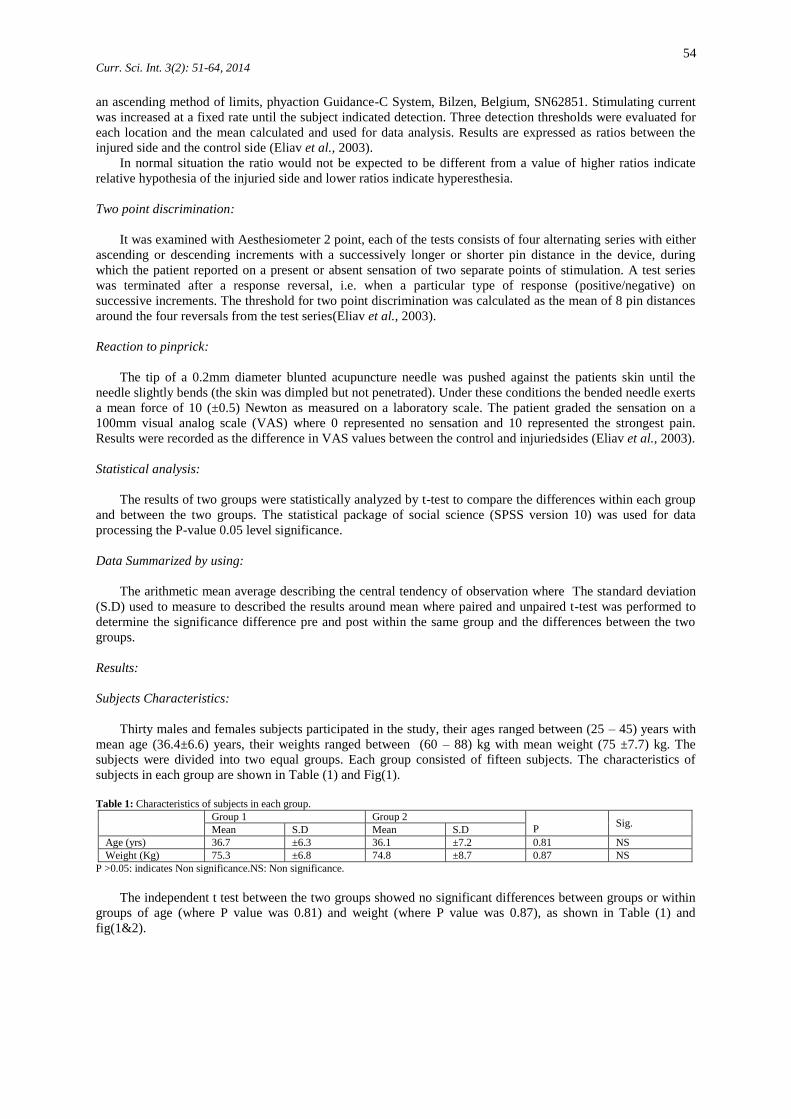
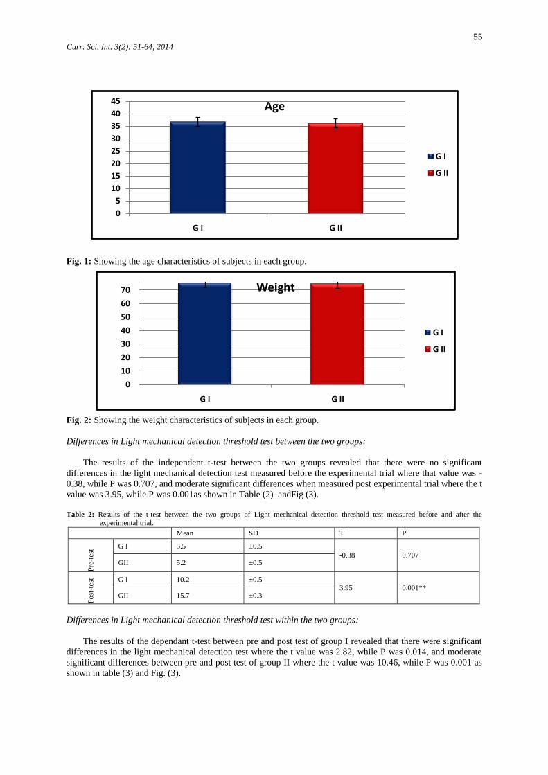
Thirty males and females subjects participated in the study, their ages ranged between (25 – 45) years with
mean age (36.4±6.6) years, their weights ranged between (60 – 88) kg with mean weight (75 ±7.7) kg. The
subjects were divided into two equal groups. Each group consisted of fifteen subjects. The characteristics of
subjects in each group are shown in Table (1) and Fig(1).
Table 1: Characteristics of subjects in each group.
Group 1 Group 2 P
Sig. Mean S.D Mean S.D
Age (yrs) 36.7 ±6.3 36.1 ±7.2 0.81 NS
Weight (Kg) 75.3 ±6.8 74.8 ±8.7 0.87 NS
P >0.05: indicates Non significance.NS: Non significance.
The independent t test between the two groups showed no significant differences between groups or within
groups of age (where P value was 0.81) and weight (where P value was 0.87), as shown in Table (1) and
fig(1&2).
55 Curr. Sci. Int. 3(2): 51-64, 2014
Fig. 1: Showing the age characteristics of subjects in each group.
Fig. 2: Showing the weight characteristics of subjects in each group.
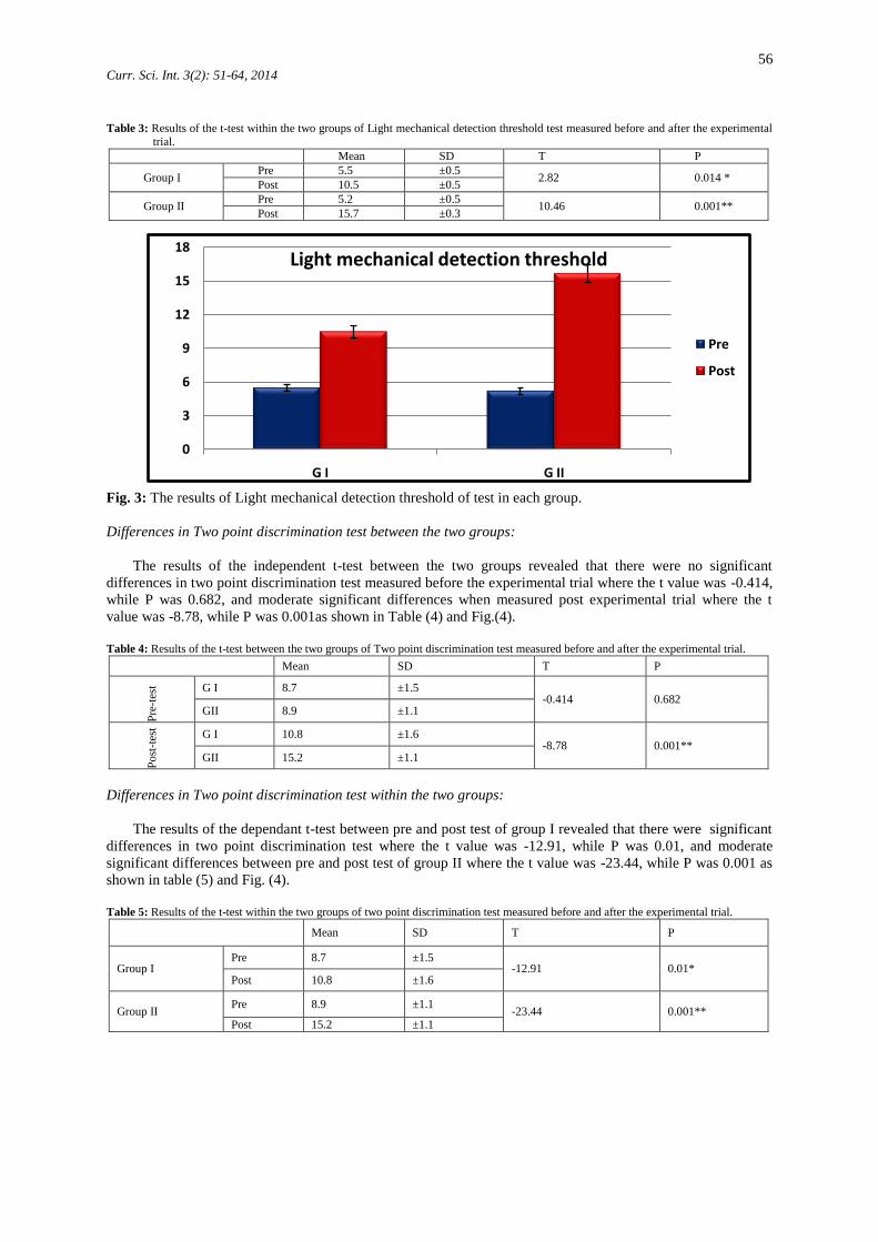
Differences in Light mechanical detection threshold test between the two groups:
The results of the independent t-test between the two groups revealed that there were no significant
differences in the light mechanical detection test measured before the experimental trial where that value was -
0.38, while P was 0.707, and moderate significant differences when measured post experimental trial where the t
value was 3.95, while P was 0.001as shown in Table (2) andFig (3).
Table 2: Results of the t-test between the two groups of Light mechanical detection threshold test measured before and after the
experimental trial.
Mean SD T P
Pre
-tes
t G I 5.5 ±0.5
-0.38 0.707 GII 5.2 ±0.5
Po
st-t
est G I 10.2 ±0.5
3.95 0.001** GII 15.7 ±0.3
Differences in Light mechanical detection threshold test within the two groups:
The results of the dependant t-test between pre and post test of group I revealed that there were significant
differences in the light mechanical detection test where the t value was 2.82, while P was 0.014, and moderate
significant differences between pre and post test of group II where the t value was 10.46, while P was 0.001 as
shown in table (3) and Fig. (3).
0
10
20
30
40
50
60
70
G I G II
Weight
G I
G II
0
5
10
15
20
25
30
35
40
45
G I G II
Age
G I
G II
56 Curr. Sci. Int. 3(2): 51-64, 2014
Table 3: Results of the t-test within the two groups of Light mechanical detection threshold test measured before and after the experimental
trial.
Mean SD T P
Group I Pre 5.5 ±0.5
2.82 0.014 * Post 10.5 ±0.5
Group II Pre 5.2 ±0.5
10.46 0.001** Post 15.7 ±0.3
Fig. 3: The results of Light mechanical detection threshold of test in each group.
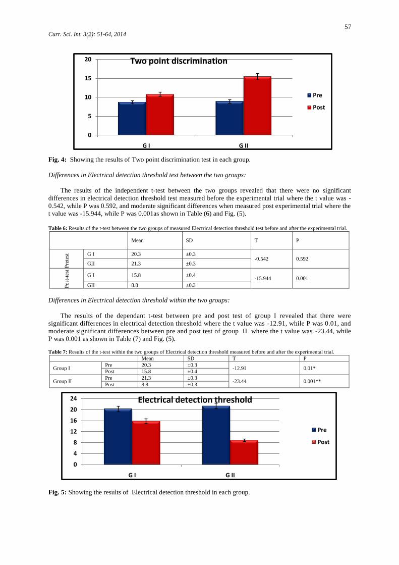
Differences in Two point discrimination test between the two groups:
The results of the independent t-test between the two groups revealed that there were no significant
differences in two point discrimination test measured before the experimental trial where the t value was -0.414,
while P was 0.682, and moderate significant differences when measured post experimental trial where the t
value was -8.78, while P was 0.001as shown in Table (4) and Fig.(4).
Table 4: Results of the t-test between the two groups of Two point discrimination test measured before and after the experimental trial.
Mean SD T P
Pre
-tes
t G I 8.7 ±1.5 -0.414 0.682
GII 8.9 ±1.1
Po
st-t
est
G I 10.8 ±1.6 -8.78 0.001**
GII 15.2 ±1.1
Differences in Two point discrimination test within the two groups:
The results of the dependant t-test between pre and post test of group I revealed that there were significant
differences in two point discrimination test where the t value was -12.91, while P was 0.01, and moderate
significant differences between pre and post test of group II where the t value was -23.44, while P was 0.001 as
shown in table (5) and Fig. (4).
Table 5: Results of the t-test within the two groups of two point discrimination test measured before and after the experimental trial.
Mean SD T P
Group I Pre 8.7 ±1.5
-12.91 0.01* Post 10.8 ±1.6
Group II Pre 8.9 ±1.1
-23.44 0.001** Post 15.2 ±1.1
0
3
6
9
12
15
18
G I G II
Light mechanical detection threshold
Pre
Post
57 Curr. Sci. Int. 3(2): 51-64, 2014
Fig. 4: Showing the results of Two point discrimination test in each group.
Differences in Electrical detection threshold test between the two groups:
The results of the independent t-test between the two groups revealed that there were no significant
differences in electrical detection threshold test measured before the experimental trial where the t value was -
0.542, while P was 0.592, and moderate significant differences when measured post experimental trial where the
t value was -15.944, while P was 0.001as shown in Table (6) and Fig. (5).
Table 6: Results of the t-test between the two groups of measured Electrical detection threshold test before and after the experimental trial.
Mean SD T P
Pre
test
G I 20.3 ±0.3 -0.542 0.592
GII 21.3 ±0.3
Po
st-t
est
G I 15.8 ±0.4 -15.944 0.001
GII 8.8 ±0.3
Differences in Electrical detection threshold within the two groups:
The results of the dependant t-test between pre and post test of group I revealed that there were
significant differences in electrical detection threshold where the t value was -12.91, while P was 0.01, and
moderate significant differences between pre and post test of group II where the t value was -23.44, while
P was 0.001 as shown in Table (7) and Fig. (5).
Table 7: Results of the t-test within the two groups of Electrical detection threshold measured before and after the experimental trial.
Mean SD T P
Group I Pre 20.3 ±0.3
-12.91 0.01* Post 15.8 ±0.4
Group II Pre 21.3 ±0.3
-23.44 0.001** Post 8.8 ±0.3
Fig. 5: Showing the results of Electrical detection threshold in each group.
0
5
10
15
20
G I G II
Two point discrimination
Pre
Post
0
4
8
12
16
20
24
G I G II
Electrical detection threshold
Pre
Post
58 Curr. Sci. Int. 3(2): 51-64, 2014
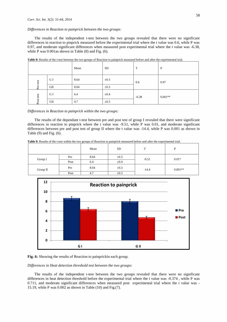
Differences in Reaction to painprick between the two groups:
The results of the independent t-test between the two groups revealed that there were no significant
differences in reaction to pinprick measured before the experimental trial where the t value was 0.6, while P was
0.97, and moderate significant differences when measured post experimental trial where the t value was -6.38,
while P was 0.001as shown in Table (8) and Fig. (6).
Table 8: Results of the t-test between the two groups of Reaction to painprick measured before and after the experimental trial.
Mean SD T P
Pre
-tes
t G I 8.64 ±0.3 0.6 0.97
GII 8.04 ±0.3
Po
st-t
est G I 6.4 ±0.4
-6.38 0.001**
GII 4.7 ±0.3
Differences in Reaction to painprick within the two groups:
The results of the dependant t-test between pre and post test of group I revealed that there were significant
differences in reaction to pinprick where the t value was -9.51, while P was 0.01, and moderate significant
differences between pre and post test of group II where the t value was -14.4, while P was 0.001 as shown in
Table (9) and Fig. (6).
Table 9: Results of the t-test within the two groups of Reaction to painprick measured before and after the experimental trial.
Mean SD T P
Group I Pre 8.64 ±0.3
-9.51 0.01* Post 6.4 ±0.4
Group II Pre 8.04 ±0.3
-14.4 0.001**
Post 4.7 ±0.3
Fig. 6: Showing the results of Reaction to painprickin each group.
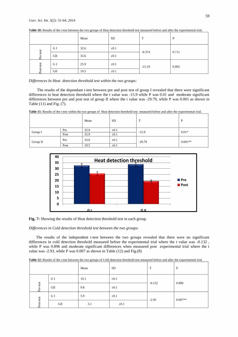
Differences in Heat detection threshold test between the two groups:
The results of the independent t-test between the two groups revealed that there were no significant
differences in heat detection threshold before the experimental trial where the t value was -0.374 , while P was
0.711, and moderate significant differences when measured post experimental trial where the t value was -
15.19, while P was 0.002 as shown in Table (10) and Fig.(7).
0
2
4
6
8
10
12
G I G II
Reaction to painprick
Pre
Post
59 Curr. Sci. Int. 3(2): 51-64, 2014
Table 10: Results of the t-test between the two groups of Heat detection threshold test measured before and after the experimental trial.
Mean SD T P P
re-t
est G I 32.6 ±0.1
-0.374 0.711
GII 33.6 ±0.1
Po
st-t
est
G I 25.9 ±0.1 -15.19 0.002
GII 19.5 ±0.1
Differences In Heat detection threshold test within the two groups:
The results of the dependant t-test between pre and post test of group I revealed that there were significant
differences in heat detection threshold where the t value was -15.9 while P was 0.01 and moderate significant
differences between pre and post test of group II where the t value was -29.79, while P was 0.001 as shown in
Table (11) and Fig. (7).
Table 11: Results of the t-test within the two groups of Heat detection threshold test measured before and after the experimental trial.
Mean SD T P
Group I Pre 32.6 ±0.1
-15.9 0.01* Post 25.9 ±0.1
Group II Pre 33.6 ±0.1
-29.79 0.001** Post 19.5 ±0.1
Fig. 7: Showing the results of Heat detection threshold test in each group.
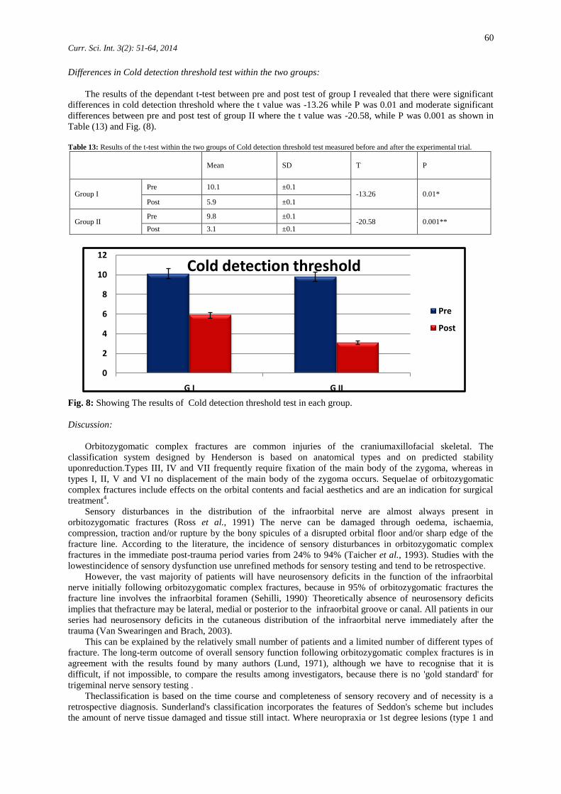
Differences in Cold detection threshold test between the two groups:
The results of the independent t-test between the two groups revealed that there were no significant
differences in cold detection threshold measured before the experimental trial where the t value was -0.132 ,
while P was 0.896 and moderate significant differences when measured post experimental trial where the t
value was -2.93, while P was 0.007 as shown in Table (12) and Fig.(8)
Table 12: Results of the t-test between the two groups of Cold detection threshold test measured before and after the experimental trial.
Mean SD T P
Pre
-tes
t
G I 10.1 ±0.1
-0.132 0.896
GII 9.8 ±0.1
Po
st-t
est G I 5.9 ±0.1
-2.93 0.007**
GII 3.1 ±0.1
0
5
10
15
20
25
30
35
40
G I G II
Heat detection threshold
PrePost
60 Curr. Sci. Int. 3(2): 51-64, 2014
Differences in Cold detection threshold test within the two groups:
The results of the dependant t-test between pre and post test of group I revealed that there were significant
differences in cold detection threshold where the t value was -13.26 while P was 0.01 and moderate significant
differences between pre and post test of group II where the t value was -20.58, while P was 0.001 as shown in
Table (13) and Fig. (8).
Table 13: Results of the t-test within the two groups of Cold detection threshold test measured before and after the experimental trial.
Mean SD T P
Group I Pre 10.1 ±0.1
-13.26 0.01* Post 5.9 ±0.1
Group II Pre 9.8 ±0.1
-20.58 0.001** Post 3.1 ±0.1
Fig. 8: Showing The results of Cold detection threshold test in each group.
Discussion:
Orbitozygomatic complex fractures are common injuries of the craniumaxillofacial skeletal. The
classification system designed by Henderson is based on anatomical types and on predicted stability
uponreduction.Types III, IV and VII frequently require fixation of the main body of the zygoma, whereas in
types I, II, V and VI no displacement of the main body of the zygoma occurs. Sequelae of orbitozygomatic
complex fractures include effects on the orbital contents and facial aesthetics and are an indication for surgical
treatment4.
Sensory disturbances in the distribution of the infraorbital nerve are almost always present in
orbitozygomatic fractures (Ross et al., 1991) The nerve can be damaged through oedema, ischaemia,
compression, traction and/or rupture by the bony spicules of a disrupted orbital floor and/or sharp edge of the
fracture line. According to the literature, the incidence of sensory disturbances in orbitozygomatic complex
fractures in the immediate post-trauma period varies from 24% to 94% (Taicher et al., 1993). Studies with the
lowestincidence of sensory dysfunction use unrefined methods for sensory testing and tend to be retrospective.
However, the vast majority of patients will have neurosensory deficits in the function of the infraorbital
nerve initially following orbitozygomatic complex fractures, because in 95% of orbitozygomatic fractures the
fracture line involves the infraorbital foramen (Sehilli, 1990). Theoretically absence of neurosensory deficits
implies that thefracture may be lateral, medial or posterior to the infraorbital groove or canal. All patients in our
series had neurosensory deficits in the cutaneous distribution of the infraorbital nerve immediately after the
trauma (Van Swearingen and Brach, 2003).
This can be explained by the relatively small number of patients and a limited number of different types of
fracture. The long-term outcome of overall sensory function following orbitozygomatic complex fractures is in
agreement with the results found by many authors (Lund, 1971), although we have to recognise that it is
difficult, if not impossible, to compare the results among investigators, because there is no 'gold standard' for
trigeminal nerve sensory testing .
Theclassification is based on the time course and completeness of sensory recovery and of necessity is a
retrospective diagnosis. Sunderland's classification incorporates the features of Seddon's scheme but includes
the amount of nerve tissue damaged and tissue still intact. Where neuropraxia or 1st degree lesions (type 1 and
0
2
4
6
8
10
12
G I G II
Cold detection threshold
Pre
Post
61 Curr. Sci. Int. 3(2): 51-64, 2014
2) exist, return to normal sensory function occurs within 1 week following nerve injury, 1st degree (type 3)
takes 1 to 2 months for complete recovery, but is far earlier than can be explained by axonal regeneration.
Complete recovery occurs in 2-4 months in an axonotmesis or 2nd degree nerve injury (Sunderland,1991).
A neurotmesis or 3rd, 4th or 5th degree nerve injury will show incomplete recovery of sensory function.
Whereas 4th and 5th degree nerve injuries will have a poor prognosis for spontaneous recovery, 3rd degree
nerve injury may show partial return of sensation within 3-5 months after the trauma (Zingg et al., 1991).
The most likely cause for 3rd and 4th degree injuries include severe traction or compression. Not all nerve
fibreshav the same susceptibility to compression injuries and ischaemia. The A/q (myelinated) fibres,
responsible for mechanoception (touch), are more susceptible to compression and ischaemia than the A6
(myelinated) and C(unmyelinated) fibres (pain, temperature) (Wiesenbaugh, 1970).
Following a compression injury it is quite possible to have a deficit in mechanoception (light touch, moving
touch),but intact nociception (pinprick) and thermal discrimination. In our study this phenomenon is reflected
by the fact that in 10 % of the cases complete recovery of neurosensory function occurred for only some of the
sensory modalities tested )Beurskens and Heymans, 2006).
Assuming that no transection or rupture of the entire infraorbital nerve trunk had occurred in our series, 36
% of patients had 3rd or 4th degree nerve injuries. Pathophysiologically this means loss of axonal continuity and
endoneurial tubes. In 4th degree injury it also means interruption of the epineurium (Zachariades et al., 1990).
Although it is impossible to translate these results to the initial trauma to the nerve, we can report that in at
least 18 cases (36 %) these severe nerve injuries were present at the time of injury, as they were still present at
6-9 months after the trauma. This should influence the management of orbitozygomatic complex fractures in
general, which has already been indicated by Jungell and Lindqvist (1987). In their study of 68 patients with
zygomatic complex fractures, 9 patients (13.2 %) underwent surgery to the nerve and 6 patients experienced
some improvement (Tajima,1997).
In another study, results from neuromicrosurgery to the inferaorbital nerve indicate a high degree of
successful regeneration, with complete return of sensation in the distribution of the infraorbital nerve in 6 out of
7 cases (Souyris et al., 1989). The indications for neuromicrosurgery are strongest for the neurotmesis or 3rd,
4th and 5th degree nerve injuries.
In cases with a zygoma fracture who were treate d only by bone elevation, 49% of all cases have
permanent damage of the infraorbital nerve and at the same time > 50% of these patients have re-displacement
of the zygoma as reported by6. In our series, the incidence was higher 77.8 %.
The reason for the damage to the nerve is not only the compression of the nerve for a short period by the
displaced zygoma but probably also from additional splits of the orbital floor, along the infraorbital canal. With
regard to fixation of unstable zygomaticfractures in relation to sensory recovery of the infraorbital nerve,
miniplateosteosynthesis is recommended as opposed to wire fixation in all unstable zygomatic bone fractures
where there is displacement (Manand Bax, 1988).
The incidence of postoperative infraorbital nerve sequelae is diminished by 50 % in unstable zygomatic
fractures when treated by osteosynthesis with miniplates(Mozsary and Middleton,1938). This is in agreement
with our findings of only 12.5 % persistent neurosensory deficits after open reduction and fixation with plate
osteosynthesis.
The results regarding orbital floor reconstruction inwhich 25 % of the cases had persistent neurosensory
deficits are in agreement with the study byKirkegaard et al. (1986), who found that 30 % of the patients had
persistent reduced sensation in the infraorbital nerve distribution area. An incidence of 22 % of long-term
neurosensory deficits in patients operated on for unilateral orbital floor fractures.
Although a neurosensory deficit in the distribution of the infraorbital nerve is not regarded as an indication
for surgical treatment of orbitozygomatic complex fractures, in the absence of other significant symptoms, 44.4
% of the patients who did not undergo surgery sustained persistent neurosensory deficits. Surgery within 1 week
after the trauma will improve the sensory function of the infraorbital nerve (Rohrich, 1991).
Despite the fact that not all of these patients will seek treatment for alteration in sensation, depending on the
nature of the sensory disturbances, there may be a subgroup of patients who would benefit from early surgical
treatment to prevent long-term nerve dysaesthesia. Furthermore, open reduction and fixation of an
orbitozygomatic complex fracture offers a better prognosis for complete recovery of inferaorbital nerve function
than elevation only with or without K-wire fixation (Nordgaard,1996).
Furthermore, early neuromicrosurgical intervention on the infraorbital nerve is indicated in one-third of all
orbitozygomatic complex fractures, in particular in type III fractures, because of a high percentage of 3rd or 4th
degree nerve injuries according to Sunderland's classification. Our study evaluated the recovery rate of
subjective sensory modalities. A study including more quantitative neurological examination, such as cutaneous
pressure thresholds (Coulson et al., 2006). two-point discrimination and vibratory threshold measurements may
give a clearer indication as to choice of treatment.
Our study showed that there was a strong relationship between the application of Neuromuscular electrical
nerve stimulation and the improvement of pain parameter. These results were confirmed those obtained
62 Curr. Sci. Int. 3(2): 51-64, 2014
byBeurskens et al. (2006) who applied the electric stimulation with 20 Hz on two patients suffering from facial
pain and there was a mild improvement in pain control.
In our study, we agree with other authors who mentioned that the Orofacial function in IO patients has
some typical features in the mechanism of sensorydisturbance but differ individually according to the extent
and location of trauma. After the assessment of different Orofacial parameters by EMG methods in 49 patients
they found that, there was pain, dysesthesia and sensory changes. They related these changes to the facial
sensation, eye movement, delayed reaction time, change detection to heat and cold (Schultze et al., 1999).
The present study is in a accordance withFogaca et al. (2004) who concluded that NMES 0.4Hz with a
physical therapy program seemed to address most of IO patients problems as function of orofacial and also had
the ability to improve many of mouth and eye functions.
In this experimental study, the analysis of mean values before and after the treatment program by
Aesthiometer showed that, there was a significant difference between the two groups with the best results for
GII, this means that NMES 18 Hz, 30% intensity and 15 min duration gave the best results in IO nerve lesion
following orbitozygomatic fractures.
Our findings of this experimental study are closely agreement and supported by the findings of Shiau et al.
(1995) who applied NMES with different frequencies 10Hz, 20Hz and 30 Hz on 30 IO patients. They were
divided into 3 equal groups accord to degree of orofacial disabilities. The treatment program was applied for 2
months, 3 sessions per week, 30% intensities and 20 min in duration. GI was treated by 10 Hz, GII by 20 Hz and
GIII by 30Hz. finally the results showed that there were moderate significant differences in GII, mild
improvements in GI and no improvements in GI.
The statistical findings of this work were supported bySchultze et al. (1999) when they conducted a case
report on female traumatic IO nerve lesion. She has suffered from supraorbital and infraorbital sensory
dysesthesia. The orofacial training for this patient required a continuous help from the therapist to assist the
patient on pain control and improve Orofacial function. She received an additional therapy by NMES 18 Hz, 3
times per week, 20 min for 6 weeks. Her Orofacial ability was assessed every two weeks. At the end of the
second week she required only an intermittent help instead of a firm continuous support. After four weeks, She
required a minimal help and after the six weeks she required only a verbal support and the patient was able to do
all Orofacial functions independently (McGimpey et al., 2000).
Also, the results of this work are accepted by the work ofCoulson et al. (2006) when they applied NMES 20
Hz on 9 traumatic SO and IO nerve lesions and assessed sensory changes by disk criminator. The treatment
program was applied for 10 weeks. The results showed that there was an improvement in ADL activities. These
results are supported when they examined the beneficial effects of NMES 24 Hz on 5 male patients suffering
from traumatic SO&IO. The main problems of patients were pain. They were treated for 8 weeks, day after day.
Their statistics revealed that there were significant improvement in pain control.
In our work, there is improvement in two point discimination in both groups with the best results for NMES
group. This coincides with the work of who applied NMES 25Hz on 27 post traumatic IO patients suffering
from limited mechanical detection sensation threshold (two point discrimination) as measured by disk
criminators. They were divided into two equal groups, GI was a control group treated by traditional physical
therapy program while GII was an experimental group which received the same exercise program and NMES 25
Hz for 2 months, each session 20 min, day after day. After the treatment program, there was significant
differences in NMES group as compared to non NMES group (Benoliel et al., 2002).
Finally, in this experimental study the analysis of mean values before and after the treatment by NMES
showed that there were significant differences within the two groups with the best results of GII. These results
mean that NMES 20 Hz, 50% intensity and 15 min duration have given the best results according to
Aesthesiometer, monofilaments, dental bud and pain scale. This prognosis may be a result of optimal
application of NMES technique, suitable frequency, appropriate intensity and optimum duration.
Summary:
The purpose of the study was designed to clarify the modern trends of neurosensory rehabilitation in
treatment of infraorbital nerve changes following zygomatic fractures. In this respect, neurosensory function
was assessed with calibrated nylon monofilaments, electrical stimulation, heat detection thresholds and response
to pin prick in the infraorbital, supraorbital and mental nerve regions in both sexes. Subjects, thirty males and
females were the same degree of fractures severity according to Henderson's classification (grade1, 2& 3), their
age ranged from 25-45years old and their weight ranged from 60-88Kg.They were randomly divided into two
equal groups (G1and G2). G1, consists of 15 patients of both sexes and was treated by surgical procedures
(reduction and fixation) and G2 consists of 15 patients was treated by the same surgical procedures and
electrical neuromuscular stimulation with exercises therapy program. Vital signs as blood pressure, body
temperature, pulse rate and respiratory rate measured before and after the treatment sessions. Assessments,
visual analogue scale was used to measure degree of pain, Semmes-Weinstein monofilaments was used to
63 Curr. Sci. Int. 3(2): 51-64, 2014
measure the light touch sensation, Aesthesiometer 2 point calliper was used to measure the two point
discrimination sensation, Peltier probe was used to measure heat detection sensation. Moreover, by the use an
ascending test of phyaction guidance-c system to measure the electrical detection threshold. Statistically the
results for all groups were analyzed by t-test to compare the differences between the two groups. The statistical
package of social sciences used for data processing using the p-value 0.05 as a level of significance. Results,
showed that there were significant improvements in all variables in both groups in a favour to G2. Therefore, we
concluded that, the use of surgical procedures combined with neuromuscular electrical stimulation and exercises
program were the good method and open a new link to improve the recovery of infraorbital nerve sensory
changes following zygomatic fractures.
Recommendations:
With the limitation of this study and from the obtained statistical results further investigations and research
studies are recommended as further studies should be attempted to describe the effect of NMES on speech,
cognition and facial expression with different frequencies and intensities of stimulation to treat many different
disabilities in different neurological diseases.
References
Becelli, R., A. Carboni, G. Cerulli, M. Perugini and G. Iannetti, 2002. Delayed and inadequately treated malar
fractures: evolution in the treatment, presentation and review of literature. Aesthetic Plast. Surg., 26: 134-
38.
Benoliel, R., A. Wilensky and M. Tal, 2002. Application of pro-inflamatory agent to the orbital portion of the
rat infraorbital nerve induces changes indicative of ongoing trigeminal pain. Pain, 99: 567-78.
Beurskens, C.H.G. and P.G. Heymans, 2006. Mime Therapy Improves Facial Symmetry in People With Long-
Term Facial Nerve Paresis: A Randomized Controlled Trial. Australian Journal of Physiotherapy, 52: 177-
183.
Beurskens, C.H.G., P.G. Heymans, R.A.B. Oostendorp, 2006. Stability of Benefits of Mime Therapy in
Sequelae of Facial Nerve Paresis During a 1-Year Period. Otology &Neurotology, Inc., 27(7): 1037-1042.
Coulson, S.E., R.D. Adams, N.J. O’Dwyer, G.R. Croxson, 2006. Physiotherapy Rehabilitation of The Smile
after Long-Term Facial Nerve Palsy using Video Self-Modeling and Implementation Intentions. American
Academy of otolaryngology-Head and Neck Surgery, 134: 48-55.
Eliav, E., S. Teich, D. Nitzan, D. Raziq and M. Tal, 2003. Facial arthralgia and myalgia: can they be
differentiated by trigeminal sensory assessment? Pain, 104: 481-90.
Ellies, E., A. Attar and K. Moos, 1985. An analysis of zygomatico-orbital fracture. J. oral Maxillofac. Surg., 43:
417-28.
Essick, G., 1992. Comprehensive clinical evaluation of perioral sensory function. Oral Maxillofac. Surg. clin.
North Am., 2: 503-27.
Finlay, P. and K.F. Ward-Booth, 1984. Morbidity associated with the use of antral packs and external pins in
the treatment of the unstable fracture of the zygomatic complex. Br. J. Oral Maxillofac. Surg., 22: 18-23.
Fogaca, W., M. Fereirra and A. Dellon, 2004. Infraorbital nerve injury associated with zygoma fractures:
documentation with neurosensory testing. Plast. Reconstr. Surg., 113: 834-38.
Jungell, P., and C. Lindqvist, 1987. Paraesthesia of the infraorbital nerve following fracture of the zygomatic
complex. Int. J. oral Maxillofac. Surg., 16: 363-70.
Kirkegaard, J., O. Greisen and P. Hojslet, 1986. Orbital floor fractures: early repair and results.
ClinOtolaryngol., 11: 69-73.
Lund, K., 1971. Fractures of the zygoma: a follow-up study of 62 patients. J. Oral Surg., 29: 557-560.
Man, K. and w. Bax, 1988. The influence of the mode of treatment of zygomatic bone fractures on the healing
process . Br. J. Oral Maxillofac. Surg., 24: 419-425.
McGimpey, J., A. Vaidya, P. Biagioni and P. lamey, 2000. Role of thermography in the assessment of
infraorbital nerve injury after malar fractures. Br. J. Oral Maxillofac. Surg., 38: 581-84.
Mozsary, P.G. and R. Middleton, 1983. Microsurgical reconstruction of the infraorbital nerve. J. Oral
Maxillofac. Surg., 41: 697-700.
Nordgaard, J.O., 1996. Persistent sensory disturbances and diplopia following fractures of the zygoma. Arch.
Otolaryngol., 102: 80-82.
Rohrich, R., 1991. The superiority of rigid plate fixation in the management of zygoma fractures and a longterm
follow-up clinical study. Plast. Reconstr. Surg., 96: 570-75.
Ross, B., J.M. Nedzelski, J.A. McLean, 1991. Efficacy of Feedback Training in Long-Standing Facial Nerve
Paresis. Laryngoscope., 101: 744-750.
64 Curr. Sci. Int. 3(2): 51-64, 2014
Schultze, S., M. Erbe, D. Rudolph and F. Neukam, 1999. Prospective study on post traumatic and postoperative
sensory disturbances of the inferior alveolar nerve and infraorbital nerve in mandibular and midfacial
fractures. J. Craniomaxillofac. Surg., 86: 86-93.
Sehilli, W., 1990. Treatment of zygoma fractures. Oral Maxillofac. Surg. Clin. North Am., 2: 155-69.
Shiau, J., B. Segal, I. Danys, R. Freedman, S. Scott, 1995. Long-Term Effects of Neuromuscular Rehabilitation
of Chronic Facial Paralysis. The Journal of Otolaryngology, 24(4): 217-220.
Souyris, F., P. Klersy, C. Jammet and F. Payrot, 1989. Malar bone fractures and their sequelae. J. Craniomaxillo
fac. Surg., 17: 64-68.
Sunderland, S., 1991. A classification of peripheral nerve injuries produced by loss of function. Brain, 74: 491-
99.
Taicher, S., N. Ardekian, Y. Samet, I. Shoshani and I. Kaffe, 1993. Recovery of the infraorbital nerve after
zygomatic complex fractures: a preliminary study of different treatment methods. Int. J. Oral Maxillofac.
Surg., 22: 339-341
Tajima, S., 1997. Malar bone fractures: experimental fractures on the dried skull and clinical sensory
disturbances. J. Max.-Fac. Surg., 5: 150-156.
VanSwearingen, J.M. and J.S. Brach, 2003. Changes in Facial Movement and Synkinesis with Facial
Neuromuscular Re-education. American Society of Plastic Surgeons., 111(7): 2370-2375.
Wiesenbaugh, J.M., 1970. Diagnostic evaluation of zygomatic complex fractures. J. Oral Surg., 28: 204-208.
Zachariades, N., D. Papavassiliou and I. Papademetriou, 1990. The alterations in sensitivity of the nerve
following fractures of the zygomaticomaxillary complex. J.Craniomaxillo fac. Surg., 18: 315-318.
Zingg, M., K. Chowdhury, F. Sutter, J. Reveh, 1991. Treatment of 813 zygoma-lateral orbital complex fractures.
Arch. Otolaryngol. Head Neck Surg., 117: 611-620.