Embed Size (px)
Citation preview
Peripheral Sensitization Increases Opioid ReceptorExpression and Activation by Crotalphine in RatsVanessa Olzon Zambelli1, Ana Carolina de Oliveira Fernandes1, Vanessa Pacciari Gutierrez1,
Julio Cesar Batista Ferreira2, Carlos Amilcar Parada3, Daria Mochly-Rosen4, Yara Cury1*
1 Laboratorio Especial de Dor e Sinalizacao, Instituto Butantan, Sao Paulo, SP, Brazil, 2 Departamento de Anatomia, Instituto de Ciencias Biomedicas, Universidade de Sao
Paulo, Sao Paulo, SP, Brasil, 3 Departamento de Fisiologia e Biofısica, Instituto de Biociencias (UNICAMP) Rua Monteiro Lobato, Cidade Universitaria, Campinas, SP, Brazil,
4 Department of Chemical and Systems Biology, Stanford University, School of Medicine, Stanford, California, United States of America
Abstract
Inflammation enhances the peripheral analgesic efficacy of opioid drugs, but the mechanisms involved in this phenomenonhave not been fully elucidated. Crotalphine (CRP), a peptide that was first isolated from South American rattlesnake C.d.terrificus venom, induces a potent and long-lasting anti-nociceptive effect that is mediated by the activation of peripheralopioid receptors. Because the high efficacy of CRP is only observed in the presence of inflammation, we aimed to elucidatethe mechanisms involved in the CRP anti-nociceptive effect induced by inflammation. Using real-time RT-PCR, western blotanalysis and ELISA assays, we demonstrate that the intraplantar injection of prostaglandin E2 (PGE2) increases the mRNAand protein levels of the m- and k-opioid receptors in the dorsal root ganglia (DRG) and paw tissue of rats within 3 h of theinjection. Using conformation state-sensitive antibodies that recognize activated opioid receptors, we show that PGE2, alonedoes not increase the activation of these opioid receptors but that in the presence of PGE2, the activation of specific opioidreceptors by CRP and selective m- and k-opioid receptor agonists (positive controls) increases. Furthermore, PGE2 down-regulated the expression and activation of the d-opioid receptor. CRP increased the level of activated mitogen-activatedprotein kinases in cultured DRG neurons, and this increase was dependent on the activation of protein kinase Cf. This CRPeffect was much more prominent when the cells were pretreated with PGE2. These results indicate that the expression andactivation of peripheral opioid receptors by opioid-like drugs can be up- or down-regulated in the presence of an acuteinjury and that acute tissue injury enhances the efficacy of peripheral opioids.
Citation: Zambelli VO, Fernandes ACdO, Gutierrez VP, Ferreira JCB, Parada CA, et al. (2014) Peripheral Sensitization Increases Opioid Receptor Expression andActivation by Crotalphine in Rats. PLoS ONE 9(3): e90576. doi:10.1371/journal.pone.0090576
Editor: Alexander Binshtok, The Hebrew University Medical School, Israel
Received June 25, 2013; Accepted February 4, 2014; Published March 4, 2014
Copyright: � 2014 Zambelli et al. This is an open-access article distributed under the terms of the Creative Commons Attribution License, which permitsunrestricted use, distribution, and reproduction in any medium, provided the original author and source are credited.
Funding: This work was supported by funds from the Fundacao de Amparo a Pesquisa do Estado de Sao Paulo, Brazil (FAPESP, grant numbers 07/03404-4,07/00135-2), Instituto Nacional de Ciencia e Tecnologia em Toxinologia (INCTTOX PROGRAM) of Conselho Nacional de Desenvolvimento Cientıfico e Tecnologico(CNPq, grant number 573790/2008-6)/FAPESP (grant number 2008/57898-0) and CAPES/CNPq, grant numbers 301508/2008-9 and 305345/2011-7. The fundershad no role in study design, data collection and analysis, decision to publish, or preparation of the manuscript.
Competing Interests: The authors have declared that no competing interests exist.
* E-mail: [email protected]
Introduction
Clinical and experimental results have shown that the
peripheral analgesic efficacy of opioids is enhanced in the presence
of inflammation and tissue injury [1,2]. The mechanisms
responsible for this phenomenon involves the following events:
increases in the opioid receptor mRNA level and opioid receptor
expression level [3–5], an increase in the axonal transport and
accumulation of these receptors in the peripheral sensory nerve
endings [6], and an increase in the opioid agonist binding to their
corresponding receptor [7,8]. Moreover, inflammation induces the
release of endogenous opioid peptides [1]. Studies investigating the
molecular mechanisms involved in the enhanced effects of opioid
treatments in the periphery caused by inflammation mainly focus
on the m-opioid receptor and the chronic inflammatory processes
[2,5–7,9]. Inflammation enhances the axonal transport of opioid
receptors toward the periphery, which is preceded by an increase
in opioid receptor mRNA transcription [3,10].
Previously, we demonstrated that crotalphine (CRP), a struc-
tural analogue to a novel analgesic peptide that was first identified
in the crude venom from the South American rattlesnake Crotalus
durissus terrificus [11], induces a potent (0.008–25 mg/kg, p.o.) and
long-lasting (2–5 days) anti-nociceptive effect that is mediated by
the activation of the peripheral k-opioid receptor (prostaglandin
E2-induced hyperalgesia) and the k- and d- (chronic constriction
injury of the rat sciatic nerve) opioid receptors [11–13]. The
L-arginine-nitric oxide-cGMP pathway is involved in the periph-
eral anti-nociceptive effect of CRP [13] and opioid agonists [14–
17]. The activation of opioid receptors regulates a variety of
intracellular signaling cascades, including the PI3Kc/AKT
signaling pathway [18], as well as the activation of mitogen-
activated protein kinases (MAPKs) and protein kinase C (PKC)
[19–21].
The high efficacy and long-lasting peripheral anti-nociceptive
effects of crotalphine have only been observed in the presence of
inflammation, and the mechanisms involved in the enhanced
effects of CRP in the presence of inflammation are currently
unknown. In the present study, we used prostaglandin E2 (PGE2)-
induced hyperalgesia in a rat model to characterize the anti-
nociceptive effect caused by the peripheral administration of CRP
and compared this response with the responses induced by select
opioid receptor agonists. Furthermore, we also investigated the
PLOS ONE | www.plosone.org 1 March 2014 | Volume 9 | Issue 3 | e90576
intracellular signaling pathways activated by CRP and the opioid
receptor agonists.
Experimental Procedures
AnimalsMale Wistar rats (170–190 g) were used in this study. The
animals were housed in a temperature-controlled (2162uC) and
light-controlled (12 h/12 h light/dark cycle) environment. All of
the behavioral tests were performed between 9:00 am and 4:00
pm. Standard food and water were available ad libitum. All of the
procedures were conducted in accordance with the guidelines for
the ethical use of conscious animals in pain research published by
the International Association for the Study of Pain [22] and were
approved by the Institutional Animal Care Committee of the
Butantan Institute (CEUAIB, protocol number 386/2007).
Drug administrationThe drugs [D-Ala2, N-Me-Phe4, Gly5-ol]-Enkephalin acetate
salt (1) (DAMGO, 5 mg/paw); [D-Pen2,5, p-Cl-Phe4]-Enkephalin
(DPDPE, 20 mg/paw); (2)-trans-(1S,2S)-U-50488 hydrochloride
hydrate (U 50,488, 10 mg/paw) [23]; Prostaglandin E2 (PGE2,
100 ng/paw) [23,24]; D-Phe-Cys-Tyr-D-Trp-Orn-Thr-Pen-Thr
amide (CTOP, 20 mg/paw) [25,26], nor-binaltorphimine dihy-
drochloride (nor-BNI, 50 mg/paw) [25,27]; N,N-diallyl-Tyr-Aib-
Aib-Phe-Leu (ICI 174,864, 10 mg/paw) [25], methiodide naloxone
(1 mg/Kg, subcutaneously) were used in this study [28]. These
drugs were purchased from Sigma-Aldrich, Saint Louis, MO,
USA. The doses of the agonists and antagonists used in this study
were determined based on the doses used in previous studies
and preliminary studies [25].The peptide f-pseudosubstrate
([C]SIYRRGARRWRKLYRAN; amino acids 105–121 in f-PKC) was synthesized at the peptide and nucleotide facility at
Stanford and fused to the cell permeable Tat protein transduction
domain peptide. The peptide CRP (,E-F-S-P-E-N-C-Q-G-E-S-
Q-P-C, where ,E is pyroglutamic acid and a disulfide bond is
located between 7C-14C) was synthesized by the American
Peptide Co, Sunyvalle, CA, USA (product number 331065, lot
number U07122A1, 98% purity, M.W. 1534,6 Daltons) using
Fmoc chemistry in the solid phase, as described by Konno et al.
(2008) [11]. Crotalphine (CRP) was administered using a dose of
0.6 ng/paw. The peptide was diluted in sterile saline, and the
opioid agonists and antagonists were diluted in water or saline. All
of the drugs were administered in a final volume of 50 ml by
intraplantar (i.pl.) injection.
PGE2 was injected into the hind paw to induce hyperalgesia.
CRP and the opioid agonists and antagonists were injected 2 h
after the PGE2 injection. Control animals were given the same
volume of sterile saline or water by i.pl. injection and were tested
following the same schedule described below.
PGE2-induced hyperalgesiaSterile saline containing PGE2 (100 ng/paw) was injected into
both hind paws for the behavioral tests and into one hind paw for
the biochemical experiments. A stock solution of PGE2 was
prepared by dissolving 0.5 mg of PGE2 in 1 ml of absolute
ethanol. For injection into the rat paw, an aliquot of this stock
solution was diluted in sterile saline. The percentage of ethanol in
the solution injected into the hind paw was less than 0.1%. The
pain threshold was measured before and 3 h after PGE2 injection
[25,29].
The rat paw pressure test [30] was used to evaluate
hyperalgesia. An Ugo-Basile pressure apparatus was used to
determine the pressure pain threshold before and 3 h after PGE2
injection. The testing was conducted blind to the group
designation. Briefly, an increasing amount of force (in g, 16 g/s)
was applied to the hind paw. The force needed to induce paw
withdrawal was recorded as the pain threshold. To reduce the
stress level, the rats were acclimated to the testing procedure 1–3
days before the experiment.
Biochemical studiesFor this study, ipsilateral and contralateral dorsal root ganglia
(DRG, L4-6, pooled 7–10 ganglia) and plantar tissue were
collected 3 h after the intraplantar injection of PGE2. To obtain
the plantar tissue, two longitudinal incisions were made through
the skin and muscle of the plantar aspect of the foot, from the
proximal edge of the heel to the toes, using a blade. The skin was
removed, and the adjacent subcutaneous tissue, including tendon,
was collected. Nnaıvenaıve animals were used as controls.
Reverse Transcriptase/Real-Time Polymerase Chain
Reaction. RNA was isolated from the DRGs and plantar tissue
using Trizol reagent (Gibco Life Technologies, Grand Island, NY,
USA). The RNA concentration and integrity were determined,
cDNA was synthesized using Superscript III RNase H-Reverse
Transcriptase (Invitrogen Life Technologies, Grand Island, NY,
USA) at 42uC for 50 min and real-time polymerase chain reaction
was performed. The following primers were used for gene
amplification: m-opioid receptor: sense GCCATCGGTCT-
GCCTGTAAT; antisense GAGCAGGTTCTCCCAGTAC, k-
opioid receptor: sense GTCAGAGGACAGCTTTGCAC, anti-
sense TAGCTCAGTGAAGGTACATGC, d-opioid receptor:
sense ATGGTCATGGCAGTGACC, antisense CACGCA-
GATCTTGGTCACAG, Cyclofilin: sense 5-AATGCTGGAC-
CAAACACAAA-3, antisense 5-CCTTCTTTCACCTTCC-
CAAA-3. The real-time PCR for the opioid receptors and
house-keeper gene cyclofilin were run separately, and the
amplification reactions were performed using the ABI Prism
7700 Sequence Detection System and the SYBR Green PCR
Master Mix (Applied Biosystems Life Technologies, Grand Island,
NY, USA). The results were quantified as Ct values, where Ct is
defined as the threshold cycle of the polymerase chain reaction at
which the amplified product is first detected. The expression levels
were normalized using the cyclofilin expression level as an
endogenous reference.
Immunoblot analysis. Frozen tissues were homogenized in
cold RIPA buffer (1% Igepal CA-630, 0.5% sodium deoxycholate,
0.1% SDS, 1 mM PMSF, 10 mg/ml aprotinin, 1 mM sodium
orthovanadate in PBS buffer, pH 7.4, 1:300 protease and
phosphatase inhibitor cocktail from Sigma-Aldrich, St. Louis,
MO, USA (cat. number P8340, P5726, P0044)). The samples were
centrifuged at 10,0006 g for 10 min at 4uC. The supernatant was
removed and centrifuged again. After centrifugation, the protein
concentration in the supernatant was determined using Bradford
assay [31]. Aliquots containing 80 mg of total protein were boiled
in Laemmli loading buffer (BioRad, Hercules, CA, USA) and then
loaded onto a 10% polyacrylamide gel. After separating by
electrophoresis, the proteins were transferred to a nitrocellulose
membrane (Bio-Rad, Hercules, CA, USA). The membranes were
blocked in TBST (20 mM Tris-HCL, 150 mM NaCl, and 0.1%
Tween 20) containing 5% non-fat dry milk for 2 h at room
temperature and then incubated in m-opioid receptor (C-terminus)
rabbit IgG, d-opioid receptor (C-terminus) rabbit IgG (1:1000,
Chemicon, Temecula, CA, USA, cat. numbers AB 5511, AB1560,
respectively) or k-opioid receptor-1 (N-19) goat polyclonal IgG
antibody (1:500; Santa Cruz Biotechnology, Dallas, TX, USA, cat.
number sc31779) overnight at 4uC. The membranes were then
incubated in the appropriate peroxidase-conjugated secondary
Opioid Receptors and Peripheral Sensitization
PLOS ONE | www.plosone.org 2 March 2014 | Volume 9 | Issue 3 | e90576
antibody (1:5000; anti-rabbit and anti-goat, Sigma-Aldrich, St.
Louis, MO, USA, cat. numbers A0545 and A8919, respectively)
for 90 min at room temperature and developed using enhanced
chemiluminescence (Amersham GE Healthcare Bio-Sciences
Corp.,; Piscataway, NJ, USA). The signal was detected by
autoradiography. Quantification analysis of the blots was per-
formed using the Scion Image software (Scion Corporation based
on NIH image). Targeted bands were normalized to GAPDH
(1:5000, Advanced ImmunoChemical Incorporate, Long Beach,
CA, USA).
Opioid receptor activation studies: ELISA assayIn vivo studies. Naıve and PGE2-sensitized rats were
injected with DAMGO (5 mg/paw), DPDPE (20 mg/paw), U
50,488 (10 mg/paw) or CRP (0.6 ng/paw). In the sensitized rats,
these drugs were administered 2 h after the PGE2 injection.
Approximately 1 h after the CRP or opioid receptor agonist
treatment, the animals were perfused through the heart with
phosphate-buffered saline and 4% paraformaldehyde (PFA) in
0.1 M phosphate buffer (PB, pH 7.4). The DRG (L5) and plantar
tissue were removed, post-fixed for 4 h in 4% PFA, and
transferred to a 30% sucrose solution in PB for 48 h to ensure
cryoprotection. The tissues were sectioned (14 mm) on a cryostat,
and the sections were mounted on histological glass slides covered
with silane. Two sections from each region were circled using an
ImmEdge PAP pen (Sigma-Aldrich) to form a water-proof barrier,
and the resulting well could hold approximately 80 ml of solution.
ELISA was performed in these wells using a 1:500 dilution of
primary antibody and a 1:500 dilution of secondary antibody. To
detect the activated receptor, we used antibodies that are able to
distinguish the conformational change in the N-terminus of the
opioid receptors following activation [32]. The antibodies were
purchased from Proteimax Brazil. The reaction product was
transferred to a 96-well plate, and the absorbance at 490 nm was
measured using a microplate reader (Molecular Devices, Sunny-
vale, CA, USA).2.4.3.2 Cell culture studies. These studies were
performed using cultured DRGs obtained from naıve animals.
Rats were decapitated, and all of the DRGs (45650 DRGs) from
the cervical, thoracic, lumbar and sacral levels were aseptically
removed and collected in Earl’s balanced salt solution (5 mM
KC1, 116 mM NaCI, 1 mM NaH2PO4, 8 mM Na2HPO4,
1.46 mM KH2PO4, 8 mM NaHCO3 and 1% glucose). Next, the
DRGs were digested in 0.5% collagenase (Sigma, Saint Louis,
MO, USA) in EBSS at 37uC for 45 min. The tissue was then
incubated for 15 min in EBSS containing 1% trypsin (Sigma,
Saint Louis, MO, USA), washed in 2.5% trypsin inhibitor (Sigma,
Saint Louis, MO, USA) and then triturated using a thin fire-
polished pipette in Dulbecco modified Eagle medium (DMEM,
Gibco Life Technologies, Grand Island, NY, USA) containing 1%
HEPES buffer solution, penicillin/streptomycin (1:200, Gibco,
Life Technologies, Grand Island, NY, USA) and 10% heat
inactivated fetal bovine serum (Sigma, Saint Louis, MO, USA).
Next, the cells were subjected to density gradient centrifugation at
2006g for 1 min. The resulting pellet was resuspended in DMEM,
and the cell suspension was filtered through a cell strainer (75 mm,
Becton Dickinson Labware, Franklin Lakes, NJ, USA). The DRG
cells were seeded onto 24-well culture plates at a density of
15,000 cells/well. The cells were cultured in a humidified
incubator at 37uC with 5% CO2 and 95% air. Two days after
seeding, the culture medium was changed. The cells were treated
with PGE2 (1 mM) for 15 minutes for sensitization. Vehicle was
used as a control. Cells were then treated with CRP dissolved in
medium at three different concentrations (0.001, 1 and 100 mM)
or DAMGO, DPDPE or U 50,488 (1 mM) for 15 minutes. In the
yoked control wells, only culture medium was added (vehicle
treatment). Three separate experiments were conducted on
different occasions. For each experiment, new primary cultures
were prepared under the same standard experimental conditions.
Each treatment was replicated in three different wells. In the pilot
studies, we determined the cell viability following the specific
concentrations of agonists, CRP and PGE2 used. No significant
increase in the number of trypan blue stained cells was observed
after these treatments when compared with the vehicle (data not
shown). ELISA was performed in each well as previously
described.
Intracellular signaling studies. These studies were per-
formed in DRG cell cultures obtained from naıve animals. The
cells were obtained as previously described (section 2.3.3.2). CRP
(1 mM) or U 50, 488 (1 mM) was added to the cells untreated or
pretreated with PGE2 (1 mM) as previously described. In these
studies, Nor-BNI (1 mM) was added to the culture (30 minutes
before treatment). After treatment, the cyclic GMP content was
measured using a cyclic GMP enzyme immunoassay kit purchased
from Amersham Enzymeimmunoassay Biotrak (GE Healthcare
Bio-Sciences Corp, Piscataway, NJ, USA).The phosphorylation of
Akt, ERK 1/2 and JNK was evaluated by immunoblot analysis
using the previously described method. The DRG cells were
homogenized in a lysis buffer as previously described. The cells
were incubated in primary antibody, either phosphorylated (p)
AKT-T, pERK, pJNK, or non-phosphorylated forms of these
proteins (1:1000; Cell Signaling Technology, Danvers, MA, USA),
overnight at 4uC and then incubated in HRP-conjugated
secondary antibody (1:2000; Amersham Biosciences, Arlington
Heights, IL, USA). The blots were visualized using ECL solution
for 1 min and exposed onto hyperfilms (Amersham Biosciences,
Arlington Heights, IL, USA).
Statistical analysisThe results are presented as the mean 6 SEM. For figures 1,
Fig. S1–4 the statistical evaluation of data was conducted using a
two-way analysis of variance (ANOVA) with post-hoc testing by
Tukey. One way ANOVA was used to analyze figures 2, 3, 4, 5, 6,
7, 8, 9, 10, with post-hoc testing by Tukey. A value of P,0.05 was
considered significant.
Results
Acute sensitization improves the anti-nociceptive effectof CRP and opioid receptor agonists
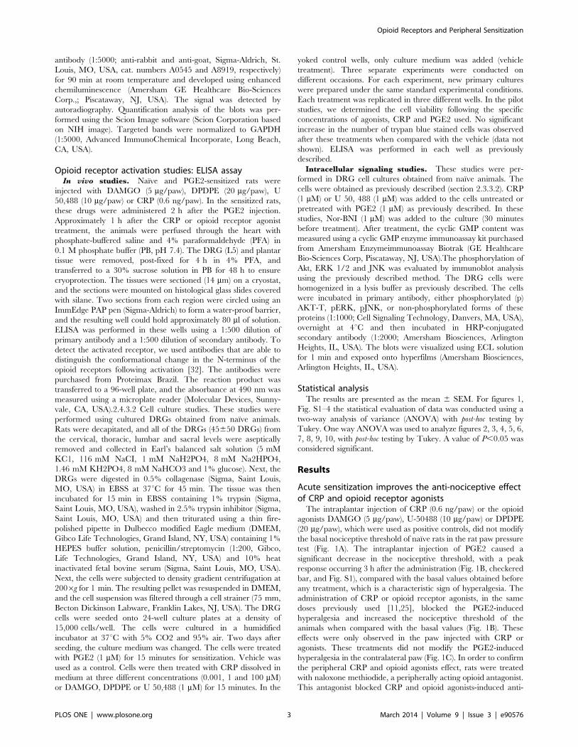
The intraplantar injection of CRP (0.6 ng/paw) or the opioid
agonists DAMGO (5 mg/paw), U-50488 (10 mg/paw) or DPDPE
(20 mg/paw), which were used as positive controls, did not modify
the basal nociceptive threshold of naıve rats in the rat paw pressure
test (Fig. 1A). The intraplantar injection of PGE2 caused a
significant decrease in the nociceptive threshold, with a peak
response occurring 3 h after the administration (Fig. 1B, checkered
bar, and Fig. S1), compared with the basal values obtained before
any treatment, which is a characteristic sign of hyperalgesia. The
administration of CRP or opioid receptor agonists, in the same
doses previously used [11,25], blocked the PGE2-induced
hyperalgesia and increased the nociceptive threshold of the
animals when compared with the basal values (Fig. 1B). These
effects were only observed in the paw injected with CRP or
agonists. These treatments did not modify the PGE2-induced
hyperalgesia in the contralateral paw (Fig. 1C). In order to confirm
the peripheral CRP and opioid agonists effect, rats were treated
with naloxone methiodide, a peripherally acting opioid antagonist.
This antagonist blocked CRP and opioid agonists-induced anti-
Opioid Receptors and Peripheral Sensitization
PLOS ONE | www.plosone.org 3 March 2014 | Volume 9 | Issue 3 | e90576
nociception (Fig. S2). In addition, the CRP or opioid receptor
agonists systemic injection, in the same doses injected intraplan-
tarly, did not alter PGE2-induced hyperalgesia (Fig. S3). Together,
these results indicate that CRP and the opioid receptor agonists, in
the doses used in this study, have a local anti-nociceptive effect in
the presence of the acute sensitization induced by PGE2.
Previously, we demonstrated that the anti-nociceptive effect of
CRP administered orally in this hyperalgesia model is mediated by
the activation of the peripheral (local) k-opioid receptor [11]. To
determine whether opioid receptors are involved in the local
(intraplantar) anti-nociceptive effect of this peptide, rats were
administered selective antagonists of the opioid receptors by i.pl.
injection. The anti-nociceptive effect of CRP was abolished in the
paw injected with nor-BNI, an antagonist of the k-opioid receptors
(Fig. 1B), but not by CTOP or ICI 174,864, m- and d-opioid
receptor antagonists, respectively (Fig. 1B). These results indicate
that the peripheral anti-nociceptive effect of CRP is mediated by
the activation of the peripheral k-opioid receptor. The opioid
receptor antagonists CTOP, ICI 174,864, and Nor-BNI blocked
the anti-nociceptive effects induced by their corresponding agonist
(DAMGO, DPDPE, and U 50,488, respectively), which confirms
the efficacy of the antagonist doses used (Fig. 1B). The antagonists,
per se, had no effect on the hyperalgesia induced by PGE2 (data not
shown).
Acute sensitization interferes with the opioid receptorlevels
To elucidate the potential mechanisms involved in the anti-
nociceptive effect of CRP and the opioid agonists observed in the
presence of prostaglandin E2 sensitization, we investigated
whether this eicosanoid treatment alters the mRNA expression
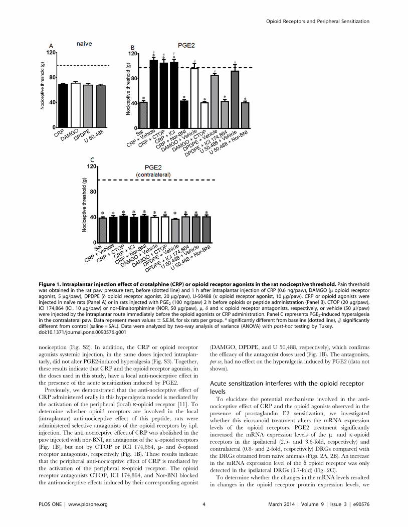
levels of the opioid receptors. PGE2 treatment significantly
increased the mRNA expression levels of the m- and k-opioid
receptors in the ipsilateral (2.5- and 3.6-fold, respectively) and
contralateral (0.8- and 2-fold, respectively) DRGs compared with
the DRGs obtained from naıve animals (Figs. 2A, 2B). An increase
in the mRNA expression level of the d opioid receptor was only
detected in the ipsilateral DRGs (3.7-fold) (Fig. 2C).
To determine whether the changes in the mRNA levels resulted
in changes in the opioid receptor protein expression levels, we
Figure 1. Intraplantar injection effect of crotalphine (CRP) or opioid receptor agonists in the rat nociceptive threshold. Pain thresholdwas obtained in the rat paw pressure test, before (dotted line) and 1 h after intraplantar injection of CRP (0.6 ng/paw), DAMGO (m opioid receptoragonist, 5 mg/paw), DPDPE (d opioid receptor agonist, 20 mg/paw), U-50488 (k opioid receptor agonist, 10 mg/paw). CRP or opioid agonists wereinjected in naıve rats (Panel A) or in rats injected with PGE2 (100 ng/paw) 2 h before opioids or peptide administration (Panel B). CTOP (20 mg/paw),ICI 174,864 (ICI, 10 mg/paw) or nor-Binaltorphimine (NOR, 50 mg/paw), m, d and k opioid receptor antagonists, respectively, or vehicle (50 ml/paw)were injected by the intraplantar route immediately before the opioid agonists or CRP administration. Panel C represents PGE2-induced hyperalgesiain the contralateral paw. Data represent mean values 6 S.E.M. for six rats per group. * significantly different from baseline (dotted line), # significantlydifferent from control (saline = SAL). Data were analyzed by two-way analysis of variance (ANOVA) with post-hoc testing by Tukey.doi:10.1371/journal.pone.0090576.g001
Opioid Receptors and Peripheral Sensitization
PLOS ONE | www.plosone.org 4 March 2014 | Volume 9 | Issue 3 | e90576
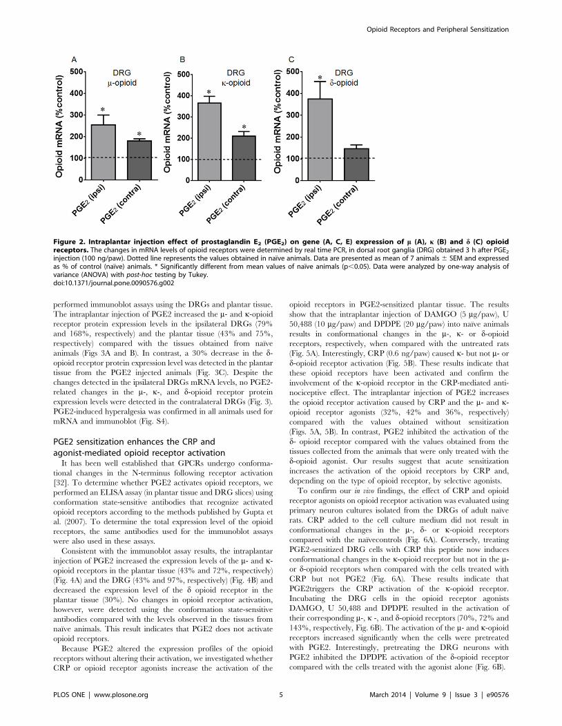
performed immunoblot assays using the DRGs and plantar tissue.
The intraplantar injection of PGE2 increased the m- and k-opioid
receptor protein expression levels in the ipsilateral DRGs (79%
and 168%, respectively) and the plantar tissue (43% and 75%,
respectively) compared with the tissues obtained from naıve
animals (Figs 3A and B). In contrast, a 30% decrease in the d-
opioid receptor protein expression level was detected in the plantar
tissue from the PGE2 injected animals (Fig. 3C). Despite the
changes detected in the ipsilateral DRGs mRNA levels, no PGE2-
related changes in the m-, k-, and d-opioid receptor protein
expression levels were detected in the contralateral DRGs (Fig. 3).
PGE2-induced hyperalgesia was confirmed in all animals used for
mRNA and immunoblot (Fig. S4).
PGE2 sensitization enhances the CRP andagonist-mediated opioid receptor activation
It has been well established that GPCRs undergo conforma-
tional changes in the N-terminus following receptor activation
[32]. To determine whether PGE2 activates opioid receptors, we
performed an ELISA assay (in plantar tissue and DRG slices) using
conformation state-sensitive antibodies that recognize activated
opioid receptors according to the methods published by Gupta et
al. (2007). To determine the total expression level of the opioid
receptors, the same antibodies used for the immunoblot assays
were also used in these assays.
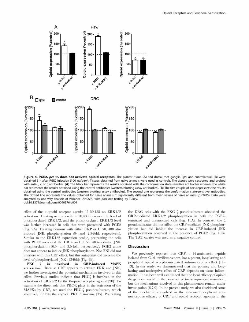
Consistent with the immunoblot assay results, the intraplantar
injection of PGE2 increased the expression levels of the m- and k-
opioid receptors in the plantar tissue (43% and 72%, respectively)
(Fig. 4A) and the DRG (43% and 97%, respectively) (Fig. 4B) and
decreased the expression level of the d opioid receptor in the
plantar tissue (30%). No changes in opioid receptor activation,
however, were detected using the conformation state-sensitive
antibodies compared with the levels observed in the tissues from
naıve animals. This result indicates that PGE2 does not activate
opioid receptors.
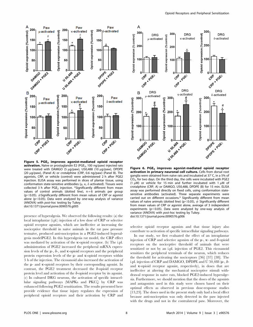
Because PGE2 altered the expression profiles of the opioid
receptors without altering their activation, we investigated whether
CRP or opioid receptor agonists increase the activation of the
opioid receptors in PGE2-sensitized plantar tissue. The results
show that the intraplantar injection of DAMGO (5 mg/paw), U
50,488 (10 mg/paw) and DPDPE (20 mg/paw) into naıve animals
results in conformational changes in the m-, k- or d-opioid
receptors, respectively, when compared with the untreated rats
(Fig. 5A). Interestingly, CRP (0.6 ng/paw) caused k- but not m- or
d-opioid receptor activation (Fig. 5B). These results indicate that
these opioid receptors have been activated and confirm the
involvement of the k-opioid receptor in the CRP-mediated anti-
nociceptive effect. The intraplantar injection of PGE2 increases
the opioid receptor activation caused by CRP and the m- and k-
opioid receptor agonists (32%, 42% and 36%, respectively)
compared with the values obtained without sensitization
(Figs. 5A, 5B). In contrast, PGE2 inhibited the activation of the
d- opioid receptor compared with the values obtained from the
tissues collected from the animals that were only treated with the
d-opioid agonist. Our results suggest that acute sensitization
increases the activation of the opioid receptors by CRP and,
depending on the type of opioid receptor, by selective agonists.
To confirm our in vivo findings, the effect of CRP and opioid
receptor agonists on opioid receptor activation was evaluated using
primary neuron cultures isolated from the DRGs of adult naıve
rats. CRP added to the cell culture medium did not result in
conformational changes in the m-, d- or k-opioid receptors
compared with the naıvecontrols (Fig. 6A). Conversely, treating
PGE2-sensitized DRG cells with CRP this peptide now induces
conformational changes in the k-opioid receptor but not in the m-
or d-opioid receptors when compared with the cells treated with
CRP but not PGE2 (Fig. 6A). These results indicate that
PGE2triggers the CRP activation of the k-opioid receptor.
Incubating the DRG cells in the opioid receptor agonists
DAMGO, U 50,488 and DPDPE resulted in the activation of
their corresponding m-, k -, and d-opioid receptors (70%, 72% and
143%, respectively, Fig. 6B). The activation of the m- and k-opioid
receptors increased significantly when the cells were pretreated
with PGE2. Interestingly, pretreating the DRG neurons with
PGE2 inhibited the DPDPE activation of the d-opioid receptor
compared with the cells treated with the agonist alone (Fig. 6B).
Figure 2. Intraplantar injection effect of prostaglandin E2 (PGE2) on gene (A, C, E) expression of m (A), k (B) and d (C) opioidreceptors. The changes in mRNA levels of opioid receptors were determined by real time PCR, in dorsal root ganglia (DRG) obtained 3 h after PGE2
injection (100 ng/paw). Dotted line represents the values obtained in naıve animals. Data are presented as mean of 7 animals 6 SEM and expressedas % of control (naıve) animals. * Significantly different from mean values of naıve animals (p,0.05). Data were analyzed by one-way analysis ofvariance (ANOVA) with post-hoc testing by Tukey.doi:10.1371/journal.pone.0090576.g002
Opioid Receptors and Peripheral Sensitization
PLOS ONE | www.plosone.org 5 March 2014 | Volume 9 | Issue 3 | e90576
PGE2 sensitization enhances the CRP-mediated k-opioidreceptor intracellular signaling
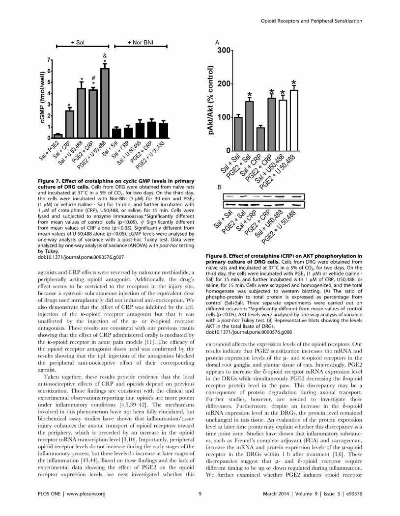
Effect on cyclic GMP level. Because the anti-nociceptive
effect of CRP is mediated by the L-arginine-nitric oxide-cGMP
pathway [12], we investigated the effect of CRP on the cyclic
GMP (cGMP) level in DRG cell cultures. CRP (1 mM) increased
the cGMP level compared with the control cells (Fig. 7), and
pretreating the cells with PGE2 (1 mM) significantly increased
(30%) the CRP-induced cGMP release. Nor-BNI, a k-opioid
receptor antagonist, blocked this effect. The k-opioid receptor
agonist U 50,488 (1 mM) was used as a positive control and
increased the cGMP level. Pretreating the cells with PGE2 further
increased (33%) the cyclic nucleotide level compared with the cells
that were treated with only the agonist (Fig. 7).
Effect on AKT activation. AKT phosphorylation is involved
in the peripheral anti-nociceptive effect of opioids, such as
morphine and U 50,488 [18,33]. Therefore, we evaluated whether
CRP increases AKT phosphorylation in PGE2-sensitized cells.
Treating DRG cells with PGE2 (1 mM) increased the AKT
phosphorylation level compared with the saline-treated (control)
cells (Fig. 8), and CRP (1 mM) did not interfere with AKT
phosphorylation compared with the control cells and the PGE2-
treated cells (Fig. 8). The k-opioid receptor agonist U 50,488
(1 mM) was used as a positive control and activated AKT (Fig. 8).
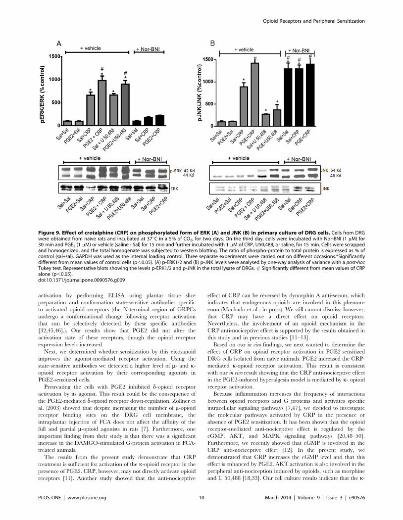
Effect on MAPK activation. The activation of the k-opioid
receptor increases the phosphorylation of MAPKs (ERK1/2 and
JNK) in neuronal and non-neuronal cells [34]. Therefore, MAPK
phosphorylation may be a useful indicator of opioid receptor
activation in DRG cells. CRP increased ERK1/2 phosphorylation
in DRG cells (Fig. 9A), and pretreating the cells with PGE2
significantly increased (1.5-fold) the CRP-mediated ERK1/2
phosphorylation (Fig. 9A). PGE2 alone, however, does not appear
to induce ERK1/2 phosphorylation. The k-opioid receptor
antagonist Nor-BNI inhibited the CRP-mediated ERK1/2 phos-
phorylation, which suggests that the k-opioid receptor is involved
in this process. Nor-BNI alone does not appear to cause ERK1/2
phosphorylation. Based on these results, we further evaluated the
Figure 3. Intraplantar injection effect of prostaglandin E2 (PGE2) on protein expression of m (A), k (B) and d (C) opioid receptors. Thechanges in protein levels of opioid receptors were determined by immunobloting, in dorsal root ganglia (DRG) and plantar tissue obtained 3 h afterPGE2 injection (100 ng/paw). Data are presented as mean 6 SEM and expressed as % of control (naıve) animals. *Significantly different from meanvalues of naıve animals, n = 6 per group (p,0.05). Data were analyzed by one-way analysis of variance (ANOVA) with post-hoc testing by Tukey.doi:10.1371/journal.pone.0090576.g003
Opioid Receptors and Peripheral Sensitization
PLOS ONE | www.plosone.org 6 March 2014 | Volume 9 | Issue 3 | e90576
effect of the k-opioid receptor agonist U 50,488 on ERK1/2
activation. Treating neurons with U 50,488 increased the level of
phosphorylated ERK1/2, and the phosphorylated ERK1/2 level
was further increased in cells that were pretreated with PGE2
(Fig. 9A). Treating neurons with either CRP or U 50, 488 also
induced JNK phosphorylation (9- and 2,5-fold, respectively).
Similar to the ERK1/2 expression profile, pretreating the cells
with PGE2 increased the CRP- and U 50, 488-mediated JNK
phosphorylation (10.5- and 3.5-fold, respectively). PGE2 alone
does not appear to induce JNK phosphorylation. Nor-BNI did not
interfere with this CRP effect, but this antagonist did increase the
level of phosphorylated JNK (13-fold) (Fig. 9B).
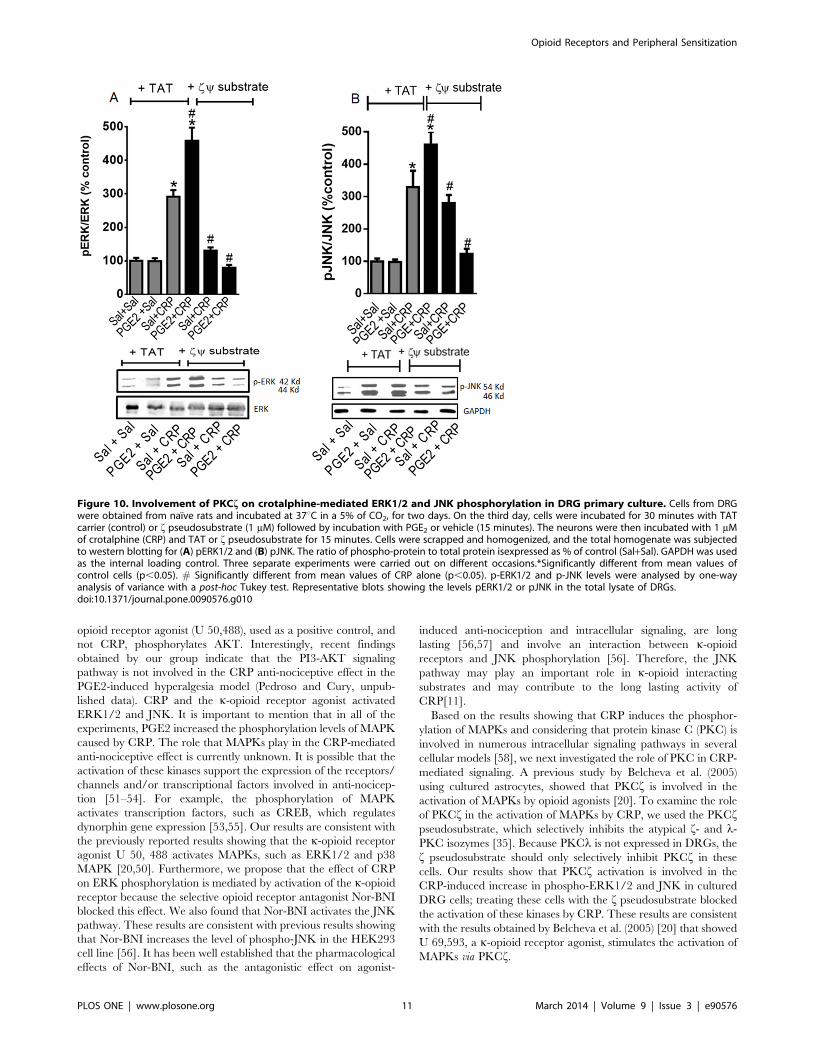
PKC f is involved in CRP-induced MAPK
activation. Because CRP appears to activate ERK and JNK,
we further investigated the potential mechanisms involved in this
effect. Previous studies indicate that PKCf is involved in the
activation of ERK1/2 by the k-opioid receptor agonist [20]. To
examine the direct role that PKC-f plays in the activation of the
MAPKs by CRP, we used the PKC-f pseudosubstrate, which
selectively inhibits the atypical PKC f isozyme [35]. Pretreating
the DRG cells with the PKC f pseudosubstrate abolished the
CRP-mediated ERK1/2 phosphorylation in both the PGE2-
sensitized and unsensitized cells (Fig. 10A). In contrast, the fpseudosubstrate did not affect the CRP-mediated JNK phosphor-
ylation but did inhibit the increase in CRP-induced JNK
phosphorylation observed in the presence of PGE2 (Fig. 10B).
The TAT carrier was used as a negative control.
Discussion
We previously reported that CRP, a 14-aminoacid peptide
isolated from C. d. terrificus venom, has a potent, long-lasting and
peripheral opioid receptor-mediated anti-nociceptive effect [11–
13]. In this study, we demonstrated that the potency and long-
lasting anti-nociceptive effect of CRP depends on tissue inflam-
mation. It has been well established that the local efficacy of opioid
drugs is enhanced in the presence of tissue injury/inflammation,
but the mechanisms involved in this phenomenon remain under
investigation [6,7,9]. In the present study, we also elucidated some
of the mechanisms involved in the increased peripheral anti-
nociceptive efficacy of CRP and opioid receptor agonists in the
Figure 4. PGE2, per se, does not activate opioid receptors. The plantar tissue (A) and dorsal root ganglia (ipsi and contralateral) (B) wereobtained 3 h after PGE2 injection (100 ng/paw). Tissues obtained from naıve animals were used as controls. The tissues were sectioned and probedwith anti-m, k or d antibodies. (A) The black bar represents the results obtained with the conformation state-sensitive antibodies whereas the whitebar represents the results obtained using the control antibodies (western blotting assay antibodies). (B) The first couple of bars represents the resultsobtained using the control antibodies (western blotting assay antibodies). The second one represents the conformation state-sensitive antibodies.The dotted line represents the values obtained for naıve animals. * Significantly different from mean values of naıve animals (p,0.05). Data wereanalyzed by one-way analysis of variance (ANOVA) with post-hoc testing by Tukey.doi:10.1371/journal.pone.0090576.g004
Opioid Receptors and Peripheral Sensitization
PLOS ONE | www.plosone.org 7 March 2014 | Volume 9 | Issue 3 | e90576
presence of hyperalgesia. We observed the following results: (a) the
local intraplantar (i.pl.) injection of a low dose of CRP or selective
opioid receptor agonists, which are ineffective at increasing the
nociceptive threshold in naıve animals in the rat paw pressure
testnaıve, produced anti-nociception in a PGE2-induced hyperal-
gesia modelPGE2. In this hyperalgesia rat model, the CRP effect
was mediated by activation of the k-opioid receptor. (b) The i.pl.
administration of PGE2 increased the peripheral mRNA expres-
sion levels of the m-, d-, and k-opioid receptors and the peripheral
protein expression levels of the m- and k-opioid receptors within
3 h of the injection. The eicosanoid also increased the activation of
the m- and k-opioid receptors by their corresponding agonist. In
contrast, the PGE2 treatment decreased the d-opioid receptor
protein level and activation of the d-opioid receptor by its agonist.
(c) In cultured DRG neurons, the activation of specific intracel-
lular signaling pathways (MAPKs and PKCf) by CRP was
enhanced following PGE2 sensitization. The results presented here
provide evidence that tissue injury regulates the expression of
peripheral opioid receptors and their activation by CRP and
selective opioid receptor agonists and that tissue injury also
contribute to activation of specific intracellular signaling pathways.
In our study, we first evaluated the effect of an intraplantar
injection of CRP and selective agonists of the m-, k- and d-opioid
receptors on the nociceptive threshold of animals that were
sensitized or not by an i.pl. injection of PGE2. This eicosanoid
sensitizes the peripheral terminals of the neuron, which reduces
the threshold for activating the nociceptors [36] [37] [38]. The
i.pl. injection of CRP and DAMGO, DPDPE and U 50,488 (m-, d-
and k-opioid receptor agonist, respectively), in doses that are
ineffective at altering the mechanical nociceptive stimuli with-
drawal response in naıve rats, blocked PGE2-induced hyperalge-
sia. Furthermore, we should mention that the doses of the agonists
and antagonists used in this study were chosen based on their
optimal effects as observed in previous dose-response studies
[13,23]. The doses used in the present study only had a local effect
because anti-nociception was only detected in the paw injected
with the drugs and not in the contralateral paw. Moreover, the
Figure 5. PGE2 improves agonist-mediated opioid receptoractivation. Naıve or prostaglandin E2 (PGE2, 100 ng/paw) injected ratswere treated with DAMGO (5 mg/paw), U50,488 (10 mg/paw), DPDPE(20 mg/paw), (Panel A) or crotalphine (CRP, 0.6 ng/paw) (Panel B). Theagonists, CRP, or vehicle (control) were administered 2 h after PGE2injection. ELISA assay was performed in slices of plantar tissue, usingconformation state-sensitive antibodies (m, k, d activated). Tissues werecollected 3 h after PGE2 injection. *Significantly different from meanvalues of control animals (dotted line), n = 6 animals per group(p,0.05). #Significantly different from mean values of CRP or agonistalone (p,0.05). Data were analyzed by one-way analysis of variance(ANOVA) with post-hoc testing by Tukey.doi:10.1371/journal.pone.0090576.g005
Figure 6. PGE2 improves agonist-mediated opioid receptoractivation in primary neuronal cell culture. Cells from dorsal rootganglia were obtained from naıve rats and incubated at 37uC, in a 5% ofCO2, for two days. On the third day, the cells were incubated with PGE2(1 mM) or vehicle for 15 min and further incubated with 1 mM ofcrotalphine (CRP, A) or DAMGO, U50,488, DPDPE (B) for 15 min. ELISAassay was performed directly on fixed cells, using conformation state-sensitive antibodies (activated). Three separate experiments werecarried out on different occasions.* Significantly different from meanvalues of naıve animals (dotted line) (p,0.05), # Significantly differentfrom mean values of CRP or agonist alone, average of 3 independentexperiments (p,0.05). Data were analyzed by one-way analysis ofvariance (ANOVA) with post-hoc testing by Tukey.doi:10.1371/journal.pone.0090576.g006
Opioid Receptors and Peripheral Sensitization
PLOS ONE | www.plosone.org 8 March 2014 | Volume 9 | Issue 3 | e90576
agonists and CRP effects were reversed by naloxone methiodide, a
peripherally acting opioid antagonist. Additionally, the drug’s
effect seems to be restricted to the receptors in the injury site,
because a systemic subcutaneous injection of the equivalent dose
of drugs used intraplantarly did not induced anti-nociception. We
also demonstrate that the effect of CRP was inhibited by the i.pl.
injection of the k-opioid receptor antagonist but that it was
unaffected by the injection of the m- or d-opioid receptor
antagonists. These results are consistent with our previous results
showing that the effect of CRP administered orally is mediated by
the k-opioid receptor in acute pain models [11]. The efficacy of
the opioid receptor antagonist doses used was confirmed by the
results showing that the i.pl. injection of the antagonists blocked
the peripheral anti-nociceptive effect of their corresponding
agonist.
Taken together, these results provide evidence that the local
anti-nociceptive effects of CRP and opioids depend on previous
sensitization. These findings are consistent with the clinical and
experimental observations reporting that opioids are more potent
under inflammatory conditions [4,5,39–42]. The mechanisms
involved in this phenomenon have not been fully elucidated, but
biochemical assay studies have shown that inflammation/tissue
injury enhances the axonal transport of opioid receptors toward
the periphery, which is preceded by an increase in the opioid
receptor mRNA transcription level [3,10]. Importantly, peripheral
opioid receptor levels do not increase during the early stages of the
inflammatory process, but these levels do increase at later stages of
the inflammation [43,44]. Based on these findings and the lack of
experimental data showing the effect of PGE2 on the opioid
receptor expression levels, we next investigated whether this
eicosanoid affects the expression levels of the opioid receptors. Our
results indicate that PGE2 sensitization increases the mRNA and
protein expression levels of the m- and k-opioid receptors in the
dorsal root ganglia and plantar tissue of rats. Interestingly, PGE2
appears to increase the d-opioid receptor mRNA expression level
in the DRGs while simultaneously PGE2 decreasing the d-opioid
receptor protein level in the paw. This discrepancy may be a
consequence of protein degradation during axonal transport.
Further studies, however, are needed to investigate these
differences. Furthermore, despite an increase in the d-opioid
mRNA expression level in the DRGs, the protein level remained
unchanged in this tissue. An evaluation of the protein expression
level at later time points may explain whether this discrepancy is a
time point issue. Studies have shown that inflammatory substanc-
es, such as Freund’s complete adjuvant (FCA) and carrageenan,
increase the mRNA and protein expression levels of the m-opioid
receptor in the DRGs within 1 h after treatment [3,6]. These
discrepancies suggest that m- and d-opioid receptor require
different timing to be up or down regulated during inflammation.
We further examined whether PGE2 induces opioid receptor
Figure 7. Effect of crotalphine on cyclic GMP levels in primaryculture of DRG cells. Cells from DRG were obtained from naıve ratsand incubated at 37uC in a 5% of CO2, for two days. On the third day,the cells were incubated with Nor-BNI (1 mM) for 30 min and PGE2
(1 mM) or vehicle (saline - Sal) for 15 min, and further incubated with1 mM of crotalphine (CRP), U50,488, or saline, for 15 min. Cells werelysed and subjected to enzyme immunoassay.*Significantly differentfrom mean values of control cells (p,0.05), # Significantly differentfrom mean values of CRP alone (p,0.05), Significantly different frommean values of U 50,488 alone (p,0.05). cGMP levels were analysed byone-way analysis of variance with a post-hoc Tukey test. Data wereanalyzed by one-way analysis of variance (ANOVA) with post-hoc testingby Tukey.doi:10.1371/journal.pone.0090576.g007 Figure 8. Effect of crotalphine (CRP) on AKT phosphorylation in
primary culture of DRG cells. Cells from DRG were obtained fromnaıve rats and incubated at 37uC in a 5% of CO2, for two days. On thethird day, the cells were incubated with PGE2 (1 mM) or vehicle (saline -Sal) for 15 min. and further incubated with 1 mM of CRP, U50,488, orsaline, for 15 min. Cells were scrapped and homogenized, and the totalhomogenate was subjected to western blotting. (A) The ratio ofphospho-protein to total protein is expressed as percentage fromcontrol (Sal+Sal). Three separate experiments were carried out ondifferent occasions.*Significantly different from mean values of controlcells (p,0.05). AKT levels were analysed by one-way analysis of variancewith a post-hoc Tukey test. (B) Representative blots showing the levelsAKT in the total lisate of DRGs.doi:10.1371/journal.pone.0090576.g008
Opioid Receptors and Peripheral Sensitization
PLOS ONE | www.plosone.org 9 March 2014 | Volume 9 | Issue 3 | e90576
activation by performing ELISA using plantar tissue slice
preparation and conformation state-sensitive antibodies specific
to activated opioid receptors (the N-terminal region of GRPCs
undergo a conformational change following receptor activation
that can be selectively detected by these specific antibodies
[32,45,46].). Our results show that PGE2 did not alter the
activation state of these receptors, though the opioid receptor
expression levels increased.
Next, we determined whether sensitization by this eicosanoid
improves the agonist-mediated receptor activation. Using the
state-sensitive antibodies we detected a higher level of m- and k-
opioid receptor activation by their corresponding agonists in
PGE2-sensitized cells.
Pretreating the cells with PGE2 inhibited d-opioid receptor
activation by its agonist. This result could be the consequence of
the PGE2-mediated d-opioid receptor down-regulation. Zollner et
al. (2003) showed that despite increasing the number of m-opioid
receptor binding sites on the DRG cell membrane, the
intraplantar injection of FCA does not affect the affinity of the
full and partial m-opioid agonists in rats [7]. Furthermore, one
important finding from their study is that there was a significant
increase in the DAMGO-stimulated G-protein activation in FCA-
treated animals.
The results from the present study demonstrate that CRP
treatment is sufficient for activation of the k-opioid receptor in the
presence of PGE2. CRP, however, may not directly activate opioid
receptors [11]. Another study showed that the anti-nociceptive
effect of CRP can be reversed by dynorphin A anti-serum, which
indicates that endogenous opioids are involved in this phenom-
enon (Machado et al., in press). We still cannot dismiss, however,
that CRP may have a direct effect on opioid receptors.
Nevertheless, the involvement of an opioid mechanism in the
CRP anti-nociceptive effect is supported by the results obtained in
this study and in previous studies [11–13].
Based on our in vivo findings, we next wanted to determine the
effect of CRP on opioid receptor activation in PGE2-sensitized
DRG cells isolated from naıve animals. PGE2 increased the CRP-
mediated k-opioid receptor activation. This result is consistent
with our in vivo result showing that the CRP anti-nociceptive effect
in the PGE2-induced hyperalgesia model is mediated by k- opioid
receptor activation.
Because inflammation increases the frequency of interactions
between opioid receptors and G proteins and activates specific
intracellular signaling pathways [7,47], we decided to investigate
the molecular pathways activated by CRP in the presence or
absence of PGE2 sensitization. It has been shown that the opioid
receptor-mediated anti-nociceptive effect is regulated by the
cGMP, AKT, and MAPK signaling pathways [20,48–50].
Furthermore, we recently showed that cGMP is involved in the
CRP anti-nociceptive effect [12]. In the present study, we
demonstrated that CRP increases the cGMP level and that this
effect is enhanced by PGE2. AKT activation is also involved in the
peripheral anti-nociception induced by opioids, such as morphine
and U 50,488 [18,33]. Our cell culture results indicate that the k-
Figure 9. Effect of crotalphine (CRP) on phosphorylated form of ERK (A) and JNK (B) in primary culture of DRG cells. Cells from DRGwere obtained from naıve rats and incubated at 37uC in a 5% of CO2, for two days. On the third day, cells were incubated with Nor-BNI (1 mM) for30 min and PGE2 (1 mM) or vehicle (saline - Sal) for 15 min and further incubated with 1 mM of CRP, U50,488, or saline, for 15 min. Cells were scrappedand homogenized, and the total homogenate was subjected to western blotting. The ratio of phospho-protein to total protein is expressed as % ofcontrol (sal+sal). GAPDH was used as the internal loading control. Three separate experiments were carried out on different occasions.*Significantlydifferent from mean values of control cells (p,0.05). (A) p-ERK1/2 and (B) p-JNK levels were analysed by one-way analysis of variance with a post-hocTukey test. Representative blots showing the levels p-ERK1/2 and p-JNK in the total lysate of DRGs. # Significantly different from mean values of CRPalone (p,0.05).doi:10.1371/journal.pone.0090576.g009
Opioid Receptors and Peripheral Sensitization
PLOS ONE | www.plosone.org 10 March 2014 | Volume 9 | Issue 3 | e90576
opioid receptor agonist (U 50,488), used as a positive control, and
not CRP, phosphorylates AKT. Interestingly, recent findings
obtained by our group indicate that the PI3-AKT signaling
pathway is not involved in the CRP anti-nociceptive effect in the
PGE2-induced hyperalgesia model (Pedroso and Cury, unpub-
lished data). CRP and the k-opioid receptor agonist activated
ERK1/2 and JNK. It is important to mention that in all of the
experiments, PGE2 increased the phosphorylation levels of MAPK
caused by CRP. The role that MAPKs play in the CRP-mediated
anti-nociceptive effect is currently unknown. It is possible that the
activation of these kinases support the expression of the receptors/
channels and/or transcriptional factors involved in anti-nocicep-
tion [51–54]. For example, the phosphorylation of MAPK
activates transcription factors, such as CREB, which regulates
dynorphin gene expression [53,55]. Our results are consistent with
the previously reported results showing that the k-opioid receptor
agonist U 50, 488 activates MAPKs, such as ERK1/2 and p38
MAPK [20,50]. Furthermore, we propose that the effect of CRP
on ERK phosphorylation is mediated by activation of the k-opioid
receptor because the selective opioid receptor antagonist Nor-BNI
blocked this effect. We also found that Nor-BNI activates the JNK
pathway. These results are consistent with previous results showing
that Nor-BNI increases the level of phospho-JNK in the HEK293
cell line [56]. It has been well established that the pharmacological
effects of Nor-BNI, such as the antagonistic effect on agonist-
induced anti-nociception and intracellular signaling, are long
lasting [56,57] and involve an interaction between k-opioid
receptors and JNK phosphorylation [56]. Therefore, the JNK
pathway may play an important role in k-opioid interacting
substrates and may contribute to the long lasting activity of
CRP[11].
Based on the results showing that CRP induces the phosphor-
ylation of MAPKs and considering that protein kinase C (PKC) is
involved in numerous intracellular signaling pathways in several
cellular models [58], we next investigated the role of PKC in CRP-
mediated signaling. A previous study by Belcheva et al. (2005)
using cultured astrocytes, showed that PKCf is involved in the
activation of MAPKs by opioid agonists [20]. To examine the role
of PKCf in the activation of MAPKs by CRP, we used the PKCfpseudosubstrate, which selectively inhibits the atypical f- and l-
PKC isozymes [35]. Because PKCl is not expressed in DRGs, the
f pseudosubstrate should only selectively inhibit PKCf in these
cells. Our results show that PKCf activation is involved in the
CRP-induced increase in phospho-ERK1/2 and JNK in cultured
DRG cells; treating these cells with the f pseudosubstrate blocked
the activation of these kinases by CRP. These results are consistent
with the results obtained by Belcheva et al. (2005) [20] that showed
U 69,593, a k-opioid receptor agonist, stimulates the activation of
MAPKs via PKCf.
Figure 10. Involvement of PKCf on crotalphine-mediated ERK1/2 and JNK phosphorylation in DRG primary culture. Cells from DRGwere obtained from naıve rats and incubated at 37uC in a 5% of CO2, for two days. On the third day, cells were incubated for 30 minutes with TATcarrier (control) or f pseudosubstrate (1 mM) followed by incubation with PGE2 or vehicle (15 minutes). The neurons were then incubated with 1 mMof crotalphine (CRP) and TAT or f pseudosubstrate for 15 minutes. Cells were scrapped and homogenized, and the total homogenate was subjectedto western blotting for (A) pERK1/2 and (B) pJNK. The ratio of phospho-protein to total protein isexpressed as % of control (Sal+Sal). GAPDH was usedas the internal loading control. Three separate experiments were carried out on different occasions.*Significantly different from mean values ofcontrol cells (p,0.05). # Significantly different from mean values of CRP alone (p,0.05). p-ERK1/2 and p-JNK levels were analysed by one-wayanalysis of variance with a post-hoc Tukey test. Representative blots showing the levels pERK1/2 or pJNK in the total lysate of DRGs.doi:10.1371/journal.pone.0090576.g010
Opioid Receptors and Peripheral Sensitization
PLOS ONE | www.plosone.org 11 March 2014 | Volume 9 | Issue 3 | e90576
In conclusion, we propose that CRP has a more potent
peripheral anti-nociceptive effect under conditions of acute
sensitization. The mechanisms responsible for this phenomenon
involve increased expression levels and the activation of opioid
receptors as well as increases in the cGMP levels, the phosphor-
ylation of MAPKs and the activation of PKCf in the periphery.
These results further elucidate the important peripheral molecular
mechanisms involved in pain control and identify interesting
therapeutic alternatives for inducing peripheral analgesia.
Supporting Information
Figure S1 Time-course for intraplantar injection ofprostaglandin E2 (PGE2) in the rat nociceptive thresh-old. Pain threshold was obtained in the rat paw pressure test,
before (time 0) and 1, 3, 5 and, 8 h after intraplantar injection of
PGE2 (100 ng/paw) or saline (control). Data represent mean
values 6 S.E.M. for six rats per group. * significantly different
from baseline, # significantly different from control. Data were
analyzed by two-way analysis of variance (ANOVA) with post-hoc
testing by Tukey.
(TIF)
Figure S2 Effect of methiodide naloxone (MN) on thelocal crotalphine (CRP) and opioid receptor agonists-induced anti-nociception. Pain threshold was obtained in the
rat paw pressure test, before (dotted line) and 3 h after intraplantar
injection of PGE2 (100 ng/paw). CRP (0.6 ng/paw), DAMGO (mopioid receptor agonist, 5 mg/paw), DPDPE (d opioid receptor
agonist, 20 mg/paw), U-50488 (k opioid receptor agonist, 10 mg/
paw) were injected 2 h after PGE2 administration. NM (1 mg/
Kg), were injected by the subcutaneous route 15 minutes before
the nociceptive threshold assessment. Data represent mean values
6 S.E.M. for five rats per group. * significantly different from
baseline (dotted line), # significantly different from control
(saline = SAL). Data were analyzed by two-way analysis of
variance (ANOVA) with post-hoc testing by Tukey.
(TIF)
Figure S3 Comparative effect between systemic andlocal injection of crotalphine (CRP) and opioid receptor
agonists. Pain threshold was obtained in the rat paw pressure
test, before (dotted line) and 3 h after intraplantar injection of
PGE2 (100 ng/paw). CRP (0.6 ng), DAMGO (m opioid receptor
agonist, 5 mg), DPDPE (d opioid receptor agonist, 20 mg),
U-50488 (k opioid receptor agonist, 10 mg) were injected by
subcutaneous (systemic) or intraplantar (local) route, 2 h after
PGE2 administration. Data represent mean values 6 S.E.M. for
five rats per group. * significantly different from baseline (dotted
line), # significantly different from control (saline = Sal). Data
were analyzed by two-way analysis of variance (ANOVA) with
post-hoc testing by Tukey. The following bars CRP intraplantar,
DAMGO intraplantar, DPDPE intraplantar and U 50,488
intraplantar were re-drawn from the Fig. S2, since this
experiments were performed in the same day.
(TIF)
Figure S4 Intraplantar injection of prostaglandin E2(PGE2) in the rat nociceptive threshold. Pain threshold was
obtained in the rat paw pressure test, before (time 0) and 3 h after
intraplantar injection of PGE2 (100 ng/paw) or saline (control).
(A) values for animals whose tissued were used for mRNA
extraction and (B) for protein extraction. Data represent mean
values 6 S.E.M. for 6–8 rats per group. * significantly different
from baseline, # significantly different from control. Data were
analyzed by two-way analysis of variance (ANOVA) with post-hoc
testing by Tukey.
(TIF)
Acknowledgments
We thank Juliana Sousa de Carvalho and Marie-Helen Disatnik for
technical assistance and Andrea S. Heimann from Proteimax Biotechnol-
ogy for critical suggestions with experiments involving the conformation
state-sensitive antibodies.
Author Contributions
Conceived and designed the experiments: VOZ YC. Performed the
experiments: VOZ VPG ACOF. Analyzed the data: VOZ YC.
Contributed reagents/materials/analysis tools: VOZ JCBF CAP DMR
YC. Wrote the paper: VOZ YC.
References
1. Stein C (1993) Peripheral mechanisms of opioid analgesia. Anesth Analg 76:
182–191.
2. Obara I, Parkitna JR, Korostynski M, Makuch W, Kaminska D, et al. (2009)
Local peripheral opioid effects and expression of opioid genes in the spinal cord
and dorsal root ganglia in neuropathic and inflammatory pain. Pain 141: 283–
291.
3. Puehler W, Zollner C, Brack A, Shaqura MA, Krause H, et al. (2004) Rapid
upregulation of mu opioid receptor mRNA in dorsal root ganglia in response to
peripheral inflammation depends on neuronal conduction. Neuroscience 129:
473–479.
4. Maekawa K, Minami M, Masuda T, Satoh M (1996) Expression of mu- and
kappa-, but not delta-, opioid receptor mRNAs is enhanced in the spinal dorsal
horn of the arthritic rats. Pain 64: 365–371.
5. Cahill CM, Morinville A, Hoffert C, O’Donnell D, Beaudet A (2003) Up-
regulation and trafficking of delta opioid receptor in a model of chronic
inflammation: implications for pain control. Pain 101: 199–208.
6. Hassan AH, Ableitner A, Stein C, Herz A (1993) Inflammation of the rat paw
enhances axonal transport of opioid receptors in the sciatic nerve and increases
their density in the inflamed tissue. Neuroscience 55: 185–195.
7. Zollner C, Shaqura MA, Bopaiah CP, Mousa S, Stein C, et al. (2003) Painful
inflammation-induced increase in mu-opioid receptor binding and G-protein
coupling in primary afferent neurons. Mol Pharmacol 64: 202–210.
8. Shaqura MA, Zollner C, Mousa SA, Stein C, Schafer M (2004) Characterization
of mu opioid receptor binding and G protein coupling in rat hypothalamus,
spinal cord, and primary afferent neurons during inflammatory pain.
J Pharmacol Exp Ther 308: 712–718.
9. Antonijevic I, Mousa SA, Schafer M, Stein C (1995) Perineurial defect and
peripheral opioid analgesia in inflammation. J Neurosci 15: 165–172.
10. Mousa SA, Zhang Q, Sitte N, Ji R, Stein C (2001) beta-Endorphin-containing
memory-cells and mu-opioid receptors undergo transport to peripheral inflamed
tissue. J Neuroimmunol 115: 71–78.
11. Konno K, Picolo G, Gutierrez VP, Brigatte P, Zambelli VO, et al. (2008)
Crotalphine, a novel potent analgesic peptide from the venom of the South
American rattlesnake Crotalus durissus terrificus. Peptides.
12. Gutierrez VP, Zambelli VO, Picolo G, Chacur M, Sampaio SC, et al. The
peripheral L-arginine-nitric oxide-cyclic GMP pathway and ATP-sensitive K
channels are involved in the antinociceptive effect of crotalphine on neuropathic
pain in rats. Behav Pharmacol 23: 14–24.
13. Gutierrez VP, Konno K, Chacur M, Sampaio SC, Picolo G, et al. (2008)
Crotalphine induces potent antinociception in neuropathic pain by acting at
peripheral opioid receptors. Eur J Pharmacol 594: 84–92.
14. Granados-Soto V, Rufino MO, Gomes Lopes LD, Ferreira SH (1997) Evidence
for the involvement of the nitric oxide-cGMP pathway in the antinociception of
morphine in the formalin test. Eur J Pharmacol 340: 177–180.
15. Sachs D, Cunha FQ, Ferreira SH (2004) Peripheral analgesic blockade of
hypernociception: activation of arginine/NO/cGMP/protein kinase G/ATP-
sensitive K+ channel pathway. Proc Natl Acad Sci U S A 101: 3680–3685.
16. Pacheco DF, Reis GM, Francischi JN, Castro MS, Perez AC, et al. (2005) delta-
Opioid receptor agonist SNC80 elicits peripheral antinociception via delta(1)
and delta(2) receptors and activation of the l-arginine/nitric oxide/cyclic GMP
pathway. Life Sci 78: 54–60.
17. Amarante LH, Duarte ID (2002) The kappa-opioid agonist (+/-)-bremazocine
elicits peripheral antinociception by activation of the L-arginine/nitric oxide/
cyclic GMP pathway. Eur J Pharmacol 454: 19–23.
18. Cunha TM, Roman-Campos D, Lotufo CM, Duarte HL, Souza GR, et al.
Morphine peripheral analgesia depends on activation of the PI3Kgamma/
Opioid Receptors and Peripheral Sensitization
PLOS ONE | www.plosone.org 12 March 2014 | Volume 9 | Issue 3 | e90576
AKT/nNOS/NO/KATP signaling pathway. Proc Natl Acad Sci U S A 107:
4442–4447.19. Law BK, Waltner-Law ME, Entingh AJ, Chytil A, Aakre ME, et al. (2000)
Salicylate-induced growth arrest is associated with inhibition of p70s6k and
down-regulation of c-myc, cyclin D1, cyclin A, and proliferating cell nuclearantigen. J Biol Chem 275: 38261–38267.
20. Belcheva MM, Clark AL, Haas PD, Serna JS, Hahn JW, et al. (2005) Mu andkappa opioid receptors activate ERK/MAPK via different protein kinase C
isoforms and secondary messengers in astrocytes. J Biol Chem 280: 27662–
27669.21. Connor M, Christie MD (1999) Opioid receptor signalling mechanisms. Clin
Exp Pharmacol Physiol 26: 493–499.22. Zimmermann M (1983) Ethical guidelines for investigations of experimental
pain in conscious animals. Pain 16: 109–110.23. Picolo G, Giorgi R, Bernardi MM, Cury Y (1998) The antinociceptive effect of
Crotalus durissus terrificus snake venom is mainly due to a supraspinally
integrated response. Toxicon 36: 223–227.24. Segond von Banchet G, Scholze A, Schaible HG (2003) Prostaglandin E2
increases the expression of the neurokinin1 receptor in adult sensory neurones inculture: a novel role of prostaglandins. Br J Pharmacol 139: 672–680.
25. Picolo G, Giorgi R, Cury Y (2000) delta-opioid receptors and nitric oxide
mediate the analgesic effect of Crotalus durissus terrificus snake venom.Eur J Pharmacol 391: 55–62.
26. Gendron L, Pintar JE, Chavkin C (2007) Essential role of mu opioid receptor inthe regulation of delta opioid receptor-mediated antihyperalgesia. Neuroscience
150: 807–817.27. Lomas LM, Barrett AC, Terner JM, Lysle DT, Picker MJ (2007) Sex differences
in the potency of kappa opioids and mixed-action opioids administered
systemically and at the site of inflammation against capsaicin-inducedhyperalgesia in rats. Psychopharmacology (Berl) 191: 273–285.
28. {Ji J, Y., Murphy AZ, Traub RJ (2007) Estrogen modulation of morphineanalgesia of visceral pain in female rats is supraspinally and peripherally
mediated. J Pain 8: 494–502.
29. Picolo G, Cury Y (2004) Peripheral neuronal nitric oxide synthase activitymediates the antinociceptive effect of Crotalus durissus terrificus snake venom, a
delta- and kappa-opioid receptor agonist. Life Sci 75: 559–573.30. Randall LO, Selitto JJ (1957) A method for measurement of analgesia activity on
inflamed tissue. Arch Inst Pharmacodyn 111: 209–219.31. Bradford MM (1976) A rapid and sensitive method for the quantitation of
microgram quantities of protein utilizing the principle of protein-dye binding.
Anal Biochem 72: 248–254.32. Gupta A, Decaillot FM, Gomes I, Tkalych O, Heimann AS, et al. (2006)
Conformation state sensitive antibodies to G-protein coupled receptors. J BiolChem.
33. Cunha TM, Souza GR, Domingues AC, Carreira EU, Lotufo CM, et al.
Stimulation of peripheral Kappa opioid receptors inhibits inflammatoryhyperalgesia via activation of the PI3Kgamma/AKT/nNOS/NO signaling
pathway. Mol Pain 8: 10.34. Bruchas MR, Chavkin C Kinase cascades and ligand-directed signaling at the
kappa opioid receptor. Psychopharmacology (Berl) 210: 137–147.35. Berra E, Diaz-Meco MT, Dominguez I, Municio MM, Sanz L, et al. (1993)
Protein kinase C zeta isoform is critical for mitogenic signal transduction. Cell
74: 555–563.36. Kwong K, Lee LY (2005) Prostaglandin E2 potentiates a TTX-resistant sodium
current in rat capsaicin-sensitive vagal pulmonary sensory neurones. J Physiol564: 437–450.
37. Southall MD, Vasko MR (2001) Prostaglandin receptor subtypes, EP3C and
EP4, mediate the prostaglandin E2-induced cAMP production and sensitizationof sensory neurons. J Biol Chem 276: 16083–16091.
38. Ferreira SH, Lorenzetti BB (1981) Prostaglandin hyperalgesia, IV: a metabolicprocess. Prostaglandins 21: 789–792.
39. Stein C, Millan MJ, Shippenberg TS, Peter K, Herz A (1989) Peripheral opioid
receptors mediating antinociception in inflammation. Evidence for involvement
of mu, delta and kappa receptors. J Pharmacol Exp Ther 248: 1269–1275.
40. Mousa SA, Machelska H, Schafer M, Stein C (2002) Immunohistochemical
localization of endomorphin-1 and endomorphin-2 in immune cells and spinal
cord in a model of inflammatory pain. J Neuroimmunol 126: 5–15.
41. Furst S, Riba P, Friedmann T, Timar J, Al-Khrasani M, et al. (2005) Peripheral
versus central antinociceptive actions of 6-amino acid-substituted derivatives of
14-O-methyloxymorphone in acute and inflammatory pain in the rat.
J Pharmacol Exp Ther 312: 609–618.
42. Nunez S, Lee JS, Zhang Y, Bai G, Ro JY (2007) Role of peripheral mu-opioid
receptors in inflammatory orofacial muscle pain. Neuroscience 146: 1346–1354.
43. Schafer M, Imai Y, Uhl GR, Stein C (1995) Inflammation enhances peripheral
mu-opioid receptor-mediated analgesia, but not mu-opioid receptor transcrip-
tion in dorsal root ganglia. Eur J Pharmacol 279: 165–169.
44. Zhou L, Zhang Q, Stein C, Schafer M (1998) Contribution of opioid receptors
on primary afferent versus sympathetic neurons to peripheral opioid analgesia.
J Pharmacol Exp Ther 286: 1000–1006.
45. Lecat S, Bucher B, Mely Y, Galzi JL (2002) Mutations in the extracellular
amino-terminal domain of the NK2 neurokinin receptor abolish cAMP signaling
but preserve intracellular calcium responses. J Biol Chem 277: 42034–42048.
46. Decaillot FM, Befort K, Filliol D, Yue S, Walker P, et al. (2003) Opioid receptor
random mutagenesis reveals a mechanism for G protein-coupled receptor
activation. Nat Struct Biol 10: 629–636.
47. Selley DE, Breivogel CS, Childers SR (1993) Modification of G protein-coupled
functions by low-pH pretreatment of membranes from NG108-15 cells: increase
in opioid agonist efficacy by decreased inactivation of G proteins. Mol
Pharmacol 44: 731–741.
48. Belcheva MM, Vogel Z, Ignatova E, Avidor-Reiss T, Zippel R, et al. (1998)
Opioid modulation of extracellular signal-regulated protein kinase activity is ras-
dependent and involves Gbetagamma subunits. J Neurochem 70: 635–645.
49. Bohn LM, Belcheva MM, Coscia CJ (2000) Mitogenic signaling via endogenous
kappa-opioid receptors in C6 glioma cells: evidence for the involvement of
protein kinase C and the mitogen-activated protein kinase signaling cascade.
J Neurochem 74: 564–573.
50. Bruchas MR, Macey TA, Lowe JD, Chavkin C (2006) Kappa opioid receptor
activation of p38 MAPK is GRK3- and arrestin-dependent in neurons and
astrocytes. J Biol Chem 281: 18081–18089.
51. Sweatt JD (2004) Mitogen-activated protein kinases in synaptic plasticity and
memory. Curr Opin Neurobiol 14: 311–317.
52. Thomas GM, Huganir RL (2004) MAPK cascade signalling and synaptic
plasticity. Nat Rev Neurosci 5: 173–183.
53. Carlezon WA, Jr., Duman RS, Nestler EJ (2005) The many faces of CREB.
Trends Neurosci 28: 436–445.
54. Bruchas MR, Xu M, Chavkin C (2008) Repeated swim stress induces kappa
opioid-mediated activation of extracellular signal-regulated kinase 1/2. Neu-
roreport 19: 1417–1422.
55. Kreibich AS, Blendy JA (2004) cAMP response element-binding protein is
required for stress but not cocaine-induced reinstatement. J Neurosci 24: 6686–
6692.
56. Bruchas MR, Yang T, Schreiber S, Defino M, Kwan SC, et al. (2007) Long-
acting kappa opioid antagonists disrupt receptor signaling and produce
noncompetitive effects by activating c-Jun N-terminal kinase. J Biol Chem
282: 29803–29811.
57. Melief EJ, Miyatake M, Bruchas MR, Chavkin C (2010) Ligand-directed c-Jun
N-terminal kinase activation disrupts opioid receptor signaling. Proc Natl Acad
Sci U S A 107: 11608–11613.
58. Velazquez KT, Mohammad H, Sweitzer SM (2007) Protein kinase C in pain:
involvement of multiple isoforms. Pharmacol Res 55: 578–589.
Opioid Receptors and Peripheral Sensitization
PLOS ONE | www.plosone.org 13 March 2014 | Volume 9 | Issue 3 | e90576