Embed Size (px)
Citation preview
Thoracic Spine Mobility Deficits ICD-9-CM: 847.1 thoracic sprain ICF codes: Activities and Participation Domain code: d4105 Bending (Tilting the back
downward or to the side, at the torso, such as in bowling or reaching down for an object)
Body Structure code: s76001 Thoracic vertebral column Body Functions code: b7101 Mobility of several joints
Common Historical Findings:
Symptoms precipitated by a trauma, strain, awkward movement, or prolonged static posture (bottom line - an identifiable mechanical stress)
Pain is usually perceived inferior and lateral to the symptomatic segment Common Impairment Findings - Related to the Reported Activity Limitation or Participation Restrictions:
Pain increases at end range of the one particular motion Palpable asymmetry of adjacent transverse processes in either thoracic spine flexion or
extension Unilateral posterior-to-anterior (PA) pressures on the involved segment reproduce the
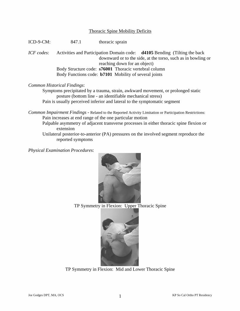
reported symptoms Physical Examination Procedures:
TP Symmetry in Flexion: Upper Thoracic Spine
TP Symmetry in Flexion: Mid and Lower Thoracic Spine
Joe Godges DPT, MA, OCS KP So Cal Ortho PT Residency 1
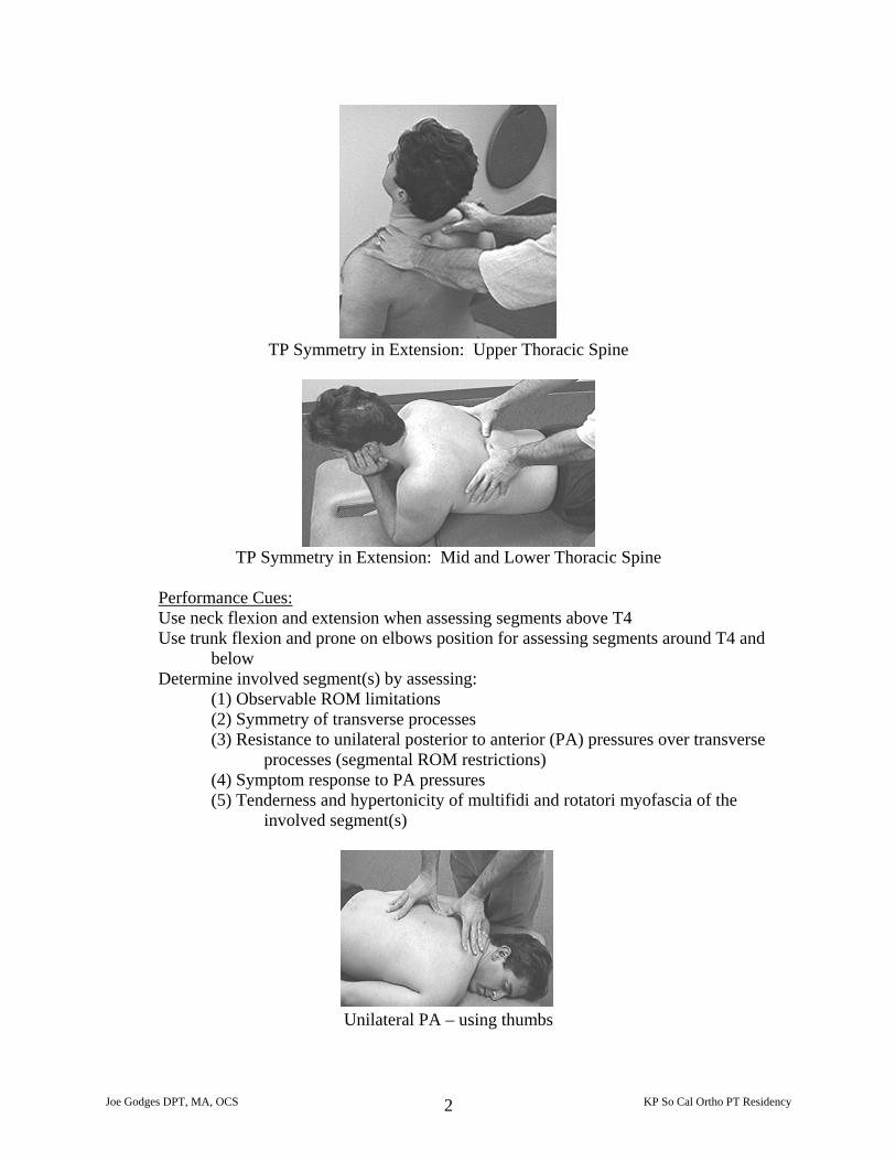
TP Symmetry in Extension: Upper Thoracic Spine
TP Symmetry in Extension: Mid and Lower Thoracic Spine
Performance Cues: Use neck flexion and extension when assessing segments above T4 Use trunk flexion and prone on elbows position for assessing segments around T4 and
below Determine involved segment(s) by assessing:
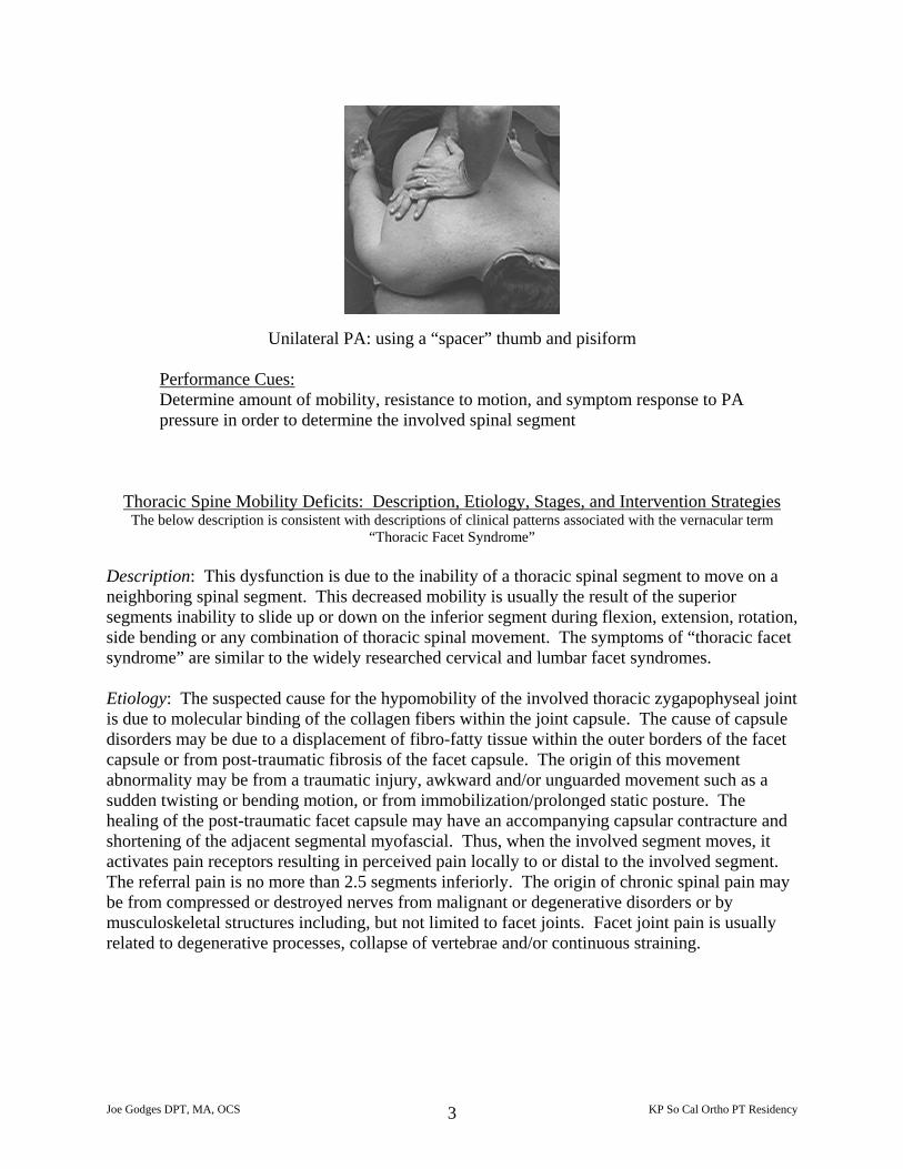
(1) Observable ROM limitations (2) Symmetry of transverse processes (3) Resistance to unilateral posterior to anterior (PA) pressures over transverse
processes (segmental ROM restrictions) (4) Symptom response to PA pressures (5) Tenderness and hypertonicity of multifidi and rotatori myofascia of the
involved segment(s)
Unilateral PA – using thumbs
Joe Godges DPT, MA, OCS KP So Cal Ortho PT Residency 2
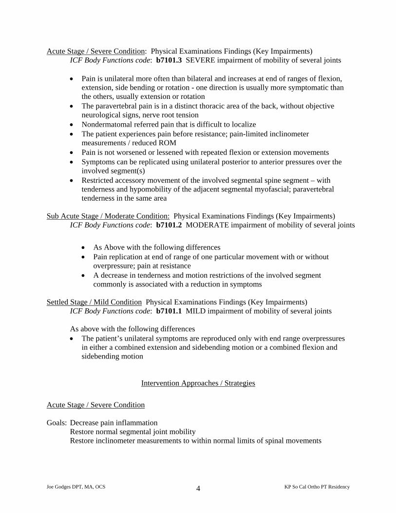
Unilateral PA: using a “spacer” thumb and pisiform
Performance Cues:Determine amount of mobility, resistance to motion, and symptom response to PA pressure in order to determine the involved spinal segment
Thoracic Spine Mobility Deficits: Description, Etiology, Stages, and Intervention Strategies The below description is consistent with descriptions of clinical patterns associated with the vernacular term
“Thoracic Facet Syndrome” Description: This dysfunction is due to the inability of a thoracic spinal segment to move on a neighboring spinal segment. This decreased mobility is usually the result of the superior segments inability to slide up or down on the inferior segment during flexion, extension, rotation, side bending or any combination of thoracic spinal movement. The symptoms of “thoracic facet syndrome” are similar to the widely researched cervical and lumbar facet syndromes. Etiology: The suspected cause for the hypomobility of the involved thoracic zygapophyseal joint is due to molecular binding of the collagen fibers within the joint capsule. The cause of capsule disorders may be due to a displacement of fibro-fatty tissue within the outer borders of the facet capsule or from post-traumatic fibrosis of the facet capsule. The origin of this movement abnormality may be from a traumatic injury, awkward and/or unguarded movement such as a sudden twisting or bending motion, or from immobilization/prolonged static posture. The healing of the post-traumatic facet capsule may have an accompanying capsular contracture and shortening of the adjacent segmental myofascial. Thus, when the involved segment moves, it activates pain receptors resulting in perceived pain locally to or distal to the involved segment. The referral pain is no more than 2.5 segments inferiorly. The origin of chronic spinal pain may be from compressed or destroyed nerves from malignant or degenerative disorders or by musculoskeletal structures including, but not limited to facet joints. Facet joint pain is usually related to degenerative processes, collapse of vertebrae and/or continuous straining.
Joe Godges DPT, MA, OCS KP So Cal Ortho PT Residency 3
Acute Stage / Severe Condition: Physical Examinations Findings (Key Impairments) ICF Body Functions code: b7101.3 SEVERE impairment of mobility of several joints
• Pain is unilateral more often than bilateral and increases at end of ranges of flexion,
extension, side bending or rotation - one direction is usually more symptomatic than the others, usually extension or rotation
• The paravertebral pain is in a distinct thoracic area of the back, without objective neurological signs, nerve root tension
• Nondermatomal referred pain that is difficult to localize • The patient experiences pain before resistance; pain-limited inclinometer
measurements / reduced ROM • Pain is not worsened or lessened with repeated flexion or extension movements • Symptoms can be replicated using unilateral posterior to anterior pressures over the
involved segment(s) • Restricted accessory movement of the involved segmental spine segment – with
tenderness and hypomobility of the adjacent segmental myofascial; paravertebral tenderness in the same area
Sub Acute Stage / Moderate Condition: Physical Examinations Findings (Key Impairments)
ICF Body Functions code: b7101.2 MODERATE impairment of mobility of several joints
• As Above with the following differences • Pain replication at end of range of one particular movement with or without
overpressure; pain at resistance • A decrease in tenderness and motion restrictions of the involved segment
commonly is associated with a reduction in symptoms Settled Stage / Mild Condition Physical Examinations Findings (Key Impairments)
ICF Body Functions code: b7101.1 MILD impairment of mobility of several joints
As above with the following differences • The patient’s unilateral symptoms are reproduced only with end range overpressures
in either a combined extension and sidebending motion or a combined flexion and sidebending motion
Intervention Approaches / Strategies
Acute Stage / Severe Condition Goals: Decrease pain inflammation
Restore normal segmental joint mobility Restore inclinometer measurements to within normal limits of spinal movements
Joe Godges DPT, MA, OCS KP So Cal Ortho PT Residency 4
• Physical Agents Electrical stimulation, ice (or heat) to provide pain relief and reduce muscle guarding
• Manual Therapy
Soft tissue mobilization primarily to multifidus and rotatores of the involved segment
Joint mobilization/manipulation using isometric mobilization and contract/relax procedures to the involved segment to reduce associated rotatores or multifi muscle guarding
Passive stretching procedures to restore normal thoracic segmental mobility to the involved segment
• Therapeutic Exercise
Instruct in exercise and functional movements to maintain the improvements in mobility gained with the soft tissue and joint manipulations
• Re-injury Prevention Instruction
Instruct the patient in efficient, painfree, motor performance of movements that are related by the patient to be the cause of the current episode of mid back pain
Sub Acute Stage / Moderate Condition Goal: Restore normal, painfree response to end of range motions or to overpressures at end ranges of rotation
• Approaches / Strategies listed above – focusing on soft tissue mobilization and joint mobilization/manipulation to normalize segmental mobility followed by mobility exercises to maintain the improvements gained from the manual procedures
Settled Stage / Mild Condition Goal: Restore normal, painfree responses to overpressures of combined extension and
sidebending and/or combined flexion and sidebending
• Approaches / Strategies listed above
• Therapeutic Exercises Instruct in stretching exercises to address the patient’s specific muscle flexibility deficits Instruct in strengthening exercises to address the patient’s specific muscle strength deficits
Joe Godges DPT, MA, OCS KP So Cal Ortho PT Residency 5
If symptoms persists (>12 months), when conservative measures fail, then the patient may consider radiofrequency facet denervation.
Intervention for High Performance / High Demand Functioning in Workers or Athletes Goal: Return to desired occupational or leisure time activities
• Approaches / Strategies listed above
• Therapeutic Exercises Encourage participation in regular low stress aerobic activities as a means to improve fitness, muscle strength and prevent recurrences
Selected References Defranca G, Levine L: The T4 syndrome. J Manipulative Physiological Therapeutics, 18:34-37 Donatelli R, Wooden MJ: Orthopedic Physical Therapy, 2nd ed. Churchill Livingston Inc, New York, 1994, pp 126 Dreyfuss P, Tibiletti C, Dreyer SJ: Thoracic zygapophyseal joint pain patterns: A study in normal volunteers. Spine 19:807-811, 1994 Flynn T. Thoracic Spine and Rib Cage Disorders. Orthopedic Physical Therapy Clinics of North America 8-1:1-20, 1999 Saunders HD, Saunders R: Evaluation, Treatment, and Prevention of Musculoskeletal Disorders: Volume I Spine, 3rd ed. The Saunders Group, Chaska, Minnesota, 1995, pp 103-105, 147-149. Schiller L. Effectiveness of Spinal Manipulation Therapy in the Treatment of Mechanical Thoracic Spine Pain: A Pilot Randomized Clinical Trial. Jour of Manipulative and Physiological Therapeutics. July/Aug 2001; 24(6): 394-401 Stolker RJ, Vervest ACM, Groen GJ. Percutaneous Facet Denervation in Chronic Thoracic Spinal Pain. Acta Neurochir (Wien). 1993; 122: 82-90
Joe Godges DPT, MA, OCS KP So Cal Ortho PT Residency 6
Thoracic Spine Manual Examination and Treatment Procedures Upper Thoracic Mobility Assessment: Physiologic Forward Bending (sitting)
Physiologic Rotation (sitting) Physiologic Sidebending (sitting)
TP Positional Symmetry in Flexion (sitting) TP Positional Symmetry in Extension (sitting)
Accessory Rotation (via transverse pressures on SPs) Accessory Rotation (via unilateral PA pressures on TPs)
Palpation/Provocation of Segmental Myofascia
Mid Thoracic Mobility Assessment: Physiologic Forward Bending (sitting)
Physiologic Sidebending (sitting)
TP Positional Symmetry in Flexion (sitting) TP Positional Symmetry in Extension (prone on elbows)
Accessory Rotation (unilateral PA pressures in flexion) Accessory Rotation (unilateral PA pressures in extension) Accessory Rotation (unilateral PA pressures in neutral)
Palpation/Provocation of Segmental Myofascia
Upper Thoracic Treatment Procedures:
Contract/Relax for restoring segmental Flexion/SB/ROT Contract/Relax for restoring segmental Extension/SB/ROT
Soft Tissue Mobilization of involved segmental myofascia
Joint Mobilization/Manipulation:
Segmental Rotation (using pisaform/scaphoid on adjacent SPs) Rotation via TPs (using “spacer” thumb and pisaform)
Rotation in Neutral (gaping – prone) Rotation/SB in Extension (closing – prone) Rotation/SB in Flexion (opening – supine or prone)
Mid Thoracic Treatment Procedures:
Contract/Relax for restoring segmental Flexion/SB/ROT Contract/Relax for restoring segmental Extension/SB/ROT
Soft Tissue Mobilization of involved segmental myofascia
Joint Mobilization/Manipulation:
Flexion/SB/ROT (opening – supine) Extension/SB/ROT (closing – prone)
Joe Godges DPT, MA, OCS KP So Cal Ortho PT Residency 7

Upper Thoracic Segmental Myofascia Soft Tissue Mobilization
Joe Godges DPT, MA, OCS KP So Cal Ortho PT Residency 8
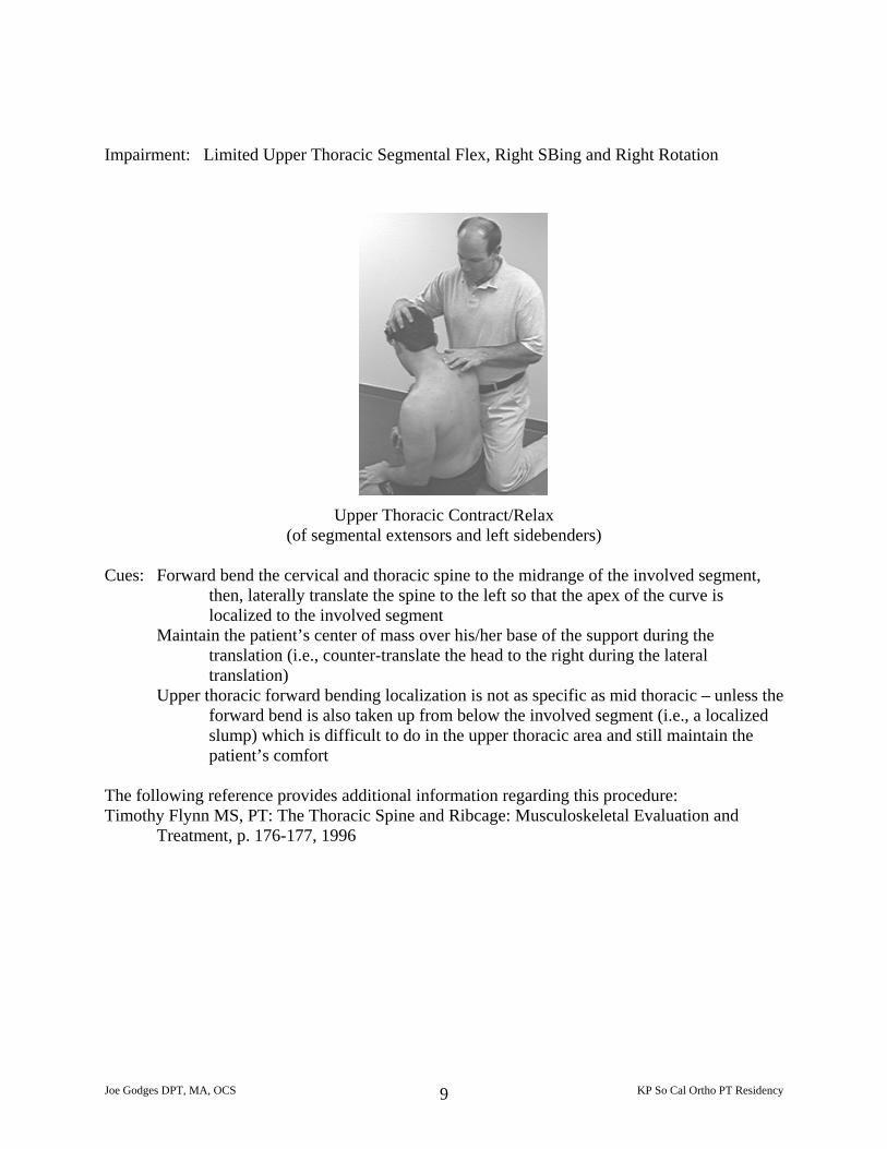
Impairment: Limited Upper Thoracic Segmental Flex, Right SBing and Right Rotation
Upper Thoracic Contract/Relax
(of segmental extensors and left sidebenders) Cues: Forward bend the cervical and thoracic spine to the midrange of the involved segment,
then, laterally translate the spine to the left so that the apex of the curve is localized to the involved segment
Maintain the patient’s center of mass over his/her base of the support during the translation (i.e., counter-translate the head to the right during the lateral translation)
Upper thoracic forward bending localization is not as specific as mid thoracic – unless the forward bend is also taken up from below the involved segment (i.e., a localized slump) which is difficult to do in the upper thoracic area and still maintain the patient’s comfort
The following reference provides additional information regarding this procedure: Timothy Flynn MS, PT: The Thoracic Spine and Ribcage: Musculoskeletal Evaluation and
Treatment, p. 176-177, 1996
Joe Godges DPT, MA, OCS KP So Cal Ortho PT Residency 9
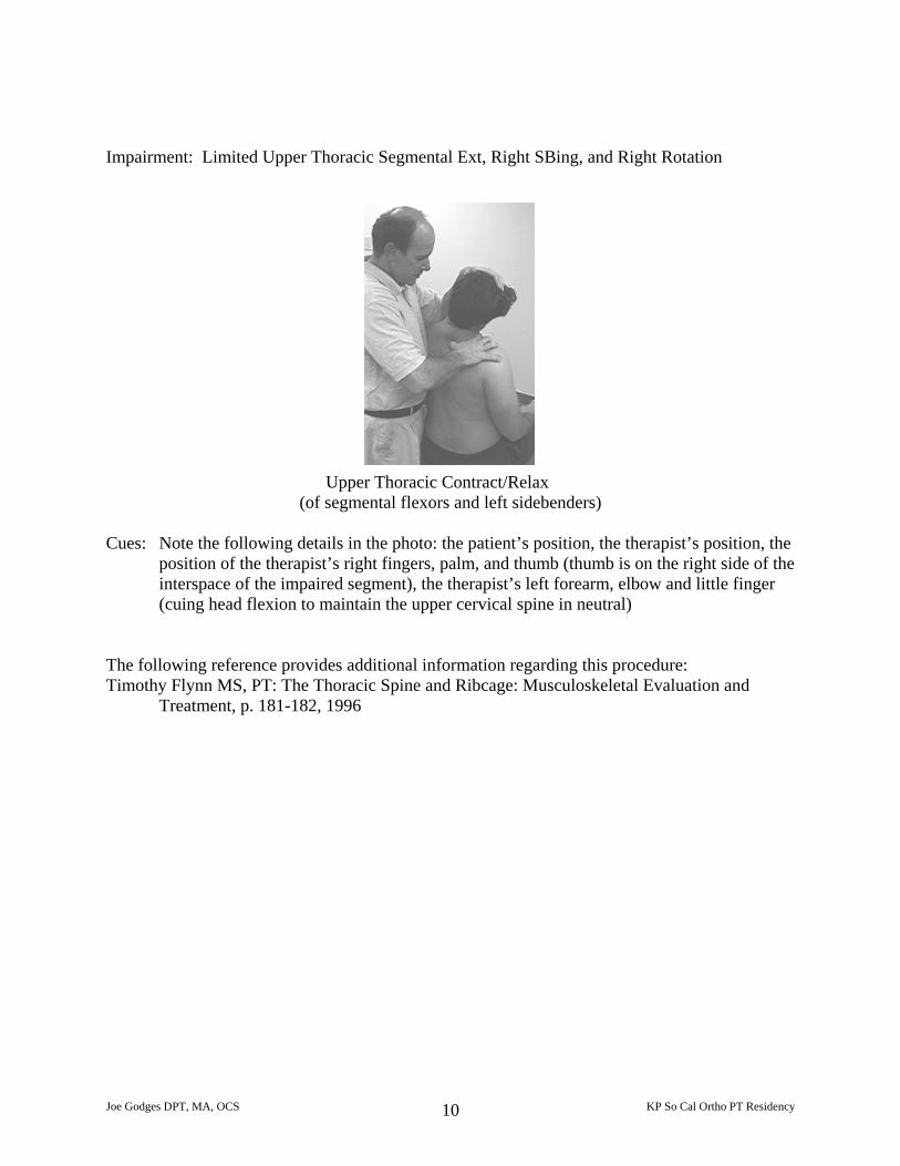
Impairment: Limited Upper Thoracic Segmental Ext, Right SBing, and Right Rotation
Upper Thoracic Contract/Relax
(of segmental flexors and left sidebenders) Cues: Note the following details in the photo: the patient’s position, the therapist’s position, the
position of the therapist’s right fingers, palm, and thumb (thumb is on the right side of the interspace of the impaired segment), the therapist’s left forearm, elbow and little finger (cuing head flexion to maintain the upper cervical spine in neutral)
The following reference provides additional information regarding this procedure: Timothy Flynn MS, PT: The Thoracic Spine and Ribcage: Musculoskeletal Evaluation and
Treatment, p. 181-182, 1996
Joe Godges DPT, MA, OCS KP So Cal Ortho PT Residency 10
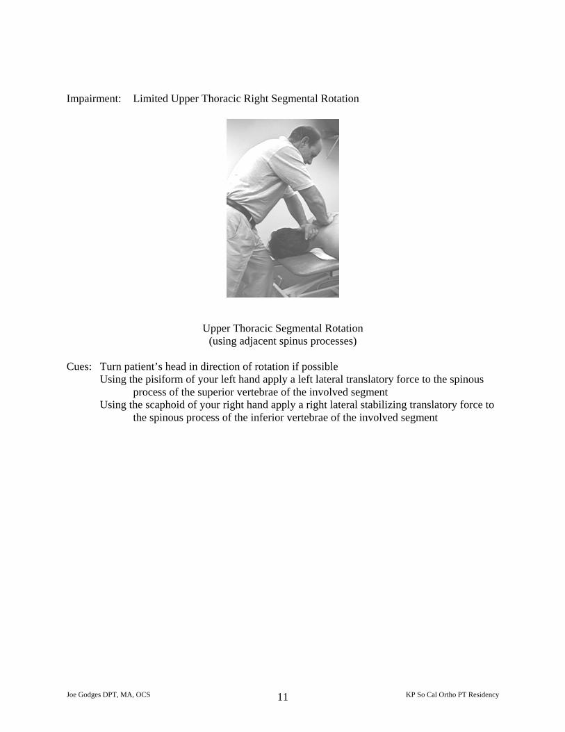
Impairment: Limited Upper Thoracic Right Segmental Rotation
Upper Thoracic Segmental Rotation
(using adjacent spinus processes) Cues: Turn patient’s head in direction of rotation if possible
Using the pisiform of your left hand apply a left lateral translatory force to the spinous process of the superior vertebrae of the involved segment
Using the scaphoid of your right hand apply a right lateral stabilizing translatory force to the spinous process of the inferior vertebrae of the involved segment
Joe Godges DPT, MA, OCS KP So Cal Ortho PT Residency 11
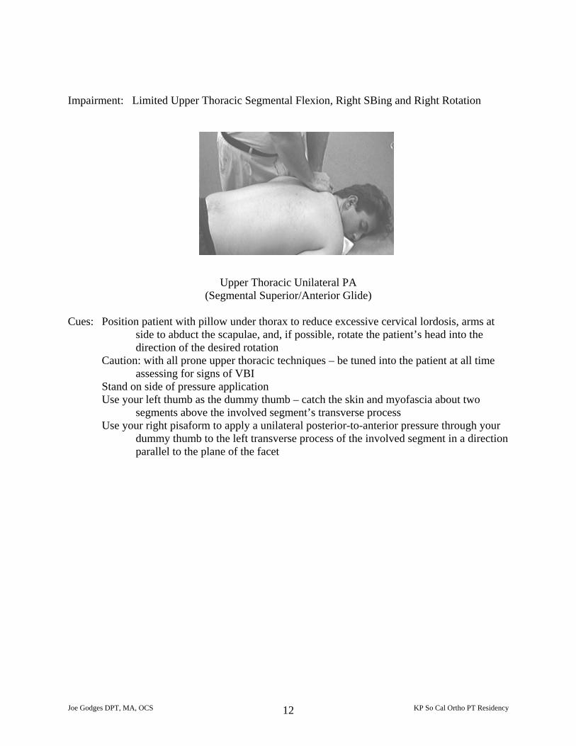
Impairment: Limited Upper Thoracic Segmental Flexion, Right SBing and Right Rotation
Upper Thoracic Unilateral PA
(Segmental Superior/Anterior Glide) Cues: Position patient with pillow under thorax to reduce excessive cervical lordosis, arms at
side to abduct the scapulae, and, if possible, rotate the patient’s head into the direction of the desired rotation
Caution: with all prone upper thoracic techniques – be tuned into the patient at all time assessing for signs of VBI
Stand on side of pressure application Use your left thumb as the dummy thumb – catch the skin and myofascia about two
segments above the involved segment’s transverse process Use your right pisaform to apply a unilateral posterior-to-anterior pressure through your
dummy thumb to the left transverse process of the involved segment in a direction parallel to the plane of the facet
Joe Godges DPT, MA, OCS KP So Cal Ortho PT Residency 12
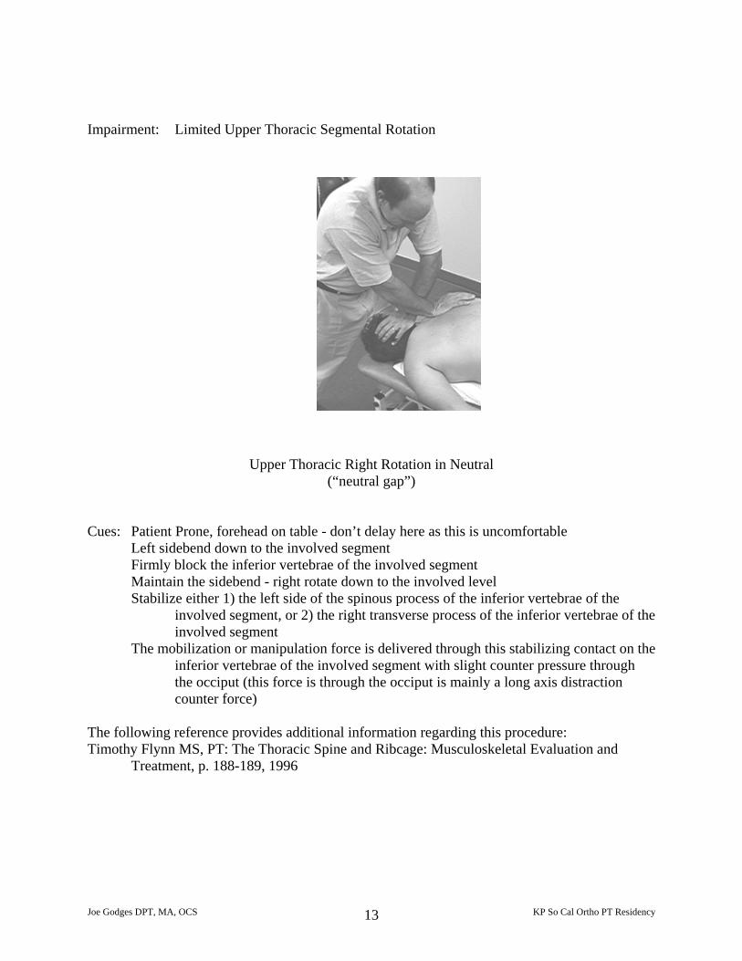
Impairment: Limited Upper Thoracic Segmental Rotation
Upper Thoracic Right Rotation in Neutral (“neutral gap”)
Cues: Patient Prone, forehead on table - don’t delay here as this is uncomfortable
Left sidebend down to the involved segment Firmly block the inferior vertebrae of the involved segment Maintain the sidebend - right rotate down to the involved level Stabilize either 1) the left side of the spinous process of the inferior vertebrae of the
involved segment, or 2) the right transverse process of the inferior vertebrae of the involved segment
The mobilization or manipulation force is delivered through this stabilizing contact on the inferior vertebrae of the involved segment with slight counter pressure through the occiput (this force is through the occiput is mainly a long axis distraction counter force)
The following reference provides additional information regarding this procedure: Timothy Flynn MS, PT: The Thoracic Spine and Ribcage: Musculoskeletal Evaluation and
Treatment, p. 188-189, 1996
Joe Godges DPT, MA, OCS KP So Cal Ortho PT Residency 13
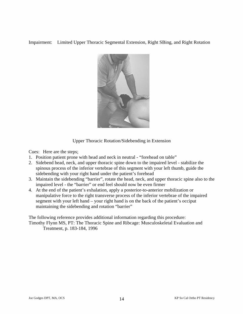
Impairment: Limited Upper Thoracic Segmental Extension, Right SBing, and Right Rotation
Upper Thoracic Rotation/Sidebending in Extension
Cues: Here are the steps; 1. Position patient prone with head and neck in neutral - “forehead on table” 2. Sidebend head, neck, and upper thoracic spine down to the impaired level - stabilize the
spinous process of the inferior vertebrae of this segment with your left thumb, guide the sidebending with your right hand under the patient’s forehead
3. Maintain the sidebending “barrier”, rotate the head, neck, and upper thoracic spine also to the impaired level - the “barrier” or end feel should now be even firmer
4. At the end of the patient’s exhalation, apply a posterior-to-anterior mobilization or manipulative force to the right transverse process of the inferior vertebrae of the impaired segment with your left hand – your right hand is on the back of the patient’s occiput maintaining the sidebending and rotation “barrier”
The following reference provides additional information regarding this procedure: Timothy Flynn MS, PT: The Thoracic Spine and Ribcage: Musculoskeletal Evaluation and
Treatment, p. 183-184, 1996
Joe Godges DPT, MA, OCS KP So Cal Ortho PT Residency 14
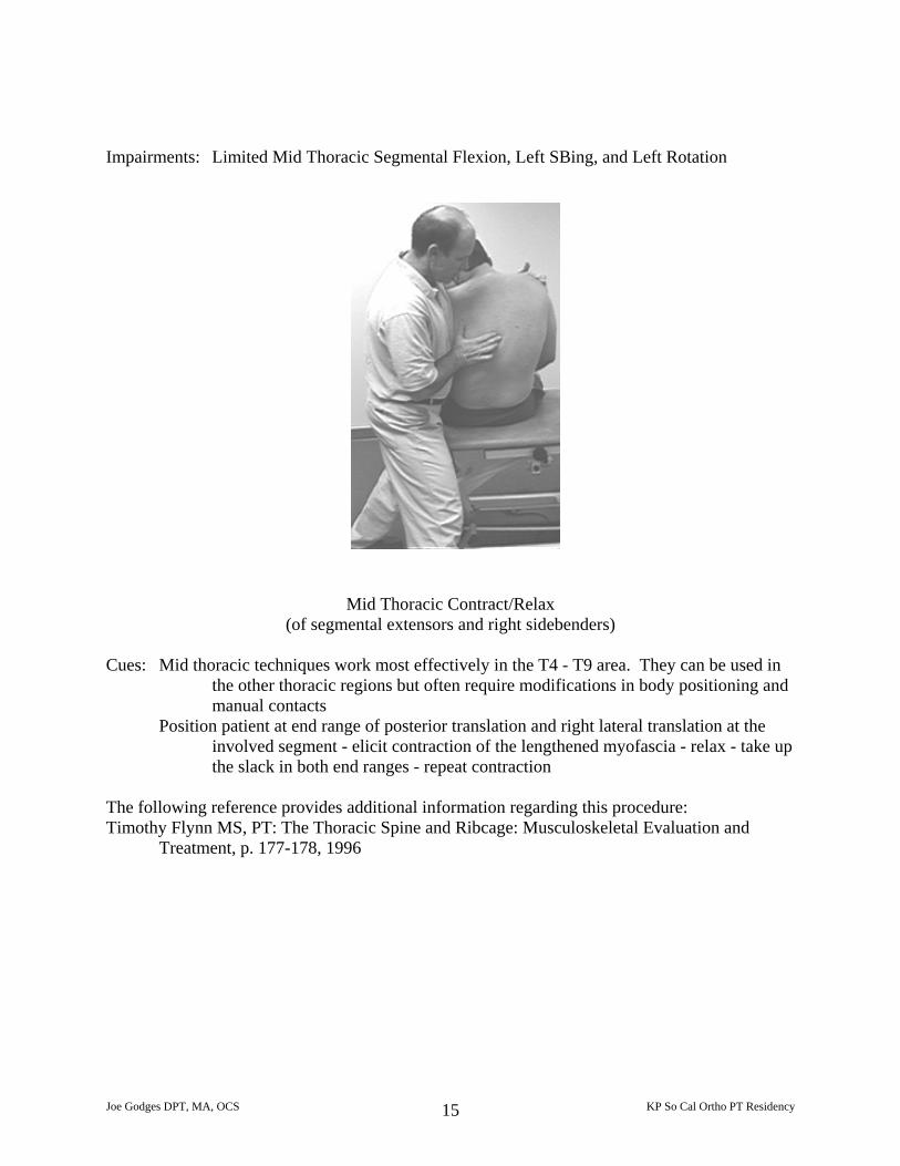
Impairments: Limited Mid Thoracic Segmental Flexion, Left SBing, and Left Rotation
Mid Thoracic Contract/Relax (of segmental extensors and right sidebenders)
Cues: Mid thoracic techniques work most effectively in the T4 - T9 area. They can be used in
the other thoracic regions but often require modifications in body positioning and manual contacts
Position patient at end range of posterior translation and right lateral translation at the involved segment - elicit contraction of the lengthened myofascia - relax - take up the slack in both end ranges - repeat contraction
The following reference provides additional information regarding this procedure: Timothy Flynn MS, PT: The Thoracic Spine and Ribcage: Musculoskeletal Evaluation and
Treatment, p. 177-178, 1996
Joe Godges DPT, MA, OCS KP So Cal Ortho PT Residency 15
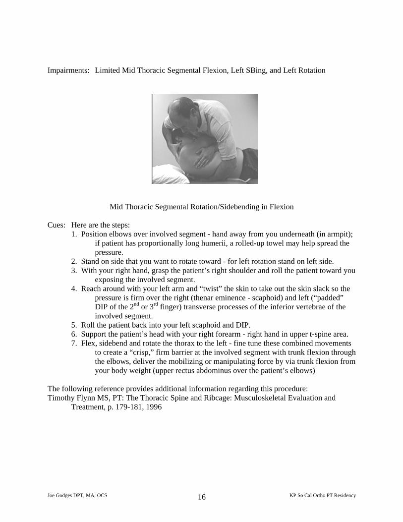
Impairments: Limited Mid Thoracic Segmental Flexion, Left SBing, and Left Rotation
Mid Thoracic Segmental Rotation/Sidebending in Flexion
Cues: Here are the steps: 1. Position elbows over involved segment - hand away from you underneath (in armpit);
if patient has proportionally long humerii, a rolled-up towel may help spread the pressure.
2. Stand on side that you want to rotate toward - for left rotation stand on left side. 3. With your right hand, grasp the patient’s right shoulder and roll the patient toward you
exposing the involved segment. 4. Reach around with your left arm and “twist” the skin to take out the skin slack so the
pressure is firm over the right (thenar eminence - scaphoid) and left (“padded” DIP of the 2nd or 3rd finger) transverse processes of the inferior vertebrae of the involved segment.
5. Roll the patient back into your left scaphoid and DIP. 6. Support the patient’s head with your right forearm - right hand in upper t-spine area. 7. Flex, sidebend and rotate the thorax to the left - fine tune these combined movements
to create a “crisp,” firm barrier at the involved segment with trunk flexion through the elbows, deliver the mobilizing or manipulating force by via trunk flexion from your body weight (upper rectus abdominus over the patient’s elbows)
The following reference provides additional information regarding this procedure: Timothy Flynn MS, PT: The Thoracic Spine and Ribcage: Musculoskeletal Evaluation and
Treatment, p. 179-181, 1996
Joe Godges DPT, MA, OCS KP So Cal Ortho PT Residency 16
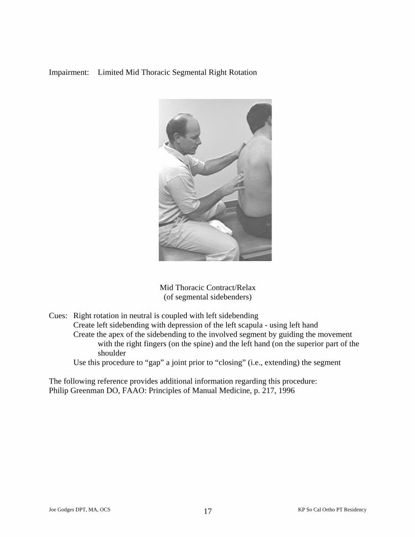
Impairment: Limited Mid Thoracic Segmental Right Rotation
Mid Thoracic Contract/Relax (of segmental sidebenders)
Cues: Right rotation in neutral is coupled with left sidebending
Create left sidebending with depression of the left scapula - using left hand Create the apex of the sidebending to the involved segment by guiding the movement
with the right fingers (on the spine) and the left hand (on the superior part of the shoulder
Use this procedure to “gap” a joint prior to “closing” (i.e., extending) the segment The following reference provides additional information regarding this procedure: Philip Greenman DO, FAAO: Principles of Manual Medicine, p. 217, 1996
Joe Godges DPT, MA, OCS KP So Cal Ortho PT Residency 17
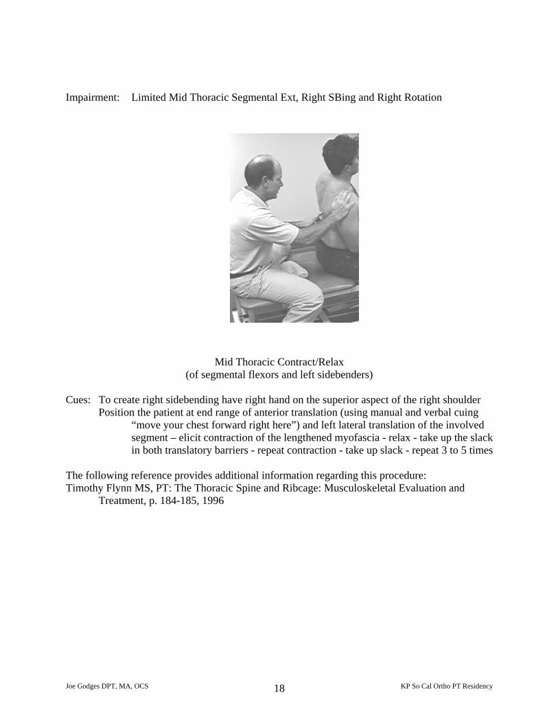
Impairment: Limited Mid Thoracic Segmental Ext, Right SBing and Right Rotation
Mid Thoracic Contract/Relax (of segmental flexors and left sidebenders)
Cues: To create right sidebending have right hand on the superior aspect of the right shoulder
Position the patient at end range of anterior translation (using manual and verbal cuing “move your chest forward right here”) and left lateral translation of the involved segment – elicit contraction of the lengthened myofascia - relax - take up the slack in both translatory barriers - repeat contraction - take up slack - repeat 3 to 5 times
The following reference provides additional information regarding this procedure: Timothy Flynn MS, PT: The Thoracic Spine and Ribcage: Musculoskeletal Evaluation and
Treatment, p. 184-185, 1996
Joe Godges DPT, MA, OCS KP So Cal Ortho PT Residency 18
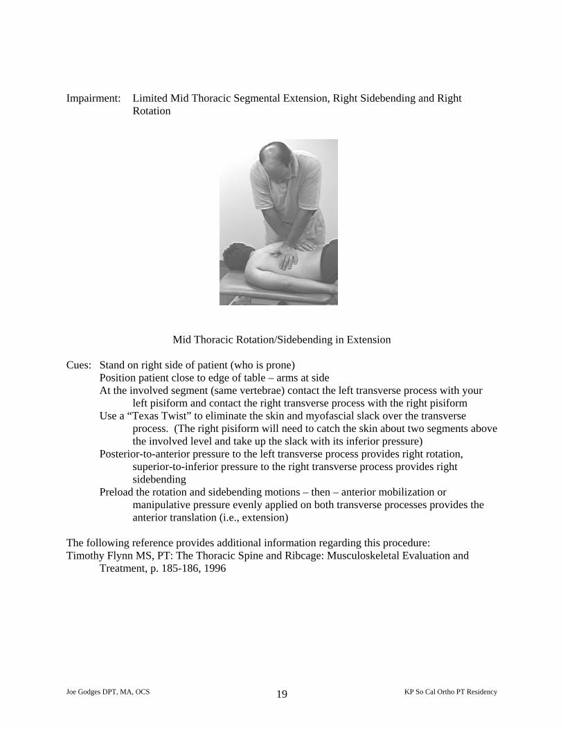
Impairment: Limited Mid Thoracic Segmental Extension, Right Sidebending and Right
Rotation
Mid Thoracic Rotation/Sidebending in Extension Cues: Stand on right side of patient (who is prone)
Position patient close to edge of table – arms at side At the involved segment (same vertebrae) contact the left transverse process with your
left pisiform and contact the right transverse process with the right pisiform Use a “Texas Twist” to eliminate the skin and myofascial slack over the transverse
process. (The right pisiform will need to catch the skin about two segments above the involved level and take up the slack with its inferior pressure)
Posterior-to-anterior pressure to the left transverse process provides right rotation, superior-to-inferior pressure to the right transverse process provides right sidebending
Preload the rotation and sidebending motions – then – anterior mobilization or manipulative pressure evenly applied on both transverse processes provides the anterior translation (i.e., extension)
The following reference provides additional information regarding this procedure: Timothy Flynn MS, PT: The Thoracic Spine and Ribcage: Musculoskeletal Evaluation and
Treatment, p. 185-186, 1996
Joe Godges DPT, MA, OCS KP So Cal Ortho PT Residency 19