Embed Size (px)
Citation preview
Percutaneous Techniques for the Diagnosis and Treatment of Localized Langerhans-Cell Histiocytosis (Eosinophilic
Granuloma of Bone)*
by ALAN W. YASKO, CHRISTINA V. FANNING, ALBERTO G. AYALA, C. HUMBERTO CARRASCO, and JOHN A. MURRAY
J Bone Joint Surg AmVolume 80(2):219-28
February 1, 1998
©1998 by The Journal of Bone and Joint Surgery, Inc.
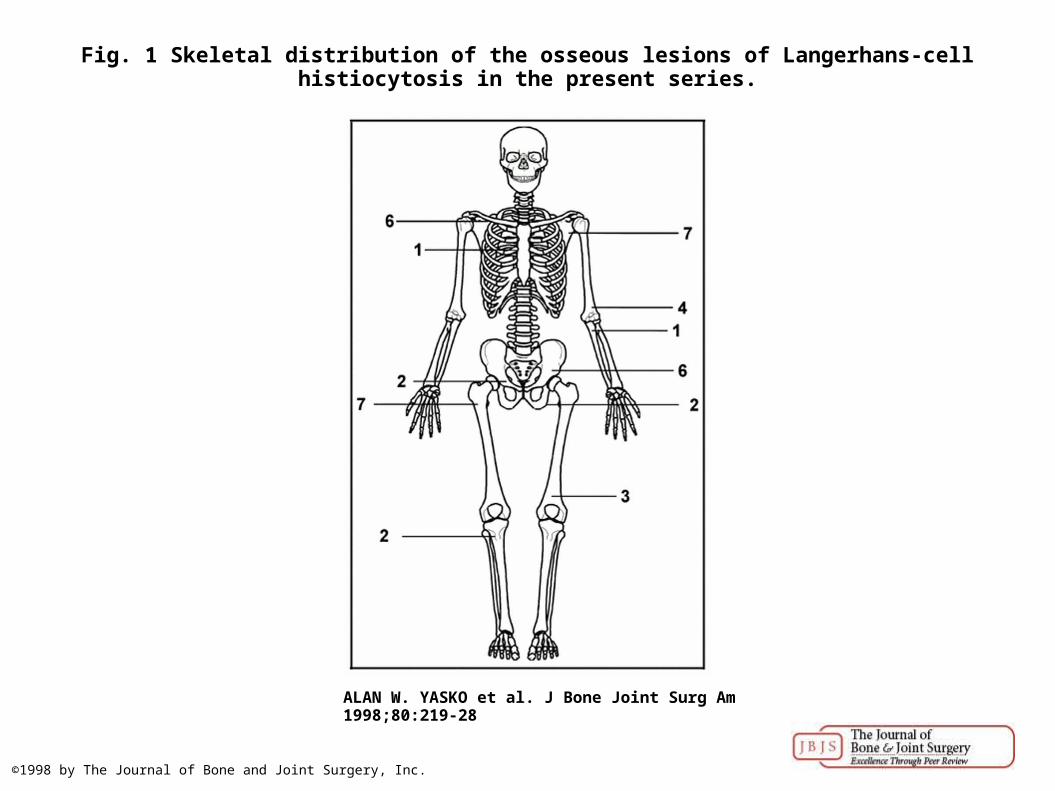
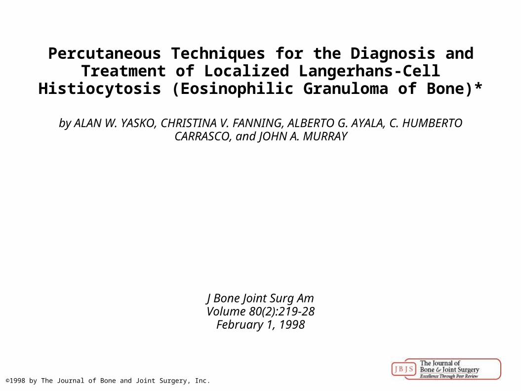
Fig. 1 Skeletal distribution of the osseous lesions of Langerhans-cell histiocytosis in the present series.
ALAN W. YASKO et al. J Bone Joint Surg Am 1998;80:219-28
©1998 by The Journal of Bone and Joint Surgery, Inc.
Fig. 2-A Cytological smear of material obtained with fine-needle aspiration, demonstrating histiocytes with abundant cytoplasm and grooved or infolded eccentric nuclei, characteristic of
Langerhans histiocytes.
ALAN W. YASKO et al. J Bone Joint Surg Am 1998;80:219-28
©1998 by The Journal of Bone and Joint Surgery, Inc.
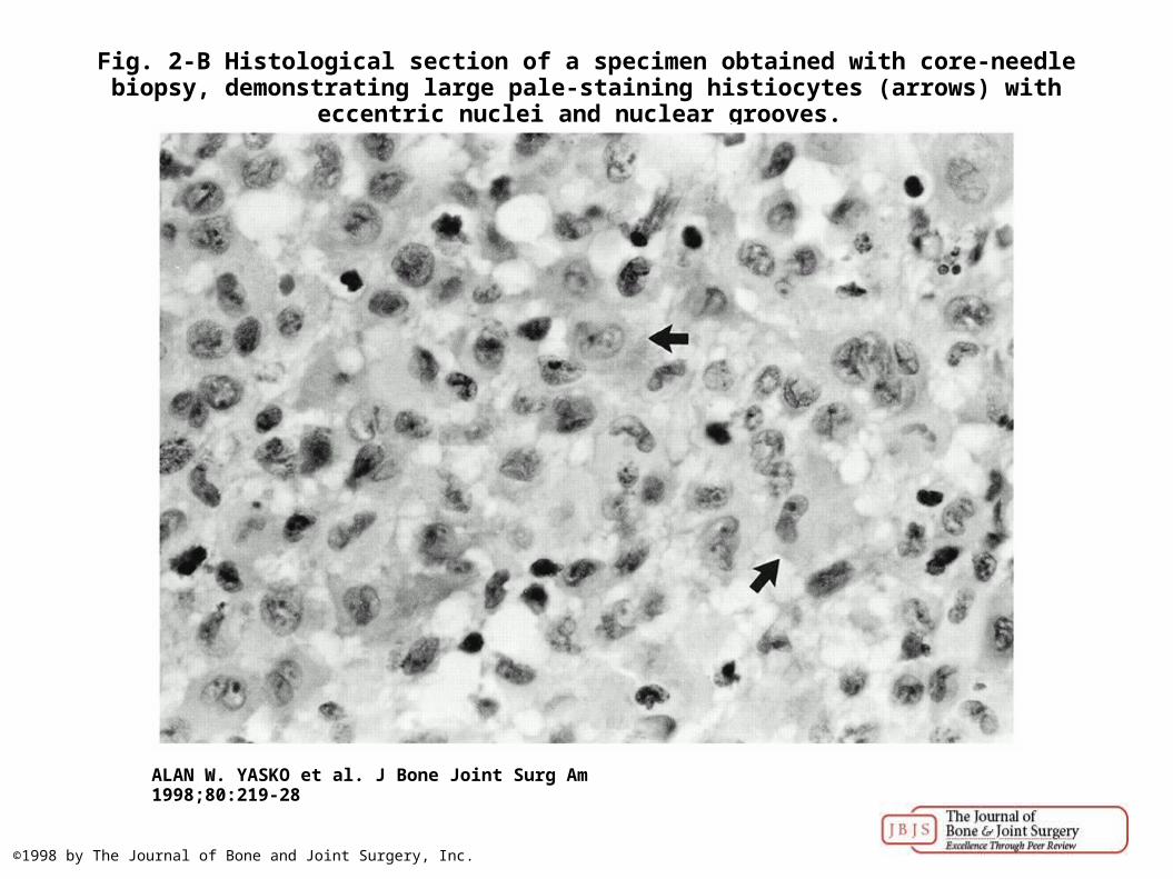
Fig. 2-B Histological section of a specimen obtained with core-needle biopsy, demonstrating large pale-staining histiocytes (arrows) with eccentric nuclei and nuclear grooves.
ALAN W. YASKO et al. J Bone Joint Surg Am 1998;80:219-28
©1998 by The Journal of Bone and Joint Surgery, Inc.
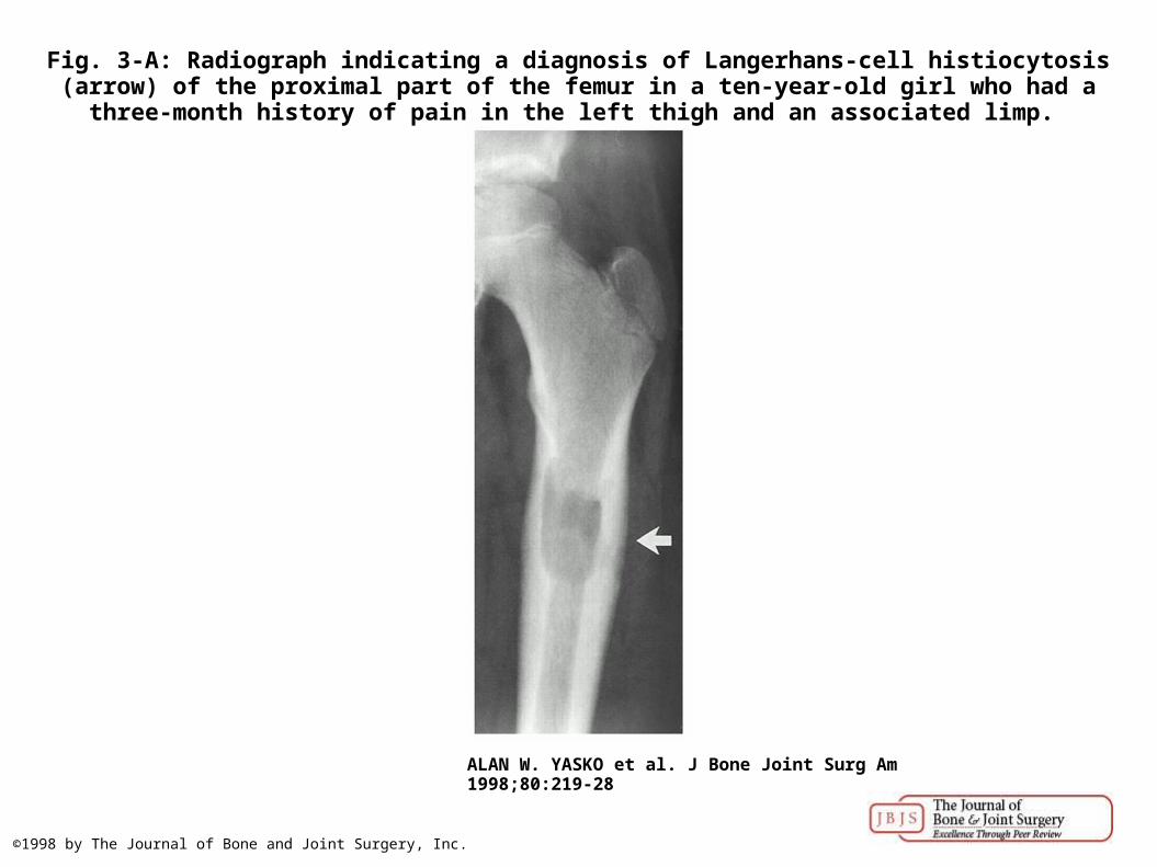
Fig. 3-A: Radiograph indicating a diagnosis of Langerhans-cell histiocytosis (arrow) of the proximal part of the femur in a ten-year-old girl who had a three-month history of pain in the left
thigh and an associated limp.
ALAN W. YASKO et al. J Bone Joint Surg Am 1998;80:219-28
©1998 by The Journal of Bone and Joint Surgery, Inc.
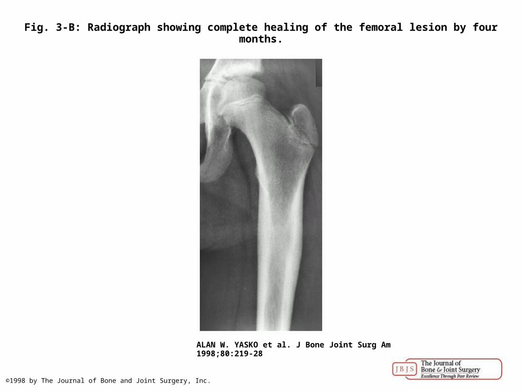
Fig. 3-B: Radiograph showing complete healing of the femoral lesion by four months.
ALAN W. YASKO et al. J Bone Joint Surg Am 1998;80:219-28
©1998 by The Journal of Bone and Joint Surgery, Inc.
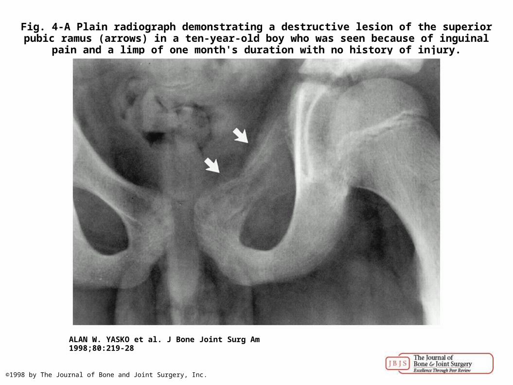
Fig. 4-A Plain radiograph demonstrating a destructive lesion of the superior pubic ramus (arrows) in a ten-year-old boy who was seen because of inguinal pain and a limp of one month's
duration with no history of injury.
ALAN W. YASKO et al. J Bone Joint Surg Am 1998;80:219-28
©1998 by The Journal of Bone and Joint Surgery, Inc.
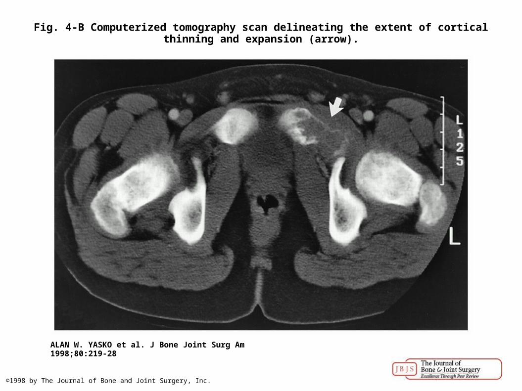
Fig. 4-B Computerized tomography scan delineating the extent of cortical thinning and expansion (arrow).
ALAN W. YASKO et al. J Bone Joint Surg Am 1998;80:219-28
©1998 by The Journal of Bone and Joint Surgery, Inc.
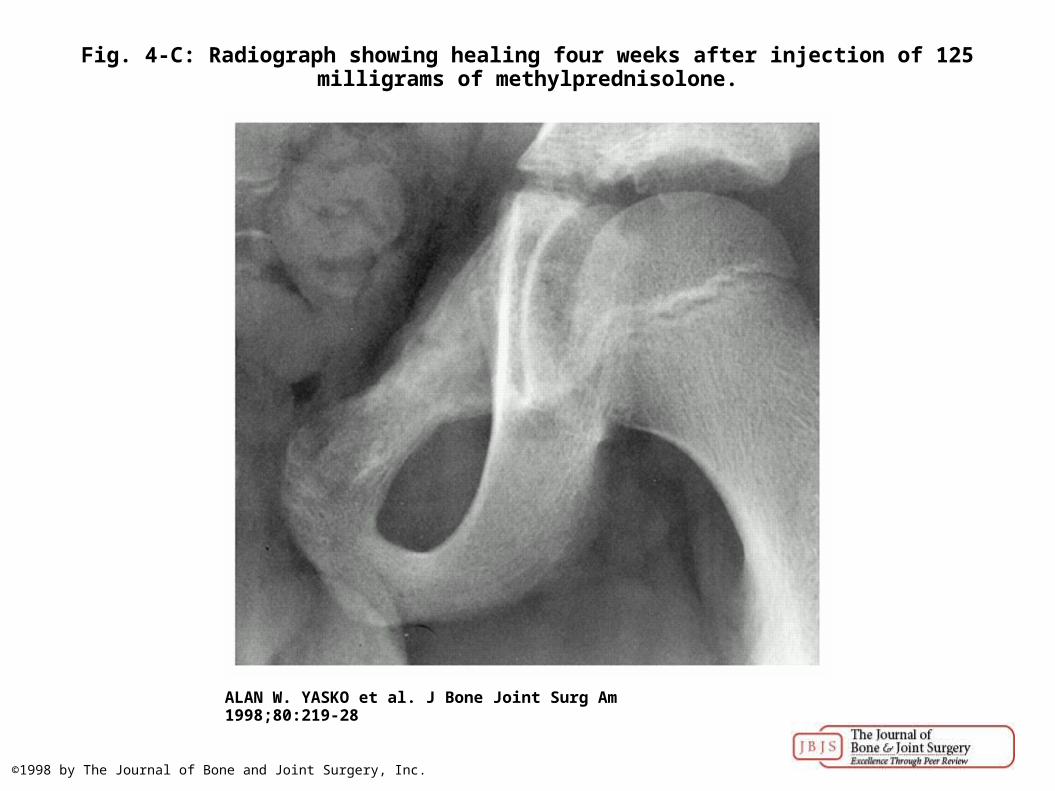
Fig. 4-C: Radiograph showing healing four weeks after injection of 125 milligrams of methylprednisolone.
ALAN W. YASKO et al. J Bone Joint Surg Am 1998;80:219-28
©1998 by The Journal of Bone and Joint Surgery, Inc.
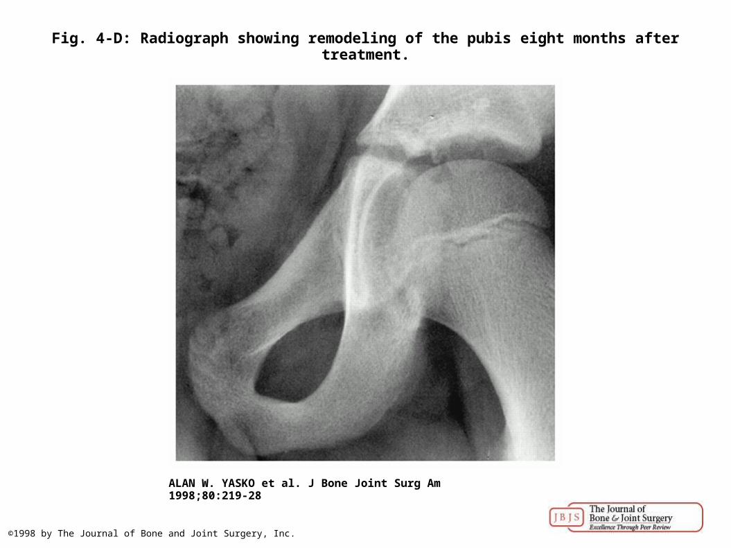
Fig. 4-D: Radiograph showing remodeling of the pubis eight months after treatment.
ALAN W. YASKO et al. J Bone Joint Surg Am 1998;80:219-28
©1998 by The Journal of Bone and Joint Surgery, Inc.
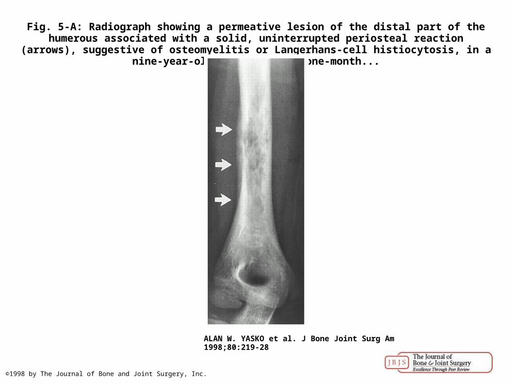
Fig. 5-A: Radiograph showing a permeative lesion of the distal part of the humerous associated with a solid, uninterrupted periosteal reaction (arrows), suggestive of osteomyelitis or
Langerhans-cell histiocytosis, in a nine-year-old boy who had a one-month...
ALAN W. YASKO et al. J Bone Joint Surg Am 1998;80:219-28
©1998 by The Journal of Bone and Joint Surgery, Inc.
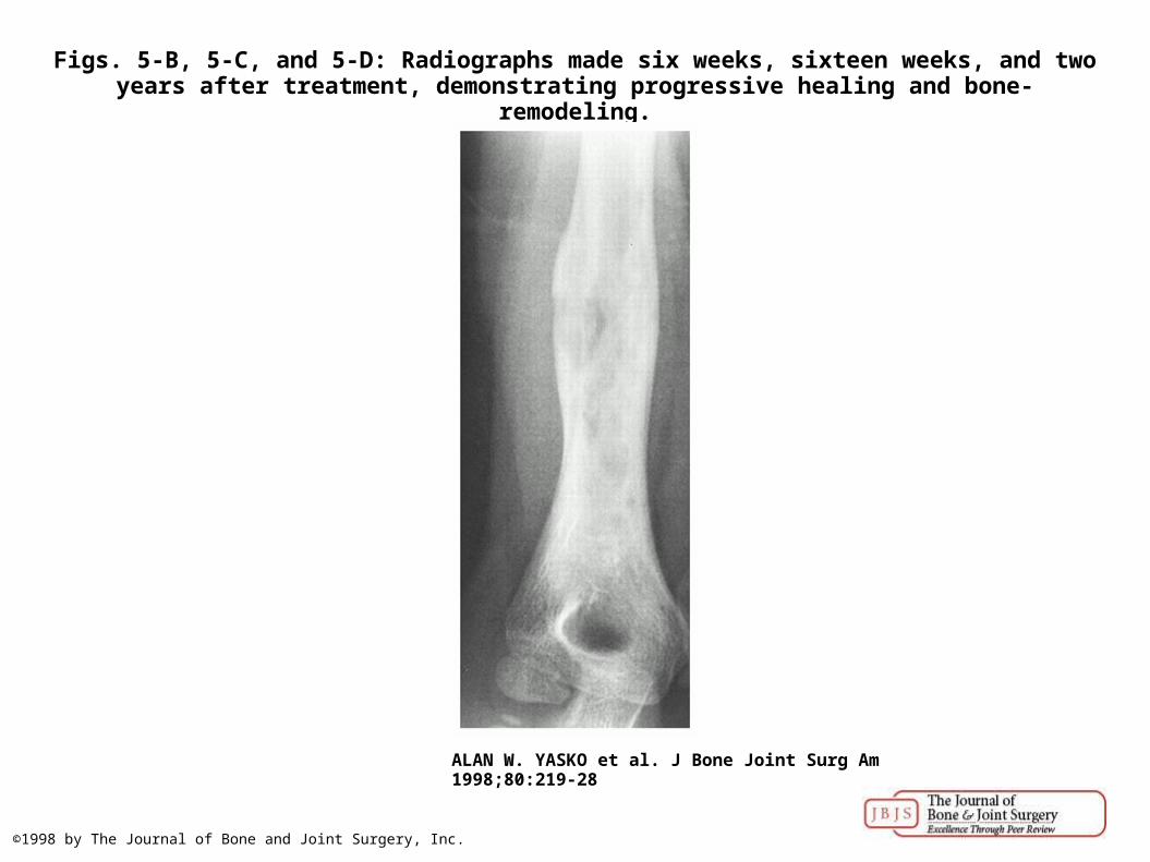
Figs. 5-B, 5-C, and 5-D: Radiographs made six weeks, sixteen weeks, and two years after treatment, demonstrating progressive healing and bone-remodeling.
ALAN W. YASKO et al. J Bone Joint Surg Am 1998;80:219-28
©1998 by The Journal of Bone and Joint Surgery, Inc.
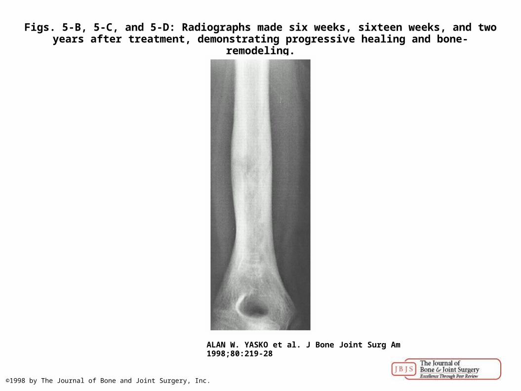
Figs. 5-B, 5-C, and 5-D: Radiographs made six weeks, sixteen weeks, and two years after treatment, demonstrating progressive healing and bone-remodeling.
ALAN W. YASKO et al. J Bone Joint Surg Am 1998;80:219-28
©1998 by The Journal of Bone and Joint Surgery, Inc.
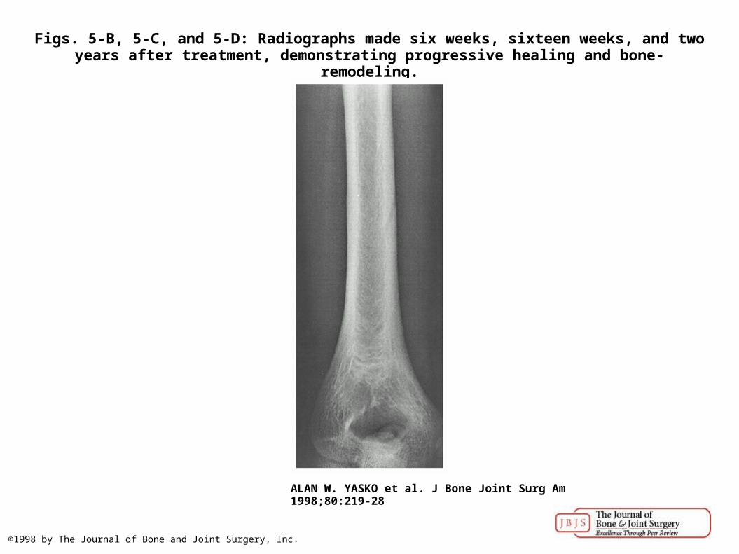
Figs. 5-B, 5-C, and 5-D: Radiographs made six weeks, sixteen weeks, and two years after treatment, demonstrating progressive healing and bone-remodeling.
ALAN W. YASKO et al. J Bone Joint Surg Am 1998;80:219-28
©1998 by The Journal of Bone and Joint Surgery, Inc.