Embed Size (px)
Citation preview
IEEE TRANSACTIONS O N BIOMEDICAL ENGINEERING. VOL. 38. NO. 7. JULY 1991 679
Perceived Locus and Intensity of Stimulation
Abstract-Two experiments investigated perceived locus and intensity for electrocutaneous stimulation. In Experiment 1, 21 subjects reported the perceived locus for various combinations of four electrode sites, two current directions, two pulse char- acteristics (single versus multiple), and two sensation levels (de- tection versus pain). In Experiment 2, 16 subjects reported the perceived locus and intensity for a wide range of current levels and two polarity conditions. The main results were 1) sensa- tions were likely to be perceived under the cathode at detection levels, but under both electrodes at intense levels; 2) the “cath- ode’, localization was gradually supplanted by “both” (“an- ode” and “cathode”) localization with increasing current; 3) subjective intensity under the cathode was greater than that under the anode; 4) the effects of cathode position on perceived locus were found for only some pairs of electrodes. These re- sults challenge the simple hypothesis that electrical stimulation of the skin through paired electrodes is perceived under the cathode.
INTRODUCTION T is well documented that electrical stimulation of the I skin has rather specialized sensory effects which are
likely due to direct excitation of the underlying afferent nerves [1]-[6]. However, the literature is not clear re- garding where the electrically induced sensation is per- ceived. Repeated physiological experiments have dem- onstrated that when a nerve is placed on two-paired electrodes, it it depolarized under the cathode and is hy- perpolarized under the anode [7], [8]. Because an action potential is generated at the depolarized site, such find- ings suggest that perceptible electrical pulses would be localized entirely or mainly under the cathode.
However, some behavioral and clinical studies have suggested that sensation for current can be perceived un- der the anode as well [9]-[12]. For example, comparing anodal and cathodal stimulation, Gibson found that an- odal stimulation was less painful and less uncomfortable than cathodal stimulation at the same subjective intensity [91.
Manuscript received November 2. 1989; revised March 29, 1990. This work was supported in part by Grant-in-Aid for Scientific Research 01510072 of the Japanese Ministry of Education, Science, and Culture. Experiment I was performed while A. Higashiyama was a visiting profes- sor at the University of Western Ontario during 198551986,
A. Higashiyama is with the Psychology Laboratory. University of Osaka Prefecture. Osaka 591, Japan.
G. B . Rollman is with the Department of Psychology, University of Western Ontario, London. Ont. N6A 5C2, Canada.
IEEE Log Number 9100508.
Atsuki Higashiyama and Gary B. Rollman
0018-9294/91/0700-0679$01 .OO @ 1991 IEEE
Electrocutaneous
Furthermore, some physiological investigations have indicated that depolarization for cells or nerves may occur under the anode, as well as the cathode [ 131-[ 161. In par- ticular, Ranck noted that when the cathode is placed on the skin surface over a nerve trunk, the nerve is depolar- ized beneath the cathode by current flowing from the an- ode to the cathode through the nerve; the region surround- ing the cathode is hyperpolarized [ 131, [ 141. The area of depolarization is generally smaller than that of hyperpo- larization, while the magnitude of depolarization is stronger than that of hyperpolarization.
Ranck suggested that the relation of depolarization and hyperpolarization is reversed for anodal stimulation [ 131, [14]. In this case, a small area of hyperpolarization, with considerable magnitude, is generated beneath the anode and is surrouded by a larger area of depolarization with weak mangitude.
Action potentials, propagated along nerve fibers, are initiated by depolarization of the cell membrane. This, taken with the facts presented above, suggests that the threshold current at which an action potential occurs is lower for the cathode than for the anode. Reviewing the papers on electrical stimulation of the mammalian central nervous system, Ranck indicated that threshold current for the anode was 1.0 to 7.7 times as intense as that for the cathode [13]. Gibson reported that at threshold level, an- odal stimulation to human skin required 1.3 to 2.0 times as much current as the cathode [9].
Localization at both detection and more intense levels has not been carefully studied, nor has the perceived mag- nitude of cathodal and anodal stimulation. The purpose of this paper is to examine perceived locus and intensity for electrical pulse stimuli delivered to the skin through paired electrodes. The cathode had the same size as the anode, so as to maintain a constant current density under each electrode. In Experiment 1, the observers were asked to report the electrode(s) under which a sensation was per- ceived for various conditions of electrode site, relative cathode and anode position, pulse number, and intensity level. Experiment 2 was an extension of Experiment 1. In addition to reporting the perceived locus for electrical pulse stimuli, the subjects judged the subjective intensity of whatever quality, touch, pressure, or pain that ap- peared under each electrode.
Three critical issues are examined in this paper. The first is the perceived locus for current pulses delivered

680 HIGASHIYAMA A N D ROLLMAN: LOCUS & INTENSITY OF ELECTRICAL STIMULATION
through two-paired electrodes. In particular, we are con- cerned with whether or how perceived locus varies as cur- rent increases. A simple physiological hypothesis, based upon statements generally presented in textbooks , sug- gests that sensations would occur only at the cathode. For example, Thompson states, “when a nerve action poten- tial is initiated, it will develop at the cathode” [17]. Grossman writes “if the membrane at the cathode is re- duced to the threshold value for the cell, the action poten- tial originates while the excitability is decreased at the anode” [ 181. An alternative hypothesis, suggested by Ranck’s model, is that electrical stimulation can be per- ceived not only under the cathode, but under the anode as well.
The second issue is to examine how perceived locus is affected by relative cathode-anode position, electrode site, and pulse characteristics. In reviewing physiological studies of electrical stimulation, Ranck notes, “there is surprisingly little data or theoretical consideration of the effects of stimulation of commonly used bipolar electrode configurations-side-by-side tips, staggered tips, and concentric electrodes. No doubt certain of these or other configurations could be used to advantage in certain cases, but it has just not been worked out” [14]. In Experiment 1 of this study, we used four electrode configurations, varying in distance, placement above common or different nerve trunks, and placement above identical or opposing arm surfaces.
The final issue is to describe how sensation magnitude for current grows under each of two-paired electrodes. In earlier studies, the subjects were usually asked to judge overall perceived intensity without regard to the possibil- ity that the perceived intensity under one electrode may differ from that under the other. If, however, sensation is not limited to the cathode site, the perceived intensity may be different at the cathode and anode. In Experiment 2 of this study, the subjects were therefore asked to judge per- ceived intensity under each electrode.
EXPERIMENT 1 Method
Subjects: The subjects were the two authors and 19 un- dergraduates of the University of Western Ontario. After ethical approval of the study was obtained, volunteer sub- jects were solicited.
Apparatus: An isolated constant current stimulation system (Frederick Haer and Co. Pulsar 6) was used with Tektronix Series 160 waveform and pulse generators. This system provided electrical monophasic positive pulses to the subject’s skin through two Grass silver electrodes of 8 mm diameter, which were filled with Cor-gel electrolyte gel.
Experimental Design: Four potentially effective vari- ables were manipulated. The first was the site of the cath- odal and anodal electrodes. Fig. 1 shows the location of electrodes on the left arm. Electrode A was placed over the ventral wrist in the region of the musculocutaneous
Fig. 1 . Location of electrodes on the left forearm: the electrodes A, B, C, and E are placed on the ventral surface and electrode D is on the dorsal surface, opposite electrode A. The center-to-center distances from A to other electrodes are given in cm. The diameter of each electrode is 8 mm.
nerve; electrode B over the ventral wrist to the right of electrode A with a center-to-center distance of 5 cm; elec- trode C over the ventral forearm with a center-to-center distance of 5 cm proximally from electrode A; electrode D over the dorsal wrist on the opposite side of electrode A; electrode E over the ventral forearm with a center-to- center distance of 17 cm proximally from electrode A. Electrodes A , C , and E ran along a straight line as shown in Fig. 1. Since electrode A was coupled with each of the other electrodes, there were four electrode pairs.
The second variable was the particular assignment of cathode and anode to each of the paired electrodes. When electrode A is paired with electrode C , either can be at- tached to the negative terminal of the constant current stimulation unit (with the other attached to the positive terminal). When A is the cathode, C is the anode and vice versa. Traditionally, in psychophysical research the prox- imal electrode C is assigned to be the cathode and the distal electrode A is assigned to be the anode, so that cur- rent flows proximally. However, careful study of the rel- ative position of the two electrodes has not been under- taken.
When A is paired with C or E, the use of the terms “cathode distal” or ‘‘cathode proximal” is unambigu- ous. The former means that the cathode is at A; the latter means that the cathode is at the other site. This same con- vention will be used to refer to pairings of A with B and D: cathode distal means the cathode is at A and cathode proximal means the cathode is at B or D.
The third variable was single versus multiple pulses.

HIGASHIYAMA A N D ROLLMAN: LOCUS & INTENSITY OF ELECTRICAL STIMULATION
~
68 I
This variable was meaningful for two reasons. First, there is ample evidence (e.g., [3]) that single pulses and pulse trains have different psychophysical effects in terms of temporal summation, loudness growth, and threshold. Second, a lengthy pulse train applied to a distal cathode can create a series of depolarizations which pass a prox- imal site while the distal site is still being stimulated by later pulses in the train. For the single-pulse condition, a pulse of 1 ms duration was delivered; for the multiple- pulse condition, 40 successive pulses of I ms duration each were provided with a stimulus-onset-asynchrony of 10 ms, resulting in a train with a total duration of 391 ms.
The fourth variable was current intensity. Perceived lo- calization, as a function of each of the first three vari- ables, was studied at both weak, detection level and strong, pain threshold level.
Procedure: The subject was seated with the left hand resting on a table. Prior to the attachment of the elec- trodes, the subject’s wrist and forearm were washed with an alcohol solution. Each of the five electrodes was then attached by a piece of surgical tape to the site indicated in Fig. 1 .
Detection threshold and pain threshold were obtained by an ascending method of limits for each of 16 (four electrode sites X two relative polarities X two pulse char- acteristics) stimulus conditions. Current was increased in step sizes of about 0.08 ma; the intertrial interval was 2.5 s. Immediately after the subject reported I ) detecting an electrical pulse(s) or 2) reaching an intensity where the stimulus became painful, he was asked to indicate under which electrodes(s) the sensation or pain had occurred. The subject was allowed to choose either or both elec- trodes. To facilitate the locus judgments, the appropriate names A , B, C , D , and E were written on the tape cov- ering each electrode. For the A-B combination, for ex- ample, the subject chose one from among the three alter- natives of “A,” “B,” and “both.”
The presentation order of electrode pairs and pulse characteristics was randomized for each subject. The pre- sentation order of the cathode distal and cathode proximal conditions was also randomized.
Results The sensation loci reported by the subjects were clas-
sified into three categories of “cathode,” “anode,” and “both,” based upon the appropriate polarities. For the A-B combination in the cathode distal condition, for ex- ample, “A” responses were classified into the “cathode” category and “B” responses into the “anode” category- Since these responses were scored independently for de- tection level and pain level, the maximum possible fre- quency for a response category was 21 for each stimulus condition (one per subject).
Detection Level: For each response category, Fisher’s exact probability tests were applied separately on four 2 (pulse) x 2 (polarity) tables, one for each site condition. For each response category, as well, Fisher’s exact prob-
ability tests or chi-square tests were performed separately on two 2 (pulse) X 4 (site) tables, one for each polarity condition. ’ The results of these preliminary tests failed to show a significant result for any table, suggesting that the single versus multiple pulse condition was independent of the other two conditions. Consequently, the data for sin- gle and multiple pulse conditions were combined.
Table I shows the frequencies of each response cate- gory taken across the pulse conditions. If the subjects se- lected locus by pure guessing, the frequency for each re- sponse category would be an average score of 14 (42/3) for any combination of site and direction conditions. Since 21 subjects provided 42 observations (two per subject), the confidence limits were estimated on the basis of a bi- nominal distribution with 21 independent trials [ 191. The 95 % confidence limits for 2 1 trials with p = 1 /3 are 2.86 and 11.3; if the frequency is 23 or more in 42 observa- tions, it is significantly higher than chance, whereas if it is six or less, it is significantly lower than chance. It is clear from the detection columns in Table I that responses designating “cathode” as the localized site were signifi- cantly more frequent than chance for six of the eight com- binations of site and relative position conditions.
For each response category, Fisher’s exact probability tests or chi-square tests were also performed on the 2 (po- larity) x 4 (site) table derived from the detection data in Table I (in this case, three 2 X 4 tables were constructed, one for each response category). I These tests revealed the following significant results for the A-E combination: 1) the “cathode” response was more frequent for the cath- ode proximal condition than the cathode distal condition, x 2 (1) = 6.81, p < 0.001; 2) the “anode” response was more frequent for the cathode distal condition than the cathode proximal condition, Fisher’s test, p < 0.001; 3) the response of “both” was more frequent for the cathode distal condition than the cathode proximal condition, Fisher’s test, p < 0.05. Other comparisons failed to yield significant differences as a function of relative polarity.
Pain Level: The same preliminary tests that were per- formed on the detection level data were applied to the data from the pain level. The results again showed that the pulse condition was independent of both site and relative position. As before, the data for single and multiple pulse conditions were combined.
The pain columns in Table I show frequencies of each response category taken across the single and multiple pulse conditions. It is clear that the “both” response was
‘Consider the two-way contingency table, in which the observed fre- quency is x,, at the ith column and jth row ( i = I , 2. . . . , n ; j = 1, 2,
, m; N = n x m). This table is reducible to N 2 x 2 contingency tables: . . .
1 X i j T,. - xij L T.j - xjj T - (T, . + T., - xjj)
where T , . = C;L I x , , . T, = E:‘= I xi,. and T = C y = , E/’=, xi,. Each of the reduced tables was subject to a statistical analysis: for the
tables containing frequencies of five or less, Fisher’s exact probability tests were applied; for the other tables, chi-square tests were used.

682 IEEE TRANSACTIONS ON BIOMEDICAL ENGINEERING. VOL. 38. NO. 7. JULY 1991
TABLE 1 FREQUENCIES OF “CATHODE,” “ANODE.” A N D “BOTH” LOCALIZATIONS
FOR ELECTRICAL PULSES AS A FUNCTION OF ELECTRODE SITE ( A PAIRED WITH B , c, D, OR E ) , RELATIVE CATHODE POSITION. A N D SENSATION
LEVEL. N = 42
Detection Pain
Cathode Cathode Cathode Cathode Localization Distal Proximal Distal Proximal
Cathode Anode Both
Cathode Anode Both
Cathode Anode Both
Cathode Anode Both
38 3 1
27 1 1 4
33 5 4
20 13 9
(A-B) 35 17 6 I I 24
(A-C) 21 14 17 2 4 26
(A-D) 32 16
9 0 1 26
W E ) 40 3
1 0 1 39
10 2
30
7 6
29
4 I
37
20 0
22
significantly more frequent than chance for seven of the eight combinations of site and relative position.
A 2 (polarity) X 4 (site) table was constructed for each response category from the pain data in Table I and was subject to a Fisher’s exact probability test or a chi-square test (see footnote on preceding page). The significant re- sults obtained from these tests were: 1 ) For the A-D com- bination, the “cathode” response was more frequent for the cathode at A than for the cathode at D , Fisher’s test, p < 0.001; 2) for the A-E combination, the “cathode” response was more frequent for the cathode proximal con- dition than for the cathode distal condition, Fisher’s test, p < 0.001; 3) for the A-E combination, the “both” re- sponse was more frequent for the cathode distal condition than for the cathode proximal condition, ~ ~ ( 1 ) = 7.02, p < 0.01. The latter two results duplicate ones found at the detection level.
EXPERIMENT 2 The results obtained from Experiment 1 did not support
the simple cathode hypothesis. Most strikingly, the sen- sation was generally perceived under the cathode at the detection level but was perceived under both electrodes at the pain level.
In an attempt to clarify further the relation between the perceived locus and sensation level for electrical stimu- lation, Experiment 2 was performed for nine current val- ues ranging from 1.3 to 3.8 times the threshold current. These current values seemed to cover the dynamic range, defined as the ratio of the strongest to the weakest stim- ulus that the subjects report without reaching tolerance levels. The subject’s task was to report the electrode(s) under which sensation was perceived and to judge the per-
ceived intensity under the effective electrode(s) they re- ported.
Method Subjects: The subjects were 16 undergraduates at the
University of Osaka Prefecture. They were paid for par- ticipation.
Apparatus: An isolated constant current stimulator (Nihonkoden SEN-7103 and SS-102J) was used to pro- vide single electrical monophasic positive pulses to the subject’s skin through two Grass silver %“-diameter electrodes, which were filled with keratin electrode paste. The pair of electrodes was constructed to fit into a plastic plate ( 1 cm wide X 20 cm long) with a center-to-center distance of 17 cm. A system comprising an Apple I1 mi- crocomputer and a Sanwa time regulator determined the time schedule of waming buzzer, foreperiod, and inter- trial interval. A trial sequence was started with a waming buzzer, followed by a 2 ms pulse to the electrodes after a foreperiod of 2.16, 3.47, or 4.81 s. The foreperiod was randomized for each trial. The intertrial interval was ap- proximately 10 s. Further details of the apparatus are pro- vided elsewhere [20].
Procedure: Each subject was seated with hidher right hand resting on a table. Prior to the attachment of the electrodes, the subject’s right arm was washed with an alcohol solution. The plastic plate was strapped to the un- derside of the arm. One electrode was placed over the ventral wrist and the other was placed over the ventral forearm near the elbow. The electrodes were each in the region of the ulnar nerve and were separated by a center- to-center distance of 17 cm.
Each subject took part in two sessions that were sepa- rated by at least three days. For half the subjects in the first session, the wrist electrode was designated as the cathode and the elbow electrode as the anode. This con- dition is the same as the cathode distal condition in Ex- periment 1 . For the remaining subjects, the polarity was reversed so that current flowed from the wrist electrode to the elbow electrode (the cathode proximal). For any sub- ject, the polarity in the second session was opposite to that designated in the first session. The subjects were not informed of the relative electrode polarities.
In each session, an absolute threshold was first deter- mined by a staircase procedure for a 2-ms single pulse. Subjects were asked to make about 30-60 yes/no judg- ments of whether current was present on the skin. If the current was detected, it would be decreased by a step (about 0.08 mA); if not, it would be increased by a step. The yesho judgments for determining a threshold were stopped when the values of stimulus current reached an asymtotic level and hovered around this level. Immedi- ately after the judgments were completed, the experimeter estimated the threshold current in accordance with stan- dard computational procedures [2 11.
In the second part of each session, the experimenter determined the nine suprathreshold current values that

HIGASHIYAMA A N D ROLLMAN: LOCUS & INTENSITY OF ELECTRICAL STIMULATION 683
were 1.3, 1.5, 1.7, 1.9, 2.2, 2.5, 2.8, 3.3, and 3.8 times as intense as the threshold current. Each of these stimuli was presented ten times, in randomized order, with the restriction that a block included nine different current val- ues. The subject was asked to report the electrode(s) un- der which the sensation was perceived and to make verbal estimates of the perceived intensity. When the sensation was perceived under one electrode, the subject reported a single number; when the sensation was perceived under both electrodes, two numbers were reported, representing perceived intensity under each electrode. When no sen- sation occured under either electrode, the subject reported “nothing. ” The subjects were instructed that the ratio of the numbers used should reflect the ratio of the subjective intensities. No modulus or standard was employed.
Results Perceived Locus: The subjects provided two types of
responses-electrode(s) under which sensations occurred (“wrist,” “elbow,” or “both”) and perceived intensity under the electrode(s). The responses about perceived lo- cus were classified into four categories of “cathode,” “anode,” “both,” and “nothing.” In this experiment, the “cathode” category is appIicable when sensation is perceived under the wrist electrode in the cathode distal condition or under the elbow electrode in the cathode proximal condition. Likewise, the “anode” category is suitable when sensation is perceived under the elbow electrode in the cathode distal condition or under the wrist
1 electrode in the cathode proximal condition. The maxi- mum possible score for each localization category was ten for each observer, reflecting the ten presentations of each combination of relative polarity and current level.
Fig. 2 shows the mean number of “cathode,” “an- ode,” and “both” responses taken across the 16 subjects as a function of current ratio (i.e., ratio of stimulus cur- rent to the threshold current), with the parameter of rela- tive polarity. Fig. 2 excludes the results of the “nothing” responses, because they amounted to only 1.04% of the total responses and were not obtained for a stimulus ratio of 2.2 or greater. Given that, in essence, three categories were available, the average score obtained by random re- sponding would be 3.3. The 95 % confidence limits for 16 independent trials with p = 1 /3 are depicted by two dot- ted horizontal lines in Fig. 2 (reflecting ten judgments per subject) where the upper limit is 5.8 and the lower limit is 1.1.
A two-way ANOVA for repeated measures was per- formed on the scores for each response category. For the “cathode” response, the main effects of polarity, F( 1, 15) = 5.36, p < 0.05, and current, F(8, 120) = 23.95, p < 0.001, were significant, and the polarity x current inter- action was significant, F(8, 120) = 3.10, p < 0.001. Taken together with Fig. 2, these results suggest that 1) “cathode” localization decreased with increasing cur- rent, 2) the cathode proximal condition generally pro-
1.5 2 3 4
STIMULUS / THRESHOLD
Fig. 2 . Mean number of localization judgments in each response category as a function of ratio of current to the threshold, with the parameter of relative polarity. Results were obtained from 16 subjects. Open marks rep- resent the cathode distal condition and filled marks represent the cathode proximal condition. Circles stand for the “both” localization; triangles for the “cathode” localization; squares for the “anode” localization. The dot- ted lines indicate the 95% confidence interval for chance performance.
duced “cathode” responses more frequently than the cathode distal condition, and 3) differences in the number of “cathode” responses between the proximal and distal conditions were greater at lower current values than at higher current values.
For the “anode” response, the main effect of current was significant, F(8, 120) = 4.21, p < 0.001, suggesting that the “anode” localization decreased with increasing current. For the “both” response, the main effect of cur- rent was significant, F(8, 120) = 27.47, p < 0.001, re- flecting an increase in dual localization with increasing current.
Perceived Intensity: To examine how perceived inten- sity for current varies as a function of relative polarity, electrode site, and current stimulus, we computed indi- vidual geometric means of the cathode sensation and the anode sensation magnitude estimates when subjects made localization judgments of “both” (and, therefore, pro- vided two intensity judgments). Since one subject pro- vided no “both” responses in one condition (she made only “cathode” responses when the cathode was proxi- mal), her data were excluded in this analysis. Therefore, the results from 15 subjects were available. The single magnitude estimates for the “cathode only” and “anode only” responses were exluded because of the small sam- ple sizes available as suggested in Fig. 2.
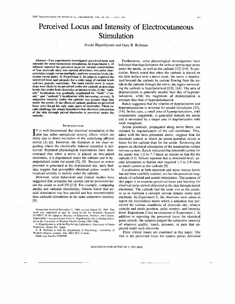
Fig. 3 shows the mean estimates for each of the “both” judgments as a function of current ratio, with relative po- larity and electrode site as the parameters.
To identify how perceived intensity (\k) under each electrode grows as current ratio (+/+,-,) increases, the power function 4! = k ( + / + O ) n was applied by the method of least squares to each of the magnitude estimates for the two loci. Separate power functions, one for perceived in- tensity under the cathode and the other for perceived in-
__
684 IEEE TRANSACTIONS ON BIOMEDICAL ENGINEERING. VOL. 38. NO. 1, JULY 1991
10 '
5 '
0.5t Elbow
1 2 3 4 5
STIMULUS J THRESHOLD
not significant, but the polarity x site interaction was sig- nificant, F(1, 14) = 5.63, p < 0.05. The latter occurred because the intercept for the cathode at the wrist (-0.17) was larger than that for a paired anode at the elbow (-0.30), and, in the reverse condition, the intercept for the cathode at the elbow (-0.14) was larger than that for the anode at the wrist (-0.46). Consequently, the overall intercept for the cathode (-0.16) was larger than that for the anode (-0.38).
GENERAL DISCUSSION The most striking finding obtained from Experiments 1
and 2 is that sensation is mainly perceived under the cath- ode at detection level, but, as current increases, it is more frequently localized under both electrodes.
Furthermore, when sensation for a particular current flow is perceived under both electrodes, the subjective in- tensity under the cathode is gnerally more intense than that under the anode. This is shown in Table 11, where for either relative polarity, the intercepts, but not the slopes, are significantly different between the two electrodes. When the wrist electrode is the cathode, the mean inter-
Fig. 3. Mean subjective intensity for each of the magnitude estimates given for dual localization as a function of current ratio. The parameters are elec- trode site and relative polarity: open circles, wrist electrode in the cathode distal condition; open triangles, elbow electrode in the cathode distal con- dition; filled triangles, wrist electrode in the cathode proximal condition: filled circles, elbow electrode in the cathode proximal condition. The cir- cles represent the judgments for anode and the triangles represent the judg-
cept for that distal locus is larger than that for the elbow anode; for the direction Of the inter- cept for the wrist anode is smaller than that for the cath- ode at the elbow. The greater subjective magnitude, con- sequent1y9 is associated with the no matter where
ments for cathode. it is sited on the arm, rather than the particular physical spot itself.
This result may parallel the observations of motor fi-
amining the effects of polarity on the twitch response of
when the cathode was close to the ulnar nerve at the wrist
TABLE I1 MEAN SLOPE AND INTERCEPT OF THE POWER FUNCTIONS FITTED TO ANODAL hers by Berger? Gravenstein, and Munson [221 who, ex-
A N D CATHODAL MAGNITUDE ESTIMATES FOR "BOTH" RESPONSES AS A FUNCTION OF RELATIVE CATHODE POSITION AND ELECTRODE SITE. THE
RESULTS WERE OBTAINED FROM 15 SUBJECTS the thumb, showed that the maximal twitch was obtained
Cathode Distal Cathode Proximal
Wrist Elbow wrist Elbow
Slope M 1 . 9 8 2.06 I .79 1.58 SD 0.59 0.70 0.64 0.5 I
Intercept M -0.17 -0.30 -0.46 -0.14 SD 0.56 0.46 0.50 0.41
tensity under the anode, were constructed for the wrist and the elbow.
Table I1 shows the mean slope (n) and intercept (log k) for each combination of relative polarity and electrode site. A two-way ANOVA performed on the slope data showed that the main effect of polarity was significant, F(1, 14) = 4.7, p < 0.05. This reflects the finding that the mean slope for the cathode distal condition (2.02) was steeper than that obtained when the cathode was proximal ( 1.68),
A similar two-way ANOVA performed on the intercept data showed that the main effects of polarity and site were
and the anode was elsewhere. The findings obtained in this study run counter to the
simple hypothesis that current is perceived exclusively under the cathode. They are, however, compatible with Ranck's model, which assumes that depolarization occurs under both cathode and anode, although to different de- grees. If Ranck's assumptions are also vakd for our paired electrodes, it may be predicted that 1) low stimulus cur- rent induces perceptible action current only under the cathode, but high stimulus current produces it under both electrodes, 2) the magnitude of depolarization is larger for the cathode than for the anode, and 3) if the magnitude of depolarization for a population of fibers is a determining factor of perceived intensity, the perceived intensity un- der the cathode is more intense than that under the anode.
Particular effects of relative polarity on perceived locus were found at the detection and pain levels for the A-E combination in Experiment 1. At both .intensities, the electrocutaneous stimulus was localized more often at the cathode when that electrode was proximal. A similar re- sult was obtained at both detection and moderate intensi- ties in Experiment 2. These findings may possibly be ac-

HIGASHIYAMA A N D ROLLMAN: LOCUS & INTENSITY OF ELECTRICAL STIMULATION
~
685
counted for in terms of collision block where the flow of the action current generated under the cathode is blocked or weakened by the flow of electrically generated anti- dromic current [23], [24]. For the cathode distal condition of the A-E combination, in which the cathode is near the wrist and the anode is near the elbow, the action current under electrode A , which is propagated through the ulnar nerve to electrode E, is likely to be weakened by the flow of stimulus current from electrode E to A . For the oppo- site condition of the A-E combination, where the cathode is more proximal than the anode, there may not be colli- sion block, because the action current flows in the same direction as the stimulus current, i.e., from the distal to the proximal site.
The effects of relative polarity on perceived locus were not obtained for the A-C combination, however, even though this pair was also positioned along the musculo- cutaneous nerve. Since the major difference between the A-C pair and the A-E pair was the distance between elec- trodes, perception of the collision block may be more likely to occur as the separation of electrodes is widened.
The previously reported exponents (slopes) of the power function for electrical stimulation have varied consider- ably, from 0.7 to beyound 3.5 [25], [26], 1271. These dif- ferences have been ascribed to the effects of the correction of power function for threshold 1281, to sensation level [20], [25], [26], [29], [30], and to regression and range effects [3 13. Table I1 shows that the mean slope for a dis- tal cathode (2.02) was steeper than that for a proximal cathode (1.69). This suggests a possibly new stimulus pa- rameter influencing the exponent of the power function.
Our results are important not only in expanding the model of electrical stimulation, but in applying electrical stimulation to therapy for pain reflief and muscular reha- bilitation. Previous clinical methods using electrical stim- ulation appear to have paid principal attention to the site of the cathode on the skin, because it has been believed that the electrical stimulation is perceived mainly at that location. These data suggest that the site of the anode de- serves equal consideration.
ACKNOWLEDGMENT The authors wish to thank L. Clohosey for her assis-
tance in collecting and analyzing the data of Experiment 1.
111
121
I31
141
REFERENCES W. D. Larkin and J . P. Reilly, ‘‘Strength/duration relationships for electrocutaneous sensitivity: Stimulation by capacitive discharges,” Percept. Psychophys., vol. 36, pp. 68-78. 1984. G. B. Rollman, “Electrocutaneous stimulation: Psychometric func- tions and temporal integration,” Percept. Psyrhophys.. vol. 5, pp.
G. B. Rollman, “Behavioral assessment of peripheral nerve func- tion,” Neurology, vol. 25, pp. 339-342, 1975. S. S. Stevens, A. S. Carton, and G. M. Shickman, “A scale of ap- parent intensity of electrical shock,” J. Exper. Psychol., vol. 56, pp.
289-293, 1969.
328-334, 1958.
W. R. Uttal, “Cutaneous sensitivity to electrical pulse stimuli,” J. Comp. Physiol. Psychol.. vol. 51, pp. 549-554, 1958. W. R. Uttal, “A comparison of neural and psychophysical responses in the somesthetic system,” J . Comp. Physiol. Psychol., vol. 52, pp.
L. A. Benton, L. L. Baker, B. Bowman, and R. L. Waters, Func- tional electrical stimulation: A practical clinical guide. 2nd ed. Downey, CA: The Prof. Staff Assoc. of the Rancho Los Amigos Hos- pital, 1981. J. B. Ranck, Jr., “Electric stimulation of neural tissue,” in Methods in Medical Research, vol. 1 1, R. F. Rusher Ed. Chicago: Year Book Publishers, 1966, pp. 262-269. R. H. Gibson, “Electrical stimulation of pain and touch,’’ ih The Skin Senses, D. R. Kenshalo, Ed. Springfield, IL: Charles C Thomas,
T. Yamamoto. Y. Yamamoto, and A. Yoshida, “Formative mecha- nisms of current concentration and breakdown phenomena dependent on direct current Row through the skin by a dry electrode,” lEEE Trans. Biomed. Eng.. vol. BME-33, pp. 396-404. 1986. J . S. Mannheimer and G. N. Lampe. Clinical Transcutaneous Elec- trical Nerve Stimulation. Philadelphia: F. A. Davis. 1984, p. 349. S. R. Berlant, “Letters to the editor,” Clin. J. Pain, vol. 4, pp. 191- 196, 1988. J . B. Ranck, Jr., “Which elements are excited in electrical stimula- tion of mammalian central nervous system: A review,” Brain Res.,
- , “Extracellular stimulation,” in Electrical Stimulation Research Techniques, M. M. Patterson and R. P. Kesner, Eds. New York: Academic, 1981. pp. 1-36. C. R. Gallistel, “Subcortical stimulation for motivation and rein- forcement,” in Electrical Stimulation Research Techniques, M. M. Patterson and R. P. Kesner, Eds. New York: Academic, 1981, pp.
J . F. Swett and C. M. Bourassa, “Electrical stimulation of peripheral nerve,” in Electrical Stimulation Research Techniques., M. M. Pat- terson and R. P. Kesner, Eds. New York: Academic, 1981, pp.
R. F. Thompson, Foundations of Physiological Psychology. New York: Harper & Row, 1967, p. 157. S. P. Grossman, A Textbook of Physiological Psychology. New York: Wiley 1967, p. 15. J. P. Ifoel, Elementary Statistics. New York: Wiley, 1960. ch. 4. A. Higashiyama and T. Tashiro, “Temporal integration of double electrical pulses,” Percept. Psychophys., vol. 43, pp. 172-178. 1988. W. J. Dixon and F. I . Massey, Introduction to Statistical Analysis. New York: McGraw-Hill, 1957. J . J. Berger, J. S. Gravenstein, and E. S. Munson, “Electrode po- larity and peripheral nerve stimulation,” Anesthesiol., vol. 56, pp.
C. van den Honert and J. T. Mortimer, “A technique for collision block of peripheral nerve: Single stimulus analysis,” IEEE Trans. Biomed. Eng., vol. BME-28, pp. 373-378, 1981. - , “A technique for collision block of peripheral nerve: Frequency dependence,” IEEE Trans. Biomed. Eng. , vol. BME-28, pp. 379- 382, 1981. A. Higashiyama. and T. Tashiro, “Magnitude estimates for electrical shock,” Japan Psy. Res., vol. 29, pp. 81-88. 1987. - , “Magn’itpde estimates for electrical pulses: Evidence for two neural mechanisms,” Percept. Psychophys.. vol. 43, pp. 537-549, 1989. G. B. Rollman and G. Harris, “The detectability, discriminability, and perceived magnitude of painful electrical shock,” Percept. Psy-
G. B. Rollman, “Electrocutaneous stimulation,” in Cutaneous Com- munication Systems and Devices. F. A. Geldard, Ed. Austin, TX: Psychonomic Soc., 1974, pp. 38-51. B. Jones, “Algebraic models for integration of painful and nonpainful electric shocks,” Percept. Psychophys., vol. 28, pp. 572-576, 1980. B. S. Rosner and W. R. G o f f , “Electrical responses of the nervous system and subjective scales of intensity.” in Contributions to Sen- sory Physiology. vol. 2. W. D. Neff, Ed. New York: Academic,
D. V. Cross, B. Tursky, and M. Lodge, “The role of regression and range effects in determination of the power function for electric shock,” Percept. Psychophys.. vol. 18, pp. 9-14, 1975.
485-490, 1959.
1968, pp. 223-261.
vol. 98, pp. 417-440, 1975.
142- 172.
244-298.
402-404, 1982.
chophys., vol. 42, pp. 257-268, 1987.
1967, pp. 169-221.
686 IEEE TRANSACTIONS ON ,BIOMEDICAL ENGINEERING, VOL. 38. NO. 7. JULY 1991
Atsuki Higashiyama was born in Hyogo Prefec- ture, Japan, 1951. He received the M.A. and Ph.D. degrees in psychology form Osaka City University, Osaka, Japan, in 1976 and 1983. re- spectively.
He joined the University of Osaka Prefecture as a Research Associate. served as an Assistant Pro- fessor from 1981 to 1988, and is currently an As- sociate Professor in the Psychology Laboratory of the College of Integrated Arts and Sciences. His principal research interests are psychophysics of
touch and pain generated by electrocutaneous stimulation and the visual perception of size, distance, and angle.
Gary B. Rollman received the Ph.D degree in
experimental psychology from the University of Pennsylvania.
He spent two years as a NIMH Postdoctoral Fellow and Visiting Lecturer at Princeton Univer- sity. and joined the University ot Western On- tario, London, Ont., in 1969. where he is Profes- sor of Psychology. He has also been a visiting professor at the University of Stockholm and the University of St. Andrews in Scotland. His re- search has emphasized psychophysical studies ot
touch, employing electrocutaneous and mechanical stimulation, and stud- ies of pain assessment and management in both laboratory and clinical set- tings.