Embed Size (px)
Citation preview

Determination of hemoglobin concentration in bloodRBC, WBC & platelet counts
Determination of MCV, MCH, MCHCDetermination of HCT/PCV
Hemolysis & fragility test of RBC
Dr. Wang Lin-linDepartment of PhysiologyZhejiang University School of Medicine
EXPERIMENT
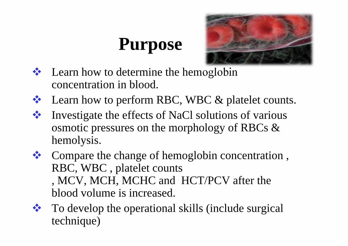
Purpose Learn how to determine the hemoglobin
concentration in blood. Learn how to perform RBC, WBC & platelet counts. Investigate the effects of NaCl solutions of various
osmotic pressures on the morphology of RBCs & hemolysis.
Compare the change of hemoglobin concentration , RBC, WBC , platelet counts, MCV, MCH, MCHC and HCT/PCV after the blood volume is increased.
To develop the operational skills (include surgical technique)

Determination of hemoglobin concentration of blood
In clinical practice, hematological informationaccumulated from a series of blood tests conducted on asmall volume of blood - even a single drop - can be ofgreat diagnostic and prognostic value.The hemoglobin content of a solution can be estimatedby measuring its color, its power of combining withoxygen or carbon monoxide, or by its iron content.The most popular methods for hemoglobin estimationin modern clincal laboratories are colorimentric.

Red Blood Cell Count
• Total RBC & WBC counts are a routine part of any physical exam, & most clinical agencies use computers to conduct these counts.
• Hemocytometer. There are three kinds of hemocytometer: Burker, Thoma & Neubauer. The improved Neubauer counting chamber is the most convenient & is now widely used clinically.
Neubauer Counting Chambers


Count the RBCs in 5 medium squares (4 at the 4 corners & 1 in the center) inside the central large square. Each border of a medium square is marked with three close parallel lines (inner, middle & outer lines). The RBC count is made only within the square bordered by the middle line.
W
WW
W
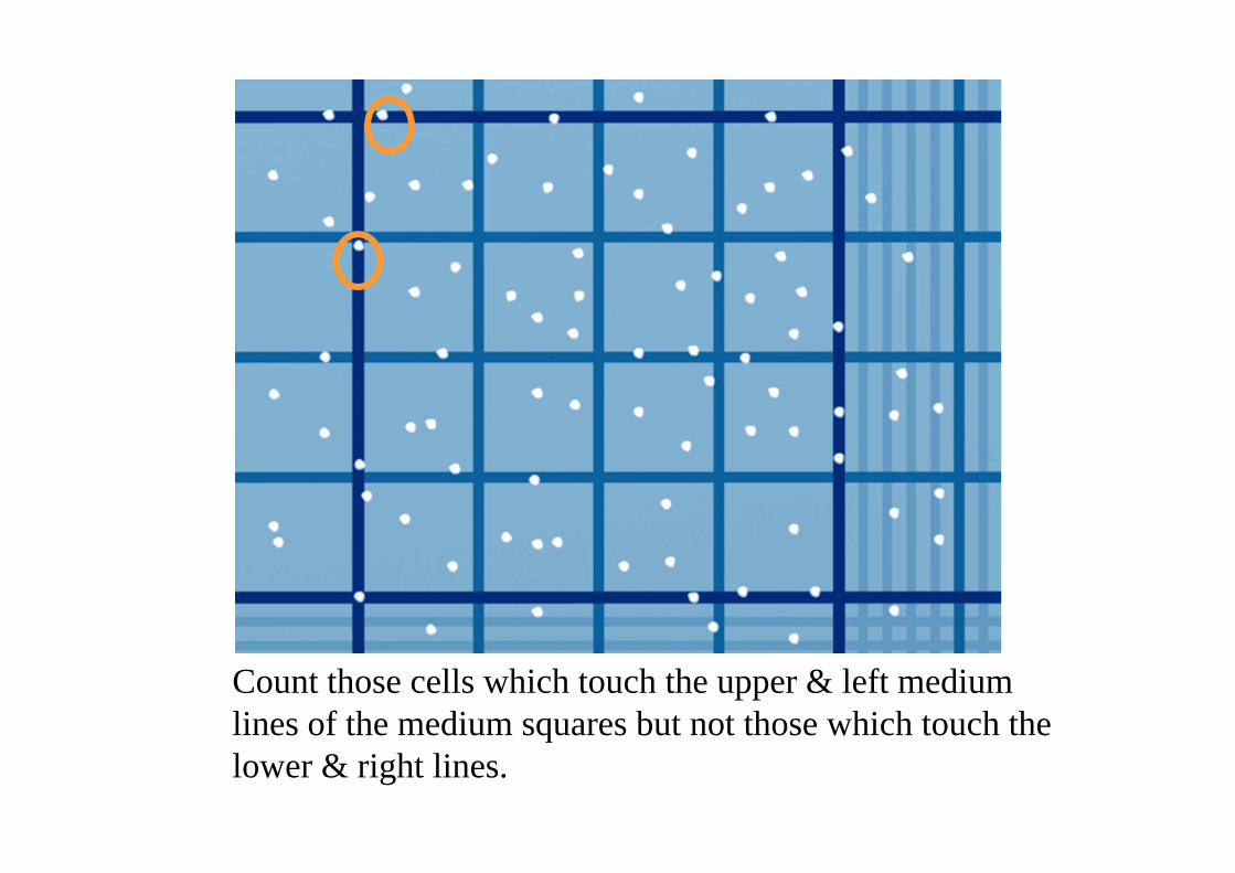
Count those cells which touch the upper & left medium lines of the medium squares but not those which touch the lower & right lines.

EXAMPLE
Total RBC count in 5 squares = 600Multiply 600 5 = 3000 (Total number of cells in central square)Multiply 3000 10 = 30,000 (Cells per mm3)Multiply 30,000 200 = 6,000,000 (Dilution factor of 200)Total number of RBC in sample is 6,000,000 cells/ml

Determination of MCV, MCH, MCHC
MCH is a calculated value and is defined as HGB/RBC giving the mean HGB concentration in the RBC population.MCHC is a calculated value and is defined as HGB/HCT.
MCV can be calculated as HCT/RBC count.


Hemolysis & fragility test of RBC
• The permeability properties of a membrane can bestudied by testing whether various compounds or agents,chemical or physical, can permeate the membrane. Onemethod of doing this is to observe the osmotic effects ofcompounds on the cell membrane.
• RBCs require a rather stable osmotic environment, butnormal RBCs resist hemolysis due to the fact that the cellsare bi-concave.

• CRENATION•
Hypertonic environment
Outflux of water

• On the other hand, in a hypotonic environment (e.g. 0.4% NaCl or distilled water), an influx of water occurs: the cells swell, the integrity of their membranes is disrupted, allowing the escape of their hemoglobin (hemolysis) which dissolves in the external medium.
• HEMOLYSIS

White Blood Cell Count• LEUKOCYTOSIS
An abnormally high WBC count, above 11,000 cells/mm3 ,may indicate bacterial or viral infection, metabolic disease, hemorrhage, or poisoning by drugs or chemicals.
• LEUKOPENIAA decrease in the white cell number below 4000 cells/mm3 may indicate typhoid fever, measles, infectious hepatitis or cirrhosis, tuberculosis, or excessive antibiotic or X-ray therapy. A person with leukopenia lacks the usual protective mechanisms we would expect to find in the immune system.

PLATELETS
• Platelets are cell fragments of large multinucleate cells (megakaryocytes) formed in the bone marrow. They appear as darkly staining, irregularly shaped bodies interspersed among the blood cells.
• The normal platelet count in blood ranges from 250,000 to 500,000 per cubic millimeter.
• Platelets are instrumental in the clotting process that occurs in plasma when blood vessels are ruptured.

Experimental Procedure• Rabbit operation(1)Catch

(2)Weigh
(3)Anaesthetize : 20% Urethane (ip) with the proportion of 5 ml/kg.
(4)Fix

(5)OperationCut off the hair on the neck. Cut and separate the skin and the hypodermis in the front of neck about 8~10 cm in length. Separate the muscles with the hemostasia clamps. Separate its trachea and the connective tissues beside the trachea to find the the left common carotid artery. .
Trachea Left common carotid artery Vagus nerve Sympathetic nerve
Depressor nerve

• Catch• Weigh• Anaesthetize• Fix• Operate• Separate the left common carotid artery• Ligate the common carotid artery near the head end• keep a ligature thread for future use). • In order to stop the blood flow, clamp the common carotid
artery near the cardiac end with an clamp. • Cut a small incision near the ligature thread.• Insert a canula into the common carotid artery from the
incision toward the heart. • Ligate and fix the canula of the common carotid artery. • Inject 1000 U/kg of heparin into the vein of outer ear
border
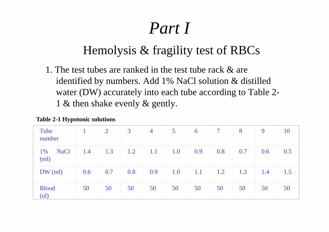
1. The test tubes are ranked in the test tube rack & are identified by numbers. Add 1% NaCl solution & distilled water (DW) accurately into each tube according to Table 2-1 & then shake evenly & gently.
Part I Hemolysis & fragility test of RBCs
Table 2-1 Hypotonic solutions
Tubenumber
1 2 3 4 5 6 7 8 9 10
1% NaCl(ml)
1.4 1.3 1.2 1.1 1.0 0.9 0.8 0.7 0.6 0.5
DW (ml) 0.6 0.7 0.8 0.9 1.0 1.1 1.2 1.3 1.4 1.5
Blood(ul)
50 50 50 50 50 50 50 50 50 50

2. Take the fresh blood, add 1 drop (50 ul) into each tube, shake evenly & gently, & stand for 30 min at room temperature.
3. Judge the results (1) The liquid of the upper level has no color & the bottom layer is muddy
red, indicating no hemolysis. (2) The upper layer is salmon pink & the bottom layer is muddy red,
indicating incomplete hemolysis. In this condition, the sodium chloride concentration reflects the minimum resistance of the RBCs.
(3) The liquid is transparent red & the bottom has no deposited cells, indicating complete hemolysis. In this condition, the sodium chloride concentration reflects the maximum resistance of the RBCs.

• Determination of hemoglobin concentration in bloodRBC, WBC & platelet countsDetermination of MCV, MCH, MCHCDetermination of HCT/PCV
• Remove the clamp and get the fresh blood sample about 0.3 ml in the tube
• Measure all above items with a CA620 Hematology Analyzer (Medonic Cell Analyzer CA620/530, Sweden)
• Detection is accomplished using the electronic impedance principle and occurs in the orifice of the transducer.(www.boule.se)
Part II
Make someone is responsible for this matter

Analyzing the Sample (open tube)
• Choose the operational mode with ‘Menu-Operate’ so that the last run sample is displayed.
• Aspirate the sample through the aspirating pipette by pressing the start lever behind
• the aspiration needle, see picture below.
• Remove the sample when the text ’Aspirating…’ isappears.• The display shows the following sequences:
Last Sample (= blank) Aspirating sequence (ca. 3 sec.)

Print the result.

• Inject 0.9% sodium chloride (100 ml ) into the vein in the ear to increase the blood volume.
• Take the fresh blood sample within 1 minute after injecting sodium chloride.
• Analyzing the Sample and compare the change of hemoglobin concentration ,RBC, WBC , platelet counts, MCV, MCH, MCHC and HCT/PCV
Part III

Catch, Weigh, Anaesthetize, FixOperation
Get blood sample
2nd
Determination Cell numberHGB….
3rd
Determination Cell number HGB….
Injection Heparin 1ml/kg
Injection NaCl 100ml to dilute the blood
1st
HemolysisObservation
Results

Requirement (一)Experimental Report
Name calss….TitleMethods and MaterialsResults
1.Stick the original data (in the report of the group leader)2.Design and complete a table for the result of two analysis of hemoglobin concentration ,RBC, WBC , platelet counts, MCV, MCH, MCHCand HCT
Discussion What is the function of RBCs? When does RBC count increase or decrease?