Embed Size (px)
Citation preview

DECEMBER 2014
UpfrontIt‘s Not You, It‘s Me, Doctor
10
In PracticeSeven Tips For Success in Cataract Patients With Blebs
37 – 38
ProfessionTranslational Research: Innovate Horizontally
44 – 48
Sitting Down WithThe New York Networker, Georgette Pascale
50 – 51
# 14
The Stars of 2014The Ophthalmologist Innovation Awards are here!18 – 26

For Retina, Cataract and Glaucoma Surgery • Newly developed vacuum and flow tri-pump system • SPEEP® mode for very precise maneuvers • Active- and gravity infusion • Up to 10’000 cuts with Twin-Blade Cutters • Double light source with color adjustable LED technology • Fully integrated 532 nm green endo-laser • Brand new phaco engine for even more efficiency and safety • Wireless, dual linear all-in-one foot switch • Intui tive user interface with Direct Access® • Designed for fast and sterile work flow • Utmost system performance thanks to latest Embedded Controller Technology
www.oertli-os4.com
The Essence of Perfection
Eckn
auer
+Sc
ho
ch A
SW

Ophthalmology Futures Forum 2014: The InterviewsFind out what makes the meeting unique – from the innovators andthe investors at the forefront of ophthalmology.
In September, The Ophthalmologist team went to Canary Wharf in London with a camera crew to cover the 2014 Ophthalmology Futures Forum.We spoke to meeting co-chair Kuldev Singh about what makes the Forum special, and some of ophthalmology’s leading figures – like Boris Malyugin, Peng Khaw, Anat Loewenstein and Tarek Sharaway – and leaders from the worlds of big pharma (Greg Kunst) and medical devices (Laurence Marsteller and Michael Mrochen) about the value of the meeting.
To see what the innovators and decision makers had to say, visit:http://top.txp.to/OFF_2014
Online this Month
Watch Juan Mura Perform Cataract Surgery in Patients with Glaucoma and a Functioning BlebIn this issue, Juan Mura offers seven top tips for successful outcomes when performing cataract surgery in eyes that have previously undergone glaucoma surgery.
To view videos of the surgery in action, head over to: https://theophthalmologist.com/issues/1114/401/
For Retina, Cataract and Glaucoma Surgery • Newly developed vacuum and flow tri-pump system • SPEEP® mode for very precise maneuvers • Active- and gravity infusion • Up to 10’000 cuts with Twin-Blade Cutters • Double light source with color adjustable LED technology • Fully integrated 532 nm green endo-laser • Brand new phaco engine for even more efficiency and safety • Wireless, dual linear all-in-one foot switch • Intui tive user interface with Direct Access® • Designed for fast and sterile work flow • Utmost system performance thanks to latest Embedded Controller Technology
www.oertli-os4.com
The Essence of Perfection
Eckn
auer
+Sc
ho
ch A
SW

Feature
18 The Ophthalmologist Innovation Awards The Innovation Awards 2014 celebrates this year’s diagnosis, therapy and surgery stars as nominated by you.
03 Online This Month
07 Editorial P, Damned P and Statistics By Mark Hillen
08 Contributors
On The Cover
Innovation hits warp speed: base image courtesy of NASA.
Upfront
10 It’s Not You, It’s Me, Doctor
11 How do People with Strabismus Locate Visual Targets?
12 The Pathway Less Traveled
13 Push, Pull, CHOMP!
14 The Smartphone Adaptor with a Social Mission
16 Fingerprick River Blindness Screening
DECEMBER 2014
UpfrontReverse transcriptase inhibitors as dry AMD therapy 10
In PracticeCataract surgery in patients with prior glaucoma surgery
34 – 36
ProfessionPractical translational research in the medical device industry
44 – 48
Sitting Down WithThe New York Networker, Georgette Pascale
46 – 48
# 14
Tech StarsThe Ophthalmologist Innovation Awards 2014 are here!18 – 26
50
Contents
16
18

ISSUE 14 - DECEMBER 2014
Editor - Mark [email protected]
Associate Editorial Director - Fedra [email protected]
Associate Editor - Roisin [email protected]
Associate Editor - Michael [email protected]
Senior Designer - Marc [email protected]
Chief Executive Officer - Andy [email protected]
Chief Operating Officer - Tracey [email protected]
Publishing Director - Neil [email protected]
Audience Development Manager - Tracey Nicholls
Digital Content Manager - David Roberts
Traffic and Administration Manager - Vicki Shinks
Mac Operator Web/Print - Peter [email protected]
Social Media / Analytics - Stephen [email protected]
Published by Texere Publishing Limited, Booths Hall,
Booths Park, Chelford Road, Knutsford, Cheshire, WA16 8GS, UK
General enquiries: [email protected]
+44 (0) 1565 752883 [email protected]
Distribution: The Ophthalmologist distributes 17,934 printed copies and 7,295 electronic copies to a targeted
European list of industry professionals. ISSN 2051-4093
36
In Practice
36 Cataract Surgery in Patients With Blebs: Seven Tips for Success Juan Mura discusses the particular challenges of performing cataract surgery on patients who have had a trabeculectomy, and ways to minimize the risk of bleb failure.
39 Lasers or Lenses? We ask Bruce Allan and Erik Mertens: in patients with moderate-to-high myopia, when do you recommend laser refractive surgery, and when do recommend a phakic IOL?
Profession
44 Translational Research: Innovate Horizontallyy Experienced innovators Pavel Zakharov, Mark Talary, Daniel Boss and Michael Mrochen explain how to bridge – or avoid – the “Valley of Death” (and more) when it comes to medical device development.
Sitting Down With
50 Georgette Pascale, President and CEO of Pascale Communications.

Only the GALILEI G4 unites Placido and Dual Scheimpflug technologies in one measurement. With the GALILEI G4, you get highly precise measurements for posterior and anterior curvature, pachymetry, Total Corneal Power, Total Corneal Wavefront and the anterior segment of your
patient’s eye. The new GALILEI G4, for first-class clinical results. The GALILEI G4 is a modular platform, which can be upgraded according to your needs. Learn more on galilei.ziemergroup.com.
FOR REFRACTIVE AND CATARACT SURGERY
Reaching a new level in corneal tomographyPatented Dual Scheimpflug system provides highly accurate pachymetry and ray-tracing, even when the measurement is decentred.
Iris-based eye motion compensationHave confidence in your follow-up measurements with realignment of maps in 3-D.
The only true solutionPlacido and Scheimpflug for highly accurate pachymetry, elevation and curvature data – in all eyes.
One platform, one solution. We simplify the daily workflow in your clinic with an all-in-one solution, from refractive to cataract surgery.
Get to know our new e-learning center: e-learning.ziemergroup.com
The_Ophthalmologist_nov2014_GALILEI_G4_Ad_210x266mm.indd 1 28.10.14 13:42

’ve been aware of David Colquhoun’s work for almost twenty years now – initially as an undergraduate, in lectures describing the UCL professor’s pioneering work on single ion channel behavior. More recently, I’ve followed
his Twitter feed, @david_colquhoun, and enjoyed reading his website, DC’s Improbable Science (www.dcscience.net), particularly for his candid and often excoriating views on metrics, university management, and alternative medicine.
One of the risks of being at the academic coalface for the best part of forty years is the development of a comprehensive understanding of statistics. DC definitely has that, and one of his tweets last month led me to his latest manuscript on arxiv.org; the first line of the abstract states, “If you use P=0.05 to suggest that you have made a discovery, you’ll be wrong at least 30 percent of the time.” The next line raises the stakes further: “If, as is often the case, experiments are underpowered, you’ll be wrong most of the time.”
Rather than try to recapitulate Colquhoun’s workings in the word count-constricted confines of the Editorial page, I’d suggest you read his manuscript and the examples within it (1). The top-line message: underpowered experiments are dangerous – false positives and false negatives accumulate to give wincingly high false discovery rates, and this only increases as (statistical) power decreases. His advice is “if you wish to keep your false discovery rate below 5 percent, you need to use a 3-sigma rule, or to insist on a P-value below 0.001,” concluding with “And *never* use the word ‘significant’.”
If you accept Colquhoun’s argument, lots of things start to make sense. The irreproducible experimental results; the disappointment of that promising drug candidate failing at Phase II; trials where homeopathy actually appeared to work – right down to the newspaper stories that “seem to link almost any nutritional supplement with almost any outcome” (2). They’re all there, because they have peer-reviewed publications to back them up. If you’re not already doing so, perhaps it’s time to view anything that reports a P-value close to 0.05 as “worth another look”, and only start considering results as beginning to be robust when the P-value approaches 0.001.
Mark HillenEditor
Editor ia l
P, Damned P and StatisticsPoor power, false positives and false negatives crucify the credibility of p<0.05
References1. D. Colquhoun, “An investigation of the false discovery rate and the misinterpretation of P values”, August 11, 2014, http://arxiv.org/abs/1407.5296.2. J.P.A. Ioannidis, “Implausible results in human nutrition research”, BMJ, 347, f6698 (2013) doi: 10.1136/bmj.f6698.

A NEW ERA HAS BEGUN,AND IT LOOKS AMAZING.Introducing TECNIS® IOL, the first and only presbyopia-correcting Extended Range of Vision IOL.
At last, your patients can enjoy increased spectacle independence with a true extended range of vision.1
• A full range of continuous, high-quality vision in all light conditions2
• Incidence of halo and glare comparable to a monofocal IOL1
• TECNIS® Symfony Toric IOL also available
The world will never look the same.
For more information, contact your Abbott Medical Opticssales representative.
1. 166 Data on File_Extended Range of Vision IOL 3-Month Study Results (NZ). 2. TECNIS® Symfony DFUTECNIS® Symfony Extended Range of Vision Lenses are indicated for primary implantation for the visual correction of aphakia and preexisting corneal astigmatism in adult patients with and without presbyopia in whom a cataractous lens has been removed by extracapsular cataract extraction, and aphakia following refractive lensectomy in presbyopic adults, who desire useful vision over a continuous range of distances including far, intermediate and near, a reduction of residual refractive cylinder, and increased spectacle independence. These devices are intended to be placed in the capsular bag. For a complete listing of precautions, warnings, and adverse events, refer to the package insert.TECNIS and TECNIS SYMFONY are trademarks owned by or licensed to Abbott Laboratories, its subsidiaries or affiliates.©2014 Abbott Medical Optics Inc., Santa Ana, CA 92705 www.AbbottMedicalOptics.com PP20140012
Contr ibutors
Juan MuraOne of the early MIGS pioneers, Juan Mura is an instructor for the Ophthalmology Department at the Universidad de Chile in Santiago, Chile. A zombie movie aficionado – particularly of George Romero’s oeuvre, with 1968’s “Night of the Living Dead” being his favorite. An experienced glaucoma surgeon, Mura also describes himself as “a good self-taught barman” and, when he turns 55, plans to open a bar called “The Bar Tender”. Read Juan’s seven tips for the best possible cataract surgery outcomes in patients with glaucoma and functioning blebs beginning on page 36.
Michael MrochenMichael Mrochen is most recently known for his pioneering CXL work, but that isn’t his first innovation. His research with Theo Seiler led to the development of both wavefront-guided and wavefront-optimized LASIK. Mrochen is the founder of IROC Science AG, a company specializing in translational research projects for medical devices in the field of ophthalmology and vision care.Read the article Michael wrote with his colleagues Pavel Zakharov, Mark S. Talary, and Daniel Boss on practical translational research in the medical device industry, starting on page 44.
Bruce AllanBruce Allan’s principal research interests are enhancing treatment accuracy in laser refractive surgery, new techniques in corneal endothelial transplantation, and early intervention and visual rehabilitation in keratoconus. An extremely prominent corneal surgeon, Allan has been a consultant ophthalmic surgeon at Moorfields in London since 1998. Outside of work, he’s both a keen sailor and an ardent football fan.
Erik MertensErik Mertens is the Medical Director of the Antwerp ophthalmic and aesthetic surgery center, Medipolis. Co-founder of the American-European Congress of Ophthalmic Surgeons, Mertens has vast experience in high-volume cataract and refractive surgery. His experience is highly sought after and he performs live operations at many national and international conferences.
Bruce and Erik’s opinions on when to choose a phakic IOL over laser eye surgery for the treatment of moderate-to-high myopia start on page 39.

A NEW ERA HAS BEGUN,AND IT LOOKS AMAZING.Introducing TECNIS® IOL, the first and only presbyopia-correcting Extended Range of Vision IOL.
At last, your patients can enjoy increased spectacle independence with a true extended range of vision.1
• A full range of continuous, high-quality vision in all light conditions2
• Incidence of halo and glare comparable to a monofocal IOL1
• TECNIS® Symfony Toric IOL also available
The world will never look the same.
For more information, contact your Abbott Medical Opticssales representative.
1. 166 Data on File_Extended Range of Vision IOL 3-Month Study Results (NZ). 2. TECNIS® Symfony DFUTECNIS® Symfony Extended Range of Vision Lenses are indicated for primary implantation for the visual correction of aphakia and preexisting corneal astigmatism in adult patients with and without presbyopia in whom a cataractous lens has been removed by extracapsular cataract extraction, and aphakia following refractive lensectomy in presbyopic adults, who desire useful vision over a continuous range of distances including far, intermediate and near, a reduction of residual refractive cylinder, and increased spectacle independence. These devices are intended to be placed in the capsular bag. For a complete listing of precautions, warnings, and adverse events, refer to the package insert.TECNIS and TECNIS SYMFONY are trademarks owned by or licensed to Abbott Laboratories, its subsidiaries or affiliates.©2014 Abbott Medical Optics Inc., Santa Ana, CA 92705 www.AbbottMedicalOptics.com PP20140012
UpfrontReporting on the innovations in medicine and surgery, the research policies and personalities that shape ophthalmology practice.
We welcome suggestions on anything that’s impactful on ophthalmology; please email [email protected]
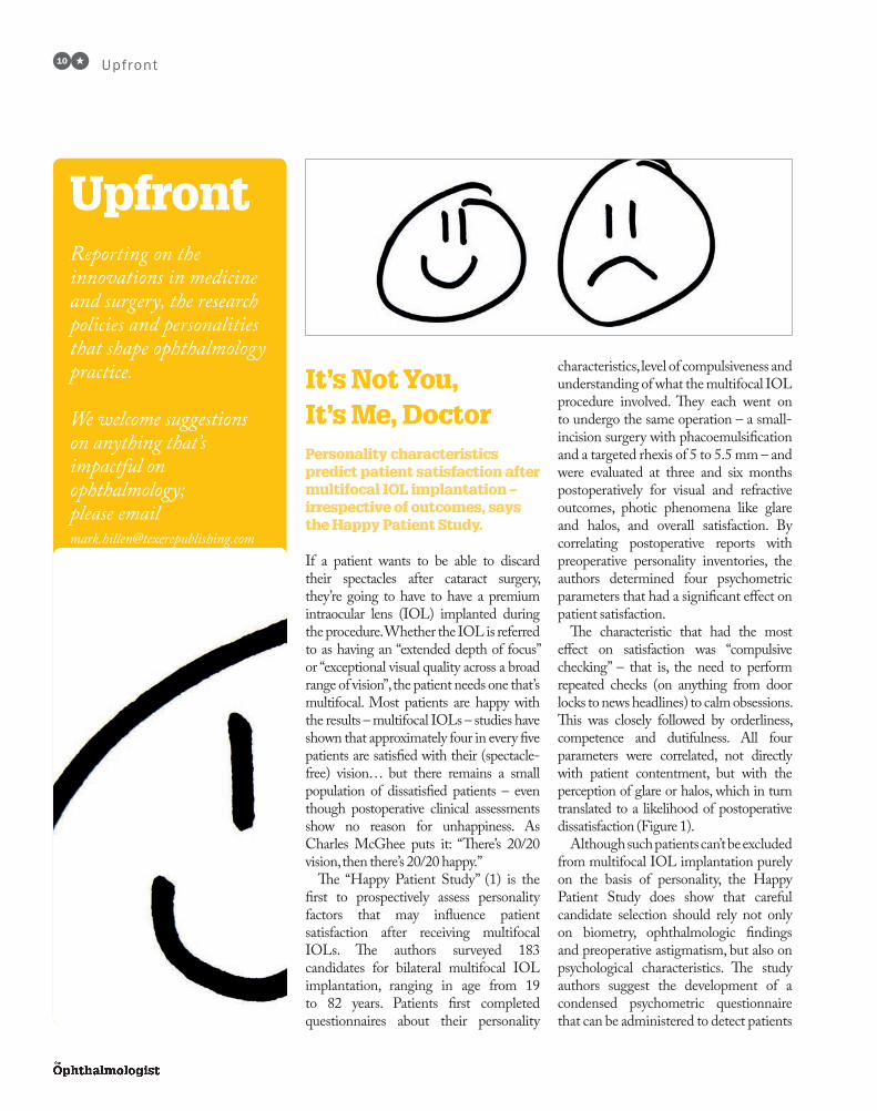
It’s Not You, It’s Me, Doctor Personality characteristics predict patient satisfaction after multifocal IOL implantation – irrespective of outcomes, says the Happy Patient Study.
If a patient wants to be able to discard their spectacles after cataract surgery, they’re going to have to have a premium intraocular lens (IOL) implanted during the procedure. Whether the IOL is referred to as having an “extended depth of focus” or “exceptional visual quality across a broad range of vision”, the patient needs one that’s multifocal. Most patients are happy with the results – multifocal IOLs – studies have shown that approximately four in every five patients are satisfied with their (spectacle-free) vision… but there remains a small population of dissatisfied patients – even though postoperative clinical assessments show no reason for unhappiness. As Charles McGhee puts it: “There’s 20/20 vision, then there’s 20/20 happy.”
The “Happy Patient Study” (1) is the first to prospectively assess personality factors that may influence patient satisfaction after receiving multifocal IOLs. The authors surveyed 183 candidates for bilateral multifocal IOL implantation, ranging in age from 19 to 82 years. Patients first completed questionnaires about their personality
characteristics, level of compulsiveness and understanding of what the multifocal IOL procedure involved. They each went on to undergo the same operation – a small-incision surgery with phacoemulsification and a targeted rhexis of 5 to 5.5 mm – and were evaluated at three and six months postoperatively for visual and refractive outcomes, photic phenomena like glare and halos, and overall satisfaction. By correlating postoperative reports with preoperative personality inventories, the authors determined four psychometric parameters that had a significant effect on patient satisfaction.
The characteristic that had the most effect on satisfaction was “compulsive checking” – that is, the need to perform repeated checks (on anything from door locks to news headlines) to calm obsessions. This was closely followed by orderliness, competence and dutifulness. All four parameters were correlated, not directly with patient contentment, but with the perception of glare or halos, which in turn translated to a likelihood of postoperative dissatisfaction (Figure 1).
Although such patients can’t be excluded from multifocal IOL implantation purely on the basis of personality, the Happy Patient Study does show that careful candidate selection should rely not only on biometry, ophthalmologic findings and preoperative astigmatism, but also on psychological characteristics. The study authors suggest the development of a condensed psychometric questionnaire that can be administered to detect patients
Upfront10

with a higher probability of postoperative dissatisfaction. By identifying people who may be unhappy even when their clinical findings are good, surgeons might be able to explain the potential side effects and the patients’ possible intolerance to them in more detail prior to surgery – which might help patients avoid the procedure if the side effects are unacceptable, and would help surgeons avoid potentially unsatisfied patients – and the can of worms that can open. MS.
Reference.1. U. Mester, T. Vaterrodt, F. Goes, et al., “Impact of Personality Characteristics on Patient Satisfaction After Multifocal Intraocular Lens Implantation: Results From the ‚Happy Patient Study‘”, J. Refract. Surg., 30, 674–678 (2014). doi :10.3928/1081597X-20140903-05.
Figure 1. Correlation of various personality characteristics with subjective disturbance by glare. *, p<0.05; **, p<0.1; NS, not significant.
Upfront 11
How do People with Strabismus Locate Visual Targets? It’s a reasonable – and until now – unanswered question: which eye is the one that tells the brain where a target lies?
Three researchers from the Laboratory for Visual Neuroscience at the University of California, San Francisco, posed a question: how do people with strabismus locate visual targets? People with strabismus (and without amblyopia) have some element of binocular vision. Their brains still receive visual input from both eyes – and can make appropriate and accurate saccades to view the target. But which eye is the one that provides the brain with the information regarding the target’s location? Is it the one
that acquires the target, or the other one?To answer the question, they devised the following experiment (1). Sixteen subjects with alternating exotropia (and no amblyopia) wore red/blue filter glasses for dichoptic stimulation while viewing stimuli on a tangent screen. The trials began with a fixation cross that was visible to either the right or the left eye. Once the subject fixated the cross, a peripheral stimulus (a spot visible only to the right or the left eye) was displayed for 200 ms. The subject was simply told that they had to look at the spot – and as it is only visible for a fifth of a second, it will have disappeared before the eye arrived. To ensure the subject remained motivated, an audible tone was generated for saccades landing within a 5° window. In 10 out of the 16 subjects, purple spots were included on the display as peripheral stimuli, in order to establish which eye
was used to fixate those targets that were potentially visible to either eye. The
researchers went on to compile binocular sensory maps that delineated the
portions of the visual field that each eye perceived, and assessed the subjects’ oculomotor behavior by randomly interleaving red, blue, and purple peripheral stimuli on
the display. What they found was
that there was a close match between suppression scotoma
maps and the eye used to acquire the peripheral stimulus – or more simply put: the target was perceived via the eye that was used to fixate it. MH
Reference1. J.R. Economides, D.L. Adams, J.C. Horton, “How do patients with strabismus locate visual targets?”, Program No. 237.03/Z31, 2014. Neuroscience Meeting Planner, Washington DC: Society for Neuroscience, 2014.

Upfront12
The Pathway Less Traveled A previously unknown anti-inflammatory effect of common HIV/AIDS drugs may offer a safe and inexpensive treatment for dry AMD
Repurposing a well-established HIV drug could be the key to treating dry age-related macular degeneration (AMD). Despite the huge prevalence of the condition, there are currently no approved agents that treat dry AMD. There are, however, a number in the pipeline, and work from Jayakrishna Ambati’s laboratory at the University of Kentucky hopes to add to that – with an existing class of drug.
Nucleoside reverse transcriptase inhibitors (NRTIs) were originally designed to treat cancer in the 1960s, then re-emerged in the late 1980s as the first effective anti-HIV agent. Now, it’s hoped that a previously undiscovered anti-inflammatory activity that this class of drugs possess can be exploited to treat dry AMD – as well as other diseases that share a common signaling pathway.
In dry AMD, a biomolecule known as Alu RNA accumulates in the retina. An overabundance of Alu RNA leads to activation of a toxic pathway – the NLRP3 inflammasome – that cause cell death of the retinal pigment epithelium (RPE). The University of Kentucky-based research group noted that Alu elements, like the HIV virus, rely on the reverse transcriptase enzyme to fulfill their life cycle (1). With that in mind, they hypothesized that NRTIs might be able to block Alu RNA-induced cytotoxicity. What they discovered, though, was more complex than that – the NRTIs did indeed prevent RPE degeneration in mice, but the drugs’
protective action occurred independently of their reverse transcriptase inhibition. Rather, NRTIs possess a previously unknown ability to block an innate immune system component known as the “inflammasome,” which facilitates the toxicity of Alu RNA in the retina. With the inflammatory pathway inhibited, the enzymes that lead to cell death remain unactivated and the RPE is protected from degeneration. Inflammasome blockade was effective in treating geographic atrophy – the late stage of dry AMD – in mouse models (Figure 1). Furthermore, it should be possible to treat wet AMD through the same pathway, suggesting that NRTIs may have a therapeutic role to play in both forms of AMD.
The benefit to using NRTIs to treat AMD is that they are already a diverse and widely used class of drugs, with several decades’ worth of collected pharmacokinetic and safety data.
“Repurposing of NRTIs could be advantageous, for one, because they are very inexpensive,” says Benjamin Fowler, lead author on the study and a postdoctoral fellow in Ambati’s laboratory. “Moreover, through decades of clinical experience, we know that some of the drugs we tested are incredibly safe. Since these NRTIs are already FDA-approved, they could be rapidly and inexpensively translated into therapies for a variety of untreatable or poorly treatable conditions.” Ambati adds, “We are excited at the prospect of testing whether NRTIs could be effective in halting the progression of AMD in patients.” MS
Reference1. B.J. Fowler, B.D. Gelfand, Y. Kim, et al., “Nucleoside reverse transcriptase inhibitors possess intrinsic anti-inflammatory activity”, Science, 346, 1000–1003 (2014). doi: 10.1126/ science.1261754.
Figure 1. Stavudine (or d4T), the NRTI used by Fowler et al. (1) to prevent RPE degeneration in mouse models of geographic atrophy.

Asia-AustraliaA2
Anterior Segment • Glaucoma • Retina
The Inaugural Asia-Australia Congress on
CONTROVERSIES IN OPHTHALMOLOGY
Anterior Segment • Glaucoma • Retina
The 6th World Congress on
CONTROVERSIES IN OPHTHALMOLOGY
MARCH 26-29
2015
FEbRuARY 5-8
2015
SORRENTO, ITALY
HO CHI MINH CIT Y, VIETNAM
www.comtecmed.com/cophy [email protected]
Follow us on:COPHyCongressCOPHyCongress
www.comtecmed.com/cophyaa [email protected]
Follow us on:COPHyCongressCOPHyCongress
THE UPCOMING COPHy CONGRESSES
SAVE THE dATES
chairs:Anat Loewenstein, israel
Neil Bressler, Usa
Push, Pull, CHOMP! A Pac-Man-style video game for the treatment of amblyopia without patching
What’s the most common treatment for amblyopia? Patching the stronger eye, forcing the weaker eye to do all the work. Is this really the best way of doing things? Perhaps not. Teng Leng Ooi, Professor of Optometry at The Ohio State University, calls this a “push-only” method of treating amblyopia, because the dominant eye remains completely unused. He’s developed what he calls a “push-pull” method that makes both eyes
work together, but still exposes the weaker eye to a more complex
set of images that generate stronger stimuli of that eye’s visual system. By forcing both eyes to cooperate (but
suppressing the dominant eye’s power), he and his group
target important pathways in the brain that must be active to produce balanced
vision. Their method taps into the neural networks
responsible for both inhibition and excitation signals governing binocular vision. “We know push-pull works,” he says. “Now it’s a question of how much better we can make it work.”
The genius part of Ooi’s method is that his push-pull training comes in the form of a computer game (Figure 1). The games feature groups of lines in different orientations; players wear red-green 3D glasses that filter images so that the dominant eye sees only a background full of horizontal lines, but the weaker eye sees bordered disks with vertical, horizontal or diagonal lines imposed upon that background. “We make sure the weak eye is seeing the contrasting images at all times,” says Ooi. “The strong
eye has stimulation, but it is cortically suppressed. That is the ‘pull’. The weak eye is ‘pushed’ to work.”
One of the games is a “cat and mouse” challenge where players direct their Pac-Man-shaped “cat” to eat scurrying disk “mice” with lines that are oriented in the same direction as the cat’s. Another shows a matrix of disks with lines in different orientations; players use cursors to line up a “master disk” to match the orientations of the lines. “In tests of these games, we’ve seen improvements in depth perception and binocular vision in people with amblyopia,” Ooi says. “The more abnormal the binocular vision is, the higher the number of training sessions needed.” It works in adults too – pilot testing on two grown-ups has demonstrated improvements in their weak-eye vision from 20/63 to 20/50 in one and from 20/25 to 20/20 in the other.
The games add an important element to amblyopia treatment: fun. In contrast with earlier training designs, where participants had to keep their eyes still and look at the same target for as much as an hour and a half, these games only require a few minutes of concentration at a time – and as patients’ playing skills improve, researchers can make the tasks more complicated or impose shorter deadlines to keep them invested in the challenge. Although in the long run patients may need to spend a long time training their eyes, games like these keep them engaged for longer, ultimately to the benefit of their vision. MS
Figure 1. Screenshot from the “cat and mouse” game to treat amblyopia

Tough on IOP.Easy on Eyes.
For Glaucoma
Nov
embe
r 201
4
The first preservative-free prostaglandin
Effective IOP-lowering (1
Low risk of hyperaemia (2
Abbreviated Prescribing Information TAFLOTAN® (tafluprost 0.0015% eye drops, solution, single-dose container). Presentation: Low-density polyethylene single-dose containers packed in foil pouch. Each single-dose container has a fill volume of 0.3 ml and there are 10 containers in each foil pouch. The following pack sizes are available: 30 x 0.3 ml and 90 x 0.3 ml. One ml of eye drops contains 15 micrograms of tafluprost. Indication: Reduction of elevated intraocular pressure in open angle glaucoma and ocular hypertension in patients who would benefit from preservative-free eye drops or who are insufficiently responsive or intolerant or contra-indicated to first line therapy, as monotherapy or as adjunctive therapy to beta-blockers. Dosage and Administration: The recommended dose is one drop of TAFLOTAN® in the conjunctival sac of the affected eye(s) once daily in the evening. Not recommended in children or adolescents (under the age of 18). In renal or hepatic impairment use with caution. Contraindications: Hypersensitivity to tafluprost or to any of the excipients. Precautions: Before treatment is initiated, patients should be informed of the possibility of eyelash growth, darkening of the eyelid skin and increased iris pigmentation. Some of these changes may be permanent, and may lead to differences in appearance between the eyes when only one eye is treated. Caution is recommended when using tafluprost in aphakic patients, pseudophakic patients with torn posterior lens capsule or anterior chamber lenses, or in patients with known risk factors for cystoid macular oedema or iritis/uveitis. There is no experience in patients with severe asthma. Such patients should therefore be treated with caution. Interactions: Specific interaction studies with other medicinal products have not been performed with tafluprost. Pregnancy: Do not use in women of childbearing age/potential unless adequate contraceptive measures are in place. Driving: Tafluprost has no influence on the ability to drive. Undesirable Effects: The most frequently reported treatment-related adverse event was ocular hyperaemia. It occurred in approximately 13% of the patients treated with preserved tafluprost and 4.1% of the patients treated with preservative-free tafluprost. Other side effects include: Common (1% to 10%): eye pruritus, eye irritation, eye pain, changes in eyelashes, dry eye, eyelash discolouration, foreign body sensation in eyes, erythema of eye lid, blurred vision, increased lacrimation, blepharal pigmentation, eye discharge, reduced visual acuity, photophobia, eyelid oedema and increased iris pigmentation and headache. Uncommon (0.1% to <1%): superficial punctate keratitis (SPK), asthenopia, conjunctival oedema, blepharitis, ocular discomfort, anterior chamber flare, conjunctival follicles, allergic conjunctivitis, anterior chamber cell, conjunctival pigmentation and abnormal sensation in eye, hypertrichosis of eyelid. Overdose: If overdose occurs, treatment should be symptomatic. Special Precautions for Storage: Store in a refrigerator (2°C - 8°C). After opening the foil pouch keep the single-dose containers in the original foil pouch, do not store above 25°C, discard an opened single-dose container with any remaining solution immediately after use. MA Holder: Santen Oy, Niittyhaankatu 20, 33720 Tampere, Finland. Date of Preparation: 11/2012.1) Taflotan lowered IOP by 6.9 - 9.7 mmHg in masked, randomized studies 1-4. 1. Uusitalo H et al. Acta Ophthalmol 2010; 88: 12-19 2. Traverso C et al. J Ocul Pharmacol Ther 2010; 26: 97-104 3. Konstas AG et al. Comparison of 24-hour efficacy with Tafluprost compared with Latanoprost in patients with primary open-single glaucoma or ocular hypertension. Abstract 5104/A2458 4. Chabi A et al. Am J Ophthalmol 2012; 153: 1187-1196 2) Low risk of hyperaemia among prostaglandins: SPC texts of preservative-free Taflotan.
Upfront14
The Smartphone Adaptor with a Social Mission Peek Retina is an adaptor for your smartphone that promises easy and high-quality fundoscopy – no matter if you’re in Scotland or sub-Saharan Africa
You might have seen the inspirational TED presentation by Peek co-founder, ophthalmologist and Clinical Lecturer at the London School of Hygiene & Tropical Medicine, Andrew Bastawrous (bit.ly/peekvision). He talked about how Peek – the Portable Eye Examination Kit – has transformed ocular health screening in rural Kenya. If you’re not aware, Peek is a set of freely available mobile apps that can enable a non-expert with a smartphone and minimal training to perform a whole suite of eye tests – from tumbling E visual acuity tests, to eye tracking, cataract assessment and, with the help of the Peek Retina adaptor (Figure 1), Fundoscopic images of the retina.
The data infrastructure that partners the app enables images and test results to be uploaded to the cloud and assessed by ophthalmologists anywhere in the world. The app uses the smartphone GPS functionality to record the coordinates where the assessments were performed, meaning that the data can be used for both epidemiological studies and patient follow-up, which isn’t always easy in rural sub-Saharan Africa. Mario Giardini, Peek co-founder and Lecturer in Digital Health at University of Strathclyde, explained, “At its core, the Peek connects patients with doctors – and we are proud to produce the tools to do that.”
The optics of Peek Retina have been refined “to a point where the autofocus features of the smartphone camera completely mitigate the complex focusing mechanisms that you’re used to with indirect ophthalmoscopy – months of mucking about in medical school learning how to use it properly have been reduce to one minute,” according to fellow Peek co-founder and ophthalmologist at the Glasgow Centre for Ophthalmic Research in Scotland, Iain Livingstone.
Giardini, the electronic and optical engineer who designed the optics and the casing, explained that “the device is now
in its sixth generation and has been used to screen over 2,000 people in the field”. Giardini described how very robust Peek Retina is, as “every device that’s gone into the field works as well today as it did when it was new”.
Perhaps the area where Peek Retina distinguishes itself the most is that it has undergone extensive clinical validation. Livingstone described the process, which involved comparing pictures from standard diabetic retinal screening cameras with those from Peek in a large cohort of patients. “The images were randomized and presented to two expert readers at Moorfields Eye Hospital, who evaluated a number of optic nerve head parameters including cup to disc ratio; then, we used the methods described by Bland and Altman to compare the two imaging methods. We’re getting really good results, and Andrew is due to publish them very soon” .
Though Peek Retina is a mature design and works well, it’s currently made by high-quality 3D printing – great for field evaluation, but a process that doesn’t scale to production volumes – although there’s certainly great demand. Bastawrous said “We’ve been getting huge levels of demand – over 180 countries have requested to use Peek in their eyecare programs. We’ve got to the point where we need to find a sustainable way of delivering Peek to people.”
Perhaps surprisingly, Peek Vision have tuned to the crowdfunding website Indiegogo to fund the commercialization of Peek Retina. We asked Andrew Bastawrous: why?
“We’ve had various options for people investing in what we’re doing, but it usually comes at a cost: people want to invest with a focus on profit,” he says. “We’re very focused on having a social impact, and so by choosing a crowdfunding campaign, we can have the social mission of what we are doing as our primary focus, without having to give away control.” MH
www.supportpeek.com
Figure 1. The Peek Retina smartphone adaptor in action.

Tough on IOP.Easy on Eyes.
For Glaucoma
Nov
embe
r 201
4
The first preservative-free prostaglandin
Effective IOP-lowering (1
Low risk of hyperaemia (2
Abbreviated Prescribing Information TAFLOTAN® (tafluprost 0.0015% eye drops, solution, single-dose container). Presentation: Low-density polyethylene single-dose containers packed in foil pouch. Each single-dose container has a fill volume of 0.3 ml and there are 10 containers in each foil pouch. The following pack sizes are available: 30 x 0.3 ml and 90 x 0.3 ml. One ml of eye drops contains 15 micrograms of tafluprost. Indication: Reduction of elevated intraocular pressure in open angle glaucoma and ocular hypertension in patients who would benefit from preservative-free eye drops or who are insufficiently responsive or intolerant or contra-indicated to first line therapy, as monotherapy or as adjunctive therapy to beta-blockers. Dosage and Administration: The recommended dose is one drop of TAFLOTAN® in the conjunctival sac of the affected eye(s) once daily in the evening. Not recommended in children or adolescents (under the age of 18). In renal or hepatic impairment use with caution. Contraindications: Hypersensitivity to tafluprost or to any of the excipients. Precautions: Before treatment is initiated, patients should be informed of the possibility of eyelash growth, darkening of the eyelid skin and increased iris pigmentation. Some of these changes may be permanent, and may lead to differences in appearance between the eyes when only one eye is treated. Caution is recommended when using tafluprost in aphakic patients, pseudophakic patients with torn posterior lens capsule or anterior chamber lenses, or in patients with known risk factors for cystoid macular oedema or iritis/uveitis. There is no experience in patients with severe asthma. Such patients should therefore be treated with caution. Interactions: Specific interaction studies with other medicinal products have not been performed with tafluprost. Pregnancy: Do not use in women of childbearing age/potential unless adequate contraceptive measures are in place. Driving: Tafluprost has no influence on the ability to drive. Undesirable Effects: The most frequently reported treatment-related adverse event was ocular hyperaemia. It occurred in approximately 13% of the patients treated with preserved tafluprost and 4.1% of the patients treated with preservative-free tafluprost. Other side effects include: Common (1% to 10%): eye pruritus, eye irritation, eye pain, changes in eyelashes, dry eye, eyelash discolouration, foreign body sensation in eyes, erythema of eye lid, blurred vision, increased lacrimation, blepharal pigmentation, eye discharge, reduced visual acuity, photophobia, eyelid oedema and increased iris pigmentation and headache. Uncommon (0.1% to <1%): superficial punctate keratitis (SPK), asthenopia, conjunctival oedema, blepharitis, ocular discomfort, anterior chamber flare, conjunctival follicles, allergic conjunctivitis, anterior chamber cell, conjunctival pigmentation and abnormal sensation in eye, hypertrichosis of eyelid. Overdose: If overdose occurs, treatment should be symptomatic. Special Precautions for Storage: Store in a refrigerator (2°C - 8°C). After opening the foil pouch keep the single-dose containers in the original foil pouch, do not store above 25°C, discard an opened single-dose container with any remaining solution immediately after use. MA Holder: Santen Oy, Niittyhaankatu 20, 33720 Tampere, Finland. Date of Preparation: 11/2012.1) Taflotan lowered IOP by 6.9 - 9.7 mmHg in masked, randomized studies 1-4. 1. Uusitalo H et al. Acta Ophthalmol 2010; 88: 12-19 2. Traverso C et al. J Ocul Pharmacol Ther 2010; 26: 97-104 3. Konstas AG et al. Comparison of 24-hour efficacy with Tafluprost compared with Latanoprost in patients with primary open-single glaucoma or ocular hypertension. Abstract 5104/A2458 4. Chabi A et al. Am J Ophthalmol 2012; 153: 1187-1196 2) Low risk of hyperaemia among prostaglandins: SPC texts of preservative-free Taflotan.

DON’T LET DRY EYE RUIN THEIR WINTER
From crisp white snow to roaring log fires, winter is a magical season. Unless, that is, you have Dry Eye. With symptoms including burning, stinging, excessive tearing and dryness, it can be tough on eyes.1–2 Fortunately, the OPTIVE® Family works effectively in either aqueous or lipid deficient Dry Eye sufferers.3-5 Recommend it to your patients and help make their winter epic.
RELIEF FOR DRY EYEWHATEVER THE SEASON
Recommended for aqueous deficiency
References:1. Zeev MS, et al. Clin Ophthalmol. 2014;8:581 -590. 2. Abelson MB, et al. Rev Ophthal. 2011;May:74-77. 3. Kaercher T, et al. Clin Ophthalmol. 2009;3:33-39. 4. Lee SY & Tong L. Optom Vis Sci. 2012;89:1654-1661. 5. Simmons PA, et al. Presented at EUCORNEA, Amsterdam, 2013. EU/0159/2014c; Date of preparation: December 2014
Recommended for lipid deficiency
Upfront16
Fingerprick River Blindness Screening New testing methods could help eliminate the neglected tropical disease
Harold Ridley’s other claim to fame is his research into River Blindness when stationed in the Gold Coast (now Ghana) in 1941. He spent a fortnight in Funsi, in the Wa East District of the country, with a battery-operated slit lamp, diagnosing and characterizing the ocular symptoms of River Blindness (Figure 1), which was eventually published in his landmark monograph, “Ocular Onchocerciasis” (1).
Contracting the disease is a disaster for patients and is one of the leading causes of preventable blindness in Africa. Infection is spread by the black fly, and is caused by the parasitic worm Onchocerca volvulus. Diagnosis and treatment is the key to prevention, but the first part can be a challenge. Although Ridley could see worms in his patients’ eyes, not all patients with onchocerciasis present in this manner. The gold-standard diagnostic test is a skin snip followed by examination of the snip in saline solution. If worms appear: the diagnosis is made. If worms don’t appear, this doesn’t give the patient the all-clear: DNA extraction and PCR screening for the worm’s genes has to then be performed.
Antibody tests would appear to be the answer – a fingerprick, drop the blood onto a immunochromatographic assay (just like a home pregnancy test) and get a result in under 20 minutes. That’s just what PATH, an international nonprofit organization have managed to develop in combination with the (US) National Institute of Allergy and Infectious
Diseases: the SD Bioline Onchocerciasis IgG4 rapid test.
David Kaslow, PATH’s vice president for product development explained why they thought this could be a game-changer: “The proven technology behind this test makes it a powerful and reliable tool in the multinational collaboration to eliminate river blindness. The availability of a rapid, point-of-care diagnostic is a harbinger of a world free of the suffering caused by this insidious parasite. What’s needed now is quick action to add this simple test to control and elimination programs.”
The US’ Centers for Disease Control and Prevention, however, published statements that are less effusive, stating “These tests cannot distinguish between past and current infections, so they are not as useful in people who lived in areas where the parasite exists, but they are useful in visitors to these areas” (2). It’s not a bug, it’s a feature, say PATH: “By detecting unique antibodies to the parasite, it quickly identifies previous exposure” (3). Given that Merck has promised to supply the treatment – the oral antiparasitic drug, ivermectin – free of charge to affected areas until the disease is eliminated, that could represent a significant chunk of people with positive-tests.
Perhaps that’s not the point. Although River Blindness has been eliminated from many regions of Africa (4) and many people have been successfully treated with ivermectin – many have not. Screening patients with a method that doesn’t require skin biopsy puts fewer people off, and if that method that is both rapid and reliable, can only be an asset in the field. It should definitely aid screening – and as that’s the first step on the path to eliminating this pernicious disease, it’s certainly a commendable endeavor on the part of PATH and its partners. MH/RM
References1. “OCULAR ONCHOCERCIASIS Including an Investigation in the Gold Coast”, Br. J. Ophthalmol. 29(Suppl), 3–58 (1945).2. Centers for Disease Control and Prevention. Parasites – Onchocerciasis (also known as River Blindness). Updated May 21, 2013. http://www. cdc.gov/parasites/onchocerciasis/diagnosis.html, accessed November 25, 2014.3. PATH “New test will combat major cause of preventable blindness in Africa”, Press Release, November 2, 2014. http://www.path.org/news/ press-room/703/, accessed November 25, 2014.4. K.L. Winthrop, J.M. Furtado, V.C. Lansingh, “River blindness: an old disease on the brink of elimination and control”, J. Glob. Infect. Dis., 3, 151–155 (2011). doi:10.4103/0974- 777X.81692.
Figure 1. Ridley often recorded his observations of the retinal fundus by watercolor painting and sketches – here is a one of fundus oculi in Onchocerciasis made in Funsi in 1944.

DON’T LET DRY EYE RUIN THEIR WINTER
From crisp white snow to roaring log fires, winter is a magical season. Unless, that is, you have Dry Eye. With symptoms including burning, stinging, excessive tearing and dryness, it can be tough on eyes.1–2 Fortunately, the OPTIVE® Family works effectively in either aqueous or lipid deficient Dry Eye sufferers.3-5 Recommend it to your patients and help make their winter epic.
RELIEF FOR DRY EYEWHATEVER THE SEASON
Recommended for aqueous deficiency
References:1. Zeev MS, et al. Clin Ophthalmol. 2014;8:581 -590. 2. Abelson MB, et al. Rev Ophthal. 2011;May:74-77. 3. Kaercher T, et al. Clin Ophthalmol. 2009;3:33-39. 4. Lee SY & Tong L. Optom Vis Sci. 2012;89:1654-1661. 5. Simmons PA, et al. Presented at EUCORNEA, Amsterdam, 2013. EU/0159/2014c; Date of preparation: December 2014
Recommended for lipid deficiency

Ophthalmology is one of the most intense incubators of medical innovation. Competition is truly driving innovation, and this is resulting in not just the
incremental improvements in products that you might expect, but also some big, game-changing leaps too.
Here we recognize a year’s worth of innovation. Apps, IOLs, vitreous cutters, imagers and lasers – the latest and greatest of these are all there.
But which one came out on top?
15Navilas 577+Tissue-friendly, standardizable, navigated microsecond pulsing therapy for retinal disease
Produced by: OD-OS (www.od-os.com)Detail: Laser energy is split up into a pulse train of low-energy pulses that stimulate the retina, but do not heat the tissue to a coagulation threshold – and as a consequence, retinal function loss and scarring can be avoided. Navilas is the only navigated microsecond pulsing therapy (NMPT) system that allows navigated application of this advanced subthreshold laser technique; the treatment area can be precisely delineated based on imported OCT thickness maps, and the aiming beam is prepositioned – compensating for eye movement and allowing complete coverage without undefined overlap.
The treatment is documented in real time, providing visual feedback about treated areas and degree of completion. An initial case series at LMU Munich performed by Marcus Kernt showed no tissue damage or retinal function loss (as expected) with this method. Contact lens-free application and comfortable infrared illumination make this a patient-friendly therapy and set Navilas 577+ apart from slit lamp-based lasers.Impact: Retinal laser is making a comeback in diabetic eye disease because of the chronic use and expense associated with anti-VEGF therapy. Initial studies indicate that laser treatment can add durability to anti-VEGF gains and reduce patient burden. The technique further refines retinal laser therapy: inter-operator variability is minimized by OCT-based planning with real-time documentation, and retinal tissue function is preserved by microsecond pulsing. NMPT has the potential to become the standard adjunct to anti-VEGF in diabetic eye disease.Judge’s comment: “Great laser to improve patients’ outcomes.”
14OCULUS BIOM readyThe world’s first single-use wide angle viewing system
Produced by: Oculus Surgical (www.oculussurgical.com)Detail: Provides the perfect view for non-contact wide angle observation for retinal surgeons without using a contact lens. Easily connected to the microscope; while the surgeon is observing the vitreous and the fundus, the BIOM ready is aligned coaxially with the operating microscope… but during extraocular surgery phases, it’s swung out of the observation beam. It incorporates the new BIOM HD Disposable Lens for unparalleled visual clarity and provides excellent depth of field for better stereopsis. The OCULUS BIOM ready comes pre-assembled with the BIOM HD Disposable Lens in a sterile blister pack.Impact: Provides the optimal balance between efficiency and high optical quality, with outstanding resolution in the periphery, often reducing indentation during laser. The depth of field is increased over other wide angle systems, allowing the surgeon to perform macula work without the need for a contact lens, saving time and money. Sterilization “down time” is decreased – increasing OR efficiency – and as it’s a single-use device, it reduces the risk of cross-contamination.Judges’ comments: “Amazing wide field view in a disposable lens.” “Improves convenience.”
Feature 19
The Judging PanelKeith Barton (Moorfields Eye Hospital, London)
Florian Kretz (IVRC, Heidelberg)
Boris Stanzel (Bonn University Eye Hospital; NEI)
Kuldev Singh (Stamford School of Medicine)
Sebastian Waldstein (Medical University of Vienna)

The Evolution of Intraocular Lens Solutions for Age Related Macular Degeneration (AMD)
AMD is the leading cause of blindness in the developed world. Until recently, the surgical options available to surgeons wishing to improve the visual outcomes of this large patient group involved complex and time consuming surgery with large incisions that had more in common with Extra Capsular Cataract Extraction than with modern surgical techniques.
2014 - EU Launch - iolAMD
Incision Size and Time Required• Injected through a 3 mm, sutureless incision.• 2 minutes longer than standard cataract operation.Description of the Device• 2 injectable, hydrophobic acrylic IOLs are placed within the eye: A high - powered biconcave IOL inside the capsular bag and a high + powered biconvex IOL in the sulcus. Method of Action• Galilean telescopic design.• Targeted Effect: 1.3× magnification and prismatic effect due to controlled misalignment of lenses created by asymmetrical haptic design of the sulcus positioned lens.
Comments• Suitable for patients with early, intermediate and late AMD as well as other forms of macular disease such as diabetic maculopathy and macular holes.• Unique, patent pending, “hyper-aspheric” design maintains excellent image quality by providing robust tolerance of relative lens positioning caused by differences in anatomy and lens offset.• Wavefront-optimized optics to minimize the effect of the aberrations inherent in high powered lenses.• Healing time comparable to standard cataract surgery.
13OPMI LUMERA 700 and RESCAN 700 Providing ophthalmic surgeons ZEISS HD-OCT images of the eye without interrupting surgery
The RESCAN 700 has a broad range of applications in anterior and posterior segment surgery.ZEISS HD-OCT images add a real-time third dimension to the visualization capabilities directly in the eyepiece of the OPMI LUMERA 700 surgical microscope. It provides surgeons with unprecedented views below the surface of the surgical field, enabling them to see more – even transparent structures – and helping them back up their decisions and improve their surgical technique, without compromising surgical workflow.
By merging two gold standards into one system, ZEISS has created a first – a surgical microscope with integrated intra-operative OCT: the OPMI LUMERA 700 and RESCAN 700.Impact: Today’s surgeons may have difficulty seeing certain anatomic details during surgery. With this new visualization tool, ophthalmologists can overcome these limits. Surgeons now can see even transparent ocular structures during surgery, monitor progress during a procedure, and verify clinical results in the OR. OCT scans can also be stored and recalled for later review and “fly through” via CALLISTO eye from ZEISS. Simply put, the new device enables better decision-making during surgery.Judge’s comment: “Enables direct control of both anterior and posterior segment procedures.”
12OrionDevice-independent OCT image analysis software
Produced by: Voxeleron (www.voxeleron.com)Detail: OCT is the standard of care in ocular disease management, but is underutilized as current software provided by the OCT manufacturers support measurements of three (of several) retinal layers at most. The retina is an extension of the central nervous system and its deeper, neuronal layers have been shown to help gauge not only the health of the eye, but also to offer direct correlates to brain structure and health. These layers are more challenging to segment, and cannot be measured with existing software. Orion addresses this need with device-independent segmentation of seven retinal layers, including the inner and outer nuclear layers, and has been validated by two independent studies. It also provides automation, speed, and intuitive interaction for an optimized workflow.Impact: Well established in ocular imaging, OCT is now poised to become an important tool in the fight against neurodegenerative diseases including ALS and Alzheimer’s. OCT is likely to become a ubiquitous, front line disease screening and management tool impacting millions of people, but only once analysis software can support it. The technology within Orion is an important step in this direction, that should help accelerate the pace of discovery in ophthalmology and neuroscience, and empower clinical researchers to study the relationship between the neuronal layers of the retina and a wide variety of neuropathies.Judge’s comment: “Good alternative in hospitals with different OCT manufacturers to have same software for evaluation.”

2006 - IOLVIP - Lenspecial, ItalyIncision Size and Time Required• Implanted through an 8 mm incision.• Up to 45 minutes longer than standard cataract operation.Description of the Device• 2 thick, rigid PMMA IOLs are placed within the eye: A high - powered biconcave IOL in the capsular bag and a high + powered biconvex IOL is placed in the anterior chamber.Method of Action• Galilean telescopic effect.
2010 - FDA approval - Implantable Miniature Telescope (IMT) - Visioncare IncIncision Size and Time Required• Implanted through a 12 mm incision. • Up to 45 minutes longer than standard cataract operation.Description of the Device• 1 large, thick, rigid device positioned within capsular bag and protrudes through pupil. Method of Action• Galilean telescopic effect.
2012 - EU Launch - IOL Revolution - Lenspecial, ItalyIncision Size and Time Required• Implanted through an 8 mm incision. • Up to 60 minutes longer than standard cataract operation.Description of the Device• 2 thick, rigid PMMA IOLs are placed within the eye: A high - powered biconcave IOL and a high + powered biconvex IOL are positioned within a separately implanted silicone gutter inside the capsular bag.Method of Action• Galilean telescopic effect.
11WIOL-CF: Bioanalogic IOLA bioanalogic polyfocal IOL for correction of cataract and presbyopia
Produced by: Medicem (www.medicem.com)Detail: Building on the heritage of Otto Wichterle, the inventor of hydrogel contact lenses, Medicem has leveraged over 20 years of scientific and clinical research into its proprietary WIGEL hydrogel material that has been specially developed for intraocular applications, creating the WIOL-CF, the first bioanalogic polyfocal IOL for correction of cataract and presbyopia. By mimicking the natural crystalline lens in material, size and design, WIOL-CF is designed to deliver visual quality at all distances. Smooth hyperbolic aspheric optics, with no
multifocal refractive or diffractive zones, in combination with biocompatible material allows the patient to perceive a natural transition of vision, while maintaining contrast sensitivity and long-term functionality.Impact: The market for presbyopia correcting (PC) IOLs has grown dramatically, more than doubling between 2008 and 2012, but PC-IOLs still only represent around 3 percent of total IOLs implanted globally. Many surgeons believe this is due to performance limitations and negative trade-offs such as optical phenomena and low contrast sensitivity, restricting the candidate patients for PC-IOL implantation to only very highly motivated patients who wish to be spectacle-free. Bioanalogic WIOL-CF offers an appealing solution to these problems and holds the potential to substantially grow the PC-IOL market.Judge’s comments: “A new concept of polyfocal IOL to create spectacle independence.”
10Tecnis Symfony Extended Range of Vision IOLA presbyopia-correcting IOL which corrects by extending range of vision rather than using traditional multifocal technology
Produced by: Abbott Medical Optics (www.abbottmedicaloptics.com)Detail: The first and currently only lens in a new range of presbyopia correcting IOLs (PC-IOLs) that extends the range of vision to correct presbyopia. The unique and proprietary design combines diffractive echelette and achromatic designs, elongating the range of defocus while correcting chromatic aberration to improve image quality. This results in truly continuous vision that could provide independence from spectacles for most tasks. Importantly, the extended range of vision is accomplished with an incidence of glare and halo comparable to a monofocal IOL. In a recent study, patients achieved visual acuity of 20/20 or better
across 1.5 D of defocus and 20/40 or better across a 2.5 D range of defocus.Impact: The Tecnis Symfony offers cataract patients with presbyopia an opportunity to achieve spectacle independence after surgery and employs technology that can provide quality vision at all distances. With strong visual performance and a low incidence of dysphotopsias, it may increase the appeal of surgical presbyopia correction – globally, an estimated 15.4 million patients with cataract are candidates for PC-IOLs, but only around 4.5 percent are expected to receive them. Because it has strong visual performance and a low incidence of dysphotopsias, the Tecnis Symfony IOL may increase the appeal of surgical presbyopia correction, thereby allowing more cataract patients to benefit from the advantages of advanced, PC-IOL technology.

8IC-8 small aperture IOLAn IOL that provides extended depth of focus by exploiting the small aperture principle
Produced by: AcuFocus (www.acufocus.com)Detail: A single-piece hydrophobic-acrylic IOL with an embedded mask measuring 3.23 mm in total diameter, with a central aperture of 1.36 mm. The mask contains 3,200 micro perforations to minimize diffraction effects. The IOL extends depth of focus through the use of the small aperture principle – the mask only allows the central paraxial light rays to reach the retina and restricts the defocused light that reduces image quality. The IOL is implanted monocularly, and the fellow eye can remain phakic if the crystalline lens is clear, or be implanted
with a high quality monofocal if lens opacity is present.Impact: When a patient undergoes cataract surgery they are typically treated with a monofocal IOL – limiting their ability to see clearly at distance only. To view objects up close these patients require reading glasses. This is why presbyopia-correcting IOLs were developed – to address this need. However, today’s multifocal and accommodating lenses come with significant limitations such as photic phenomena, incomplete range of vision or unpredictable functionality. The small aperture is a proven method for improving range and provides patients with continuous functional vision from near-to-far. Glare and halos complaints are minimized as there aren’t competing focal points (as there are with multifocal IOLs), meaning that this IOL has the potential to change the way we treat patients with cataract and provide a reliable vision correction method.Judge’s comments: “Simple and Novel”
1994FDA approves Alcon’s AcrySof® 3-Piece Intraocular Lens (IOL)representing the first time a material had been developed specifically for an IOL
2000Going from AcrySof® 3-Piece Intraocular Lens (IOL) to AcrySof® Single-Piece IOL. With a unique haptic design, this evolution of AcrySof® lenses allowed surgeons to reduce their incision sizes, create new implantation techniques and significantly improve their patient outcomes.
2002Launch of Alcon’s proprietary blue light filtering chromophore. In FDA clinical trials, the bio-optic design with BLF chromophore significantly outperformed the clear, UV-only control lens. Over time, studies have indicated ocular health protection and improved functional vision
2004The Single-Piece IOL is taken to a new level with the aspheric design of the AcrySof® IQ Aspheric IOL. AcrySof® IQ is proven to provide optimal visual performance through reduced spherical and total order aberrations, improved functional vision, and increased mesopic contrast sensitivity
9KXL II SystemTopography-guided custom cross-linking for refractive applications
Produced by: Avedro (www.avedro.com)Detail: By leveraging the fundamentals of traditional cross-linking for keratoconus, Avedro developed a new application – the use of accelerated cross-linking alone as a refractive treatment. The process, photorefractive intrastromal CXL (PiXL), is performed using Avedro’s KXL II system. The treatment requires no cutting or contact – it just involves the activation of riboflavin eye drops with ultraviolet light.
Corneal topography is integrated with real-time eye tracking and programmable UV-A illumination patterns, allowing treatment that is customized for each individual patient. CE marked, and in clinical use since April 2014, PiXL has now
been performed on over 150 patients across 17 centers, with encouraging early results. The technique has the potential to treat post-cataract ametropia, provide non-surgical refractive correction, and maintain or improve corneal biomechanical integrity.Impact: PiXL is the first procedure to offer non-surgical refractive correction with cross-linking alone, thus eliminating the corneal weakening that is inherent in all traditional approaches to refractive surgery – and has the potential to be the most significant development in refractive correction since LASIK surgery. PiXL could potentially be used for two of the largest applications in ophthalmology: post-cataract ametropia and primary refractive correction – which represent 10 million procedures each year, and have a combined yearly revenue potential of $2 billion. There are over 90 million patients in the US with myopic error of -2.4 D or less, 99 percent of whom do not have laser refractive surgery. With PiXL, refractive practices have the potential to offer those patients a non-surgical solution to their vision correction needs, which could truly be revolutionary.
A Continued Commitment to Innovation
As the global leader in ophthalmology, Alcon will never stop pursuing new technologies, new tools and new techniques to help surgeons around the world continually improve their patient outcomes and address unmet medical needs. With continuous innovations for cataract surgery always on the horizon, the future of ophthalmology looks bright.
CONFIDENCE

2004AcrySof® ReSTOR® +4 .0 D IOL– With the launch of Alcon’s first multifocal lens, designed for people suffering from cataract and presbyopia, patients can benefit from reduced dependence on glasses for all distances
2006Alcon expands its cataract treatment options with the launch of AcrySof® Toric IOL for cataract patients with astigmatism. Biomechanics of the Stableforce® haptics and the unique biomaterial allows for optimal stability of the AcrySof® IOL in the eye
2008A new cutting edge launch the AcrySof® IQ ReSTOR®+3.0 D IOL.The new apodized structure is engineered to send optimal light to near and distance focal points for ideal performance and efficient light energy management for a broader range of vision
2012The newest members of the ReSTOR® Family: AcrySof ® IQ ReSTOR®+2.5 D IOL & the AcrySof® IQ ReSTOR® Multifocal Toric +2.5 D IOLDesigned for patients with distant dominant lifestyles who desire the opportunity for decreased spectacle dependence. Astigmatic patients can benefit from the astigmatism-correcting power of the AcrySof ® IQ ReSTOR® +2.5 D Toric version
2011Continuous improvements in the AcrySof ® manufacturing process have resulted in significant reduction in microvacuole formation. AcrySof ® IOLs manufactured today correspond to “Grade 0” on the Miyata glistening scale.
201420-year anniversary of Alcon’s AcrySof® IOL platform
7SP.eyeIntravitreal injection assistant device, combining control and sharps safety
Produced by: Salar Surgical (www.salarsurgical.co.uk)Detail: SP.eye provides three-dimensional control of needle position with respect to the limbus, depth and angle of injection, and is the first device to feature integrated sharps safety, with both passive needle tip protection and active locking. It is supplied mounted on a standard 30 G needle, and can immediately be integrated into existing workflow patterns.Impact: SP.eye increases safety for both patient and clinician, and allows nurses and other non-surgeons to deliver repeatable injections with confidence. This complements the shift towards nurse injectors, and could increase capacity and efficiency in medical retina clinics; fitting directly into existing workflow patterns. It can be used with both ranibizumab and aflibercept, and is compatible with push fit, Luer lock and proprietary pre-loaded syringe systems. It is also the first sharps safe intravitreal injection device, and therefore the only one to comply with European regulations on reduction of needle stick injuries.Judge’s comment: “A clever and useful device.”
6LipiView IIThe only device available which assesses both meibomian gland function and structure
Produced by: TearScience (www.tearscience.com)Detail: LipiView II assists clinicians by improving the diagnosis of meibomian gland dysfunction (MGD) through the provision of an unparalleled objective examination of patients’ tear film lipid layer thickness, blink profile, and dynamic meibomian imaging (DMI). DMI simultaneously employs dynamic surface illumination and adaptive transillumination, eliminating glare and providing auto-adjustment for lid thickness. Dual mode imaging delivers a more accurate visualization of the meibomian gland structure – allowing physicians to evaluate gland structure, measure lipid layer thickness and determine partial blinking with confidence.Impact: To properly diagnose and educate patients, eyecare providers must examine gland structure and function with as much accuracy as possible. LipiView’s complete, detailed gland images (made possible by their proprietary illumination technology) also helps convey to patients the importance of treating the glands before the disease progresses. Around 90 percent of all dry eye can be attributed to MGD, and with 300 million people suffering from dry eye worldwide, MGD may represent one of the largest patient segments that could present to an eyecare practice. LipiView II’s sophisticated visualization helps clinicians identify root etiology in patients with dry eye, take proper management steps and improve their quality of life.Judge’s comment: “Novel and interesting”

5UNO Colorline MACH2 Vitreous CutterA double bladed vitreous cutter for fast core vitrectomy with no traction and safe shaving
Produced by: Geuder AG (www.geuder.com)Detail: The MACH2 double-blade vitreous cutter improves can considerably improve the performance of vitrectomy. The guillotine blade carries out two cuts per work step, meaning that compared to single-blade vitreous cutters, it slices the vitreous into smaller pieces – improving not only vitrectomy performance, but aspiration performance and flow rate too – and has the happy side-effect of increased blade durability. Furthermore, even when you’re cutting near the periphery of the retina, the blade performance is so good, the retina remains virtually completely immobile.
You also get a smoother cut – when using a single blade the surgeon must control two independent parameters, vacuum/flow and cut rate, and the higher the cut rate, the lower the aspiration flow. With a single blade, the aspiration window is, cumulatively, closed for longer – but with the MACH2, the aspiration window remains permanently open and decouples cut rate from aspiration flow, resulting in faster core vitrectomy and fully controllable vitreous shaving.Impact: The constant, high flow, along with a permanently open cutting window, an any-time adjustable flow, and double blades that allow for up to 12,000 cuts per minute helps the surgeon perform a fast and safe core vitrectomy with minimal traction at the vitreous base – and makes duty-cycle management obsolete. The pulse-free action also results in predictable behavior of the retina and therefore increased patient safety. The MACH2 also provides optimal complication management in complex indications such as trauma, organized vitreous, vitreous hemorrhage or luxated lenses.Judge’s comment: “Sounds like an obvious thing to do – but a significant advance.”
4Icare HOMEA tonometer for 24 hour IOP self-monitoring
Produced by: Icare (www.icaretonometer.com)Detail: Designed for home use in patients with (or clinical suspicion of ) glaucoma, who need regular 24-hour IOP monitoring per their ophthalmologist’s recommendation. The device is based on a rebound measuring principle that requires no topical anesthesia, air or specialized skills. It includes a number of features that make it easy to use: automatic measuring sequence, intelligent positioning and automatic OD/OS recognition display for ease of use. The results are not displayed to the patient and can only be retrieved by a healthcare professional using Icare LINK software.Impact: One of the first methods for 24 hour IOP monitoring outside the clinic, Icare HOME enables IOP self-monitoring anywhere and at any time. The concept of continuous monitoring should provide more information for the ophthalmologist – and comfort for the patient. A better understanding of the patients’ IOP profile may also enable the ophthalmologist to further optimize their patients’ medication regimens, enabling improved IOP control and clinical outcomes.Judge’s comment: “Great innovation for IOP self-monitoring”
Feature24

3ViaOpta NavAn app to help blind and low vision people with their mobility
Produced by: Novartis International AG (www.novartis.com)Detail: The aim of the app is to allow blind or low vision individuals to move independently, with the ability to walk to a destination and have information useful to facilitating their orientation while they are moving. The user can enter a destination and get turn-by-turn directions, and waypoints can be added to improve the effectiveness of the calculated route. While moving, the app will also give information on junctions as well as distances and directions. The user can query the app at any time for their position and get it in terms of street address. A list of junctions around the user, with the corresponding distances and bearings can also be obtained. The information is provided by text to speech or if it’s running, the screen reader.Impact: Commercial navigators are of considerable value to people with low vision, but there are a number of features that are either unavailable or inaccessible to these users. ViaOpta Nav
picks up the slack. For example, pedestrians with low vision often need to know their position in order to avoid getting lost, to feel safe and be aware. They also find it extremely useful knowing information about the next junction, even if it is that they only need to cross it, without having to take a turn. The app (available on iOS and Android systems) gives this information using an extremely easy and immediate user interface for a person with vision loss – and could be an indispensable to thousands of visually impaired people in their daily mobility tasks.Judges’ comments: “Important public health impact.”“Great opportunity for visually impaired people.”
Feature 25
2Cassini Total Corneal Astigmatism Measures the total corneal astigmatism – anterior and posterior – for optimal selection and alignment of premium IOLs
Produced by: i-Optics (www.i-optics.com)Detail: Failing to take into account posterior corneal astigmatism
(PCA) during cataract surgery may lead to incorrect estimation of total corneal astigmatism (TCA). Research has shown that selecting toric IOLs based on anterior corneal measurements could lead to over-correction in eyes that have with-the-rule astigmatism and under-correction in eyes that have against-the-rule astigmatism – but there seems to be a large variety in the relationship between anterior and posterior astigmatism.
What this means is that patients undergoing cataract surgery would benefit from individual measurements of the TCA (anterior and posterior) rather than using a generic nomogram. Cassini provides the personalized data that enables the ophthalmologist to create unique, personalized surgical plans for each and every patient.Impact: A study by Warren Hill demonstrated that over 50 percent of patients with cataract patients have anterior corneal astigmatism that falls within the range correctable by toric IOL – but nomogram estimates of PCA can lead to intra or post-operative refractive surprises. Taking into account the individualized measurements of TCA means that physicians are better able to select the most appropriate lens and axis for their patient, and provide more confidence and better outcomes – which should ultimately lead to a higher volume of premium patients coming to your clinic.Judge’s comment: “Improves refractive outcomes. Period.”

1iolAMDThe world’s first micro-incision, injectable telescopic implant
Produced by: London Eye Hospital Pharma (www.iolamd.com)Detail: The iolAMD procedure involves two advanced hydrophobic acrylic IOLs being injected into the eye using modern surgical techniques via a 3 mm, sutureless incision. The two lenses work together and act like a Galilean telescope, gently magnifying the image entering the eye and diverting it to a healthier part of the retina. The section of healthier retina then takes over the role of the macula and provides the iolAMD patient with significantly improved vision. The magnification achieved is around 1.3×, which allows for bilateral implantation, and while visual acuity is improved, visual field is maintained.
The iolAMD lenses contain patented hyper-aspheric surfaces and unique wavefront characteristics that reduce the optical distortions that are normally associated with high powered lenses, as well as creating an increased tolerance of relative lens positioning. This additional positional tolerance
maintains image quality, even if the lenses end up slightly closer or further apart due to the physiological variances of each individual eye.Impact: iolAMD is a new IOL system for the treatment of early, intermediate and end-stage dry AMD and other macular pathologies including diabetic maculopathy, macular holes, myopic degeneration and hereditary retinal diseases such as Stargardt’s and Best’s.
AMD is the leading cause of blindness in the developed world and there is currently no cure, but iolAMD can restore central vision in this large group of potential patients, greatly improving their quality of life.
Invented by leading eye surgeon Bobby Qureshi and developed by optical physicist r Pablo Artal, iolAMD represents a huge leap forward in patient safety and postoperative optical performance. This procedure is as safe as routine cataract surgery and can be used as an exciting new alternative to monofocal lens implants in appropriate patients with macular disease.Judges’ comments: “This technology could allow blind individuals to see better.”“Big benefit for patients with AMD. Combines two principles of magnification and paramacular image.”
Feature26

Innovation Showcase
Sponsored Section
Oraya Therapy is a simple, non-invasive procedure, performed on an outpatient basis, and is intended as a one-time procedure.
Nearly 200 patients have now been commercially treated with the Oraya Therapy, currently available in nine treatment centres across Germany, Switzerland, and the United Kingdom.
The INTREPID study, initiated in April 2011, was a sham-controlled, double-masked trial to evaluate the effectiveness and safety of a one-time radiation therapy in conjunction with as-needed anti-VEGF injections for the treatment of wet AMD. The primary and secondary end points were met. The multi-national study included sites in Austria, Czech Republic, Germany, Italy and the United Kingdom and demonstrated reduced injections in a targeted patient population which obtained a 45% reduction in injections in two years.
Full results of the 3-year safety evaluation from the INTREPID study were presented in September 2014 at the 14th EURETINA Congress, with physicians from three countries discussing their clinical experiences. The 2-year paper, published ahead of print in Ophthalmology, September 2014, is available on line.
Oraya Therapy Availability Expands for Wet AMD Patients The Oraya Therapy™ Stereotactic Radiotherapy for Wet AMD uses low-voltage, stereotactic, highly targeted X-rays to reduce anti-VEGF injections while maintaining vision.
Oraya Therapeutics, Inc. develops innovative and non-invasive therapies for diseases of the eye. Founded in 2007, investors include Essex Woodlands Health Ventures, Domain Associates, and Scale Venture Partners.
For more, visit www.orayainc.com
Oraya Therapy Treatment Room

Innovation Showcase
Sponsored Section
Advances in femtosecond laser technology have led UK company, Malosa Medical, to develop a unique surgical instrument in collaboration with one of the world’s leading refractive surgeons.
The instrument, which features both a pocketing hook and a unique separator tip, was developed alongside Professor Dan Reinstein specifically for the ReLEx® SMILE refractive procedure.
The SMILE technique allows a flapless extraction of an intra-stromal lenticule through a single 2mm incision, minimising disruption to the biomechanics of the cornea and maintaining the structural integrity of the anterior stroma.
The benefits of the Malosa tip design lie in the distribution of the vector forces in separating the stromal tissue. The thicker shaft and tip allow a more axial force and a higher force of separation while the expanded tip allows lenticule edge separation near the small 2mm incision without stressing or stretching the wound.
The inclusion of a pocketing hook to open the access means that the entire procedure can be performed with a single instrument.
The Reinstein Lenticule Separator from Malosa Medical.The first single-use instrument designed specifically for the pioneering SMILE procedure
Malosa Medical are specialist manufacturers of high quality single-use surgical instruments and procedure packs. With a wholly British-owned factory near Shanghai and UK-based packing and warehousing facility, Malosa deliver the best quality at factory direct prices.
A video of the instrument in action can be found at www.malosa.com/SMILE

First introduced in 2010, the OCULUS Corvis® ST shows pictures nobody has ever seen before: a high-speed Scheimpflug camera records the movement of the cornea!
These pictures open doors in a scientific and ophthalmological sense and open up possibilities in diagnosis of numerous diseases. Whereas classic tonometers merely calculate pressure values, the OCULUS Corvis® ST creates in only one second over 4,300 detailed ultra-high-speed Scheimpflug images of the deforming cornea. These videos provide ophthalmologists
highly precise tonometric values along with a completely new view of corneal biomechanical properties.
OCULUS Optikgeräte GmbH is a family run business located in Germany since 1895. OCULUS consistently develops, produces and manufactures an extensive range of products at their headquarters in Wetzlar. www.oculus.de
OCULUS Corvis® ST – the world’s first seeing tonometer Corvis® ST records the reaction of the cornea to a defined air pulse using a newly developed high-speed Scheimpflug camera which takes over 4,300 images per second.
www.oculus.de
Innovation Showcase
Sponsored Section
The ability to show and measure the entire anterior segment of the eye was a challenging concept. The result was presented at the AAO 2002. It was and still is a quantum leap for eye diagnosis: the OCULUS Pentacam®.
The Pentacam® is an automatically rotating Scheimpflug camera without any Placido limitations. The unrevealed accuracy of the results as well as the unique and intuitive software tools made the Pentacam® technology the worldwide standard of care for cataract and refractive practices. Just to mention the unique Belin/Ambrósio software for early
ectasia detection, the Indices Report with normative data, and the Cataract Pre-OP Display for customized premium IOL selection. Free software updates keep the Pentacam® always up to date – and there is more to come!
OCULUS Optikgeräte GmbH is a family run business located in Germany since 1895. OCULUS consistently develops, produces and manufactures an extensive range of products at their headquarters in Wetzlar.
OCULUS Pentacam® - The Gold Standard in Anterior Segment Tomography The Pentacam® offers a quick and comprehensive overview of the anterior segment and provides pachymetry, topography, height and curvature data for the front and back surfaces of the cornea.

Innovation Showcase
Sponsored Section
The Retina Implant Alpha IMS is a subfoveal implant, consisting of a small chip just 3mm² with 1,500 micro photo-diodes. Using this implant, it is arithmetically possible to regain a field of vision of 10°-12° and a decimal visual acuity of 0.04 (corresponding to a resolution of 25').
Incident light is captured dot by dot by photo-diodes and converted into electrical signals, so that at each point of the microchip a corresponding electrical charge is passed to the bipolar cells of the retina. In this way, the correct retinotopy of the connected bipolar cells is used to the full.
The actual 70 μm thick microchip is placed subretinally under the macula lutea. This placement uses the natural microsaccades to refresh a static image. The angle of view of the eye is also used. Objects are recorded without any head movements. This principle has so far provided the best spatial resolution and visual acuity.
Retina Implant Alpha IMS restores sight to blind peopleThe Retina Implant Alpha IMS is a CE–certified subretinal implant, which is placed in the region of the macula lutea. In blind people, the function of destroyed photo-receptors is replaced by electronic light receptors.
Retina Implant AG is the leading developer of subretinal implants for blind patients. They began implanting human patients in 2005 and started a larger clinical trial in 2010. In 2013, their subretinal implant technology received CE mark.
http://www.retina-implant.de/

Innovation Showcase
Sponsored Section
CataRhex 3® offers the latest phaco technology, from clear lens exchange to the hardest lenses. The easyPhaco®Technology is available for 1.6, 2.2 and 2.8mm incisions with unparalleled efficiency and chamber stability. The newly developed CortexModeTM makes I/A with capsule cleaning noticeably safer and faster. Anterior vitrectomies can be precisely controlled with the high-precision flow control and the pneumatic Twinac cutting instrument and thanks to the integrated air pressure compressor they are available at all times. The HFDS® (High Frequency Deep Sclerotomy) ab interno function provides MIGS technology which elegantly enables combined glaucoma and phaco surgery. Efficiency and safety start with ease of operation. Displays and tactile keys are visible at a glance, all connections can be operated from the front, with the ergonomic multi-function pedal immediately responding to any foot movement.
For further information visit www.oertli-catarhex3.com
CataRhex 3® - Phaco without limitsWeighing as little as 5 kg and fitting in any pilot case, the phaco machine can be mounted on any IV pole with a click – it is thus the epitome of portable equipment.
For over 50 years Oertli has been successfully developing, producing and selling surgical equipment that enables doctors and OR personnel to work in a safer, easier and more efficient way. The company is an independent owned family business and is headquartered in Berneck, Switzerland.
www.oertli-instruments.com

Please welcome Medicel’s first single-use screw-type Injector ACCUJECTTM SCREW.
• fully disposable• Easy to prefold any backloaded
hydrophobic lens or toploadedhydrophilic lens
• Push function for quick advancement• Screw function for controlled lens
injection• Available cartridge sizes for incisions
of 1.8 mm to 3.2 mm
For further information pleas visit www.medicel.com
ACCUJECTTM SCREW
For smooth, gentle and controlled injections
Medicel AG
9427 Wolfhalden
SWITZERLAND
Tel. +41 71 727 10 50
www.medicel.com
Promoting Toric IOLs can be careful balancing act; on the one hand you want your patients to benefit from astigmatic correction in order to achieve optimal vision. At the same time you want to avoid refractive surprises and disappointed patients at all costs. With its unique and patented specular reflection technology, Cassini measures anterior and posterior astigmatism and provides you critical data to understand the role of posterior astigmatism for every patient individually.
Cassini Total Corneal Astigmatism helps you to reduce refractive surprises and increases your confidence. So your patients can benefit from optimal visual outcomes.
Reducing refractive surprises during refractive cataract surgery?Ignoring posterior corneal astigmatism during cataract surgery may lead to incorrect estimation of Total Corneal Astigmatism. Cassini Total Corneal Astigmatism (TCA) will help surgeons to understand the impact of posterior astigmatism on the magnitude and alignment of Toric IOL’s.
Cassini is an i-Optics product - i-Optics pioneers smart and superior eye diagnosis solutions that are affordable, fast and user-friendly for care providers worldwide to serve their patients best.
http://i-optics.com/
Innovation Showcase
Sponsored Section

Please welcome Medicel’s first single-use screw-type Injector ACCUJECTTM SCREW.
• fully disposable• Easy to prefold any backloaded
hydrophobic lens or toploadedhydrophilic lens
• Push function for quick advancement• Screw function for controlled lens
injection• Available cartridge sizes for incisions
of 1.8 mm to 3.2 mm
For further information pleas visit www.medicel.com
ACCUJECTTM SCREW
For smooth, gentle and controlled injections
Medicel AG
9427 Wolfhalden
SWITZERLAND
Tel. +41 71 727 10 50
www.medicel.com
This innovative laser system operates at an impressive repetition rate of 1050 Hz - currently the highest of all excimer lasers on the market - giving an extremely short ablation time of 1.3 seconds per dioptre. Another innovative feature of the AMARIS 1050RS is active 7D eye tracking in space and time. The Latency-Free Tracking considers the time factor, i.e. the seventh dimension. With this feature, eye movements occurring during the period between acquisition of the eyetracker image and triggering of the subsequent laser pulses are anticipated and pre-compensated. This results in zero
latency for the laser system as a whole, which means even greater safety during laser treatment.
The family company SCHWIND eye-tech-solutions develops, produces and markets a comprehensive product portfolio for the treatment of ametropia and corneal diseases
www.eye-tech-solutions.com
1050 Hz Repetition Rate and 7D Eye Tracking The SCHWIND AMARIS 1050RS is the new flagship of SCHWIND‘s excimer laser portfolio and strengthens the leading role played by AMARIS technology.
Innovation Showcase
Sponsored Section

GERMANY
SWITZERLAND
Luebeck
Essen
Freiburg
Basel
Cologne
UK
Manchester
Shef�eld
Birmingham
www.orayainc.com
©2014 Oraya Therapeutics, Inc. All rights reserved. PMAF2014-209The IRay is a CE-marked medical device. The IRay is not available for sale in the United States. For additional details, including safety and risk information, please see the Oraya website at www.orayainc.com. Oraya Therapy and I-Guide are trademarks and IRay is a registered trademark of Oraya Therapeutics, Inc. Stratus is a trademark of Carl Zeiss Meditec, Inc.
Fewer injections. More locations.Now available in three countries at nine locations, the Oraya Therapy™ Stereotactic Radiotherapy for Wet AMD is reducing injections while maintaining vision* for more Wet AMD patients. This non-invasive, low-energy X-ray therapy is a simple outpatient procedure and is intended as a one-time treatment.
Learn how fewer injections and more locations can help your wet AMD patients. For more information about the Oraya Therapy and how to select patients for whom the treatment can be most effective, please visit www.orayainc.com/selectionkit.
*The INTREPID 2-year results were presented at the 2013 EURETINA Congress in Hamburg, Germany. The targeted population includes wet AMD patients with lesion size ≤4 mm and macular volume >7.4 mm3 as measured by Stratus OCT™.

InPracticeSurgical Procedures
DiagnosisNew Drugs
30-38Cataract Surgery in Patients withBlebs: Seven Tips for SuccessWith the right approach, you can reduce the risk of post-procedural bleb failure when removing cataracts in patients with glaucoma – Juan Mura details his seven tips for success…
39-41Lasers or Lenses?Phakic IOLs and excimer lasers are great options for myopia correction. pIOLs occupy the high (myopia ground), laser the low ground. At what point does it switch?Bruce Allan and Erik Mertens tell us what they would do.

Cataract Surgery in Patients with Blebs: Seven Tips for SuccessThe right approach can reduce the risks of post-procedural bleb failure when removing cataracts from glaucomatous eyes that have undergone prior trabeculectomy.
By Juan Mura
Performing cataract surgery in patients with glaucoma is almost inevitably more problematic than doing the procedure in similar patients without the disease. You may find pupil abnormalities, pseudo-exfoliation, posterior synechiae, weak or loose zonules, glaucoma medications might have given rise to a brittle capsule… and all are associated with intraoperative complications, including vitreous loss. It’s always been the case that doing the
surgery is not the easiest option. During my residency program, one of the most challenging decisions I faced was: do I offer an extracapsular cataract extraction to patients with glaucoma and a pre-existing, functioning trabeculectomy? If I don’t, the cataract remains; if I do, I risk inducing bleb failure. It was also a difficult call in patients without previous glaucoma surgery, as the conjunctival manipulation risked poorer prognosis with any future filtering surgery.
Today’s cataract surgery in patients with glaucoma can be different…Today, cataract surgery can be a good, initial surgical option for reducing intraocular pressure (IOP) in some patients with glaucoma (1). We can get IOP reductions in the range of 2–4 mmHg, with potentially greater reductions in patients with higher pre-operative pressures, and the procedure tends to produce the best results in patients with primary angle closure glaucoma.
When we add micro-invasive glaucoma surgery (MIGS) to the cataract extraction, we can often – without the need for medication – reduce IOP down to around 15 mmHg. This is a great advance – even the ASCRS recognized this, awarding the 2014 Binkhorst medal to the glaucoma surgeon and investigator, Ike Ahmed, for his MIGS work. I would say that MIGS is here to stay: the surgery causes minimal trauma, has demonstrated a good safety profile, has no impact on refractive outcomes, and patients experience rapid recoveries. The future of MIGS looks promising, and with it the outcomes of the patients that are suitable to receive it.
… but still difficult if prior trabeculectomy has been performedCataract surgery in eyes previously operated for glaucoma is a different story – particularly those with prior trabeculectomy or tube shunt surgery, and the approach to those patients is necessarily different.
Here are seven key concepts you should bear in mind:
1. Cataract surgery in eyes with functioning filtration blebs increases the risk of bleb failureEven nowadays phacoemulsification in an eye with a functioning trabeculectomy risks causing the trabeculectomy failure, principally due to scarring, inflammation, or complications thereof (2,3). When you consider that half of all patients who undergo trabeculectomy develop significant cataract within 5 years, it’s extremely important to have a comprehensive discussion with them about the risks involved, to ensure you truly have informed consent for the procedure.
2. Cataract surgery in eyes that have already undergone tube shunt surgery won’t reduce IOPWhen you’re performing cataract surgery in such eyes, you need to ensure that the tube shunt remains fully functional (4,5). Consider flushing the tube – or even a plate revision, in cases of borderline function.
3. The shorter the time between trabeculectomy and cataract surgery, the greater the risk of bleb failureThe optimum timing is not known, but a delay of one to two years is protective against bleb failure (2,3). Understandably, there are many circumstances where cataract surgery has to be performed during the “high risk for bleb failure” period – but the general principle is: postpone the cataract surgery for as long as you can.
4. The higher the IOP prior to cataract surgery, the greater the risk of bleb failureThe literature reports that 10–61 percent of trabeculectomies fail between 12–36 months after cataract surgery – but if you look more closely at the data, some of those trabeculectomies had already failed prior
In Pract ice36
At a Glance• The eyes of patients with glaucoma have always been challenging to operate on, as the disease and its medications can both induce adverse anatomical changes• If a patient has a functioning bleb, and needs cataract surgery, inflammation and subsequent scarring can jeopardize the bleb• There are a number of ways you can minimize the risk of bleb failure – and I list them in this article• Glaucoma treatment has been rapidly evolving, and certainly the advancing technologies should start to improve our outcomes really soon.

In Pract ice 37
to the procedure – as many as 40 percent of cases in some studies (2,3). Clearly, we need to identify blebs with borderline function, as they are more susceptible to scarring and failing.
How do you achieve this? Ultrasound biomicroscopy (UBM) or anterior segment (AS)-OCT (6,7), if you can (Figures 1 and 2) – with UBM, you want to see a visible route under the scleral flap and a low-reflective bleb – if you cannot visualize a tract under the flap or you see a highly reflective bleb surface – the bleb is not working properly (8). With AS-OCT, you’re looking for multiform reflectivity, multiple internal layers, subconjunctival separation and microcysts as a sign that the bleb is functioning.
Having said that, you can’t blame cataract surgery for producing bleb failure in a patient with a previous trabeculectomy, who was using four drugs
to keep IOP around 21 mmHg before phacoemulsification.
Nevertheless, if you are able to identify those borderline blebs, you can use injections of 5-FU or mitomycin C (to reduce scarring; Online Video 1) – or even needling (Online Video 2) to keep them functioning. In patients with underfunctioning blebs, simultaneous phacoemulsification and needling has been reported with bleb recovery in 89 percent of patients (9).
5. The more atraumatic the cataract surgery, the less risk of bleb failureYou really need to do as little intraoperative iris manipulation as possible, in order to minimize the risk of inflammation and bleb scarring (2,3). The key point here is to stay away from the bleb. When performing cataract surgery, use a temporal, clear corneal approach, ensuring that the sites
are at least three clock hours apart. In essence: do as little as possible – but
be prepared for everything!
6. Use aggressive anti-inflammatory treatment in the postoperative periodI usually use a pretty aggressive post-operative anti-inflammatory regimen: steroids for eight to twelve weeks (with slow tapering), and I always prescribe non-steroidal anti-inflammatory drugs (NSAIDs) for about three weeks after surgery, principally to dampen down any iris manipulation-related inflammation. I’d also suggest you consider 5-FU injections in the postoperative period in order to increase the success of the bleb.
7. Improve your gonioscopy skills!MIGS is advancing at a breathtaking pace – the technology is here to stay, and with the data that’s currently coming in, it’s easy

to envisage that the future treatment of glaucoma will include the implantation of a MIGS device, with or without cataract surgery – so your gonioscopy will need to be in tip-top condition when performing these procedures!
Dealing with cataracts in patients with glaucoma has always been challenging, and
remains, even today, a difficult situation that needs careful and considerable attention in order to deal with it. But even today, if you make the right choices, most of the time, you can get good results. The future looks even brighter – the field of MIGS is expanding, many new devices and therapeutic options for glaucoma are
under development, which holds promise of even better outcomes compared to the ones available today.
Juan Mura is an instructor for the Ophthalmology Department at the Universidad de Chile in Santiago, Chile.
References1. S.L. Mansberger, M.O. Gorfon, H. Jampel, et al., “Reduction in Intraocular Pressure after Cataract Extraction: The Ocular Hypertension Treatment Study”, Ophthalmology 119, 1826– 1831 (2012). doi:10.1016/j.ophtha.2012.02.050.2. R. Husain, S. Liang, P.J. Foster, et al., “Cataract surgery after trabeculectomy: the effect on trabeculectomy function”, Arch. Ophthalmol., 130, 165-170 (2012). doi: 10.1001/ archophthalmol.2011.329.3. R.G. Mathew, I.E. Murdoch, “The silent enemy: a review of cataract in relation to glaucoma and trabeculectomy surgery”, Br. J. Ophthalmol. 95, 1350-1354 (2011). doi: 10.1136/ bjo.2010.194811.4. H.Y. Patel, H.V. Danesh-Meyer, “Incidence and management of cataract after glaucoma surgery”, Curr. Opin. Ophthalmol. 24, 15-20 (2013). doi: 10.1097/ICU.0b013e32835ab55f.5. C.A. Bhattacharyya, D. WuDunn, V. Lakhani et al., “Cataract surgery after tube shunts”, J. Glaucoma, 9, 453-457 (2000).6. X. Wang, H. Zhang, S. Li, N. Wang, “The effects of phacoemulsification on intraocular pressure and ultrasound biomicroscopic image of filtering bleb in eyes with cataract and functioning filtering blebs”, Eye, 23, 112-116 (2009).7. T. Yamamoto, T. Sakuma, Y. Kitazawa, “An ultrasound biomicroscopic study of filtering blebs after mitomycin C trabeculectomy”, Ophthalmology, 102, 1770-1776 (1995).8. M.B. Khamar, S.R Soni, S.V. Mehta et al., “Morphology of functioning trabeculectomy blebs using anterior segment optical coherence tomography”, Indian J. Ophthalmol., 62, 711–714 (2014). doi:10.4103/0301-4738.136227.9. N. Kasahara, S.A. Sibayan, M.H. Montenegro, et al., “Corneal incision phacoemulsification and internal bleb revision”, Ophthalmic Surg. Lasers, 27, 361-366 (1996).
Figure 1. (a) Representative AS-OCT image of bleb wall showing uniform reflectivity in the bleb wall, Hyper-reflective areas are visible as a dense white area within the bleb wall. The asterisk marks the single cavity for aqueous drainage and the rest of the bleb wall shows uniform reflectivity. (b) Multiform reflectivity of the bleb. The arrows indicate multiple tiny cavities of aqueous drainage, thereby generating multiform reflectivity of the bleb wall. Adapted from (8).
Figure 2. Representative AS-OCT images of eyes with multiform reflectivity, showing (a) multiple internal layers (arrows); (b) subconjunctival separation (arrows); (c) and presence of microcysts (circles). Adapted from (8).

In Pract ice 39
Lasers or Lenses?Deciding between laser vision correction and intraocular lens implantation is a question of technological advances, surgeons’ preferences and patient education
By Michael Schubert
The goal of refractive surgery for myopia is to safely and stably achieve a patient’s desired refractive state. There’s more than one way to accomplish this, though – either by laser refractive surgery or by implantation of a phakic intraocular lens (pIOL). Sometimes, the options are clear.
Though there are some instances where ophthalmologists show clear preferences – such as laser surgery for patients with low myopia, or pIOLs for people with high myopia or who are otherwise ineligible for laser surgery – in many cases, patients occupy a grey area in which either procedure could be appropriate. How, then, do ophthalmologists decide which type of refractive surgery to recommend? At what degree of myopia is a phakic lens preferable to laser ablation? Official guidance as to which option is the most appropriate varies across European countries but in general, where there’s no clear option, there’s no clear guidance – the final decision comes down to the ophthalmologist’s advice, and the patient’s preference.
A Cochrane review has recently been published that compared laser refractive surgery to pIOL implantation for the correction of moderate-to-high myopia (1). The review included three clinical trials that involved a total of 228 eyes, and found that following:
• There was no significant difference between either group in the chance of patients having 20/20 (or better) uncorrected visual acuity
• pIOL surgery was considered safer to excimer laser surgical correction as it resulted in significantly less loss of best spectacle corrected visual acuity (BSCVA) at 12 months postoperatively
• Phakic IOL surgery appears to result in better contrast sensitivity than excimer laser correction
• Phakic IOL surgery also scored more highly on patient satisfaction/ preference questionnaires.
• Neither technique resulted in any complication that caused a loss of final BSCVA
So is the matter settled then? The Cochrane Review’s authors, Allon Barsam and Bruce Allan stated that these data should be examined with caution: the available evidence is of relatively poor quality, and called for more randomized, controlled trials that are adequately powered for subgroup analyses and have longer follow-up times are necessary to reach definite conclusions. We spoke to two eminent ophthalmologists to canvass their views on the matter: Bruce Allan, and Erik Mertens.
Bruce AllanYour Cochrane review has been updated – but no additional trial data has been added over the last version. What’s changed in terms of its recommendations? Very little. Very few randomized trials of phakic implants versus laser correction in refractive surgery have been performed, and of those that have, a lot of them are quite old. It’s old data, and that’s one of the problems of the review. But the other big problem with the review is that you’re comparing apples with pears, in a way. The patients see this straight away. You’re comparing an intervention, laser, where all of the complications and the risk are front-loaded in the early post-operative period, versus an intraocular technique
Figure 1. Visian ICL lens implant, in position behind the iris and in front of the natural lens.

In Pract ice40
that leaves an implant in the eye, for which there are question marks over long-term safety. So although we did definitely demonstrate advantages for vision for pIOL recipients from the comparison with the trial data that does exist, you’ve got to bear in mind that these trials only looked at short-term outcomes, and so the question for the patient is bigger than that. What is the best option for the long term? That question, you can’t answer from randomized trials for obvious reasons. You can’t do trials with a 50-year follow-up!
Having said this, some pIOLs have greater long-term risks than others. The ICL, which is now the most widely implanted pIOL, has an excellent safety record stretching back to 1992. The only real concern for ICL recipients, and this is theoretical, is that patients may develop a cataract earlier than they would have done otherwise. Less than 1 in 20 patients will develop a cataract within 5 years of ICL implantation. Cataract surgery is technically straightforward after ICL implantation, and still leaves patients with their myopia corrected. So, in reality, any long-term risk associated with ICL implantation is very well contained. Wearing contact lenses is often difficult for patients with higher prescriptions, and has its own risks. Informed patients understand these arguments well, and, interestingly, ICL recipients scored lower than contact lens wearers with the same level of myopia for ongoing concern over eye health in a recent questionnaire study.
A patient walks into your clinic. When is a pIOL the better option for them?It’s a better option outside the range for LASIK. In our practice, we use the SCHWIND Amaris laser, which is very accurate and produces very nice results, and we treat up to 10 D of myopia with that, and up to 4 D of hyperopia (depending on corneal steepness). Outside that range, you’re looking at a lens.
The other big group of patients in whom you prefer a pIOL are those ineligible for LASIK because of ocular surface problems, or keratoconus. Those are the main decision-guiding factors in my head.
Low-power pIOLs are extremely useful in patients that have reached the limits of refractive surgery, either in ablation depth or other reasons that you wouldn’t want to laser a second time, and they can be useful for eliminating residual refractive error in those patients.
Is the fact that you can take the pIOLs out valuable?It’s a valuable thing to have. It’s a good answer to patients that start out with the perception that LASIK for patients with 10 Diopters of myopia is a non-aggressive procedure! It works well, but if you start out with a preconception that an implant is more invasive than LASIK, you need to factor in the pathway back out of it. The fact is that you rarely need to back out of these procedures. Why would you? I can’t think of a single time where I’ve had to take an ICL out for dysphotopsia symptoms, and I’ve been implanting them for over ten years. It’s a theoretical benefit, yes, but it’s no more than that.
Do you see many – any – cases where you’ve needed to remove a pIOL?I currently implant STAAR Visian ICLs, and the good thing about this ICL versus other phakic implants is safety. Endothelial attrition has been a problem with pIOLs in the past, but it’s not a big problem for the long-term in most
patients using this ICL. However, there’s still a question mark over cataracts in ICL-receiving patients, who need to know that they might develop a cataract earlier in life than they otherwise would have if they’ve had an ICL implanted. That’s not true for laser refractive surgery.
Are there any other cases where you might use a pIOL/ICL?There’s definitely a crossover between ICL implantation and refractive lens exchange. The one thing that’s shifted in my own practice – and this may well be a general trend – is that I’m much more keen to use ICLs in patients in the 45 to 55 year age-group now, than previously. I think these patients still have useful accommodation, whereas in the FDA study (1), I believe that the cut-off was 45 years, and ICLs weren’t implanted in presbyopic patients at all. I’ve had good results in implanting people in that age-range recently – and I think there’s much more of a shift towards that, and away from refractive lens exchange, particularly in the high myopes in that age-group. Part of that move has been informed by the risk of retinal detachment – it’s not increased in ICL recipients, whereas there is this big debate at the moment about the risk of retinal detachment after refractive lens exchanges.
Bruce Allan is a Consultant Ophthalmic Surgeon at Moorfields Eye Hospital and is the Service Director of the Refractive Surgery Service at Moorfields. Allan has performed laser refractive surgery since the early 1990s and has implanted pIOLs since 2003.
References1. ICL in Treatment of Myopia (ITM) Study Group, “United States Food and Drug Administration clinical trial of the implantable collamer lens (ICL) for moderate to high myopia: three-year follow-up”, Ophthalmology, 111, 1683–1692 (2004).
“I can’t think of a single time where I’ve had to take an ICL out for dysphotopsia symptoms”

Erik MertensHow long have you been implanting pIOLs?Since 2002 – I’ve implanted more than 3,000 since then.
What’s your patient selection protocol? Who are the candidates for pIOLs implantation?Every patient is a candidate for a phakic lens – all diopters of myopia, from -1 to -20 – unless there is a contraindication. But of course, even laser refractive surgery has contraindications. My problem with laser refractive surgery is that it can, in many cases, induce dry eye symptoms. Also, we see that – compared with corneal laser surgery – the refractive stability over time is better with phakic lenses.
What has been your experience with the Visian ICL implant?Consistently good results. Before the Visian ICL CentraFLOW (V4c) model came out, we had to make an iridotomy or a surgical iridectomy. The iridotomy could be bothersome for the patient; it could be uncomfortable, and light entering through the iridotomy could sometimes cause visual symptoms. We’ve been working with the V4c for the last 3 years now, and we’ve never seen that problem since we switched.
The number of ICLs we’ve implanted has grown exponentially; to begin with, my practice had 6 percent of patients receiving pIOLs and 94 percent receiving laser vision correction (LVC) surgeries.
Now it’s 92 percent ICL and 8 percent LVC. It has completely flipped from 12 years ago.
One of the concerns raised about pIOLs is the paucity of long-term safety data from clinical trials. You have implanted more ICLs than most. What has been your experience in that regard?
The first phakic IOL that I implanted was in 1992, so I’ve been implanting them for well over 20 years. There used to be problems with endothelial cell attrition with posterior chamber phakic lenses, but (unlike today’s anterior chamber IOLs) that’s no longer a problem with the current Visian ICLs. We’ve had over 3 years’ experience with the V4c, and we have not had a single case of an eye losing a line or more – all were either the same or gained a line.
And cataract? I haven’t seen a single case of cataract yet with the V4c either. The risk factors for cataract development in these cases are low vaulting, myopia over 12 D and being over the age of 40 years. I have cases in all of those three categories, and yet I’ve seen no problems so far with the V4c – zero cataracts. The development of the hole in the center of the ICL’s optic – KS-AquaPORT – which encourages normal aqueous circulation in the V4c, is probably why we haven’t.
Some patients perceive “laser is better”, and view ICL implantation as “more invasive”. How do you inform patients of the options?When a patient calls our clinic wanting refractive surgery, we send them a brochure that mentions the possibility of phakic lenses, and we position the phakic lens in the same section as laser surgery. Also, the more phakic implants we do, the more our staff see the benefits over laser vision correction. So when the patient is going through their pre-operative examination, they talk with the staff, and speaking from
their own experience, they can tell the patient that phakic lenses give a better quality of vision than laser vision correction, with fewer side effects.
What drove you to start implanting ever-greater proportions of ICLs relative to laser vision correction?Dry eye is almost always present with LASIK. When I see a patient one month after LASIK surgery, they say “I’m happy, but…”. There’s always a “but”. You don’t always see that with an ICL. Actually, one of my optometrists told me that when you do ICL surgery, one day post-operatively, patients don’t complain, but one day after laser surgery, patients have many questions. They may see 20/20, but they’re still squinting their eyes. It’s not about seeing 20/20, there’s also the comfort and ease in achieving that result… it’s very important.
Some ophthalmologists perceive that pIOL implantation has a lower profit margin than laser vision correction surgery. Is that your experience?Well, I do not agree. When you use the femtosecond laser, you have the patient interface, you have yearly cost for the laser – for the excimer and the femtosecond laser – and these costs add up. I don’t think that it’s more profitable to do laser surgery.
Where do you see refractive surgery going in the future?I think you need to preserve the cornea. It’s a difficult part of the eye, it’s elastic, and more and more patients are complaining about dry eye without having corneal surgery done, so touching the cornea is not the best option. I think in the future, the best option will be intraocular surgery in all cases.
Erik Mertens is Medical Director of the Medipolis Eye Center in Antwerp, Belgium. A hugely experienced ophthalmic surgeon, Mertens has been implanting pIOLs since 2002.
In Pract ice 41

The indispensable tool for anterior segment analysis
• 3D Cataract Analysis & PNS
• Iris Camera and automatic HWTW
• Holladay Report for post-refractive IOL calculations
• Anterior & posterior topography & elevation maps
• Support for decision making regarding premium IOL, toric IOL and aspheric IOL for corneal wavefront correction
• Belin/Ambrosio Display for early Keratoconus detection – NEW! Now for hyperopes too!
• NEW! Cataract pre-op display for corneal optical quality assessment
OCULUS Pentacam®/Pentacam® HR
www.oculus.de
Plea
se n
ote:
The
ava
ilabi
lity
of t
he p
rodu
cts
and
feat
ures
may
diff
er in
you
r co
untr
y.
Spec
ifica
tions
and
des
ign
are
subj
ect
to c
hang
e. P
leas
e co
ntac
t yo
ur lo
cal d
istr
ibut
or f
or d
etai
ls.
The Ophthalmologist Pentacam Produkt 210x266 e 03.14.indd 1 04.03.2014 12:33:23

ProfessionYour career
Your businessYour life
44-48Translational Research: Innovate HorizontallyExperienced innovators Pavel Zakharov, Mark Talary, Daniel Boss and Michael Mrochen explain how to bridge – or avoid – the “Valley of Death” (and more) when it comes to medical device development.

Translational Research: Innovate Horizontally Bridging the “Valley of Death” of expertise for medical device development
By Pavel Zakharov, Mark S. Talary, Daniel Boss and Michael Mrochen
When it comes to technological innovation, the medical device industry is a peculiar place. In most other fields of development, the critical factor for successful development and commercialization of a technology is the technology itself: it will succeed on its own merits. But if the technology has a medical application, a number of additional factors come into play: regulatory requirements, reimbursement policies, and ultimately user acceptance can all have a decisive impact on an innovation’s destiny and dictate its financial success.
Disruptive medical device innovation Harvard Business School’s Professor of Business Administration, Clayton Christensen, and his co-authors Jerome Grossman and Jason Hwang, proposed the disruptive innovation model (1) – a particularly helpful method of understanding technology adoption across many domains. It states that one can expect an underperforming – but promising – technology to be adopted first in the least demanding market niche. Multiple development iterations over time improve the product, which can result in an expanded market presence – and the product’s market success is determined by its fitness for purpose. In the most extreme case, such a product can eventually completely seize the market.
The difficulty we have with products with medical applications is getting it to the market in the first place (let alone finding the least-demanding niche or undergoing the iterative improvements that will hopefully lead to a market-conquering device). In addition to the regulatory and reimbursement scheme hurdles that need to be cleared, if a product deviates from an established medical procedure, it will meet additional resistance that needs to be overcome. But disruptive innovation is almost never a small evolutionary change to the status quo.
There are two distinct pathways to innovation: the first is the classical bottom-up, or vertical translation model (Figure 1), where the developed technology is actively “pushed” to the potential users; the second is the horizontal translation model (Figure 2), where a technology is “pulled” from other domains to provide a potential solution. Though each pathway type is a crucial step towards efficient translational research, it’s important to understand the specific challenges of each process.
The vertical approachWhen it comes to medical devices, you can’t rely on the classical bottom-up
innovation model, which is comprised of a basic research phase (usually performed in academia) where the fundamental physical or biological mechanisms are investigated for a better understanding of the underlying theory. This phase is typically funded by public research programs such as the US National Institutes of Health (NIH) or the European research councils. Funds are awarded for research that satisfies the conditions of originality and the robustness of the proposed methodology. But after that, it’s assumed that commercial, privately-funded institutions will step in at the applied research stage. As the developed technology is actively pushed to potential users, this strategy is known as a “push” type of technology adoption. It’s evident that this product development method is increasingly failing to generate useful health innovations. The pharmaceutical industry is a great example. Research councils have supported countless discoveries in genomics and biology, and have funded initial breakthroughs in such potentially game-changing fields as nanotechnology and stem cell therapies… but the translation of these basic findings to the development of new drugs with clinical applications has been, as one Nature Editorial put it, “disturbingly limited” (2). The same piece stated, “The uncomfortable truth is that scientists and clinicians have been unable to convert advances in biological basic pure research into therapies or resolve why these conversion attempts so often don’t succeed.”
In the 1990s, even the US Congress recognized that there was lack of substantial industry involvement in medical innovation, and supported a doubling of the NIH’s health research budget – as long as that research had the principal objective of improving the health of the nation (3). This was the foundation for a translational research strategy intended to promote the links between basic laboratory investigations, clinical
At a Glance• Disruptive innovation is almost never a small evolutionary change to the status quo• There are two approaches – vertical translation and horizontal translation: neither is easy, but…• Horizontal translation provides a better template for medical devices to follow than the traditional, big pharma vertical approach• IOLs, corneal laser surgery and OCT imaging are all examples of what can be achieved when horizontal translational approaches are adopted.
Profession44

investigations, applications of clinical trial outcomes in community settings, and decisions about public health policy. Practically, this meant funding those prioritized multi- and trans-disciplinary approaches for translating research discoveries into clinical practice in order to improve health outcomes (3) – and it ended up creating a new discipline: translational science.
The success of the translational science approach depends on the ability to establish appropriate (and cooperative) relationships between those with bench-science expertise in academia and those who understand unmet clinical needs and how to apply the product “at the bedside” – the healthcare provider. This creates a “continuum of innovation” (3) from basic to applied science and back again. In some respects, though, the paradigm applied (traditional or translational) doesn’t matter in terms of innovation: advances are driven from the bottom up, towards the user – it’s known as the “vertical translation pathway” (Figure 1).
It’s no surprise that the average time it takes for the pharmaceutical industry to develop a drug is 10–15 years. “Bench to bedside” is a slow process, and there are a number of reasons inherent to the industry for this.
The first: every step along the path from the lab bench towards a marketable drug is associated with an increase in complexity as it progresses from molecular understanding through to clinical application in humans (4,5) – so lengthy innovation cycles are understandable. It’s costly too; in the early 2000s, the average cost of drug development – including the cost of failures – was estimated at $1.2 billion (6), and today’s costs are even greater.
Complexity is only part of the story. Tech transfer from academia to industry for product development and manufacture has historically been fraught with complications. Differences in aims, working cultures, timetables, expectations
and outcomes between both communities have often led to considerable difficulties in communication and collaboration – and serve to create gaps along the knowledge transfer path (7).
To understand why this is the case, you need to understand that the factors motivating academia and industry are disparate. Both act to improve the lives of patients. Universities reward their researchers for basic discoveries and the publication of results. Applied, commercial research needs to financially benefit the organization funding the work – and they need patents to commercialize their intellectual property, meaning there’s a considerable need for secrecy within the organization before they reveal their discoveries to the world and their competitors in a patent.
Innovations then have to pass through the “Valley of Death” to make it to commercial reality; this is where a project has progressed past the point where public funding is available… but private funding is still out of reach. Why? Risk-averse investors typically prefer to invest at a later stage of development, when most of the technical risks are minimized and returns on investment can be better predicted (6). Many technically and scientifically brilliant concepts have failed at this stage.
When scientists drive innovation, they may start from a point that makes clinical application difficult. They often follow a “backwards” process where the innovation itself justifies the research, but this method of technology adoption – and the resulting need to push the technology to its potential users – can lead to a product whose clinical outcomes are unable to meet regulatory requirements.
Lab-derived innovations need to prove their functionality and usability before they can enter the clinic – but without the expertise of people actively involved in clinical work, this is a difficult task. If new technologies aren’t up to the challenge, regulatory and reimbursement procedures
can also suffer. As Thomas Fogarty once said, “what a doctor wants, what they say they want, what they need, and what they will pay for, are all different things” (7).
These challenges, taken together, make the innovator-driven approach to drug development comparable to a climb up a very steep mountain through multiple levels of technology sophistication on the pathway from bench to bedside. However, this is not the only technology translation pathway. There are several examples of successful innovations in ophthalmology that moves technology between domains of comparable level of sophistication: “horizontal translation” (Figure 2), which is usually driven by healthcare practitioners.
The horizontal approachIn the realm of medical device technology, though, there’s a better way to accomplish technology transfer. Whereas pharmaceuticals are usually designed for a specific disease, medical devices typically apply technology developed in other areas (chip manufacturing, photochemistry, optics, electronics, and so on) that might not be regulated to the same degree (8). In this case, innovators involved in clinical application can
Figure 1. Vertical technology path: Innovator is pushing the technology upwards through multiple levels of increasing complexity to the user. At the end of the journey he might face the fact that there is no application for the technology or invention.

NEW ICARE HOME SELF-TONOMETER FOR EASY 24H IOP MONITORING BY OPHTHALMOLOGIST RECOMMENDATION
initiate a “horizontal translation” of the technology. For instance, a clinician who spots a problem in his practice may search for solutions in other domains; when he identifies a potential solution, he pulls the technology to the medical domain. In this example, the innovator is responding to the pull of technology, as opposed to the push of strategy characteristic of vertical translation. Because the clinician is both the initiator and the end-user in this context of innovation, he is more likely to take into account scientific and economic considerations that make commercial success more feasible (6,9,10).
Many new technologies have found their way into ophthalmology by horizontal translation. Much of this success was achieved through multiple iterations or adaptations of existing technologies that have resulted in products with clear ophthalmic health benefits.
The First IOLThe inventor of intraocular lenses (IOLs), Harold Ridley, was a British ophthalmic surgeon in the Second World War. He was dissatisfied with the cataract surgery of the era because it left patients aphakic and dependent on poorly performing spectacles. While treating pilots in World War II, he noticed that splinters of acrylic from aircraft cockpit canopies did not trigger rejection alike glass splinters did when lodged in patients’ eyes – which led him to propose the use of artificial lenses to correct cataracts. Lacking appropriate
chemical knowledge, Ridley joined forces with scientists John Pike of Rayner and John Holt of ICI to develop a suitable form of acrylic (11). Five years later, Ridley implanted the first IOL into an aphakic woman with 20 D of hyperopia. Following surgery, the patient was 14 D nearsighted. Though the power of the IOL was clearly wrong – and power calculation was corrected and refined for subsequent procedures – Ridley proved that an artificial lens could be successfully implanted into the eye. From concept to product, the innovation of the first IOL took only 5 years to establish the beginnings of what turned out to be a new norm for cataract surgery.
Corneal laser surgeryIn 1981, Rangaswamy Sr inivasan discovered that an ultraviolet excimer laser could precisely etch living tissue without damaging the surrounding area. He called this phenomenon ablative photodecomposition (APD). In 1983, Srinivasan collaborated with Steve Trokel to develop APD to etch the cornea, which resulted in the refractive procedure we now know as LASIK. Since its introduction, millions of people have taken advantage of LASIK surgery to reduce their dependency on corrective lenses.
Optical Coherence TomographyIn 1988, Fercher adopted low-coherence interferometry, used to characterize optical fibers, for in vivo eye length
measurements (12). The adoption of this and other interferometric techniques for ophthalmology eventually led to the development of a tomographic technique, known as optical coherence tomography (OCT), for in vitro ocular tissue imaging. The availability of low-cost components designed for the telecommunications industry contributed to the simplicity of adapting the technology for clinical experiments, and groups involved in the development of OCT were linked with clinical research institutions from the beginning.
Implementation of the horizontal translation pathwayInsight is only the beginning of innovation – but the hard work required to turn an idea into a finished product is often left out of success stories. The innovation pathway involves several development stages with continual validation (“Are we building the right thing?”) and verification (“Are we building it right?”). Those stages are:
A. Exploratory investigations,B. Technical and clinical
proof-of-concept, C. Product development, D. Product launch and life cycle
management, E. Post-market surveillance and
clinical investigations, F. Product improvements.
Academia usually focuses on basic science and exploratory investigations; it’s industry’s job to create commercial success by establishing products with long-term uses and beneficial outcomes. The technical and clinical proof-of-concept stage (B), which involves a combination of skills to demonstrate the feasibility of an idea, links the two. The advantage of horizontal innovation is that feasibility can be evaluated relatively inexpensively during the early exploratory stage (A).
A crucial part of the validation process
Figure 2. Horizontal technology path: innovation is pulled between domains at comparable levels of complexity, which helps generate better mutual understanding between innovator and user.

NEW ICARE HOME SELF-TONOMETER FOR EASY 24H IOP MONITORING BY OPHTHALMOLOGIST RECOMMENDATION

is asking: Does the device fulfill its clinical purpose according to the intended use with beneficial outcomes and acceptable risks? In the past this might include either a literature review of equivalent devices at the end of the development process, but in the future will increasingly require a clinical demonstration of efficacy and safety. Working with a clinical practitioner passionate about the technology, who can find an appropriate application in ophthalmology, allows the demand for successful validation and verification to be user-driven; otherwise, it will be technology-driven and its wide range of potential uses may result in poor clinical utility. The most successful medical device products focus compelling technology on a very specific clinical need, considering the intended use of the device, its expected clinical success, its potential risks and patient safety, and its usability from a clinical perspective.
Once user needs are fully understood, the next step is to design the device and specify its system requirements. During the engineering and testing phases, interim reviews of the design verify that it meets those needs, while a final validation test at the end of the development phase ensures that the device has been built correctly and that it fulfills the user requirements. Then it can progress to legal review (for instance, in Europe, under the medical device directives), a step that shouldn’t be underestimated, as it can substantially influence development costs and timelines. Finally, the new device can enter clinical use – at which point the main concerns become manufacturing, marketing, regulatory activities related to post-market surveillance and ongoing product improvements.
Even in a horizontal translation pathway, companies only embrace technology that has already successfully passed technical and clinical proof-of-concept, as it maximizes the revenue-to-risk metric.
Innovators may be able to perform, at most, some of the stage A requirements, but then find themselves in the “Valley of Death” between stages A and B. For a confident innovator, this is an excellent opportunity to develop a technology as a private startup company until it can be taken over by a large industrial player. Financially, this “Valley of Death” can be bridged with the help of private investment or venture capital – but it’s difficult to acquire the expertise and finances needed to develop and execute clinical trials and validate a proof-of-concept within the lifespan of a startup. Support from a partner with required expertise can play a decisive role in translating the technology from innovator to industry.
It’s not easy, but it’s viableHorizontal translational research is no easy task, but despite this, it’s a more viable strategy than the questionable eventual success of vertical translation. In recent decades, great health improvements suggest that the benefits of medical technology far outweigh its costs. Yet healthcare expenditure on medical technology remains relatively small and more or less constant over time. Bearing in mind increasing budget constraints, patient and clinical expectations, and the shift from private to public funding, there’s a rising need for high-quality products with observable “bedside” outcomes. The promotion and support of horizontal translational research is the key to creating reliable products with clear benefits for patients, clinicians and society.
The classical duo of academia and industry can benefit from an experienced third party to bridge the gap between their two worlds.
Michael Mrochen is the founder of IROC Science AG, Zurich, Switzerland, a company specializing in translational research projects for medical devices in the field of ophthalmology and vision care.
Pavel Zakharov, Mark S. Talary and Daniel Boss are consultants at IROC Science AG.
References1. C. M. Christensen, J. H. Grossman, and J. Hwang, The Innovator’s Prescription. McGraw Hill (2009).2. Editorial. “Hope in translation,” Nature, 467, 499 (2010). doi: 10.1038/467499a.3. UHealth, “What Is Translational Research?” http://mscti.med.miami.edu/program/what-is- translational-research/, accessed September 30, 2014.4. J. Cunha-Vaz and E. Zrenner, “Special Report Translational Research in Ophthalmology - A European Perspective,” Eur. Ophthalmic Rev., 5, 13–15, (2011).5. EurovisionNet, “White Paper on Translational Research in Ophthalmology and Vision Sciences in the European Union” (2012). http://www. vision-research.eu/index.php?id=728, accessed November 26, 2014.6. Pharmaceutical Research and Manufacturers of America, “Biopharmaceutical Research Industry Profile” (2013). http://www.phrma.org/sites/ default/files/pdf/PhRMA%20Profile%202013. pdf, accessed November 26, 2014.7. T. R. Kucklick, “The Medical Device R&D Handbook”, CRC Press (2006).8. WHO, “Introduction & Medical Devices,” in Medical devices: managing the mismatch (2010). http://apps.who.int/medicinedocs/documents/ s17704en/s17704en.pdf, accessed November 26, 2014.9. J. Wilkinson, “The medical technology industry in Europe,” (2011), http://bit.ly/EUCOMED, accessed November 26, 2014. 10. WHO, “Public health needs,” in Medical devices: managing the mismatch, (2010). http://apps.who. int/medicinedocs/en/d/Js17704en/, accessed November 26, 2014. 11. H. Ridley, “Intra-ocular acrylic lenses; a recent development in the surgery of cataract”, Br. J. Ophthalmol., 36, 113–22, (1952).12. A. F. Fercher, K. Mengedoht, and W. Werner, “Eye- length measurement by interferometry with partially coherent light”, Opt. Lett., 13, 186–8, (1988).
Profession48

The New York NetworkerSitting Down With…Georgette Pascale, President and CEO of Pascale Communications, LLC.
How did you begin working in ophthalmology?It’s a very interesting story. I come from a background in fashion and sports – I went to the New York Fashion Institute of Technology – and I did public relations in those areas before I fell into healthcare PR. I’d just been laid off from a high-tech Silicon Valley PR firm and I was trying to pay my rent in Brooklyn, not knowing what I was going to do next, when I got a call from a former colleague. They said, “Hey, this company really needs someone to promote an eyedrop; would you be interested?” and I said, “Sure, I’ll take it.” And I just fell in love with ophthalmology.
The eye drop was Alocril, from Allergan, which I ended up building – along with other brands – into over a million-dollar business as the director of healthcare practice at the company. I loved it. I read all the publications on the subway; I got to know all the key opinion leaders and interviewed them all. I was kind of self-taught, because there wasn’t really anyone to mentor me in the role. But the more I did within the ophthalmic space, the more people I got to know – including many people from advocacy groups. I thought, “I can really help promote what everyone does” – and so I did.
What project that you helped to promote stands out the most?I think the best example is Ophthalmic Women Leaders (OWL). One of the best things we did was help them bring more rising stars and true public relations to the organization. Sometimes groups like that aren’t as known as they deserve to be, so I really wanted to make sure that didn’t happen to OWL – I wanted to bring in more social media, get people a bit more
involved and make the organization better-known. I love being on their Board and the Pascale team enjoys helping to get the word out by promoting their webinars and profiling their members. People like Marsha Link and Jan Beiting are well-known in ophthalmology, but people weren’t necessarily aware of how closely they’re affiliated with OWL. That’s changing; I love working with these amazing women.
It seems to be harder for women to break into ophthalmology – how do you do it?Work very hard, but also make yourself known. For example, I came in from another background, so I brought a different type of energy – really fast-paced, “we’re going to get this done and we’re going to get this done, and did you guys think of doing this?” I think some people just don’t do that because they’re so mired down in the details. You really have to break through in terms of coming in with something different.
Ultimately, though, there are high-level people that are kind of the gods of ophthalmology. I wish more were the goddesses, which is what we’re trying to change.
Is it frustrating to have to go through channels like the press instead of speaking directly to ophthalmologists?It can be, but we couldn’t do what we do without the press. To me, they’re really our customers, and that’s how we look at it. You have to pitch differently to the Wall Street Journal than to the doctors – and I think we try to go to the press with different angles. It’s not just about throwing press releases against the wall and seeing what sticks, but more about
getting people interested. To interest ophthalmologists, I might talk about a patient case study or a surgical technique that’s new and different. A lot of people go to ophthalmologists and say, “Can you talk about this laser? How do you use it?” and it’s very bland. We try to bring a different energy to the way we present things; we want to bring a little positivity into it. “How can you increase patient flow in your practice? How can you make your practice work better, or use social media, or use tools to really push your practice forward?” It should be about what you’re bringing to the patient and how it’s affecting your practice.
What are the most important things ophthalmologists can do to enhance how they write?Try to communicate visually – show people what’s going on. It doesn’t have to be fancy, it just has to be an image that conveys what you’re doing. Speak in layman’s terms more. Collaborate – it’s not all about you, and there are other people in your practice who really know your patients, maybe even better than you do. Work with those people, or with a PR firm, or a medical writer, or with your “fans” – include other people who can help make your story better.
Sitt ing Down With 51
“You really have to break through
in terms of coming in with something
different.”

CAPTURING THEPRESERVATIVE-FREEPOWER FOR LONG-TERMGLAUCOMA THERAPY!
Coming soon...
141028_Pre-marketing-Advert_V4_SAT-02-01-14-004_RZ.indd 1 28.10.14 17:38