Embed Size (px)
Citation preview
Pathology of the female genital tract
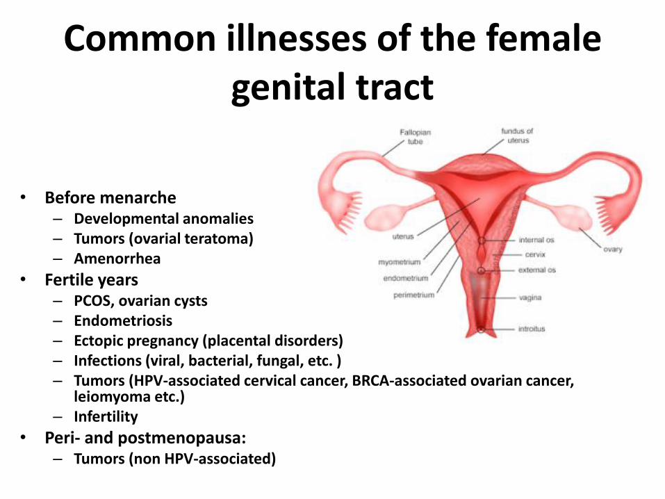
Common illnesses of the female genital tract
• Before menarche – Developmental anomalies – Tumors (ovarial teratoma) – Amenorrhea
• Fertile years – PCOS, ovarian cysts – Endometriosis – Ectopic pregnancy (placental disorders) – Infections (viral, bacterial, fungal, etc. ) – Tumors (HPV-associated cervical cancer, BRCA-associated ovarian cancer,
leiomyoma etc.) – Infertility
• Peri- and postmenopausa: – Tumors (non HPV-associated)
Common symptoms
- Menstrual disorders: stronger, painful or irregular
- Postmenopausal bleeding (endometrium cc!)
- Dyspareunia
- Lower abdominal pain (younger patients endometriosis!)
- Ascites (ovarian cancer)
- Change of discharge (infections)
- Effects of hormone secreting ovarian tumors (estrogen, testosterone)
Tumors: vulva and vagina
- Precancerous lesions - Vulva
- HPV: VIN I-III, LSIL/HSIL - lichen sclerosus (leukoplakia – white plaques)
- Vagina - HPV: VAIN I-III, LSIL/HSIL
- Vulva and vagina carcinoma: 90% squamous cell
carcinoma - Vagina: sarcoma botryoides= embryonal
rhabdomyosarcoma
Tumors: cervix
• Precancerous lesion: LSIL/HSIL (HPV!)
• Invasive cervical carcinoma: mostly squamous cell carcinoma
• Prevention: Vaccine for HPV and regular cervical carcinoma screening
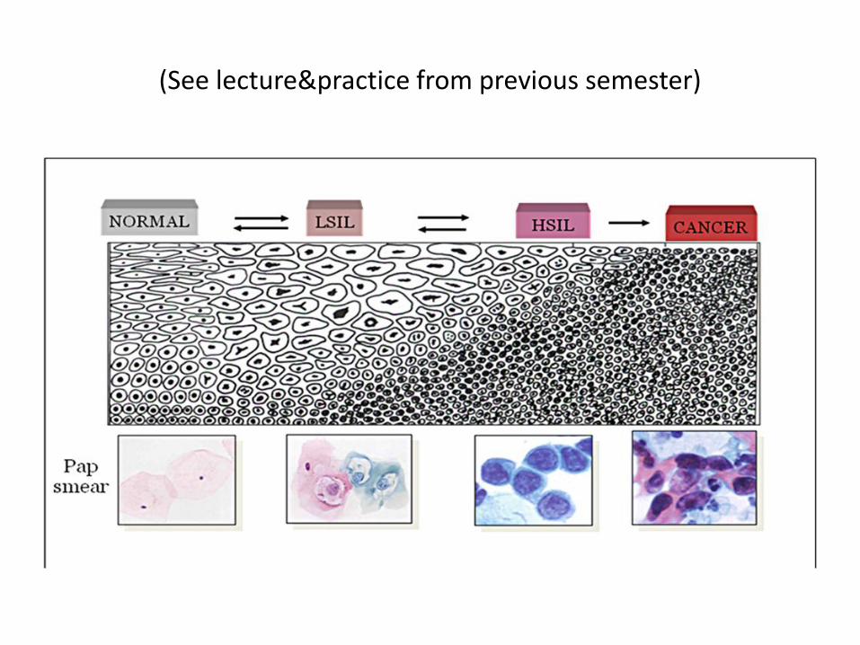
(See lecture&practice from previous semester)
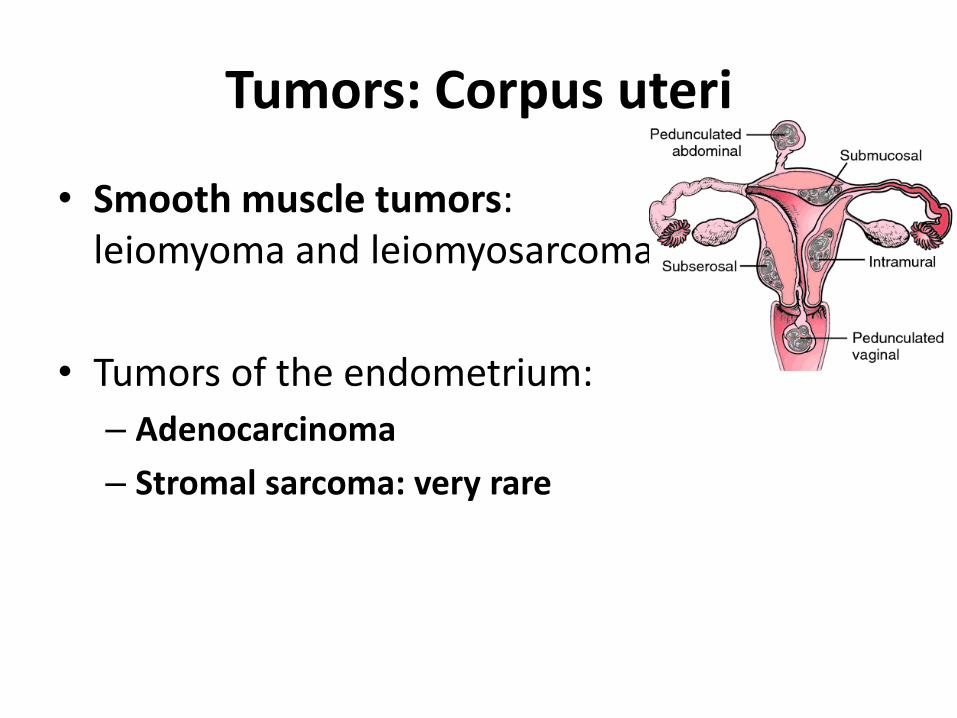
Tumors: Corpus uteri
• Smooth muscle tumors: leiomyoma and leiomyosarcoma
• Tumors of the endometrium:
– Adenocarcinoma
– Stromal sarcoma: very rare
Tumors: ovaries
• Epithelial tumors:
– Serous
– Mucinous (can contain endocervical, intestinal and endometrial epithelium) >>> pseudomyxoma peritonei
– Endometrioid tumors
– Brenner tumor
– Benign, borderline and malignant forms!
Tumors: ovaries
• Germ cell tumors – Teratomas (benign mature, malignant immature, special: struma
ovarii) – Dysgerminoma – Choriocarcinoma – Yolk sac tumor
• Sex cord- stromal tumors – derived from the sex cord of the embryonic gonad – Granulosa - theca cell tumors – Fibrothecomas – Sertoli – Leydig cell tumors
• Metastasis – Mostly bilateral – Krukenberg tumor : signet cell carcinoma of the stomach
Practice slides
• Ectopic pregnancy
• Endometriosis
• Endometrium hyperplasia
• Endometrium carcinoma
• Follicular cyst
• Ovarial tumors
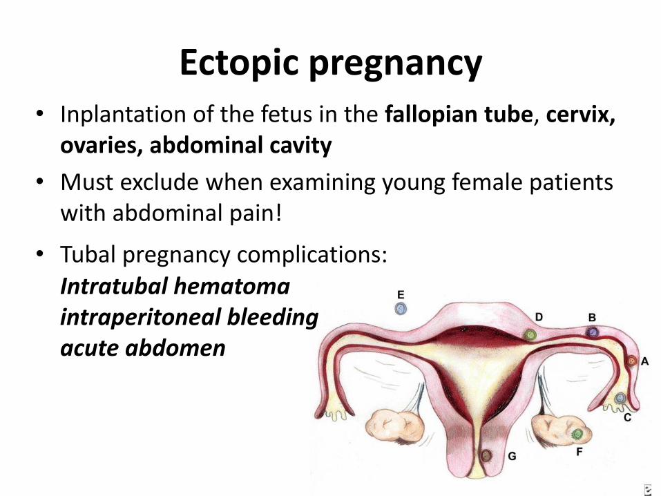
Ectopic pregnancy • Inplantation of the fetus in the fallopian tube, cervix,
ovaries, abdominal cavity
• Must exclude when examining young female patients with abdominal pain!
• Tubal pregnancy complications: Intratubal hematoma intraperitoneal bleeding acute abdomen
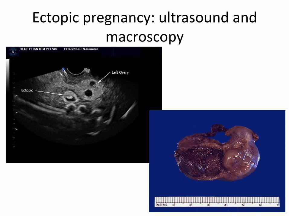
Ectopic pregnancy: ultrasound and macroscopy
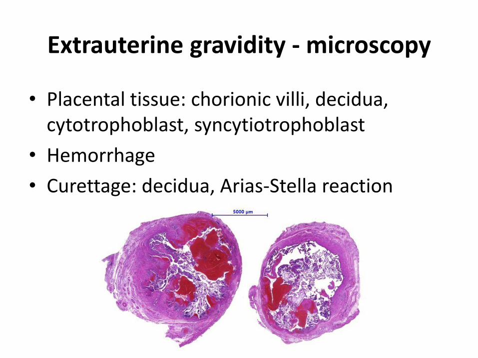
Extrauterine gravidity - microscopy
• Placental tissue: chorionic villi, decidua, cytotrophoblast, syncytiotrophoblast
• Hemorrhage
• Curettage: decidua, Arias-Stella reaction
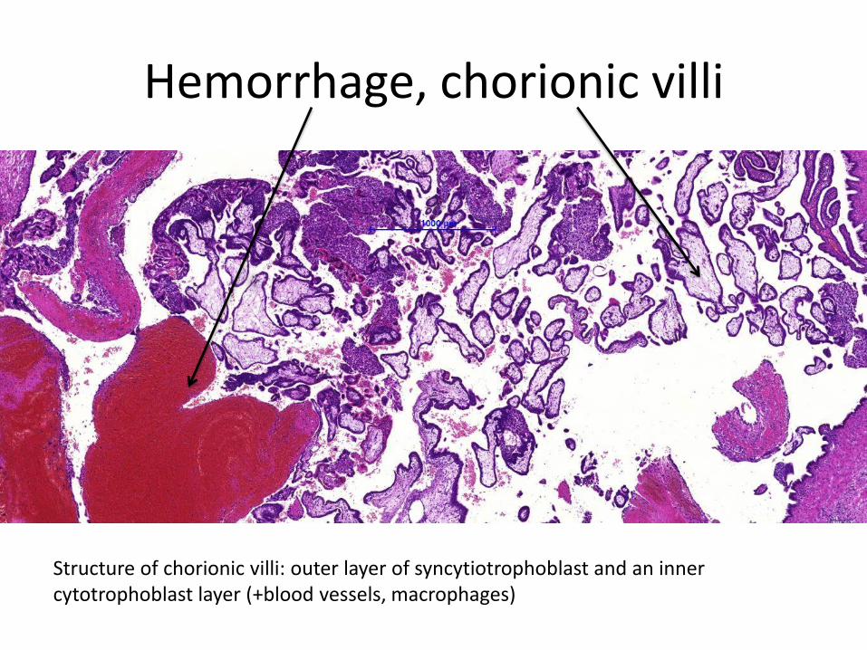
Hemorrhage, chorionic villi
Structure of chorionic villi: outer layer of syncytiotrophoblast and an inner cytotrophoblast layer (+blood vessels, macrophages)
Endometriosis
• Common illness of young women • Presence of functioning endometrial tissue in an
atypical localization • Difficult to treat • Common cause of infertility • Development:
- Retrograde menstruation through the fallopian tubes, with subsequent implantation of endometrial tissue in the peritoneum (regurgitation theory) - Hematogenous spread of endometrial tissue during menstruation (vascular invasion theory) - endometrium arises directly from coelomic epithelium (metaplastic theory)
Endometriosis
• Symptoms: lower abdominal pain, that increases with menstrual cycle pain
• Localization: – Uterus (deeper layers): adenomyosis – Fallopian tube: infertility – Ovaries: chocolate cyst (colour due to hemosiderin from
previous bleedings) – Peritoneum: adhesion, pain – Cesarian section scar – Inguinal canal – DIE (deep infiltrating endometriosis): rectum (hematochezia),
bladder (macrohaematuria), vaginal wall, sacroiliac ligaments – Extra pelvical organs (rare)
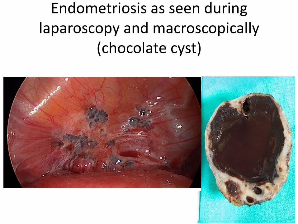
Endometriosis as seen during laparoscopy and macroscopically
(chocolate cyst)
Endometriosis - microscopy
• Endometrial epithelium
• Endometrial stroma
• Haemosiderin (macrophages)
Barna pigment: hemosziderin!
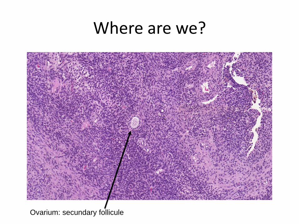
Where are we?
Ovarium: secundary follicule

Haemosiderin Endometrium stroma and
epithelium
Endometrium hyperplasia
- Simplex hyperplasia (without atypia) - Complex hyperplasia (without atypia) - Hyperplasia with atypia = EIN: endometrial
intraepithelial neoplasma
• Cause: Prolonged estrogen stimulation (anovulation, PCOS, estrogen secreting ovarian tumor, hormone containing medication, obesity)
• Symptoms: irregular bleeding • Endometrium carcinoma risk increases with the
severity of the atypia
Simplex hyperplasia of the endometrium
• Macroscopy: Endometrium is thickened or polyp formation
• Microscopy: Gland/stroma ratio increased, simplex (=round/oval) often cystic glands (not confluent)
– Without cellular atypia!
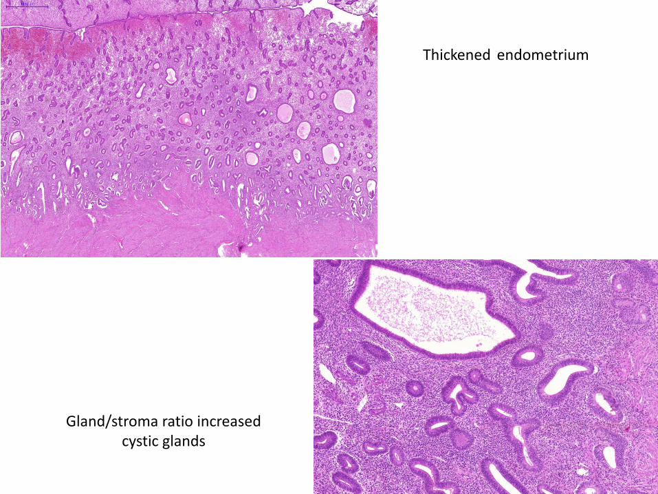
Gland/stroma ratio increased cystic glands
Thickened endometrium
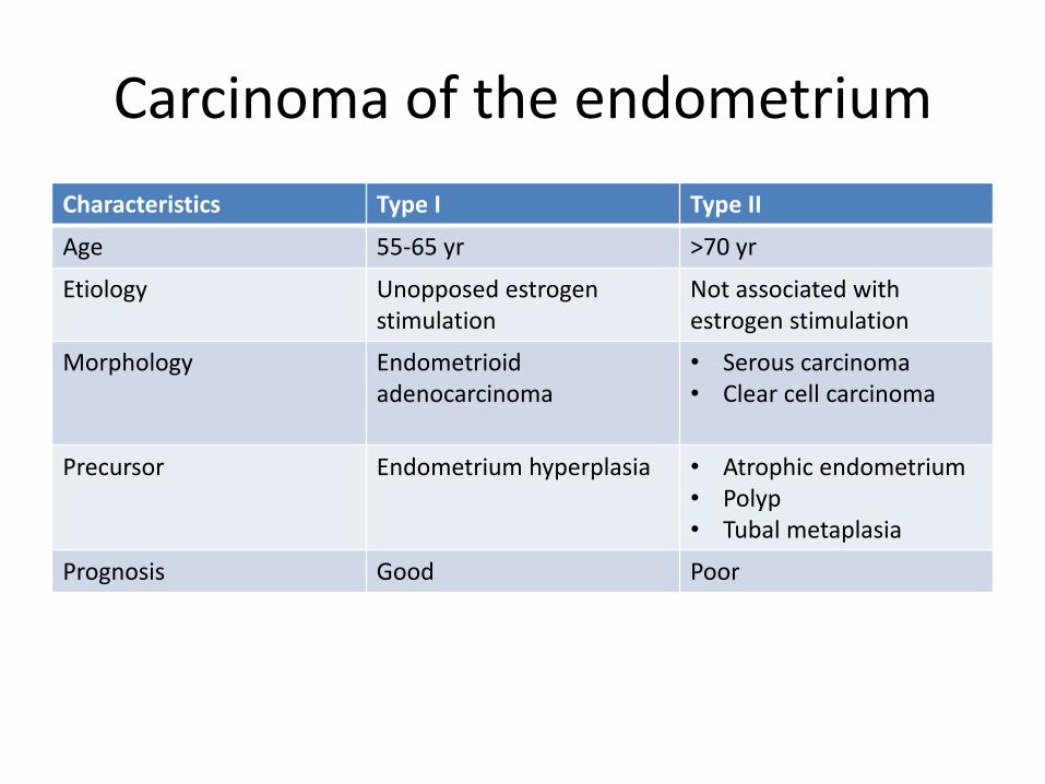
Carcinoma of the endometrium
Characteristics Type I Type II
Age 55-65 yr >70 yr
Etiology Unopposed estrogen stimulation
Not associated with estrogen stimulation
Morphology Endometrioid adenocarcinoma
• Serous carcinoma • Clear cell carcinoma
Precursor Endometrium hyperplasia • Atrophic endometrium • Polyp • Tubal metaplasia
Prognosis Good Poor
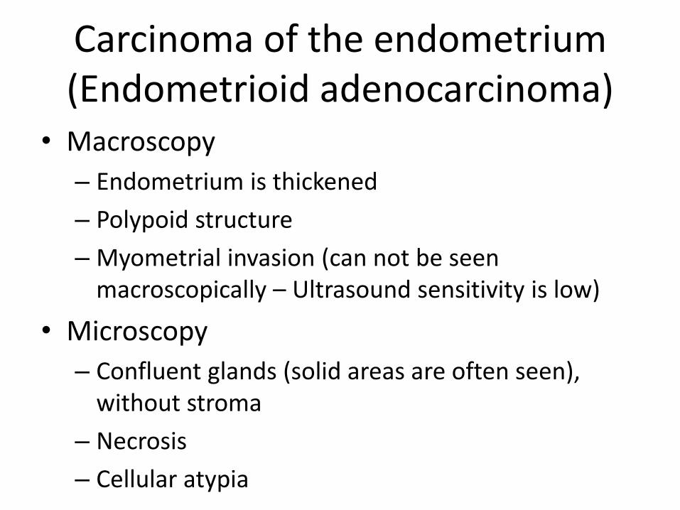
Carcinoma of the endometrium (Endometrioid adenocarcinoma)
• Macroscopy
– Endometrium is thickened
– Polypoid structure
– Myometrial invasion (can not be seen macroscopically – Ultrasound sensitivity is low)
• Microscopy
– Confluent glands (solid areas are often seen), without stroma
– Necrosis
– Cellular atypia
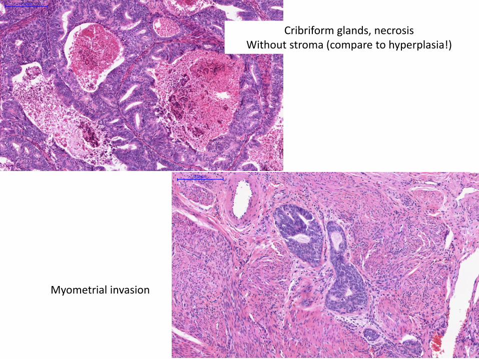
Myometrial invasion
Cribriform glands, necrosis Without stroma (compare to hyperplasia!)
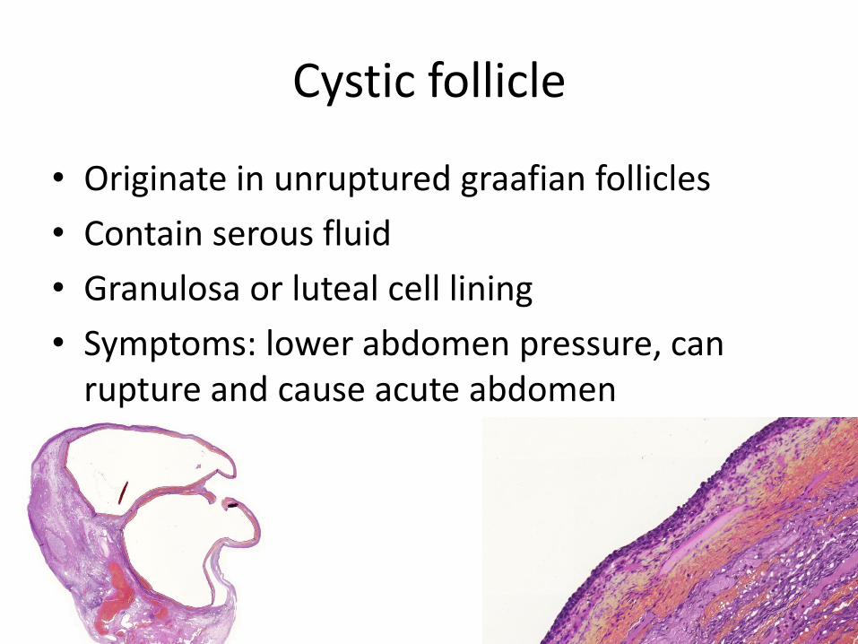
Cystic follicle
• Originate in unruptured graafian follicles
• Contain serous fluid
• Granulosa or luteal cell lining
• Symptoms: lower abdomen pressure, can rupture and cause acute abdomen

Cystadenoma mucinosum
• Benign ovarial tumor • Can be large, multilocular structure (solid areas
are suspicious for malignancy) • Microscopy
– Benign: Cystic wall is thin, lined with a single layer of columnar epithelium, without atypia
– Malignant: complex papillary proliferation, cellular atypia, invasion (peritoneal spread: pseudomyxoma peritonei)
– Borderline: structural and cellular atypia, without invasion!
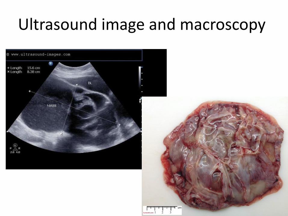
Ultrasound image and macroscopy
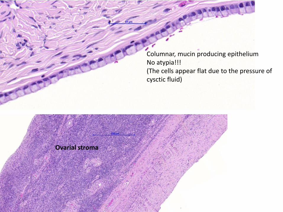
Columnar, mucin producing epithelium No atypia!!! (The cells appear flat due to the pressure of cysctic fluid)
Ovarial stroma
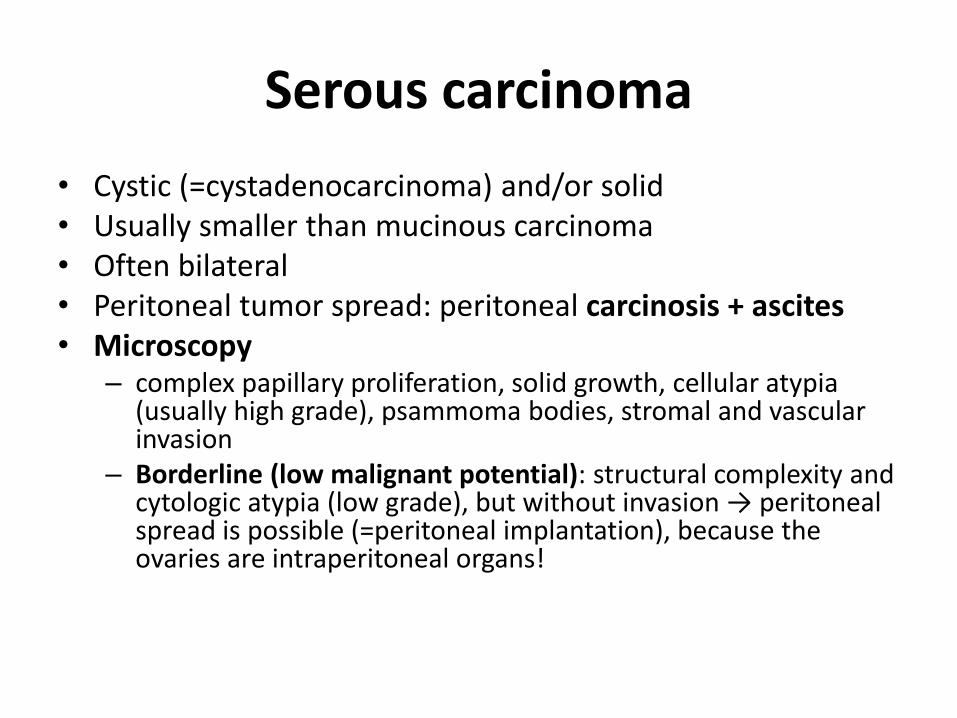
Serous carcinoma
• Cystic (=cystadenocarcinoma) and/or solid • Usually smaller than mucinous carcinoma • Often bilateral • Peritoneal tumor spread: peritoneal carcinosis + ascites • Microscopy
– complex papillary proliferation, solid growth, cellular atypia (usually high grade), psammoma bodies, stromal and vascular invasion
– Borderline (low malignant potential): structural complexity and cytologic atypia (low grade), but without invasion → peritoneal spread is possible (=peritoneal implantation), because the ovaries are intraperitoneal organs!
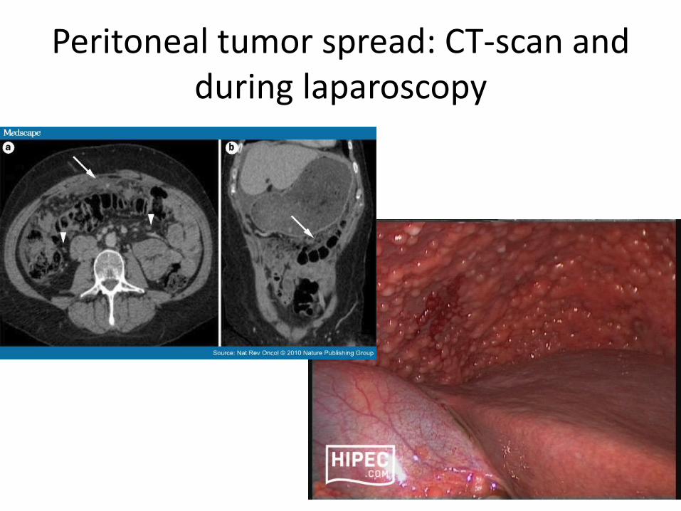
Peritoneal tumor spread: CT-scan and during laparoscopy
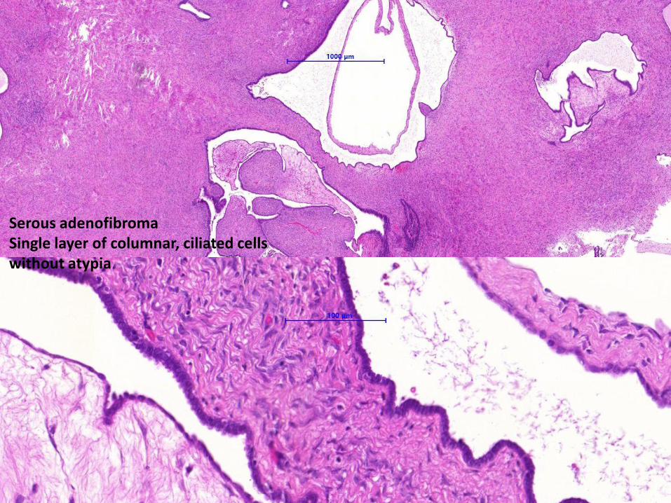
Serous adenofibroma Single layer of columnar, ciliated cells without atypia
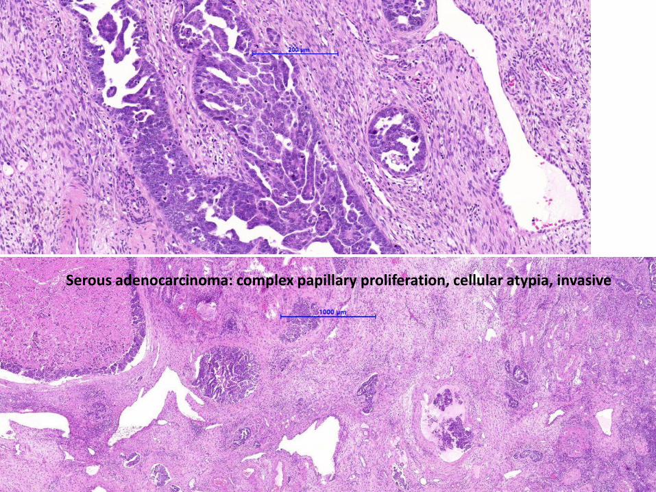
Serous adenocarcinoma: complex papillary proliferation, cellular atypia, invasive
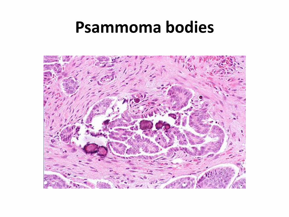
Psammoma bodies