Embed Size (px)
Citation preview
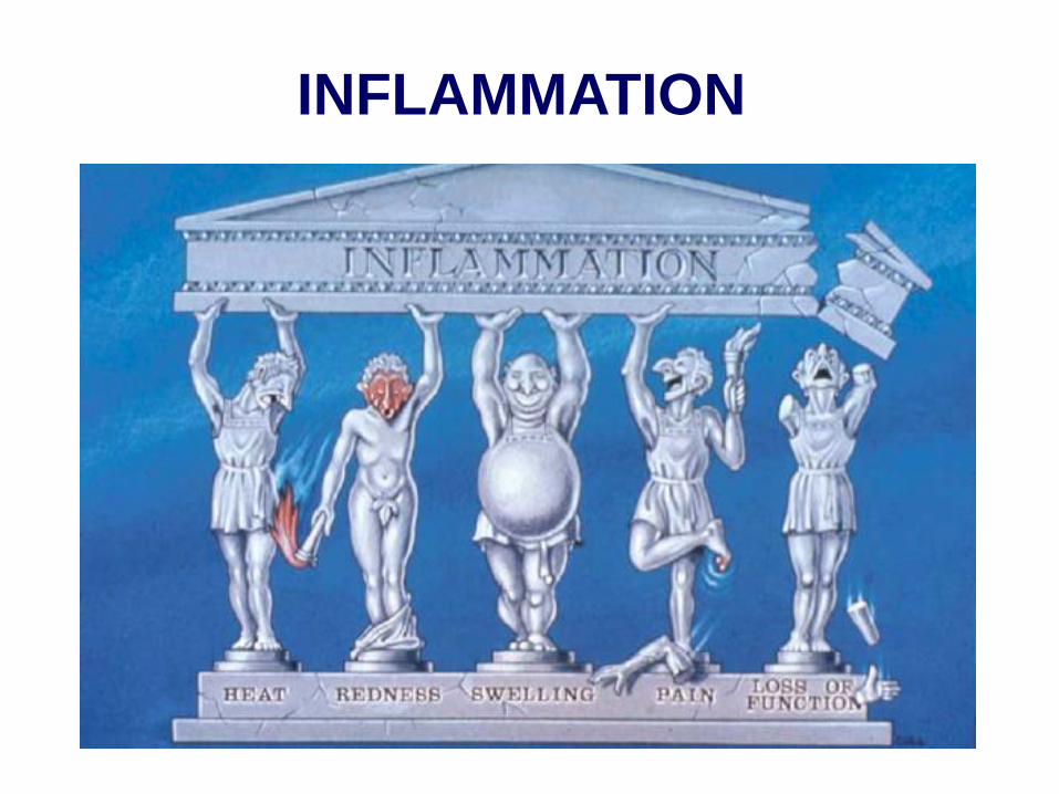
INFLAMMATION
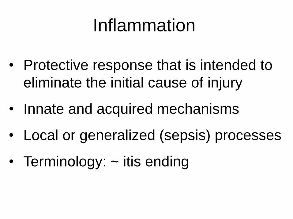
• Protective response that is intended to
eliminate the initial cause of injury
• Innate and acquired mechanisms
• Local or generalized (sepsis) processes
• Terminology: ~ itis ending
Inflammation
Etiology
• Physical effects:
• Extreme temperature, electric shock,
ionization, physical injury, etc.
• Chemical agents:
• Metabolic substances, acids, alkalis drugs,
tissue necrosis
• Microorganisms:
• Bacteria, viruses, fungal infections,
parasites, immune cells and
immunocomplexes
Types of inflammation
• acute
– Duration: few minutes to few days
– Exsudate rich in fluid and plasma proteins and mainly
neutrophil granulocytes
• subacute
– Transition between acute and chronic inflammation
– Mixed cellular infiltration
• chronic
– Duration: few days to years
– Lymphocytes, macrophages, vascular proliferation
and fibrosis
Signs and symptoms
• Rubor (redness): arteriole dilatation + venous
outflow impairment dilation of capillary loops, stasis
• Calor (warm)
• Dolor (painful): vasodilatative and permeability
increasing substances
• Tumor: increased fluid outflow
- exsudate (inflammation, protein > 3 mg/ml)
- transsudate (stasis, increased hydrostatic
pressure)
• Functio laesa
Outcome of acute inflammation
• Resolution – regeneration, reparation
• Secondary infections
• Hematogenous spread - sepsis
• Development of chronic inflammation
• Scarring - fibrosis
Morphologic patterns of acute inflammation
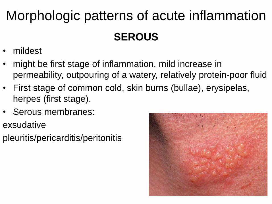
SEROUS
• mildest
• might be first stage of inflammation, mild increase in
permeability, outpouring of a watery, relatively protein-poor fluid
• First stage of common cold, skin burns (bullae), erysipelas,
herpes (first stage).
• Serous membranes:
exsudative
pleuritis/pericarditis/peritonitis
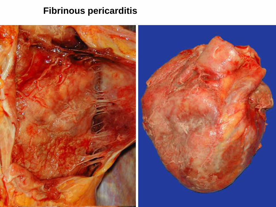
• greater vascular permeability that allows large molecules
(such as fibrinogen) to pass the endothelial barrier.
extravascular fibrin
• Greyish, sticky exsudate, which is removable
• Pericarditis sicca: friction rub; uremia (cor villosum/’bread
and butter’)
• Fibrinous pleuritis: painful, above lung infarction, diffusely as
a complication of pneumonia
• Fibrinous peritonitis
FIBRINOUS
Fibrinous pericarditis
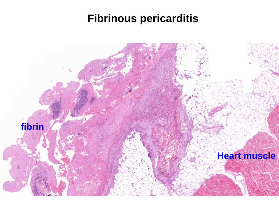
Fibrinous pericarditis
Heart muscle
fibrin
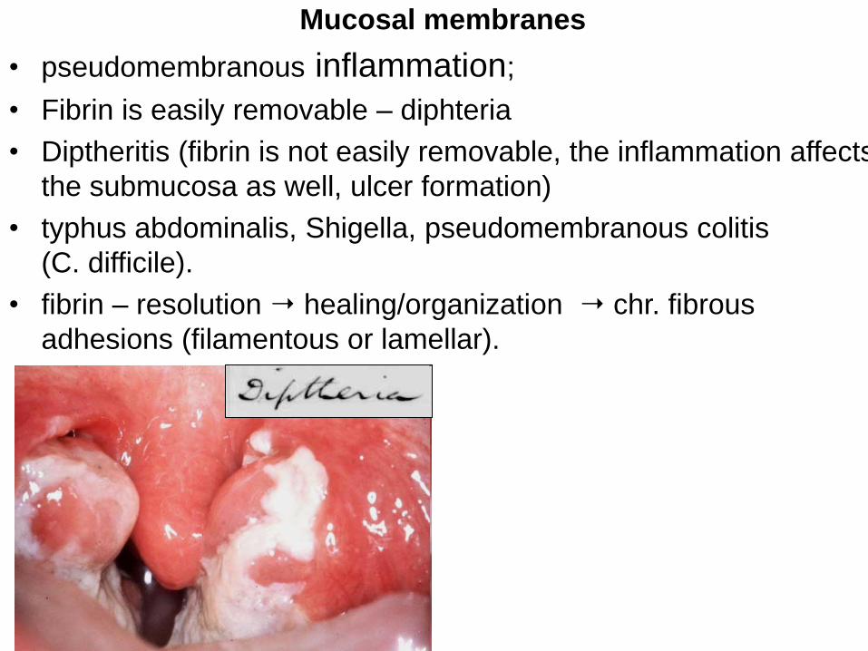
Mucosal membranes
• pseudomembranous inflammation;
• Fibrin is easily removable – diphteria
• Diptheritis (fibrin is not easily removable, the inflammation affects
the submucosa as well, ulcer formation)
• typhus abdominalis, Shigella, pseudomembranous colitis
(C. difficile).
• fibrin – resolution healing/organization chr. fibrous
adhesions (filamentous or lamellar).
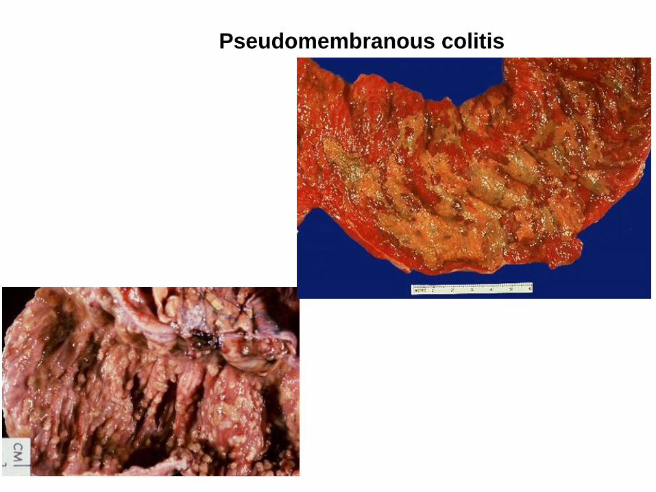
Pseudomembranous colitis
• Pus: neutrophyl granulocytes + necrotic cells and cell debris
• Folliculitis, furuncule, carbuncule, pustule (blisters filled with
pus in the epidermis, purulent meningitis
• Abscess: Localised form of acute purulent inflammation
forming a pus filled cavity
• Empyema: Pus in a preformed cavity (pyocephalus,
pyometra, pyosalpinx, pyonephros…)
• Phlegmon: Diffuse form of acute purulent inflammation,
spreading within the tissues
PURULENT
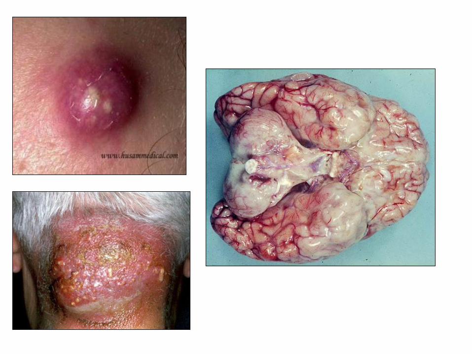
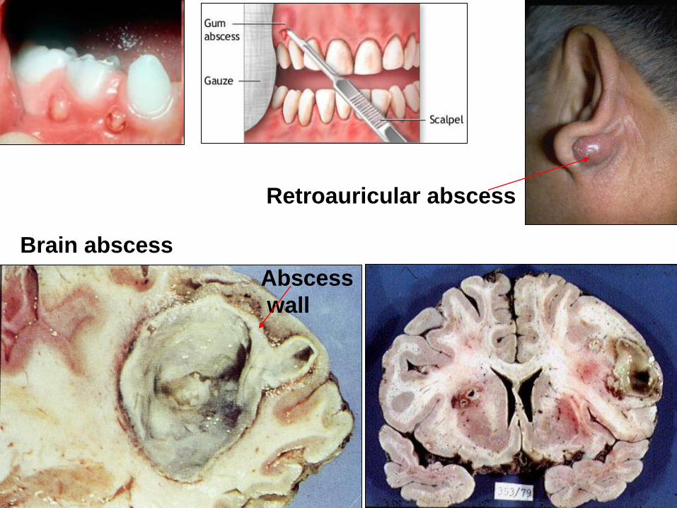
Brain abscess
Abscess
wall
Retroauricular abscess
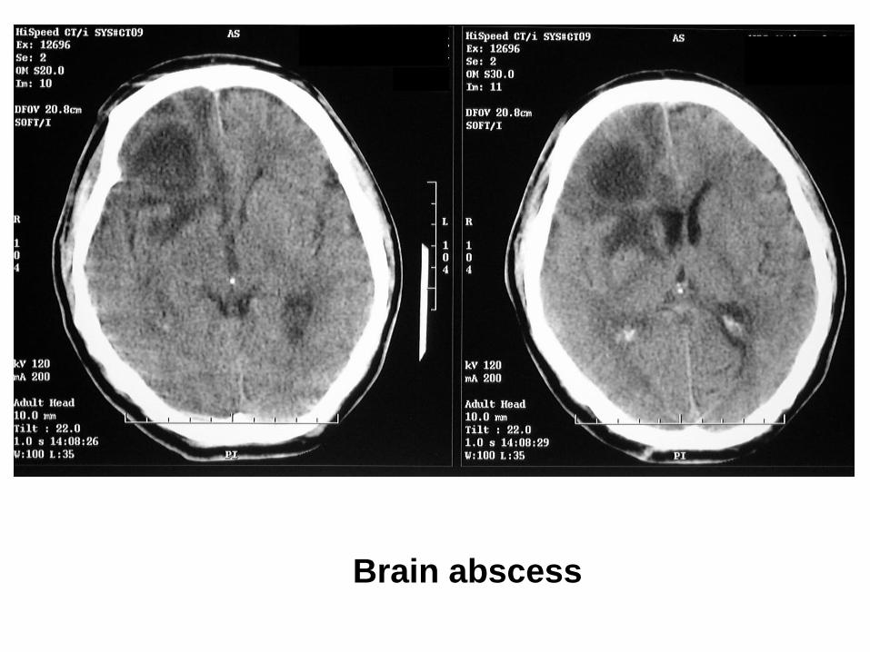
Brain abscess
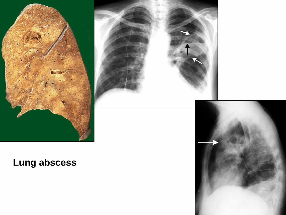
Lung abscess
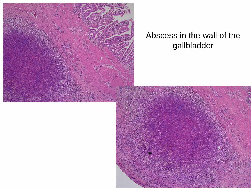
Abscess in the wall of the
gallbladder
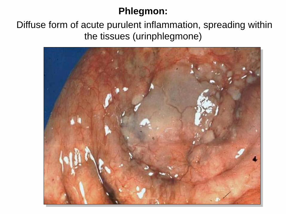
Phlegmon:
Diffuse form of acute purulent inflammation, spreading within
the tissues (urinphlegmone)
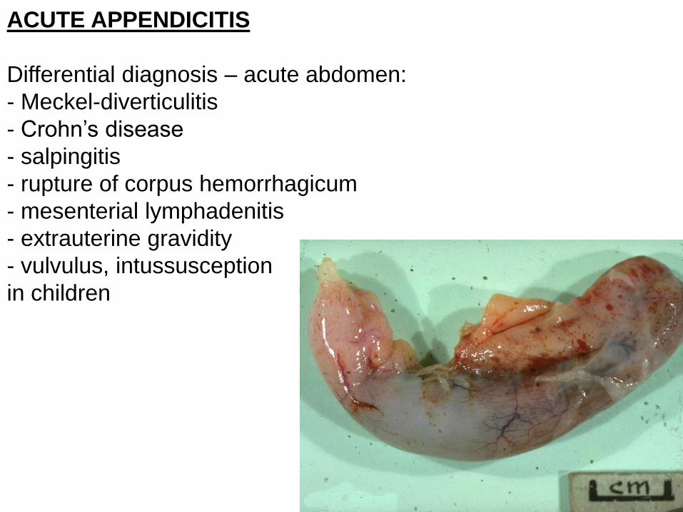
ACUTE APPENDICITIS
Differential diagnosis – acute abdomen:
- Meckel-diverticulitis
- Crohn’s disease
- salpingitis
- rupture of corpus hemorrhagicum
- mesenterial lymphadenitis
- extrauterine gravidity
- vulvulus, intussusception
in children
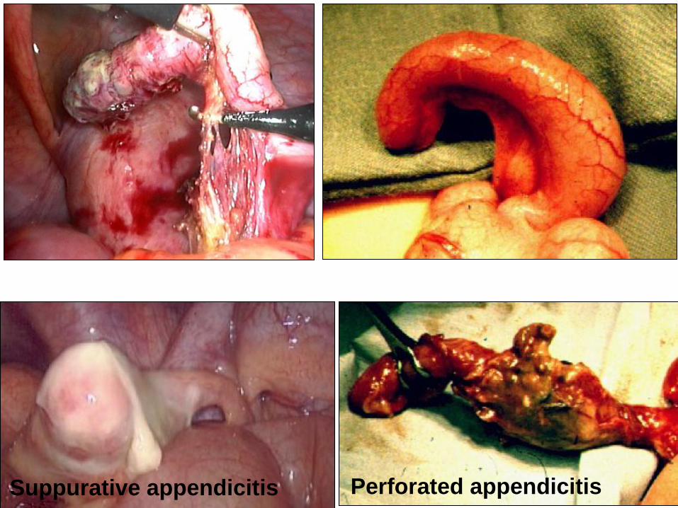
Perforated appendicitis Suppurative appendicitis
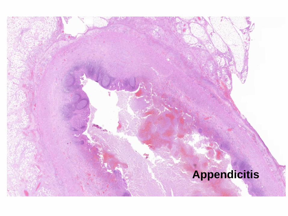
Appendicitis
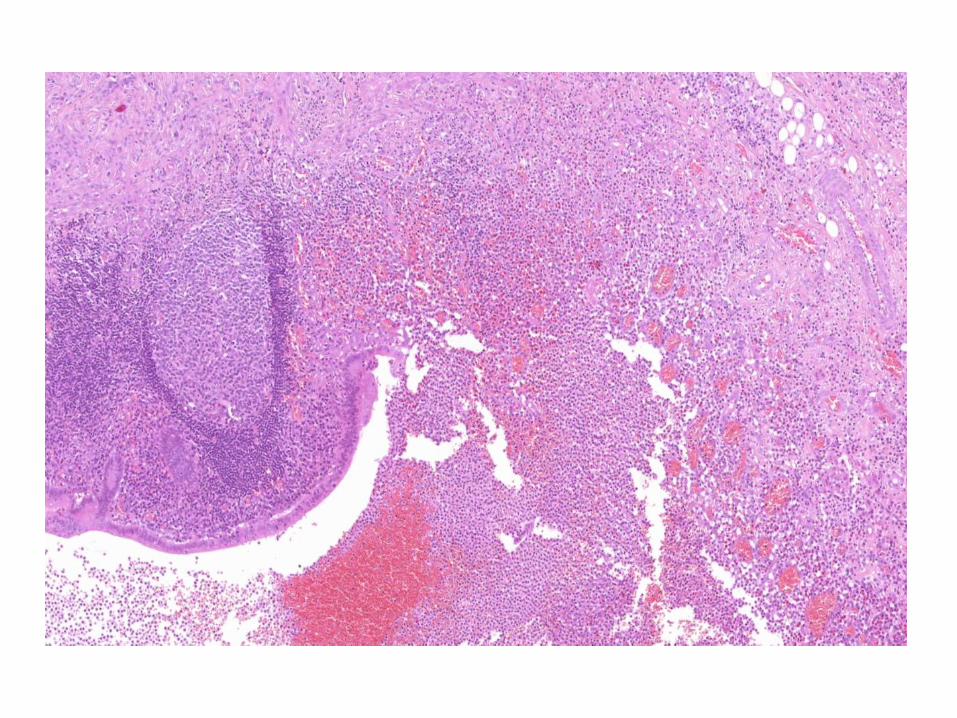
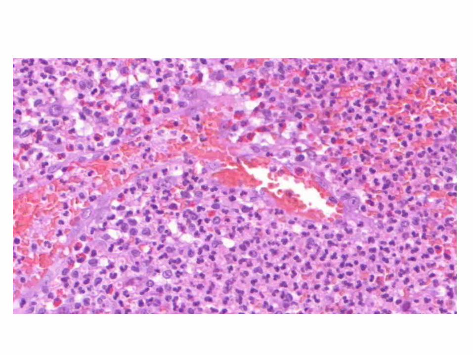
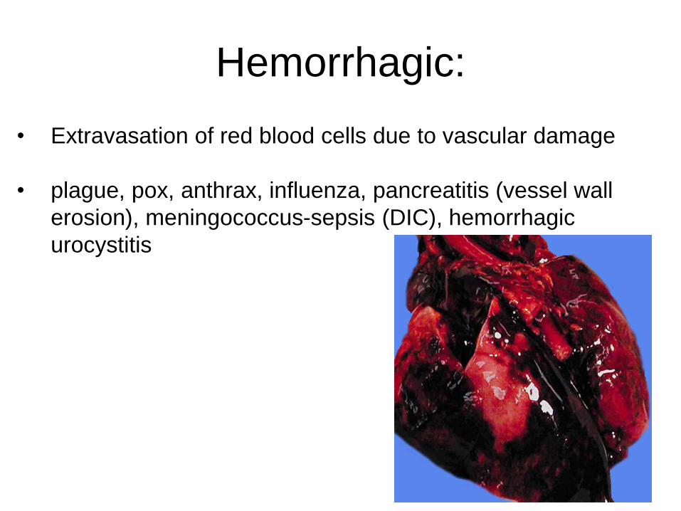
Hemorrhagic:
• Extravasation of red blood cells due to vascular damage
• plague, pox, anthrax, influenza, pancreatitis (vessel wall
erosion), meningococcus-sepsis (DIC), hemorrhagic
urocystitis
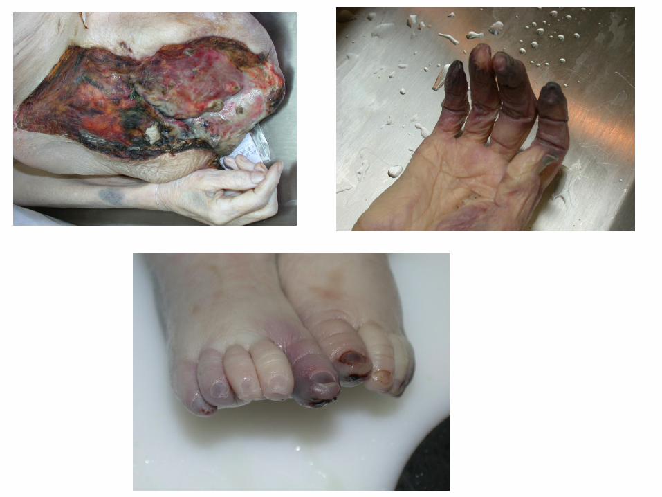
Gangrenous:
• Immune system is not effective/local hemodynamic
disorder
• Extensive tissue damage
• arteriosclerosis, diabetes (arterial occlusion): dry
gangrene superimposed bacterial infection wet
gangrene
• in the lungs after aspiration
• as a complication of tumors
• In healthy individuals: gangrenous appendicitis,
gangrenous cholecystitis
• gasgangrene: emphysematous gangrene caused by
C. perfringens
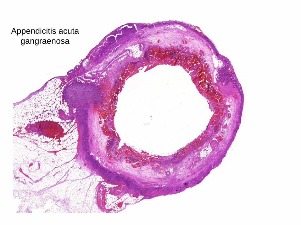
Appendicitis acuta
gangraenosa
Chronic inflammation
• Mononuclear cell infiltration (lymphocyte,
macrophage, plasmacell)
• Tissue damage
• Reparation – angiogenesis and fibrosis
are seen together
Causes:
• Persistant infection (TB, syphilis)
• Hypersensitivity reactions
• Continuous exposure to toxic agents
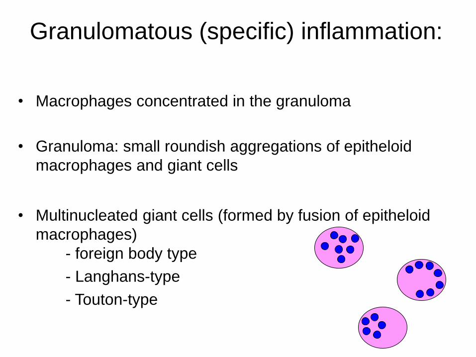
Granulomatous (specific) inflammation:
• Macrophages concentrated in the granuloma
• Granuloma: small roundish aggregations of epitheloid
macrophages and giant cells
• Multinucleated giant cells (formed by fusion of epitheloid
macrophages)
- foreign body type
- Langhans-type
- Touton-type
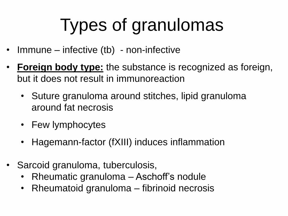
• Immune – infective (tb) - non-infective
• Foreign body type: the substance is recognized as foreign,
but it does not result in immunoreaction
• Suture granuloma around stitches, lipid granuloma
around fat necrosis
• Few lymphocytes
• Hagemann-factor (fXIII) induces inflammation
• Sarcoid granuloma, tuberculosis,
• Rheumatic granuloma – Aschoff’s nodule
• Rheumatoid granuloma – fibrinoid necrosis
Types of granulomas
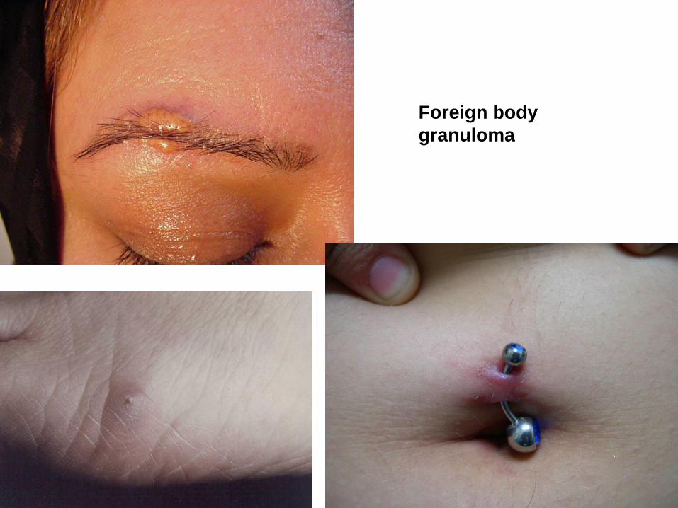
Foreign body
granuloma
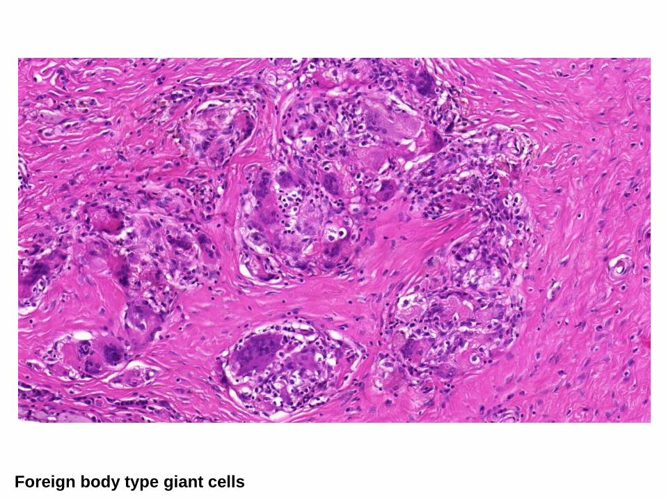
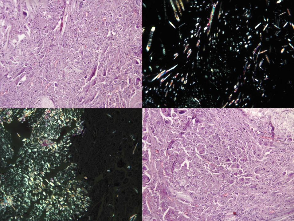
Foreign body type giant cells
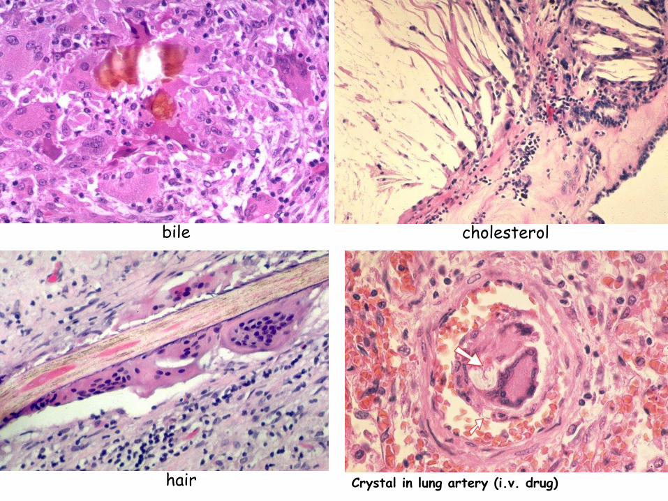
bile cholesterol
hair Crystal in lung artery (i.v. drug)
Wound healing
• Primary
– Sterile, cut wounds, with sharp edge
• Secondary
- damaged wounds with loss of tissue, infected wounds
Phases:
-Exsudative: wound exsudate, edema
-Resorptive: complement system (6 hours),
cellular immunity (12 hours)
-Proliferative: 3rd day, granulation tissue
-Reparative: epithel migration, reepithelisation, macrophages,
collagen, scar
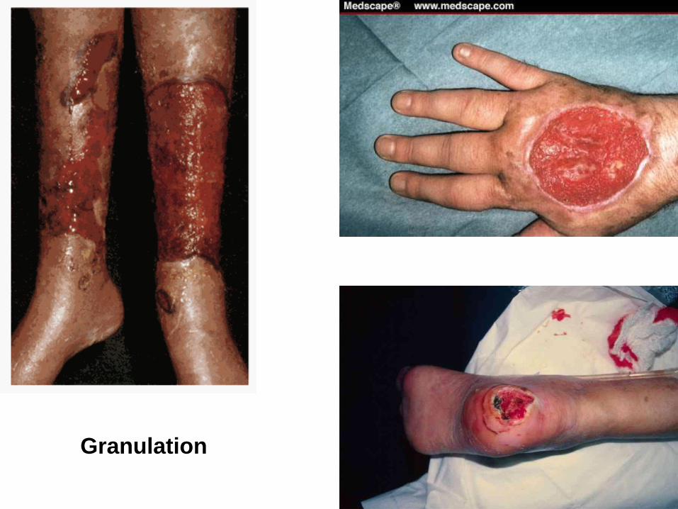
Granulation
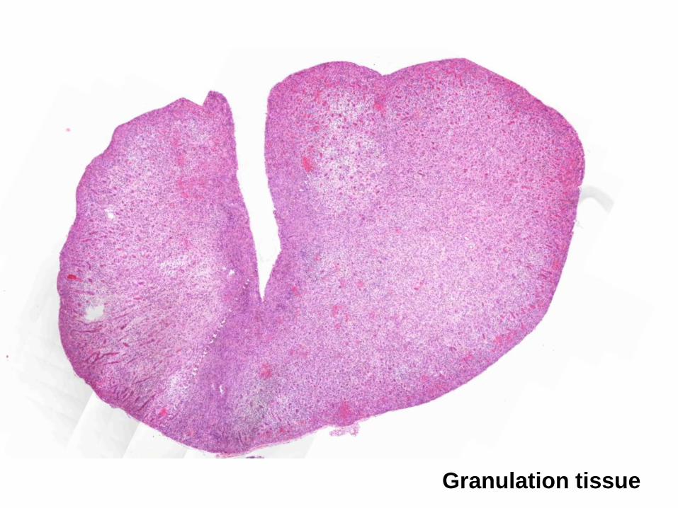
Granulation tissue
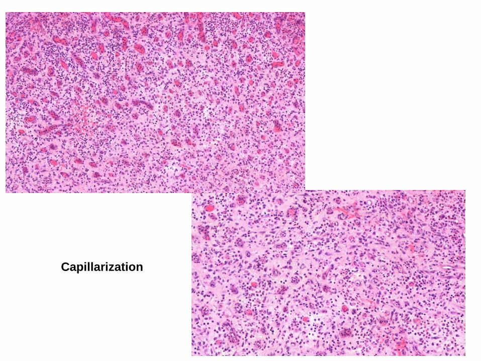
Capillarization
Complications of wound healing:
• rupture
• infection
• granuloma formation
• traumatic epithelial cyst
• seroma
• keloid: too much scar tissue
• caro luxurians: too much granulation tissue