Embed Size (px)
Citation preview
Tishk International University
Science Faculty
Medical Analysis Department
Pathology
Fourth Grade- Spring Semester 2020-2021
Dr. Jalal A. Jalal
Assistant Professor of Pathology
Neoplasia-2, Characteristics
of neoplasms
CHARACTERISTICS OF BENIGN AND MALIGNANT
NEOPLASMS
1. Differentiation and Anaplasia:
• The differentiation of parenchymal cells refers to
the extent to which they resemble their
comparable normal cells morphologically and
functionally.
• Benign neoplasms are composed of well-
differentiated cells that closely resemble their
normal counterparts.
• In benign tumors, mitoses are extremely scant in
number and are of normal configuration.
2
• Malignant neoplasms are characterized by a wide
range of parenchymal cell differentiation, from
well differentiated to completely undifferentiated.
• Between the two extremes lie tumors referred to
as moderately well differentiated.
• Malignant neoplasms that are composed of
undifferentiated cells are said to be anaplastic.
• Lack of differentiation, or anaplasia, is
considered a hallmark of malignancy.
3
Anaplastic cells display:
A. Marked pleomorphism (i.e., marked variation in
size and shape of cells).
B. Characteristically the nuclei are extremely
hyperchromatic (darkly stained) and large.
C. The nuclear-to-cytoplasmic ratio may approach 1
: 1 instead of the normal 1 : 4 or 1 : 6.
D. Giant cells may be formed and possess either
one large nucleus or several nuclei.
4
E. Anaplastic nuclei are variable and bizarre in
size and shape. The chromatin is coarse and
clumped, and nucleoli may be prominent.
F. Mitoses are often numerous and distinctly
atypical; sometimes appear as tripolar or
quadripolar forms.
G. anaplastic cells usually fail to develop
recognizable patterns of orientation to one
another (i.e., they lose normal polarity).
6
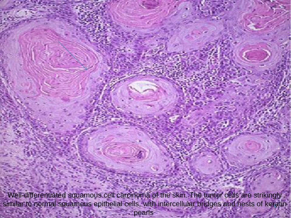
Well-differentiated squamous cell carcinoma of the skin. The tumor cells are strikingly
similar to normal squamous epithelial cells, with intercellular bridges and nests of keratin
pearls
7
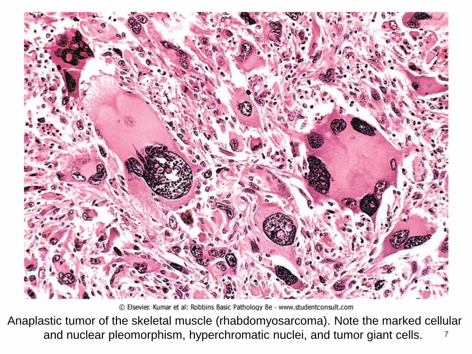
Anaplastic tumor of the skeletal muscle (rhabdomyosarcoma). Note the marked cellular
and nuclear pleomorphism, hyperchromatic nuclei, and tumor giant cells.
8
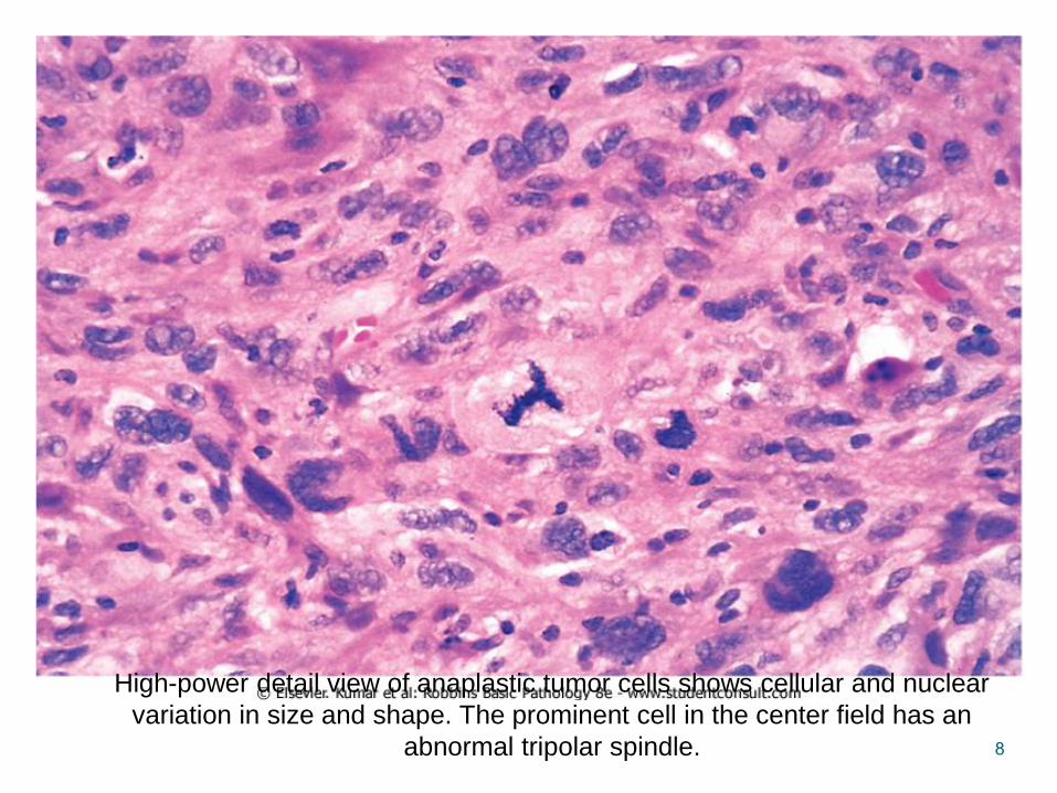
High-power detail view of anaplastic tumor cells shows cellular and nuclear
variation in size and shape. The prominent cell in the center field has an
abnormal tripolar spindle.
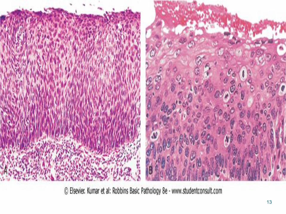
Dysplasia
• A term used to describe disorderly but non-neoplastic proliferation & its encounteredprincipally in the epithelia.
• It is a loss in the uniformity of individual cellsand in their architectural orientation.
9
• Dysplastic cells exhibit considerablepleomorphism and often possesshyperchromatic nuclei that are abnormallylarge for the size of the cell.
• Mitotic figures are more abundant than usual &frequently the mitoses appear in abnormallocations within the epithelium.
• e.g. In dysplastic stratified squamous epithelium,mitoses are not confined to the basal layers,where they normally occur, but may appear at alllevels and even in surface cells
• When dysplastic changes are marked and involvethe entire thickness of the epithelium, the lesionis referred to as carcinoma in situ, a pre-invasivestage of cancer.
11
• Dysplastic changes are often found adjacent tofoci of malignant transformation, and long-termstudies of cigarette smokers show thatepithelial dysplasia almost invariably antedatesthe appearance of cancer.
• The term dysplasia does not indicate cancer,and dysplasias do not necessarily progress tocancer.
13
2. Rate of Growth
• Most benign tumors grow slowly, and most
cancers grow much faster, eventually spreading
locally and to distant sites (metastasizing) and
causing death.
• The rate of growth of malignant tumors correlates
in general with their level of differentiation.
• In other words, rapidly growing tumors tend to be
poorly differentiated.
14
Rapidly growing malignant tumors often contain
central areas of ischemic necrosis because the
tumor blood supply, derived from the host, fails to
cope with the oxygen needs of the expanding
mass of cells.
3. Local Invasion
• A benign neoplasm remains localized at its site
of origin.
• It does not have the capacity to infiltrate, invade,
or metastasize to distant sites, like malignant
neoplasms.
• most benign neoplasms develop an enclosing
fibrous capsule that separates them from the
host tissue.
16
• Cancers grow by progressive infiltration,
invasion, destruction, and penetration of the
surrounding tissue.
• They do not develop well-defined capsules.
• Next to the development of metastases, local
invasiveness is the most reliable feature that
distinguishes malignant from benign tumors.
18
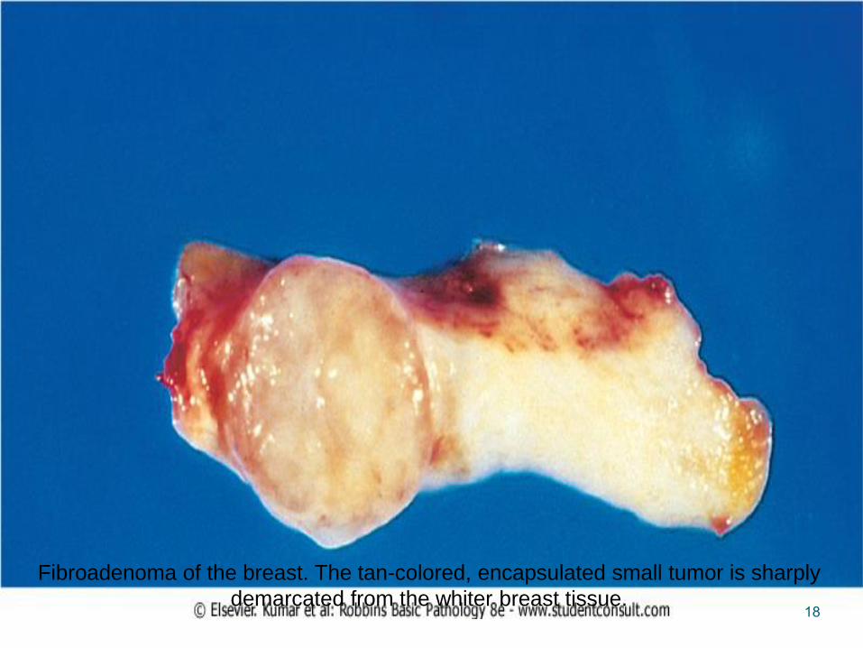
Fibroadenoma of the breast. The tan-colored, encapsulated small tumor is sharply
demarcated from the whiter breast tissue.
19
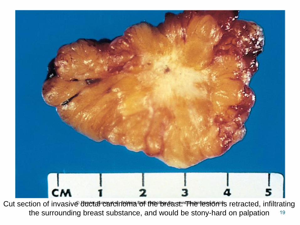
Cut section of invasive ductal carcinoma of the breast. The lesion is retracted, infiltrating
the surrounding breast substance, and would be stony-hard on palpation
4. Metastasis
• Means the development of secondary implants
discontinuous with the primary tumor, in remote
tissues.
• The properties of invasiveness and, even more
so, metastasis, definitely identify a neoplasm as
malignant.
• In general, the more anaplastic and the larger the
primary neoplasm, the more likely is metastatic
spread
20
21
Pathways of spread
Malignant neoplasms disseminate by one of three
pathways:
(1) seeding within body cavities.
(2) lymphatic spread.
(3) hematogenous spread.
22
1. Spread by seeding:
• occurs when neoplasms invade a natural body cavity.
• This mode of dissemination is particularly characteristic
of cancers of the ovary, which often cover the peritoneal
surfaces widely.
23
2. Lymphatic spread:
• is more typical of carcinomas.
• The pattern of lymph node involvement depends
principally on the site of the primary neoplasm
and the natural pathways of lymphatic drainage of
the site.
• Carcinoma of the breast usually arises in the
upper outer quadrant and first spreads to the
axillary nodes
24
3. Hematogenous spread:
• is the most feared consequence of a cancer.
• It is the favored pathway for sarcomas, but
carcinomas use it as well.
• As might be expected, arteries are penetrated
less readily than are veins.
• Since all portal area drainage flows to the liver,
and all caval blood flows to the lungs, the liver
and lungs are the most frequently involved
secondary sites in hematogenous dissemination.
25
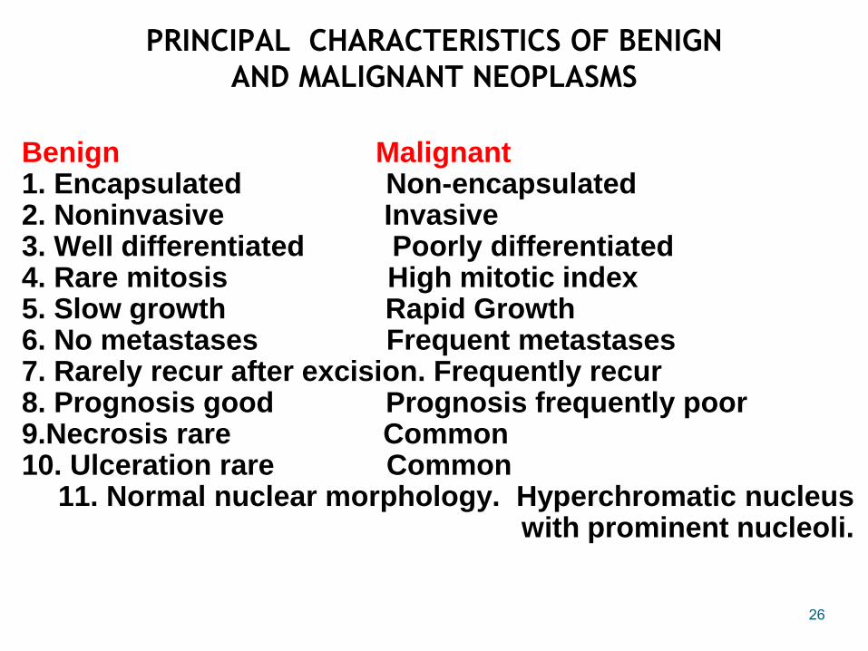
PRINCIPAL CHARACTERISTICS OF BENIGN
AND MALIGNANT NEOPLASMS
Benign Malignant1. Encapsulated Non-encapsulated2. Noninvasive Invasive3. Well differentiated Poorly differentiated4. Rare mitosis High mitotic index5. Slow growth Rapid Growth6. No metastases Frequent metastases7. Rarely recur after excision. Frequently recur8. Prognosis good Prognosis frequently poor9.Necrosis rare Common10. Ulceration rare Common
11. Normal nuclear morphology. Hyperchromatic nucleus with prominent nucleoli.
26

30