Embed Size (px)
Citation preview
General Pathology Histogenetic Classification
of Neoplasms
Neuroectodermal& „Other“ Neoplasms
Jaroslava DuškováInst. Pathol. ,1st Med. Faculty, Charles Univ. Praguehttp://www1.lf1.cuni.cz/~jdusk/
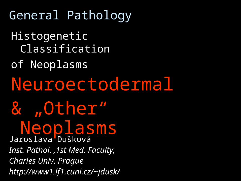
NEOPLASIA – classification
HISTOGENETIC mesenchymal epithelial neuroectodermal mixed, teratoma choriocarcinoma mesothelioma
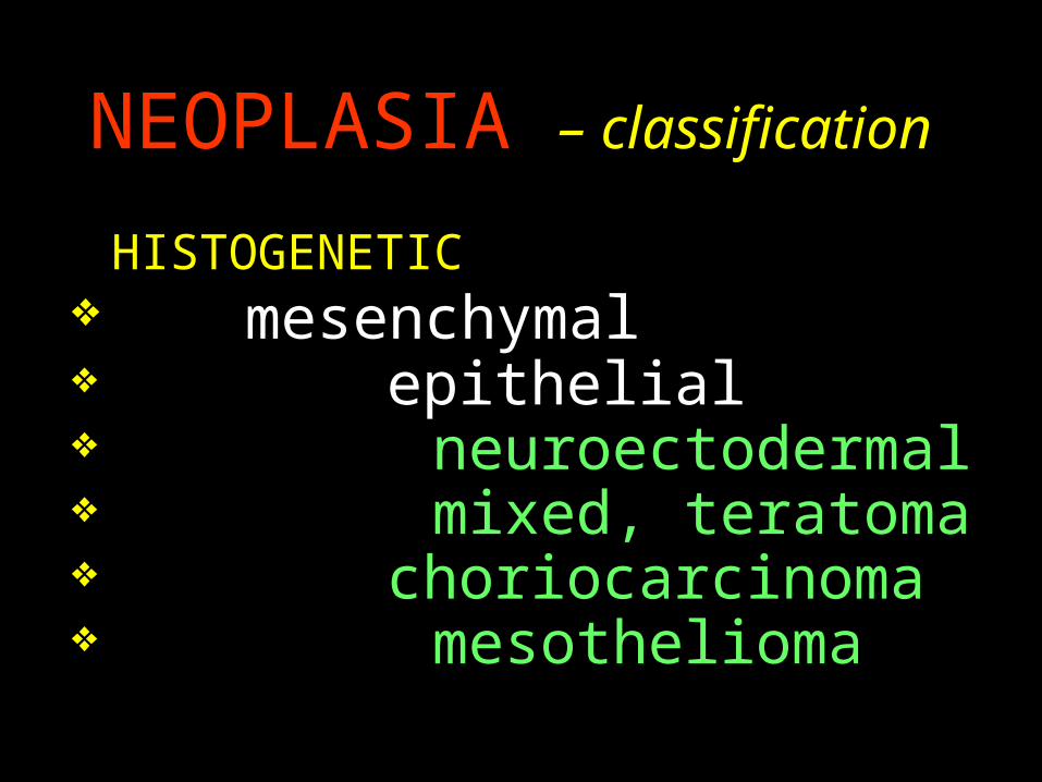
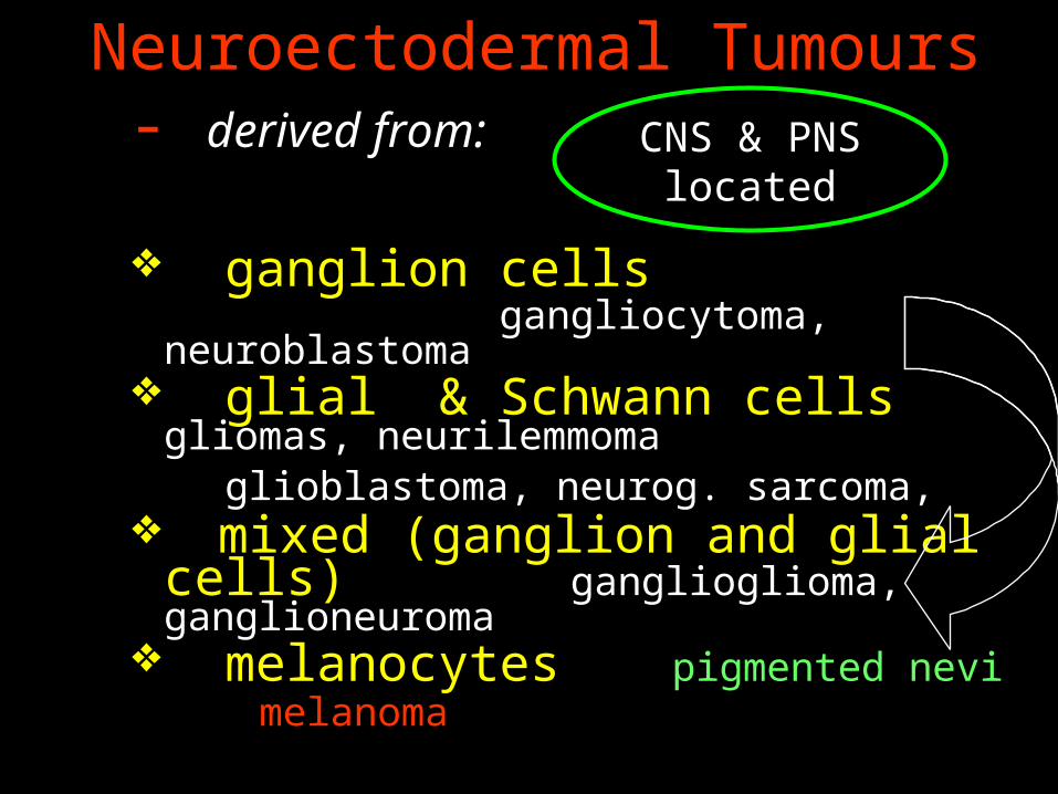
Neuroectodermal Tumours - derived from:
ganglion cells gangliocytoma, neuroblastoma
glial & Schwann cells gliomas, neurilemmoma
glioblastoma, neurog. sarcoma, mixed (ganglion and glial cells)
ganglioglioma, ganglioneuroma melanocytes
pigmented nevimelanoma
CNS & PNS located
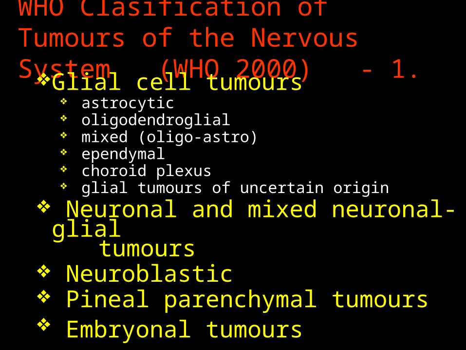
WHO Clasification of Tumours of the Nervous System (WHO 2000) - 1.
Glial cell tumours astrocytic oligodendroglial mixed (oligo-astro) ependymal choroid plexus glial tumours of uncertain origin
Neuronal and mixed neuronal-glial
tumours Neuroblastic Pineal parenchymal tumours Embryonal tumours
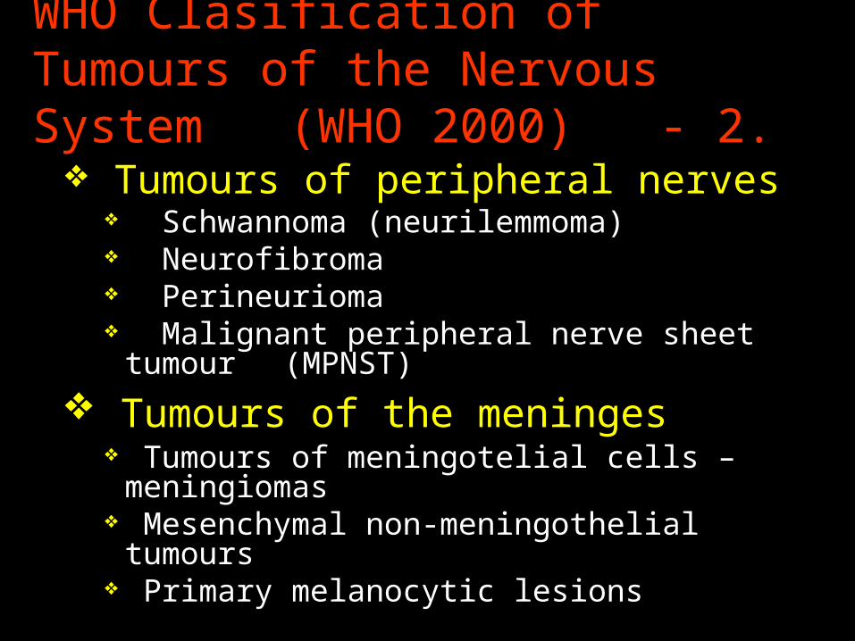
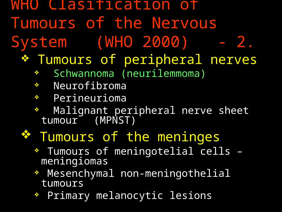
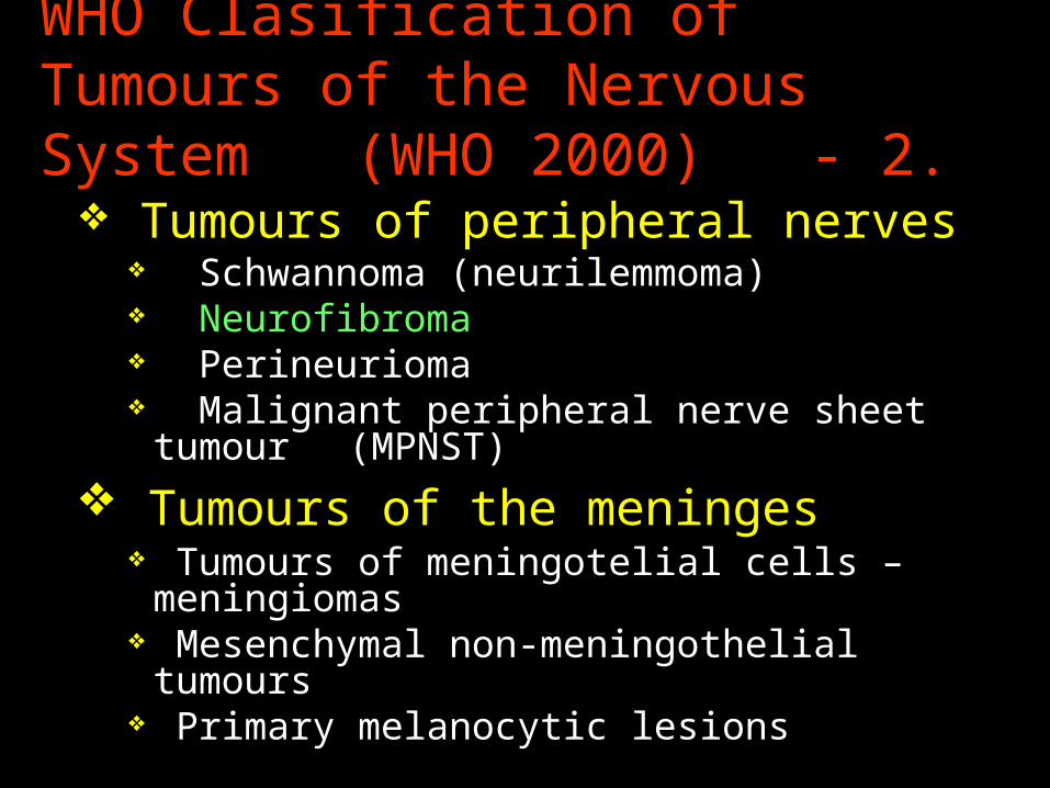
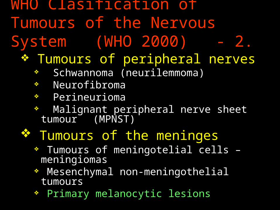
WHO Clasification of Tumours of the Nervous System (WHO 2000) - 2.
Tumours of peripheral nerves Schwannoma (neurilemmoma) Neurofibroma Perineurioma Malignant peripheral nerve sheet tumour
(MPNST)
Tumours of the meninges Tumours of meningotelial cells – meningiomas Mesenchymal non-meningothelial tumours Primary melanocytic lesions
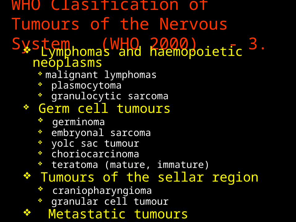
WHO Clasification of Tumours of the Nervous System (WHO 2000) - 3.
Lymphomas and haemopoietic neoplasms malignant lymphomas plasmocytoma granulocytic sarcoma
Germ cell tumours germinoma embryonal sarcoma yolc sac tumour choriocarcinoma teratoma (mature, immature)
Tumours of the sellar region craniopharyngioma granular cell tumour
Metastatic tumours
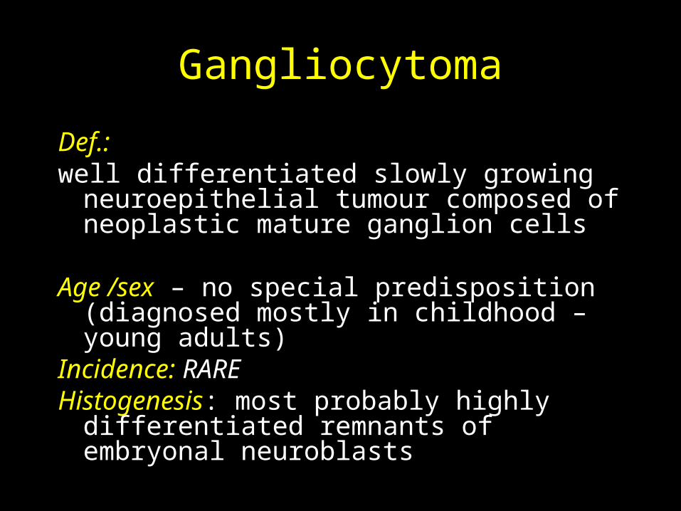
Gangliocytoma
Def.: well differentiated slowly growing
neuroepithelial tumour composed of neoplastic mature ganglion cells
Age /sex – no special predisposition (diagnosed mostly in childhood – young adults)
Incidence: RAREHistogenesis: most probably highly
differentiated remnants of embryonal neuroblasts
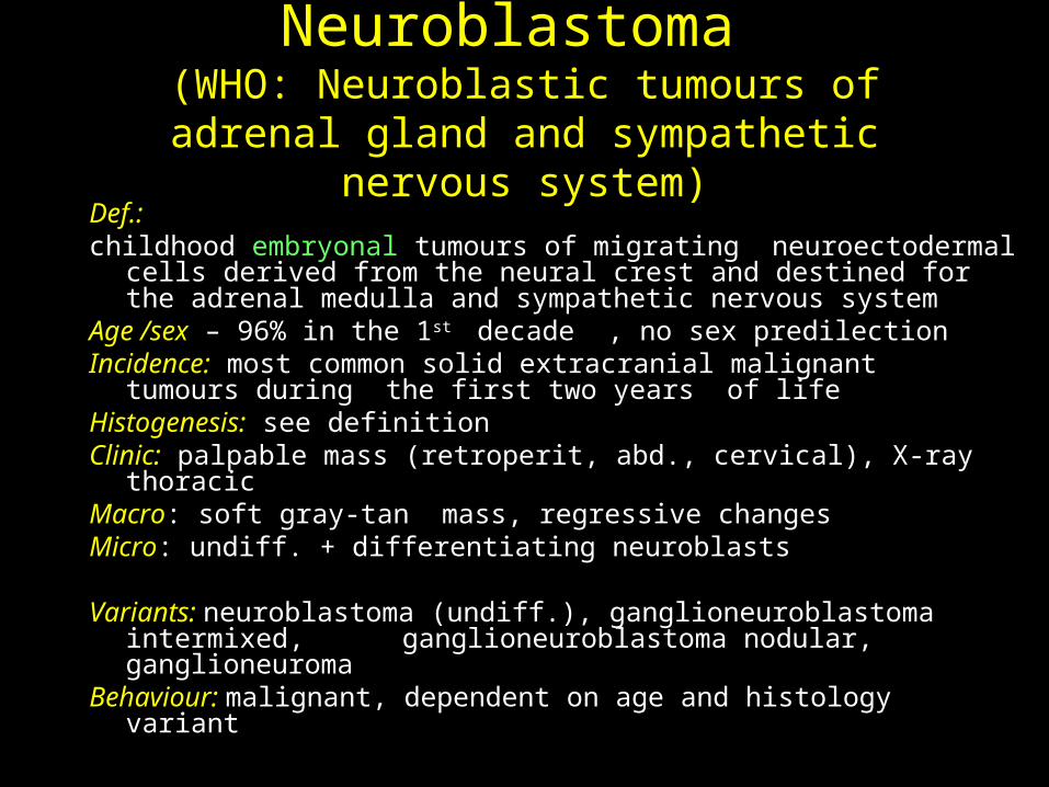
Neuroblastoma (WHO: Neuroblastic tumours of adrenal gland
and sympathetic nervous system)Def.: childhood embryonal tumours of migrating neuroectodermal cells
derived from the neural crest and destined for the adrenal medulla and sympathetic nervous system
Age /sex – 96% in the 1st decade , no sex predilectionIncidence: most common solid extracranial malignant tumours during
the first two years of lifeHistogenesis: see definitionClinic: palpable mass (retroperit, abd., cervical), X-ray thoracicMacro: soft gray-tan mass, regressive changesMicro: undiff. + differentiating neuroblasts
Variants: neuroblastoma (undiff.), ganglioneuroblastoma intermixed, ganglioneuroblastoma nodular, ganglioneuroma
Behaviour: malignant, dependent on age and histology variant

Neuroectodermal Tumours - derived from:
ganglion cells gangliocytoma, neuroblastoma
glial & Schwann cells gliomas, neurilemmoma
glioblastoma, neurog. sarcoma, mixed (ganglion and glial cells)
ganglioglioma, ganglioneuroma melanocytes
pigmented nevimelanoma
CNS & PNS located
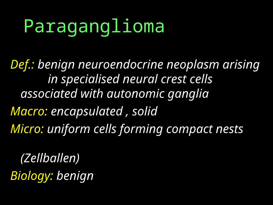
Paraganglioma
Def.: benign neuroendocrine neoplasm arising in specialised neural crest cells
associated with autonomic ganglia
Macro: encapsulated , solid
Micro: uniform cells forming compact nests
(Zellballen)
Biology: benign
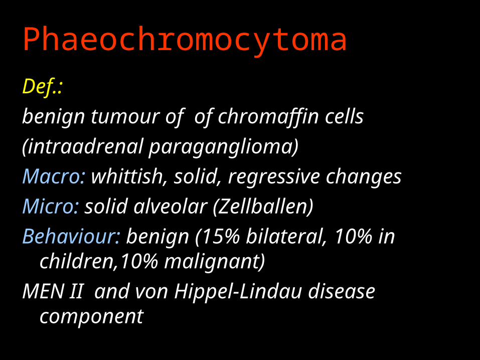
PhaeochromocytomaDef.:
benign tumour of of chromaffin cells
(intraadrenal paraganglioma)
Macro: whittish, solid, regressive changes
Micro: solid alveolar (Zellballen)
Behaviour: benign (15% bilateral, 10% in children,10% malignant)
MEN II and von Hippel-Lindau disease component
WHO Clasification of Tumours of the Nervous System (WHO 2000) - 2.
Tumours of peripheral nerves Schwannoma (neurilemmoma) Neurofibroma Perineurioma Malignant peripheral nerve sheet tumour
(MPNST)
Tumours of the meninges Tumours of meningotelial cells – meningiomas Mesenchymal non-meningothelial tumours Primary melanocytic lesions
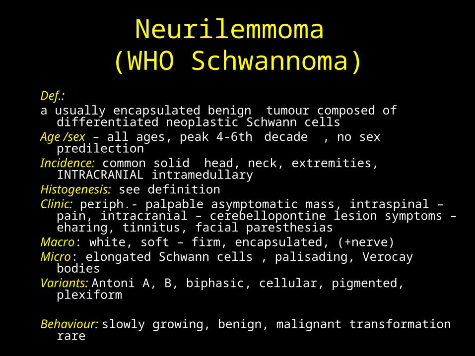
Neurilemmoma (WHO Schwannoma)
Def.: a usually encapsulated benign tumour composed of differentiated
neoplastic Schwann cellsAge /sex – all ages, peak 4-6th decade , no sex predilectionIncidence: common solid head, neck, extremities, INTRACRANIAL
intramedullaryHistogenesis: see definitionClinic: periph.- palpable asymptomatic mass, intraspinal – pain,
intracranial – cerebellopontine lesion symptoms – eharing, tinnitus, facial paresthesias
Macro: white, soft – firm, encapsulated, (+nerve)Micro: elongated Schwann cells , palisading, Verocay bodiesVariants: Antoni A, B, biphasic, cellular, pigmented, plexiform
Behaviour: slowly growing, benign, malignant transformation rare
WHO Clasification of Tumours of the Nervous System (WHO 2000) - 2.
Tumours of peripheral nerves Schwannoma (neurilemmoma) Neurofibroma Perineurioma Malignant peripheral nerve sheet tumour
(MPNST)
Tumours of the meninges Tumours of meningotelial cells – meningiomas Mesenchymal non-meningothelial tumours Primary melanocytic lesions
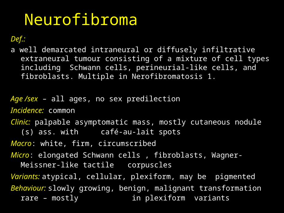
Neurofibroma Def.:
a well demarcated intraneural or diffusely infiltrative extraneural tumour consisting of a mixture of cell types including Schwann cells, perineurial-like cells, and fibroblasts. Multiple in Nerofibromatosis 1.
Age /sex – all ages, no sex predilection
Incidence: common
Clinic: palpable asymptomatic mass, mostly cutaneous nodule (s) ass. with café-au-lait spots
Macro: white, firm, circumscribed
Micro: elongated Schwann cells , fibroblasts, Wagner-Meissner-like tactile corpuscles
Variants: atypical, cellular, plexiform, may be pigmented
Behaviour: slowly growing, benign, malignant transformation rare – mostly in plexiform variants

Neuroectodermal Tumours - derived from:
ganglion cells gangliocytoma, neuroblastoma
glial & Schwann cells gliomas, neurilemmoma
glioblastoma, neurog. sarcoma, mixed (ganglion and glial cells)
ganglioglioma, ganglioneuroma melanocytes
pigmented nevimelanoma
CNS & PNS located
Neuroectodermal Tumours - derived from:
ganglion cells gangliocytoma, neuroblastoma
glial & Schwann cells gliomas, neurilemmoma
glioblastoma, neurog. sarcoma, mixed (ganglion and glial cells)
ganglioglioma, ganglioneuroma melanocytes
pigmented nevimelanoma
CNS & PNS located
WHO Clasification of Tumours of the Nervous System (WHO 2000) - 2.
Tumours of peripheral nerves Schwannoma (neurilemmoma) Neurofibroma Perineurioma Malignant peripheral nerve sheet tumour
(MPNST)
Tumours of the meninges Tumours of meningotelial cells – meningiomas Mesenchymal non-meningothelial tumours Primary melanocytic lesions
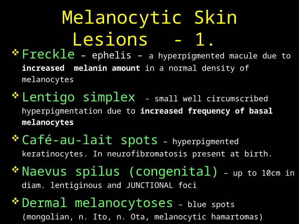
Melanocytic Skin Lesions - 1. Freckle – ephelis – a hyperpigmented macule due to increased
melanin amount in a normal density of melanocytes
Lentigo simplex - small well circumscribed hyperpigmentation
due to increased frequency of basal melanocytes
Café-au-lait spots – hyperpigmented keratinocytes. In
neurofibromatosis present at birth.
Naevus spilus (congenital) – up to 10cm in diam.
lentiginous and JUNCTIONAL foci
Dermal melanocytoses – blue spots (mongolian, n. Ito, n.
Ota, melanocytic hamartomas)
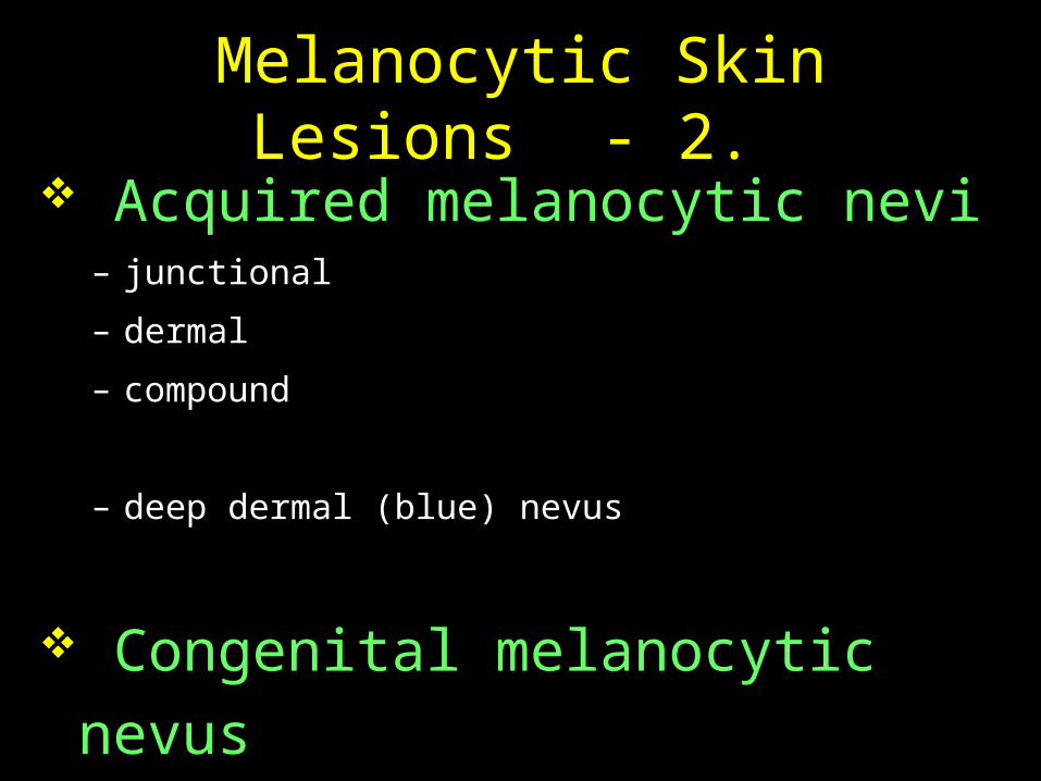
Melanocytic Skin Lesions - 2. Acquired melanocytic nevi
– junctional
– dermal
– compound
– deep dermal (blue) nevus
Congenital melanocytic nevus
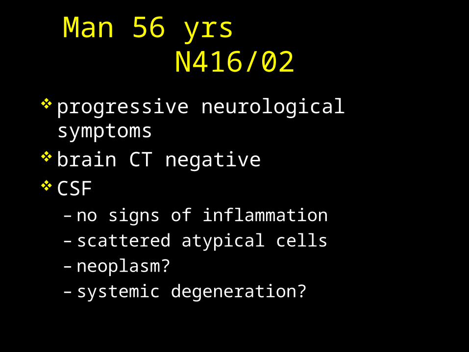
Man 56 yrs N416/02
progressive neurological symptoms brain CT negative CSF
– no signs of inflammation – scattered atypical cells – neoplasm? – systemic degeneration?
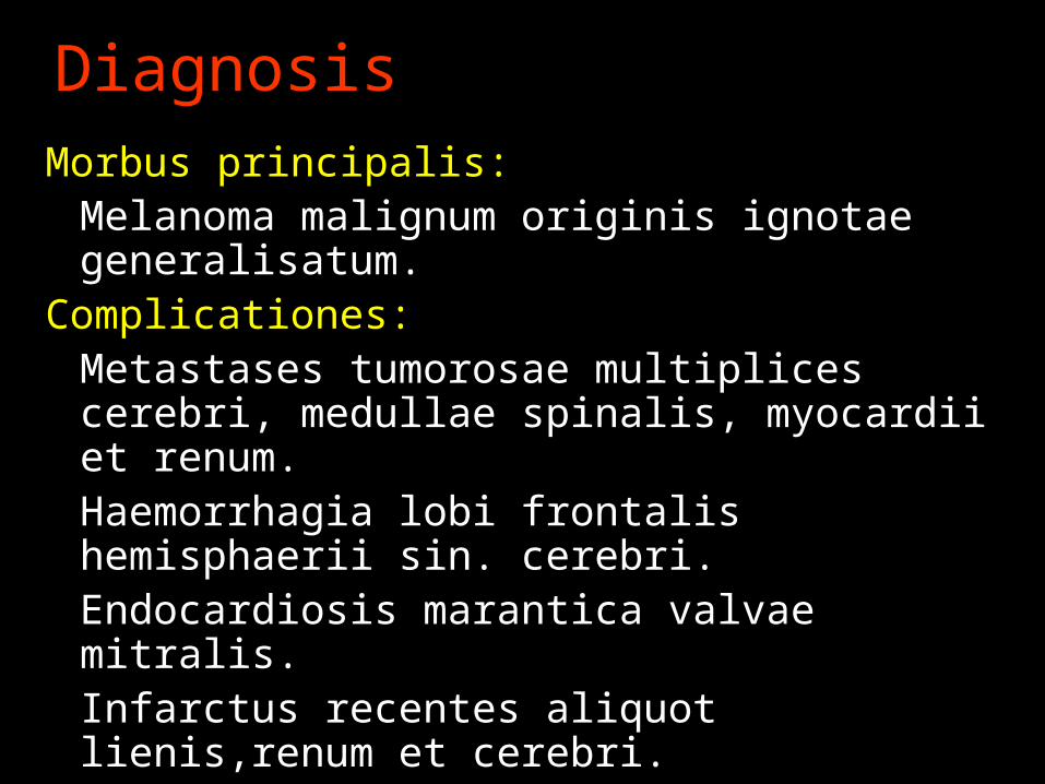
Diagnosis
Morbus principalis:Melanoma malignum originis ignotae generalisatum.
Complicationes:Metastases tumorosae multiplices cerebri, medullae spinalis, myocardii et renum.Haemorrhagia lobi frontalis hemisphaerii sin. cerebri.Endocardiosis marantica valvae mitralis.Infarctus recentes aliquot lienis,renum et cerebri.
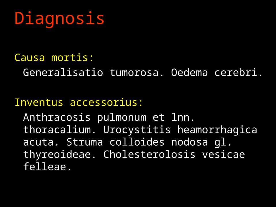
Diagnosis
Causa mortis:
Generalisatio tumorosa. Oedema cerebri.
Inventus accessorius:
Anthracosis pulmonum et lnn. thoracalium. Urocystitis heamorrhagica acuta. Struma colloides nodosa gl. thyreoideae. Cholesterolosis vesicae felleae.
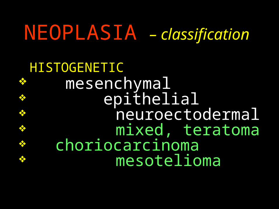
NEOPLASIA – classification
HISTOGENETIC mesenchymal epithelial neuroectodermal mixed, teratoma choriocarcinoma mesotelioma
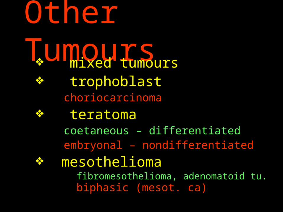
Other Tumours mixed tumours trophoblast
choriocarcinoma
teratomacoetaneous – differentiatedembryonal – nondifferentiated
mesothelioma fibromesothelioma, adenomatoid tu. biphasic (mesot. ca)
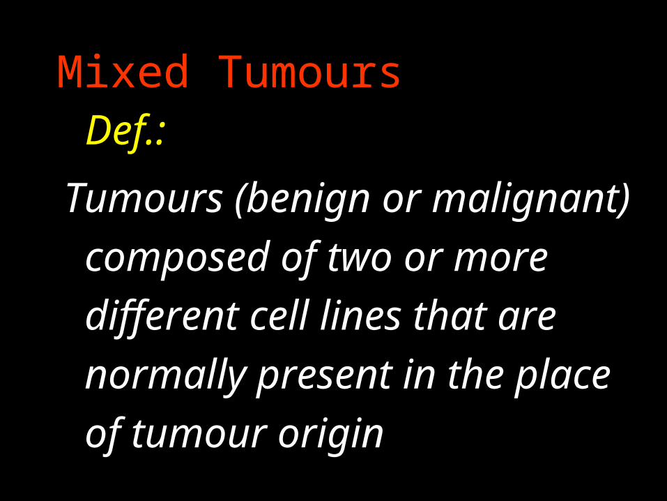
Mixed Tumours Def.:
Tumours (benign or malignant)
composed of two or more
different cell lines that are
normally present in the place
of tumour origin
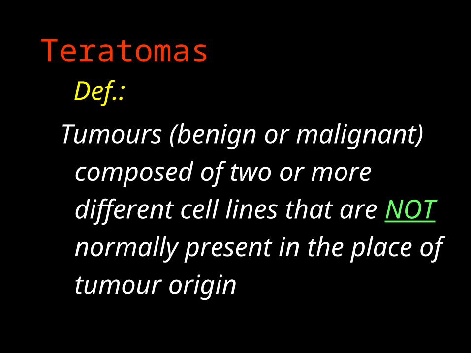
Teratomas Def.:
Tumours (benign or malignant)
composed of two or more
different cell lines that are NOT
normally present in the place of
tumour origin
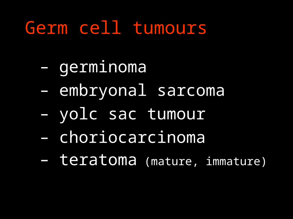
Germ cell tumours
– germinoma
– embryonal sarcoma
– yolc sac tumour
– choriocarcinoma– teratoma (mature, immature)