Embed Size (px)
Citation preview

Leticia Quintanilla-Martinez
Pathology of aggressive lymphomas
Institute of Pathology


• Aggressive B-cell lymphoid neoplasms
– Major changes that impact how cases should
be evaludated and diagnosed (pathologist)
– Therapeutic implications (hematologist)
– New biological information
Changes in the new 2016 WHO


Diffuse large B-cell lymphoma, NOS
Definition: Diffuse large B-cell lymphoma is a neoplasm of large B lymphoid cells more than twice the size of a normal lymphocyte and with diffuse growth pattern. DLBCL is clinically, morpholgically and biologically a heterogeneous disease reflected in the highly variable clinical course
WHO 2008

Diffuse large B-cell morphology, NOS
Anaplastic morphology is independent of ALK expression

Centroblastic
T-cell rich
Immunoblastic
Mediastinal LBCL
Diffuse large B-cell lymphomas
Diffuse large B-cell lymphoma (DLBCL), NOS
Germinal centre B-cell subtype
Activated B-cell subtype
T-cell/histiocyte-rich large B-cell lymphoma
Primary DLBCL of the CNS
Primary cutaneous DLBCL, leg type
EBV-positive DLBCL, NOS
EBV-positive mucocutaneous ulcer
DLBCL associated with chronic inflammation
Fibrin-associated diffuse large B-cell lymphoma
Lymphomatoid granulomatosis, grade 1,2
Lymphomatoid granulomatosis, grade 3
Primary mediatinal (thymic) large B-cell lymphoma
Intravascular large B-cell lymphoma
ALK-positive large B-cell lymphoma
Revised 4th edition WHO classification
Diffuse large B-cell lymphomas
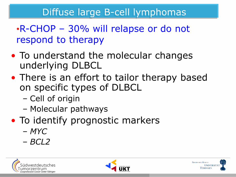
• To understand the molecular changes underlying DLBCL
• There is an effort to tailor therapy based on specific types of DLBCL – Cell of origin
– Molecular pathways
• To identify prognostic markers – MYC
– BCL2
•R-CHOP – 30% will relapse or do not respond to therapy

Diffuse large B-cell lymphomas, molecular signature
Alizadeh et al, Nature 2000; 403:503
• The most frequent category
representing 25-40% of all lymphomas

Diffuse large B-cell lymphomas
50% 50%
CD10+ BCL6+
Highly mutated IG
Ongoing SHM
BCR activated B-cells
MUM1+FOXP1+
Activation of the NF-kB pathway
15% of all cases remain unclassifiable

Diffuse large B-cell lymphomas Determining the cell of origin
Rosenwald et al, NEJM 2002;346:1938

Hans Algorithm for molecular classification of DLBCL
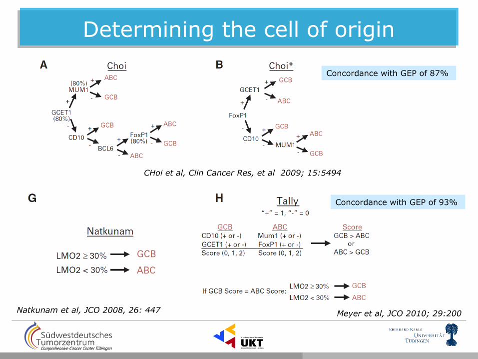
Determining the cell of origin
Meyer et al, JCO 2010; 29:200
CHoi et al, Clin Cancer Res, et al 2009; 15:5494
Natkunam et al, JCO 2008, 26: 447
Concordance with GEP of 93%
Concordance with GEP of 87%

WHO 2016 determining the cell of origin
Quintanilla-Martinez, Hematological Oncology 2015
It is acceptable to investigate the cell of origin in DLBCL with IHC algorithms
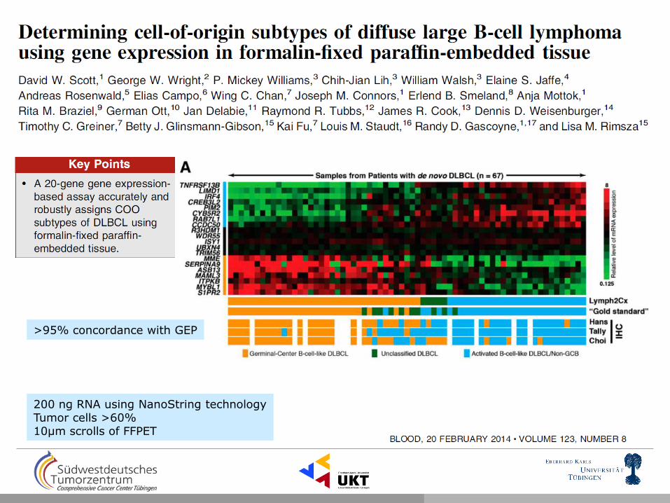
>95% concordance with GEP
200 ng RNA using NanoString technology Tumor cells >60% 10µm scrolls of FFPET

Pasqualucci L et al. Nat Genet 2011.
Recurrent somatic mutations in DLBCL
Aberrant histone/ chromatin modification

Constitutive BCR and NF-kB signaling
BCR
Ag
LYN
SYK BTK
TLR/IL-1R CD40
CD40L
21%
CD79A/B
PKCb
MAPK/ERK P13K/AKT CARD11
BCL10
MALT1
MYD88
NF-kB
IRF4
BCL6
JAK/STAT Interferon P38/MAPK
10%
30-37%
A20
PRDM1/BLIMP1
30%
25-32% 25%
ABC-DLBCL
BTK-i
MALT-i
NF-kB-i
Lenalidomide
BCL6-i


Prognostic importance of MYC translocation in DLBCL
Aukema et al., Blood 2011,117:2319 Salaverria et al., JCO 2011
Ott et al, Blood 2013;122:3884-91 Karube K & Campo E, Semin Haematol 2015;52:97
MYC break-apart probe

DLBCL morphology with MYC translocation
Reference DH/TH (%)
MYC-R (%)
Type of DH % GCB type
Niitsu 2009 5% 11% BCL2 84%
Johnson 2012 5% 12% BCL2 64%
Green 2012 6% 11% BCL2 91%
Akyurek 2012 3% 6% BCL2 & BCL6 71%
Visco 2013 2% 8% BCL2 88%
Valera 2013 4% 7% BCL2 & BCL6 71%
Hu 2013 3% N/A BCL2 90%
Tzankov 2014 3-4% 9% BCL2 & BCL6 80%*
Copie-Bergman 2015 6% 8.8% BCL2 & BCL6 N/A
*BCL6 DH only 50% GCB >90% have a GCB phenotype
BCL2 R- 60-70% BCL6 R – 6% Triple hit: 15-20%

Johnson NA et al Blood 2009;114:2273-2279
MYC partner matters? IG vs non-IG
Morphology matters? BCLU vs DLBCL
BCL2 expression matters
Not all MYC translocated DLBCL are the same. Modulators of prognosis
MYC -
MYC+ non-IG
MYC+IG
P=0.04
Johnson NA et al Blood 2009;114:2273-2279
Pedersen et al, European J Haematol 2014;92-42-49 Copie-Bergman et al, Blood 2015

When to test MYC in DLBCL?
CD10
• Selection of cases • Clinical presentation
– Extensive disease, CNS involvement, BM involvement and Leukemic presentation
• Morphology – All BCL-U morphology
– All blastoid morphology
– DLBCL of GCB type • Ki67 is not a good parameter • BCL2 > 50% • MYC > 40%
• FISH – Start with break-apart probes for MYC
followed for BCL2 and BCL6.

DLBCL with MYC expression without translocation
MYC protein expression is more frequent than genetic alteration in DLBCL
Author
Cases#
MYC/BCL2+
Johnson 2012 136 18%
Green 2012 185 29%
Horn 2013 141 28%
Hu 2013 466 34%
Valera 2013 120 27%
• MYC FISH rearranged in DLBCL 5-12%
• MYC IHC+ 29-64% •MYC+/BCL2+ IHC: 30%
MYC BCL2

DLBCL with expression of MYC and BCL2
Johnson et al
„Double expressor“

ABC vs GCB “double expressors”
MYC/BCL2 coexpression, rather than cell-of-origin is a better predictor of prognosis in DLBCL
Double expression of BCL2/MYC is more frequently observed in non-GC phenotype (66% vs 39%)

• Changes in the 2016 revised WHO Distinction of GCB vs ABC/non-GCB type required
with use of immunohistochemical algorithm acceptable, may affect therapy
Coexpression of MYC and BCL2 considered new prognostic marker (double expressor lymphoma) (MYC>40% and BCL2>50%)
Mutational landscape better understood and might become part of the foundation for optimal patient care.
Diffuse Large B-cell lymphoma
Prognostic factors • Immunophenotypic: MYC/BCL2 IHC • Genetic: MYC, BCL2, BCL6 rearrangements

“Grey zone lymphomas”
• Not an entitiy, but provisional groups
that awaits further studies
2008 WHO Classification
BCLU

Differential diagnosis of DLBCL
Burkitt lymphoma DLBCL BCL-U

Differential diagnosis of BL, B-unclassifiable, and DLBCL
Burkitt CD10+ BCL6+ BCL2- MYC expression + MIB1>98% MYC-R simple BCL2/BCL6-R – ID3/TCF3 mutations + EBV+/-
BCL-U category CD10+ BCL6+/- BCL2+ MYC expression + MIB1<90% MYC-R complex (54%) BCL2/BCL6-R frequent ID3 mutations rare EBV-
GCB-DLBCL CD10+ BCL6+ BCL2+/- MYC expression +/- MIB1 variable MYC-R complex (5-15%) BCL2/BCL6-R (2-6%) ID3/TCF mutations - EBV-

WHO 2016 Update High grade B-cell lymphoma, NOS
WHO 4th edition 2008:
– B-cell lymphoma, unclassifiable, with features intermediate between diffuse large B-cell lymphoma and Burkitt lymphoma (BCLU)
• Cases morphologically resembling BL but with morphological and phenotypical deviations
• Includes but not limited to DHL.
WHO 4th edition update 2016:
– High grade B-cell lymphomas
• High-grade B-cell lymphomas with MYC and BCL2 and/or BCL6 rearrangements (double or triple-hit)
– Specifiy whether DLBCL, blastoid or BCLU morphology
– Cases of FL or LBL with DH are not included!
• High-grade B-cell lymphoma, NOS
– Cases with BCLU or blastoid morphology or other high-grade features and no DH

Diagnostic approach to high grade B-cell lymphoma
BCLu
The morphology appearance should be noted in a commnent
HGBL with double or triple hit
HGBL, nos
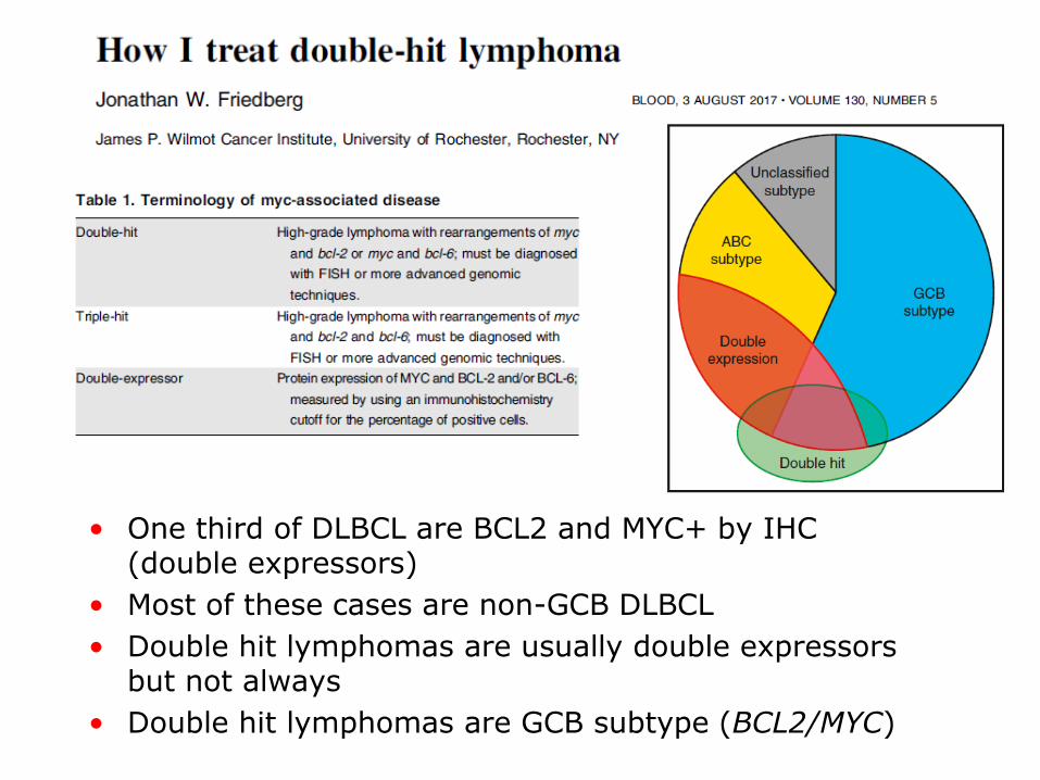
• One third of DLBCL are BCL2 and MYC+ by IHC (double expressors)
• Most of these cases are non-GCB DLBCL
• Double hit lymphomas are usually double expressors but not always
• Double hit lymphomas are GCB subtype (BCL2/MYC)

Definition: Highly aggressive lymphoma often presenting in extranodal sites, composed of monomorphic medium-sized B-cells with basopohilic cytoplasm and numerous mitotic figures. Epidemiology: Endemic, sporadic and immunodeficiency associated Genetics: MYC translocation with simple karyotype, 100% EBV in endemic cases.
Break in c-myc locus
Burkitt lymphoma
Dalla-Favera R, Science 1983;219:963

Classical Burkitt lymphoma without MYC alterations
•Approx 3-10% of all BLs including endemic and pediatric
•Considered cases that can be missed with FISH
Cryptic insertions of IG into MYC locus
distal 5´and 3´breaks
•Recurrent 11q alterations
Candidate genes FLI1, ETS1
Aukema et al., Blood 2011,117:2319 Salaverria et al., JCO 2011 Salaverria et al, Blood 2014;123:1187
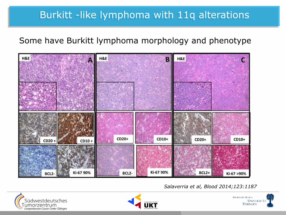
Burkitt -like lymphoma with 11q alterations
Salaverria et al, Blood 2014;123:1187
Some have Burkitt lymphoma morphology and phenotype
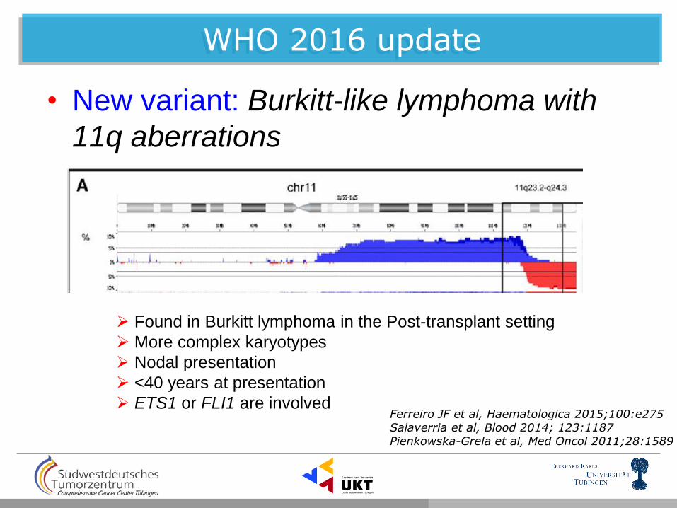
WHO 2016 update
• New variant: Burkitt-like lymphoma with
11q aberrations
Ferreiro JF et al, Haematologica 2015;100:e275 Salaverria et al, Blood 2014; 123:1187 Pienkowska-Grela et al, Med Oncol 2011;28:1589
Found in Burkitt lymphoma in the Post-transplant setting
More complex karyotypes
Nodal presentation
<40 years at presentation
ETS1 or FLI1 are involved
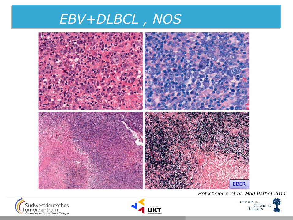
Hofscheier A et al, Mod Pathol 2011
EBV+DLBCL , NOS
EBER


• EBV+ circumscribed ulcerative lesions
– Immunosuppression associated
• Azathioprine
• Methotrexate
• Cyclosporin-a
– Age-related immunosenescence
• Clinical presentation: Oropharyngeal mucosa,
skin or gastrointestinal tract
• Morphology: Resembles CHL
• Prognosis: excellent. Some cases regressed spontaneously

DLBCL with plasma cell phenotype
• ALK-positive DLBCL – Predominates in male, adults
– The cells have plasmablastic morphology and phenotype
– The t(2;17) involving ALK and clathrin
– CD30 is negative
• Plasmablastic lymphoma (PBL) – Aggressive neoplasm
– Immunodeficiency, HIV
– EBV associated
– MYC translocation
• Primary effusion lymphoma (PEL) – Young males with HIV infection
– Human Herpes virus 8 (HHV-8) Kaposi sarcoma herpes virus
– Usually co-infection with EBV

CD20 MUM1 EBER LMP1
Plasmablastic lymphoma

• Definition:
• PEL is a large cell lymphoma usually presenting as serous effusions without detectable mass.
• It is universally associated with HHV8.
• It presents in immunodeficiency patients, mainly HIV+.
• Usually coinfected with EBV
WHO 2008, Said J and Cesarman E HHV8 (LANA) EBER
Primary effusion lymphoma
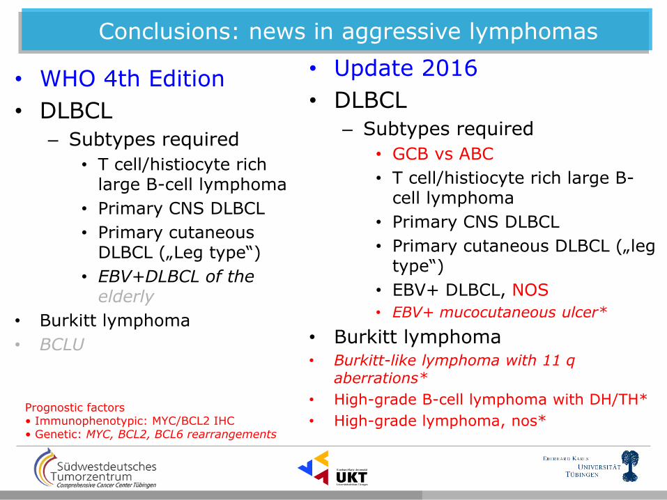
• WHO 4th Edition
• DLBCL
– Subtypes required
• T cell/histiocyte rich large B-cell lymphoma
• Primary CNS DLBCL
• Primary cutaneous DLBCL („Leg type“)
• EBV+DLBCL of the elderly
• Burkitt lymphoma
• BCLU
• Update 2016
• DLBCL
– Subtypes required
• GCB vs ABC
• T cell/histiocyte rich large B-cell lymphoma
• Primary CNS DLBCL
• Primary cutaneous DLBCL („leg type“)
• EBV+ DLBCL, NOS
• EBV+ mucocutaneous ulcer*
• Burkitt lymphoma • Burkitt-like lymphoma with 11 q
aberrations*
• High-grade B-cell lymphoma with DH/TH*
• High-grade lymphoma, nos* Prognostic factors • Immunophenotypic: MYC/BCL2 IHC • Genetic: MYC, BCL2, BCL6 rearrangements
Conclusions: news in aggressive lymphomas

• The diagnosis of DLBCL needs to integrate, standard morphology, IHC, molecular techniques – Distinctiction between GCB and ABC is required
• RNA technology might have a role in the near future • IHC algorithms (Hans, Tally, etc)
– IHC for MYC/BCL2 is required – FISH analysis if possible in all GCB-type, if not follow two-step approach
• Mutation analysis and targeted therapy will certainly influence the diagnosis and treatment of DLBCL in the near future
• BTKi (Ibrutinib) or Bortezomib for relapsed ABC-DLBCL • EZH2i or BCL6i for relapsed GCB-DLBCL
Stay tuned for DLBCL with single MYC rearrangements and
Other modulators of prognosis in DH lymphomas and the role of specific somatic mutations
Conclusions