Embed Size (px)
Citation preview

A C T A T H E R I O L O G I C A
VOL. XII, 30: 445—452 BIAŁOWIEŻA 30.XII.1967
Zygmunt P O D G U R N I A K
Pathological Lesions in the European Bison
Caused by Foot-and-Mouth Disease in Polish Reservations
Bisoniana X X V I
[With 12 Figs . & 1 Table]
Thirty f ive European bisons died during the epizootic of foot-and--mouth disease in polish reservations (1953—1954). The post mortem examinations revealed lesions characteristic for foot-and-mouth disease, namely: freshly burst secondary vesicles, surface erosions, and in case of secondary infection, extensive necrotic lesions. The above lesions were observed in the mucous membrane of the alimentary tract (oral cavity, esophagus, rumen) and on the skin of interdigital space. In 9 cases the primary process of foot-and-mouth disease was complicated by bronchial pneumonia, purulent broncho-pneumonia, fungal invasion, actinomycotic pneumonia, abscesses in different organs and tissues or bone fractures.
There is very little published data on foot-and-mouth disease in the European bison. Thoroughful work of T o m e s c u , G h e o r g h i u & I s o p e s c u (19bu; does not mention the foot-and-mouth disease in this species, whi le S k o r o m o -c h o w (1954) mentions only that similarly to many other species European bisons are susceptible to the virus of foot-and-mouth disease.
This disease was observed in the European bison ( F o l m e r , 1939; J a c z e w -s k i , 1959; 1960) in the american bison ( U r b a i n , 1939; H e d i g e r „ 1940; S t a ś - k i e w i c z , 1946) and in hybrids of the two species. Specially valuable are detailed observations of J a c z e w s k i (1959; 1960) on the course of the foot-and-mouth disease epizootic of the European bison in 1953 and 1954 in Polish reservations. It is interesting that in natural conditions in the Białowieża Primaeval Forest the foot-and-mouth disease was observed three times between 1906 and 1909 but the mortality of sick animals was only 5%> ( W r ó b l e w s k i , 1927).
The above mentioned papers were concerned predominantly with the clinical and epizootic aspects of the disease.
During the epizootic of foot-and-mouth disease in the polish reservations Ło-
1445]
446 Z. Podgurniak
puszna, Pszczyna and Niepołomice (1953—1954) a total of 35 bisons died '). As de-monstrated in later studies the foot-and-mouth disease in Pszczyna and Niepoło- mice was caused by the A 5 variant of the virus ( B a r a n o w s k i , 1956>.
In the Łopuszna reservation the first symptons of foot-and-mouth disease w e r e noticed on July 6, 1953. Eight days later (July 14, 1953) the female bison »Puma« died. During the following 15 day f ive more bisons died. About 5 months after the outbreak of the disease (December 18, 1953) female- »Tatra« died and three months later »Pustak«, a male the last bison in the reservation died (Table 1). The latter two animals survived the disease but had serious complications of its primary process ( J a c z e w s k i , 1959) which were the indirect cause of death.
In the Pszczyna reservation the first symptoms of the disease (lack of appetite, slow rumination and sluggishness) were observed on December 20̂ 1953. Male bison »Platanek« died already on December 21, 1953. Sixteen bisons died in the seven following days. On January 4, 1954 female »Pleśnianka« died whi le the last bison from the herd, female >Plusia« died on January 19, 1954 (Table 1).
On April 19, 1954 in one of the three enclosures of the Niepołomice reservation the female bison »Puszcza« was noticed to be sad, without appetite and with slight salivation. Similar symptoms were noticed on April 21 in »Pura«, »Puzon« and »Purchawka«. During the following f e w days the disease attacked the remaining 'Ó bisons in this enclosure. > Puszcza« died on April 23 and 3 more bisons died during the following 8 days (»Puzon«, »Purchawka« and »Pura«). Three more animals suc-cumbed between May 20 and May 28 (»Pustybór«, »Pułkownik < and »Pukajka«) and three months after the outbreak of the disease male »Pułan« died. The disease did not transfer to the remaining two enclosures (Table 1).
In 33 of 34 cases autopsy revealed pronounced lesions indicating in-fection with foot-and-mouth disease virus. The process of the disease was accompanied by more or less typically expressed lesions in the mucous membrane of the upper segment of the alimentary tract (mucous membrane of the tongue, the lips, the gums, the hard and soft palate) rarely in the mucous membrane of further parts of the alimen-tary tract (esophagus, rumen, abomasum) an in the skin of intsrdigital space.
In animals which died during the first few days of the disease, surface erosions in the mucous membrane of the dorsal and ventral surface of the tongue were found usually on the lateral edges and the tip. These erosions had uneven, shreded edge and a more or less bright red center, or else smooth edge and finely granular, brown-grey center. Appearance of the latter erosions was indicating a beginning of regenerative pro-cesses in the epithelium. In similar cases, in animals which died some-what later, spots in which secondary vesicles burst were covered with completely regenerated epithelium (providing that there was no second-
') This number also includes the female bison »Puma« which was autopsied by the staff of the local veterinary service; the results of the autopsy and additional tests were not available to the author and therefore this case is omitted in the listing of lesions.
Pathological lesions caused by foot-and-mouth disease 447
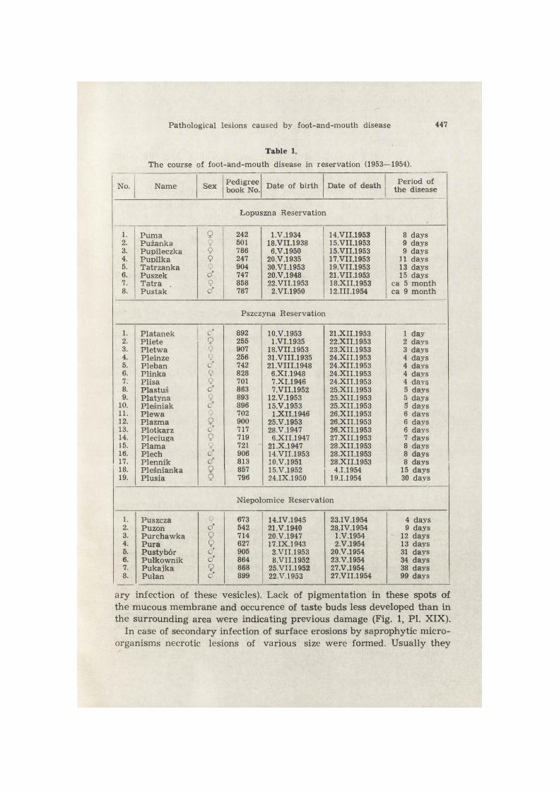
Table 1.
The course of foot-and-mouth disease in reservation (1953—1954).
No. Name Sex Pedigree book No. Date of birth Date of death Period of
the disease
Łopuszna Reservation
1. 2. 3. 4. 5. 6. 7. 8.
Puma Pużanka Pupileczka Pupilka Tatrzanka Puszek Tatra . Pustak
9 9 9 9 9 Ö* 9 o"
242 501 786 247 904 747 858 787
1.V.1934 18.VII.1938 6.V.1950
20.V.1935 30.VI.1953 20.V.1948 22.VII.1953
2.VI.1950
14.VII.1953 15.VII.1953 15.VII.1953 17.VII.1953 19.VII.1953 21.VII.1953 18.XII.1953 12.III.1954
8 days 9 days 9 days
11 days 13 days 15 days
ca 5 month ca 9 month
Pszczyna Reservation
1. 2. 3. 4. 5. 6. 7. 8. 9.
10. 11. 12. 13. 14. 15. 16. 17. 18. 19.
Platanek Pliete Płetwa Pleinze Pleban Plinka Plisa Plastuś Platyna Pleśniak Plewa Plazma Plotkarz Pleciuga Plama Piech Plennik Pleśnianka Plusia
c" 9 9 9 o" 9 9 o* 9 ć 9 9 c" 9 9 o" cf 9 9
892 255 907 256 742 828 701 863 893 896 702 900 717 719 721 906 813 857 796
10.V.1953 1.VI.1935
18.VII.1953 31.VIII.1935 21.VIII.1948
6.XI.1948 7.XI.1946 7.VII.1952
12.V.1953 15.V.1953
1.XII.1946 25.V.1953 28.V.1947
6.XII.1947 21 .X.1947 14.VII.1953 10.V.1951 15.V.1952 24.IX.1950
21.XII.1953 22.XII.1953 23.XII.1953 24.XII.1953 24.XII.1953 24.XII.1953 24.XII.1953 25.XII.1953 25.XII.1953 25.XII.1953 26.XII.1953 26.XII.1953 26.XII.1953 27.XII.1953 28.XII.1953 28.XII.1953 28.XII.1953
4.1.1954 19.1.1954
1 day 2 days 3 days 4 days 4 days 4 days 4 days 5 days 5 days 5 days 6 days 6 days 6 days 7 days 8 days 8 days 8 days
15 days 30 days
Niepołomice Reservation
1. 2. 3. 4. 5. 6. 7. 8.
Puszcza Puzon Purchawka Pura Pustybór Pułkownik Puka.ika Pułan
9 o" 9 9 c" o" 9 0*
673 542 714 627 905 864 868 899
14.IV.1945 21.V.1940 20.V.1947 17.IX.1943
3.VII.1953 8.VII.1952
25.VII.1952 22.V.1953
23.IV.1954 28.IV.1954
1.V.1954 2.V.1954
20.V.1954 23.V.1954 27.V.1954 27.VII.1954
4 days 9 days
12 days 13 days 31 days 34 days 38 days 99 days
ary infection of these vesicles). Lack of pigmentation in these spots of the mucous membrane and occurence of taste buds less developed than in the surrounding area were indicating previous damage (Fig. 1, Pl. XIX).
In case of secondary infection of surface erosions by saprophytic micro-organisms necrotic lesions of various size were formed. Usually they

448 Z. Podgurniak
were round with a diameter of 3—4 cm and reaching deep into the muscles of the tongue (Fig. 2). The most pronounced lesions were ob-served in a male »Pulan« which died 99 days after the outbreak of the foot-and-mouth disease in the herd. The lesion covered about Vs of the dorsal surface and both lateral margins as well as the underlying muscle of the tongue. On the dorsal part of the tongue there was a large (8X 10 cm) and about 1 cm deep loss of tissue irregularly shaped with roll--like edges elevated above the surrounding undamaged surface of the mucous membrane with some scars. The center was mostly smooth, but in the middle where the erosion was H-shaped (Fig. 3) and deeper, reaching about half of the tongue's thickness, the center was rough covered with a necrotic spongy mass.
Freshly burst secondary vesicles, surface erosions, or traces of healed erosions, or else (in case of complications) deep ulcerations were found in the mucous membrane of the lips with similar frequency. Similar changes were relatively rare in the mucous membrane of the hard and soft palate (»Plisa«, »Plewa«, »Puszcza«, »Pustybor«, »Purchawka«), of the gums (»Pliete«, »Pleciuga«, »Puszcza«, »Pustybor«), or of the nares (»Puszek«, »Plewa«, »Plazma«, »Pleciuga«, »Pura«, »Pustybor«) there were numerous and relatively deep erosions on the tops of the folds in the mucous membrane of the thoracic portion of the esophagus. These erosions had roll-like and elevated edges and a grey-brown, relatively dry, scab-like mass deep at the center. One of the erosions measured 3X1.5 cm and was covered by dry thick scab, easy to separate from the surface (Fig. 4).
In five bisons (»Plesnianka«, »Puszcza«, »Puzon«, »Pura«, »Pustybor«) lesions similar to those observed in the mouth were found in the mucous membrane of the ruminal pillars. In »Puszcza« which died on the fourth day of the disease, the ruminal pillars were covered with numerous round or irregular shallow erosions measuring from 0.3 to 2.0 cm. Their edges were uneven shreded with remnants of burst vesicles. Some of the erosions were covered with thin brown-grey or brown-red scabs tightly adhering to the membrane. In the animals which died later during the disease (»Plesnianka«, »Pura«) the process of epithelium regeneration was well pronounced. The edges of the erosions were smooth and even; the surface of the erosions usually free from scabs, smooth or finely granular ranging from pink to bluish red in colour. Lesions in the mucous membrane of the ruminal pillars were much more pronounced in »Pustybor« which died a month after the outbreak of the foot-and--mouth disease in the reservation. Besides round small erosions not exceeding 1 cm of diameter, there were vast (3X4 cm) deep ulcerations reaching the muscle layer (Fig. 5).
Pathological lesions caused by foot-and-mouth disease 449
Lesions in the skin of the interdigital space were observed in 30 of 34 cases, in both front and hind limbs. There were either fresh erosions with brown scabs covering red foci without epithelium, or older erosions in the stage of regenerating the epithelium or else scars after healed erosions. Similar changes were much less common in the coronary region. In four cases (»Plusia«, »Pukajka«, »Pułan«, »Pustybór«) com-plications of the primary process of the disease were observed on the skin of interdigital space, namely deep ulcerations and abscesses. »Pu-kajka« had an abscess the size of a wallnut in connective tissue, between second digits above the interdigital space of the right hind limb. This abscess did not have a tendency to encyst itself (Fig. 6 — Plate XIX).
Edema, congestion and extravasation were relatively frequently ob-served in the mucous membrane of abomasum (18 mases) and small intestine (7 cases).
In the heart muscle autopsy revealed indications of cloudy swelling in the majority of the cases. In two cases (»Tatrzanka«, »Pleśniak«) there was a simultaneous occurence of cloudy swelling and fatty degene-ration, the so called tigre heart. In some cases small extravasations were observed under epicard especially in the region of vascular grooves. Also an increased amount of serous fluid in the pericardial sac was observed.
Fatty degeneration of the liver was frequent and in six bisons (»Pupi-leczka«, »Pupilka«, »Tatra«, »Puszcza«, »Purchawka«, »Pura«) this process was so advanced that even quite large fragments of the liver did not sink when placed in water.
In the 7 months old »Platanek« which died in the Pszczyna reservat-ion on the second day of the disease no lesions characteristic of the foot--and-mouth disease were detected. In the mucous membrane of the oral cavity and in the skin of interdigital space there were no vesicles and no resulting erosion. Post mortem examination revealed only congestion of the mucous membrane of the lips, the presence of numerous petechiae in the tracheal mucosa, under the visceral pleura, under the epicard and in the mesentery as well as catarrhal gastritis. Advanced processes of post mortem autolysis (autopsy was done 3 days after death) made any histopathologic studies impossible. The above described picture of lesions indicated an acute course of the disease without vesicle formation. This is known to occur in the course of the malignant form of foot-and--mouth disease in cattle and especially in calves.
Frequently, namely in 9 cases, the primary process of the disease was complicated by bronchial pneumonia or purulent bronch-pneumonia (»Tatra«, »Pułkownik«, »Pułan«, »Pukajka«, »Pustybór«, »Pleśnianka«), purulent broncho-pneumonia caused by mycotic organisms from the gen. Aspergillus (»Plusia« — P o d g u r n i a k , 1962), by an abscess in

450 Z. Podgurniak
the subcutaneous connective tissue and in the muscles (>Pulan«) and by bone fracture (»Pustak«, »Tatra«).
Secondary vesicle formation in the epithelium of the mouth and ruminal pillars (Fig. 7) was observed in the histopathologic picture of animals which died after the first few days of the disease, similar to the situation sometimes found in cattle. The vesicles contained serous fluid with dead epithelial cells, neutrophilic polymorphonuclear granulocytes and single red blood cells. The prickle cells of the epithelium imme-diately adjacent to the secondary vesicle were subject to the so called »ballooning degeneration« (Fig. 8, PI. XX).
Serous exudation appears among the cells of this layer causing their loosening and manifestation of bridges between cells. Subsequently the prickle cells swell, become more or less round, the links between them disappear and the cells are transformed into spherical bodies (ballooning degeneration). Serous exudation and neutrophilic polymorphonuclear granulocytes fill the spaces formed between degenerated cells. Accumul-ation of fluid increases the pressure in the vesicle; the cornified layer becomes thin and eventually breaks. An erosion partially covered by shreds of necrotic epithelium is formed in place of the burst vesicle. The surface of the erosion is later covered with a yellowish-brown scab formed from dried exudation. The epithelium starts to heal beneath the scab and after 5—8 days is completely regenerated. However, fibrinous and purulent inflammantion of deeper layers of the mucous membrane occurs in case of a secondary infection. A relatively deeper erosion is formed which, under favourable conditions, can heal in some two weeks (Fig. 9); or else, the necrotic process spreads to surrounding tissue causing for example the above described lesions of the tongue (»Pusty-bór«, »Pułan«).
In animals which died during the first few days of the disease cloudy swelling of muscle fibres was the most common lesion of the heart muscle. However, it was often accompanied by leucocytic infiltration and perivascular proliferation of cells from the adventitia of vessels (Fig. 10). In animals which died in the later course of the disease de-generative changes were accompanied by exudations of variable size composed of limphoidal cells, histiocytes and plasma cells with con-comitant degeneration of muscle fibers and the proliferation of con-nective tissue (Fig. 11). The character of these exudations indicates an advanced stage of phagocytosis and the beginning of regeneration processes. In four bisons which died 13—39 days after the outbreak of foot-and-mouth disease in the Niepołomice reservation (»Pura«, »Pusty-bór«, »Pułkownik«, »Pukajka«) small deposits of calcium salts were

Zmiany anatomopatologiczne u żubrów w przebiegu pryszczycy 451
found in fibers of the heart muscle showing hyaline degeneration. These foci (centers) were surrounded with either young fibers of connective tissue or with elements of non specific granulation (Fig. 12 — Plate XX).
REFERENCES
1. B a r a n o w s k i C., 1956: Diagnozowanie typów pryszczycy i ich rozmieszcze-nie w Polsce. Med. wet., 12, 7: 395—396. Lublin.
2. F o l m e r C. J., 1939: Aphtae episooticae among the Wisents (Bison bonasus L.) and the Cross-bred Wisents (Bison bonasus L. X Bison bison) of the Royal Zoological Society "Natura Artis Magistra" at Amsterdam in the autum of 1937. Bijd. Dierk., 27: 53—60. Leiden.
3. H e d i g e r H., 1940: Uber Maul und Klauenseuche bei Zootieren. Zool. Garten, 1940/41 (N.F.) 12: 291—299. Leipzig.
4. J a c z e w s k i Z„ 1959: Spostrzeżenia z zakresu opieki weterynaryjnej w re-zerwatach żubrów w latach 1952—1954. Roczn. Nauk. roln., 69-E, 2: 297—318. Warszawa.
5. J a c z e w s k i Z., 1960: Beobachtungen bei der Maul — und Klauenseuche in polnischen Wisentreserwaten. Z. Jagdwiss., 6, 3: 100—107. Hamburg.
6. P o d g u r n i a k Z., 1960: Grzybica płuc żubra i renifera. Pat. pol., 11, 4: 401— 406. Warszawa.
7. P o d g u r n i a k Z., 1962: Przypadek promienicy płuc u żubra. Med. wet., 18, 7: 422—424. Lublin.
8. S k o r o m o c h o w A., 1954: Pryszczyca. Państwowe Wyd. Roln. i Leśne, 56. Warszawa.
9. S t a ś k i e w i c z G., 1946: Enzootia pryszczycy u bizonów i żubrobizonów w zwierzyńcu w Smardzewicach w r. 1938. Med. wet., 2, 11: 513—515. Lublin.
10. U r b a i n A., B u l l i e r P. & N o u v e l J., 1938: Au sujet d'une petite épisoo-tie de f ièvre aphteuse ayant sévi sur des animaux sauvage en captivité. Bull. Acad. vét. Fr„ 11: 59—73. Paris.
11. T o m e s c u V., G h e o r g h i u J. & I s o p e s c u J., 1960: Febra aphtosâ (Stu-dio monografie). Editura Academici Republici Populäre Romine. Bucuresti.
12. W r ó b l e w s k i K., 1927: Żubr Puszczy Białowieskiej. Wydawnictwo Polskie, 1—232 + XVI. Poznań.
Received, March 3, 1967. Warsaw Agricultural University, Department of Pathological Anatomy, Warszawa, Grochowska 272.
Zygmunt PODGURNIAK
OBRAZ ZMIAN ANATOMOPATOLOGICZNYCH U ŻUBRÓW W PRZEBIEGU PRYSZCZYCY W POLSKICH REZERWATACH
Streszczenie
W okresie (1953—1954) epizootii pryszczycy w 3 rezerwatach żubra w Polsce (Ło-puszna, Pszczyna, Niepołomice) padło ogółem 35 sztuk. Wyginął cały stan pogło-wia żubrów w rezerwatach Łopuszna (8 sztuk) i Pszczyna (19 sztuk), zaś w jednej

452 Z. Podgurniak
z trzech zagród w Niepołomicach, w której przebywało do czasu choroby 14 żu-brów, zostało przy życiu zaledwie 3 sztuki.
W 32 przypadkach stwierdzono sekcyjnie charakterystyczne dla pryszczycy zmiany w górnym odcinku przewodu pokarmowego (w błonie śluzowej języka, warg, dziąseł, policzków), rzadko w błonie śluzowej dalszych odcinków przewodu pokarmowego (przełyk, żwacz) oraz w skórze szpar międzyracicowych. W zależ-ności od czasu jaki upłynął od zakażenia do śmierci zwierzęcia obserwowano albo świeżo pęknięte pęcherzyki wtórne, powierzchowne nadżerki i miejsca po ich w y -gojeniu, albo w przypadku wtórnego ich zakażenia mniej lub bardziej rozległe głę-bokie ogniska martwicy. W mięśniu sercowym w większości przypadków wystę-powało zwyrodnienie miąższowe, w dwóch przypadkach t.zw. serce tygrysie, w y -broczyny pod nasierdziem, w worku osierdziowym zwiększona ilość płynu surowi-czego. Histologicznie obserwowano w mięśniu sercowym obok zmian wstecznych (zwyrodnienie miąższowe, szkliste, ogniska martwicy) procesy zapalne z występo-waniem nacieków drobnokomórkowych.
Stosunkowo często stwierdzano powikłania pierwotnego procesu pryszczycowego (nieżytowe, nieżytowo-ropne zapalenie płuc, grzybica i promienica płuc, ropnie w różnych tkankach i narządach, złamania kości).
EXPLANATION OF PLATES
Plate XIX.
Fig. 1. »Plesnianka« — Tongue — traces of healed surface erosions. Fig. 2. »Pustybor«. Tongue — deep round necrotic lesion on the edge.
Fig. 3. »Pulan«. Tongue — extens ive erosion on the dorsal surface of the tongue with roll-l ike edges. In the middle H-shaped necrosis reaching down to half of
the tongues thickness. Fig. 4. »Pustybor«, Esophagus — deep ulceration of mucous membrane reaching
the muscle layer. Fig, 5. »Pustyb6r«. Rumen — extens ive deep ulceration of mucous membrane
reaching the muscle layer and smaller erosion, 1 cm in diameter. Fig. 6. »Pukajka«. Hoof — interdigital space abscess in the connective t issue (size
of a wallnutX
Plate XX.
Fig. 7. »Puszcza«. Rumen — fragments of two secondary vesicular lesions in the mucous membrane of the ruminal pillars. Neutrophilic polymorphonuclear granulo-cytes, single red blood cells and prickle cells of epithelium are seen; magnif. oc. —
4 X , ob. — 40 X . Fig. 8. »Pliete«. Tongue — so called balloning degeneration of prickle cells in the mucous membrane. Cells become loosened and round. Exudation of neutrophilic polymorphonuclear granulocytes between the cells. Magnif. oc. — 4 X, ob. — 16 X . Fig. 9. »Purchawka«. Tongue — healed erosion in the mucous membrane. Magnif.
oc. — 4 X, ob. — 6.3 X . Fig. 10. »Plazma«. Heart muscle — cloudy swelling. Leucocytic infiltration and perivascular proliferation of cells from the vascular adventitia. Magnif. oc. — 4 X„
ob. — 40 X . Fig. 11. »Tatra«. Heart muscle — extensive infiltrations of l imphoidal cells, histio-cytes and plasma cells. Atrophy of muscle fibers. Magnif. oc. 2.5 X , ob. — 40 X. Fig. 12. »Pukajka«. Heart muscle — small deposits of calcium salts in hyaline degenerated muscle f ibers and extens ive lymphocytic infiltration. Magnif. oc. —
2.5 X , ob. — 16 X.

ACTA THERIOLOGICA Vol. XII, 30. Plate XIX.
Z. Pocgurniak auctor phot,

ACTA THERIOLOGICA Vol. XII, 30. P l a t e XX.
Z. Podgurniak auctor phot.


![Paper Class XII [EnthuseCourse(XI XII)]](https://img.pdfslide.us/doc/110x75/577cc7ee1a28aba711a1e761/paper-class-xii-enthusecoursexi-xii.jpg)