Embed Size (px)
Citation preview
The Iraqi Journal of Veterinary Medicine, 37(2): 178 -187. 2013
871
Pathological changes of immunized mice with Trichophyton mentagrophyte
lyophilized antigen Radam, S. A. and Faleh, E. B.
Department of Pathology, College of Veterinary Medicine, University of Baghdad, Iraq.
E-Mail: [email protected]
Accepted on: 27/3/2013
Summary
The study was carried to investigate the pathological effect of lyophilized antigen of
Trichophyton mentagrophytes in mice. Fifty mice were divided into three groups. The first group 20
mice were immunized subcutaneous (s/c) with 0.5 ml of T.mentagrophyte antigen 20 µgm/ml, by
two doses, 14 day intervals, between them. The second group 20 mice and third group 10 mice
considered as positive and negative control groups respectively. After 30 days post immunization
first and second groups were challenged intradermal I/d. with 0.1 ml of fungal suspension contain
(1×10⁷ ml) of viable virulence T.mentagrophyte while the third group injected intraperitoneally I/P.
with 0.5 ml of sterile phosphate buffer saline. All mice of the first and second groups were
sacrificed at (5, 14, 30 and 60) days post challenge for gross and histopathological examination.
Histopathologically the second group showed epidermal hyperkeratosis with appearance of crust
lesions seen with abscess formation especially in early stage of lesion, while the main feature of
advance cases were characterized by folliculitis with fungal hyphae invasion in all epidermal and
dermal layers together with eosinophilic infiltration. Mild pathological changes were seen in the 1st
immunized group characterized by infiltration of mononuclear cells mainly macrophages in dermal
connective tissue with dense proliferation of collagenous fibers , appearance of young fibroblasts
together with cellular hypodermal infiltration of eosinophil no evidence of clear follicular lesions
were seen. Lyophilized antigen of T.mentagrophyte can be considered as an effective immunogen
for protecting mice against T.mentagrophyte infection and it is synchronized with its dose.
Keywords: T.mentagrophyte lyophilized, mice, pathology.
------------------------------------------------------------------------------------------------------------------------
Introduction
Trichophyton mentagrophytes is a group
of the most common zoonotic worldwide
fungi generally grow only in keratinized
tissues such a hair, nails and outer layers of
skin, The fungus adheres, proliferates usually
digging into the epidermis and entering
through hair follicles causing an infection that
may vary from mild to very intense (1 and 2),
these fungi produce keratinases, proteolytic
enzymes that enable them to hydrolyze keratin
(3) where T.mentagrophytes contacts to living
cells or areas of inflammation but mucus
membranes are not affected (4).
The skin lesions are usually characterized
by inflammation that is most severe at the
edges, with erythema, scaling and occasionally
blister formation, central clearing is sometimes
seen particularly in tinea corporis, and this
results in the formation of a classic ringworm
lesion (5 - 7). The lyophilized vaccine used to
increase the resistance of vaccinated animals
to experimental challenge infection. The
lyophilized vaccine is designed to protect (i.e.,
increase resistance to infection) bearing
animals against T.mentagrophytes infection
and there by decrease zoonotic infection
exposure to their human attendants. This
broader lyophilized antigenic base and the
antigenic cross protection between
dermatophyte genera and species (8). The aims
of this study was to investigate the
pathological effected of lyophilized antigen of
tem entire aphids a mice.
Materials and Methods
The isolate of T.mentagrophyte was
obtained from the Microbiology department in
the College of Veterinary Medicine/Baghdad
University; which was confirmed by the
macroscopical, microscopical and biochemical
tests which maintained in dextrose agar for
preparation of lyophilized antigens according
to (8) and the concentration of protein was
measured by Biurete method. A total number
of 50 mice from both sexes with ages ranged
The Iraqi Journal of Veterinary Medicine, 37(2): 178 -187. 2013
871
from (4–8) weeks old which were obtained
from the (National Center of Researches and
Drugs Monitor in Baghdad), then divided into
three groups. The 1st group 20 mice
immunized by T.mentagrophyte antigen, 2nd
group 20 mice and third group 10 mice
considered as positive and negative control
groups respectively.
Whole Killed Lyophilized Antigen was
prepared from sediment of centrifuged virulent
T.mentagrophyte according to (8), after that it
was sonicated above antigen and kept it under
4˚C until it used in a skin test according to Al-
Haddad, (9). The first group was immunized
by 0.5 ml subcutaneously each ml contanin 10
µgm/ml of T.mentagrophyte antigen, the
second group considered as positive control
while the third group gave 0.5 ml of phosphate
buffer saline as control negative. After two
weeks the animals of the 1st group gave same
dose of immunization as a booster dose first
and second groups were challenge at day 30 at
dose 0.1 ml of fungal suspension contain
1×10⁷ cell/ml of virulence T.mentagrophyte,
between (5, 14, 30 and 60) days. All animals
were scarified for gross and histopathological
examination of skin, about 1cm3 was taken and
fixed in 10% formalin saline for
histopathology section which was dose
according to (10).
Results and Discussion
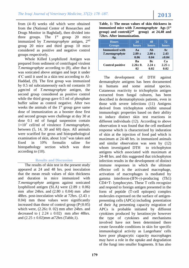
The results of skin test in the present study
appeared at 24 and 48 hrs. post inoculation
that the mean result values of skin thickness
and duration in mice immunized with
T.mentagrophyte antigens against sonicated
lyophilized antigen (SLA) were (2.89 ± 0.06)
mm after 24hrs. and (2.80 ± 0.04) mm after
48hrs. post-inoculation while at 72hrs. (2.43 ±
0.04) mm these values were significantly
increased than those of control group (P≤0.05)
which were, (2.26± 0. 02) mm after 24hrs. and
decreased to ( 2.24 ± 0.02) mm after 48hrs.
and (2.25 ± 0.02)mm at72hrs (Table,1).
Table, 1: The mean values of skin thickness in
immunized mice with T.mentagrophyte Ags (1st
group) and control(2nd group) at 24,48 and
72hrs. After immunization.
The development of DTH against
dermatophyte antigens has been documented
in humans and some animal species.
Cutaneous reactivity to trichophytin antigen
extracted from fungal cultures, has been
described in dermatophytosis patients, even in
those with severe infections (11) Antigens
derived from trichophyton exhibit unusual
immunologic properties based on their ability
to induce distinct skin test reactions in
different individuals (12). According to above
observation it was found that the cell mediated
response which is characterized by induration
of skin at the injection of food pad which is
maximum at 24-48 hrs. in immunized animals
and similar observation was seen by (12)
whom investigated DTH to trichophyton
antigens which associated with maximum at
24-48 hrs. and this suggested that trichophyton
infection results in the development of distinct
immune responses in which the ultimate
effector cell is the activated macrophage,
activation of macrophages is mediated by
gamma interferon-(IFN-γ)-producing (Th1)
CD4+T- lymphocytes. These T cells recognize
and respond to foreign antigen presented in the
form of peptide (T-cell epitopes) complex
molecules expressed on the surface of antigen-
presenting cells (APCs) including potentiation
of their Ag presenting capacity migration of
APCs is probable initiated by different
cytokines produced by keratinocyte however
the type of cytokines and mechanisms
involved have not been determined there
create favorable conditions in skin for specific
immunological activity as Langerhans' cells
have poor phagocytic capacity macrophages
may have a role in the uptake and degradation
of the fungi into smaller fragments. It has also
72
hours
48
hours
24
hours
Hours
Groups
Ac
2.43
±0.04
Ab
2.80 ±
0.04
Aa
2.89 ±
0.06
Immunized with
T.mentagrophyte
Ag.
Ca
2.25 ±
0.02
Bc
2.24 ±
0.02
Ba
2.26± 0.
02
Control positive
The Iraqi Journal of Veterinary Medicine, 37(2): 178 -187. 2013
811
been proposed that keratinocytes, which are
able to phagocytize and degrade antigens, may
process antigens that can be transferred to
Langerhans' cells and directly presented to T
cells. They are in a unique position to capture
exogenous antigen upon exposure to antigens,
they migrate as veiled cells to lymph nodes
draining the skin, where the antigens are
presented to T-lymphocytes in a major
histocompatibility complex (MHC) class II-
restricted fashion (13).
At 72 hrs. the result showed that the mean
thickness was lessor than 24 – 48 hrs., and this
observation was in consistence to recent study
by (14) which explained this decline in median
thickness may be possible indicate that T.
mentagrophytes antigens induced release of
suppressor cells and/or factor, which inhibited
development of cellular immune response
suppression of the hypersensitivity to
dermatophyte Ags could be an important
mechanisms for resolving cutaneous
T.mentogrophytes infection and limiting tissue
damage (15 and 16).
The cellular branch of the immune
system is crucial for protective immunity
against dermatophyte infections, vaccination
stimulated a cellular immune response, as
assessed by a skin test and aleukocyte
migration inhibition test (17). Although
several studies (18) showed that infection with
dermatophyte presented two patterns of
cellular immune response that an acute
inflammatory response correlated with positive
DTH skin test to Trichophyton and clearing of
infection.
The main clinical signs in the 2nd group
observed during 30-60 days post-inoculation
with heavy fungal isolation from external
organ (skin) was characterized by ruffled
haircoat, scaly ovoid type lesions with crusty
edge and patch of hair loss mostly seen on the
back and itching was reported these results
may indicate that the animal exposed to an
infective dose of highly virulent
T.mentagrophyte while in the immunized
group (1st group) there is no fungal isolation as
in (Table, 2).
Table, 2: Show fungal isolation in immunized and
non-immunized mice with T.mentagrophyte Ags (1st
group) and control(2nd group) after 5 ,14, 30,and 60
day post challenge. Day
60
Day
30
Day
14
Day
5
No.
Days
Groups
_ _ _ _
20
Immunized with
T.mentagrophyte
++++ ++ + _ 01 Control positive
- negative growth, + mild growth ++ moderate
growth +++ heavy growth ++++ very heavy growth.
In regard to fungal isolation our present
observation is similar to that noticed by (19)
who explained that most rodents infected with
T.mentogrophytes are asymptomatic or have
few clinical signs, in mice, partial or complete
areas of alopecia, erythema, scale, and scab
may be seen, often on the tails (20) because
the T.mentogrophytes have the ability to
invade keratinized layer of the skin mainly the
stratum corneum and produce proteolytic
enzymes that enable them to hydrolyze keratin
to induce active infection caused by this
organisms (3 and 21).
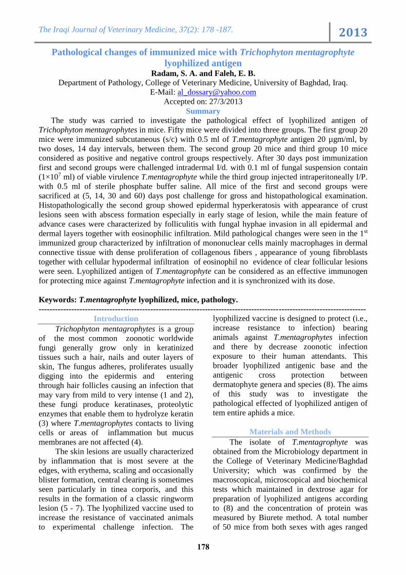
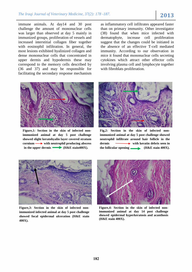
The first microscopic observation in the
skin at 5 day was focal epidermal
hyperkeratosis with moderate
polymorphonuclear cells infiltration in sub
epidermal and upper dermal with evidence of
abscess formation together with appearance of
slight keratohyaline layer covered the stratum
cornium (Fig. 1) .In another section there was
evidence of follicular invasion by neutrophilic
infiltration around the hair follicle (Fig. 2)
together with focal of epidermal ulceration
contain necrotic debris (Fig. 3).
At day14 post challenge cutaneous lesion
revealed epidermal hyperkeratosis and
acanthosis (Fig. 4) in addition to present
balloonic degeneration of some prickle cell
accompanied with formation clumps of
pyknotic nuclei nearly adjacent to stratum
cornium (Fig. 5) with sever dilation and
destruction of some hair follicle (Fig. 6) as
well as dermal odema which characterized by
fragmentation of dermal collagen fiber at 30
days post challenge together with mononuclear
cells infiltration around some degenerated hair
follicle (Fig. 7), in another section follicular
destruction appeared with degenerated keratin
that replace by compact mass of red purplish
microfilament hyphae mainly seen in the
The Iraqi Journal of Veterinary Medicine, 37(2): 178 -187. 2013
818
portal end and basement membrane of hair
follicle (Fig. 8) as well as fragmentation and
separation of dermal collagen with
eosinophilic infiltration and congested blood
vessels (perivascular dermititis) (Fig. 9 a and
b).
At day 60 post challenge the main
characteristic pathological changes was dense
fungal growth with hyphae that appeared red
with PAS stain spreading in both dermal and
epidermal involving hair follicle (Fig. 10).
The immunized group at 5 day was
characterized by no clear pathological changes
post challenge, except fragment separation of
dermal collagen fiber with edema and
congestion of blood vessels with scattered
infiltrate of young fibroblast seen mainly at 14
day (Fig. 11) as well as polymorph nuclear
cells around some degenerated follicles. At 30
day the lesion characterized by slight
acanthosis with fibrotic and granulated tissue
(evidence of dermal healing) associated with
intact hair follicle (Fig. 12 and 13).
At day 60 showed massive
mononuclear cells infiltration in dermis and
hypodermis (fatty dermis) with proliferation
of collagen fiber in the upper dermis together
with vascular dermatitis (Fig. 14) in another
section there was the epidermal acanthosis
accompanied with intracellular odema together
with vascular dermatitis (Fig. 15) in addition
to present follicular plugging with slight
perivascular dermatitis (Fig. 16) also the
result reveals intense proliferation of
mononuclear cells in dermal and hypodermal
tissue with proliferation of hair follicle and
sebaceous gland (Fig. 17), as well as dilation
and congestion of blood vessels. There is no
clear characteristic change in the epidermis.
The histopathology of murine
dermatophytosis showed presence of
cutaneous abscess and ulceration to primary
infection with T.mentogrophytes during 5 days
post challenge which closely compaired with
description of primary irritant dermatitis (22
and 23). The presence of these abscesses could
be a consequence of the fungal components
over the complement system, once it has been
reported that T. mentagrophytes activates this
system generating anaphylotoxin C5a, even in
non-immune animals, through the alternate via
(24). Many of these infected individuals have
no cell-mediated immunity against
Trichophyton antigens and do not develop
DTH evaluated through the intradermic test
with trichophyton. It has also been suggested
in these cases that polymorph nuclear cells
migrate to the area of follicle rupture after
complement activation and C5a generation
besides C5a, soluble factors released by
keratinocytes, including IL8, have also been
involved in this process (25). The necrotic
feature in the upper dermis which may be
initiated by production of toxic product by this
fungi (26 and 27). A similar histopathological
picture is observed in guinea pigs skin when
skin sensitizing hapten (DNCB) when toxic
concentration is applied to the skin of non-
sensitized animals (22 and 26), the area of
sever epidermal necrosis are invaded by
neutrophils (28). The role of neutrophils in the
defense mechanisms against dermatophytes is
not totally clear. It has been demonstrated in
experimental models that neutrophil
infiltration occurs before the peak of infection
(29) and these are capable of inhibiting fungal
multiplication, even in the absence of immune
response (30). Therefore, the presence of
neutrophils during primary T.mentogrophytes
infection (day 5) could also be attributed to
early tissue damage caused by expression of
contact hypersensitivity (22). Because neither
granulocytes nor monocytes were observed in
physical contact with hyphae in lesion
biopsies, the elimination of the fungi may be
mediated by soluble fungi static serum
mediators, as postulated by (31). Contact
sensitivity would damage the epithelial barrier
causing the release of fungi static or fungicidal
factors (from serum or cells), contact
sensitivity is thought to be mediated by the
inter action of peripheral T-lymphocyte with
soluble foreign antigens which diffused into
viable tissues (32). This mechanism could also
provide a rapid anamnestic recognition of a
microbial invasion of the keratinized layers of
the skin (33-35). Authors (H) was
demonstrated that in immune animals, as well
as non-immune, the time required for lesions
to resolve was longer when leukopeny was
induced concomitantly with inflammation. The
delayed cure was more noticeable in non-
The Iraqi Journal of Veterinary Medicine, 37(2): 178 -187. 2013
810
immune animals. At day14 and 30 post
challenge the amount of mononuclear cells
was larger than observed at day 5 mainly in
immunized groups, proliferation of vessels and
increased interstitial collagen fiber together
with eosinophil infiltration. In general, the
most lesions exhibited hyalinized collagen and
dense mononuclear cells that concentrated in
upper dermis and hypodermis these may
correspond to the memory cells described by
(36 and 37) and may be responsible for
facilitating the secondary response mechanism
as inflammatory cell infiltrates appeared faster
than on primary immunity. Other investigator
(38) found that when mice infected with
dermatophyte, increase cell proliferation
suggest that the changes could be initiated in
the absence of an effective T-cell mediated
immunity. According to our observation in
mice it found that mononuclear cells secreting
cytokines which attract other effector cells
involving plasma cell and lymphocyte together
with fibroblats proliferation.
Figure,1: Section in the skin of infected non-
immunized animal at day 5 post challenge
showed slight keratohyalin layer covered stratum
cornium with neutrophil producing abscess
in the upper dermis (H&E stain400X).
Fig,2: Section in the skin of infected non-
immunized animal at day 5 post challenge showed
neutrophil infiltrate around hair follicle in the
dermis with keratin debris seen in
the follicular opening (H&E stain 400X).
Figure,3: Section in the skin of infected non-
immunized infected animal at day 5 post challenge
showed focal epidermal ulceration (H&E stain
400X).
Figure,4: Section in the skin of infected non-
immunized animal at day 14 post challenge
showed epidermal hyperkeratosis and acanthosis
(H&E stain 400X).

The Iraqi Journal of Veterinary Medicine, 37(2): 178 -187. 2013
811
Figure, 9a: Histological section in the skin of
infected non- immunized animal at day 30 post
challenge showed hypodermal odema with
eosinophilic infiltration and congested blood vessel
(H&E stain 400X).
Figure,5: Section in the skin of infected non-
immunized animal at day 14-post challenge
showed epidermal hyperkeratosis and acanthosis
In addition to present balloonic
degeneration of some prickle cell accompanied
with formation clumps of pyknotic nuclei
(H&E stain 400X).
Figure,6: Section in the skin of infected non-
immunized animal at day 30 post challenge
showed sever dilation and degeneration of hair
follicles (H&E stain 400X).
Figure, 7: Section in the skin of infected non-
immunized animal at day 30 post challenge
showed mononuclear cells infiltration around
degenerated hair follicle (H&E stain 400X).
Figure, 8: Section in the skin of infected non-
immunized animal at day 30post challenge show
hair follicle invasion with red purplish
microfilament hyphae with dermal edema
(PAS stain 400X).
Figure, 9b: section in the skin of non- immunized
animal at day 30 post challenge showed
eosinophilic infiltration in hypodermis with
appearance of some destructed follicle (PAS stain
400X).

The Iraqi Journal of Veterinary Medicine, 37(2): 178 -187. 2013
811
Figure, 10: Section in the skin of infected non-
immunized animal at day 60 post challenge showed
dense fungal growth with hyphae that stained red with
PAS stain spreading in both dermal and epidermal
involving hair follicle (PAS stain400X).
Figure, 11: Section in the skin of immunized animal
at day 14 post challenge showed proliferation of
dermal collagen fiber with appearance of young
fibroblast (H&E stain 400X).
Figure,12: Section in skin of immunized animal at
day 30 post challenge showed slight acanthosis
with fibrotic and granulated tissue(dermal
healing)with intact hair follicle (H&E
Stain400X).
Figure,13: Section in skin of immunized animal
at day 30 post challenge showed fibrotic and
granulated tissue with moderate MNCs
infiltration (H&E Stain400X).
Figure, 14: Section in skin of immunized animal
at day 60 post challenge showed mononuclear
cells infiltration in dermis and hypodermis with
proliferation of collagen fiber in the upper dermis
together with vascular dermatitis (H&E Stain
400X).
Figure, 15: section in skin of immunized animal
at day 60 post challenge showed intense epidermal
acanthosis accompanied with
intracellular odema together with
vascular dermatitis (H&E Stain 400X). .

The Iraqi Journal of Veterinary Medicine, 37(2): 178 -187. 2013
811
Figure, 17: Section in skin of immunized animal
at 60 day post challenge show reveals intense
proliferation of MNCs in dermal and
hypodermal tissue with proliferation of hair
follicle and sebaceous gland as well as dilation
and congestion of blood vessels (H&E Stain
400X).
References
1. Weitzman, I. and Summerbell, R.C. (1995).
The dermatophytes. Clin Microbiol. Rev.,
8:240–259.
2. Romani, L. (2007). Immunity to fungi. In:
Kavanagh K. ed. New Insights in
Medicinal Mycology. Dordrecht:
Springer:1-18.
3. Kaufman, G.; Horwitz, B.A.; Duek. L.;
Ullman, Y. and Berdicevsky, I. (2007).
Infection stages of the dermatophyte
pathogen Trichophyton: microscopic
characterization and proteolytic enzymes.
Med Mycol., 45:149-55.
4. A
iello, S.E. and Mays, A. (1998). The Merck
veterinary manual. 8th ed. Dermatophytosis;
p626; Whitehouse, NJ: Merck and Co1419.
5. Devroey, C. (1985). Epidemiology of
ringworm (Dermatophytose). Dermatol.,
4:185-200.
6. Hainer, B.L. (2003). Dermatophyte
infections. Pratical. Therapeutics, 67:101-
108.
7. Dahdah, M.J. and Sher, R.K. (2008).
Dermatophytes. Current Fungal Infection
Reports, 2:81-86.
8. Pier, A.C. (1994). Broad spectrum
dermatophyte vaccine. January 11, www.antitope.co.uk.
9. Al-Haddad, Z.A. (2009). Immunopath-
ological study of C.neoformans isolated
from human and cows milk in mice. MSc
Thesis, College of Veterinary Medicine,
Baghdad University.
10. Bancroft, J.D. and Stevens, A. (1982).
Theory and practice of Histological
Techniques. 2nd ed. Churchill Livingstone:
483-516.
11. Tagami, H.; Kudoh, K. and Takematsu, H.
(1989). Immune defense mechanisms in
dermatophytosis. In: international
symposium of the research center for
pathogenic fungi and microbial mycosis,
Chiba Univ., Proceedings, PP: 119-122.
12. Khosravi, A.R.; Shokri, H. and Mansouri,
P. (2012). Immediated hypersensitivity and
serum IgE antibody response in patient with
dermatophytosis. Asian Pac J Allergy
Immunol., 30(1):40-47.
13. Hay, R.J.; Calderon, R.A.; Collins, M.J.
(1983). Experimental dermatophytosis:the
clinical and histopathologic features of a
mouse model using Trichophyton
quinckeanum (mouse favus). J Invest
Dermatol., 81:270–274.
14. de Arruda, M.S.; Gilioli, S. and Vilani-
Moreno, F.R. (2001). Experimental
Dermatophytosis in Hamsters inoculated
with Trichophyton mentagrophytes in the
cheek pouch. Rev Inst Med Trop Sao
Paulo., 43(1):29-32.
Figure,16. Section in skin of immunized animal at
day 60 post challenge showed follicular plugging
with slight perivascular dermatitis (H&E
Stain400X).
The Iraqi Journal of Veterinary Medicine, 37(2): 178 -187. 2013
811
15. Turk, J. L.; Polak, L. and Parker, D. (1976).
Control mechanisms in delayed
hypersensitivity. Br. Med. Bull., 32:165-
170.
16. Green, F. and Balish, E. (1979).
Suppression of in vitro lymphocyte
transformation during, Infect. Immun.,
26:554-562.
17. Rippon, A. (1988). Medical Mycology: the
pathogenic fungi and the pathogenic
Actinomycosis, W. B. Saunders Company,
p:231.
18. Sandro, R. A. (2008). Immunology of
dermatophytosis. Mycopathologia., 166(5-
6):277-283.
19. Issa, N. A. and Zangana, I. K. (2009).
Isolation of Trichophytont mentagrophytes
var mentogrophytes from naturally infected
laboratory albino rats:experimental
infection and treatment in rabbits. Iraqi J. of
Vet. Sci., 23(2): 29-34.
20. Laber-Lavid, K.; Swindle, M.M.; Flecknell,
P. (1996). Hand book of rodentiand rabbit
medicine. 1st ed. Pergamon. Pp 21-22.
21. Barbara, J.H., John, K; Sally, J. R.; Alice,
S. W and Richard C. T. (1987). Clinical and
Pathogenic Microbiology. C.V. Mosby, St.
Louis, P: 618.
22. Medenica, M. and Rostenberg, A. (1971).
Acomparative light and electron
microscope study of primary irritant contact
dermatitis and allergic contact dermatitis. J.
Invest. Dermatol., 56:269-271.
23. Swan, J.W.; Dahl, M.V.; Coppo, P.A. and
hammerschmidt, D.E. (1983). Complement
activation by Trychophyton rubrum. J.
invest. Derm., 80:156-158.
24. Dahil, M.V. (1994). Dermatophytosis and
the immune response. J. Amer. Acad.
Derm., 31(l): 34-41.
25. Kerl, H.G.; Berg, S. and Braun-Falco, O.
(1974). Quantitative and qualitative
dynamics of the epidermal and cellular
inflammatory reaction in primary toxic and
allergic dinitrochlorobenzene contact
dermatitis in guinea pigs. Arch. Dermatol.
Forsch., 249:207-226.
26. Green, F.; Anderson, J.W. and Balish, E.
(1980). Cutaneous basophil hypersensitivity
and contact sensitivity after cutaneous
Trichophyton mentagrophytes infection.
Infect Immun., 29(2):758-767.
27. Minocha, V.; Pasricha, J.S.; Mohaptra, L.N.
and Kandhari, K.C. (1972). Proteolytic
activity of dermatophytes and its role in the
pathogenesis of skin lesions, Sabouraudia,
10:79-85.
28. Dvorak, H.F. (1974). Delayed
hypersensitivity, p.291-345. In Zweifach,
B.W. et al. (ed).The inflammatory process,
Vol 3. Academic Press, Inc., New York.
29. Woodfolk, J.A. and Plattts-mills, T.A.
(1998). The immune response to
dermatophytes. Res. Immunol., 149:436-
445.
30. Grappel, S.F.; Bishop, C.T. and Blank, F.
(1974). Immunology of dermatophytes and
dermatophytosis. Bact. Rev., 38:222-250.
31. King, R.D.; Khan, H.A.; Foye, J.C.;
Greenberg, J.H.; and Jones, H.E. (1975).
Trasferrin, iron and dermatophytes. I.
Serum inhibitory component definitively
identified as unsaturated transferrin. J. Lab.
Clin. Med., 86:204-212.
32. Streilein, J. (1978). Lymphocyte traffic T-
cell malignancies and the skin. J. Invest.
Dermatol., 71:167-171.
33. Turk, J.L. and Parker, D. (1973). Further
studies on B-lymphocyte suppression,
indicating a possible mechanism for Jones.
Mote hypersensitivity. Immunology, 24:
751-758.
34. Rocklin, R.E. (1976). Modulation of
cellular-immune response in vivo and in
vitro by histamine reseptor bearing
lymphocytes. J. Clin. Invest., 57:1061-
1058.
35. Polak, L. and Rinck, C. (1977). Effect of
elimination of suppressor cells on the
development of DNCB contact sensitivity
in guinea pigs. Immunology, 33:305-311.
36. Poulain, D.T.; ronchia, G.; Vernes, A.;
Delabre, M. and Biguet, J. (1980).
The Iraqi Journal of Veterinary Medicine, 37(2): 178 -187. 2013
817
Experimental study of resistance to
infection by Trichophyton mentagrophytes.
Demonstration of memory skin cells. J.
Invest. Dermatol., 74:205-209.
37. Hay, R. J.; Calderon, R.A. and Mackenzie,
C.D. (1988). Experimental dermatophytosis
in mice: Correlation between light and
electron microscope in primary, Secondary
and chronic infection. Br. J. Exp.
Pathol., 69(5): 703–716.
38. Green, F.; Lee, K.W.; Balish, E. (1982).
Chronic Trichophyton mentagrophytes
dermatophytosis of guinea pigs skin grafts
on nude mice. J. Invest. Dermatol., 79:125-
129.
Trichophyton mentagrophytes التغيرات المرضية في الفئران الممنعة بمستضد الفطرالشعري المجفد
سرى عايد ردام وانعام بدرفالح
العراق -جامعة بغداد –كلية الطب البيطري –فرع الامراض
الخلاصة
فأر قسمت 05المجفد في الفئران وقد تم استخدام T. mentagrophyteاجري البحث لدراسة التأثيرات المرضية لمستضد
02مل تحت الجلد من مستضد الشعروية الحاوي على 2.0( فأرمنعت مرتين ب 02. المجموعة الاولى)الى ثلاث مجاميع
( فأراعتبرتا مجموعتي 02( والثالثة )02قداره اسبوعين بين الجرعتين. المجموعتين الثانية)مايكروغرام/ مل وبفاصل زمني م
مل في 2.0يوم من التمنيع حقنت المجموعة الاولى والثانية بجرعةالتحدي والبالغة 02سيطرة موجبة وسالبة على التوالي. بعد
2.0الضاري. اما المجموعة الثالثة حقنت ب sgrophyteT.mentaمن فطر 027 ×0الادمة من العالق الفطري الحاوي على
( بعد 05و 5501,00المحلول الملحي المتعادل. جميع حيوانات المجموعتين الاولى والثانية قتلت في الايام ) مل بمنطقة الخلب من
في التقرنفرط الثانية المرضي النسيجي في المجموعةالفحص أظهر. على التغيرات العيانية والمجهريةللتعرف جرعة التحدي
خاصة في المراحل الاولية من ظهور الافة،في حين ان الصفة الرئيسية تكوين خراجملاحظة منطقة البشرة مع ظهور القشرة و
مصحوبة بارتشاح البشرة و الادمة معا لطبقتيمع غزو الخيوط الفطرية الجريبة التي تتميز بها الآفة في الحالات المتقدمة التهاب
ولاسيما البلاعم اةالخلايا وحيدة النوأرتشاح . اما التغيرات المرضية في المجموعة الاولى الممنعة كانت معتدلة تمثلت ب حمضاتال
الحمضات في منطقة ارتشاح فضلا عن الناضجة ظهور الخلايا الليفية و مع زيادة الألياف الكولاجينية بكثافة الكبيرة في الادمة
يعد مستضد الشعروية مستضد فعال لحماية الفئران من الاصابة يمكن ان .الآفات الجرابيةعلى حدوث دليلكذلك لا يوجد اللحمة
بالفطريات الشعروية وله تأثير تناغمي مع الجرعة المعطاة.
المرضية. التغيراتالفئران , المجفد, الفطرالشعري المستضد الكلمات المفتاحية:


![A History of [Un]Immunized Diseases](https://img.pdfslide.us/doc/110x75/55a75a391a28ab71458b4756/a-history-of-unimmunized-diseases.jpg)