Embed Size (px)
Citation preview
~ 1108 ~
Journal of Entomology and Zoology Studies 2018; 6(1): 1108-1111 E-ISSN: 2320-7078 P-ISSN: 2349-6800 JEZS 2018; 6(1): 1108-1111 © 2018 JEZS Received: 16-11-2017 Accepted: 20-12-2017
Hanadi J Al-Zubaidi Department of Pathology and Poultry disease, AL-Kufa University College of Veterinary Medicine, Najaf, Iraq Inam B Falih Department of Pathology and Poultry disease, Baghdad University College of Veterinary Medicine, Baghdad, Iraq Correspondence Hanadi J Al-Zubaidi Department of Pathology and Poultry disease, AL-Kufa University College of Veterinary Medicine, Najaf, Iraq
Immunological and pathological effect of lactoferrin against murine leishmaniasis
Hanadi J Al-Zubaidi and Inam B Falih
Abstract The total numbers of animals in the experimental 40 mice of both sexes divided into 2 groups: The first group (30 mice) was immunized with lactoferrin orally with a dose of 0.25mg/10g (B.w.) in a volume of 0.1 ml daily for 10 days and then challenged with L. major. The 2nd group (10 mice) were non-immunized serve as a positive control. Then each group sacrificed by chloroform overdosing and blood was collected to measure serum IgG through by using ELISA Kit (mouse IgG Elisa Kite). The IgG levels were measured both in mice immunized with lactoferrin and in healthy controls and the results were then compared with each other. In our study, IgG levels were significantly higher in sera than in sera of the control group (P<0.001) in the mean values of the group that immunized by lactoferrin which was (3.378±0.171) as compared with control group (PBS) (0.282±0.035). The pathological lesion varied between moderate acanthosis with slight basal cells degeneration together with dermal blood vessels dilation accompanied by neutrophils in their lumen, as well as proliferation of dermal collagen fiber in another section MNCs infiltration with few PMNs leukocytes mainly around hair follicle. Another section, the upper dermis showed collagen fibers proliferation. Keywords: Leishmania major, lactoferrin, mice, igg, histopathology 1. Introduction Leishmaniasis is a major parasitic disease that effect on human health and society caused by genus of leishmania. Their prevalence has been widespread closed to the poorest countries of the world, and they receive less care than other infectious diseases such as malaria, tuberculosis and AIDSF [1]. or the aggressive of individual species, their organ preference and the immune status host determine the course of the disease. There are a variety of clinical features that are determined by site of sand fly bites which is cutaneous leishmaniasis, visceral Leishmaniasis, destructive mucositis and mucosal leishmaniasis [2]. There are two species present in Iraq: L.tropica which is the agent of anthroponotic cutaneous leishmaniasis (ACL) and L.major which is the agent of zoonotic cutaneous leishmaniasis (ZCL). Both ACL and ZCL were reported as causative agents of leishmaniasis in Iraq, but the ACL is found mainly in the suburban areas [3]. In general the cutaneous leishmaniasis causes skin lesions and the lesions typically evolve from papules to nodal plaques of ulcerative lesions, with elevated boundaries and central depression, which can be covered with a scab or crust. Some lesions continue as nodules. The lesions usually are painless but can be painful, especially if ulcerative lesions become infected with bacteria [4]. Immune responses are designed to interact with the environment to protect the host against pathogenic invaders and conferring a state of health through effective elimination infectious agents (bacteria, viruses, fungi and parasites) and modulation of systemic responses that include host immunological surveillance. Recent research has identified lactoferrins [5]. Milk and Colostrum are a vital nutritional source for the offspring of humans and all mammals. In addition to its nutritional value, it is a rich source of proteins, including lactoferrin (Lf) [6]. The aims of the study: Due to a little researches about the anti-parasitic effect of lactoferrine, the current research is an attempt to Investigate the immunmodulatory effect of lactoferrine against murine leishmaniasis 2. Materials and Methods 2.1 Experimental design The total numbers of animals in the experimental 40 mice of both sexes divided into 3groups: The first group (30 mice) was immunized with lactoferrin orally with a dose of 0.25mg/10g
~ 1109 ~
Journal of Entomology and Zoology Studies
(B.w.) in a volume of 0.1 ml daily for 10 days and then challenged with L. major. The 2nd group (10 mice) was non-immunized serve as a positive control. In 30 days of the first immunization 10 mice from each 1st and 3rd group were sacrificed by chloroform overdosing and blood was collected to measure serum IgG through by using ELISA Kit (mouse IgG Elisa Kite). The remaining animals were challenged with L.major 0.5ml\mouse subcutaneous (S\c) containing (1x10⁷) to investigate the histopathological changes. 2.2 Statistical analysis: Data were analyzed using the program SPSS.22 (statistic package for social science) and use Excel.10 program. A P values of 0.05 or less were considered to be significant. 3. Results 3.1 ELISA test for detection levels of (IgG) titer IgG levels were measured both in mice immunized with lactoferrin and in healthy controls. and the results were then compared with each other (Table 1) In our study, IgG levels were significantly higher in sera than in sera of the control group (P<0.001) in the mean values of the group that immunized by lactoferrin which was (3.378±0.171) as compared with control group (PBS) (0.282±0.035).
Table 1: Evaluation of IgG antibody from mice immunized with Bovine lactoferrin (bLf)
Animal group Mice immunized group Mice Control group IgG titer 3.378±0.171 0.282±0.035 p-values P<0.001
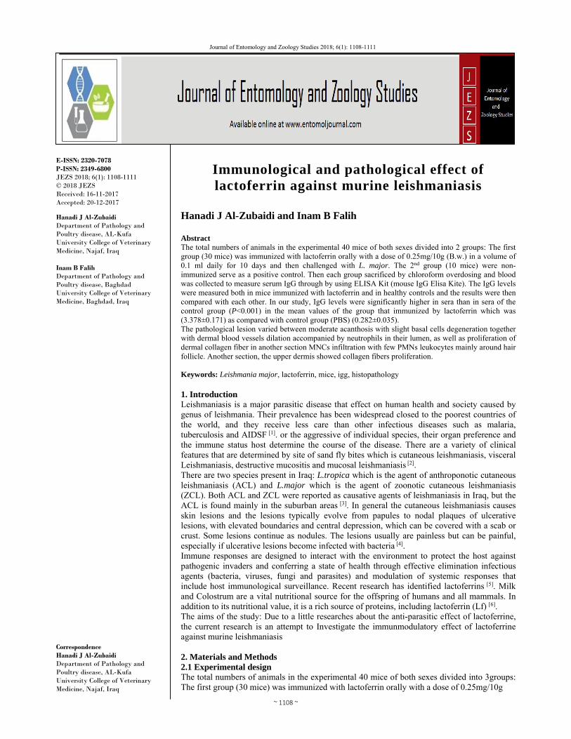
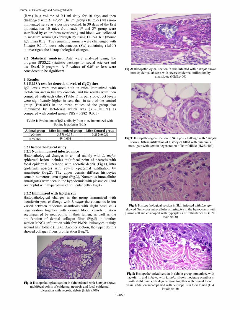
3.2 Histopathological study 3.2.1 Non immunized infected mice Histopathological changes in animal mainly with L. major epidermal lesion includes multifocal point of necrosis with focal epidermal ulceration with necrotic debris (Fig.1), intra epidermal abscess with severe epidermal infiltration by amastigote (Fig.2). The upper dermis diffuses histocytes contain numerous amastigote (Fig.3), Numerous intracellular amastigotes were seen in the hypodermis with plasma cell and eosinophil with hyperplasia of follicular cells (Fig.4). 3.2.2 Immunized with lactoferrin Histopathological changes in the group immunized with lactoferrin post challenge with L.major the cutaneous lesion varied between moderate acanthosis with slight basal cells degeneration together with dermal blood vessels dilation accompanied by neutrophils in their lumen, as well as the proliferation of dermal collagen fiber (Fig.5) in another section MNCs infiltration with few PMNs leukocytes mainly around hair follicle (Fig.6). Another section, the upper dermis showed collagen fibers proliferation (Fig.7).
Fig 1: Histopathological section in skin infected with L.major shows multifocal points of epidermal necrosis and focal epidermal
ulceration with necrotic debris (H&E x400)
Fig 2: Histopathological section in skin infected with L.major shows intra epidermal abscess with severe epidermal infiltration by
amastigote (H&Ex400)
Fig 3: Histopathological section in Skin post challenge with L.major shows Diffuse infiltration of histocytes filled with numerous
amastigote with keratin degeneration of hair follicle (H&Ex400)
Fig 4: Histopathological section in Skin infected with L.major showed Numerous intracellular amastigotes in the hypodermis with
plasma cell and eosinophil with hyperplasia of follicular cells. (H&E stain x400)
Fig 5: Histopathological section in skin in group immunized with lactoferrin and infected with L.major shows moderate acanthosis with slight basal cells degeneration together with dermal blood
vessels dilation accompanied with neutrophils in their lumen (H & Estain x400)
~ 1110 ~
Journal of Entomology and Zoology Studies
Fig 6: Histopathological section in skin in group immunized with lactoferrin and infected with L.major shows MNCs infiltration with
few PMNs leukocytes mainly around hair follicle (H&E x400)
Fig 7: Histopathological section in skin in group immunized with lactoferrin and infected with L.major showed the upper dermis
showed collagen fibers proliferation (H&E x400) 4. Discussion According to El-Loly and Mahfouz whom explained that young of many mammalian species are born without an effective immune system, therefore the Igs and LF (Lactoferrin) exhibit antimicrobial activity and protect the neonate from infection until their own immune system has developed [11]. One of the first factors released by neutrophils upon encounter with pathogens and contributes to innate activation by directing the development of adaptive responses, this is consistent with our results that lactoferrin had a significant effect on the IgG level in serum of mice (P<0.001), and a similar finding was found by [12] in his study both Serum lactoferrin and immunoglobulin G (IgG) concentrations are correlated and increase in healthy animals after ingestion of colostrum. Also Proinflammatory cytokines, Tumor necrosis factor alpha (TNF-α), (Interleukin) IL-6, and IL-1β, can be modulated by lactoferrin, either to increase [13] or decrease [14] production depends upon the type of insult recognized by the immune system in concert with local environmental conditions. In pathological examinations Our data represent focal epidermal ulceration with necrotic debris this agree with [15] Who have been reported In leishmaniasis, the epidermis may be either ulcerated with marked secondary changes and dermal reaction or, alternatively the epidermis may be quite unmarkable and overlying a dense dermal infiltrate. The infiltrate is typically dense and contains predominantly histocytes, lymphocytes and plasma cells. While, the major skin lesion at a later period characterized by severe epidermal and dermal invasion associated with deeper penetration of Leishmania amastigetes this may attributed to TH1 (T helper cell type 1) cytokines TNF (Tumor necrosis factor) and IFNγ (Interferon-gamma) as well as TNF is a cofactor for macrophage activation, and TNF receptor-deficient mice are more susceptible to L. major infections [16].
Progressive cutaneous leishmania lesions have been reported in the current experirmental study mainly associated with epidermal abscess containing a number of neutrophils with intracellular amastigotes together with follicle rupture this observation may indicate that Neutrophils are rapidly recruited to the site of Leishmania infection is consistent with
[17] and parasites promastigotes are killed by neutrophil extracellular traps (NETs) consistent with [18]. However, several studies showed that Lactoferrin modulates the phagocytic capacity of neutrophils and macrophages in the resolution of infections [19] and this evidence is in agreement with present study expressed moderate to severe MNCs and PMNs infiltration in the examined tissue there are several parasites in which Lf and its peptides have been used to control infection such as Toxoplasma gondii, amoebiasis. 5. Conclusions: Lactoferrin is effective for treating and presents suitable oral immune prevention therapy against Leishmaniasis.Lactoferrin oral immunization of Leishmania with (0.25mg/10g B.w.) for 10 days induced inhibition and reduction in Leishmania growth and load. 6. Acknowledgement I would like to thank dr. kifah F. Hasson Al-Shaba/ Microbiology; Ezzate Hasson Ajeena /Zoology/phyiology and my sisters,especially Dr. Diyar Jaafar (Ophthalmologist. M.B.Ch.b / C.A.B. Ophth /FICO). For their kindness, love &support. 7. References 1. Salam N, Al-Shaqha WM, Azzi A. Leishmaniasis in the
Middle East: Incidence and Epidemiology. PLoS Negl Trop Dis. 2014; 8(10):e3208.
2. Reithinger R; Dujardin JC, Louzir H, Pirmez C, Alexander B, Brooker S. Cutaneous leishmaniasis. Lancet Infect Dis. 2007; 7(9): 581-596.
3. World Health Organization. Communicable Disease Working Group on Emergencies, HQ Division of Communicable Disease Control, EMRO, WHO OFFICE, Baghdad. WHO Office, Baghdad. Communicable Disease Toolkit, IRAQ CRISIS, 2003. WHO 2003, 3944. www.who.int/diseasecontrol_emergencies/toolkits/Iraq_profile_ok.pdf.
4. Aronson N, Barbara L, Michael L, Richard P, Rogelio L, Peter W, Edgar M. Diagnosis and Treatment of Leishmaniasis: Clinical Practice Guidelines by the Infectious Diseases Society of America (IDSA) and the American Society of Tropical Medicine and Hygiene, 2016.
5. Actor J, Hwang S, Kruzel M. Lactoferrin as a natural immune modulator. Curr Pharm Des. 2009; 15(17):1956-73.
6. Jenssen H, Hancock R. Antimicrobial properties of lactoferrin. Biochimie. 2009; 91:19-29.
7. Younis N, Aboody AL, Clinical Isolation B, Molecular Diagnosis of cutaneous Leishmaniasis by using KDNA gene PCR at some Baghdad Hospital. 2017; 155/4/m.
8. Kagan I, Norman L. In Manual of clinical microbiology AM. Soc. Microbiology, Washington. 1970; 453-486.
9. Dawson R, Elliot D, Elliot W, Jons K. Data for biochemic research 2ed., Claredenpress, Oxford. 1969, 508.
10. Wakabayashi H, Kurokawa M, Shin K, Teraguchi S, Tamura Y, Shiraki K. Oral lactoferrin prevents body weight loss and increases cytokine responses during

~ 1111 ~
Journal of Entomology and Zoology Studies
herpes simplex virus type1 infection of mice. Biosci Biotechnol Biochem. 2004; 68:537-44.
11. El-Loly Ml, Mahfouz M. Lactoferrin in Relation to Biological Functions and Applications: A Review. International Journal of Dairy Science. 2011; 6:79-111.
12. Barton MH, Hurley D, Norton N, Heusner G, Costa L, Jones S, et al. Serum lactoferrin and immunoglobulin G concentrations in healthy or ill neonatal foals and healthy adult horses. Journal Vet Intern Med. 2006; 20(6):1457-62.
13. Zimecki M, Wlaszczyk A, Wojciechowski R, Dawiskiba J, Kruzel M. Lactoferrin regulates the immune responses in post-surgical patients. Arch Immunol Ther Exp (Warsz). 2001; 49:325-33.
14. Zimecki M, Dawiskiba J, Zawirska B, Krawczyk Z, Kruzel M. Bovine lactoferrin decreases histopathological changes in the liver and regulates cytokine production by splenocytes of obstructive jaundiced rats. Inflamm Res. 2003; 52:305-10.
15. Patrick E. Leishmaniasis pathology. Derm Net New Zealand, 2013.
16. Wilhelm P, Ritter U, Labbow S, Donhauser N, Röllinghoff M, Bogdan C, et al. Rapidly fatal leishmaniasis in resistant C57BL/6 mice lacking TNF. J Immunol. 2001; 4012-9.
17. Guimaraes-Costa AB, Michelle TC, Giselle S, Rodrigo PP, Soares Fernanda N, Elvira M. Saraiva Leishmania amazonensis promastigotes induce and are killed by neutrophilextracellular traps. Proc. Natl Acad. Sci. USA. 2009; 106:6748-6753.
18. Dobson CM. Experimental investigation of protein folding and misfolding. Methods. 2004; 34(1):4-14.
19. Legrand D, Pierce A, Elass E, Carpentier M, Mariller C, Mazurier J. Lactoferrin structure and functions. Adv Exp Med Biol. 2008; 606:163-94.
20. Fatoux-Ardore M, Peysselon F, Weiss A, Bastien P, Pratlong F, Ricard-Blum S. Large-scale investigation of Leishmania interaction networks with host extracellular matrix by surface plasmon resonance imaging. Infect Immun United States. 2014; 82:594-606.
21. Dunn LL, Rahmanto YS, Richardson DR. Iron uptake and metabolism in the new millennium. Trends Cell Biol. 2007; 17(2):93-100.
22. Abreu-Silva AL, Calabrese KS, Mortara RA, Tedesco RC, Cardoso FO, Carvalho LO. Et al. Extracellular matrix alterations in experimental murine Leishmania (L.) amazonensis infection. Parasitology. 2004; 385-90.