Embed Size (px)
Citation preview

CLINICAL MICROBIOLOGY REVIEWS,0893-8512/00/$04.0010
Apr. 2000, p. 318–331 Vol. 13, No. 2
Copyright © 2000, American Society for Microbiology. All Rights Reserved.
Pathogenesis of Intestinal Amebiasis: From Molecules to DiseaseMARTHA ESPINOSA-CANTELLANO* AND ADOLFO MARTINEZ-PALOMO
Center for Research and Advanced Studies, Mexico City, Mexico
INTRODUCTION .......................................................................................................................................................318ACUTE INTESTINAL AMEBIASIS.........................................................................................................................319
Macroscopic Lesions ..............................................................................................................................................319Microscopic Findings .............................................................................................................................................320
PATHOGENESIS OF INTESTINAL AMEBIASIS................................................................................................320Nonspecific Lesion ..................................................................................................................................................320Mucopenic Depression ...........................................................................................................................................320Early Invasive Lesions with Superficial Ulceration ...........................................................................................322
Superficial epithelial erosion ............................................................................................................................322(i) Gal-GalNAc lectin .....................................................................................................................................322(ii) Amebapores...............................................................................................................................................323
Interglandular foci of microinvasion ...............................................................................................................323(i) Invasion through locomotion...................................................................................................................323(ii) Degradation of ECM components by cysteine proteases....................................................................324
Neutrophil infiltration of the lamina propria.................................................................................................325Role of neutrophils .............................................................................................................................................326
Late Invasive Lesion with Deep Ulceration ........................................................................................................326Role of macrophages ..........................................................................................................................................327
Granulating Ulcer...................................................................................................................................................328CONCLUSIONS .........................................................................................................................................................328ACKNOWLEDGMENTS ...........................................................................................................................................328REFERENCES ............................................................................................................................................................328
INTRODUCTION
Amebiasis is the infection of the human gastrointestinaltract by Entamoeba histolytica, a protozoan parasite that iscapable of invading the intestinal mucosa and may spread toother organs, mainly the liver. Entamoeba dispar, an amebamorphologically similar to E. histolytica that also colonizes thehuman gut, has been recognized recently as a separate specieswith no invasive potential (8, 35, 41, 90). The acceptance of E.dispar as a distinct but closely related protozoan species hashad profound implications for the epidemiology of amebiasis,since most asymptomatic infections found worldwide are nowattributed to this noninvasive ameba.
Currently, there is no low-cost laboratory test available forthe differentiation of E. histolytica from E. dispar infections.The development of this valuable diagnostic tool for use inclinical laboratories and large-scale epidemiological studieshas been made a priority (8) and is the subject of intenseresearch (2, 20, 57, 60, 63, 96, 129, 161, 170). Preliminary dataobtained from the application of these methods confirm thepresence of E. dispar in most asymptomatic amebic infections,although E. histolytica asymptomatic colonization is not un-common (19, 58, 59, 130, 171). Of note, the prevalence rates ofboth species in different geographical areas is still difficult toestimate due to the small number of samples analyzed.
Invasive amebiasis due to E. histolytica is more common indeveloping countries. In areas of endemic infection, a varietyof conditions including ignorance, poverty, overcrowding, in-adequate and contaminated water supplies, and poor sanita-
tion favor direct fecal-oral transmission of amebas from oneperson to another. Being responsible for approximately 70thousand deaths annually, amebiasis is the fourth leading causeof death due to a protozoan infection after malaria, Chagas’disease, and leishmaniasis and the third cause of morbidity inthis organism group after malaria and trichomoniasis, accord-ing to recent World Health Organization estimates (177).
The motile form of E. histolytica, the trophozoite, lives in thelumen of the large intestine, where it multiplies and differen-tiates into the cyst, the resistant form responsible for the trans-mission of the infection. Cysts are excreted in stools and maybe ingested by a new host via contaminated food or water. Theparasite excysts in the terminal ileum, with each emergingquadrinucleate trophozoite giving rise to eight uninucleatedtrophozoites. Trophozoites may invade the colonic mucosa andcause dysentery and, through spreading via the bloodstream,may give rise to extraintestinal lesions, mainly liver abscesses.
Depending on the affected organ, the clinical manifestationsof amebiasis are intestinal or extraintestinal. There are fourclinical forms of invasive intestinal amebiasis, all of which aregenerally acute: dysentery or bloody diarrhea, fulminating co-litis, amebic appendicitis, and ameboma of the colon. Dysen-teric and diarrheic syndromes account for 90% of cases ofinvasive intestinal amebiasis. Patients with dysentery have anaverage of three to five mucosanguineous evacuations per day,with moderate colic pain preceding discharge, and they haverectal tenesmus. In patients with bloody diarrhea, evacuationsare also few but the stools are composed of liquid fecal mate-rial stained with blood. While there is moderate colic pain,there is no rectal tenesmus. Fever and systemic manifestationsare generally absent. These syndromes constitute the classicambulatory dysentery and can easily be distinguished from thatof bacterial origin, where the patient frequently complains of
* Corresponding author. Mailing address: Department of Experi-mental Pathology, Cinvestav, Av. IPN 2508 esq. Ticoman, Col. SanPedro Zacatenco, 07360 Mexico, D.F. Phone: (52) 57 47 38 00 ext.5660. Fax: (52) 57 47 98 90. E-mail: [email protected].
318
systemic signs and symptoms such as fever, chills, headache,malaise, anorexia, nausea, vomiting, cramping abdominal pain,and tenesmus (reviewed in reference 89).
Although E. histolytica can infect almost every organ of thebody, the most frequent form of extraintestinal amebiasis is theamebic liver abscess. This condition, which results from themigration of trophozoites from the colon to the liver throughthe portal circulation, is 10 times more common in adults thanin children and 3 times more frequent in males than in females(144). In general, the onset is abrupt, with pain in the righthypochondrium radiating toward the right shoulder and scap-ular area. The pain usually increases with deep breathing, withcoughing, and while stepping on the right foot during walking.When the abscess is localized to the right lobe, symptomsinclude an irritative cough that is sometimes productive and apleuritic type of chest pain. Abscesses in the upper left lobe cancause epigastric, sometimes dyspneic pain, at times spreadingto the base of the neck and to one or both shoulders. Feverbetween 38 to 40°C is found in 85 to 90% of patients withamebic liver abscess. The patient commonly has chills andprofuse sweating in the afternoon and at night. Other symp-toms include anorexia, nausea, vomiting, diarrhea (with orwithout blood), and dysentery. On physical examination, thecardinal sign of amebic liver abscess is painful hepatomegaly.Digital pressure and fist percussion will often produce intensepain in the liver region. On palpation, the liver is soft andsmooth, in contrast to the rough, hard, irregular character ofthe liver in patients with cirrhosis and hepatocarcinoma. Jaun-dice is present in 8% of the patients who respond well totreatment. When jaundice is severe, multiple abscesses shouldbe suspected. Diarrhea or dysentery is seen in fewer thanone-third of patients. Complications of amebic liver abscessinclude perforation to the pericardial space, pleura, or perito-neal cavity (reviewed in reference 89).
The diagnosis of invasive intestinal amebiasis is still based onthe microscopic identification of E. histolytica trophozoites inrectal smears or recently evacuated stools and on the results ofrectosigmoidoscopy. Trophozoites are most likely to be foundin the bloody mucus and in the yellowish exudate covering themucosal ulcerations obtained during rectosigmoidoscopy. Di-agnostic problems arise when only cysts are identified in stoolsof healthy or diarrheic individuals. A commercially availablelaboratory test based on the identification of specific E. histo-lytica antigens in stool (60) is able to discriminate E. histolyticafrom E. dispar cysts (W. A. Petri, unpublished observations).However, the high cost and lack of knowledge of this test havehindered its use in clinical laboratories, especially in countrieswhere amebiasis is endemic. Until these new diagnostic testsare widely available to clinical laboratories, these samplesshould be reported as containing E. histolytica/E. dispar (8).
The diagnosis of amebic liver abscess is sometimes difficult.In areas of endemic infection or when there is a history oftravel to such places, amebic abscess should be suspected inpatients with spiking fever, weight loss, and abdominal pain inthe upper right quadrant or epigastrium and in patients withtenderness in the liver area. The presence of leukocytosis, ahigh alkaline phosphatase level, and an elevated right dia-phragm suggest a hepatic abscess. The diagnosis is confirmedby ultrasonography or by computed tomography (CT) scans.The CT scan is the most precise method for identifying hepaticabscesses, especially when they are small, and following intra-venous injection of contrasting agents, it is of great value in thedifferential diagnosis of other focal lesions of the liver (144).The high cost of this method, however, limits its use to caseswhen there are doubts about the diagnosis.
Serological tests for antiamebic antibodies are positive in
approximately 75% of patients with invasive colonic amebiasisand in over 90% of patients with amebic liver abscesses. Of alltests available, the Centers for Disease Control and Preventionhas chosen the enzyme immunoassay as its standard serologicalreference test for amebiasis. However, in areas of endemicinfection, the high prevalence of antiamebic antibodies in thegeneral population reduces the usefulness of serological testsfor diagnosis (reviewed in reference 89).
The powerful lytic activity of E. histolytica, for which theparasite was named, has inspired a variety of approachesaimed at understanding the pathogenesis of invasive amebiasis.Most studies have focused on a single factor in an attempt todissect the multiple mechanisms used by the parasite that ul-timately result in tissue destruction. The aim of the presentreview is to provide an overview of the pathological lesions ofhuman intestinal amebiasis and to discuss recent advances inthe study of the molecular mechanisms of amebic pathogenic-ity.
ACUTE INTESTINAL AMEBIASIS
Intestinal invasive amebiasis may be associated with a varietyof anatomical alterations such as acute ulcerative colitis, toxicmegacolon, ameboma, or amebic appendicitis. Amebic ulcer-ative colitis is by far the most frequent and is thus the focus ofthis discussion.
Macroscopic Lesions
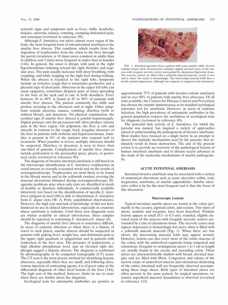
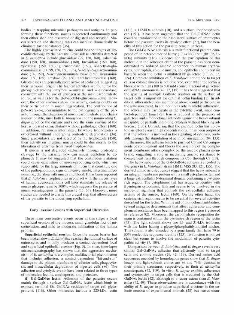
Typical intestinal amebic ulcers are found in the colon (pri-marily in the cecum), sigmoid colon, and rectum. Two types ofulcers, nodular and irregular, have been described. Nodularlesions appear as small (0.1- to 0.5-cm), rounded, slightly ele-vated areas of the mucosa with irregular necrotic centers sur-rounded by a rim of edematous tissue. The necrotic center mayappear depressed or hemorrhagic but more often is filled witha yellowish mucoid material (Fig. 1). When there are fewulcers, the intervening mucosal folds may appear normal.However, lesions can also cover most of the entire mucosa ofthe colon, with the uninvolved segments being congested andedematous. Irregular or serpinginous ulcers 1 to 5 cm in lengthare usually found in the cecum and ascending colon. Theseulcers are characteristically shallow with broad, elevated mar-gins and are filled with fibrin. Congestion and edema of thenarrow strips of uninvolved mucosa and edematous thickeningof the entire intestinal wall are frequently observed accompa-nying these large ulcers. Both types of intestinal ulcers areoften present in the same patient. In surgical specimens, ex-tensive superficial mucosal denudation is observed (reviewedin reference 113).
FIG. 1. Intestinal specimen from a patient with acute amebic colitis. Severalnodular lesions show characteristic rounded, slightly elevated areas of the mu-cosa with irregular necrotic centers surrounded by edematous hyperemic tissue.The necrotic centers are filled with a yellowish mucoid material, except in twoulcers, where the center is hemorrhagic. The intervening mucosal folds have amostly normal appearance, although one segment is congested and edematous.
VOL. 13, 2000 PATHOGENESIS OF INTESTINAL AMEBIASIS 319
Microscopic Findings
The microscopic changes characteristic of amebic ulcerativecolitis have been studied in human rectal biopsy specimens.Little has been added to the now classical description by Pra-thap and Gilman, published in 1970, who found five types oflesions that seem to correspond to the progression of damagecaused by E. histolytica trophozoites (120).
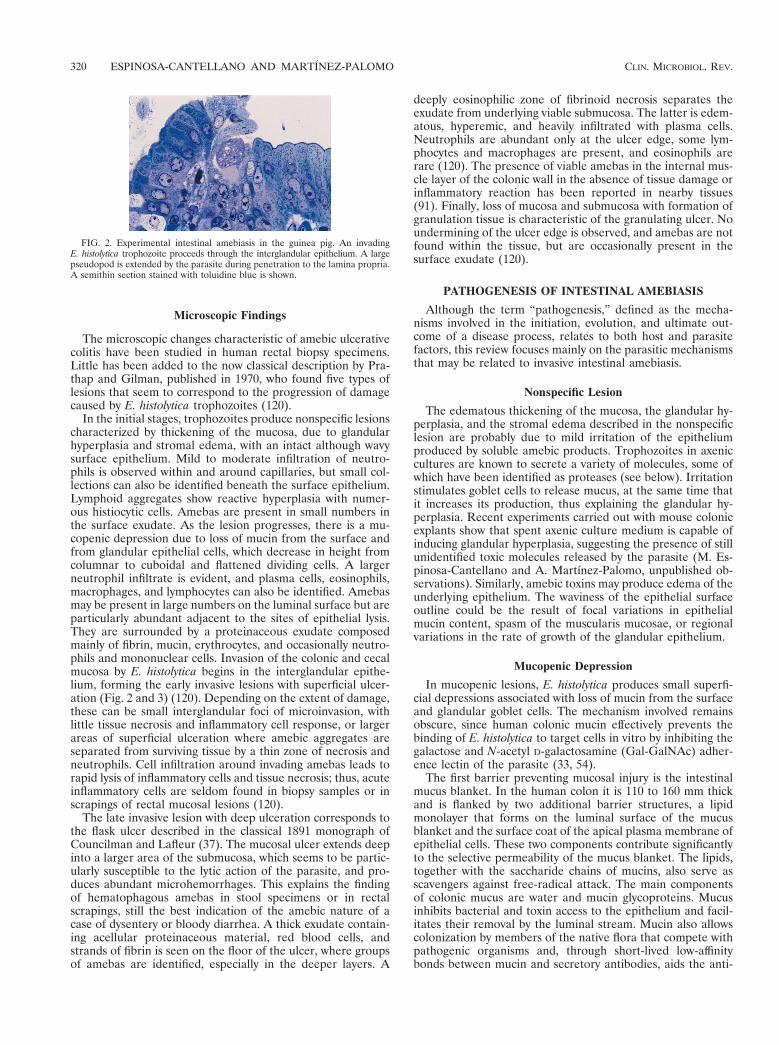
In the initial stages, trophozoites produce nonspecific lesionscharacterized by thickening of the mucosa, due to glandularhyperplasia and stromal edema, with an intact although wavysurface epithelium. Mild to moderate infiltration of neutro-phils is observed within and around capillaries, but small col-lections can also be identified beneath the surface epithelium.Lymphoid aggregates show reactive hyperplasia with numer-ous histiocytic cells. Amebas are present in small numbers inthe surface exudate. As the lesion progresses, there is a mu-copenic depression due to loss of mucin from the surface andfrom glandular epithelial cells, which decrease in height fromcolumnar to cuboidal and flattened dividing cells. A largerneutrophil infiltrate is evident, and plasma cells, eosinophils,macrophages, and lymphocytes can also be identified. Amebasmay be present in large numbers on the luminal surface but areparticularly abundant adjacent to the sites of epithelial lysis.They are surrounded by a proteinaceous exudate composedmainly of fibrin, mucin, erythrocytes, and occasionally neutro-phils and mononuclear cells. Invasion of the colonic and cecalmucosa by E. histolytica begins in the interglandular epithe-lium, forming the early invasive lesions with superficial ulcer-ation (Fig. 2 and 3) (120). Depending on the extent of damage,these can be small interglandular foci of microinvasion, withlittle tissue necrosis and inflammatory cell response, or largerareas of superficial ulceration where amebic aggregates areseparated from surviving tissue by a thin zone of necrosis andneutrophils. Cell infiltration around invading amebas leads torapid lysis of inflammatory cells and tissue necrosis; thus, acuteinflammatory cells are seldom found in biopsy samples or inscrapings of rectal mucosal lesions (120).
The late invasive lesion with deep ulceration corresponds tothe flask ulcer described in the classical 1891 monograph ofCouncilman and Lafleur (37). The mucosal ulcer extends deepinto a larger area of the submucosa, which seems to be partic-ularly susceptible to the lytic action of the parasite, and pro-duces abundant microhemorrhages. This explains the findingof hematophagous amebas in stool specimens or in rectalscrapings, still the best indication of the amebic nature of acase of dysentery or bloody diarrhea. A thick exudate contain-ing acellular proteinaceous material, red blood cells, andstrands of fibrin is seen on the floor of the ulcer, where groupsof amebas are identified, especially in the deeper layers. A
deeply eosinophilic zone of fibrinoid necrosis separates theexudate from underlying viable submucosa. The latter is edem-atous, hyperemic, and heavily infiltrated with plasma cells.Neutrophils are abundant only at the ulcer edge, some lym-phocytes and macrophages are present, and eosinophils arerare (120). The presence of viable amebas in the internal mus-cle layer of the colonic wall in the absence of tissue damage orinflammatory reaction has been reported in nearby tissues(91). Finally, loss of mucosa and submucosa with formation ofgranulation tissue is characteristic of the granulating ulcer. Noundermining of the ulcer edge is observed, and amebas are notfound within the tissue, but are occasionally present in thesurface exudate (120).
PATHOGENESIS OF INTESTINAL AMEBIASIS
Although the term “pathogenesis,” defined as the mecha-nisms involved in the initiation, evolution, and ultimate out-come of a disease process, relates to both host and parasitefactors, this review focuses mainly on the parasitic mechanismsthat may be related to invasive intestinal amebiasis.
Nonspecific Lesion
The edematous thickening of the mucosa, the glandular hy-perplasia, and the stromal edema described in the nonspecificlesion are probably due to mild irritation of the epitheliumproduced by soluble amebic products. Trophozoites in axeniccultures are known to secrete a variety of molecules, some ofwhich have been identified as proteases (see below). Irritationstimulates goblet cells to release mucus, at the same time thatit increases its production, thus explaining the glandular hy-perplasia. Recent experiments carried out with mouse colonicexplants show that spent axenic culture medium is capable ofinducing glandular hyperplasia, suggesting the presence of stillunidentified toxic molecules released by the parasite (M. Es-pinosa-Cantellano and A. Martınez-Palomo, unpublished ob-servations). Similarly, amebic toxins may produce edema of theunderlying epithelium. The waviness of the epithelial surfaceoutline could be the result of focal variations in epithelialmucin content, spasm of the muscularis mucosae, or regionalvariations in the rate of growth of the glandular epithelium.
Mucopenic Depression
In mucopenic lesions, E. histolytica produces small superfi-cial depressions associated with loss of mucin from the surfaceand glandular goblet cells. The mechanism involved remainsobscure, since human colonic mucin effectively prevents thebinding of E. histolytica to target cells in vitro by inhibiting thegalactose and N-acetyl D-galactosamine (Gal-GalNAc) adher-ence lectin of the parasite (33, 54).
The first barrier preventing mucosal injury is the intestinalmucus blanket. In the human colon it is 110 to 160 mm thickand is flanked by two additional barrier structures, a lipidmonolayer that forms on the luminal surface of the mucusblanket and the surface coat of the apical plasma membrane ofepithelial cells. These two components contribute significantlyto the selective permeability of the mucus blanket. The lipids,together with the saccharide chains of mucins, also serve asscavengers against free-radical attack. The main componentsof colonic mucus are water and mucin glycoproteins. Mucusinhibits bacterial and toxin access to the epithelium and facil-itates their removal by the luminal stream. Mucin also allowscolonization by members of the native flora that compete withpathogenic organisms and, through short-lived low-affinitybonds between mucin and secretory antibodies, aids the anti-
FIG. 2. Experimental intestinal amebiasis in the guinea pig. An invadingE. histolytica trophozoite proceeds through the interglandular epithelium. A largepseudopod is extended by the parasite during penetration to the lamina propria.A semithin section stained with toluidine blue is shown.
320 ESPINOSA-CANTELLANO AND MARTINEZ-PALOMO CLIN. MICROBIOL. REV.
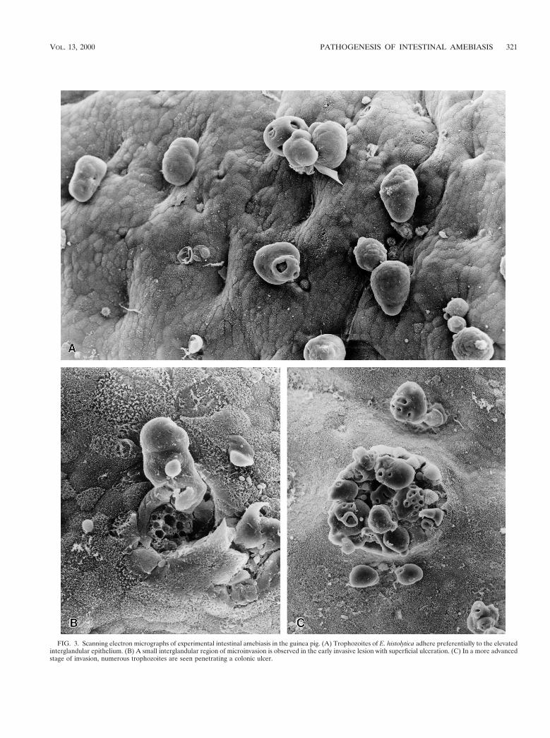
FIG. 3. Scanning electron micrographs of experimental intestinal amebiasis in the guinea pig. (A) Trophozoites of E. histolytica adhere preferentially to the elevatedinterglandular epithelium. (B) A small interglandular region of microinvasion is observed in the early invasive lesion with superficial ulceration. (C) In a more advancedstage of invasion, numerous trophozoites are seen penetrating a colonic ulcer.
VOL. 13, 2000 PATHOGENESIS OF INTESTINAL AMEBIASIS 321
bodies in trapping microbial pathogens and antigens. In per-forming these functions, mucus is secreted continuously andthen either shed and discarded or digested and recycled. Mu-cus secretion and shedding rates can increase dramatically toeliminate toxic substances (36).
The highly glycosylated mucins could be the targets of gly-cosidic cleavage by the parasite. Glycosidase activities detectedin E. histolytica include glucosidase (99, 150, 160), galactosi-dase (150, 160), mannosidase (160), fucosidase (150, 160),xylosidase (150, 160), glucuronidase (160), N-acetyl-D-glu-cosaminidase (14, 99, 150, 160, 176), N-acetyl-D-galactosamini-dase (14, 150), N-acetylneuraminate lyase (160), neuramini-dase (160, 165), amylase (99, 160), and hyaluronidase (160).Glycosidases are generally more active at acidic pH, suggestingtheir lysosomal origin. The highest activities are found for theglycogen-degrading enzymes a-amylase and a-glucosidase,consistent with the use of glycogen as the main carbohydratesource, as well as for the b-N-acetyl-D-glucosaminidase. How-ever, the other enzymes show low activity, casting doubts ontheir participation in mucin degradation. The contribution ofb-N-acetyl-D-glucosaminidase to the pathogenicity of the par-asite through the digestion of mucin carbohydrate side chainsis questionable, since both E. histolytica and the noninvading E.dispar produce the enzyme and since the same enzyme of thehost is present extracellularly without pathologic effect (160).In addition, rat mucin internalized by whole trophozoites isexocytosed without undergoing proteolytic degradation (34).Since glycosidases are not secreted by the trophozoite (150),their activity on intestinal mucus could be due mostly to theliberation of enzymes from lysed trophozoites.
If mucin is not degraded exclusively through proteolyticcleavage by the parasite, how can mucus depletion be ex-plained? It may be suggested that the continuous irritationcould cause exhaustion of mucus-producing cells, which areresponsible for the large amounts of mucus that constitute oneof the pathognomonic signs of invasive amebic intestinal infec-tions, i.e., diarrhea with mucus and blood. It has been reportedthat E. histolytica trophozoites in contact with the mucus layerof human adenocarcinoma cell lines stimulate the release ofmucus glycoproteins by 300%, which suggests the presence ofmucin secretagogues in the parasite (15, 66). However, morestudies are needed to clarify this crucial step that allows accessof the parasite to the underlying epithelium.
Early Invasive Lesions with Superficial Ulceration
Three main consecutive events occur at this stage: a focalsuperficial erosion of the mucosa, small glandular foci of mi-croinvasion, and mild to moderate infiltration of the laminapropria.
Superficial epithelial erosion. Once the mucus barrier hasbeen broken down, E. histolytica reaches the luminal surface ofenterocytes and initially produces a contact-dependent focaland superficial epithelial erosion (Fig. 3). In vitro, time-lapsemicrocinematography has shown that the aggressive mecha-nism of E. histolytica is a complex multifactorial phenomenonthat includes adhesion, a contact-dependent “hit-and-run”damage to the plasma membrane of effector cells, phagocyto-sis, and intracellular degradation of ingested cells (86). Theadhesion and cytolytic events have been related to three typesof molecules: lectins, amebapores, and proteases.
(i) Gal-GalNAc lectin. Adhesion of the parasite occursmainly through a surface Gal-GalNAc lectin which binds toexposed terminal Gal-GalNAc residues of target cell glyco-proteins (116). Other molecules include a 220-kDa lectin
(131), a 112-kDa adhesin (10), and a surface lipophosphogly-can (151). It has been suggested that the Gal-GalNAc lectincould be translocated to the basolateral surface of enterocytesbefore the parasite exerts its cytolytic effect (77), but the ben-efits of this action for the parasite remain unclear.
The Gal-GalNAc adhesin is a multifunctional protein com-posed of an heterodimer of heavy (170-kDa) and light (35/31-kDa) subunits (116). Evidence for the participation of thismolecule in the adhesion event of the parasite has been dem-onstrated by reduced amebic adherence to human erythro-cytes, neutrophils, colonic mucins, and epithelia and to certainbacteria when the lectin is inhibited by galactose (17, 29, 33,124). Complete inhibition of E. histolytica adherence to targetcells or colonic mucins is not observed, even when the lectin isblocked with high (100 to 500 mM) concentrations of galactoseor GalNAc monomers (42, 77, 115). It has been suggested thatthe spacing of multiple GalNAc residues on the surface oftarget cells is important for optimal lectin binding (1). In ad-dition, other molecules (mentioned above) could participate inthe adhesion event. In addition to its role in amebic adherence,the adhesin may participate in the cytolytic event, since con-tact-dependent target cell lysis is reduced in the presence ofgalactose and a monoclonal antibody against the heavy subunitis capable of partially inhibiting cytolysis without blocking ad-herence (133). Considering that the purified lectin has no cy-totoxic effect even at high concentrations, it has been proposedthat the adhesin is involved in the signaling of cytolysis, prob-ably through the stimulation of actin polymerization (84, 133).Furthermore, the adhesin binds to purified C8 and C9 compo-nents of complement and blocks the assembly of the comple-ment membrane attack complex on the amebic plasma mem-brane, suggesting a role in mediating amebic resistance tocomplement lysis through components C5b through C9 (18).
The heavy subunit of the Gal-GalNAc adhesin is encoded byfive genes in E. histolytica strain HM1:IMSS (83, 121, 155). Thederived amino acid sequences suggest that the heavy subunit isan integral membrane protein with a small cytoplasmic tail anda large extracellular N-terminal domain containing a cysteine-rich region. The cytoplasmic tail has sequence identity withb2-integrin cytoplasmic tails and seems to be involved in theinside-out signaling that controls the extracellular adhesiveactivity of the amebic lectin (169). On the other hand, thecysteine-rich region seems to be essential for several activitiesdescribed for the lectin. With the aid of monoclonal antibodies,several antigenic determinants that affect adherence and com-plement resistance have been mapped to this region (reviewedin reference 92). Moreover, the carbohydrate recognition do-main is contained within the cysteine-rich region of the lectin(43). The light subunit shows two 35- and 31-kDa isoforms,with the latter having a glycosylphosphatidylinositol anchor.This subunit is also encoded by a gene family that have 79 to85% nucleotide sequence identity (123). Its function is not yetclear but seems to involve the modulation of parasite cyto-pathic activity (7, 109).
Comparison between E. histolytica and E. dispar reveals verysimilar Gal-GalNAc adhesins that efficiently bind to targetcells and colonic mucins (29, 42, 119). Derived amino acidsequences encoded by homologous genes show that E. disparheavy- and light-subunit clones are 86 and 79% identical intheir primary structures, respectively, to their E. histolyticacounterparts (42, 119). In vitro, E. dispar exhibits adherenceand cytotoxicity to target cells that is mediated by the Gal-GalNAc lectin (42), although to a lesser extent than E. histo-lytica (42, 49). These observations are in accordance with theability of E. dispar to produce superficial erosions in the co-lonic mucosa of experimental animals (28) but leaves unan-
322 ESPINOSA-CANTELLANO AND MARTINEZ-PALOMO CLIN. MICROBIOL. REV.
swered the question why this ameba does not invade any fur-ther.
(ii) Amebapores. Once E. histolytica establishes contact withmammalian cells in vitro, a rapid cytolytic event takes placethat results in swelling, surface blebbing, and lysis of the inad-vertent target cell, including lymphocytes, polymorphonuclearleukocytes, and macrophages, leaving the parasite unharmed.The similarity of this event to the perforin-mediated lysis oftarget cells by cytotoxic T lymphocytes (162) initially suggestedthe participation of a channel-forming protein called the ame-bapore, whose activity had been identified in E. histolyticalysates (82, 132, 178).
The amebapore of E. histolytica is a channel-forming peptideof 77 amino acid residues, which has now been purified; theprotein has been sequenced, and the respective genes havebeen cloned (68, 69). Three isoforms, amebapores A, B, and C,are present at a ratio of 35:10:1, respectively, with the genesshowing 35 to 57% deduced amino acid sequence identity. Themolecules share six cysteine residues at identical positions anda histidine residue near the C terminus. Structural modelingsuggests a compact tertiary structure composed of four a-he-lical structures stabilized by three disulfide bonds (72). Thus,amebapores are different from the much larger (65- to 70-kDa)perforins that contain three amphipathic segments, two a-he-lices and one b-sheet (162). However, similarities at the struc-tural and functional levels have been found between ameba-pores and NK-lysin, a polypeptide present in natural killer(NK) cells and cytotoxic T lymphocytes of pigs (reviewed inreferences 73 and 75).
Like other pore-forming peptides, amebapores are readilysoluble but are capable of rapidly changing into a membrane-inserted stage (75). Early observations suggested that the mol-ecule forms multistate channels with similar properties tothose found in the barrel-stave aggregates of toxins such asalamethacin (65). With the elucidation of the primary andsecondary structures, it is now believed that amebapores ag-gregate through the arrangement of their amphipathic a-heli-ces. In a model proposed by Andra and Leippe (5), ameba-pores bind to negatively charged phospholipids via protonatedlysine residues; this is followed by the insertion of the peptideinto the lipid bilayer driven by the negative membrane poten-tial of the target membrane. Oligomerization of the peptideoccurs with the participation of a key histidine residue (His75),that may either interact with another monomer through theformation of hydrogen bonds or stabilize the predicted fourtha-helix. The oligomer forms a channel through the plasmamembrane, allowing the passage of water, ions, and other smallmolecules and thus lysing the target cell.
In vitro, amebapores exert cytolytic activity against severalhuman cultured cell lines. Amebapore C seems to be the mosteffective, while amebapore A is not efficient in lysing erythro-cytes. In addition, the peptides show potent antibacterial ac-tivity against gram-positive bacteria by damaging their surfacemembranes. Damage to the outer membrane-shielded gram-negative bacteria requires high concentrations of amebaporeor removal of the wall with lysozyme (6, 71, 72). With the useof synthetic peptides, of the four a-helices present in the mol-ecule, helix 3 was found to be responsible for the membranepenetration, displaying the highest antibacterial activity. Inter-estingly, helix 3 is also the most highly conserved domain in thethree isoforms (71). The peptide derived from isoform C wasthe most active of several synthetic peptides studied, reachinga magnitude of activity in the range of the whole molecule (6).
Amebapores are localized in cytoplasmic vesicles, as evi-denced by positive immunofluorescence staining and by thepresence of typical signal peptides of intracellular transport in
its primary translation products. The peptides show maximumactivity at acidic pH, which is consistent with previous obser-vations that lysis of target cells by E. histolytica required a pHof 5.0 within amebic vesicles (69).
A peptide homologous to E. histolytica amebapore A hasbeen identified in E. dispar (70). The molecules have commonstructural and functional properties, such as insertion into neg-atively charged liposomes, highest activity at low pH, localiza-tion in cytoplasmic vesicles, 95% identity of primary structures,and a high degree of similarity of secondary-structure predic-tions. In spite of these similarities, the specific activity of the E.dispar amebapore is 60% lower than that of the one in E.histolytica. This may be related to a shortened amphipathichelix in the former (70).
Surprisingly, in spite of all the advances in the biochemistryand molecular biology of amebapores, their participation inthe cytolytic event produced by E. histolytica has not yet beendemonstrated. Amebapores are not spontaneously secretedfrom viable trophozoites (74). Whether the molecule is able toinsert into target cell membranes upon adherence in vivo re-mains to be established.
The presence of pore-forming activity in the noninvasive E.dispar suggests that the primary function of amebapores is todestroy phagocytosed bacteria, the main intestinal source ofnutrients of amebas; thus, they have a similar function to de-fensins found in mammalian phagocytes that kill bacteria andfungi to prevent intracellular microbial growth within digestivevacuoles (reviewed in references 75 and 158). The anaerobicenvironment found in the colon may favor oxygen-independentmechanisms as a means of destroying bacteria, rather thanusing oxygen metabolites and nitric oxide. Amebapores andother proteins, like the recently characterized ameba lysozymethat colocalizes to the same cytoplasmic granules of ameba-pores (61, 105), could synergistically enhance the antibacterialactivity.
Interglandular foci of microinvasion. Focal superficial epi-thelial erosions produced by E. histolytica in humans are fol-lowed by small interglandular foci of microinvasion. Electronmicroscopy studies of experimentally infected rodents haveconfirmed the invasion of trophozoites through the interglan-dular epithelium (Fig. 3), where pronounced shedding of des-quamating epithelial cells takes place. The superficial desqua-mation seems to render this area particularly susceptible toinvasion, which occurs through an active displacement of ame-bas with large pseudopodia extending toward the basal epithe-lial layers (88, 149). During their passage to deeper layers ofthe intestine, trophozoites must lyse surrounding cells anddegrade the extracellular matrix (ECM) components of thecolonic mucosa. Thus, this stage of lesion is characterized bycontinuing lysis of cells, penetration through locomotion, andproteolytic degradation of ECM (Fig. 2). The way in which theparasite exerts its cytolytic action is described above. The lasttwo events, locomotion and ECM degradation, seem to beclosely related and possibly occur through cycles of anchorageto ECM components, forward movement, and degradation ofECM.
(i) Invasion through locomotion. In mammals, cell locomo-tion involves at least three processes: (i) extension of a leadingedge, (ii) attachment to the substrate surface through adhesionplaques, and (iii) pulling forward of the remainder of the cell.The motile force underlying cytoplasmic streaming is a Ca21-activated interaction between actin and myosin, similar to theone described for skeletal muscle contraction. In E. histolytica,actin polymerization occurs during pseudopod extension (47).No reports are available on fluctuations in intracellular Ca21
concentration ([Ca21]i) during locomotion, but the presence of
VOL. 13, 2000 PATHOGENESIS OF INTESTINAL AMEBIASIS 323

myosin II in leading lamellae (47) suggests that the mechanismfor leading-edge extension in amebas is similar to that in mam-malian motile cells.
Interaction of E. histolytica trophozoites with fibronectin(FN) and other extracellular matrix substrates induces the for-mation of adhesion plaques. Ligand recognition and bindingseem to involve a 37-kDa FN-binding protein (152) and a140-kDa integrin-like receptor (153, 154). Isolated amebic ad-hesion plaques are composed mainly of actin filaments and atleast four actin binding proteins: vinculin, a-actinin, tropomy-osin, and myosin I (166). Vinculin may serve as a link betweenactin and an integral component during anchoring of the cy-toskeleton to the plasma membrane in higher eukaryotes. Itsidentification in amebic adhesion plaques suggests a similarfunction in this parasite. The presence of a-actinin in E. his-tolytica had already been reported (13). However, the iden-tification of this gel-forming protein in adhesion plaquessuggests a role in the stabilization of actin filaments. Inmammalian cells, tropomyosin is not present in adhesionplaques but is found in stress fibers, which are absent in ame-bas. In muscle cells, this cofilamentous protein sterically blocksthe interaction of actin and myosin but shifts its position whenthe [Ca21]i is raised, thus allowing contraction to take place.As discussed below, adhesion of E. histolytica trophozoites toFN induces a sustained rise of [Ca21]i. The unexpected findingin amebic adhesion plaques of myosin I, a molecule known toparticipate in the intracellular transport of vesicles in highereukaryotes, awaits further explanation.
The mechanism of the third process involved in cell motility,pulling forward of the remainder of the cell, is still obscure inE. histolytica. During locomotion, the cell body of amebasmove toward the pseudopod, leaving a trailing uroid. Actin andmyosin II have been identified in the uroid (9), but no regu-latory proteins have been reported.
The most mysterious aspect of locomotion, however, is re-lated to its control. Only recently have signaling pathways in E.histolytica started to be explored. As stated above, interactionof E. histolytica with FN induces a marked reorganization ofthe actin cytoskeleton, with the formation of adhesion plaques.FN binding transduces information into the trophozoites, ac-tivating a protein kinase C (PKC) pathway with the productionof inositol triphosphate and the phosphorylation of severalproteins, probably including PKC itself (137). Activation ofPKC through binding of FN or direct stimulation with phorbolmyristate acetate shows a direct relationship to actin polymer-ization and assembly of various actin binding proteins in theadhesion plaques (reviewed in reference 94). Although thetargets of PKC have not been identified, its activation shedssome light on one signaling pathway in the parasite.
A second signaling pathway seems to occur via a focal ad-hesion kinase, pp125FAK, identified in immunoblots of isolatedamebic adhesion plaques induced by FN (166). Moreover, ad-hesion of E. histolytica trophozoites to collagen induces thephosphorylation of several proteins, one of which has beenidentified as pp125FAK, further suggesting its participation insignal transduction (111). It has been proposed that activationof pp125FAK might initiate a signal transduction pathway thatactivates the mitogen-activated protein kinase cascade, allow-ing the flow of information from the ECM to the cell interior.This is supported by the identification of p42MAPK in tyrosine-phosphorylated polypeptides induced by collagen-stimulatedtrophozoites (111).
Incubation of trophozoites with FN produces a sustainedrise in [Ca21]i, which promotes the stabilization of adhesionplaques and focal contacts by polymerizing soluble actin. Ex-ternal Ca21 influx is responsible for the increased [Ca21]i,
since cytoplasmic Ca21 stores are rapidly depleted (30). In theabsence of Ca21 following the addition of chelating agents,poor adhesion of trophozoites is observed. Since tropomyosinhas been identified in isolated adhesion plaques, it is possiblethat this protein regulates actin-myosin interactions, as occursin muscle cells, where increased Ca21 levels induce a confor-mational change in troponin that shifts the position of tropo-myosin, thus allowing the interaction between actin and myo-sin.
Recent studies indicate that actin organization might havedifferent levels of regulation, namely, changes in actin geneexpression and balance between the monomeric G-actin andpolymerized F-actin configurations. Treatment of E. histolyticatrophozoites with drugs known to increase cytoplasmic cyclicAMP levels (forskolin and dibutyryl cyclic AMP) or to activatePKC (phorbol myristate acetate) produces a shift in the actinequilibrium to the F-actin form, which in turn increases actinmRNA levels (85). This suggests an actin feedback-regulatorymechanism, previously described in other cells, where the geneproduct and the configuration of the microfilaments could di-rectly regulate actin gene expression (85). Evidence for an-other regulatory mechanism in actin organization is providedby the recent isolation and cDNA cloning of the E. histolyticaprofilin basic isoform (16). Profilin acts as a buffer in actinorganization by either inhibiting or promoting actin filamentformation.
(ii) Degradation of ECM components by cysteine proteases.Once regarded as a passive support element for cells andtissues, the ECM is now considered a highly active tissue thatparticipates in cellular migration, proliferation, differentiation,and immune system signaling. In the human colon, the base-ment lamina underlying the epithelium consists mainly of typeIV collagens, laminins, and proteoglycans and contains a num-ber of basement lamina-associated molecules such as fibronec-tin, tenascin C, and entactin (114). In addition, fibronectin,laminin, and collagens type I, III, IV, and VI have been iden-tified in the lamina propria and muscularis mucosae (3).
The amebic adhesion plaques described above seem to beimportant not only in the adhesion and locomotion of theparasite but also in the degradation of ECM components. Thisis supported by the detection of several protease activities inisolated adhesion plaques (166).
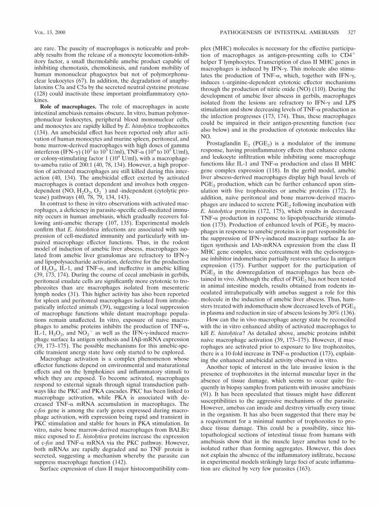
Cysteine proteases are the most abundant proteases in theparasite. Potent cysteine protease activities ranging from 16 to116 kDa are found in amebic extracts electrophoresed on sub-strate gels (11, 38, 108, 112, 138, 147). Cell fractionation stud-ies indicate that although present in the cytosol, they are en-riched in the plasma and internal membranes. In addition,secreted protease activities of 16, 26, and 56 kDa have beenreported (64, 80, 81). A total of six distinct genes (EhCP1through EhCP6) encoding prepro forms of cysteine protein-ases have been identified. The enzymes encoded by three ofthese genes have been purified and characterized, namely,EhCP1 (ACP3; previously known as amebapain) (138, 156),EhCP2 (ACP2; earlier reported as histolysin) (80, 157), andEhCP5, a membrane-bound protease (62). These three en-zymes, all with a molecular mass of ;30 kDa, account for;90% of cysteine protease transcripts and for virtually allcysteine protease activity found in E. histolytica lysates (24). Itremains to be established whether the different molecularmasses reported for other protease activities represent differ-ent enzymes or the expression of different states of one of thesix genes identified (97).
Amebic cysteine proteases are active against a variety ofsubstrates and increased activity has been reported in clones ofhigh virulence (103). Of the ECM components that E. histo-
324 ESPINOSA-CANTELLANO AND MARTINEZ-PALOMO CLIN. MICROBIOL. REV.
lytica encounters during colonic invasion, laminin, collagentypes I and IV, and FN are good targets for amebapain, his-tolysin, EhCP5 membrane-bound protease, and the neutral56-kDa protease (Table 1).
E. dispar seems to possess four homologous cysteine pro-tease genes, although none of the enzymes (EdCP2, EdCP3,EdCP4, and EdCP6) have been purified. In addition, the pres-ence of the neutral 56-kDa protease in E. dispar has not beenreported yet. If E. dispar indeed lacks several of the mostpotent E. histolytica cysteine proteases (EdCP1, EdCP5, andthe neutral protease), it is possible that this difference couldpartially explain its noninvasive nature.
In addition, E. histolytica possesses a membrane-bound met-allocollagenase that degrades collagen types I and III and ismore active against the former (100). The degree of collag-enolytic activity has been linked to the virulence of differentisolates (53, 101, 145, 164). Activation of the enzyme appar-ently results in translocation of the enzyme from internal mem-branes to the plasma membrane (87).
In vitro, incubation of trophozoites on collagen-coated wellsinduces the formation and release of electron-dense granules
(EDGs) through a cytoskeleton-driven, Ca21-calmodulin-de-pendent mechanism (87, 102, 146). In addition to a collageno-lytic activity, EDGs seem to contain at least 25 polypeptideswith acidic pIs (6.06 to 6.59), 9 gelatinase activities, actin, smallmolecules including inorganic phosphate (Pi) and pyrophos-phate (PPi), and several ions. Six of these polypeptides (108,106, 104, 97, 68, and 59 kDa) and two protease activities of 40and 85 kDa have been claimed to be detected in EDGs but notin total-trophozoite extracts (76). Whether some of the above-mentioned components represent contamination with cyto-plasmic molecules not found in EDGs remains to be estab-lished.
In summary, E. histolytica possesses the necessary machineryto degrade the ECM components it encounters during inva-sion. However, the participation of the different proteases dur-ing in vivo infections has yet to be demonstrated.
Neutrophil infiltration of the lamina propria. The last ob-servation of the early invasive lesion with superficial ulcerationis a mild to moderate infiltration of the lamina propria. At thisstage, cell infiltration around invading amebas leads to rapidlysis of inflammatory cells and tissue necrosis. These observa-
TABLE 1. Cysteine proteases of E. histolytica
Enzyme Size pH Substrates Other features Reference(s)
Amebapain (EhCP1 orACP3)
27 6 2 kDa,a 24 kDab 5.0–8.5 Human laminin and FN, bovinecollagen type I, and humancollagens types IV (70-kDacomponent) and V (a1-chain)
Multicopy gene, high levelsof expression
24, 139–141,156, 157
Histolysin (EhCP2 orACP2)
26–29 kDa,a 23.6kDab
5.5–9.5 Human collagen type IV,bovine nasal cartilage; notactive against rabbit skincollagen type I or elastin
Single-copy gene, high levelsof expression
24, 80, 157
EhCP3 or ACP1 NAc NA NA Only primary structureavailable; single copy-gene, low levels ofexpression
24, 25, 95, 125
EhCP4 33,726 Dab NA NA Only primary structureavailable; multicopy gene,very low levels ofexpression
24
Membrane bound protease(EhCP5)
30 kDa,a 24 kDab 6.0–8.0 FN, immunoglobulin G, C3and C9 components ofcomplement
Single-copy gene, high levelsof expression; highcontent of hydrophobicregions can explainmembrane association
12, 24, 62
EhCP6 35,324 Dab NA NA Only primary structureavailable; single-copygene, very low levels ofexpression
24
Major neutral protease 56 kDaa 6.0–7.0 Rat-derived laminin,fibronectin, collagen type I,immunoglobulin G, C3 (a-chain) and C5 componentsof complement, andanaphylatoxins C3a and C5a
Secreted enzyme 64, 126–128,159
Hemoglobinase 82 kDaa 7.0 Human, bovine, and porcinehemoglobin
Two additional minor bandsof 116 and 21 kDa withhemoglobinase activityare described
147
a Reported molecular mass calculated after gel electrophoresis run with known molecular mass standards.b Reported molecular mass deduced from the amino acid sequence of the enzyme.c NA, not available.
VOL. 13, 2000 PATHOGENESIS OF INTESTINAL AMEBIASIS 325

tions have been confirmed in rodent models of intestinal ame-biasis (88, 149).
Neutrophils, representing over 90% of the circulating gran-ulocytes, respond to a variety of cytokines and soluble factorsduring the inflammatory response. C5a, the cleavage productof complement component C5, is one of the most potent che-motactic and activation molecules for phagocytes. In amebicintestinal lesions, however, the participation of C5a in theneutrophil infiltration of the lamina propria is uncertain, sinceE. histolytica trophozoites release a neutral cysteine proteasethat degrades this molecule (128).
In recent years it has become evident that gut inflammationis not only the result of the immune system response to anintestinal insult but also the result of a complex interplay ofimmune and nonimmune cell interactions that involve epithe-lial, mesenchymal, endothelial, and nerve cells, as well as com-ponents of the ECM (51). Studies on mucosal immunity haveprovided growing evidence that intestinal epithelial cells, forinstance, constitutively express or can be induced to express anumber of immunologically active cytokines and soluble fac-tors, including interleukin-8 (IL-8), monocyte chemoattractantprotein 1, granulocyte-macrophage colony-stimulating factor,and tumor necrosis factor alpha (TNF-a) (93). Thus, in addi-tion to their fundamental absorptive and secretory functions,intestinal epithelial cells are integral and essential componentsof the host’s innate and acquired immune system.
Neutrophils are rapidly recruited and activated in responseto the proinflammatory cytokine IL-8. In human colon epithe-lial cell lines, E. histolytica trophozoites increase the secretionof IL-8 and TNF-a (45, 179). At least three possible mecha-nisms have been described for the upregulation of IL-8. Somecolon epithelial cell lines increase the secretion of IL-8 inresponse to the preformed IL-1a released from the cells lysedby E. histolytica. In other cell lines lacking preformed IL-1a,increased IL-8 production seems to be mediated by the adher-ence of trophozoites through the Gal-GalNAc lectin by amechanism that involves, at least partially, an increase in[Ca21]i (45). In a third mechanism, E. histolytica secreted prod-ucts could induce the secretion of IL-8 in the absence of tro-phozoite contact (179). Whether any of these mechanisms takeplace during natural intestinal infection remains to be estab-lished, but further support for the importance of intestinalepithelial cells as producers of inflammatory cytokines comesfrom studies using a severe combined immunodeficient (SCID)mouse-human intestinal xenograft model (148). In this model,increased IL-1a and IL-8 production and extensive neutro-philic infiltration followed infection of the xenograft with E.histolytica. Moreover, both cytokines were of epithelial originfrom the human intestinal xenograft (148).
Finally, a chemoattractant activity for neutrophils has beenfound in E. histolytica (32). Although the molecule responsiblefor this activity has not been purified, enzymatic treatmentsuggests that it corresponds to a membrane-bound peptide.
Role of neutrophils. Cell infiltration around invading ame-bas leads to rapid lysis of inflammatory cells followed by tissuenecrosis. The reasons for the inability of neutrophils to destroyE. histolytica are unknown. In other parasitic infections, neu-trophils kill invading microorganisms by both O2-dependentand -independent mechanisms; they produce an intense oxida-tive burst, and their secretory granules contain highly cytotoxicmolecules. E. histolytica could avoid the oxidative burstthrough an iron-containing superoxide dismutase that can beinduced by superoxide anions to produce H2O2 (22). In addi-tion, a bifunctional NADPH:flavin oxidoreductase containingNADPH-dependent disulfide reductase and H2O2-formingNADPH oxidase activities could aid in the detoxification of
hydroperoxides produced during an oxidative stress (23, 27).Although the ameba does not contain catalase, a cysteine-rich29-kDa protein in amebas has been shown to eliminate H2O2(21, 26, 52). There is no evidence, however, on how E. histo-lytica might escape the action of the cytotoxic molecules re-leased by the neutrophils. The parasite must possess still un-explored mechanisms of protection against its own lyticpeptidases, but whether it uses them to avoid attack by externalcytotoxic molecules is unknown.
Alternatively, it could be speculated that neutrophils presentin human E. histolytica lesions are not properly activated. Asstated above, humans are the only natural host for E. histo-lytica. Unfortunately, no experimental model has been devel-oped to date that reproduces the invasive intestinal amebiclesions seen in human intestinal amebiasis. Although the re-sults are highly reproducible with the best model available, thegerbil, they can only mimic the initial stages of infection butresolve in 96 h (149). This has been interpreted as an innateresistance to E. histolytica. Recent studies using the model ofinduction of amebic liver abscess have started to explore thecellular basis of this innate resistance in mice (167). Followingintrahepatic inoculation of trophozoites, neutropenic mice de-veloped larger liver lesions than did normal mice, suggesting arole for neutrophils, albeit partial, in the mechanism of resis-tance in these rodents (167). Also, in the highly susceptiblehamster liver model, in contrast to what occurs with E. histo-lytica, neutrophils effectively eliminate E. dispar within 24 h(48). Comparison of the responses induced by the two speciesof amebas in experimental liver and intestinal infection mightshed some light on their strikingly different pathogenic behav-iors.
Neutrophils not only fail to resist E. histolytica but may infact contribute to host tissue damage through their destructionand release of cytotoxic granules (88, 163). In vitro, humanneutrophils are killed even by low-virulence strains of E. his-tolytica in a 400:1 ratio of neutrophils to amebas, althoughmany of these parasites succumb in the process. Highly virulentstrains, however, emerge victorious after incubations with3,000 neutrophils per ameba (55).
In addition to the contribution of neutrophils, amebas play adirect role in tissue necrosis. Evidence for this is available onlyfrom the model of induction of amebic liver abscess, where theparasite is able to produce liver damage in the absence of aninflammatory reaction in neutropenic mice (167). In the samereport (167), images suggesting apoptosis of hepatocytes closeto the inflammatory infiltrate or in areas of the liver paren-chyma distant from amebas or inflammatory cells are shown.
Late Invasive Lesion with Deep Ulceration
As invasion progresses, the mucosal ulcer extends deep intoa larger area of the submucosa. Once the interglandular epi-thelium has surrendered to amebic penetration, the underlyingtissue offers little resistance, allowing extension of the ulcerinto the typical flask form. While continuing the invasion pro-cess through degradation of ECM and locomotion, the parasitemight use its hemoglobinases to digest phagocytosed erythro-cytes, thus obtaining the necessary iron for its survival (147).As a result of lysed cells and necrosis, the observed thickexudate containing acellular proteinaceous material, red bloodcells, and strands of fibrin is explained, as is the deeply eosin-ophilic zone of fibrinoid necrosis separating the exudate fromthe underlying viable submucosa.
At this stage, infiltration by abundant neutrophils and somelymphocytes and macrophages is observed, while eosinophils
326 ESPINOSA-CANTELLANO AND MARTINEZ-PALOMO CLIN. MICROBIOL. REV.
are rare. The paucity of macrophages is noticeable and prob-ably results from the release of a monocyte locomotion-inhib-itory factor, a small thermolabile amebic product capable ofinhibiting chemotaxis, chemokinesis, and random mobility ofhuman mononuclear phagocytes but not of polymorphonu-clear leukocytes (67). In addition, the degradation of anaphy-latoxins C3a and C5a by the secreted neutral cysteine protease(128) could inactivate these important proinflammatory cyto-kines.
Role of macrophages. The role of macrophages in acuteintestinal amebiasis remains obscure. In vitro, human polymor-phonuclear leukocytes, peripheral blood mononuclear cells,and monocytes are rapidly killed by E. histolytica trophozoites(134). An amebicidal effect has been reported only after acti-vation of human monocytes and murine spleen, peritoneal, andbone marrow-derived macrophages with high doses of gammainterferon (IFN-g) (102 to 103 U/ml), TNF-a (104 to 105 U/ml),or colony-stimulating factor 1 (104 U/ml), with a macrophage-to-ameba ratio of 200:1 (40, 78, 134). However, a high propor-tion of activated macrophages are still killed during this inter-action (40, 134). The amebicidal effect exerted by activatedmacrophages is contact dependent and involves both oxygen-dependent (NO, H2O2, O2
2) and -independent (cytolytic pro-tease) pathways (40, 78, 79, 134, 143).
In contrast to these in vitro observations with activated mac-rophages, a deficiency in parasite-specific cell-mediated immu-nity occurs in human amebiasis, which gradually recovers fol-lowing anti-amebic therapy (107, 135). Experimental modelsconfirm that E. histolytica infections are associated with sup-pression of cell-mediated immunity and particularly with im-paired macrophage effector functions. Thus, in the rodentmodel of induction of amebic liver abscess, macrophages iso-lated from amebic liver granulomas are refractory to IFN-gand lipopolysaccharide activation, defective for the productionof H2O2, IL-1, and TNF-a, and ineffective in amebic killing(39, 173, 174). During the course of cecal amebiasis in gerbils,peritoneal exudate cells are significantly more cytotoxic to tro-phozoites than are macrophages isolated from mesentericlymph nodes (31). This higher activity has also been reportedfor spleen and peritoneal macrophages isolated from intrahe-patically infected animals (39), suggesting a local suppressionof macrophage functions while distant macrophage popula-tions remain unaffected. In vitro, exposure of naive macro-phages to amebic proteins inhibits the production of TNF-a,IL-1, H2O2, and NO2
2 as well as the IFN-g-induced macro-phage surface Ia antigen synthesis and IAb-mRNA expression(39, 173–175). The possible mechanisms for this amebic-spe-cific transient anergy state have only started to be explored.
Macrophage activation is a complex phenomenon whoseeffector functions depend on environmental and maturationaleffects and on the lymphokines and inflammatory stimuli towhich they are exposed. To become activated, macrophagesrespond to external signals through signal transduction path-ways like the PKC and PKA cascades. PKC has been linked tomacrophage activation, while PKA is associated with de-creased TNF-a mRNA accumulation in macrophages. Thec-fos gene is among the early genes expressed during macro-phage activation, with expression being rapid and transient inPKC stimulation and stable for hours in PKA stimulation. Invitro, naive bone marrow-derived macrophages from BALB/cmice exposed to E. histolytica proteins increase the expressionof c-fos and TNF-a mRNA via the PKC pathway. However,both mRNAs are rapidly degraded and no TNF protein issecreted, suggesting a mechanism whereby the parasite cansuppress macrophage function (142).
Surface expression of class II major histocompatibility com-
plex (MHC) molecules is necessary for the effective participa-tion of macrophages as antigen-presenting cells to CD41
helper T lymphocytes. Transcription of class II MHC genes inmacrophages is induced by IFN-g. This molecule also stimu-lates the production of TNF-a, which, together with IFN-g,induces L-arginine-dependent cytotoxic effector mechanismsthrough the production of nitric oxide (NO) (110). During thedevelopment of amebic liver abscess in gerbils, macrophagesisolated from the lesions are refractory to IFN-g and LPSstimulation and show decreasing levels of TNF-a production asthe infection progresses (173, 174). Thus, these macrophagescould be impaired in their antigen-presenting function (seealso below) and in the production of cytotoxic molecules likeNO.
Prostaglandin E2 (PGE2) is a modulator of the immuneresponse, having proinflammatory effects that enhance edemaand leukocyte infiltration while inhibiting some macrophagefunctions like IL-1 and TNF-a production and class II MHCgene complex expression (118). In the gerbil model, amebicliver abscess-derived macrophages display high basal levels ofPGE2 production, which can be further enhanced upon stim-ulation with live trophozoites or amebic proteins (172). Inaddition, naive peritoneal and bone marrow-derived macro-phages are induced to secrete PGE2 following incubation withE. histolytica proteins (172, 175), which results in decreasedTNF-a production in response to lipopolysaccharide stimula-tion (173). Production of enhanced levels of PGE2 by macro-phages in response to amebic proteins is in part responsible forthe suppression of IFN-g-induced macrophage surface Ia an-tigen synthesis and IAb-mRNA expression from the class IIMHC gene complex, since cotreatment with the cyclooxygen-ase inhibitor indomethacin partially restores surface Ia antigenexpression (175). Further support for the participation ofPGE2 in the downregulation of macrophages has been ob-tained in vivo. Although the effect of PGE2 has not been testedin animal intestine models, results obtained from rodents in-oculated intrahepatically with amebas suggest a role for thismolecule in the induction of amebic liver abscess. Thus, ham-sters treated with indomethacin show decreased levels of PGE2
in plasma and reduction in size of abscess lesions by 30% (136).How can the in vivo macrophage anergy state be reconciled
with the in vitro enhanced ability of activated macrophages tokill E. histolytica? As detailed above, amebic proteins inhibitnaive macrophage activation (39, 173–175). However, if mac-rophages are activated prior to exposure to live trophozoites,there is a 10-fold increase in TNF-a production (173), explain-ing the enhanced amebicidal activity observed in vitro.
Another topic of interest in the late invasive lesion is thepresence of trophozoites in the internal muscular layer in theabsence of tissue damage, which seems to occur quite fre-quently in biopsy samples from patients with invasive amebiasis(91). It has been speculated that tissues might have differentsusceptibilities to the aggressive mechanisms of the parasite.However, amebas can invade and destroy virtually every tissuein the organism. It has also been suggested that there may bea requirement for a minimal number of trophozoites to pro-duce tissue damage. This could be a possibility, since his-topathological sections of intestinal tissue from humans withamebiasis show that in the muscle layer amebas tend to beisolated rather than forming aggregates. However, this doesnot explain the absence of the inflammatory infiltrate, becausein experimental models strikingly large foci of acute inflamma-tion are elicited by very few parasites (163).
VOL. 13, 2000 PATHOGENESIS OF INTESTINAL AMEBIASIS 327
Granulating Ulcer
The formation of granulation tissue is characterized by theproliferation of small vessels and usually indicates healing of alesion. Since the reports describing this stage of amebic lesionsin humans do not provide data on the clinical status of thepatient, it is difficult to interpret whether this repair processoccurs spontaneously or is secondary to antiamebic chemo-therapy. It is known that if properly treated, invasive amebiclesions in humans, whether localized in the large intestine,liver, or skin, almost invariably heal without the formation ofscar tissue. The mechanism of this striking repair process is stillunknown.
CONCLUSIONS
Lysis of the colonic mucosa in intestinal amebiasis has beenrelated to a variety of molecules produced by E. histolytica:adhesins, amebapores, and proteases. A multifunctional ad-herence lectin allows the parasite attachment to the colonicmucus blanket, thereby avoiding elimination through the in-testinal stream. The lectin is also involved in signaling cytolysisand in blocking the deposition of the harmful membrane attackcomplex of complement, and it could participate in the anchor-age of the ameba to proteoglycans during the invasion process.The amebapores of E. histolytica, small but potent peptides,destroy ingested bacteria that serve as the main nutrients forthe parasite in the otherwise nutrient-scarce colonic environ-ment. Their participation in the cytolytic event has not yet beenproven. Proteases can be used to degrade the extracellularmatrix during invasion and aid in the lysis of target cells. Inaddition, interesting mechanisms of parasitic modulation ofthe host immune response are starting to be unravelled. Themain targets of this modulation appear to be neutrophils andmacrophages, which, although recruited at the site of the le-sion, are unable to abort infection.
In spite of these advances, many questions must be answeredbefore we can fully understand the sequence of events duringamebic invasion of the colon. To name but a few, the signal forthe initiation of the invasion process, the mechanism of elim-ination of the mucus barrier, the role of EDG, and the in vivoparticipation of the different molecules in this multifactorialprocess have not been demonstrated. The recent advances inamebic transfection technology (56, 98, 104, 106, 122, 168) willundoubtedly shed new light on the involvement of specificmolecules in the pathogenic mechanism. The uncertainty ofthe ploidy of E. histolytica has hindered the production of geneknockout clones like those produced for the study of thepathogenicity of Toxoplasma gondii and Leishmania spp. (44,46). However, inhibition of expression of a surface antigen hasalready been achieved through transfection and transcriptionof its antisense RNA (4). In vivo infections with these modifiedtrophozoites in current or in newly developed animal models(knockout or SCID mice) would also help our understandingof the process. Finally, much work needs to be done to clarifythe complex signalling pathways of the parasite and to explainthe modulation of the host immune response that results in theestablishment and continuation of infection.
ACKNOWLEDGMENTS
We thank Graham Clark and John Ackers for critically reading themanuscript.
Part of the work described in this review was supported by theConsejo Nacional de Ciencia y Tecnologıa (Conacyt grant 3694P-M9607), the Fundacion Miguel Aleman, and the Fundacion Mexicanapara la Salud.
REFERENCES
1. Adler, P., S. J. Wood, Y. C. Lee, R. T. Lee, W. A. Petri, Jr., and R. L.Schnaar. 1995. High affinity binding of the Entamoeba histolytica lectin topolyvalent N-acetylgalactosaminides. J. Biol. Chem. 270:5164–5171.
2. Aguirre, A., D. C. Warhurst, F. Guhl, and I. A. Frame. 1995. Polymerasechain reaction-solution hybridization enzyme-linked immunoassay (PCR-SHELA) for the differential diagnosis of pathogenic and non-pathogenicEntamoeba histolytica. Trans. R. Soc. Trop. Med. Hyg. 89:187–188.
3. Aigner, T., D. Neureiter, S. Muller, G. Kuspert, J. Belke, and T. Kirchner.1997. Extracellular matrix composition and gene expression in collagenouscolitis. Gastroenterology 113:136–143.
4. Alon, R. N., R. Bracha, and D. Mirelman. 1997. Transfection of Entamoebadispar: inhibition of expression of the lysine-rich 30 kDa surface antigen bythe transcription of its antisense RNA. Arch. Med. Res. 28:S52–S55.
5. Andra, J., and M. Leippe. 1994. Pore-forming peptide of Entamoeba his-tolytica: significance of positively charged amino acid residues for its modeof action. FEBS Lett. 354:97–102.
6. Andra, J., O. Berninghausen, J. Wulfken, and M. Leippe. 1996. Shortenedamoebapore analogs with enhanced antibacterial and cytolytic activity.FEBS Lett. 385:96–100.
7. Ankri, S., F. Padilla-Vaca, T. Stolarsky, L. Koole, U. Katz, and D. Mire-lman. 1999. Antisense inhibition of expression of the light subunit (35 kDa)of the Gal/GalNAc lectin complex inhibits Entamoeba histolytica virulence.Mol. Microbiol. 33:327–337.
8. Anonymous. 1997. Entamoeba taxonomy. Bull. W. H. O. 75:291–292.9. Arhets, P., P. Gounon, P. Sansonetti, and N. Guillen. 1995. Myosin-II is
involved in capping and uroid formation in the human pathogen Entamoebahistolytica. Infect. Immun. 63:4358–4367.
10. Arroyo, R., and E. Orozco. 1987. Localization and identification of anEntamoeba histolytica adhesin. Mol. Biochem. Parasitol. 23:151–158.
11. Avila, E. E., M. Sanchez-Garza, and J. Calderon. 1985. Entamoeba histo-lytica and E. invadens: sulfhydryl-dependent proteolytic activity. J. Proto-zool. 32:163–166.
12. Avila, E. E., and J. Calderon. 1993. Entamoeba histolytica trophozoites: asurface-associated cysteine protease. Exp. Parasitol. 76:232–241.
13. Bailey, G. B., P. S. Shen, M. J. Beanan, and N. E. McCoomer. 1992. Actinassociated proteins of Entamoeba histolytica. Arch. Med. Res. 23:129–132.
14. Beanan, M. J., and G. B. Bailey. 1995. The primary structure of an Ent-amoeba histolytica b-hexosaminidase A subunit. J. Eukaryot. Microbiol.42:632–636.
15. Belley, A., K. Keller, M. Goettke, and K. Chadee. 1999. Intestinal mucins incolonization and host defense against pathogens. Am. J. Trop. Med. Hyg.60:10–15.
16. Binder, M., S. Ortner, H. Erben, O. Scheiner, G. Wiedermann, R. Valenta,and M. Duchene. 1995. The basic isoform of profilin in pathogenic Ent-amoeba histolytica. cDNA cloning, heterologous expression, and actin-bind-ing properties. Eur. J. Biochem. 233:976–981.
17. Bracha, R., and D. Mirelman. 1984. Virulence of Entamoeba histolyticatrophozoites. Effects of bacteria, microaerobic conditions and metronida-zole. J. Exp. Med. 160:353–386.
18. Braga, L. L., H. Ninomiya, J. J. McCoy, S. Eacker, T. Wiedmer, C. Pham,S. Wood, P. J. Sims, and W. A. Petri. 1992. Inhibition of the complementmembrane attack complex by the galactose-specific adhesin of Entamoebahistolytica. J. Clin. Investig. 90:1131–1137.
19. Braga, L. L., A. A. M. Lima, C. L. Sears, R. D. Newman, T. Wuhib, C. A.Paiva, R. L. Guerrant, and B. J. Mann. 1996. Seroepidemiology of Ent-amoeba histolytica in a slum in Northeastern Brazil. Am. J. Trop. Med. Hyg.55:693–697.
20. Britten, D., S. N. Wilson, R. McNerney, A. H. Moody, P. L. Chiodini, andJ. P. Ackers. 1997. An improved colorimetric PCR-based method for de-tection and differentiation of Entamoeba histolytica and Entamoeba disparin feces. J. Clin. Microbiol. 35:1108–1111.
21. Bruchhaus, I., and E. Tannich. 1993. Analysis of the genomic sequenceencoding the 29-kDa cysteine-rich protein of Entamoeba histolytica. Trop.Med. Parasitol. 44:116–118.
22. Bruchhaus, I., and E. Tannich. 1994. Induction of an iron-containing su-peroxide dismutase in Entamoeba histolytica by a superoxide anion-gener-ating system or by iron chelation. Mol. Biochem. Parasitol. 67:281–288.
23. Bruchhaus, I., and E. Tannich. 1995. Identification of an Entamoeba his-tolytica gene encoding a protein homologous to prokaryotic disulphideoxidoreductases. Mol. Biochem. Parasitol. 70:187–191.
24. Bruchhaus, I., T. Jacobs, M. Leippe, and E. Tannich. 1996. Entamoebahistolytica and Entamoeba dispar: differences in numbers and expression ofcysteine proteinase genes. Mol. Microbiol. 22:255–263.
25. Bruchhaus, I., and E. Tannich. 1996. A gene highly homologous to ACP1encoding cysteine proteinase 3 in Entamoeba histolytica is present andexpressed in E. dispar. Parasitol. Res. 82:189–192.
26. Bruchhaus, I., S. Richter, and E. Tannich. 1997. Removal of hydrogenperoxide by the 29kDa protein of Entamoeba histolytica. Biochem. J. 326:785–789.
27. Bruchhaus, I., S. Richter, and E. Tannich. 1998. Recombinant expressionand biochemical characterization of an NADPH:flavin oxidoreductase from
328 ESPINOSA-CANTELLANO AND MARTINEZ-PALOMO CLIN. MICROBIOL. REV.
Entamoeba histolytica. Biochem. J. 330:1217–1221.28. Brumpt, E. 1925. Etude sommaire de l’ “Entamoeba dispar” n. sp. Amibe a
kystes quadrinuclees, parasite de l’homme. Bull. Acad. Med. (Paris) 94:943–952.
29. Burchard, G. D., and R. Bilke. 1992. Adherence of pathogenic and non-pathogenic Entamoeba histolytica strains to neutrophils. Parasitol. Res. 78:146–153.
30. Carbajal, M. E., R. Manning-Cela, A. Pina, E. Franco, and I. Meza. 1996.Fibronectin-induced intracellular calcium rise in Entamoeba histolytica tro-phozoites: effect on adhesion and the actin cytoskeleton. Exp. Parasitol.82:11–20.
31. Chadee, K., E. Meerovitch, and F. Moreau. 1985. In vitro and in vivointeraction between trophozoites of Entamoeba histolytica and gerbil lym-phoid cells. Infect. Immun. 49:828–832.
32. Chadee, K., F. Moreau, and E. Meerovitch. 1987. Entamoeba histolytica:chemoattractant activity for gerbil neutrophils in vivo and in vitro. Exp.Parasitol. 64:12–23.
33. Chadee, K., W. A. Petri, D. J. Innes, and J. I. Ravdin. 1987. Rat and humancolonic mucins bind to and inhibit the adherence lectin of Entamoebahistolytica. J. Clin. Investig. 80:1245–1254.
34. Chadee, K., M. L. Johnson, E. Orozco, W. A. Petri, and J. I. Ravdin. 1988.Binding and internalization of rat colonic mucins by the galactose/N-acetyl-D-galactosamine adherence lectin of E. histolytica. J. Infect. Dis. 158:398–406.
35. Clark, C. G. 1998. Entamoeba dispar, an organism reborn. Trans. R. Soc.Trop. Med. Hyg. 92:361–364.
36. Cone, R. A. 1999. Mucus, p. 43–64. In P. L. Ogra, J. Mestecky, M. E. Lamm,W. Strober, J. Bienenstock, and J. R. McGhee (ed.), Mucosal immunology.Academic Press, Inc., San Diego, Calif.
37. Councilman, W. T., and H. A. Lafleur. 1891. Amebic dysentery. JohnsHopkins Hosp. Rep. 2:395–548.
38. De Meester, F., E. Shaw, H. Scholze, T. Stolarsky, and D. Mirelman. 1990.Specific labeling of cysteine proteinases in pathogenic and nonpathogenicEntamoeba histolytica. Infect. Immun. 58:1396–1401.
39. Denis, M., and K. Chadee. 1988. In vitro and in vivo studies of macrophagefunctions in amebiasis. Infect. Immun. 56:3126–3131.
40. Denis, M., and K. Chadee. 1989. Cytokine activation of murine macro-phages for in vitro killing of Entamoeba histolytica trophozoites. Infect.Immun. 57:1750–1756.
41. Diamond, L. S., and C. G. Clark. 1993. A redescription of Entamoebahistolytica Schaudinn, 1903 (emended Walker, 1911) separating it fromEntamoeba dispar Brumpt, 1925. J. Eukaryot. Microbiol. 40:340–344.
42. Dodson, J. M., C. G. Clark, L. A. Lockhart, B. M. Leo, J. W. Schroeder, andB. J. Mann. 1997. Comparison of adherence, cytotoxicity, and Gal/GalNAclectin gene structure in Entamoeba histolytica and Entamoeba dispar. Para-sitol. Int. 46:225–235.
43. Dodson, J. M., P. W. Lenkowski, A. C. Eubanks, T. F. G. H. Jackson, J.Napodano, D. M. Lyerly, L. A. Lockhart, B. J. Mann, and W. A. Petri. 1999.Infection and immunity mediated by the carbohydrate recognition domainof the Entamoeba histolytica Gal/GalNAc lectin. J. Infect. Dis. 179:460–466.
44. Donald, R. G., and D. S. Roos. 1998. Gene knock-outs and allelic replace-ments in Toxoplasma gondii: HXGPRT as a selectable marker for hit-and-run mutagenesis. Mol. Biochem. Parasitol. 91:295–305.
45. Eckmann, L., S. L. Reed, J. R. Smith, and M. F. Kagnoff. 1995. Entamoebahistolytica trophozoites induce an inflammatory cytokine response by cul-tured human cells through the paracrine action of cytolytically releasedinterleukin-1a. J. Clin. Investig. 96:1269–1279.
46. Engers, H. D., R. Bergquist, and F. Modabber. 1996. Progress on vaccinesagainst parasites. Dev. Biol. Stand. 87:73–84.
47. Espinosa-Cantellano, M., and A. Martınez-Palomo. 1994. Entamoeba his-tolytica: mechanism of surface receptor capping. Exp. Parasitol. 79:424–435.
48. Espinosa-Cantellano, M., G. Castanon Gutierrez, and A. Martınez-Palomo. 1997. In vivo pathogenesis of Entamoeba dispar. Arch. Med. Res.28:204–206.
49. Espinosa-Cantellano, M., A. Gonzalez-Robles, B. Chavez, G. Castanon, C.Arguello, A. Lazaro-Haller, and A. Martınez-Palomo. 1998. Entamoebadispar: ultrastructure, surface properties, and cytopathic effect. J. Eukaryot.Microbiol. 45:265–272.
50. Reference deleted.51. Fiocchi, C. 1997. Intestinal inflammation: a complex interplay of immune
and nonimmune cell interactions. Am. J. Physiol. 273:G769–G775.52. Flores, B. M., M. A. Batzer, M. A. Stein, C. Petersen, D. L. Diedrich, and
B. E. Torian. 1993. Structural analysis and demonstration of the 29 kDaantigen of pathogenic Entamoeba histolytica as the major accessible freethiol-containing surface protein. Mol. Microbiol. 7:755–763.
53. Gadasi, H., and E. Kessler. 1983. Correlation of virulence and collageno-lytic activity in Entamoeba histolytica. Infect. Immun. 39:528–531.
54. Gottke, M. U., K. Keller, A. Belley, R. M. Garcia, M. A. Hollingsworth,D. R. Mack, and K. Chadee. 1998. Functional heterogeneity of colonicadenocarcinoma mucins for inhibition of Entamoeba histolytica adherenceto target cells. J. Eukaryot. Microbiol. 45:17S–23S.
55. Guerrant, R. L., J. Brush, J. I. Ravdin, J. A. Sullivan, and G. L. Mandell.
1981. Interaction between Entamoeba histolytica and human polymorpho-nuclear neutrophils. J. Infect. Dis. 143:83–93.
56. Hamann, L., R. Nickel, and E. Tannich. 1995. Transfection and continuousexpression of heterologous genes in the protozoan parasite Entamoebahistolytica. Proc. Natl. Acad. Sci. USA 92:8975–8979.
57. Haque, R., L. M. Neville, S. Wood, and W. A. Petri, Jr. 1994. Detection ofEntamoeba histolytica and E. dispar directly in stool. Am. J. Trop. Med.Hyg. 50:595–596.
58. Haque, R., A. S. G. Faruque, P. Hahn, D. M. Lyerly, and W. A. Petri. 1997.Entamoeba histolytica and Entamoeba dispar infection in children in Bang-ladesh. J. Infect. Dis. 175:734–736.
59. Haque, R., A. S. G. Faruque, and W. A. Petri. 1997. Entamoeba histolyticaand Entamoeba dispar infection in children in Bangladesh. Arch. Med. Res.28:S317–S318.
60. Haque, R., I. K. M. Ali, S. Akther, and W. A. Petri, Jr. 1998. Comparisonof PCR, isoenzyme analysis, and antigen detection for diagnosis of Ent-amoeba histolytica infection. J. Clin. Microbiol. 36:449–452.
61. Jacobs, T., and M. Leippe. 1995. Purification and molecular cloning of amajor antibacterial protein of the protozoan parasite Entamoeba histolyticawith lysozyme-like properties. Eur. J. Biochem. 231:831–838.
62. Jacobs, T., I. Bruchhaus, T. Dandekar, E. Tannich, and M. Leippe. 1998.Isolation and molecular characterization of a surface-bound proteinase ofEntamoeba histolytica. Mol. Microbiol. 27:269–276.
63. Katzwinkel-Wladarsch, S., T. Loscher, and H. Rinder. 1994. Direct ampli-fication and differentiation of pathogenic and nonpathogenic Entamoebahistolytica DNA from stool specimens. Am. J. Trop. Med. Hyg. 51:115–118.
64. Keene, W. E., M. G. Petitt, S. Allen, and J. H. McKerrow. 1986. The majorneutral proteinase of Entamoeba histolytica. J. Exp. Med. 163:536–549.
65. Keller, F., W. Hanke, D. Trissl, and T. Bakker-Grunwald. 1989. Pore-forming protein from Entamoeba histolytica forms voltage- and pH-con-trolled multi-state channels with properties similar to those of the barrel-stave aggregates. Biochim. Biophys. Acta 982:89–93.
66. Keller, K., M. Olivier, and K. Chadee. 1992. The fast release of mucinsecretion from human colonic cells induced by Entamoeba histolytica isdependent on contact and protein kinase C activation. Arch. Med. Res.23:217–221.
67. Kretschmer, R., M. L. Collado, M. G. Pacheco, M. C. Salinas, M. Lopez-Osuna, M. Lecuona, E. M. Castro, and J. Arellano. 1985. Inhibition ofhuman monocyte locomotion by products of axenically grown E. histolytica.Parasite Immunol. 7:527–543.
68. Leippe, M., S. Ebel, O. L. Schoenberger, R. D. Horstman, and H. J.Muller-Eberhard. 1991. Pore-forming peptide of pathogenic Entamoebahistolytica. Proc. Natl. Acad. Sci. USA 88:7659–7663.
69. Leippe, M., E. Tannich, R. Nickel, G. van der Goot, F. Pattus, R. D.Horstmann, and H. J. Muller-Eberhard. 1992. Primary and secondarystructure of the pore-forming peptide of pathogenic Entamoeba histolytica.EMBO J. 11:3501–3506.
70. Leippe, M., E. Bahr, E. Tannich, and R. D. Horstmann. 1993. Comparisonof pore-forming peptides from pathogenic and nonpathogenic Entamoebahistolytica. Mol. Biochem. Parasitol. 59:101–110.
71. Leippe, M., J. Andra, and H. J. Muller-Eberhard. 1994. Cytolytic andantibacterial activity of synthetic peptides derived from amoebapore, thepore-forming peptide of Entamoeba histolytica. Proc. Natl. Acad. Sci. USA91:2602–2606.
72. Leippe, M., J. Andra, R. Nickel, E. Tannich, and H. J. Muller-Eberhard.1994. Amoebapores, a family of membranolytic peptides from cytoplasmicgranules of Entamoeba histolytica: isolation, primary structure, and poreformation in bacterial cytoplasmic membranes. Mol. Microbiol. 14:895–904.
73. Leippe, M. 1995. Ancient weapons: NK-lysin is a mammalian homolog topore-forming peptides of a protozoan parasite. Cell 83:17–18.
74. Leippe, M., H. J. Sievertsen, E. Tannich, and R. D. Horstmann. 1995.Spontaneous release of cysteine proteinases but not of pore-forming pep-tides by viable Entamoeba histolytica. Parasitology 111:569–574.
75. Leippe, M. 1997. Amoebapores. Parasitol. Today 13:178–183.76. Leon, G., C. Fiori, P. Das, M. Moreno, R. Tovar, J. L. Sanchez-Salas, and
M. L. Munoz. 1997. Electron probe analysis and biochemical characteriza-tion of electron-dense granules secreted by Entamoeba histolytica. Mol.Biochem. Parasitol. 85:233–242.
77. Leroy, A., G. Debruyne, M. Mareel, C. Nokkaew, G. Bailey, and H. Nelis.1995. Contact-dependent transfer of the galactose-specific lectin of Ent-amoeba histolytica to the lateral surface of enterocytes in culture. Infect.Immun. 63:4253–4260.
78. Lin, J. Y., and K. Chadee. 1992. Macrophage cytotoxicity against Entamoe-ba histolytica trophozoites is mediated by nitric oxide from L-arginine.J. Immunol. 148:3999–4005.
79. Lin, J. Y., R. Seguin, K. Keller, and K. Chadee. 1994. Tumor necrosis factoralpha augments nitric oxide-dependent macrophage cytotoxicity againstEntamoeba histolytica by enhanced expression of the nitric oxide synthasegene. Infect. Immun. 62:1534–1541.
80. Luaces, A. L., and A. J. Barret. 1988. Affinity purification and biochemicalcharacterization of histolysin, the major cysteine protease of Entamoebahistolytica. Biochem. J. 250:903–909.
VOL. 13, 2000 PATHOGENESIS OF INTESTINAL AMEBIASIS 329

81. Lushbaugh, W. B., A. F. Hofbauer, and F. E. Pittman. 1985. Entamoebahistolytica: purification of cathepsin B. Exp. Parasitol. 59:328–336.
82. Lynch, E. C., I. Rosenberg, and C. Gitler. 1982. An ion-channel formingprotein produced by Entamoeba histolytica. EMBO J. 1:801–804.
83. Mann, B. J., B. E. Torian, T. S. Vedvick, and W. A. Petri. 1991. Sequenceof a cysteine-rich galactose-specific lectin of Entamoeba histolytica. Proc.Natl. Acad. Sci. USA 88:3248–3252.
84. Mann, B. J., and L. A. Lockhart. 1998. Molecular analysis of the Gal/GalNAc adhesin of Entamoeba histolytica. J. Eukaryot. Microbiol. 45:13S–16S.
85. Manning-Cela, R., and I. Meza. 1997. Up-regulation of actin mRNA andreorganization of the cytoskeleton in Entamoeba histolytica trophozoites. J.Eukaryot. Microbiol. 44:18–24.
86. Martınez-Palomo, A., A. Gonzalez-Robles, B. Chavez, E. Orozco, S. Fer-nandez-Castelo, and A. Cervantes. 1985. Structural bases of the cytolyticmechanisms of Entamoeba histolytica. J. Protozool. 32:166–175.
87. Martınez-Palomo, A., I. Meza, B. Chavez, J. L. Rosales-Encina, M. L.Munoz, A. Gonzalez-Robles, and M. Rojkind. 1987. Entamoeba histolytica:activation and release of membrane dense bodies, p. 371–376. In K. P.Chang and D. Snary (ed.), Host-parasite cellular and molecular interactionsin protozoal infections. Springer-Verlag KG, Heidelberg, Germany.
88. Martınez-Palomo, A., V. Tsutsumi, F. Anaya-Velazquez, and A. Gonzalez-Robles. 1989. Ultrastructure of experimental intestinal invasive amebiasis.Am. J. Trop. Med. Hyg. 41:273–279.
89. Martınez-Palomo, A., and M. Espinosa Cantellano. 1998. Intestinal amoe-bae, p. 157–177. In F. E. G. Cox, J. P. Kreier, and D. Wakelin (ed.), Topley& Wilson’s microbiology and microbial infections. Edward Arnold, NewYork, N.Y.
90. Martınez-Palomo, A., and M. Espinosa-Cantellano. 1998. Amoebiasis: newunderstanding and new goals. Parasitol. Today 14:1–3.
91. Masliah, E., and R. Perez-Tamayo. 1984. Nota sobre la histopatologıa de laamibiasis invasora del intestino grueso. Patologia 22:233–245.
92. McCoy, J. J., B. J. Mann, and W. A. Petri. 1994. Adherence and cytotoxicityof Entamoeba histolytica or how lectins let parasites stick around. Infect.Immun. 62:3045–3050.
93. McGee, D. W. 1999. Inflammation and mucosal cytokine production, p.559–573. In P. L. Ogra, J. Mestecky, M. E. Lamm, W. Strober, J. Bienen-stock, and J. R. McGhee (ed.), Mucosal immunology. Academic Press,Inc., San Diego, Calif.
94. Meza, I. 2000. Extracellular matrix-induced signaling in Entamoeba histo-lytica: its role in invasiveness. Parasitol. Today 16:23–28.
95. Mirelman, D., Y. Nuchamowitz, B. Bohm-Gloning, and B. Walderich. 1996.A homologue of the cysteine proteinase gene (ACP1 or Eh-CPp3) ofpathogenic Entamoeba histolytica is present in non-pathogenic E. disparstrains. Mol. Biochem. Parasitol. 78:47–54.
96. Mirelman, D., Y. Nuchamowitz, and T. Stolarsky. 1997. Comparison of useof enzyme-linked immunosorbent assay-based kits and PCR amplificationof rRNA genes for simultaneous detection of Entamoeba histolytica and E.dispar. J. Clin. Microbiol. 35:2405–2407.
97. Montfort, I., R. Perez-Tamayo, R. Perez-Montfort, A. G. Canto, and A.Olivos. 1994. Purification and immunologic characterization of a 30-kDacysteine proteinase of Entamoeba histolytica. Parasitol. Res. 80:607–613.
98. Moshitch-Moshkovitch, S., T. Stolarsky, D. Mirelman, and R. N. Alon.1996. Stable episomal transfection and gene expression in Entamoeba dis-par. Mol. Biochem. Parasitol. 83:257–261.
99. Muller, F. W., A. Franz, and E. Werries. 1988. Secretory hydrolases of E.histolytica. J. Protozool. 35:291–295.
100. Munoz, M. L., J. Calderon, and M. Rojkind. 1982. The collagenase ofEntamoeba histolytica. J. Exp. Med. 155:42–51.
101. Munoz, M. L., M. Rojkind, J. Calderon, M. Tanimoto, S. Arias-Negrete,and A. Martınez-Palomo. 1984. Entamoeba histolytica: collagenolytic activ-ity and virulence. J. Protozool. 31:468–470.
102. Munoz, M. L., M. A. Moreno, J. N. Perez-Garcia, G. R. Tovar, and V. I.Hernandez. 1991. Possible role of calmodulin in the secretion of Entamoebahistolytica electron-dense granules containing collagenase. Mol. Microbiol.5:1707–1714.
103. Navarro-Garcıa, F., L. Chavez-Duenas, V. Tsutsumi, F. Posadas del Rıo,and R. Lopez-Revilla. 1995. Entamoeba histolytica: increase of enterotoxic-ity and of 53- and 75-kDa cysteine proteinases in a clone of higher viru-lence. Exp. Parasitol. 80:361–372.
104. Nickel, R., and E. Tannich. 1994. Transfection and transient expression ofchloramphenicol acetyltransferase gene in the protozoan parasite Ent-amoeba histolytica. Proc. Natl. Acad. Sci. USA 91:7095–7098.
105. Nickel, R., T. Jacobs, and M. Leippe. 1998. Molecular characterization ofan exceptionally acidic lysozyme-like protein from the protozoon Ent-amoeba histolytica. FEBS Lett. 437:153–157.
106. Olvera, A., F. Olvera, R. R. Vines, F. Recillas-Targa, P. M. Lizardi, S. Dhar,S. Bhattacharya, W. Petri, and A. Alagon. 1997. Stable transfection ofEntamoeba histolytica trophozoites by lipofection. Arch. Med. Res. 28:S49–S51.
107. Ortiz-Ortiz, L., G. Zamacona, B. Sepulveda, and N. R. Capın. 1975. Cell-
mediated immunity in patients with amebic abscess of the liver. Clin. Im-munol. Immunopathol. 4:127–134.
108. Ostoa-Saloma, P., N. Cabrera, I. Becker, and R. Perez-Montfort. 1989.Proteinases of Entamoeba histolytica associated with different subcellularfractions. Mol. Biochem. Parasitol. 32:133–144.
109. Padilla-Vaca, F., S. Ankri, R. Bracha, L. A. Koole, and D. Mirelman. 1999.Down regulation of Entamoeba histolytica virulence by monoxenic cultiva-tion with Escherichia coli O55 is related to a decrease in expression of thelight (35-kilodalton) subunit of the Gal/GalNAc lectin. Infect. Immun.67:2096–2102.
110. Paulnock, D. M. 1994. The molecular biology of macrophage activation, p.47–62. In B. S. Zwilling and T. K. Eisenstein (ed.), Macrophage-pathogeninteractions. Marcel Dekker, Inc., New York, N.Y.
111. Perez, E., M. L. Munoz, and A. Ortega. 1996. Entamoeba histolytica: in-volvement of pp125FAK in collagen-induced signal transduction. Exp. Para-sitol. 82:164–170.
112. Perez-Montfort, R., P. Ostoa-Saloma, L. Velazquez-Medina, I. Montfort,and I. Becker. 1987. Catalytic classes of proteinases of Entamoeba histo-lytica. Mol. Biochem. Parasitol. 26:87–98.
113. Perez-Tamayo, R. 1986. Pathology of amebiasis, p. 45–94. In A. Martınez-Palomo (ed.), Amebiasis. Elsevier Science Publishing, Amsterdam, TheNetherlands.
114. Perreault, N., F. E. Herring-Gillam, N. Desloges, I. Belanger, L. P. Pageot,and J. F. Beaulieu. 1998. Epithelial vs mesenchymal contribution to theextracellular matrix in the human intestine. Biochem. Biophys. Res. Com-mun. 248:121–126.
115. Petri, W. A., R. D. Smith, P. H. Schlesinger, C. F. Murphy, and J. I. Ravdin.1987. Isolation of the galactose-binding lectin that mediates the in vitroadherence of Entamoeba histolytica. J. Clin. Investig. 80:1238–1244.
116. Petri, W. A., M. D. Chapman, T. Snodgrass, B. J. Mann, J. Broman, andJ. I. Ravdin. 1989. Subunit structure of the galactose and N-acetyl-D-galac-tosamine-inhibitable adherence lectin of Entamoeba histolytica. J. Biol.Chem. 264:3007–3012.
117. Reference deleted.118. Phipps, R. P., S. H. Stein, and R. L. Roper. 1991. A new view of prosta-
glandin E regulation of the immune response. Immunol. Today 12:349–352.119. Pillai, D. R., D. Britten, J. P. Ackers, J. I. Ravdin, and K. C. Kain. 1997. A
gene homologous to hgl2 of Entamoeba histolytica is present and expressedin Entamoeba dispar. Mol. Biochem. Parasitol. 87:101–105.
120. Prathap, K., and R. Gilman. 1970. The histopathology of acute intestinalamebiasis. A rectal biopsy study. Am. J. Pathol. 60:229–245.
121. Purdy, J. E., B. J. Mann, E. C. Shigart, and W. A. Petri, Jr. 1993. Analysisof the gene family encoding the Entamoeba histolytica galactose-specificadhesin 170-kDa subunit. Mol. Biochem. Parasitol. 62:53–60.
122. Purdy, J. E., B. J. Mann, L. T. Pho, and W. A. Petri, Jr. 1994. Transienttransfection of the enteric parasite Entamoeba histolytica and expression offirefly luciferase. Proc. Natl. Acad. Sci. USA 91:7099–7103.
123. Ramakrishnan, G., B. D. Ragland, J. E. Purdy, and B. J. Mann. 1996.Physical mapping and expression of gene families encoding the N-acetylD-galactosamine adherence lectin of Entamoeba histolytica. Mol. Microbiol.19:91–100.
124. Ravdin, J. I., and R. L. Guerrant. 1981. Role of adherence in cytopathicmechanisms of Entamoeba histolytica. Study with mammalian tissue culturecells and human erythrocytes. J. Clin. Investig. 68:1305–1313.
125. Reed, S., J. Bouvier, A. Sikes Pollack, J. C. Engel, M. Brown, K. Hirata, X.Que, A. Eakin, P. Hagblom, F. Gillin, and J. H. McKerrow. 1993. Cloningof a virulence factor of Entamoeba histolytica pathogenic strains possess aunique cysteine proteinase gene. J. Clin. Investig. 91:1532–1540.
126. Reed, S. L., W. E. Keene, and J. H. McKerrow. 1989. Thiol proteinaseexpression and pathogenicity of Entamoeba histolytica. J. Clin. Microbiol.27:2772–2777.
127. Reed, S. L., W. E. Keene, J. H. McKerrow, and I. Gigli. 1989. Cleavage ofC3 by a neutral cysteine proteinase of Entamoeba histolytica. J. Immunol.143:189.
128. Reed, S. L., J. A. Ember, D. S. Herdman, R. G. DiScipio, T. E. Hugli, andI. Gigli. 1995. The extracellular neutral cysteine proteinase of Entamoebahistolytica degrades anaphylatoxins C3a and C5a. J. Immunol. 155:266–274.
129. Rivera, W. L., H. Tachibana, M. R. A. Silva-Tahat, H. Uenura, and H.Kanbara. 1996. Differentiation of Entamoeba histolytica and E. dispar DNAfrom cysts present in stool specimens by polymerase chain reaction: its fieldapplication in the Philippines. Parasitol. Res. 82:585–589.
130. Rivera, W. L., H. Tachibana, and H. Kanbara. 1998. Field study on thedistribution of Entamoeba histolytica and Entamoeba dispar in the NorthernPhilippines as detected by the polymerase chain reaction. Am. J. Trop.Med. Hyg. 59:916–921.
131. Rosales-Encina, J. L., I. Meza, A. Lopez de Leon, P. Talamas-Rohana, andM. Rojkind. 1987. Isolation of a 220 kDa protein with lectin properties froma virulent strain of Entamoeba histolytica. J. Infect. Dis. 156:790–797.
132. Rosenberg, I., and C. Gitler. 1985. Subcellular fractionation of amoebaporeand plasma membrane components of Entamoeba histolytica using self-generated Percoll gradients. Mol. Biochem. Parasitol. 14:231–248.
133. Saffer, L. D., and W. A. Petri, Jr. 1991. Role of the galactose lectin of
330 ESPINOSA-CANTELLANO AND MARTINEZ-PALOMO CLIN. MICROBIOL. REV.
Entamoeba histolytica in adherence-dependent killing of mammalian cells.Infect. Immun. 59:4681–4683.
134. Salata, R. A., R. D. Pearson, and J. I. Ravdin. 1985. Interaction of humanleukocytes and Entamoeba histolytica: killing of virulent amebae by theactivated macrophage. J. Clin. Investig. 76:491–499.
135. Salata, R. A., A. Martınez-Palomo, L. Canales, H. W. Murray, N. Trevino,and J. I. Ravdin. 1990. Suppression of T-lymphocyte responses to Ent-amoeba histolytica antigen by immune sera. Infect. Immun. 58:3941–3946.
136. Sanchez Ramırez, B., B. Escalante, J. L. Rosales Encina, and P. TalamasRohana. 1997. Role of prostaglandin E2 on amoebic liver abscess formationin hamsters. Prostaglandins 53:411–421.
137. Santiago, A., M. E. Carbajal, G. Benitez-King, and I. Meza. 1994. Ent-amoeba histolytica: PKC transduction pathway activation in the trophozo-ite-fibronectin interaction. Exp. Parasitol. 79:436–444.
138. Scholze, H., and E. Werries. 1984. A weakly acidic protease has a powerfulproteolytic activity in Entamoeba histolytica. Mol. Biochem. Parasitol. 11:293–300.
139. Scholze, H., and W. Schulte. 1988. On the specificity of a cysteine protein-ase from Entamoeba histolytica. Biomed. Biochim. Acta 47:115–123.
140. Scholze, H., and E. Tannich. 1994. Cysteine endopeptidases of Entamoebahistolytica. Methods Enzymol. 244:512–523.
141. Schulte, W., and H. Scholze. 1989. Action of the major protease fromEntamoeba histolytica on proteins of the extracellular matrix. J. Protozool.36:538–543.
142. Seguin, R., K. Keller, and K. Chadee. 1995. Entamoeba histolytica stimu-lates the unstable transcription of c-fos and tumor necrosis factor-a mes-senger RNA by protein kinase C signal transduction in macrophages. Im-munology 86:49–57.
143. Seguin, R., B. J. Mann, K. Keller, and K. Chadee. 1997. The tumor necrosisfactor alpha-stimulating region of galactose-inhibitable lectin of Entamoebahistolytica activates gamma interferon-primed macrophages for amebicidalactivity mediated by nitric oxide. Infect. Immun. 65:2522–2527.
144. Sepulveda, B., and N. Trevino-Garcıa Manzo. 1986. Clinical manifestationsand diagnosis of amebiasis, p. 169–188. In A. Martınez-Palomo (ed.), Ame-biasis. Elsevier Science Publishing, Amsterdam, The Netherlands.
145. Serrano, J. J., M. de la Garza, M. A. Moreno, R. Tovar, G. Leon, V.Tsutsumi, and M. Munoz. 1994. Entamoeba histolytica: electron-densegranule secretion, collagenase activity and virulence are altered in thecytoskeleton mutant BG-3. Mol. Microbiol. 11:787–792.
146. Serrano, J. J., M. de la Garza, M. Reyes, G. Leon, R. Tovar, and M. L.Munoz. 1996. Entamoeba histolytica: proteinase secretion induced by col-lagen type I is dependent on cytoskeleton integrity. Parasitol. Res. 82:200–205.
147. Serrano-Luna, J. J., E. Negrete, M. Reyes, and M. de la Garza. 1998.Entamoeba histolytica HM1:IMSS: hemoglobin-degrading neutral cysteineproteases. Exp. Parasitol. 89:71–77.
148. Seydel, K. B., E. Li, P. E. Swanson, and S. L. Stanley. 1997. Humanintestinal epithelial cells produce proinflammatory cytokines in response toinfection in a SCID mouse-human intestinal xenograft model of amebiasis.Infect. Immun. 65:1631–1639.
149. Shibayama, M., F. Navarro-Garcia, R. Lopez-Revilla, A. Martınez-Palomo,and V. Tsutsumi. 1997. In vivo and in vitro experimental intestinal amebi-asis in Mongolian gerbils (Meriones unguiculatus). Parasitol. Res. 83:170–176.
150. Spice, W. M., and J. P. Ackers. 1998. The effects of Entamoeba histolyticalysates on human colonic mucins. J. Eukaryot. Microbiol. 45:24S–27S.
151. Stanley, S. L., H. Huizenga, and E. Li. 1992. Isolation and partial charac-terization of a surface glycoconjugate of Entamoeba histolytica. Mol. Bio-chem. Parasitol. 50:127–138.
152. Talamas-Rohana, P., and I. Meza. 1988. Interaction between pathogenicamebas and fibronectin: substrate degradation and changes in cytoskeletonorganization. J. Cell Biol. 106:1787–1794.
153. Talamas-Rohana, P., V. I. Hernandez, and J. L. Rosales-Encina. 1994. A b1integrin-like molecule in Entamoeba histolytica. Trans. R. Soc. Trop. Med.Hyg. 88:596–599.
154. Talamas-Rohana, P., V. I. Hernandez-Ramırez, J. N. Perez-Garcıa, and J.Ventura-Juarez. 1998. Entamoeba histolytica contains a b1 integrin-likemolecule similar to fibronectin receptors from eukaryotic cells. J. Eukaryot.Microbiol. 45:356–360.
155. Tannich, E., F. Ebert, and R. D. Horstmann. 1991. Primary structure of the170-kDa surface lectin of pathogenic Entamoeba histolytica. Proc. Natl.Acad. Sci. USA 88:1849–1853.
156. Tannich, E., H. Scholze, R. Nickel, and R. D. Horstmann. 1991. Homolo-
gous cysteine proteinases of pathogenic and nonpathogenic Entamoebahistolytica: differences in structure and expression. J. Biol. Chem. 266:4798–4803.
157. Tannich, E., R. Nickel, H. Buss, and R. D. Horstmann. 1992. Mapping andpartial sequencing of the genes coding for two different cysteine proteinasesin pathogenic Entamoeba histolytica. Mol. Biochem. Parasitol. 54:109–111.
158. Tannich, E. 1998. Entamoeba histolytica and E. dispar: comparison of mol-ecules considered important for host tissue destruction. Trans. R. Soc.Trop. Med. Hyg. 92:593–596.
159. Tran, V. Q., D. S. Herdman, B. E. Torian, and S. L. Reed. 1998. The neutralcysteine proteinase of Entamoeba histolytica degrades IgG and prevents itsbinding. J. Infect. Dis. 177:508–511.
160. Trissl, D. 1983. Glycosidases of E. histolytica. Z. Parasitenkd. 69:291–298.161. Troll, H., H. Marti, and N. Weiss. 1997. Simple differential detection of
Entamoeba histolytica and Entamoeba dispar in fresh stool specimens bysodium acetate-acetic acid-formalin concentration and PCR. J. Clin. Mi-crobiol. 35:1701–1705.
162. Tschopp, J., and M. Nabholz. 1990. Perforin-mediated target cell lysis bycytotoxic T lymphocytes. Annu. Rev. Immunol. 8:279–302.
163. Tsutsumi, V., R. Mena-Lopez, F. Anaya-Velazquez, and A. Martınez-Palomo. 1984. Cellular bases of experimental amebic liver abscess forma-tion. Am. J. Pathol. 117:81–91.
164. Tsutsumi, V., A. Ramırez-Rosales, H. Lanz-Mendoza, M. Shibayama, B.Chavez, E. Rangel-Lopez, and A. Martınez-Palomo. 1992. Entamoeba his-tolytica: erythrophagocytosis, collagenolysis, and liver abscess production asvirulence markers. Trans. R. Soc. Trop. Med. Hyg. 86:170–172.
165. Udezulu, I. A., and G. J. Leitch. 1987. A membrane-associated neuramin-idase in Entamoeba histolytica trophozoites. Infect. Immun. 55:181–186.
166. Vazquez, J., E. Franco, G. Reyes, and I. Meza. 1995. Characterization ofadhesion plates induced by the interaction of Entamoeba histolytica tropho-zoites with fibronectin. Cell Motil. Cytoskeleton 32:37–45.
167. Velazquez, C., M. Shibayama-Salas, J. Aguirre-Garcıa, V. Tsutsumi, and J.Calderon. 1998. Role of neutrophils in innate resistance to Entamoebahistolytica liver infection in mice. Parasite Immunol. 20:255–262.
168. Vines, R. R., J. E. Purdy, B. D. Ragland, J. Samuelson, B. J. Mann, andW. A. Petri, Jr. 1995. Stable episomal transfection of Entamoeba histolytica.Mol. Biochem. Parasitol. 71:265–267.
169. Vines, R. R., G. Ramakrishnan, J. B. Rogers, L. A. Lockhart, B. J. Mann,and W. A. Petri, Jr. 1998. Regulation of adherence and virulence by theEntamoeba histolytica lectin cytoplasmic domain, which contains a b2 inte-grin motif. Mol. Biol. Cell 9:2069–2079.
170. Walderich, B., L. Mueller, R. Bracha, J. Knobloch, and G. D. Burchard.1997. A new method for isolation and differentiation of native Entamoebahistolytica and E. dispar cysts from fecal samples. Parasitol. Res. 83:719–721.
171. Walderich, B., A. Weber, and J. Knobloch. 1997. Differentiation of Ent-amoeba histolytica and Entamoeba dispar from German travelers and resi-dents of endemic areas. Am. J. Trop. Med. Hyg. 57:70–74.
172. Wang, W., and K. Chadee. 1992. Entamoeba histolytica alters arachidonicacid metabolism in macrophages in vitro and in vivo. Immunology 76:242–250.
173. Wang, W., K. Keller, and K. Chadee. 1992. Modulation of tumor necrosisfactor production by macrophages in Entamoeba histolytica infection. In-fect. Immun. 60:3169–3174.
174. Wang, W., K. Keller, and K. Chadee. 1994. Entamoeba histolytica modulatesthe nitric oxide synthase gene and nitric oxide production by macrophagesfor cytotoxicity against amoebae and tumour cells. Immunology 83:601–610.
175. Wang, W., and K. Chadee. 1995. Entamoeba histolytica suppresses gammainterferon-induced macrophage class II major histocompatibility complexIa molecule and I-Ab mRNA expression by a prostaglandin E2-dependentmechanism. Infect. Immun. 63:1089–1094.
176. Werries, E., P. Nebinger, and A. Franz. 1983. Degradation of biogenicoligosaccharides by b-N-acetyl-D-glucosaminidase secreted by E. histolytica.Mol. Biochem. Parasitol. 7:127–140.
177. World Health Organization. 1998. The World Health Report 1998. Life inthe 21st century: a vision for all. World Health Organization, Geneva,Switzerland.
178. Young, J. D.-E., T. M. Young, L. P. Lu, J. C. Unkeless, and Z. A. Cohn.1982. Characterization of a membrane pore-forming protein from Ent-amoeba histolytica. J. Exp. Med. 156:1677–1690.
179. Yu, Y., and K. Chadee. 1997. Entamoeba histolytica stimulates interleukin 8from human colonic epithelial cells without parasite-enterocyte contact.Gastroenterology 112:1536–1547.
VOL. 13, 2000 PATHOGENESIS OF INTESTINAL AMEBIASIS 331