Embed Size (px)
Citation preview

Partial Trisomy 17q22-qter and Partial MonosomyXq27-qter in a Girl With a De Novo UnbalancedTranslocation Due to a Postzygotic Error:Case Report and Review of the Literature on PartialTrisomy 17qter
C. Sarri,1* J. Gyftodimou,1 D. Avramopoulos,1 M. Grigoriadou,1 W. Pedersen,5 E. Pandelia,1C. Pangalos,2 D. Abazis,2 G. Kitsos,3 D. Vassilopoulos,4 K. Brøndum-Nielsen,5 and M.B. Petersen1
1Department of Genetics, Institute of Child Health, Athens, Greece2Diagnostic Genetic Center, Athens, Greece3University Eye Clinic of Ioannina, Ioannina, Greece4University Department of Neurology, Athens, Greece5Department of Medical Genetics, John F. Kennedy Institute, Glostrup, Denmark
Partial trisomy 17q22-qter is a rare but well-recognized clinical entity. We present a caseof partial trisomy for the long arm of chro-mosome 17, which was detected in a femaleinfant with severe psychomotor and so-matic retardation, Stargardt disease, shortlimbs, and numerous minor anomalies. Dif-ferential chromosomal staining demon-strated an excess of genetic material on thelong arm of the late replicating X chromo-some. FISH and DNA polymorphism analy-sis showed that the extra material belongedto the distal part of the long arm of chromo-some 17 and that there was a partial mono-somy of the distal part of the long arm of thederivative X chromosome. The breakpointregions of this translocation were identifiedby molecular analysis using polymorphicmicrosatellite markers on human chromo-somes 17 and X. The origin of the abnormalX chromosome was found to be paternal,whereas the origin of the duplicated part ofchromosome 17 was maternal. The unbal-anced translocation between the paternal Xand the maternal chromosome 17 is, there-fore, suggested to be due to a postzygoticerror. Am. J. Med. Genet. 70:87–94, 1997.© 1997 Wiley-Liss, Inc.
KEY WORDS: chromosome 17; partial tri-somy 17q; de novo unbal-
anced translocation; postzy-gotic error; Stargardt disease
INTRODUCTION
Abnormalities of chromosome 17 are relatively un-common, apart from those found in leukemias such aschronic myelocytic leukemia frequently showing an iso-chromosome 17q during the blastic crisis [Nakagawa etal., 1992]. Complete trisomy 17 has been recorded inspontaneous abortions but with an extremely low inci-dence, suggesting that nondisjunction of chromosomes17 occurs rarely, or that the loss of such conceptusesoccurs in early unrecognized pregnancies [Warburtonet al., 1980].
Until now, 28 liveborn cases and two prenatal diag-noses of partial trisomy for the distal region of 17qwere reported. Most of these were due to adjacent-1segregation of reciprocal translocations, which were de-rived from either one of the parents [Hagemeijer et al.,1977; Berberich et al., 1978; Turleau et al., 1979; Ya-mamoto et al., 1979; Gallien et al., 1981; Shawe et al.,1983; Naccache et al., 1984; Bridge et al., 1985; Robb etal., 1987; Lenzini et al., 1988; Caine et al., 1989; Ohdoet al., 1989; Cotter and Stewart, 1990; Nunez et al.,1993]. Three cases were due to a familial chromosome17 inversion [Greenberg et al., 1986; Kingston et al.,1996]. A few were de novo events [Fryns et al., 1979;Parcheta et al., 1985; King et al., 1991]. Another pre-vious case of trisomy 17q was interpreted as the resultof tandem duplication of band 17q25 largely on thegrounds of clinical findings [Orye and Van Bever,1985]. Shimizu et al. [1988] reported the first knowncase of inverted tandem duplication of 17q24-q25 of denovo origin. Mosaicism regarding direct duplication17q also has been reported recently in a liveborn infant
*Correspondence to: Catherine Sarri, Ph.D., Department of Ge-netics, Institute of Child Health, ‘‘Aghia Sophia’’ Children’s Hos-pital, GR-11527 Athens, Greece.
Received 7 December 1995; Accepted 18 July 1996
American Journal of Medical Genetics 70:87–94 (1997)
© 1997 Wiley-Liss, Inc.

[Serotkin et al., 1988], as well as in a fetus duringprenatal diagnosis where the abnormality developedafter differentiation of embryonic and trophoblasticcells [King et al., 1991]. Salamanca-Gomez and Ar-mendares [1975] have reported the first case of trisomy17q due to isochromosome 17q.
Duplication of the distal portion of 17q is apparentlyquite rare. The reported cases have involved bandsq21, q22, q23, q24, q25-qter [Hagemeijer et al., 1977;Berberich et al., 1978; Fryns et al., 1979; Turleau et al.,1979; Gallien et al., 1981; Shawe et al., 1983; Naccacheet al., 1984; Bridge et al., 1985; Orye and Van Bever,1985; Parcheta et al., 1985; Greenberg et al., 1986;Robb et al., 1987; Lenzini et al., 1988; Serotkin et al.,1988; Shimizu et al., 1988; Caine et al., 1989; Ohdo etal., 1989; Cotter and Stewart, 1990; King et al., 1991;Nunez et al., 1993; Kingston et al., 1996]. Identificationof additional material on a chromosome by cytogenetictechniques alone is often difficult when the extra ma-terial cannot be traced to a parental balanced translo-cation. The smaller the length of extra material, themore difficult becomes the problem of identification.Chromosomal aberrations involving small segments ofchromosomes often create serious diagnostic problems.
We present the clinical details of a girl with partialtrisomy of the distal part of 17q and partial monosomy
of the distal part of Xq. Use of FISH and DNA analysispermitted the identification of the chromosome 17 andX breakpoint regions and completed the cytogeneticevaluation.
CLINICAL REPORT
The proposita (Fig. 1) was born to healthy, noncon-sanguineous parents who already had a healthy daugh-ter. There were no previous miscarriages. Table Ishows the physical measurements. There was a singleumbilical artery. The patient was referred at 10 daysbecause of minor anomalies and was re-evaluated atthe ages of 2 6/12 and 3 4/12 years. The examinationsdemonstrated psychomotor retardation, muscular hy-potonia, short stature with rhizomelic limb shortnesshirsutism, very wide anterior fontanelle, frontal boss-ing, temporal ‘‘retraction,’’ facial and cranial asymme-try, widow’s peak, hypertelorism, upslanted palpebralfissures, broad and depressed nasal bridge, broad andshort nose, poorly delineated philtrum, wide mouthwith thin lips, downturned corners of the mouth, ap-parently low-set, posteriorly angulated, malformedears, micro-retrognathia, very low posterior hairlinewith short and webbed neck, widely spaced nipples,and anteriorly placed anus. Table II shows the psycho-
Fig. 1. Clinical photographs of the proposita with partial trisomy 17q. a–d: At 10 days of age (notice hirsutism, skin laxity, low set-posteriorlyrotated-malformed ear, clitoris hypertrophy. e and f: At 13 months of age (frontal bossing, widow’s peak, hypertelorism, broad nasal bridge, wide mouth,thin upper lip, corners of the mouth).
88 Sarri et al.

motor development. The first teeth erupted at 20months.
Ophthalmologic examination at 2 years showed Star-gardt maculopathy. Results of the laboratory investi-gations were normal with the exception of G6PD en-zyme which was only ∼10% (32U/1012RBC) of the nor-mal value (146–376U/1012RBC). Analysis of the G6PDenzyme in the parents showed partial deficiency in themother (112U/1012RBC) and normal value in the father(210U/1012RBC).
CYTOGENETIC ANALYSIS
Chromosome analysis was carried out on standardPHA-stimulated blood cultures. The following bandingtechniques were applied to air-dried slides: RHG, highresolution GTG banding, and QFQ, according to thenomenclature [ISCN, 1995]. The replication pattern of
the X chromosome was studied using the BrdU-pulse(RTBG) technique. With this technique, late replicat-ing chromosomes appear more elongated and less con-trasted than the early replicating ones.
FLUORESCENCE IN SITU HYBRIDIZATION
FISH was done using commercially available biotin-dUTP labelled probes according to the manufacturer’sinstructions. Whole chromosome paint 17 and X (Cam-bio) and 17p telomere cosmid probe (Oncor) were used.
TABLE III. Microsatellite DNA Markers From Chromosomes17 and Xq in a Child With a De Novo Unbalanced
Translocation and in Her Parents*
Locus
Genotypes
Fa Mo Prob
D17S849 13 12 23D17S804 12 22 22D17S799 13 23 13D17S783 12 12 11D17S798 12 22 12D17S800 13 12 11D17S806 11 23 12D17S788 13 23 133D17S809 23 13 333D17S787 22 13 112D17S808 12 12 122D17S794 13 22 223D17S789 13 12 111D17S801 12 33 133D17S784 11 11 111
DXS995 1 21 12DXS1002 1 22 12DXS990 2 12 22DXS1106 3 12 23DXS1072 1 11 11DXS1210 3 12 23DXS984 1 11 1/11DXS1227 2 12 1DXS1193 3 12 1
G6PD 1 11 1/11F8 1 12 2
*The loci on chromosomes 17 and X are ordered in columns correspondingto their relative order based on linkage analysis [Gyapay et al., 1994]. Thelast two loci in the X chromosome column (G6PD and F8) are not incorpo-rated in the genetic linkage map, but are physically localised to Xqter. Thethree genotypes correspond to father, mother, and proband, respectively.The numbers in the genotypes represent the different alleles at a specificlocus.
TABLE I. Physical Measurements of a Child With Partial Trisomy 17q*
Birth 10 days 2 6/12 years 3 4/12 years
Weight (g) 2,940 (25th centile) NM 10.100 (3rd centile) 11.680 (3rd centile)Length (cm) 49.5 (25th centile) 82.5 (3rd centile) 86.5 (3rd centile)Head circumference (cm) 34.5 (50th centile) NM 47.0 (3rd centile)Fontanelle 8 × 8.5 (»3rd SD) 4.5 × 5.5 2.2 × 4.3Span NM 78 NMLower segment NM 36 (normal) NMChest circumference 33 (50th centile) NM NMInternipple distance 9.7 (97th centile) NM NMSternum 6 (−1 SD) NM NMPalm length NM 5.6 (3rd centile) NMMiddle finger length NM 4.0 (3rd centile) NMFoot length NM 11.2 (‹3rd centile) NM
*NM 4 not measured; SD 4 standard deviation.
TABLE II. Psychomotor Development of a Child With PartialTrisomy 17q
MilestonesYears (if not
otherwise defined)
Head support 2 10/12 (partially)Rolls over 2 4/12Ability to sit Not yetTries to get up to sit position 3 3/12Stays with support 2 11/12Creeping Not yetParachute reactions Not yetGeneralized hypotonia PersistsFollows with the eyes 2 11/12Conjugate eye movements Not alwaysSocial smile 2 10/12Turns to voice 3 1/12Cries out with pleasure 3 3/12Repetitive vowel and consonant sounds NeverImitation of sounds/word repeat 2 11/12Four words with meaning 2 6/12Brings objects to the mouth 7–8 monthsReaches for object 1 4/12Passes from hand to hand 1 4/12Bangs 2 cubes held in hands 3 4/12Claps the hands 2 6/12Waves ‘‘bye-bye’’ 3 4/12Releases an object upon request Not yetFollows simple instructions Not yetChewing Not yetSphincter control Not yet
Partial Trisomy 17q 89

The biotin-labelled probes were detected with FITC(fluorescein isothiocyanate)-conjugated avidin (VectorLaboratories), and amplified by two rounds of biotinyl-ated anti-avidin antibodies (Vector Lab), and the chro-mosomes were counterstained with DAPI and prop-idium iodine. Preparations were mounted in antifadingagent (Perma Fluor, Immunon) and observed under afluorescence microscope (filter combination Zeiss4302295).
MOLECULAR ANALYSIS
For molecular analysis, polymorphic microsatelliteDNA markers from human chromosomes 17 and Xwere used (Table III). Genomic DNA was extractedfrom EDTA-anticoagulated blood by a salting-out pro-cedure [Miller et al., 1988]. Primers flanking microsat-ellites on human chromosomes 17 and Xqter were aspublished elsewhere [Gyapay et al., 1994; Lalloz et al.,1991; Theune et al., 1991]. Polymerase chain reaction(PCR) amplification of genomic DNA with end-labeling
of primer, polyacrylamide gel electrophoresis of theamplification products, and autoradiography were per-formed according to protocols published previously[Economou et al., 1990; Petersen et al., 1990]. The scor-ing of polymorphic alleles was done as described else-where [Petersen et al., 1991].
RESULTS
Cytogenetic analysis showed 46 chromosomes in allcells. One of the X chromosomes was found to be ab-normal: at the distal end of the long arm a small seg-ment of extra material was present (Fig. 2a). The X-segment of the rearranged chromosome was late repli-cating in R-banded preparations following BrdUincorporation, but the distal segment was early repli-cating. The parents had normal chromosomes.
The FISH technique with the use of whole chromo-some paints X and 17 demonstrated that the extrachromosomal material, which was present on theder(X), was not derived from chromosome X but from
Fig. 2. a: Partial karyotype of the proband with GTG-banding. b: Ideogram of the probable breakpoint regions of the chromosomes 17 and X.
90 Sarri et al.
chromosome 17 (Fig. 3A). The whole chromosome paint17 did not give information about the part of chromo-some 17 involved on the der(X). FISH with the 17ptelomere cosmid probe (Fig. 3B) and the Miller-Diekercosmid/chromosome 17 alpha-satellite probes gave nosignals on the der(X). Therefore, it was concluded thatthe extra material did not belong to 17pter or to thecentromere region of chromosome 17. FISH techniquewith the combination of RHG and high resolution GTGbanding demonstrated the following karyotype:46,X,der(X),t(X;17)(q27;q22) de novo (Fig. 2b).
Proximal chromosome Xq microsatellites showed twocopies of the chromosome with one paternal and onematernal allele present, whereas three distal microsat-ellites showed non-inheritance of a paternal allele,thus indicating that a partial monosomy X was present(Table III, Fig. 4b). This also showed that the chromo-some X part of the derivative chromosome was of pa-ternal origin. The breakpoint on chromosome X wasfound to be between markers DXS1210 and DXS1227(Table III). A microsatellite at the G6PD locus was notinformative, but it should be noticed that the enzymeactivity in the proposita was only 10% of the normalvalue, which can be explained by inheritance of a pa-
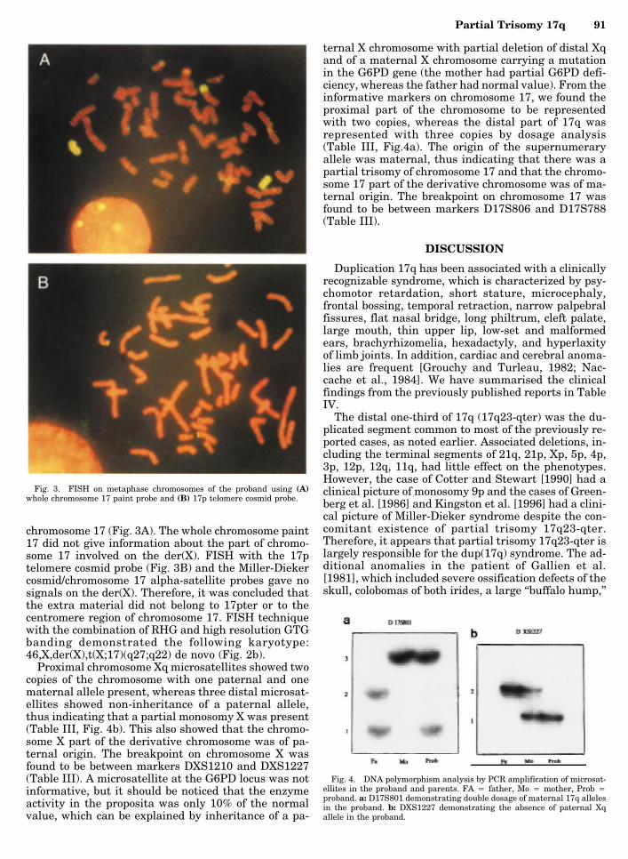
ternal X chromosome with partial deletion of distal Xqand of a maternal X chromosome carrying a mutationin the G6PD gene (the mother had partial G6PD defi-ciency, whereas the father had normal value). From theinformative markers on chromosome 17, we found theproximal part of the chromosome to be representedwith two copies, whereas the distal part of 17q wasrepresented with three copies by dosage analysis(Table III, Fig.4a). The origin of the supernumeraryallele was maternal, thus indicating that there was apartial trisomy of chromosome 17 and that the chromo-some 17 part of the derivative chromosome was of ma-ternal origin. The breakpoint on chromosome 17 wasfound to be between markers D17S806 and D17S788(Table III).
DISCUSSION
Duplication 17q has been associated with a clinicallyrecognizable syndrome, which is characterized by psy-chomotor retardation, short stature, microcephaly,frontal bossing, temporal retraction, narrow palpebralfissures, flat nasal bridge, long philtrum, cleft palate,large mouth, thin upper lip, low-set and malformedears, brachyrhizomelia, hexadactyly, and hyperlaxityof limb joints. In addition, cardiac and cerebral anoma-lies are frequent [Grouchy and Turleau, 1982; Nac-cache et al., 1984]. We have summarised the clinicalfindings from the previously published reports in TableIV.
The distal one-third of 17q (17q23-qter) was the du-plicated segment common to most of the previously re-ported cases, as noted earlier. Associated deletions, in-cluding the terminal segments of 21q, 21p, Xp, 5p, 4p,3p, 12p, 12q, 11q, had little effect on the phenotypes.However, the case of Cotter and Stewart [1990] had aclinical picture of monosomy 9p and the cases of Green-berg et al. [1986] and Kingston et al. [1996] had a clini-cal picture of Miller-Dieker syndrome despite the con-comitant existence of partial trisomy 17q23-qter.Therefore, it appears that partial trisomy 17q23-qter islargely responsible for the dup(17q) syndrome. The ad-ditional anomalies in the patient of Gallien et al.[1981], which included severe ossification defects of theskull, colobomas of both irides, a large ‘‘buffalo hump,’’
Fig. 3. FISH on metaphase chromosomes of the proband using (A)whole chromosome 17 paint probe and (B) 17p telomere cosmid probe.
Fig. 4. DNA polymorphism analysis by PCR amplification of microsat-ellites in the proband and parents. FA 4 father, Mo 4 mother, Prob 4proband. a: D17S801 demonstrating double dosage of maternal 17q allelesin the proband. b: DXS1227 demonstrating the absence of paternal Xqallele in the proband.
Partial Trisomy 17q 91

omphalocele, and labia majora with the appearance ofscrotal folds, were most probably due to a larger dupli-cated segment (17q21-qter). It seems from the previousclinical reports that duplication 17q predisposes tomidline defects. CNS malformations ranging from ho-loprosencephaly to Dandy-Walker defect, as well asoral clefting, cardiac septal defects, omphalocele, andabsent vagina were found. These findings are in agree-ment with the postulate of Opitz and Gilbert’s [1982]that midline development is less buffered than that ofparamedian structures.
In our case, the autosomal segment 17q22-qter,translocated onto the inactivated der(X), was alwaysearly replicating. This indicates that the multiple ab-normalities of our patient actually represent a partialtrisomy 17q syndrome. In unbalanced X-autosomaltranslocations, the translocated X chromosome is usu-ally inactivated [Geerkens et al., 1994; Hagemeijer etal., 1977]. It is possible that the extension of the inac-tivation from the translocated X, by a spreading effectwould reduce or even suppress the chromosomal dis-
equilibrium. It is generally known that in the presenceof a structural abnormality of the X chromosome, inac-tivation could occur at random, but usually is followedby a cellular selection favoring the better genetic bal-ance. In our case the abnormal X chromosome is inac-tivated in blood, but it is unknown whether it is inac-tivated in other tissues. According to Hagemeijer et al.[1977], the inactivation pattern observed in one tissueis not necessarily characteristic of the whole indi-vidual.
In our case the use of FISH in combination with mo-lecular analysis with polymorphic microsatellite DNAmarkers identified the breakpoints of the derivativechromosome between D17S806 and D17S788 for chro-mosome 17 and between DXS1210 and DXS1227 forthe X chromosome. According to the molecular analy-sis, the origin of the abnormal X chromosome was pa-ternal, whereas the origin of the duplicated part ofchromosome 17 was maternal. The unbalanced trans-location between the paternal X and the maternal chro-mosome 17 is therefore suggested to be due to a postzy-
TABLE IV. Summary of Clinical Findings From Previously Published Reports of Partial Trisomy 17q Compared to Our Patient
Published reports(number of patients 4 30)
Ourpatient
Sex m 4 15/30 f 4 15/30 f
Findings Positive NegativeNot described
or unclearNot
relevant
Short stature 22 0 8 0 +Psychomotor retardation 24 0 0 6 +Hypotonia 8 3 17 2 +Microcephaly/large anterior fontanelle 16/7 1/0 13/21 0/2 −/+Frontal bossing 18 2 10 0 +Bitemporal narrowing 17 2 11 0 +Facial/cranial asymmetry 13 6 11 0 +Widow’s peak 7 6 17 0 +Hypertelorism/broad nasal bridge 12/1 2/3 16/26 0 +/+Upslanted/downslanted palpebral fissures 4/3 12/2 14/25 0 +/−Epicanthus 12 6 12 0 −Flat nasal bridge/broad nose 15/2 4/0 11/28 0 +/+Poorly delineated philtrum 4 0 26 0 +Wide mouth/thin upper lip 9/10 5/0 16/20 0 +/+Down-turned corners of the mouth 11 3 16 0 +Cleft lip and (or) palate/bifid uvula 10/2 9/0 11/28 0 −/−Highly arched palate 13 7 10 0 −Micrognathia 17 5 8 0 +Low set ears 17 2 11 0 +Malformed ears 6 0 24 0 +Short neck 15 5 10 0 +Webbed neck 14 6 10 0 +Low posterior hairline 16 2 12 0 +Kyphoscoliosis 8 0 20 2 −Widely spaced nipples 13 2 13 2 +Cryptorchidism 8 2 4 16Proximal limb shortness 15 4 11 0 +Polydactyly of hands and/or feet 11 8 11 0 −Syndactyly of fingers and/or toes 10 8 12 0 −Hyperlaxity of limb joints 9 3 16 2 −Hirsutism 4 0 26 0 +CNS abnormalities 11 2 17 0 +Ophthalmological problems 4 0 24 2 +Heart defects 13a 4 13 0 −Kidney abnormalities 8 5 17 0 −Gastrointestinal abnormalities 7 0 23 0 −Genital abnormalities 8 0 22 0 +
aIncluding three cases with heart murmur.
92 Sarri et al.

gotic error, possibly a translocation in a cell with tri-somy 17 of maternal origin and with subsequent loss ofthe reciprocal product of the translocation. It is inter-esting that the translocated 17q segment is not inacti-vated. This could be due to the suggested postzygoticorigin of the translocation, perhaps after X inactivationhas taken place. The present case makes a new addi-tion to the expanding category of mitotically derivedchromosome abnormalities, as previously described insome cases of trisomy 21 [Antonarakis et al., 1993],trisomy 18 [Fisher et al., 1993], trisomy 8 [Grigoriadouet al., 1995; James and Jacobs, 1996], homologous Rob-ertsonian translocations and isochromosomes [Blouinet al., 1994; Robinson et al., 1994], and disomy/trisomymosaicism [DeBrasi et al., 1995; Pangalos et al., 1994;Robinson et al. 1995]. Apparently, our case is the firstnon-Robertsonian translocation in which this kind ofobservation has been made.
To the best of our knowledge, this is the first case ofpartial trisomy 17q that is identified by molecularanalysis and the second case that is identified by FISH.The first such case was reported by Kingston et al.[1996] in a male with Miller-Dieker syndrome and aduplication 17q25-qter, owing to a familial chromo-some 17 inversion. Our patient is also the third pub-lished case in which the partial trisomy probably is dueto a postzygotic error. The first such case was reportedby Serotkin et al. [1988] in a female infant with mul-tiple anomalies who was mosaic for duplication 17q21-qter, owing to a direct tandem duplication. The secondcase was reported by King et al. [1991] in a femaleinfant born after prenatal diagnosis of mosaic partialtrisomy 17q21.1-qter. However, these two cases werenot studied by FISH or molecular analysis.
The patient described in this report presented withStargardt disease, a macular degeneration of early on-set, and a rapidly progressive course. This condition isgenetically heterogeneous and can be transmitted asan autosomal recessive trait (one locus mapped to chro-mosome 1) or as an autosomal dominant trait (locimapped to chromosomes 6 and 13) [Rosenfeld et al.,1994]. To the best of our knowledge, Stargardt diseasehas not been described in association with chromosom-al aberrations involving chromosome 17, and we pro-pose that a further locus for Stargardt disease maymap to either the breakpoint at 17q22 or to the dupli-cated segment 17q22-qter.
In conclusion, cases of partial trisomy 17q includingthe present one show some variability in clinical ex-pression, related to the extent of the 17q duplication. Astudy of more patients is needed to refine the pheno-typic mapping of chromosome 17 and to correlate dif-ferent clinical syndromes with the extent of the 17qduplication. It is also important that chromosomal ab-normalities are studied by molecular analysis in orderto discover the underlying mechanisms of formation.
ACKNOWLEDGMENTS
Most of the primers flanking microsatellite markerswere kindly supplied by Dr. Claes Wadelius (Univer-sity of Uppsala, Sweden), supported by the Council ofthe Nordic Ministers. The secretarial help of Mrs. As-
pasia Giannakou and the technical help of Mrs. Geor-gia Karadima are gratefully acknowledged.
REFERENCES
Antonarakis SE, Avramopoulos D, Blouin JL, Talbot CC, Schinzel AA(1993): Mitotic errors somatic cells cause trisomy 21 in about 4.5% ofcases and are not associated with advanced maternal age. Nat Genet3:146–150.
Berberich MS, Carey JC, Lauce HJ, Hall BD (1978): Duplication (partialtrisomy) of the distal long arm of chromosome 17: A new clinicallyrecognizable disorder. BD:OAS XIV:287–295.
Blouin JL, Binkert F, Antonarakis SE (1994): Biparental inheritance ofchromosome 21 polymorphic markers indicates that some Robertso-nian translocations t(21:21) occur postzygotically. Am J Med Genet49:363–368.
Bridge J, Sanger W, Mosher G, Buehler B, Hearty C, Olney A, Fordyce R(1985): Partial duplication of distal 17q. Am J Med Genet 22:229–235.
Caine A, Knapton DM, Mueller RF, Gongdon PJ, Haigh D (1989): Dupli-cation of distal 17q from a maternal translocation: An additional casewith some unique features. J Med Genet 26:577–579.
Cotter PD, Stewart NL (1990): Partial trisomy 17q and monosomy 9p dueto a familial translocation. Ann Genet 33:231–233.
DeBrasi D, Genuardi M, D’Agostino A, Calvieri F, Tozzi C, Varrone S, NeriG (1995): Double autosomal/gonosomal mosaic aneuploidy: Study ofnondisjunction in two cases with trisomy of chromosome 8. Hum Genet95:519–525.
Economou EP, Bergen AW, Warren AC, Antonarakis SE (1990): Thepolydeoxyadenylate tract of Alu repetitive elements is polymorphic inthe human genome. Proc Natl Acad Sci USA 87:2951–2954.
Fisher JM, Harvey JF, Lindenbaum RH, Boyd PA, Jacobs PA (1993): Mo-lecular studies of trisomy 18. Am J Hum Genet 52:1139–1144.
Fryns JP, Parloir C, Van den Berghe H (1979): Partial trisomy 17q. Karyo-type: 46, XY, der(21), t(17;21)(q22;p13). Hum Genet 49:361–364.
Gallien JU, Neu RL, Wynn RJ, Steinberg-Warren N, Bannerman RM(1981): Brief clinical report: An infant with duplication of 17q21-17qter.Am J Med Genet 8:111–115.
Geerkens C, Just W, Vogel W (1994): Deletions of Xq and growth deficit: Areview. Am J Med Genet 50:105–113.
Greenberg F, Stratton RF, Lockhart LH, Elder FFB, Dobyns WB, Ledbet-ter DH (1986): Familial Miller-Dieker syndrome associated with peri-centric inversion of chromosome 17. Am J Med Genet 23:853–859.
Grigoriadou M, Bugge M, Avramopoulos D, Kitsiou-Tzeli S, Anneren G,Hertz JM, Lacombe D, Tsezou A, Galla-Voumvouraki A, Clausen N,Vassilopoulos D, Brondum-Nielsen K, Petersen MB (1995): Nondis-junction studies in trisomy 8. Am J Hum Genet 57:A114.
Grouchy J de, Turleau C (1982): ‘‘Atlas des Maladies Chromosomiques.’’ 2e
ed. Paris: Expansion Scientifique Francaise, pp 288–291.
Gyapay G, Morisette J, Vignal A, Dib C, Fizames C, Milleseau P, Marc S,Bernandi G, Lathrop M, Weissenbach J (1994): The 1993–94 Genethonhuman genetic linkage map. Nat Genet 7:246–339.
Hagemeijer A, Hoovers J, Smit EME, Bootsma D (1977): Replication pat-tern of the X chromosomes in three X/autosomal translocations. Cyto-genet Cell Genet 18:333–348.
ISCN (1995): ‘‘An International System for Human Cytogenetic Nomen-clature,’’ Mitelman F (ed). Basel: S. Karger.
James RS, Jacobs PA (1996): Molecular studies of the aetiology of trisomy8 in spontaneous abortions and the liveborn population. Hum Genet97:283–286.
King PA, Ghosh A, Tang M (1991): Mosaic partial trisomy 17q2. J MedGenet 28:641–643.
Kingston HM, Ledbetter DH, Tomlin PI, Gaunt KL (1996): Miller-Diekersyndrome resulting from rearrangement of a familial chromosome 17inversion detected by fluorescence in situ hybridization. J Med Genet33:69–72.
Lalloz MR, McVey JH, Pattinson JK, Tuddenham EGD (1991): Haemo-philia: A diagnosis by analysis of a hypervariable dinucleotide repeatwithin the factor VIII gene. Lancet 338:207–211.
Lenzini E, Leszi A, Artifoni L, Casellato R, Tenconi R, Baccichetti C (1988):Partial duplication of 17 long arm. Ann Genet 31:175–180.
Miller SA, Dykes DD, Polesky HF (1988): A simple salting out procedure
Partial Trisomy 17q 93

for extracting DNA from human nucleated cells. Nucl Acids Res 16:1215.
Naccache NF, Vianna-Morgante AM, Richieri-Costa A (1984): Brief clinicalreport: Duplication of distal 17q: Report of an observation. Am J MedGenet 17:633–639.
Nakagawa H, Inazawa J, Misawa S, Tanaka S, Takashima T, Taniwaki M,Abe T, Kashima K (1992): Detection of i(17q) chromosome by FISHwith a chromosome 17 alpha satellite DNA probe. Cancer Genet Cyto-genet 62:140–143.
Nunez PB, Rolon A, Lizcano LA, Rivera H (1993): Pure trisomy 17q from a17;21 translocation. Genet Couns 4:227–229.
Ohdo S, Madocoro H, Sonoda T, Ohba KI (1989): Sibs lacking characteristicfeatures of duplication of distal 17q. J Med Genet 26:465–468.
Opitz JM, Gilbert EF (1982): Editorial comment: CNS anomalies and themidline as a ‘‘developmental field.’’ Am J Med Genet 12:443–455.
Orye E, Van Bever H (1985): De novo distal trisomy 17q. Ann Genet 28:61–62.
Pangalos C, Avramopoulos D, Blouin JL, Raoul O, de Blois MC, Prieur M,Schinzel AA, Gika M, Abazis D, Antonarakis SE (1994): Understandingthe mechanism(s) of mosaic trisomy 21 by using DNA polymorphismanalysis. Am J Hum Genet 54:473–481.
Parcheta B, Skawinsk W, Wisniews L, Piontek E, Gutkowsk A, WermensK (1985): A new case of partial trisomy 17 long arm: Densitometricanalysis of aberrations. Eur J Ped 143:314–316.
Petersen MB, Economou EP, Slaugenhaupt SA, Chakravarti A, Antonara-kis SE (1990): Linkage analysis of the human HMG14 gene on chro-mosome 21 using a GT dinucleotide repeat as polymorphic marker.Genomics 7:136–138.
Petersen MB, Schinzel AA, Binkert F, Tranebjaerg L, Mikkelsen M, CollinsFA, Economou EP, Antonarakis SE (1991): Use of short sequence re-peat DNA polymorphisms after PCR amplification to detect the paren-tal origin of the additional chromosome 21 in Down syndrome. Am JHum Genet 48:65–71.
Robb A, Forsyth I, Tolmie J (1987): Partial trisomy 17q and a generalisedbone dysplasia in a 12 week fetus. J Med Genet 24:502–504.
Robinson WP, Bernasconi F, Basaran S, Yuksel-Apak M, Neri G, ServilleF, Balicek P, Haluza R, Farah LMS, Luleci G, Schinzel AA (1994): Asomatic origin of homologous Robertsonian translocations and isochro-mosomes. Am J Hum Genet 54:290–302.
Robinson WP, Binkert F, Bernasconi F, Lorda-Sanchez I, Werder EA,Schinzel AA (1995): Molecular studies of chromosomal mosaicism:Relative frequency of chromosome gain or loss and possible role of cellselection. Am J Hum Genet 56:444–451.
Rosenfeld PJ, Mckusick VA, Amberger JS, Dryja TP (1994): Recent ad-vances in the gene map of inherited eye disorders:primary hereditarydiseases of the retina, choroid and vitreous. J Med Genet 31:903–915.
Salamanca-Gomez F, Armendares S (1975): Identification of isochromo-some 17 in a girl with mental retardation and congenital malforma-tions. Ann Genet 18:235–238.
Serotkin A, Stamberg J, Waber L (1988): Duplication 17q mosaicism: Aninfant with features of Ellis-van Creveld syndrome. J Med Genet 25:258–260.
Shawe DJ, Fear C, Appleyard WJ (1983): A child with partial trisomy ofchromosome 17 and partial monosomy of chromosome 3:46,XY,der(3),t(3;17)(p25;q23). J Med Genet 20:383–385.
Shimizu T, Ikeuchi T, Shinohara T, Ohba S, Miyaguchi H, Akiyama T,Shibata T (1988): Distal trisomy of chromosome 17q due to invertedtandem duplication. Clin Genet 33:311–314.
Theune S, Fung J, Todd S, Sakaguchi AY, Naylor SL (1991): PCR primersfor human chromosomes: reagents for the rapid analysis of somatic cellhybrids. Genomics 9:511–516.
Turleau C, Grouchy J de, Bouvert JP (1979): Distal trisomy 17q. ClinGenet 15:54–57.
Warburton D, Stein Z, Kline Z, Susser M (1980): Chromosome abnormali-ties in spontaneous abortion data from the New York City study. InPorter IH, Hook EB (eds): ‘‘Human Embryonic and Fetal Death,’’ NewYork: Academic Press, pp.261–287.
Yamamoto Y, Endo Y, Kurok Y (1979): A case of partial trisomy 17 result-ing from X-autosomal translocation. J Med Genet 16:395–399.
94 Sarri et al.