Embed Size (px)
Citation preview

Part 1The Thorax
ECA1 7/18/06 6:30 PM Page 1
ECA1 7/18/06 6:30 PM Page 2
Surface anatomy and surface markings
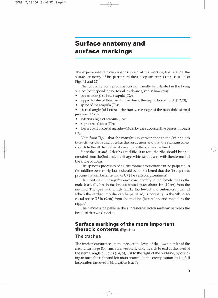
The experienced clinician spends much of his working life relating thesurface anatomy of his patients to their deep structures (Fig. 1; see also Figs. 11 and 22).
The following bony prominences can usually be palpated in the livingsubject (corresponding vertebral levels are given in brackets):•◊◊superior angle of the scapula (T2);•◊◊upper border of the manubrium sterni, the suprasternal notch (T2/3);•◊◊spine of the scapula (T3);•◊◊sternal angle (of Louis) — the transverse ridge at the manubrio-sternaljunction (T4/5);•◊◊inferior angle of scapula (T8);•◊◊xiphisternal joint (T9);•◊◊lowest part of costal margin—10th rib (the subcostal line passes throughL3).
Note from Fig. 1 that the manubrium corresponds to the 3rd and 4ththoracic vertebrae and overlies the aortic arch, and that the sternum corre-sponds to the 5th to 8th vertebrae and neatly overlies the heart.
Since the 1st and 12th ribs are difficult to feel, the ribs should be enu-merated from the 2nd costal cartilage, which articulates with the sternum atthe angle of Louis.
The spinous processes of all the thoracic vertebrae can be palpated inthe midline posteriorly, but it should be remembered that the first spinousprocess that can be felt is that of C7 (the vertebra prominens).
The position of the nipple varies considerably in the female, but in themale it usually lies in the 4th intercostal space about 4in (10cm) from themidline. The apex beat, which marks the lowest and outermost point atwhich the cardiac impulse can be palpated, is normally in the 5th inter-costal space 3.5in (9cm) from the midline (just below and medial to thenipple).
The trachea is palpable in the suprasternal notch midway between theheads of the two clavicles.
Surface markings of the more importantthoracic contents (Figs 2–4)
The trachea
The trachea commences in the neck at the level of the lower border of thecricoid cartilage (C6) and runs vertically downwards to end at the level ofthe sternal angle of Louis (T4/5), just to the right of the mid-line, by divid-ing to form the right and left main bronchi. In the erect position and in fullinspiration the level of bifurcation is at T6.
3
ECA1 7/18/06 6:30 PM Page 3
4 The Thorax
Fig. 1◊Lateral view of thethorax—its surfacemarkings and vertebrallevels. (Note that theangle of Louis (T4/5)demarcates the superiormediastinum, the uppermargin of the heart andthe beginning and end ofthe aortic arch.)
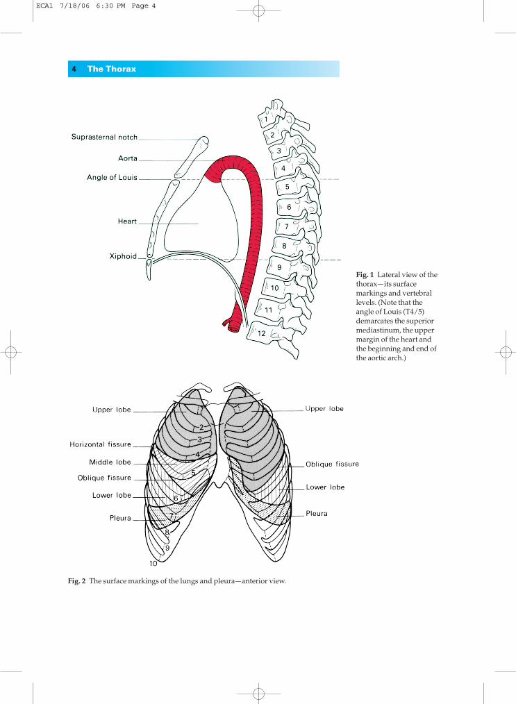
Fig. 2◊The surface markings of the lungs and pleura—anterior view.
ECA1 7/18/06 6:30 PM Page 4
Surface anatomy and surface markings 5
The pleura
The cervical pleura can be marked out on the surface by a curved line drawnfrom the sternoclavicular joint to the junction of the medial and middlethirds of the clavicle; the apex of the pleura is about 1 in (2.5cm) above the clavicle. This fact is easily explained by the oblique slope of the first rib.It is important because the pleura can be wounded (with consequent
Fig. 3◊The surfacemarkings of the lungsand pleura—posteriorview.
Fig. 4◊The surfacemarkings of the heart (seetext).
ECA1 7/18/06 6:30 PM Page 5
pneumothorax) by a stab wound — and this includes the surgeon’s knifeand the anaesthetist’s needle—above the clavicle.
The lines of pleural reflexion pass from behind the sternoclavicular jointon each side to meet in the midline at the 2nd costal cartilage (the angle ofLouis). The right pleural edge then passes vertically downwards to the 6thcostal cartilage and then crosses:•◊◊the 8th rib in the midclavicular line;•◊◊the 10th rib in the midaxillary line;•◊◊the 12th rib at the lateral border of the erector spinae.
On the left side the pleural edge arches laterally at the 4th costal carti-lage and descends lateral to the border of the sternum, due, of course, to itslateral displacement by the heart; apart from this, its relationships are thoseof the right side.
The pleura actually descends just below the 12th rib margin at itsmedial extremity — or even below the edge of the 11th rib if the 12th isunusually short; obviously in this situation the pleura may be opened acci-dentally in making a loin incision to expose the kidney, perform an adrena-lectomy or to drain a subphrenic abscess.
The lungs
The surface projection of the lung is somewhat less extensive than that ofthe parietal pleura as outlined above, and in addition it varies quite consid-erably with the phase of respiration. The apex of the lung closely follows theline of the cervical pleura and the surface marking of the anterior border of theright lung corresponds to that of the right mediastinal pleura. On the leftside, however, the anterior border has a distinct notch (the cardiac notch)which passes behind the 5th and 6th costal cartilages. The lower border of thelung has an excursion of as much as 2–3in (5–8cm) in the extremes of respi-ration, but in the neutral position (midway between inspiration and expira-tion) it lies along a line which crosses the 6th rib in the midclavicular line,the 8th rib in the midaxillary line, and reaches the 10th rib adjacent to thevertebral column posteriorly.
The oblique fissure, which divides the lung into upper and lower lobes, isindicated on the surface by a line drawn obliquely downwards and out-wards from 1in (2.5cm) lateral to the spine of the 5th thoracic vertebra tothe 6th costal cartilage about 1.5in (4cm) from the midline. This can be rep-resented approximately by abducting the shoulder to its full extent; the lineof the oblique fissure then corresponds to the position of the medial borderof the scapula.
The surface markings of the transverse fissure (separating the middle andupper lobes of the right lung) is a line drawn horizontally along the 4thcostal cartilage and meeting the oblique fissure where the latter crosses the5th rib.
The heart
The outline of the heart can be represented on the surface by the irregular quadrangle bounded by the following four points (Fig. 4):
6 The Thorax
ECA1 7/18/06 6:30 PM Page 6
The thoracic cage 7
1◊◊the 2nd left costal cartilage 0.5in (12mm) from the edge of the sternum;2◊◊the 3rd right costal cartilage 0.5in (12mm) from the sternal edge;3◊◊the 6th right costal cartilage 0.5in (12mm) from the sternum; 4◊◊the 5th left intercostal space 3.5in (9cm) from the midline (correspond-ing to the apex beat).
The left border of the heart (indicated by the curved line joining points 1 and 4) is formed almost entirely by the left ventricle (the auricularappendage of the left atrium peeping around this border superiorly), thelower border (the horizontal line joining points 3 and 4) corresponds to theright ventricle and the apical part of the left ventricle; the right border(marked by the line joining points 2 and 3) is formed by the right atrium(see Fig. 24a).
A good guide to the size and position of your own heart is given byplacing your clenched right fist palmar surface down immediately inferiorto the manubriosternal junction. Note that the heart is about the size of thesubject’s fist, lies behind the body of the sternum (therefore anterior to tho-racic vertebrae 5–8), and bulges over to the left side.
The surface markings of the vessels of the thoracic wall are of im-portance if these structures are to be avoided in performing aspiration of the chest. The internal thoracic (internal mammary) vessels run verticallydownwards behind the costal cartilages half an inch from the lateral border of the sternum. The intercostal vessels lie immediately below their corresponding ribs (the vein above the artery) so that it is safe to pass a needle immediately above a rib, dangerous to pass it immediately below(see Fig. 8).
The thoracic cage
The thoracic cage is formed by the vertebral column behind, the ribs andintercostal spaces on either side and the sternum and costal cartilages infront. Above, it communicates through the ‘thoracic inlet’ with the root of the neck; below, it is separated from the abdominal cavity by thediaphragm (Fig. 1).
The thoracic vertebrae
See ‘vertebral column’, page 327.
The ribs
The greater part of the thoracic cage is formed by the twelve pairs of ribs. Of these, the first seven are connected anteriorly by way of their costal cartilages to the sternum, the cartilages of the 8th, 9th and 10th articulateeach with the cartilage of the rib above (‘false ribs’) and the last two ribs arefree anteriorly (‘floating ribs’).
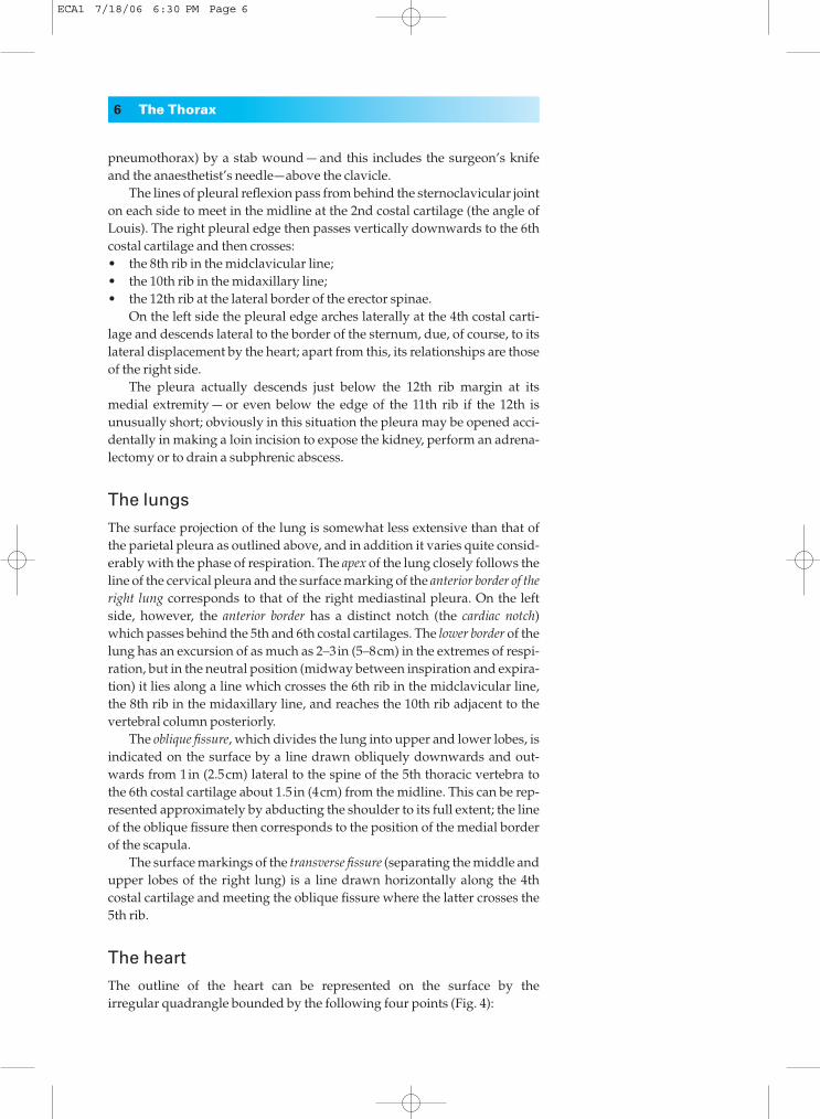
Each typical rib (Fig. 5) has a head bearing two articular facets, for
ECA1 7/18/06 6:30 PM Page 7
articulation with the numerically corresponding vertebra and the vertebraabove, a stout neck, which gives attachment to the costotransverse liga-ments, a tubercle with a rough non-articular portion and a smooth facet, forarticulation with the transverse process of the corresponding vertebra, anda long shaft flattened from side to side and divided into two parts by the‘angle’ of the rib. The angle demarcates the lateral limit of attachment of theerector spinae muscle.
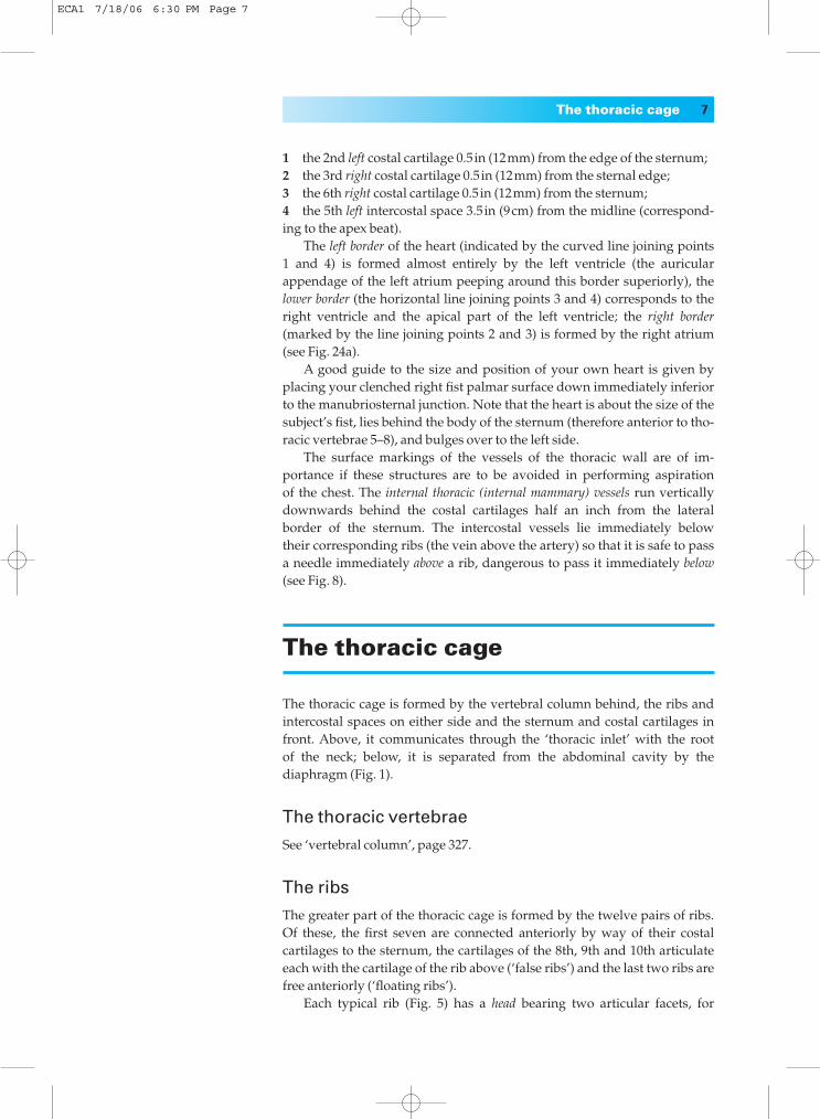
The following are the significant features of the ‘atypical’ ribs.1st Rib (Fig. 6). This is flattened from above downwards. It is not
only the flattest but also the shortest and most curvaceous of all the ribs. Ithas a prominent tubercle on the inner border of its upper surface for the
8 The Thorax
Fig. 5◊A typical rib.
Fig. 6◊Structures crossing the first rib.
ECA1 7/18/06 6:30 PM Page 8
insertion of scalenus anterior. In front of this tubercle, the subclavian veincrosses the rib; behind the tubercle is the subclavian groove where the subcla-vian artery and lowest trunk of the brachial plexus lie in relation to thebone. It is here that the anaesthetist can infiltrate the plexus with localanaesthetic.
Crossing the neck of the first rib from the medial to the lateral side arethe sympathetic trunk, the superior intercostal artery (from the costocervi-cal trunk) and the large branch of the first thoracic nerve to the brachialplexus.
The 2nd rib is much less curved than the 1st and about twice as long.The 10th rib has only one articular facet on the head.The 11th and 12th ribs are short, have no tubercles and only a single facet
on the head. The 11th rib has a slight angle and a shallow subcostal groove;the 12th has neither of these features.
Clinical features
Rib fractures
The chest wall of the child is highly elastic and therefore fractures of the ribin children are rare. In adults, the ribs may be fractured by direct violence orindirectly by crushing injuries; in the latter the rib tends to give way at itsweakest part in the region of its angle. Not unnaturally, the upper two ribs,which are protected by the clavicle, and the lower two ribs, which are unat-tached and therefore swing free, are the least commonly injured.
In a severe crush injury to the chest several ribs may fracture in frontand behind so that a whole segment of the thoracic cage becomes torn free(‘stove-in chest’). With each inspiration this loose flap sucks in, with eachexpiration it blows out, thus undergoing paradoxical respiratory move-ment. The associated swinging movements of the mediastinum producesevere shock and this injury calls for urgent treatment by insertion of achest drain with underwater seal, followed by endotracheal intubation, ortracheostomy, combined with positive pressure respiration.
Coarctation of the aorta (see Fig. 34b and page 41)
In coarctation of the aorta, the intercostal arteries derived from the aortareceive blood from the superior intercostals (from the costocervical trunk ofthe subclavian artery), from the anterior intercostal branches of the internalthoracic artery (arising from the subclavian artery) and from the arteriesanastomosing around the scapula. Together with the communicationbetween the internal thoracic and inferior epigastric arteries, they providethe principal collaterals between the aorta above and below the block. Inconsequence, the intercostal arteries undergo dilatation and tortuosity anderode the lower borders of the corresponding ribs to give the characteristicirregular notching of the ribs, which is very useful in the radiographic confir-mation of this lesion.
The thoracic cage 9
ECA1 7/18/06 6:30 PM Page 9
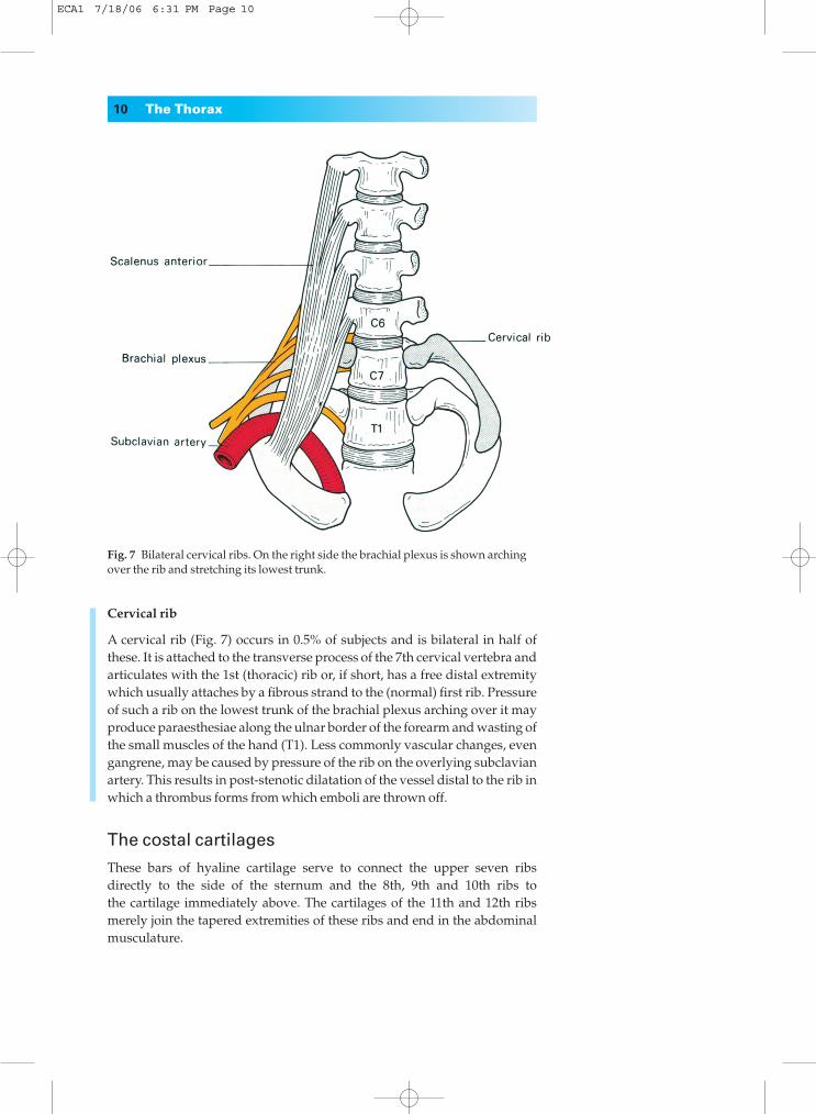
Fig. 7◊Bilateral cervical ribs. On the right side the brachial plexus is shown archingover the rib and stretching its lowest trunk.
Cervical rib
A cervical rib (Fig. 7) occurs in 0.5% of subjects and is bilateral in half ofthese. It is attached to the transverse process of the 7th cervical vertebra andarticulates with the 1st (thoracic) rib or, if short, has a free distal extremitywhich usually attaches by a fibrous strand to the (normal) first rib. Pressureof such a rib on the lowest trunk of the brachial plexus arching over it mayproduce paraesthesiae along the ulnar border of the forearm and wasting ofthe small muscles of the hand (T1). Less commonly vascular changes, evengangrene, may be caused by pressure of the rib on the overlying subclavianartery. This results in post-stenotic dilatation of the vessel distal to the rib inwhich a thrombus forms from which emboli are thrown off.
The costal cartilages
These bars of hyaline cartilage serve to connect the upper seven ribsdirectly to the side of the sternum and the 8th, 9th and 10th ribs to the cartilage immediately above. The cartilages of the 11th and 12th ribsmerely join the tapered extremities of these ribs and end in the abdominalmusculature.
10 The Thorax
ECA1 7/18/06 6:31 PM Page 10
Clinical features
1◊◊The cartilage adds considerable resilience to the thoracic cage and pro-tects the sternum and ribs from more frequent fracture.2◊◊In old age (and sometimes also in young adults) the costal cartilagesundergo progressive ossification; they then become radio-opaque and maygive rise to some confusion when examining a chest radiograph of anelderly patient.
The sternum
This dagger-shaped bone, which forms the anterior part of the thoraciccage, consists of three parts. The manubrium is roughly triangular in outlineand provides articulation for the clavicles and for the first and upper part ofthe 2nd costal cartilages on either side. It is situated opposite the 3rd and4th thoracic vertebrae. Opposite the disc between T4 and T5 it articulates atan oblique angle at the manubriosternal joint (the angle of Louis), with thebody of the sternum (placed opposite T5 to T8). This is composed of four partsor ‘sternebrae’ which fuse between puberty and 25 years of age. Its lateralborder is notched to receive part of the 2nd and the 3rd to the 7th costal car-tilage. The xiphoid process is the smallest part of the sternum and usuallyremains cartilaginous well into adult life. The cartilaginous manu-briosternal joint and that between the xiphoid and the body of the sternummay also become ossified after the age of 30.
Clinical features
1◊◊The attachment of the elastic costal cartilages largely protects thesternum from injury, but indirect violence accompanying fracture disloca-tion of the thoracic spine may be associated with a sternal fracture. Directviolence to the sternum may lead to displacement of the relatively mobilebody of the sternum backwards from the relatively fixed manubrium.2◊◊In a sternal puncture a wide-bore needle is pushed through the thinlayer of cortical bone covering the sternum into the highly vascular spongybone beneath, and a specimen of bone marrow aspirated with a syringe.3◊◊In operations on the thymus gland, and occasionally for a retrosternalgoitre, it is necessary to split the manubrium in the midline in order to gainaccess to the superior mediastinum. A complete vertical split of the wholesternum is one of the standard approaches to the heart and great vesselsused in modern cardiac surgery.
The intercostal spaces
There are slight variations between the different intercostal spaces, but typi-cally each space contains three muscles, comparable to those of the abdomi-nal wall, and an associated neurovascular bundle (Fig. 8). The muscles are:
The thoracic cage 11
ECA1 7/18/06 6:31 PM Page 11
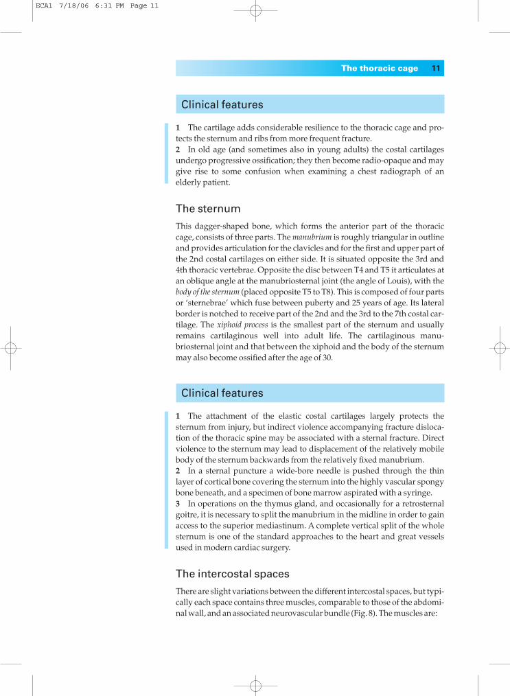
1◊◊the external intercostal, the fibres of which pass downwards and forwards from the rib above to the rib below and reach from the vertebraebehind to the costochondral junction in front, where muscle is replaced bythe anterior intercostal membrane;2◊◊the internal intercostal, which runs downwards and backwards from thesternum to the angles of the ribs where it becomes the posterior intercostalmembrane; 3◊◊the innermost intercostal, which is only incompletely separated from theinternal intercostal muscle by the neurovascular bundle.The fibres of this sheet cross more than one intercostal space and it may beincomplete. Anteriorly it has a more distinct portion which is fan-like inshape, termed the transversus thoracis (or sternocostalis), which spreadsupwards from the posterior aspect of the lower sternum to insert onto theinner surfaces of the second to the sixth costal cartilages.
Just as in the abdomen, the nerves and vessels of the thoracic wall liebetween the middle and innermost layers of muscles. This neurovascularbundle consists, from above downwards, of vein, artery and nerve, the veinlying in a groove on the undersurface of the corresponding rib (remember—v,a,n).
The vessels comprise the posterior and anterior intercostals.The posterior intercostal arteries of the lower nine spaces are branches of
the thoracic aorta, while the first two are derived from the superior inter-costal branch of the costocervical trunk, the only branch of the second partof the subclavian artery. Each runs forward in the subcostal groove to anas-tomose with the anterior intercostal artery. Each has a number of branchesto adjacent muscles, to the skin and to the spinal cord. The correspondingveins are mostly tributaries of the azygos and hemiazygos veins. The firstposterior intercostal vein drains into the brachiocephalic or vertebral vein.
12 The Thorax
Fig. 8◊The relationship of an intercostal space.(Note that a needlepassed into the chestimmediately above a rib will avoid theneurovascular bundle.)
ECA1 7/18/06 6:31 PM Page 12
On the left, the 2nd and 3rd veins often join to form a superior intercostalvein, which crosses the aortic arch to drain into the left brachiocephalicvein.
The anterior intercostal arteries are branches of the internal thoracic artery(1st–6th space) or of its musculophrenic branch (7th–9th spaces). Thelowest two spaces have only posterior arteries. Perforating branches piercethe upper five or six intercostal spaces; those of the 2nd–4th spaces are largein the female and supply the breast.
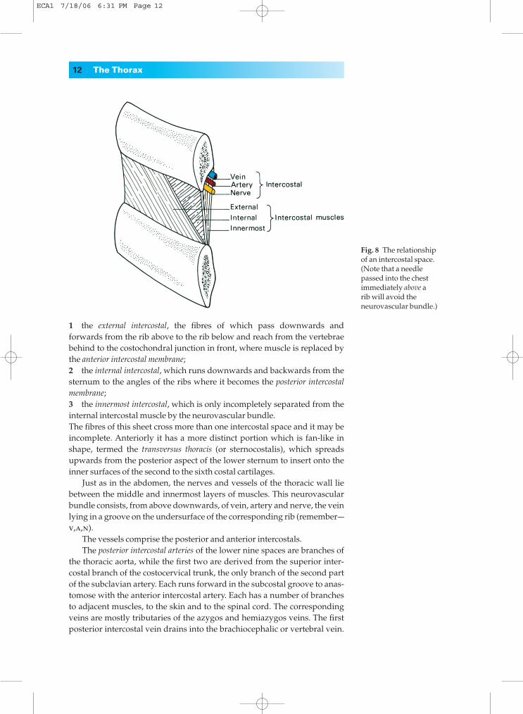
The intercostal nerves are the anterior primary rami of the thoracicnerves, each of which gives off a collateral muscular branch and lateral andanterior cutaneous branches for the innervation of the thoracic and abdom-inal walls (Fig. 9).
Clinical features
1◊◊Local irritation of the intercostal nerves by such conditions as Pott’sdisease of the thoracic vertebrae (tuberculosis) may give rise to pain whichis referred to the front of the chest or abdomen in the region of the periph-eral termination of the nerves.2◊◊Local anaesthesia of an intercostal space is easily produced by infiltra-tion around the intercostal nerve trunk and its collateral branch — a proce-dure known as intercostal nerve block.
The thoracic cage 13
Fig. 9◊Diagram of a typical spinal nerve and its body-wall relationships. On the leftside the sites of eruption of a tuberculous cold abscess tracking forwards from adiseased vertebra are shown—these occur at the points of emergence of thecutaneous branches.
ECA1 7/18/06 6:31 PM Page 13
3◊◊In a conventional posterolateral thoracotomy (e.g. for a pulmonary lobectomy) an incision is made along the line of the 5th or 6th rib; theperiosteum over a segment of the rib is elevated, thus protecting the neu-rovascular bundle, and the rib is excised. Access to the lung or medi-astinum is then gained though the intercostal space, which can be openedout considerably owing to the elasticity of the thoracic cage.4◊◊Pus from the region of the vertebral column tends to track around thethorax along the course of the neurovascular bundle and to ‘point’ to thethree sites of exit of the cutaneous branches of the intercostal nerves, whichare lateral to erector spinae (sacrospinalis), in the midaxillary line and justlateral to the sternum (Fig. 9).
The diaphragmThe diaphragm is the dome-shaped septum dividing the thoracic from theabdominal cavity. It comprises two portions: a peripheral muscular partwhich arises from the margins of the thoracic outlet and a centrally placedaponeurosis (Fig. 10).
The muscular fibres are arranged in three parts.1◊◊A vertebral part from the crura and from the arcuate ligaments. The rightcrus arises from the front of the bodies of the upper three lumbar vertebraeand intervertebral discs; the left crus is only attached to the first two verte-brae. The arcuate ligaments are a series of fibrous arches, the medial being athickening of the fascia covering psoas major and the lateral of fascia overly-ing quadratus lumborum. The fibrous medial borders of the two crura forma median arcuate ligament over the front of the aorta.2◊◊A costal part is attached to the inner aspect of the lower six ribs and costalcartilages.3◊◊A sternal portion consists of two small slips from the deep surface of thexiphisternum.
The central tendon, into which the muscular fibres are inserted, is trefoilin shape and is partially fused with the undersurface of the pericardium.
The diaphragm receives its entire motor supply from the phrenic nerve(C3, 4, 5) whose long course from the neck follows the embryologicalmigration of the muscle of the diaphragm from the cervical region (seebelow). Injury or operative division of this nerve results in paralysis andelevation of the corresponding half of the diaphragm.
Radiographically, paralysis of the diaphragm is recognized by its eleva-tion and paradoxical movement; instead of descending on inspiration it isforced upwards by pressure from the abdominal viscera.
The sensory nerve fibres from the central part of the diaphragm also run in the phrenic nerve, hence irritation of the diaphragmatic pleura (inpleurisy) or of the peritoneum on the undersurface of the diaphragm bysubphrenic collections of pus or blood produces referred pain in the corre-sponding cutaneous area, the shoulder-tip.
The peripheral part of the diaphragm, including the crura, receivessensory fibres from the lower intercostal nerves.
14 The Thorax
ECA1 7/18/06 6:31 PM Page 14
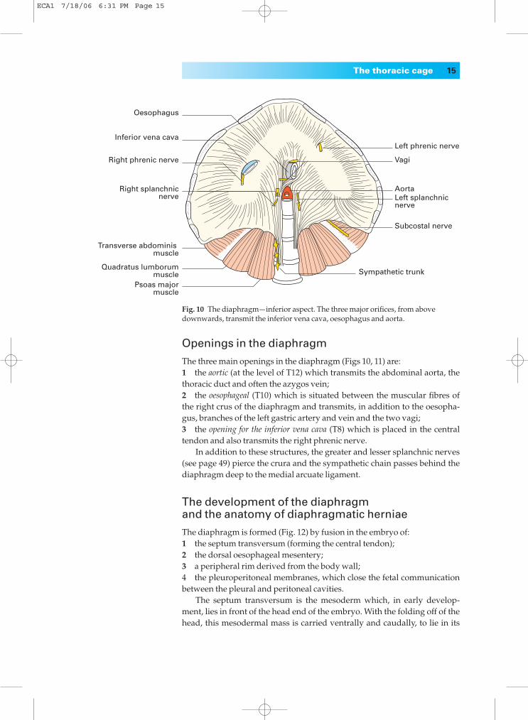
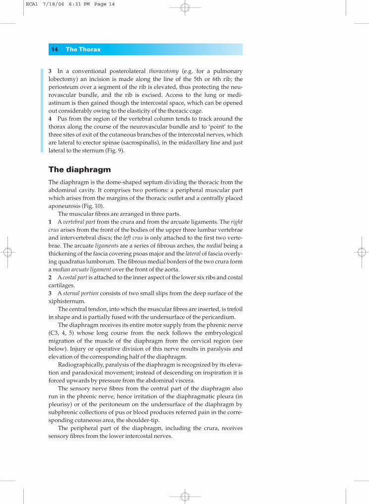
Openings in the diaphragm
The three main openings in the diaphragm (Figs 10, 11) are:1◊◊the aortic (at the level of T12) which transmits the abdominal aorta, thethoracic duct and often the azygos vein;2◊◊the oesophageal (T10) which is situated between the muscular fibres ofthe right crus of the diaphragm and transmits, in addition to the oesopha-gus, branches of the left gastric artery and vein and the two vagi;3◊◊the opening for the inferior vena cava (T8) which is placed in the centraltendon and also transmits the right phrenic nerve.
In addition to these structures, the greater and lesser splanchnic nerves(see page 49) pierce the crura and the sympathetic chain passes behind thediaphragm deep to the medial arcuate ligament.
The development of the diaphragm and the anatomy of diaphragmatic herniae
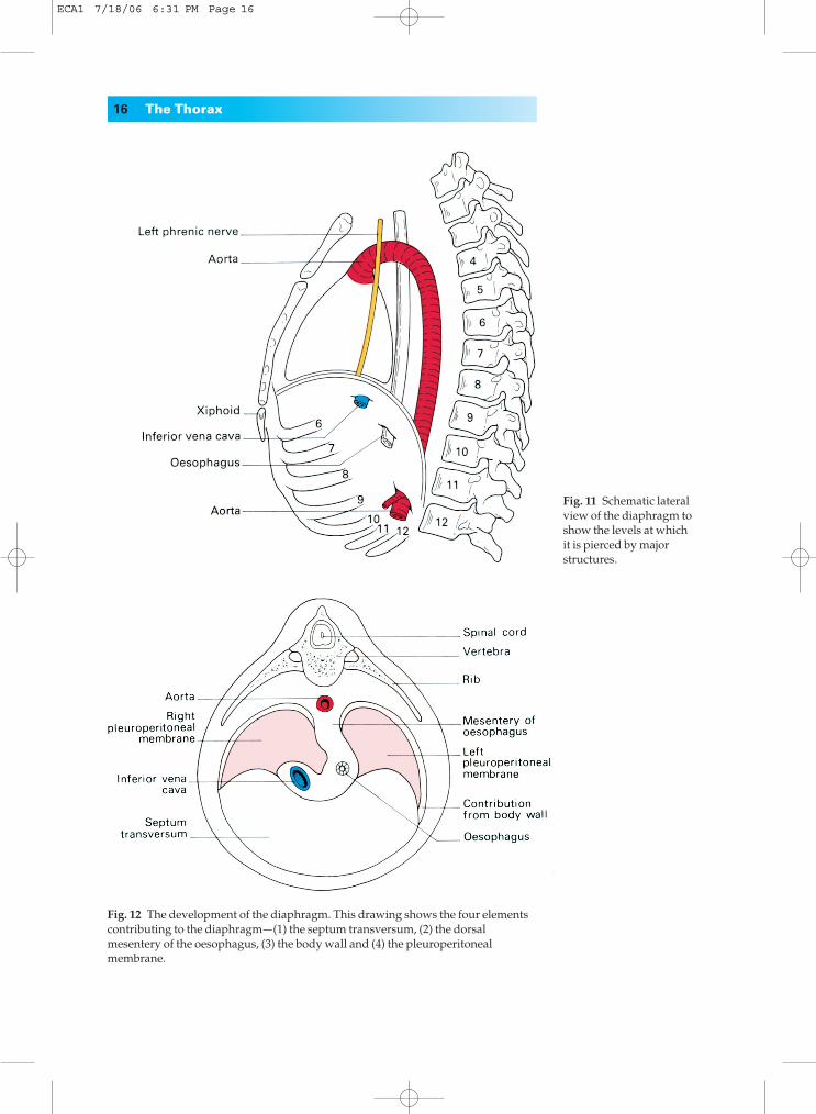
The diaphragm is formed (Fig. 12) by fusion in the embryo of:1◊◊the septum transversum (forming the central tendon);2◊◊the dorsal oesophageal mesentery;3◊◊a peripheral rim derived from the body wall;4◊◊the pleuroperitoneal membranes, which close the fetal communicationbetween the pleural and peritoneal cavities.
The septum transversum is the mesoderm which, in early develop-ment, lies in front of the head end of the embryo. With the folding off of thehead, this mesodermal mass is carried ventrally and caudally, to lie in its
The thoracic cage 15
Oesophagus
Left phrenic nerve
Vagi
AortaLeft splanchnic nerve
Subcostal nerve
Sympathetic trunk
Inferior vena cava
Right phrenic nerve
Right splanchnicnerve
Transverse abdominis muscle
Quadratus lumborummuscle
Psoas majormuscle
Fig. 10◊The diaphragm—inferior aspect. The three major orifices, from abovedownwards, transmit the inferior vena cava, oesophagus and aorta.
ECA1 7/18/06 6:31 PM Page 15
16 The Thorax
Fig. 11◊Schematic lateralview of the diaphragm toshow the levels at whichit is pierced by majorstructures.
Fig. 12◊The development of the diaphragm. This drawing shows the four elementscontributing to the diaphragm—(1) the septum transversum, (2) the dorsalmesentery of the oesophagus, (3) the body wall and (4) the pleuroperitonealmembrane.
ECA1 7/18/06 6:31 PM Page 16
definitive position at the anterior part of the diaphragm. During this migra-tion, the cervical myotomes and nerves contribute muscle and nervesupply respectively, thus accounting for the long course of the phrenicnerve (C3, 4 and 5) from the neck to the diaphragm.
With such a complex embryological story, one may be surprised toknow that congenital abnormalities of the diaphragm are unusual.
However, a number of defects may occur, giving rise to a variety of con-genital herniae through the diaphragm. These may be:1◊◊through the foramen of Morgagni; anteriorly between the xiphoid andcostal origins;2◊◊through the foramen of Bochdalek — the pleuroperitoneal canal — lyingposteriorly;3◊◊through a deficiency of the whole central tendon (occasionally such ahernia may be traumatic in origin);4◊◊through a congenitally large oesophageal hiatus.
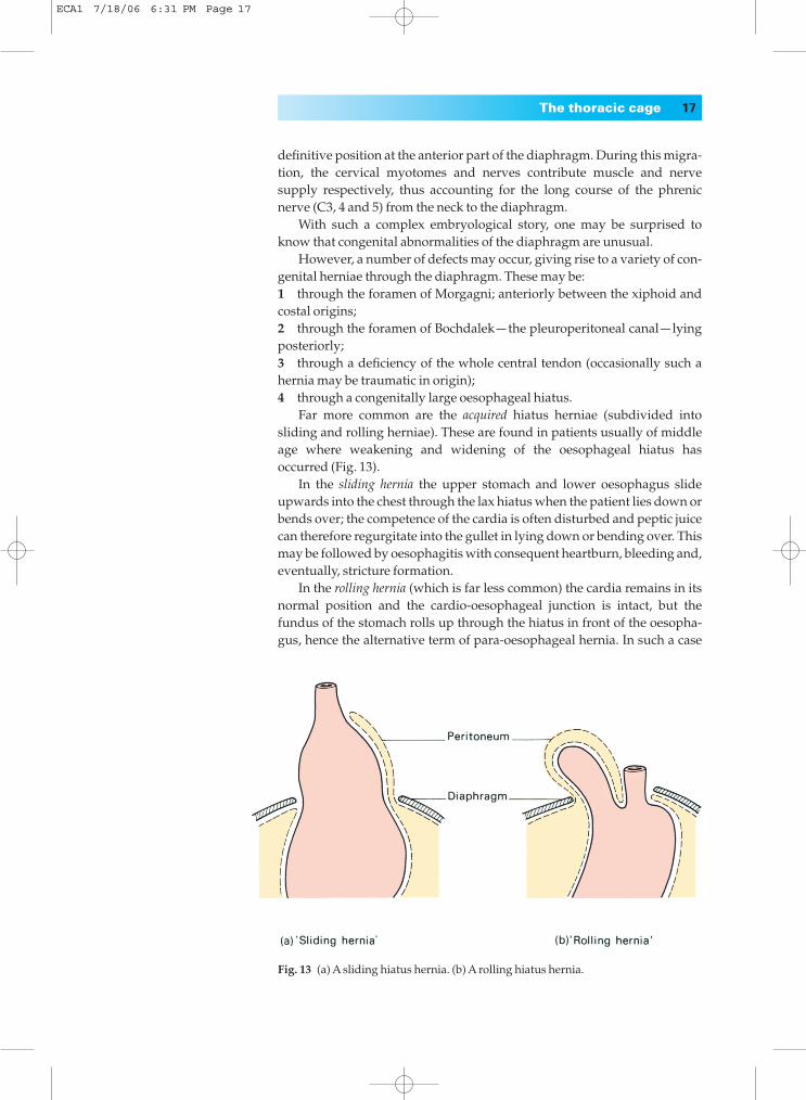
Far more common are the acquired hiatus herniae (subdivided intosliding and rolling herniae). These are found in patients usually of middleage where weakening and widening of the oesophageal hiatus hasoccurred (Fig. 13).
In the sliding hernia the upper stomach and lower oesophagus slideupwards into the chest through the lax hiatus when the patient lies down orbends over; the competence of the cardia is often disturbed and peptic juicecan therefore regurgitate into the gullet in lying down or bending over. Thismay be followed by oesophagitis with consequent heartburn, bleeding and,eventually, stricture formation.
In the rolling hernia (which is far less common) the cardia remains in itsnormal position and the cardio-oesophageal junction is intact, but thefundus of the stomach rolls up through the hiatus in front of the oesopha-gus, hence the alternative term of para-oesophageal hernia. In such a case
The thoracic cage 17
Fig. 13◊(a) A sliding hiatus hernia. (b) A rolling hiatus hernia.
ECA1 7/18/06 6:31 PM Page 17
there may be epigastric discomfort, flatulence and even dysphagia, but noregurgitation because the cardiac mechanism is undisturbed.
The movements of respiration
During inspiration the movements of the chest wall and diaphragm resultin an increase in all diameters of the thorax. This, in turn, brings about anincrease in the negative intrapleural pressure and an expansion of the lungtissue. Conversely, in expiration the relaxation of the respiratory musclesand the elastic recoil of the lung reduce the thoracic capacity and force airout of the lungs.
In quiet inspiration the first rib remains relatively fixed, but contractionof the external and internal intercostals elevates and, at the same time,everts the succeeding ribs. In the case of the 2nd–7th ribs this principallyincreases the anteroposterior diameter of the thorax (by the forward thrust of the sternum), like a pump handle. The corresponding movementof the lower ribs raises the costal margin and leads mainly to an increase inthe transverse diameter of the thorax, like a bucket handle. The depth of thethorax is increased by the contraction of the diaphragm which draws downits central tendon. Normal quiet expiration, brought about by elastic recoil ofthe elevated ribs, is aided by the tone of the abdominal musculature which,acting through the contained viscera, forces the diaphragm upwards.
In deep and in forced inspiration additional muscles attached to thechest wall are called into play (e.g. scalenus anterior, sternocleidomastoid,serratus anterior and pectoralis major) to increase further the capacity ofthe thorax. Similarly, in deep expiration, forced contraction of the abdomi-nal muscles aids the normal expulsive factors described above.
The pleuraeThe two pleural cavities are totally separate from each other (Fig. 2). Eachpleura consists of two layers: a visceral layer intimately related to the surfaceof the lung, and a parietal layer lining the inner aspect of the chest wall, theupper surface of the diaphragm and the sides of the pericardium and medi-astinum. The two layers are continuous in front and behind the root of thelung, but below this the pleura hangs down in a loose fold, the pulmonaryligament, which forms a ‘dead-space’ for distension of the pulmonary veins.The surface markings of the pleura and lungs have already been describedin the section on surface anatomy.
Notice that the lungs do not occupy all the available space in the pleuralcavity even in forced inspiration.
Clinical features
1◊◊Normally the two pleural layers are in close apposition and the spacebetween them is only a potential one. It may, however, fill with air (pneu-mothorax), blood (haemothorax) or pus (empyema).
18 The Thorax
ECA1 7/18/06 6:31 PM Page 18


![[PPT]Chapter 11 Surface Anatomy - Gavilan College -> …hhh.gavilan.edu/rmorales/documents/ch12lect_000.ppt · Web viewChapter 12 Surface Anatomy Surface Anatomy of Head Surface Anatomy](https://img.pdfslide.us/doc/110x75/5b29e6677f8b9ad8298b5149/pptchapter-11-surface-anatomy-gavilan-college-hhh-web-viewchapter-12.jpg)