Embed Size (px)
Citation preview
P-Selectin Glycoprotein Ligand 1 Is a Ligand for L-Selectin on Neutrophils, Monocytes, and CD34 + Hematopoietic Progenitor Cells Olivier Spertini, Anne-Sophie Cordey, Natacha Monai, Laura Giuffr~, and Marc Schapira Division of Hematology and the Hematology Central Laboratory, University of Lausanne, 1011-CHUV Lausanne, Switzerland
Abstract. Selectins play a critical role in initiating leu- kocyte binding to vascular endothelium. In addition, in vitro experiments have shown that neutrophils use L-selectin to roll on adherent neutrophils, suggesting that they express a nonvascular L-selectin ligand. Using a L-selectin/IgM heavy chain (Ix) chimeric protein as an immunocytological probe, we show here that L-selectin can bind to neutrophils, monocytes, CD34 ÷ hematopoi- etic progenitors, and HL-60 and KG-1 myeloid cells. The interaction between L-selectin and teukocytes was protease sensitive and calcium dependent, and abol- ished by cell treatment with neuraminidase, chlorate, or O-sialoglycoprotein endopeptidase. These results revealed common features between leukocyte L-selec- tin ligand and the mucin-like P-selectin glycoprotein ligand 1 (PSGL-1), which mediates neutrophil rolling on P- and E-selectin. The possibility that PSGL-1 could be a ligand for L-selectin was further supported by the ability of P-selectin/Ix chimera to inhibit L-selectin/Ix
binding to leukocytes and by the complete inhibition of both selectin interactions with myeloid cells treated with mocarhagin, a cobra venom metalloproteinase that cleaves the amino terminus of PSGL-1 at Tyr-51. Finally, the abrogation of L- and P-selectin binding to myeloid cells treated with a polyclonal antibody, raised against a peptide corresponding to the amino acid resi- dues 42-56 of PSGL-1, indicated that L- and P-selectin interact with a domain located at the amino-terminal end of PSGL-1. The ability of the anti-PSGL-1 mAb PL-1 to inhibit L- and P-selectin binding to KG-1 cells further supported that possibility. Thus, apart from be- ing involved in neutrophil rolling on P- and E-selectin, PSGL-1 also plays a critical role in mediating neutro- phil attachment to adherent neutrophils. Interaction between L-selectin and PSGL-1 may be of major im- portance for increasing leukocyte recruitment at in- flammatory sites.
S EVERAL adhesion molecules are involved in the regu-
lation of leukocyte homing into tissues. Selectins initiate neutrophil rolling along vascular endothe-
lium at sites of inflammation, whereas integrins and immu- noglobulin-like adhesion molecules have a more impor- tant role in subsequent steps of leukocyte migration into tissues (2, 10, 24, 25, 53, 59, 65-67). L-selectin is expressed by most circulating leukocytes and hematopoietic progeni- tors, whereas E-selectin is expressed by endothelium acti- vated by cytokines or endotoxin (8, 9, 29, 30). P-selectin is contained in intracytoplasmic granules and is rapidly translocated to platelet or endothelial surfaces after cell exposure to thrombin or histamine (14, 19, 28, 31, 32).
The amino-terminal lectin domain of selectins interacts in a calcium-dependent reaction with a large variety of carbohydrate glycoconjugates (1, 3, 20, 33, 45, 48, 53, 56, 57, 61, 64). Some oligosaccharides, such as the tetrasaccha-
Address all correspondence to Olivier Spertini, Division of Hematology, University of Lausanne, 1011-CHUV Lausanne, Switzerland.
ride sialyl Lewis x (sLeX) 1, bind to the three selectins, whereas other carbohydrates react only with one or two of them (12, 64). Several biological ligands for selectins have now been identified (7, 20, 33, 45, 53), most of them being mucin-like glycoproteins with many serine or threonine residues that are potential sites for attachment of O-linked glycans. Sialylation and fucosylation are essential for the function of these sialomucins. Sulfation was also shown to be required for the interaction of L-selectin with endothe- lial ligands such as CD34 and GlyCAM-1 (6, 15, 16, 18, 21) or for P-selectin binding to its major ligand on leukocytes, the P-selectin glycoprotein ligand 1 (PSGL-1) (27, 44, 47, 68). PSGL-1 is a disulfide-linked homodimer comprised of two ~120-kD subunits, which is expressed by most human leukocytes (35, 37, 46, 63, 69). The first 41 amino acid resi- dues of this mucin-like glycoprotein contain an 18-residue signal peptide and a propeptide extending from residues 19-41. After cleavage of the propeptide at position 41,
1. Abbrevia t ions used in this paper: sLe x, sialyl LewisX; PSGL-1, P-selectin glycoprotein ligand 1.
© The Rockefeller University Press, 0021-9525/96/10/523/9 $2.00 The Journal of Cell Biology, Volume 135, Number 2, October 1996 523-531 523
on April 9, 2019jcb.rupress.org Downloaded from http://doi.org/10.1083/jcb.135.2.523Published Online: 15 October, 1996 | Supp Info:
Glu 42 becomes the amino-terminal end residue of mature PSGL-1 (27, 46). PSGL-1 contains many clustered sialylated O-linked glycans extended with poly-N-lactosamine termi- nating in sLe x, as well as tyrosine sulfation sites at residues 46, 48, and 51 that are required for its interaction with P-selectin (27, 35-37, 39, 44, 46, 47, 68). PSGL-1 also inter- acts with E-selectin (4, 41) and plays an essential role in mediating neutrophil rolling on P- and E-selectin-express- ing cells (37, 41, 42). An additional ligand for E-selectin, E-selectin ligand 1, may also support the attachment of neutrophils to activated endothelium (23, 55).
In vitro studies have shown that neutrophils can roll via L-selectin on the surface of previously arrested neutro- phils, suggesting that these cells could express a nonvascu- lar ligand for L-selectin (5). Additional observations have indicated that a ligand for L-selectin is also present on the CD34 ÷ KG-1 myeloid cell line (40). Indeed, function- blocking anti-L-selectin mAbs were found to inhibit lym- phocyte binding to KG-1 cells. Since this interaction was not dependent on CD34 expression, it was suggested that L-selectin could interact with a ligand distinct from that glycoprotein (40).
Using an L-selectin/IgM heavy chain (Ix) chimeric pro- tein, we show in the present study that CD34 ÷ hematopoi- etic progenitors, neutrophils, monocytes, and KG-1 and HL-60 cells express a ligand for L-selectin. In addition, we present a detailed biochemical and immunochemical char- acterization of this L-selectin ligand.
Materials and Methods
Antibodies Anti-L-selectin mAbs anti-LAM1-3, -4, -10, -11 (49) were purified from hybridoma culture supernatants on protein A using the MAPP-II kit (Bio Rad Laboratories, Irvine, CA). Anti-E-selectin mAb H18/7 (8) was a gift from F.W. Luscinskas (Brigham and Women's Hospital, Harvard Medical School, Boston, MA), and anti-P-selectin mAb G1, anti-PSGL-1 mAb PL-1, and PL-2 (31, 37) were gifts from R.P. McEver and K.L. Moore (Warren Medical Research Institute, University of Oklahoma Health Sci- ences Center, Oklahoma City, OK). Phycoerythrin-conjugated mAb anti- CD34 (QBEND) was from Becton Dickinson (Basel, Switzerland). Rab- bit anti-PSGL-1 (42-56) antibody was raised against a 15-met peptide (QATEYEYLDYDFLEPE) corresponding to the amino acid residues 42-56 of PSGL-1 (46). The specificity of this antibody was established by experiments showing that it reacted with recombinant PSGL-1 expressed by COS-7 or CHO cells transfected with PSGL-1 cDNA (gift from D. Sako, Genetics Institute, Cambridge, MA) (46) but not with mock-trans- fected COS-7 or CHO cells. Like PL-1 mAb, it immunoprecipitated a pro- tein from [35S]Met metabolically labeled KG-1 cells that migrated, in 7.5% SDS polyacrylamide gels, with a Mr of ~240 kD under nonreducing condi- tions and with a Mr of ~120 kD under reducing conditions. The binding of anti-PSGL-1 (42-56) antibody to KG-1 cells was partially inhibited by PL-1 mAb but not by PL-2 mAb. This is in agreement with the recog- nition by PL-1 mAb of an epitope on PSGL-1 spanning residues 49~52, whereas PL-2 mAb binds to a region within residues 188-235 (26). Ant i - PSGL-1 (42-56) antibody was purified on protein A-Sepharose or on the immunizing 15-mer peptide (QATEYEYLDYDFLEPE) coupled to SulfoLink TM (Pierce Chemical Co., Rockford, IL). Identical results were obtained with purified anti-PSGL-1 (42-56) antibody or with di- luted antiserum (1:100). Rabbit IgG were purified from serum using pro- tein A-Sepharose.
Construction of cDNAs Encoding for Chimeric Selectins and pCD411x Sequences encoding the lectin domain, the EGF-like domain, the first two short consensus repeats, and the membrane proximal region of L-selectin
were amplified by PCR using synthetic oligonucleotides. The sequence of the forward primer was GCCTCCCGGGACCTCACCATGGGCTGCA- GAAGA. The reverse primer contained an artificial splice donor site and its sequence was GCCTCCCGGGTATACTFACCGTTATAATCAC- CCTCC. L-selectin eDNA (60) was amplified by conducting 35 cycles (1 min at 94°C, 2 min at 45°C, and 1 min 30" at 55°C). The PCR mixture (100 I~1) contained L-selectin eDNA (10 ng), PCR buffer, Taq polymerase (1 U), primers (0.5 ~M), and dNTP (0.8 mM). PCR buffer, Taq polymerase, and dNTP were obtained from Perkin-Elmer Corp. (Cetus, CA). The PCR product was first subcloned in the PCR-Script TM vector (Stratagene, La Jolla, CA) and then in a plasmid containing the CH2, CH3, and CH4 do- mains of IgM heavy chain (p,) in genomic configuration (kindly provided by A. Traunecker, Basel Institute for Immunology, Basel, Switzerland) (62). After digestion with Notl and XhoI, the resulting pL-selectin//.L frag- ment was subcloned into the pcDNA I expression vector (Invitrogen, San Diego, CA) and used to transiently transfect COS-7 cells by the DEAE dextran method. Transfected cells were cultured in serum-free medium (Optimem; Gibco BRL, Basel, Switzerland). After 4 d of culture, cell su- pernatants were filtered and concentrated by ultrafiltration. E- and P-selec- fin/p~ chimeric cDNAs were constructed by replacing L-selectin coding sequences in pcDNAI L-selectin/l~ vector by eDNA fragments encoding the lectin domain, the EGF-like domain, and the first two short consensus repeats of E- or P-selectin. In addition, to facilitate subcloning, a SmaI site was introduced in oligomers used to prime E-selectin cDNA for the PCR. Similarly, a HindIII site was introduced in the primers used to amplify P-selectin eDNA. E-selectin eDNA (9) was amplified using a AAC- CCGGGGAAGTCATGATI~GCTTCACAG forward primer. The re- verse primer AACCCGGGTCTACCTTTACACGTTGGCTT contained a splice donor site at its 3' end. P-selectin eDNA (19) amplification was carried out using a C G C T A A G C T T A G A G G A G A T G G C C A A C T G C - C A A A T A 5' oligomer, the reverse primer being TATAAAGCTTACT- CACC'IqTACACACFGGGGC. Amplification of E- and P-selectin cDNAs was performed using the same cycling parameters as for L-seleetin eDNA. For construction of CD4/Ix cDNA, a cDNA fragment encoding the first two amino-terminal domains of CD4 was substituted for the L-selectin coding sequence in pcDNAI L-selectin/ix cDNA. The cDNA fragment en- coding the first two amino-terminal domains of CD4 was kindly provided by A. Traunecker.
The concentration of L-selectin/~ chimera in COS cell culture medium was measured by ELISA as previously described (51, 52, 58). The concen- tration of the other chimera was also determined by ELISA, using a goat anti-human IgM heavy chain antibody to capture the chimeric protein (Vector Laboratories, Inc., Burlingame, CA). The presence of the chi- mera was detected with a biotinylated goat ant i-human IgM heavy chain antibody (Vector Laboratories, Inc.), avidin-HRP (Pierce, BA oud Belier- land, Holland) and O-phenylendiamine (0.125%, wt/vol; Sigma Chemical Co., St. Louis, MO), in 0.1 M citrate buffer, pH 4.5, as substrate. The con- centration of the chimera was determined using L-selectin/;~ chimera as standard. Samples were run in triplicate, diluted at 1:500 to 1:5,000 to ob- tain a measure in the linear range of our assay. Absorbance at 490 nm was measured using an ELISA reader (MR 5000; Dynatech Laboratories, Inc., Chantilly, VA).
Cell Samples Heparinized blood was obtained from normal donors. Peripheral blood mononuclear cells were prepared by centrifugation on Ficoll-Hypaque. Neutrophils were isolated from FicoI1-Hypaque pellets by dextran sedi- mentation followed by erythrocyte hypotonic lysis with ice-cold 0.2% (wt/ vol) NaC1. Monocytes were prepared by adherence of mononuclear cells on gelatin (1%)-coated plastic flasks. After two washes, monocytes were detached with PBS containing 5 mM EDTA. Monocytes were washed again with RPMI 1640 (Gibco BRL) and kept on ice until use. The KG-1 and HL60 cell lines were gifts from Drs. R. Peters and S. Carrel (Centre Pluridisciplinaire d'Oncologie and Ludwig Institute for Cancer Research, Lausanne, Switzerland). KG-I and HL60-cells were cultured in RPMI 1640 containing 10% FCS. CD34 + cells were isolated from umbilical cord blood by centrifugation on FicoU-Hypaque and positive selection with anti-CD34-conjugated immunomagnetic beads. CD34 ÷ cell purification was performed using the procedure described in the progenitor cell isola- tion kit (OBEN/10 CD34; Miltenyi Biotec GmBH, Bergisch Gladbach, Germany). After a 15-min incubation at 4°C, mononuclear cells were ap- plied to the appropriate MACS column, and CD34 ÷ cells were captured using a magnet (SuperMACS; Miltenyi Biotec GmBH). After preincuba- tion with the OKT-3 anti-CD3 mouse mAb, T lymphocytes were isolated
The Journal of Cell Biology, Volume 135, 1996 524
from peripheral blood mononuclear cells by positive selection using im- munomagnetic beads conjugated with goat anti-mouse antibody. The cell suspension obtained by this method contained 90% CD3 ÷ lymphocytes, as determined by immunostaining with a phycoerythrin-conjugated anti- CD3 mAb (UCHT-1; Becton Dickinson).
lmmunofluorescence Analysis One- or two-color flow eytometric analysis was performed using cells washed and resuspended in PBS containing 1% albumin, lmmunostaining was carried out by cell incubation for 20 min at 4°C with appropriate FITC- and phycoerythrin-conjugated mAbs or chimeric proteins, mAbs and chimeric proteins were used at optimal concentrations in PBS supple- mented with 1% albumin and 1 mM CaC12. Cell surface binding of chi- meric proteins was detected using a polyclonal FITC-conjugated rabbit ant i-human IgM heavy chain antibody (Dako, Glostrup, Denmark). Flow cytometry was performed with a cytofluorimeter (EPICS Profile; Coulter Electronics, Inc., Hialeah, FL). Mononuclear cells were gated by forward- and side-scatter signals. A total of 5,000 cells was analyzed in experiments involving monocytes, neutrophils, or lymphocytes. At least 10,000 events were analyzed for the characterization of CD34 + cells. T lymphocytes were identified by CD3 expression. Monocytes were identified by CD14 expression. CD34 + cells were identified by coexpression of CD34 and CD45.
Chimera Cross-blocking Experiments Neutrophils, monocytes, KG-1 cells, or HL-60 cells (2 x 10⁵ cells) were preincubated for 30 min with one- to threefold saturating concentrations of E-selectin/p., P-selectin/p., or CD4/~ chimera. Cell suspensions were then incubated for 30 min on ice with optimal concentrations of L-selec- tin/p~ chimera. After washing, L-selectin/p~ cellular binding was revealed using FITC-conjugated anti-LAM1-5 mAb and flow cytometry (see above).
Enzyme Treatments In experiments using sialidase, live cells were incubated with Vibrio chol- erae neuraminidase (150 to 500 mU/ml for 20 rain at 37°C) (Boehringer Mannheim GmBH, Mannheim, Germany) in RPMI 1640 medium con- taining 10 mM Hepes. Endoglycosidase F and O-sialoglycoprotease (Boeh- ringer Mannheim GmBH) were used at 42 U/ml and 0.8 mg/ml, respec- tively, in the same medium. Peptide N glycosidase F was used at 33 U/ml. Aerobacter aerogenes arylsulfatase (type V1; Sigma Chemical Co.) was used at 5 U/ml. Digestion with glycosidase and sulfatase was performed for 45 min at 37°C. In control experiments, cells were incubated in the ab- sence of enzymes. Enzyme treatment did not change the expression of surface molecules such as CD13, CD33, or human histocompatibility leu- kocyte antigen class I. CSLEX-1 mAb binding to KG-1 cells was com- pletely inhibited by neuraminidase. Mocarhagin was a gift from M.C. Berndt (Baker Institute, Victoria, Australia). Purified nentrophils, HL-60 cells, and KG-1 cells were incubated for 45 rain at 37°C with 8 p.g/ml mocarhagin in RPMI medium. After three washes, cells were incubated with L-selectin/~, P-selectin/p, E-selectin/p., or CD4/p, Chimeric protein binding was evaluated using FITC-conjugated rabbit antibody against hu- man IgM heavy chain and flow cytometry (see above).
Neutrophil-Neutrophil Binding Assay The neutrophil-neutrophil binding assay used in this study was based on the methods of Oxley et al. (40) and Stamper and Woodruff (54). Neutrophil cytospins were prepared by centrifuging 2.0 x 10⁵ neutro- phils in a polysiloxane circle (2.2-cm-diam) on glass slides. Cytospins were then dried at room temperature. Neutrophil suspensions (4 x 10⁶ cells) were incubated for 15 min at 4°C in 200 ~1 of medium (RPMI/1% FCS) containing mAbs (20 ~g/ml) or polyclonal antibodies (1.0 mg/ml). Cytospins were preincubated for 15 min with antibodies. Neutrophil sus- pensions were then added to the cytospins. After 20 min of incubation at 4°C under rotation at 72 rpm, nonadherent cells were discarded, and petri dishes were placed vertically in PBS/2% glutaraldhehyde. After washing, the number of adherent neutrophils was determined by counting six to eight microscopic fields (0.25 mm 2 per field). Results were expressed as the mean -+ 1 SD.
Statistical Analysis Differences between groups were assessed using the paired t test.
Results
Neutrophils, Monocytes, KG-1, HL-60, and CD34 + Hematopoietic Progenitor Cells Express a Ligand for L-Selectin
L-selectin/ix, P-selectin/ix, and E-selectin/ix chimera were found to strongly react with neutrophils, monocytes, and HL-60 cells (Fig. 1, solid lines), whereas CD4/IX did not (Fig. 1, dotted lines). The specificity of selectin binding to leukocytes was demonstrated by showing the ability of adhesion-blocking mAbs to inhibit binding of the three selectin/ix chimeric proteins (Fig. 1, dashed lines). Anti- L-selectin LAM1-3 or -4 mAbs (49) abolished L-selectin/ix binding (Fig. 1, dashed lines, column 1), anti-P-selectin mAb G1 (31) completely inhibited P-selectin/ix cell bind- ing (Fig. 1, dashed lines, column 2), and anti-E-selectin mAb H18/7 (8) completely inhibited E-selectin/ix cell bind- ing (Fig. 1, dashed lines, column 3). Further evidence for the specificity of selectin binding was provided by experi- ments revealing that the binding of E- or P-selectin/ix was not inhibited by the presence of the blocking anti-L-selectin mAb LAM1-3 (100 Ixg/ml). In addition, anti-LAM1-11, an mAb that recognizes a nonfunctional epitope of L-selec- tin, did not inhibit binding of L-selectin/ix (not shown) (49). In keeping with the calcium dependence of selectin- carbohydrate interactions, chimera cell binding was com- pletely abrogated by addition of 5 mM EDTA or EGTA (Fig. 1, dotted lines). Cell specificity of L-selectin/ix, P-selec- tin/ix, and E-selectin/ix binding was shown by observations indicating that only a low percentage (5-7%) of peripheral blood T lymphocytes reacted with the chimeric receptor proteins (Fig. 1). Several studies have indicated that L-selec- tin contributes to rolling on E-selectin (22, 41). With the assay system used in this study, no interaction was detect- able between L- and E- or P-selectin. Thus, treatment of neutrophils with the anti-LAM1-3 or anti-LAM1-4 mAbs, or complete shedding of L-selectin after cell exposure to phorbol-ester (100 ng/ml for 60 min at 37°C), had no effect on E- or P-selectin/ix neutrophil binding (not shown).
A recent study suggested that a ligand for L-selectin could be expressed on the CD34 ÷ hematopoietic cell line, KG-1 (40). L- and P-selectin/ix strongly reacted with KG-1 cells, whereas E-selectin binding was weaker (Fig. 1). Since selectins could be involved in the regulation of stem cells homing into bone marrow (13, 69), the interaction of chimeric selectins with purified CD34 ÷ hematopoietic pro- genitors was examined (Fig. 1). In agreement with earlier results, most CD34 ÷ cells (50-60%) expressed a ligand for P-selectin/ix. Moreover, a strong interaction was demon- strated with L-selectin/ix, with >30% of CD34 ÷ cells ex- pressing a ligand for this receptor. In contrast, only a low percentage of CD34 ÷ ceils bound E-selectin/ix (Fig. 1).
Sialylation, Sulfation, and O-Glycosylation Are Essential for the Function of L-Selectin Ligand
Cell exposure to chymotrypsin rapidly abolished L- and P-selectin binding to neutrophils, KG-1, or HL-60 cells (data
Spertini et al. P-Selectin Glycoprotein Ligand I Is a Ligand for CD62L 525
L-selectin P-selectin E-selectin
I
0
Fluorescence intensity (log scale) Figure 1. Interaction of L-, P-, and E-selectin with neutrophils, monocytes, CD3 + lymphocytes, KG-I, HL-60, and CD34 + he- matopoietic progenitors. L-, P-, and E-selectin/l~ chimera (solid lines) were used as immunocytological probes, and CD4/I~ chi- mera was used as unreactive isotype-matched control (not shown). Se]ectin chimera were used at optimal concentration for immunostaining, and binding was revealed by indirect immuno- fluorescence analysis. The binding of chimeric selectins was abol- ished by the presence of 5 mM EDTA in RPMI 1640 medium (dotted lines) or by treatment with adhesion-blocking mAb (dashed lines). Anti-L-selectin mAb LAM 1-3, anti-P-selectin mAb G1, and anti-E-selectin mAb H18/7 were used to inhibit L-, P-, and E-selectin binding.
not shown), indicating that L- and P-selectin counter-recep- tors are located on cell surface glycoproteins and not on glycolipids. Additional experiments investigated the role of sulfate groups and sialic acid residues, which are known to be essential for the interaction of L-selectin with several mucin-like glycoprotein ligands (15-18). Exposure of KG-1 cells to Aerobacter aerogenes aryl-sulfatase decreased L- and P-selectin binding, suggesting a role for sulfate residues in the interaction of these two selectins with KG-1 cells (Fig. 2, lower panels). The importance of sulfate residues for L- and P-selectin binding to KG-1 cells was demonstrated by cul- turing KG-1 cells in medium containing sodium chlorate, an inhibitor of ATP-sulfurylase and sulfate biosynthesis. To inhibit the sulfation of most newly synthesized cell sur- face proteins, KG-1 cells were pretreated for 15 min with chymotrypsin, and then cultured for 24 h in R P M I me- dium containing 100 mM sodium chlorate. Control ceils were submitted to the same pretreatment, and then cul-
(b
L-selectin
I, k ' i :
chlorate
sulfatase
P-selectin
chlorate
sulfatase
. -
"A .1 ~,~.
I
Fluorescence intensity (log scale) Figure 2. L- and P-selectin ligands require sulfate residues for function. After pretreatment with chymotrypsin, sulfation was in- hibited by culturing KG-1 cells for 24 h in RPMI medium/10% FCS containing 100 mM sodium chlorate. In experiments of de- sulfation, KG-1 cells were exposed to aryl-sulfatase for 45 min at 37°C. (Solid histograms) Fluorescence intensity of control cells stained with saturating concentrations of L- or P-selectin/l~; (dashed lines) staining after inhibition of sulfation with chlorate or desulfation with sulfatase; (dotted histograms) staining ob- tained with L- or P-selectin/ix in medium containing 5 mM EDTA. Control cells, like cells cultured in the presence of chlor- ate, were pretreated with chymotrypsin for 15 min, and then cul- tured for 24 h in RPMI medium/10% FCS before staining with L- or P-selectin/~.
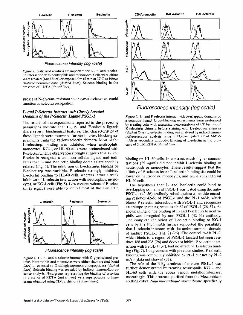
tured in R P M I 1640/10% FCS without chlorate. P- and L-selectin chimera brightly stained KG-1 cells initially pre- treated with chymotrypsin, and then cultured for 24 h in RPMI medium/10% FCS (Fig. 2, solid lines, upper panels). In contrast, culturing KG-1 cells in medium containing so- dium chlorate completely abrogated L- and P-selectin binding (Fig. 2, dashed lines, upper panels). This treatment did not affect cell viability or the expression of other sur- face molecules such as CD13, CD33, or H L A - D R (not shown). The role of sialic acid residues was examined by exposing neutrophils, monocytes, KG-1, and HL-60 cells to Vibrio cholera neuraminidase. Sialidase abolished L- and E-selectin interactions with the cells, suggesting that sialic acid residues have a critical role in the function of L- and E-selectin ligands on neutrophils (Fig. 3). P-selectin bind- ing to neuraminidase-treated cells was only partially inhib- ited.
The experiments illustrated in Fig. 4 examined whether L-, P-, and E-selectin interact with O-glycosylated cell sur- face proteins. Neutrophils, monocytes, KG-I , and HL-60 cells treated with O-sialoglycoprotease completely abro- gated cellular binding of L-, P-, and E-selectin (Fig. 4). These results indicate that O-glycosylated proteins are es- sential for L-, P-, and E-selectin binding. In contrast, treat- ment of neutrophils or KG-1 cells with endoglycosidase F or peptide N glycosidase F did not significantly inhibit L-, P-, or E-selectin cellular binding (data not shown). However, this result does not exclude the possibility that a
The Journal of Cell Biology, Volume 135, 1996 526
L-selectin P-selectin E-selectin
Fluorescence intensity (log scale)
Figure 3. Sialic acid residues are important for L-, P-, and E-selec- tin interaction with neutrophils and monocytes. Cells were either sham treated (solid lines) or exposed for 45 min at 37°C to Vibrio cholerae neuraminidase (dashed lines). Selectin binding in the presence of EDTA (dotted lines).
subset of N-glycans, resistant to enzymatic cleavage, could function in selectin recognition.
L- and P-Selectin Interact with Closely Located Domains of the P-Selectin Ligand PSGL-1
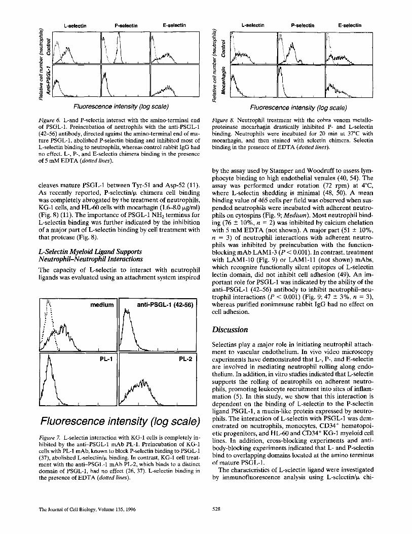
The results of the experiments reported in the preceding paragraphs indicate that L-, P-, and E-selectin ligands share several biochemical features. The characteristics of these ligands were examined further in cross-blocking ex- periments using the various selectin chimera. Most of the L-selectinhx binding was inhibited when neutrophils, monocytes, KG-1, or HL-60 cells were preincubated with P-selectin/p~. This observation strongly suggests that L- and P-selectin recognize a common cellular ligand and indi- cates that L- and P-selectin binding domains are spatially related (Fig. 5). The inhibition of L-selectin/ix binding by E-selectin/Ix was variable. E-selectin strongly inhibited L-selectin binding to HL-60 cells, whereas it was a weak inhibitor of L-selectin interaction with neutrophils, mono- cytes, or KG-1 cells (Fig. 5). Low concentrations of E-selec- tin (3 ixg/ml) were able to inhibit most of the L-selectin
L-selectin P-selectin E-selectin
j
;i
L I ~ • J
' ~ t~"
Fluorescence intensity (log scale)
Figure 4. L-, P-, and E-selectin interact with O-glycosylated pro- teins. Neutrophils and monocytes were either sham treated (solid lines) or exposed to O-sialoglycoprotein endopeptidase (dashed lines). Selectin binding was revealed by indirect immunofluores- cence analysis. Histograms representing the binding of selectins in presence of E D T A (not shown) were superposable to histo- grams obtained using CD4/Ix chimera (dotted lines).
CD4/L-selectin P-/L-selectin E-/L-selectin
t '% l
Fluorescence intensity (log scale) Figure 5. L- and P-selectin interact with overlapping domains of a common ligand. Cross-blocking experiments were performed by treating cells with saturating concentrations of CD4/IX, P-, or E-selectin/ix chimera before staining with L-selectin/ix chimera (dashed lines). L-selectin binding was analyzed by indirect immu- nofluorescence analysis using FITC-conjugated anti-LAM1-5 mAb as secondary antibody. Binding of L-selectin in the pres- ence of 5 mM EDTA (dotted lines).
binding on HL-60 cells. In contrast, much higher concen- trations (25 i~g/ml) did not inhibit L-selectin binding to neutrophils or monocytes. These results suggest that the affinity of E-selectin for an L-selectin binding site could be lower on neutrophils, monocytes, and KG-1 cells than on HL-60 cells.
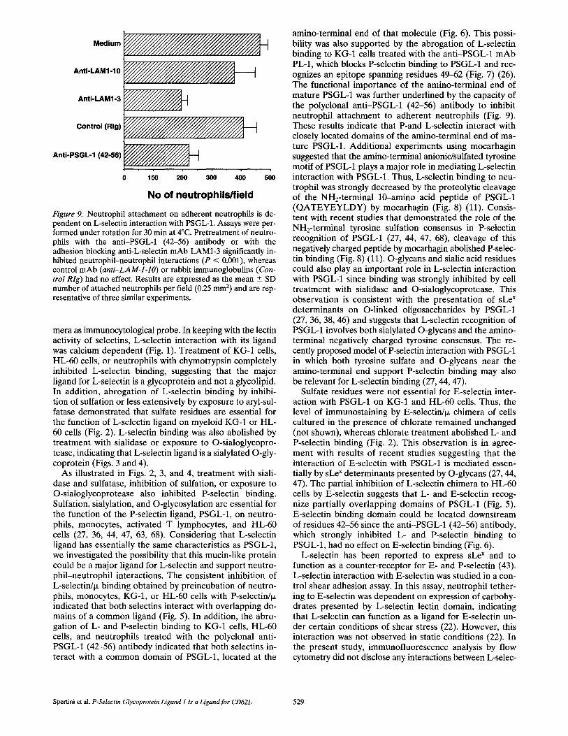
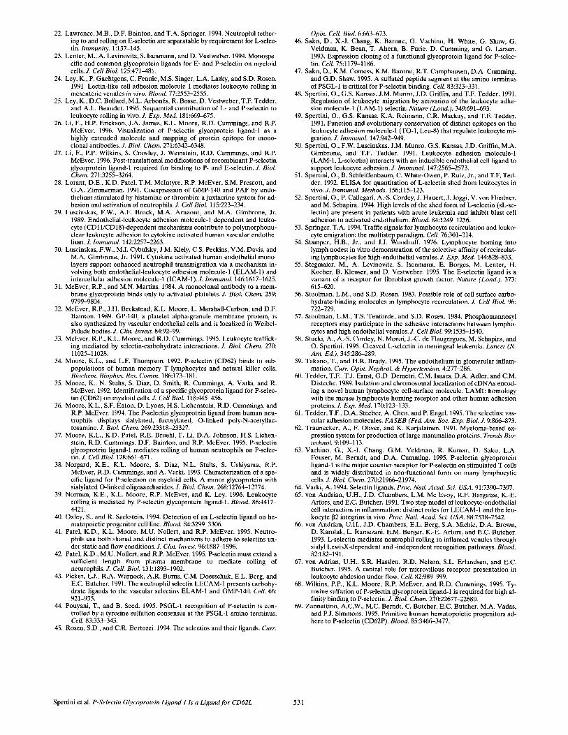
The hypothesis that L- and P-selectin could bind to overlapping domains of PSGL-1 was tested using the anti- PSGL-1 (42-56) antibody raised against a peptide encod- ing residues 42-56 of PSGL-1 and the PL-1 mAb, which blocks P-selectin interaction with PSGL-1 and recognizes an epitope spanning residues 49-62 of PSGL-1 (26, 37). As shown in Fig. 6, the binding of L- and P-selectin to neutro- phils was abrogated by anti-PSGL-1 (42-56) antibody. The complete inhibition of L-selectin binding to KG-1 cells by the PL-1 mAb further supported the possibility that L-selectin interacts with the amino-terminal domain of mature PSGL-1 (Fig. 7) (26). The control mAb PL-2, which binds to a region of PSGL-1 located between resi- dues 188 and 235 (26) and does not inhibit P-selectin inter- action with PSGL-1 (37), had no effect on L-selectin bind- ing (Fig. 7). In agreement with previous studies, P-selectin binding was completely inhibited by PL-1 but not by PL-2 mAb (data not shown) (37).
The role of the NH2 terminus of mature PSGL-1 was further demonstrated by treating neutrophils, KG-1, and HL-60 cells with the cobra venom metalloproteinase, mocarhagin. This protease, purified from the Mozambican spitting cobra, Naja mocambique mocambique, specifically
Spertini et al. P-Selectin Glycoprotein Ligand I Is a Ligand for CD62L 527
L-selectin
A
P-selectin E-selectin
j
Fluorescence intensity (log scale)
Figure 6. L-and P-selectin interact with the amino-terminal end of PSGL-1. Preincubation of neutrophils with the anti-PSGL-1 (42-56) antibody, directed against the amino-terminal end of ma- ture PSGL-1, abolished P-selectin binding and inhibited most of L-selectin binding to neutrophils, whereas control rabbit IgG had no effect• L-, P-, and E-selectin chimera binding in the presence of 5 mM EDTA (dotted lines)•
cleaves mature PSGL-1 between Tyr-51 and Asp-52 (11). As recently reported, P-selectin/ix chimera cell binding was completely abrogated by the treatment of neutrophils, KG-1 cells, and HL-60 cells with mocarhagin (1.6-8.0 ixg/ml) (Fig. 8) (11). The importance of PSGL-1 NH2 terminus for L-selectin binding was further indicated by the inhibition of a major part of L-selectin binding by cell treatment with that protease (Fig. 8).
L-Selectin Myeloid Ligand Supports Neutrophil-Neutrophil Interactions
The capacity of L-selectin to interact with neutrophil ligands was evaluated using an attachment system inspired
medium
PL-1
r i
anti-PSGL-1 (42-56)
PL-2
Fluorescence intensity (log scale) Figure 7. L-selectin interaction with KG-1 cells is completely in- hibited by the anti-PSGL-1 mAb PL-1. Preincubation of KG-1 cells with PL-1 mAb, known to block P-selectin binding to PSGL-1 (37), abolished L-selectin/ix binding• In contrast, KG-1 cell treat- ment with the anti-PSGL-1 mAb PL-2, which binds to a distinct domain of PSGL-1, had no effect (26, 37), L-selectin binding in the presence of EDTA (dotted lines)•
L-selectin P-selectin E-selectln
, \ I I
Fluorescence intensity (log scale)
Figure 8. Neutrophil treatment with the cobra venom metallo- proteinase mocarhagin drastically inhibited P- and L-selectin binding. Neutrophils were incubated for 20 min at 37°C with mocarhagin, and then stained with selectin chimera. Selectin binding in the presence of EDTA (dotted lines).
by the assay used by Stamper and Woodruff to assess lym- phocyte binding to high endothelial venules (40, 54). The assay was performed under rotation (72 rpm) at 4°C, where L-selectin shedding is minimal (48, 50). A mean binding value of 465 cells per field was observed when sus- pended neutrophils were incubated with adherent neutro- phils on cytospins (Fig. 9; Medium)• Most neutrophil bind- ing (76 --- 10%, n = 2) was inhibited by calcium chelation with 5 mM E D T A (not shown)• A major part (51 - 10%, n = 3) of neutrophil interactions with adherent neutro- phils was inhibited by preincubation with the function- blocking mAb LAM1-3 (P < 0.001). In contrast, treatment with LAMI-10 (Fig. 9) or L A M I - l l (not shown) mAbs, which recognize functionally silent epitopes of L-selectin lectin domain, did not inhibit cell adhesion (49). An im- portant role for PSGL-1 was indicated by the ability of the anti-PSGL-1 (42-56) antibody to inhibit neutrophil-neu- trophil interactions (P < 0.001) (Fig. 9; 47 ___ 3%, n = 3), whereas purified nonimmune rabbit IgG had no effect on cell adhesion.
Discussion
Selectins play a major role in initiating neutrophil attach- ment to vascular endothelium. In vivo video microscopy experiments have demonstrated that L-, P-, and E-selectin are involved in mediating neutrophil rolling along endo- thelium. In addition, in vitro studies indicated that L-selectin supports the rolling of neutrophils on adherent neutro- phils, promoting leukocyte recruitment into sites of inflam- mation (5). In this study, we show that this interaction is dependent on the binding of L-selectin to the P-selectin ligand PSGL-1, a mucin-like protein expressed by neutro- phils. The interaction of L-selectin with PSGL-1 was dem- onstrated on neutrophils, monocytes, CD34 ÷ hematopoi- etic progenitors, and HL-60 and CD34 ÷ KG-1 myeloid cell lines• In addition, cross-blocking experiments and anti- body-blocking experiments indicated that L- and P-selectin bind to overlapping domains located at the amino terminus of mature PSGL-1.
The characteristics of L-selectin ligand were investigated by immunofluorescence analysis using L-selectin/ix chi-
The Journal of Cell Biology, Volume 135, 1996 528
Medium ~ / / ~
AnII-LAMI-10 ~ ~
AnIi-LAMI-3 ~ . ~
Control (Rig) ~ / ~
AnU-PSGL-1 (42-56) ~ ~
0 100 200 300 400 500
No of neutrophils/field
Figure 9. Neutrophil attachment on adherent neutrophils is de- pendent on L-selectin interaction with PSGL-1. Assays were per- formed under rotation for 30 min at 4°C. Pretreatment of neutro- phils with the anti-PSGL-1 (42-56) antibody or with the adhesion blocking anti-L-selectin mAb LAM1-3 significantly in- hibited neutrophil-neutrophil interactions (P < 0.001), whereas control mAb (anti-LAM-l-lO) or rabbit immunoglobulins (Con- trol Rig) had no effect. Results are expressed as the mean --_ SD number of attached neutrophils per field (0.25 mm 2) and are rep- resentative of three similar experiments.
mera as immunocytological probe. In keeping with the lectin activity of selectins, L-selectin interaction with its ligand was calcium dependent (Fig. 1). Treatment of KG-1 cells, HL-60 cells, or neutrophils with chymotrypsin completely inhibited L-selectin binding, suggesting that the major ligand for L-selectin is a glycoprotein and not a glycolipid. In addition, abrogation of L-selectin binding by inhibi- tion of sulfation or less extensively by exposure to aryl-sul- fatase demonstrated that sulfate residues are essential for the function of L-selectin ligand on myeloid KG-1 or HL- 60 cells (Fig. 2). L-selectin binding was also abolished by treatment with sialidase or exposure to O-sialoglycopro- tease, indicating that L-selectin ligand is a sialylated O-gly- coprotein (Figs. 3 and 4).
As illustrated in Figs. 2, 3, and 4, treatment with siali- dase and sulfatase, inhibition of sulfation, or exposure to O-sialoglycoprotease also inhibited P-selectin binding. Sulfation, sialylation, and O-glycosylation are essential for the function of the P-selectin ligand, PSGL-1, on neutro- phils, monocytes, activated T lymphocytes, and HL-60 cells (27, 36, 44, 47, 63, 68). Considering that L-selectin ligand has essentially the same characteristics as PSGL-1, we investigated the possibility that this mucin-like protein could be a major ligand for L-selectin and support neutro- phil-neutrophil interactions. The consistent inhibition of L-selectin/~ binding obtained by preincubation of neutro- phils, monocytes, KG-1, or HL-60 cells with P-selectinhx indicated that both selectins interact with overlapping do- mains of a common ligand (Fig. 5). In addition, the abro- gation of L- and P-selectin binding to KG-1 cells, HL-60 cells, and neutrophils treated with the polyclonal anti- PSGL-1 (42-56) antibody indicated that both selectins in- teract with a common domain of PSGL-1, located at the
amino-terminal end of that molecule (Fig. 6). This possi- bility was also supported by the abrogation of L-selectin binding to KG-1 cells treated with the anti-PSGL-1 mAb PL-1, which blocks P-selectin binding to PSGL-1 and rec- ognizes an epitope spanning residues 49--62 (Fig. 7) (26). The functional importance of the amino-terminal end of mature PSGL-1 was further underlined by the capacity of the polyclonal anti-PSGL-1 (42-56) antibody to inhibit neutrophil attachment to adherent neutrophils (Fig. 9). These results indicate that P-and L-selectin interact with closely located domains of the amino-terminal end of ma- ture PSGL-1. Additional experiments using mocarhagin suggested that the amino-terminal anionic/sulfated tyrosine motif of PSGL-1 plays a major role in mediating L-selectin interaction with PSGL-1. Thus, L-selectin binding to neu- trophil was strongly decreased by the proteolytic cleavage of the NHz-terminal 10-amino acid peptide of PSGL-1 (QATEYEYLDY) by mocarhagin (Fig. 8) (11). Consis- tent with recent studies that demonstrated the role of the NH2-terminal tyrosine sulfation consensus in P-selectin recognition of PSGL-1 (27, 44, 47, 68), cleavage of this negatively charged peptide by mocarhagin abolished P-selec- tin binding (Fig. 8) (11). O-glycans and sialic acid residues could also play an important role in L-selectin interaction with PSGL-1 since binding was strongly inhibited by cell treatment with sialidase and O-sialoglycoprotease. This observation is consistent with the presentation of sLe x determinants on O-linked oligosaccharides by PSGL-1 (27, 36, 38, 46) and suggests that L-selectin recognition of PSGL-1 involves both sialylated O-glycans and the amino- terminal negatively charged tyrosine consensus. The re- cently proposed model of P-selectin interaction with PSGL-1 in which both tyrosine sulfate and O-glycans near the amino-terminal end support P-selectin binding may also be relevant for L-selectin binding (27, 44, 47).
Sulfate residues were not essential for E-selectin inter- action with PSGL-1 on KG-1 and HL-60 cells. Thus, the level of immunostaining by E-selectin/~ chimera of cells cultured in the presence of chlorate remained unchanged (not shown), whereas chlorate treatment abolished L- and P-selectin binding (Fig. 2). This observation is in agree- ment with results of recent studies suggesting that the interaction of E-selectin with PSGL-1 is mediated essen- tially by sLe x determinants presented by O-glycans (27, 44, 47). The partial inhibition of L-selectin chimera to HL-60 cells by E-selectin suggests that L- and E-selectin recog- nize partially overlapping domains of PSGL-1 (Fig. 5). E-selectin binding domain could be located downstream of residues 42-56 since the anti-PSGL-1 (42-56) antibody, which strongly inhibited L- and P-selectin binding to PSGL-1, had no effect on E-selectin binding (Fig. 6).
L-selectin has been reported to express sLe x and to function as a counter-receptor for E- and P-selectin (43). L-selectin interaction with E-selectin was studied in a con- trol shear adhesion assay. In this assay, neutrophil tether- ing to E-selectin was dependent on expression of carbohy- drates presented by L-selectin lectin domain, indicating that L-selectin can function as a ligand for E-selectin un- der certain conditions of shear stress (22). However, this interaction was not observed in static conditions (22). In the present study, immunofluorescence analysis by flow cytometry did not disclose any interactions between L-selec-
Spertini et al. P-Selectin Glycoprotein Ligand 1 Is a Ligand for CD62L 529

tin expressed by neutrophils and the P- or E-selectin/tx chi- mera. E- and P-selectin binding to neutrophils was not inhibited by the function-blocking anti-LAM1-3 or anti- LAM1-4 mAbs that block L-selectin tethering on E-selec- tin under flow conditions (not shown) (22). In addition, the shedding of L-selectin observed after exposure of neu- trophils to phorbol-ester did not affect E- or P-selectin binding (data not shown). Thus, L-selectin was not a major ligand for E- or P-selectin in the conditions used in this study. With different conditions, L-selectin could function as a ligand for E- or P-selectin (22, 41, 43).
A small percentage of peripheral blood T lymphocytes (~7%) interacted with the L-selectin/tx chimera (Fig. 1). Future studies will be required to characterize more pre- cisely the immunophenotype and function of these T cells. P-selectin/tx also bound with a few T lymphocytes (~7%). These data are in agreement with those of others who identified a ligand for P-selectin on ~12% of peripheral blood lymphocytes (34). Interestingly, using anti-PSGL-1 mAbs, PSGL-1 expression was observed on >90% of pe- ripheral blood T lymphocytes and on the majority of lym- phoid cell lines (37, 63). This observation suggests that PSGL-1 is constitutively expressed by most lymphocytes in a form unable to interact with P-selectin. T lymphocyte activation increased P-selectin binding, whereas cell sur- face expression of PSGL-1 remained unchanged, suggest- ing that activation-dependent posttranslational events contribute to the expression of functional PSGL-1 (37, 63). Similarly, L-selectin reactivity with T lymphocytes could increase after cell activation.
The expression of a ligand for L-selectin was demon- strated on 35% of CD34 ÷ hematopoietic progenitors. P-selectin interacted with 55% of CD34 ÷ hematopoietic progenitors, whereas only a few CD34 ÷ cells bound to E-selectin. The nature of the ligand for L-, E-, and P-selec- tin on CD34 ÷ stem cells has not been characterized. How- ever, the demonstration of PSGL-1 mRNA expression (69) and surface expression of this molecule by ~30% of CD34 ÷ cells (data not shown) suggest that PSGL-1 could be important in mediating interactions of stem cells with selectins. In particular, PSGL-1 could be involved in regu- lating the migration of hematopoietic progenitors by at- taching them to selectins expressed by bone marrow en- dothelium (69). Recently, altered hematopoiesis was observed in P- and E-selectin-deficient mice underlining the role of vascular selectins in regulating hematopoiesis (13). Additional studies will be needed to determine the role of PSGL-1 in regulating stem cell circulation and homing into the bone marrow. The importance of L-selec- tin interaction with PSGL-1 in regulating stem cell homing remains also to be established.
In conclusion, this study extends our knowledge on the role of L-selectin in leukocyte migration and provides evi- dence that the mucin-like glycoprotein PSGL-1 regulates neutrophil-neutrophil and leukocyte-endothelial interac- tions, promoting leukocyte recruitment at sites of inflam- mation. Moreover, by demonstrating that L-, P-, and E-selec- tin interact with PSGL-1, our results emphasize the role of this mucin-like glycoprotein as a major protagonist in the inflammatory reaction. Furthermore, the expression of PSGL-1 by CD34 ÷ hematopoietic progenitors and most lymphocytes suggests that this counter-receptor could also
be involved in stem cell homing in the bone marrow and contribute to regulating the immune response.
We are grateful to Dr. Phil ippe Schneider, Dr. Jean-Daniel Tissot, and the
staff of the Centre de Transfusion Sanguine at Lausanne for providing blood samples. We also thank Mrs. M. P6clard for technical assistance.
This work was supported by grant 31-43235.95 from the Swiss National Foundat ion for Scientific Research.
Received for publicat ion 13 February 1996 and in revised form 14 June
1996.
References
1. Alon, R., T. Feizi, C.-T. Yuen, R.C. Fuhlbrigge, and T.A. Springer. 1995. Glycolipid ligands for selectins support leukocyte tethering and rolling under physiologic flow conditions. J. Immunol, 154:5356-5366.
2. Arbones, M.L., D.C. Ord, K. Ley, H. Radich, C. Maynard-Curry, D.J. Cap- pon, and T.F. Tedder. 1994. Lymphocyte homing and leukocyte rolling and migration are impaired in L-selectin (CD62L) deficient mice. Immu- nity. t:247-260.
3. Aruffo, A., W. Kolanus, G. Walz, P. Fredman, and B. Seed. 1991. CD62/P- selectin recognition of myeloid and tumor cell sulfatides. Cell. 67:35-44.
4. Asa, D., L. Raycroft, L. Ma, P.A. Aeed, P.S. Kaytes, A.P. Elhammer, and J.-G. Geng. 1995. The P-selectin glycoprotein ligand functions as a com- mon human leukocyte ligand for P- and E-selectins. J. Biol. Chem. 270: 11663-11670.
5. Bargatze, R., S. Kurk, E. Butcher, and M. Jutila. 1994. Neutrophils roll on adherent neutrophils bound to cytokine-induced endothelial cells via L-selectin on the rolling cells. J. Exp. Med. 180:1785-1792.
6. Baumhueter, S., M.S. Singer, W. Henzel, S. Hemmerich, M. Renz, S.D. Rosen, and L.A. Lasky. 1993. Binding of L-selectin to the vascular sialo- mucin CD34. Science (Wash. DC). 262:436-438.
7. Berg, E.L., L.M. McEvoy, C. Berlin, R.F. Bargatze, and E.C. Butcher. 1993. L-selectin mediated lymphocyte rolling on MadCAM-1. Nature ( Lond. ). 366:695-698.
8. Bevilacqua, M.P., J.S. Pober, D.L. Mendrick, R.S. Cotran, and M.A. Gim- brone, Jr. 1987. Identification of an inducible endothelial-leukocyte ad- hesion molecule. Proc. Natl. Acad. Sci. USA. 84:9238-9243.
9. Bevilacqua, M.P., S. Stengelin, M.A. Gimbrone, Jr., and B. Seed. 1989. En- dothelial leukocyte adhesion molecule 1: an inducible receptor for neu- trophils related to complement regulatory proteins and lectins. Science (Wash. DC). 243:1160--1164.
10. Carlos, T.C., and J.M. Harlan. 1994. Leukocyte-endothelial adhesion mole- cules. Blood. 84:2068-2101.
11. Deluca, M., L.C. Dunlop, R.K. Andrews, J.V. Flannery, R. Ettling, D.A. Cumming, M. Veldman, and M.C. Berndt. 1995. A novel cobra venom metalloproteinase, mocarhagin, cleaves a 10-amino acid peptide from the mature N terminus of P-selectin glycoprotein ligand receptor, PSGL-1, and abolishes P-selectin binding. J. Biol. Chem. 270:26734-26737.
12. Foxall, C., S.R. Watson, D. Dowbenko, C. Fennie, L.A. Lasky, M. Kiso, A. Hasegawa, D. Asa, and B.K. Brandley. 1992. The three members of the selectin receptor family recognize a common carbohydrate epitope, the sialyl Lewis x oligosaccharide. J. Cell, Biol. 117:895-902.
13. Frenette, P.S., T.N. Mayadas, H. Rayburn, R.O. Hynes, and D.D. Wagner. 1996. Susceptibility to infection and altered hematopoiesis in mice defi- cient in both P- and E-selectins. Cell. 84:563-574.
14. Geng, J.G., M.P. Bevilacqua, K.L. Moore, T.M. Mclntyre, S.M. Prescott, J.M. Kim, G.A. Bliss, G.A. Zimmerman, and R.P. McEver. 1990. Rapid neutrophil adhesion to activated endothelium mediated by GMP-140. Nature (Lond.). 343:757-760.
15. Hemmerich, S., and S.D. Rosen. 1994.6'-sulfated sialyl Lewis x is a major capping group of GIyCAM-1. Biochemistry. 33:4830-4835.
16. Hemmerich, S., E.C. Butcher, and S.D. Rosen. 1994. Sulfation-dependent recognition of high endothelial venules (HEV)-ligands by L-selectin and MECA 79, an adhesion-blocking monoclonal antibody. J. Exp. Med. 180: 2219-2226.
17. Imai, Y., M.S. Singer, C. Fennie, L.A. Lasky, and S.D. Rosen. 1991. Identi- fication of a carbohydrate-based endothelial ligand for a lymphocyte homing receptor. J. Cell Biol. 113:1213-1221.
18. Imai, Y., L.A. Lasky, and S.D. Rosen. 1993. Sulphation requirement for GIyCAM-1, an endothelial ligand for L-selectin. Nature (Lond.). 361: 555-557.
19. Larsen, E., E.A. Celi, G.E. Gilbert, B.C. Furie, J.K. Ethan, R. Bonfanti, D.D. Wagner, and B. Furie. 1989. PADGEM protein: a receptor that me- diates the interaction of activated platelets with neutrophils and mono- cytes. Cell, 59:305-312.
20. Lasky, L. 1995. Selectin-carbohydrate interactions and the initiation of the inflammatory response. Annu. Rev. Biochem. 64:113-139.
21. Lasky, L.A., M.S. Singer, D. Dowbenko, Y. Imai, W.J. Henzel, C. Grimley, C. Fennie, N. Gillett, S.R. Watson, and S.D. Rosen. 1992. An endothelial ligand for L-selectin is a novel mucin-like molecule. Cell. 69:927-938.
The Journal of Cell Biology, Volume 135, 1996 530
22. Lawrence, M.B., D.F. Bainton, and T.A. Springer. 1994. Neutrophil tether- ing to and rolling on E-selectin are separatable by requirement for L-selec- tin. Immunity. 1:137-145.
23. Lenter, M., A. Levinovitz, S. Isenmann, and D. Vestweber. 1994. Monospe- cific and common glycoprotein ligands for E- and P-selectin on myeloid cells. J. Cell Biol. 125:471-481.
24. Ley, K., P. Gaehtgens, C. Fennie, M.S. Singer, L.A. Lasky, and S.D. Rosen. 1991. Lectin-like cell adhesion molecule 1 mediates leukocyte rolling in mesenteric venules in vivo. Blood. 77:2553-2555.
25. Ley, K., D.C. Bullard, M.L. Arbonrs, R. Bosse, D. Vestweber, T.F. Tedder, and A.L. Beaudet. 1995. Sequential contribution of L- and P-selectin to leukocyte rolling in vivo. J. Exp. Med. 181:669-675.
26. Li, F., H.P. Erickson, J.A. James, K.L. Moore, R.D. Cummings, and R.P. McEver. 1996. Visualization of P-selectin glycoprotein ligand-1 as a highly extended molecule and mapping of protein epitope for mono- clonal antibodies. J. BioL Chem. 271:6342-6348.
27. Li, F., P.P. Wilkins, S. Crawley, J. Weinstein, R.D. Cummings, and R.P. McEver. 1996. Post-translational modifications of recombinant P-selectin glycoprotein ligand-1 required for binding to P- and E-selectin. J. Biol. Chem. 271:3255-3264.
28. Lorant, D.E., K.D. Patel, T.M. Mclntyre, R.P. McEver, S.M. Prescott, and G.A. Zimmerman. 1991. Coexpression of GMP-140 and PAF by endo- thelium stimulated by histamine or thrombin: a juxtacrine system for ad- hesion and activation of neutrophils. J. Cell Biol. 115:223-234.
29. Luscinskas, F.W., A.F. Brock, M.A. Arnaout, and M.A. Gimbrone, Jr. 1989. Endothelial-leukocyte adhesion molecule-1 dependent and leuko- cyte (CD11/CD18)-dependent mechanisms contribute to polymorphonu- clear leukocyte adhesion to cytokine-activated human vascular endothe- lium. J. lmmunol. 142:2257-2263.
30. Luscinskas, F.W., M.I. Cybulsky, J.M. Kiely, C.S. Peckins, V.M. Davis, and M.A. Gimbrone, Jr. 1991. Cytokine-activated human endothelial mono- layers support enhanced neutrophil transmigration via a mechanism in- volving both endothelial-leukocyte adhesion molecule-1 (ELAM-1) and intercellular adhesion molecule-1 (ICAM-1). J. Immunol, 146:1617-1625.
31. McEver, R.P., and M.N. Martins. 1984. A monoclonal antibody to a mem- brane glycoprotein binds only to activated platelets. J. Biol. Chem. 259: 9799-9804.
32. McEver, R.P., J.H. Beckstead, K.L. Moore, L. Marshall-Carlson, and D.F. Bainton. 1989. GP-140, a platelet alpha-granule membrane protein, is also synthesized by vascular endothelial cells and is localized in Weibel- Palade bodies. J. Clin. Invest. 84:92-99.
33. McEver, R.P., K.L. Moore, and R.D. Cummings. 1995. Leukocyte traffick- ing mediated by selectin-carbohydrate interactions. Z Biol, Chem. 270: 11025-11028.
34. Moore, K.L., and L.F. Thompson. 1992. P-selectin (CD62) binds to sub- populations of human memory T lymphocytes and natural killer cells. Biochem. Biophys. Res. Comm. 186:173-181.
35. Moore, K., N. Stults, S. Diaz, D. Smith, R. Cummings, A. Varki, and R. McEver. 1992. Identification of a specific glycoprotein ligand for P-selec- tin (CD62) on myeloid cells. J. Cell Biol. 118:445-456.
36. Moore, K.L., S.F. Eaton, D. Lyons, H.S. Lichenstein, R.D. Cummings, and R.P. McEver. 1994. The P-selectin glycoprotein ligand from human neu- trophils displays sialylated, fucosylated, O-linked poly-N-acetyllac- tosamine. J. Biol. Chem. 269:23318-23327.
37. Moore, K.L., K.D. Patel, R.E. Bruehl, F. Li, D.A. Johnson, H.S. Lichen- stein, R.D. Cummings, D.F. Bainton, and R.P. McEver. 1995. P-selectin glycoprotein ligand-1 mediates rolling of human neutrophils on P-selec- tin. Z Cell BioL 128:661-671.
38. Norgard, K.E., K.L. Moore, S. Diaz, N.L. Stults, S. Ushiyama, R.P. McEver, R.D. Cummings, and A. Varki. 1993. Characterization of a spe- cific ligand for P-selection on myeloid cells. A minor glycoprotein with sialylated O-linked oligosaccharides. Z Biol. Chem. 268:12764-12774.
39. Norman, K.E., K.L. Moore, R.P. McEver, and K. Ley. 1996. Leukocyte rolling is mediated by P-selectin glycoprotein ligand-1. Blood. 86:4417- 4421.
40. Oxley, S., and R. Sackstein. 1994. Detection of an L-selectin ligand on he- matopoietic progenitor cell line. Blood. 84:3299-3306.
41. Patel, K.D., K.L. Moore, M.U. Nollert, and R.P. McEver. 1995. Neutro- phils use both shared and distinct mechanisms to adhere to selectins un- der static and flow conditions. Z Clin. Invest. 96:1887-1896.
42. Patel, K.D., M.U. Nollert, and R.P. McEver. 1995. P-selectin must extend a sufficient length from plasma membrane to mediate rolling of neturophils. J. Cell Biol. 131:1893-1902.
43. Picker, L.,I., R.A. Warnock, A.R. Burns, C.M. Doerschuk, E.L. Berg, and E.C. Butcher. 1991. The neutrophil selectin LECAM-1 presents carbohy- drate ligands to the vascular selectins ELAM-1 and GMP-140. Cell 66: 921-935.
44. Pouyani, T., and B. Seed. 1995. PSGL-1 recognition of P-selectin is con- trolled by a tyrosine sulfation consensus at the PSGL-1 amino terminus. Cell. 83:333-343.
45. Rosen, S.D., and C.R. Bertozzi. 1994. The selectins and their ligands. Curr.
Opin. CeIL Biol. 6:663-673. 46. Sako, D., X.-J. Chang, K. Barone, G. Vachino, H. White, G. Shaw, G.
Veldman, K. Bean, T. Ahern, B. Furie, D. Cumming, and G. Larsen. 1993. Expression cloning of a functional glycoprotein ligand for P-selec- tin. Cell. 75:1179-1186.
47. Sako, D., K.M. Comess, K.M. Barone, R.T. Camphausen, D.A. Cumming, and G.D. Shaw. 1995. A sulfated peptide segment at the amino terminus of PSGL-1 is critical for P-selectin binding. Cell 83:323-331.
48. Spertini, O., G.S. Kansas, J.M. Munro, J.D. Griffin, and T.F. Tedder. 1991. Regulation of leukocyte migration by activation of the leukocyte adhe- sion molecule-1 (LAM-1) selectin. Nature (Lond.). 349:691-693.
49. Spertini, O., G.S. Kansas, K.A. Reimann, C.R. Mackay, and T.F. Tedder. 1991. Function and evolutionary conservation of distinct epitopes on the leukocyte adhesion molecule-1 (TQ-1, Leu-8) that regulate leukocyte mi- gration. J. lmmunol. 147:942-949.
50. Spertini, O., F.W. Luscinskas, J.M. Munro, G.S. Kansas, J.D. Griffin, M.A. Gimbrone, and T.F. Tedder. 1991. Leukocyte adhesion molecule-1 (LAM-1, L-selectin) interacts with an inducible endothelial cell ligand to support leukocyte adhesion. Z lmmunol. 147:2565-2573.
51. Spertini, O., B. Schleiffenbaum, C. White-Owen, P. Ruiz, Jr., and T.F. Ted- def. 1992. ELISA for quantitation of L-selectin shed from leukocytes in vivo. J. lmmunol. Methods. 156:115-123.
52. Spertini, O., P. Callegari, A.-S. Cordey, ,i. Hauert, J. Joggi, V. von Fliedner, and M. Schapira. 1994. High levels of the shed form of L-selectin (sL-se- lectin) are present in patients with acute leukemia and inhibit blast cell adhesion to activated endothelium. Blood. 84:1249-1256.
53. Springer, T.A. 1994. Traffic signals for lymphocyte recirculation and leuko- cyte emigration: the multistep paradigm. Cell, 76:301-314.
54. Stamper, H.B., Jr., and J.J. Woodruff. 1976. Lymphocyte homing into lymph nodes: in vitro demonstration of the selective affinity of recirculat- ing lymphocytes for high-endothelial venules. J. Exp. Med. 144:828-833.
55. Stegmaier, M., A. Levinovitz, S. Isenmann, E. Borges, M. Lenter, H. Kocher, B. Kleuser, and D. Vestweber. 1995. The E-selectin ligand is a variant of a receptor for fibroblast growth factor. Nature (Lond.). 373: 615-620.
56. Stoolman, L.M., and S.D. Rosen. 1983. Possible role of cell surface carbo- hydrate-binding molecules in lymphocyte recirculation. J. Cell Biol. 96: 722-729.
57. Stoolman, L.M., T.S. Tenforde, and S.D. Rosen. 1984. Phosphomannosyl receptors may participate in the adhesive interactions between lympho- cytes and high endothelial venules. J. Cell Biol. 99:1535-1540.
58. Stucki, A., A.-S. Cordey, N. Monai, J.-C. de Flaugergues, M. Schapira, and O. Spertini. 1995. Cleaved L-selectin in meningeal leukemia. Lancet (N. Am. Ed.). 345:286-289.
59. Takano, T., and H.R. Brady. 1995. The endothelium in glomerular inflam- mation. Curt Opin. Nephrol. & Hypertension. 4:277-286.
60. Tedder, T.F., T.J. Ernst, G.D. Demetri, C.M. Isaacs, D.A. Adler, and C.M. Disteche. 1989. Isolation and chromosomal localization of cDNAs encod- ing a novel human lymphocyte cell-surface molecule, LAMI: homology with the mouse lymphocyte homing receptor and other human adhesion proteins. J. Exp. Med. 170:123-133.
61. Tedder, T.F., D.A. Steeber, A. Chen, and P. Engel. 1995. The selectins: vas- cular adhesion molecules. FASEB (Fed. Am. Soc. Exp. Biol. J. 9:866~873.
62. Traunecker, A., F. Oliver, and K. Karjalainen. 1991. Myeloma-based ex- pression system for production of large mammalian proteins. Trends Bio- technol. 9:109-113.
63. Vachino, G., X.-,I. Chang, G.M. Veldman, R. Kumar, D. Sako, L.A. Fouser, M. Berndt, and D.A. Cumming. 1995. P-selectin glycoprotein ligand-1 is the major counter-receptor for P-selectin on stimulated T cells and is widely distributed in non-functional form on many lymphocytic cells. Z Biol. Chem. 270:21966-21974.
64. Varki, A. 1994. Selectin ligands. Proc. Natl. Acad. Sci. USA. 91:7390-7397. 65. von Andrian, U.H., .I.D. Chambers, L.M. Mc Evoy, R.F. Bargatze, K.-E.
Arfors, and E.C. Butcher. 1991. Two step model of leukocyte-endothelial cell interaction in inflammation: distinct roles for LECAM-1 and the leu- kocyte 132 integrins in vivo. Proc. Natl. Acad. Sci. USA. 88:7538-7542.
66. yon Andrian, U.H., J.D. Chambers, E.L. Berg, S.A. Michie, D.A. Brown, D. Karolak, L. Ramezani, E.M. Berger, K.-E. Arfors, and E.C. Butcher. 1993. L-selectin mediates neutrophil rolling in inflamed venules through sialyl LewisX-dependent and -independent recognition pathways. Blood. 82:182-191.
67. von Adrian, U.H., S.R. Hasslen, R.D. Nelson, S.L. Erlandsen, and E.C. Butcher. 1995. A central role for microvillous receptor presentation in leukocyte ahdesion under flow. Cell, 82:989-999.
68. Wilkins, P.P., K.L. Moore, R.P. McEver, and R.D. Cummings. 1995. Ty- rosine sulfation of P-selectin glycoprotein ligand-1 is required for high af- finity binding to P-selectin. J. Biol. Chem. 270:22677-22680.
69. Zannettino, A.C.W., M.C. Berndt, C. Butcher, E.C. Butcher, M.A. Vadas, and P.J. Simmons. 1995. Primitive human hematopoietic progenitors ad- here to P-selectin (CD62P). Blood. 85:3466--3477.
Spertini et al. P-Selectin Glycoprotein Ligand I Is a Ligand for CD62L 531