Embed Size (px)
Citation preview
of March 22, 2018.This information is current as
Rollingby Allosteric Antibodies Affects Leukocyte Heterotropic Modulation of Selectin Affinity
Rupert Hallmann, Erhard Hohenester and Konrad BuscherSebastian B. Riese, Christian Kuehne, Thomas F. Tedder,
http://www.jimmunol.org/content/192/4/1862doi: 10.4049/jimmunol.1302147January 2014;
2014; 192:1862-1869; Prepublished online 15J Immunol
MaterialSupplementary
7.DCSupplementalhttp://www.jimmunol.org/content/suppl/2014/01/16/jimmunol.130214
Referenceshttp://www.jimmunol.org/content/192/4/1862.full#ref-list-1
, 25 of which you can access for free at: cites 46 articlesThis article
average*
4 weeks from acceptance to publicationFast Publication! •
Every submission reviewed by practicing scientistsNo Triage! •
from submission to initial decisionRapid Reviews! 30 days* •
Submit online. ?The JIWhy
Subscriptionhttp://jimmunol.org/subscription
is online at: The Journal of ImmunologyInformation about subscribing to
Permissionshttp://www.aai.org/About/Publications/JI/copyright.htmlSubmit copyright permission requests at:
Email Alertshttp://jimmunol.org/alertsReceive free email-alerts when new articles cite this article. Sign up at:
Print ISSN: 0022-1767 Online ISSN: 1550-6606. Immunologists, Inc. All rights reserved.Copyright © 2014 by The American Association of1451 Rockville Pike, Suite 650, Rockville, MD 20852The American Association of Immunologists, Inc.,
is published twice each month byThe Journal of Immunology
by guest on March 22, 2018
http://ww
w.jim
munol.org/
Dow
nloaded from
by guest on March 22, 2018
http://ww
w.jim
munol.org/
Dow
nloaded from
The Journal of Immunology
Heterotropic Modulation of Selectin Affinity by AllostericAntibodies Affects Leukocyte Rolling
Sebastian B. Riese,* Christian Kuehne,* Thomas F. Tedder,† Rupert Hallmann,‡
Erhard Hohenester,x and Konrad Buscher‡,{
Selectins are a family of adhesion receptors designed for efficient leukocyte tethering to the endothelium under shear. As a key
property to resist premature bond disruption, selectin adhesiveness is enhanced by tensile forces that promote the conversion
of a bent into an extended conformation of the N-terminal lectin and epidermal growth factor–like domains. Conformation-
specific Abs have been invaluable in deciphering the activation mechanism of integrins, but similar reagents are not available for
selectins. In this study, we show that the anti-human L-selectin mAbs DREG-55 and LAM1-5 but not DREG-56, DREG-200, or
LAM1-1 heterotropically modulate adhesion presumably by stabilizing the extended receptor conformation. Force-free affinity
assays, flow chamber, and microkinetic studies reveal a ligand-specific modulation of L-selectin affinity by DREG-55 mAb,
resulting in a dramatic decrease of rolling velocity under flow. Furthermore, secondary tethering of polymorphonuclear cells
was blocked by DREG-200 but significantly boosted by DREG-55 mAb. The results emphasize the need for a new classification for
selectin Abs and introduce the new concept of heterotropic modulation of receptor function. The Journal of Immunology, 2014,
192: 1862–1869.
Leukocyte tethering to and rolling on the vascular endothe-lium represents the first step of the adhesion cascade andis mediated by the selectin receptor family in most phys-
iological and pathological conditions (1).E-, P-, and L-selectin are calcium-dependent type I adhesion
receptors. They consist of an N-terminal lectin domain followed byan epidermal growth factor (EGF)–like domain, a varying numberof short consensus repeats, a single transmembrane domain, anda short intracellular tail (2). A common minimal ligand determi-nant was identified as the tetrasaccharide sialyl Lewis x (sLex) withterminal a2,3-linked sialic acid and a1,3-linked fucose units thatdecorate a variety of O-glycans, for example, the leukocyte-expressedP-selectin glycoprotein ligand 1 (PSGL-1). In most inflammatoryconditions, E- and P-selectin are major counterreceptors for PSGL-1,but also trans-interactions with L-selectin (CD62L) on passing
leukocytes were found to be relevant for mediating secondarycapture (3, 4). In lymphoid tissue, particularly in high endothelialvenules (HEVs), the predominant ligand entity for L-selectin–mediated rolling is peripheral lymph node addressin (PNAd), amolecular complex of different sialomucins (5). Importantly, onlysLex with sulfated N-acetylglucosamine on PNAd shows L-selectinbinding activity (5). The great variety of different ligands, selectinexpression patterns, and relevant posttranslational modificationsreflects the precise tissue- and cell type–specific manner of leu-kocyte recruitment.By nature, the bonds that bind selectin to endothelial- or leukocyte-
expressed ligands are subjected to high tensile forces imposed byhydrodynamic flow. Cell flattening (6), microvillus receptor pre-sentation (7, 8), the formation of upstream membrane tethers, anddownstream slings (9) describe cell adaptions to rolling under highshear. Importantly, also intrinsic receptor binding properties ef-fectively modulate bond stability. A threshold of shear force is re-quired for L-selectin–mediated binding, which was the first indi-cation of the striking role of blood flow on selectin mechanics(10). Leukocyte rolling on immobilized ligands requires selectinsto engage in fast but transient ligand interactions with high asso-ciation (Kon) and dissociation rates (Koff) (11). Surprisingly, it wasdemonstrated that tensile forces enhance selectin-mediated adhesionand stabilize cell rolling by decreasing Koff in low shear conditions(12, 13), promoting the formation of so-called “catch bonds”The first study on altered L-selectin receptor function detected
affinity changes upon leukocyte activation, but the precise mecha-nism remained unresolved (14). Domain-swapping experiments sug-gested a role for the EGF-like domain in ligand binding (15, 16),and crystal structure analysis subsequently revealed a flexiblehinge between the N-terminal lectin and EGF-like domain ofselectins (17, 18). Whereas sLex is bound by a bent conformation ofP-selectin, cocrystallization with PSGL-1 glycopeptide revealed anextended conformation (17). The transition from the bent to theextended state involves several subdomain movements in the lectindomain (19). One major component of this allosteric pathway is the83–89 loop that relocates in close vicinity to the ligand-bindinginterface. Thereby new noncovalent interactions are formed, in-
*Institute of Laboratory Medicine, Clinical Chemistry and Pathobiochemistry,Charite–University of Medicine Berlin, 10117 Berlin, Germany; †Department ofImmunology, Duke University Medical Center, Durham, NC 27710; ‡Institute of Phys-iological Chemistry and Pathobiochemistry, University of Muenster, 48149 Muenster,Germany; xDepartment of Life Sciences, Imperial College London, London SW7 2AZ,United Kingdom; and {Department of Anesthesiology, Intensive Care, and PainMedicine, University of Muenster, 48149 Muenster, Germany
Received for publication August 14, 2013. Accepted for publication December 18,2013.
This work was supported by Interdisziplinares Zentrum fur Klinische Forschung GrantSEED 04/12 of the University of Muenster (to K.B.). E.H. is a Wellcome Trust SeniorResearch Fellow in Basic Biomedical Science (Grant 083942/Z/07/Z).
Experiments were performed by S.B.R., K.B., and C.K.; T.F.T. provided LAM Abs;E.H. analyzed the structural implications; K.B. designed the study and analyzed data;and K.B. and R.H. wrote the manuscript.
Address correspondence and reprint requests to Dr. Konrad Buscher, Institute ofPhysiological Chemistry and Pathobiochemistry, University of Muenster, WaldeyerStrasse 15, 48149 Muenster, Germany. E-mail address: [email protected]
The online version of this article contains supplemental material.
Abbreviations used in this article: EGF, epidermal growth factor; FOV, field of view;HEV, high endothelial venule; PBL, primary blood lymphocyte; PMN, polymorpho-nuclear leukocyte; PNAd, peripheral lymph node addressin; PPME, polyphosphomo-noester; PSGL-1, P-selectin glycoprotein ligand 1; SCR, short consensus repeat;sLex, sialyl Lewis x (NeuAc-a2,3-Gal-b1,4-(Fuc-a1,3)GlcNAcb1-R).
Copyright� 2014 by TheAmericanAssociation of Immunologists, Inc. 0022-1767/14/$16.00
www.jimmunol.org/cgi/doi/10.4049/jimmunol.1302147
by guest on March 22, 2018
http://ww
w.jim
munol.org/
Dow
nloaded from
cluding Glu88 ligation to the calcium ion and the PSGL-1 fucoseunit, and Arg85 binding to a sulfated tyrosine of the PSGL-1 poly-peptide. A second sulfate tyrosine is bound by His114 in P-selectin.The corresponding residue in L-selectin is alanine, a substitutionthat partially explains the lower affinity of L-selectin for PSGL-1(20). To date, L-selectin crystal data are available only for theunbound state (PDB 3CFW), but the high phylogenetic conservationand molecular dynamic simulations suggest fundamentally similarligand-binding modes for all selectins (21).Tensile forces acting on a selectin/ligand complex favor the ex-
tended conformation, aligning the long axis of receptor with thedirection of the force applied (21, 22). It is thought that this prop-erty gives rise to catch bonds; however, there is no clear consensusabout the underlying mechanism. In the “allosteric model,” pivo-ting about the EGF–lectin interdomain hinge causes a restructur-ing of the distal ligand binding interface to a high-affinity con-formation (19, 22). In contrast, the “sliding-rebinding” model isbased on alignment of the lectin domain with the acting force,thereby enabling repetitive contacts to several carbohydrate epi-topes on the same ligand (21, 23).In all studies on selectin mechanochemistry, only genetically
modified receptors have been studied (21, 22, 24). This is due tothe fact that, in contrast to, for example, integrins, no reagents areknown that modulate specific conformational states of selectins.We discovered that the anti–L-selectin mAbs DREG-55 and LAM1-5 dramatically reduce L-selectin–mediated lymphocyte rolling ve-locity under shear, consistent with the selective binding to a high-affinity conformation of L-selectin. These data introduce the newconcept of heterotropic modulation of selectin affinity and dem-onstrate how such anti-selectin mAbs can provide further insightson the structure–function relationship. Moreover, our data highlightthe need for a new classification of anti-selectin mAbs accordingto their distinct stimulatory or inhibitory activities.
Materials and MethodsReagents and Abs
HBSS containing Ca2+ and Mg2+ was obtained from Invitrogen. DREG-55and DREG-200 mAb hybridomas were a gift from E. Butcher (StanfordUniversity). DREG-56 (sc-18851) was purchased from Santa Cruz Bio-technology, and the secondary Ab goat anti-human IgG (Fc specific)–FITC(F9512) was from Sigma-Aldrich. Mouse anti-human L-selectin LAM1-1and LAM1-5 mAbs were collected from ascites fluid (16). Purified humanL-selectin–, PSGL-1–, and E-selectin–Fc chimeras were obtained fromR&D Systems. PNAd extracts from human tonsils were provided byS. Rosen (University of California, San Francisco). Streptavidin wasfrom Calbiochem and biotinylated sLex-polyacrylamide was from Lecinity(Moscow, Russia). DREG-55 Fab fragments were generated with an IgG1
Fab purification kit according to the manufacturer’s instructions (ThermoScientific). SDS-PAGE under reducing and nonreducing conditions con-firmed the generation of monovalent Fab and the exclusion of Fc.
Cell isolation and culture
The Jurkat cell line clone E6-1 (American Type Culture Colllection) wascultured in RPMI 1640 medium supplemented with 10% FCS and penicillin/streptomycin at 37˚C and 5% CO2. EDTA-anticoagulated blood wasobtained from healthy volunteers and separated by a Ficoll 1.077 and1.119 g/ml gradient. The isolation yielded .90% viable human primaryblood lymphocytes (PBLs) and polymorphonuclear leukocytes (PMNs)as verified by flow cytometry. Isolated cells were kept on ice for a max-imum of 4 h.
L-selectin–coated microspheres
Carboxylated polystyrol microspheres (beads, 6 mm diameter) were cou-pled to protein G using a carbodiimide coupling kit (Polysciences). HumanL-selectin–Fc (complete extracellular domain of L-selectin) was incubatedwith the beads for 1 h at room temperature, subsequently blocked by 2 mg/ml BSA in PBS, and successful coating was verified by flow cytometry.Protein G–coupled beads without addition of L-selectin–Fc did not showany interaction in PSGL-1–coated flow chambers under shear.
Flow cytometry–based affinity assay
Jurkat cells (3 3 106) were washed in calcium-free PBS and resuspendedin HBSS buffer. Then, 10 mg/ml DREG-55 mAb, DREG-200 mAb or PBS(untreated), and 10 mg/ml PSGL-1–Fc or E-selectin–Fc were added andincubated for 10 min at room temperature. After washing, cells wereresuspended in ice-cold PBS/1% FCS buffer followed by a 30-min incu-bation with 1:1000 anti-human IgG-FITC on ice. Flow cytometry wasperformed and the mean fluorescence intensity was calculated relative tothe sample with DREG-200 Ab (negative control).
Laminar flow assays
Ibidi m-slides VI0.1 (0.1 mm high) were coated for 2 h at room temperaturewith 30 mg/ml PSGL-1–Fc or 5 mg/ml E-selectin–Fc, unless otherwisestated, followed by 1 h blocking with 2 mg/ml BSA in PBS. Alterna-tively, PNAd was applied overnight at 4˚C. For flow chambers on sLex, 10mg/ml streptavidin coating overnight at 4˚C was followed by 20 mg/mlbiotinylated sLex-polyacrylamide coating for 2 h at room temperature.Cells (0.3 3 106/ml) in HBSS were preincubated for 5 min with the in-dicated Ab at room temperature. For representative video recordings, theAb was directly added to the distal cell suspension reservoir of the runningflow chamber system. Digital videos were recorded with an inverted phasecontrast microscope (Axiovert M200; Zeiss) equipped with a CCD camera(ORCA; Hamamatsu) at 37˚C and 310 magnification. Cells were intro-duced at a high flow rate (.20 dynes/cm2) for 1 min, and the flow rate wassubsequently adjusted to the desired shear stress level. After an equili-bration of 2 min, at least three different fields of view (FOV) were re-corded. Rolling velocities were determined by offline analysis using Fiji(25). For detachment assays, cells were allowed to settle and the flow ratewas increased stepwise to 1, 1.5, 3, 6, and 10 dynes/cm2. After 1 minequilibration, a snapshot of three different FOVs were taken and the numberof interacting cells was counted and expressed relative to 1 dyne/cm2. PMNstring formation was analyzed using 20 mg/ml E-selectin–coated chambersafter 3 min equilibration of flow and six adjacent FOVs (∼13003 1000 mm)were recorded. More than three cells aligned in the direction of the flowwith at most five cell diameters distance between two cells were considereda string.
Microkinetic velocity measurement
For motion analysis in high-temporal resolution, digital videos wereaquired at 320 or 340 magnification with 100 frames/s (Neo 5.5 sCMOScamera; Andor) at room temperature. The recorded rolling motion wasthen subjected to an automated frame-by-frame analysis using a snakemodel–based algorithm in MATLAB (MathWorks). The x-coordinateswere translated into step distances and velocities by customized Excelmacros. At 0.96 dyne/cm2 a velocity threshold of 28 mm/s was foundsuitable for discriminating between load-bearing (deceleration) and breaking(acceleration) bonds in untreated and DREG-55 Ab-treated (10 mg/ml)samples. Measurements were repeated twice in triplicates with 13–16analyzed cells per group, totaling 68–116 tether events. The logarithmicexpression of tethered cells allowed the determination of 2Koff from theslope of the linear regression. Importantly, under the experimental con-ditions used this value is only an apparent Koff owing to multibond tethers;however, it can be used for comparison of dissociation kinetics in the samecellular system.
Statistical analysis
The significance of results was determined using the Student t test. Ap value ,0.05 was considered to be statistically significant.
ResultsDREG-55 and LAM1-5 Abs induce L-selectin–mediatedslow lymphocyte rolling
L-selectin–dependent rolling was investigated in a parallel plateflow chamber on the immobilized ligand PSGL-1–Fc. UntreatedPBLs showed fast and stable rolling at 2 dynes/cm2 (Fig. 1A).Anti-human L-selectin mAb DREG-200 blocked any interactions,proving the specificity of rolling (not shown). DREG-55 mAb butnot isotype anti-human IgG1 preincubation at saturating concen-trations triggered a massive decrease in mean rolling velocity from113 to 13 mm/s. Fast rolling was directly converted into slow ho-mogeneous rolling within a subsecond period, and hardly any de-tachment of cells was observed (Supplemental Video 1). Minimal
The Journal of Immunology 1863
by guest on March 22, 2018
http://ww
w.jim
munol.org/
Dow
nloaded from
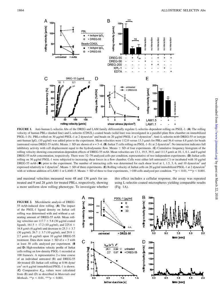
and maximal velocities measured were 48 and 136 mm/s for un-treated and 9 and 28 mm/s for treated PBLs, respectively, showinga more uniform slow rolling phenotype. To investigate whether
this effect includes a cellular response, the assay was repeatedusing L-selectin–coated microspheres yielding comparable results(Fig. 1A).
FIGURE 1. Anti-human L-selectin Abs of the DREG and LAM family differentially regulate L-selectin–dependent rolling on PSGL-1. (A) The rolling
velocity of human PBLs (dashed line) and L-selectin (CD62L)–coated beads (solid line) was investigated in a parallel plate flow chamber on immobilized
PSGL-1–Fc. PBLs rolled on 30 mg/ml PSGL-1 at 2 dynes/cm2 and beads on 20 mg/ml PSGL-1 at 3 dynes/cm2. Anti–L-selectin mAb DREG-55 or isotype
anti-human IgG1 (10 mg/ml) was added prior to the experiment. Mean velocities were 112.8 versus 13.3 mm/s for PBLs and 36.4 versus 4.8 mm/s for beads
(untreated versus DREG-55 mAb). Means6 SD are shown of n = 3–4. (B) Jurkat T cells rolling on PSGL-1–Fc at 2 dynes/cm2. No interaction indicates full
inhibitory activity with cell displacement equal to the hydrodynamic flow. Means 6 SD of four experiments. (C) Cumulative frequency histogram of the
rolling velocity showing concentration-dependent effects of DREG-55 mAb. Mean velocities are 13.1, 19.5, 39.5, and 111.5 mm/s at 10, 1, 0.1, and 0 mg/ml
DREG-55 mAb concentration, respectively. There were 32–79 analyzed cells per condition, representative of two independent experiments. (D) Jurkat cells
rolling on 30 mg/ml PSGL-1 were subjected to increasing shear forces in a flow chamber. Cells were either left untreated (s) or incubated with 10 mg/ml
DREG-55 mAb (d) prior to the experiment. The number of interacting cells was determined for each shear level at 1, 1.5, 3, 6, and 10 dynes/cm2 and
expressed relatively to 1 dyne/cm2. Means6 SD of three experiments. (E) Rolling velocity of Jurkat cells on 20 mg/ml immobilized PSGL-1 at 2 dynes/cm2
with or without addition of LAM1-1 or LAM1-5. Means6 SD of three to four experiments,.100 cells analyzed per condition. **p, 0.01, ***p, 0.001.
FIGURE 2. Microkinetic analysis of DREG-
55 mAb-induced slow rolling. (A) The impact
of the PSGL-1 ligand density on Jurkat cell
rolling was determined with and without a sat-
urating amount of DREG-55 mAb. Mean roll-
ing velocities are 117.76 5.8 (30 mg/ml coated
ligand), 161.5 6 17.2 (10 mg/ml), and 225.6 616.8 mm/s (6 mg/ml) and decrease to 25.36 3.7
(30 mg/ml), 26.7 6 3.7 (10 mg/ml), and 29.9 62.7 mm/s (6 mg/ml) upon 10 mg/ml DREG-55
treatment. Data show mean 6 SD of n = 3 with
at least 30 cells analyzed per experiment. (B
and D) High-resolution velocity profile of Jurkat
cells rolling on low-density PSGL-1 recorded at
100 frames/s. A representative 2-s time course
of an individual untreated (B) and DREG-55
Ab-treated (D) Jurkat cell rolling at 0.96 dyne/
cm2 on 6 mg/ml immobilized PSGL-1 is shown.
(C) Comparative Koff values were calculated
from (B) and (D) as described in Materials and
Methods. **p , 0.01, ***p , 0.001.
1864 ALLOSTERIC SELECTIN Abs
by guest on March 22, 2018
http://ww
w.jim
munol.org/
Dow
nloaded from
A similar phenotype was observed using human primary PMNs(not shown) and the human T lymphocyte cell line Jurkat (Fig. 1B).Importantly, monovalent DREG-55 mAb Fab fragment was suf-ficient to reduce rolling velocity, excluding Fc receptor participationand dimerization effects. Additional DREG-200 mAb, EDTA, orKPL-1 mAb (blocking anti-human PSGL-1 mAb) treatment blockedthe slow rolling interaction and induced detachment showing theL-selectin–dependent nature of this phenomenon (Fig. 1B). DREG-55 Ab titration revealed a concentration-dependent effect, with10 mg/ml yielding the lowest velocity (Fig. 1C). A further in-crease up to 100 mg/ml did not significantly alter rolling (notshown).To investigate DREG-55 Ab implications on shear resistance,
Jurkat cells were allowed to settle onto a PSGL-1–Fc-coated flowchamber in the presence or absence of DREG-55, followed by astepwise increase of shear, and the number of rolling cells at eachshear level was measured. The proportion of slow rolling Jurkatcells in the presence of DREG-55 mAb was reduced with increasingshear compared with untreated cells (Fig. 1D). In a range from 1 to6 dynes/cm2 no significant change in the rolling flux of untreatedcells was detected, whereas DREG-55 Ab treatment promoted de-tachment already at 1.5 dynes/cm2, with complete abolishment ofrolling at 6 dynes/cm. Similarly, the capture rate was significantlydecreased by DREG-55 mAb (not shown).It was reported previously that monoclonal anti-human L-selectin
Abs of the LAM family showed stimulatory L-selectin bindingactivity in a soluble binding assay using polyphosphomonoester(PPME) polysaccharide or fucoidan (16, 26). Indeed, LAM1-5Ab significantly reduced the rolling velocity of Jurkat cells onPSGL-1 by 36% whereas another member of this family, LAM1-1,exerted full inhibitory function (Fig. 1E).
Bond dissociation rates are decreased by DREG-55 mAb
Because ligand density is a crucial parameter affecting rolling me-chanics, we investigated the impact of increased ligand spacing.Dilution of immobilized PSGL-1–Fc revealed 6 mg/ml to be theminimal coating concentration required to support rolling at 2 dynes/cm2. A significant increase in rolling velocity was already observedat 10 mg/ml but more accentuated at 6 mg/ml PSGL-1–Fc usinguntreated Jurkat cells. However, DREG-55 Ab treatment triggeredsimilar slow rolling velocities at all densities of PSGL-1–Fc tested(Fig. 2A), suggesting decreased bond dissociation.Next, microkinetic analysis on low-density ligand was performed
at high temporal resolution (100 frames/s, Supplemental Video 2).Fig. 2B depicts a representative velocity profile of an individualuntreated Jurkat cell during 2 s at 0.96 dyne/cm2. Regular cell
decelerations with intermitting accelerations from 6 to 300 mm/swere observed. Notably, at the applied shear stress and frame rate,no complete cell arrest was detected. The addition of DREG-55 mAb dramatically altered the microkinetics of L-selectin–dependent rolling. Enduring slow motions ranging from completestop to 15 mm/s were interrupted by short forward movementsat medium velocity (Fig. 2D). Although the velocity patternsmost likely represent multibond interactions in this experimentalsetup, a comparative Koff was determined on the basis of tetherdurations as described in Materials and Methods, revealing analmost 10-fold increase (Fig. 2C). Step distances were markedlyreduced from 20.9 to 1.2 mm upon DREG-55 mAb treatment (notshown).
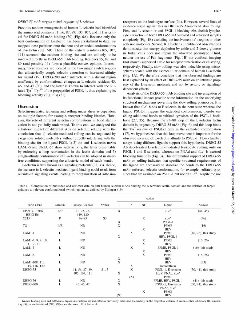
FIGURE 3. DREG-55 mAb-induced slow rolling is ligand specific. Flow
chamber assays were performed with Jurkat cells at 2 dynes/cm2 using
different immobilized L-selectin ligands. Numbers on top of the bars in-
dicate the mean rolling velocity. No interaction indicates full inhibitory
activity of DREG-55 mAb. DREG-200 Ab blocked all interactions (not
shown); n = 3–4, means 6 SD.
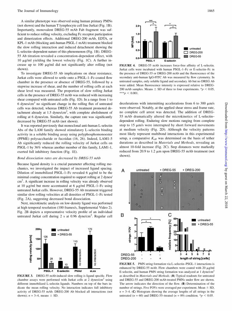
FIGURE 4. DREG-55 mAb increases force-free affinity of L-selectin.
Jurkat cells were incubated with human PSGL-1–Fc or E-selectin–Fc in
the presence of DREG-55 or DREG-200 mAb and the fluorescence of the
secondary anti-human IgG-FITC Ab was measured by flow cytometry. In
untreated samples, only soluble ligand and secondary Ab but no DREG Ab
were added. Mean fluorescence intensity is expressed relative to DREG-
200 mAb samples. Means 6 SD of three to four experiments. *p , 0.05,
***p , 0.001.
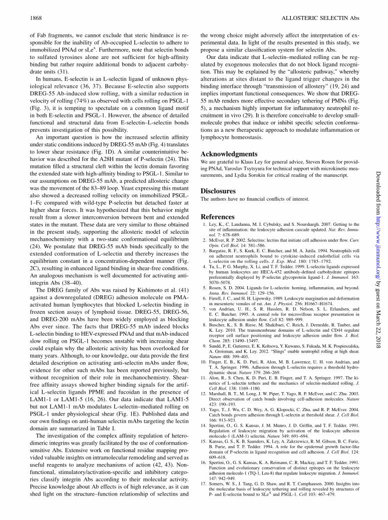
FIGURE 5. PMN string formation via L-selectin–PSGL-1 interactions is
enhanced by DREG-55 mAb. Flow chambers were coated with 20 mg/ml
E-selectin, and human PMN string formation was analyzed at 1 dyne/cm2
as described in Materials and Methods. (A) Typical readouts for untreated
and DREG-55 and DREG-200 mAb-treated PMNs under flow are shown.
The arrow indicates the direction of the flow. (B) Determination of the
number of strings. Five FOVs were averaged per experiment. Mean 6 SD,
n = 3–4. (C) Histogram showing the average lengths of all strings in the
untreated (n = 60) and DREG-55–treated (n = 89) condition. *p , 0.05.
The Journal of Immunology 1865
by guest on March 22, 2018
http://ww
w.jim
munol.org/
Dow
nloaded from
DREG-55 mAb–induced slow rolling is ligand specific
Physiological L-selectin ligand epitopes vary considerably. Whereasa crucial binding motif in PSGL-1 includes the sulfated tyrosineresidues of the protein scaffold in addition to an adjacent core2–based O-glycan capped with sLex (27), binding to HEV-expressedPNAd depends on carbohydrates from sLex with sulfated N-ace-tylglucosamine (5).Immobilized PSGL-1–Fc, E-selectin–Fc, PNAd, and sLex sup-
ported specific L-selectin–mediated rolling of Jurkat cells (Fig. 3).However, only PSGL-1–Fc and E-selectin–Fc were sufficient toallow DREG-55–induced slow rolling, whereas on PNAd andsLex all rolling cells detached. In contrast, DREG-200 Ab blockedrolling on all ligands (not shown). A similar selective modulationof rolling on PSGL-1–Fc and E-selectin–Fc was obtained usingDREG-55 mAb Fab fragments (not shown). These results indicatethat specific ligand requirements for DREG-55 mAb-inducedL-selectin–mediated slow rolling are met by PSGL-1 and E-selectinbut not by sLex or PNAd.
Shear-free affinity of L-selectin is upregulated by DREG-55 Ab
To directly address the question of force-free (shear-independent)affinity modulation, a flow cytometry–based assay for ligand af-finity was performed. Jurkat cells were incubated with or withoutmouse anti-human DREG-55 or DREG-200 Ab together with sol-uble human PSGL-1–Fc or E-selectin–Fc chimeric proteins, andthe specific binding of a secondary anti-human IgG-FITC Abwas detected. Samples with the blocking DREG-200 Ab servedas negative control. Because L-selectin bonds are characterized
by low-affinity interactions (28), the binding of soluble E-selectin–Fc or PSGL-1–Fc to untreated Jurkat cells was absent under theseexperimental conditions (Fig. 4). DREG-200 mAb did not inducesignificant changes relative to untreated samples excluding pos-sible cross-reactions of the anti-human IgG Ab to mouse DREG-Igor of DREG Abs to cellular Fc receptors. In contrast, coincubationof Jurkat cells with DREG-55 mAb led to a 1.6- and 1.4-foldhigher binding of soluble PSGL-1–Fc and E-selectin–Fc, respec-tively. This result suggests that the treatment of Jurkat cells withDREG-55 mAb exposes a higher affinity binding site on theL-selectin molecule in the absence of shear (Fig. 4).
Secondary capture of PMNs is amplified by DREG-55 mAb
Intercellular interactions between free-flowing and adherent neu-trophils occur via transient PSGL-1–L-selectin bonds and serve asa mechanism to boost neutrophil recruitment at inflammatory sites(“secondary capturing”) (29). Using isolated PMNs under flow, theformation of cells lined up in the direction of the flow (strings) canbe observed in vitro. Because L-selectin shows a higher affinity toPSGL-1 in the DREG-55 mAb modified state (Fig. 4), we inves-tigated whether there is a functional relevance in secondary capture.Therefore, untreated, DREG-55 or DREG-200 mAb Fab fragment–treated human PMNs were perfused into an E-selectin–coated flowchamber at 1 dyne/cm2. An average of approximately two stringsper FOV was found in untreated conditions, and addition of DREG-200 mAb Fab abolished most of the secondary but not primarycapture (Fig. 5A, 5B). In contrast, DREG-55 mAb Fab treatment in-tensified string formation and yielded longer strings (Fig. 5B, 5C).
FIGURE 6. Mapping of DREG Ab epitopes onto the
N-terminal lectin domain. (A) Sequence alignment of the
N-terminal portion of human selectins. The four switch
regions in the lectin domain that undergo conformational
changes upon selectin extension (19) are underlined and
labeled S1–S4. Residues involved in PSGL-1 binding are
indicated in orange (PSGL-1 polypeptide) and magenta
(PSGL-1 glycan). Residues involved in calcium binding are
marked by cyan circles. Residues whose mutation abol-
ishes mAb DREG-55 and DREG-200 binding (30) are in-
dicated in blue and green, respectively. (B) Drawings of
P-selectin crystal structures in the bent (left panel) and
extended (right panel) conformation (17). The lectin do-
main is at the top and the EGF-like domain is at the bot-
tom. The pivot at the interdomain hinge is indicated by
a filled black triangle and switch regions S2 and S3 are
labeled. The calcium ion is shown as a cyan sphere. The
PSGL-1 ligand is shown in orange (polypeptide) and ma-
genta (glycan). The two sulfated tyrosines are shown in
atomic detail. Arg85, which reorients dramatically upon
PSGL-1 binding, is shown in red. Residues whose mutation
abolishes DREG-55 and DREG-200 mAb binding are in-
dicated by blue and green Ca spheres, respectively. The
putative DREG-55 mAb epitope in the extended confor-
mation is indicated by a blue oval.
1866 ALLOSTERIC SELECTIN Abs
by guest on March 22, 2018
http://ww
w.jim
munol.org/
Dow
nloaded from
DREG-55 mAb targets switch regions of L-selectin
Previous random mutagenesis of human L-selectin had identifiedthe amino acid positions 11, 56, 87, 89, 105, 107, and 111 as criti-cal for DREG-55 mAb binding (30) (Fig. 6A). Because only thebent conformation of L-selectin has been crystallized to date, wemapped these positions onto the bent and extended conformationsof P-selectin (Fig. 6B). Three of the critical residues (105, 107,111) surround the calcium binding site and are unlikely to beinvolved directly in DREG-55 mAb binding. Residues 55, 87, and89 (and possibly 11) form a plausible convex epitope. Interest-ingly, these residues are located in the two major switch regionsthat allosterically couple selectin extension to increased affinityfor ligand (19). DREG-200 mAb interacts with a distant regionunaffected by conformational changes at L-selectin residues 45,46, and 47 (30), and the latter is known to interact with the sul-fated Tyr7 (Tyr48 of the propeptide) of PSGL-1, thus explaining itsblocking activity (Fig. 6B) (31).
DiscussionSelectin-mediated tethering and rolling under shear is dependenton multiple factors, for example, receptor-binding kinetics. How-ever, the role of different selectin conformations in bond stabili-zation is not yet fully understood. To this end, we analyzed theallosteric impact of different Abs on selectin rolling with theconclusion that 1) selectin-mediated rolling can be regulated byexogenous soluble molecules without directly interfering with thebinding site for the ligand PSGL-1; 2) the anti–L-selectin mAbsLAM1-5 and DREG-55 show such activity, the latter presumablyby enforcing a loop reorientation in the lectin domain; and 3)a high-affinity conformation of L-selectin can be adopted in shear-free conditions, supporting the allosteric model of catch bonds.L-selectin is well known as a signaling molecule (32, 33). Hence,
the increase in L-selectin–mediated ligand binding could result fromoutside–in signaling events leading to reorganization of adhesion
receptors on the leukocyte surface (34). However, several lines ofevidence argue against this in DREG-55 Ab-induced slow rolling.First, anti–L-selectin or anti–PSGL-1 blocking Abs abolish lympho-cyte interaction in both DREG-55 mAb-treated and untreated samplescompletely (Fig. 1B) excluding the involvement of integrins or otheradhesion molecules. Second, K. Buscher’s unpublished observationsdemonstrate that energy depletion by azide and 2-deoxy-glucosein Jurkat cells does not impair the observed phenotype. Third,neither the use of Fab fragments (Fig. 1B) nor confocal imaging(not shown) supported a role for receptor dimerization or clustering,respectively. Finally, slow rolling was also inducible using micro-spheres coated with the extracellular domain of human L-selectin(Fig. 1A). We therefore conclude that the observed findings arebest explained by an effect of DREG-55 mAb on an intrinsic prop-erty of the L-selectin molecule and not by avidity or signaling-dependent effects.Analysis of the DREG-55 mAb binding site and investigation of
its functional impact provide some information on the underlyingstructural mechanisms governing the slow rolling phenotype. It isknown that sLex binds to P-selectin in the bent state whereas theligand PSGL-1 triggers the extended conformation, thereby en-abling additional bonds to sulfated tyrosines of the PSGL-1 back-bone (27, 35). Because the 83–88 loop of the L-selectin lectindomain is targeted by DREG-55 mAb (Fig. 6) and this loop bindsthe Tyr7 residue of PSGL-1 only in the extended conformation(17), we hypothesized that this loop movement is important for theobserved increase of L-selectin affinity to PSGL-1. Flow chamberassays using different ligands support this hypothesis. DREG-55Ab decelerated L-selectin–mediated leukocyte rolling only onPSGL-1 and E-selectin, whereas on PNAd and sLex it exertedblocking functions (Fig. 3). This differential support of DREG-55mAb on rolling indicates that specific structural requirements ofthe ligand are necessary to stabilize the bonds to the DREG-55mAb-enforced selectin conformation, for example, sulfated tyro-sines that are available on PSGL-1 but not on sLex. Despite the use
Table I. Compilation of published and our own data on anti-human selectin mAbs binding the N-terminal lectin domain and the relation of targetepitopes to relevant conformational switch regions as defined by Springer (19)
mAb Clone Selectin Epitope Residues Switch
Action
SourcesI S NF Ligand
EP-5C7, 1.2B6,BBIG-E6
E/P 21, 22, 23,119, 120
X sLex (44, 45)
C215 P 76–83 S3 X sLex (46)X PSGL-1
TQ-1 L/E ND X PPME (16)X HEV
LAM1-1 L ND X PPME (16, 26), this studyX HEV, PSGL-1
LAM1-7, 8, 9, 10,11, 12, 13
L ND X PPME (16, 26)X HEV
LAM1-5 L ND X PPME, PSGL-1 (16), this studyX HEV
LAM1-6 L ND X PPME (16, 26)X HEV
LAM1-108, 110,115, 116, 120
L ND X HEV (33)X Intercellular
DREG-55 L 11, 56, 87, 89,105, 107, 111
S1, 3 X PSGL-1, E-selectin (30, 41), this studyX HEV, PNAd, sLex
(X) PPMEDREG-56 L ND X PPME, HEV, PSGL-1 (41), this studyDREG-200 L 45, 46, 47 X PSGL-1, E-selectin
PNAd, sLex(30, 41), this study
X PPME(X) HEV
Known binding sites and differential ligand interactions are indicated as previously published. Depending on the respective column, X means either inhibitory (I), stimula-tory (S), or nonfunctional (NF). (X)means the same effect but weak.
The Journal of Immunology 1867
by guest on March 22, 2018
http://ww
w.jim
munol.org/
Dow
nloaded from
of Fab fragments, we cannot exclude that steric hindrance is re-sponsible for the inability of Ab-occupied L-selectin to adhere toimmobilized PNAd or sLex. Furthermore, note that selectin bondsto sulfated tyrosines alone are not sufficient for high-affinitybinding but rather require additional bonds to adjacent carbohy-drate units (31).In humans, E-selectin is an L-selectin ligand of unknown phys-
iological relevance (36, 37). Because E-selectin also supportsDREG-55 Ab-induced slow rolling, with a similar reduction invelocity of rolling (74%) as observed with cells rolling on PSGL-1(Fig. 3), it is tempting to spectulate on a common ligand motifin both E-selectin and PSGL-1. However, the absence of detailedfunctional and structural data from E-selectin–L-selectin bondsprevents investigation of this possibility.An important question is how the increased selectin affinity
under static conditions induced by DREG-55 mAb (Fig. 4) translatesto lower shear resistance (Fig. 1D). A similar counterintuitive be-havior was described for the A28H mutant of P-selectin (24). Thismutation filled a structural cleft within the lectin domain favoringthe extended state with high-affinity binding to PSGL-1. Similar toour assumptions on DREG-55 mAb, a predicted allosteric changewas the movement of the 83–89 loop. Yeast expressing this mutantalso showed a decreased rolling velocity on immobilized PSGL-1–Fc compared with wild-type P-selectin but detached faster athigher shear forces. It was hypothesized that this behavior mightresult from a slower interconversion between bent and extendedstates in the mutant. These data are very similar to those obtainedin the present study, supporting the allosteric model of selectinmechanochemistry with a two-state conformational equilibrium(24). We postulate that DREG-55 mAb binds specifically to theextended conformation of L-selectin and thereby increases theequilibrium constant in a concentration-dependent manner (Fig.2C), resulting in enhanced ligand binding in shear-free conditions.An analogous mechanism is well documented for activating anti-integrin Abs (38–40).The DREG family of Abs was raised by Kishimoto et al. (41)
against a downregulated (DREG) adhesion molecule on PMA-activated human lymphocytes that blocked L-selectin binding infrozen section assays of lymphoid tissue. DREG-55, DREG-56,and DREG-200 mAbs have been widely employed as blockingAbs ever since. The facts that DREG-55 mAb indeed blocksL-selectin binding to HEV-expressed PNAd and that mAb-inducedslow rolling on PSGL-1 becomes unstable with increasing shearcould explain why the allosteric activity has been overlooked formany years. Although, to our knowledge, our data provide the firstdetailed description on activating anti-selectin mAbs under flow,evidence for other such mAbs has been reported previously, butwithout recognition of their role in mechanochemistry. Shear-free affinity assays showed higher binding signals for the artif-ical L-selectin ligands PPME and fucoidan in the presence ofLAM1-1 or LAM1-5 (16, 26). Our data indicate that LAM1-5but not LAM1-1 mAb modulates L-selectin–mediated rolling onPSGL-1 under physiological shear (Fig. 1E). Published data andour own findings on anti-human selectin mAbs targeting the lectindomain are summarized in Table I.The investigation of the complex affinity regulation of hetero-
dimeric integrins was greatly facilitated by the use of conformation-sensitive Abs. Extensive work on functional residue mapping pro-vided valuable insights on intramolecular remodeling and served asuseful reagents to analyze mechanisms of action (42, 43). Non-functional, stimulatory/activation-specific and inhibitory catego-ries classify integrin Abs according to their molecular activity.Precise knowledge about Ab effects is of high relevance, as it canshed light on the structure–function relationship of selectins and
the wrong choice might adversely affect the interpretation of ex-perimental data. In light of the results presented in this study, wepropose a similar classification system for selectin Abs.Our data indicate that L-selectin–mediated rolling can be reg-
ulated by exogenous molecules that do not block ligand recogni-tion. This may be explained by the “allosteric pathway,” wherebyalterations at sites distant to the ligand trigger changes in thebinding interface through “transmission of allostery” (19, 24) andimplies important functional consequences. We show that DREG-55 mAb renders more effective secondary tethering of PMNs (Fig.5), a mechanism highly important for inflammatory neutrophil re-cruitment in vivo (29). It is therefore conceivable to develop small-molecule probes that induce or inhibit specific selectin conforma-tions as a new therapeutic approach to modulate inflammation orlymphocyte homeostasis.
AcknowledgmentsWe are grateful to Klaus Ley for general advice, Steven Rosen for provid-
ing PNAd, Yaroslav Tsytsyura for technical support with microkinetic mea-
surements, and Lydia Sorokin for critical reading of the manuscript.
DisclosuresThe authors have no financial conflicts of interest.
References1. Ley, K., C. Laudanna, M. I. Cybulsky, and S. Nourshargh. 2007. Getting to the
site of inflammation: the leukocyte adhesion cascade updated. Nat. Rev. Immu-nol. 7: 678–689.
2. McEver, R. P. 2002. Selectins: lectins that initiate cell adhesion under flow. Curr.Opin. Cell Biol. 14: 581–586.
3. Bargatze, R. F., S. Kurk, E. C. Butcher, and M. A. Jutila. 1994. Neutrophils rollon adherent neutrophils bound to cytokine-induced endothelial cells viaL-selectin on the rolling cells. J. Exp. Med. 180: 1785–1792.
4. Tu, L., P. G. Murphy, X. Li, and T. F. Tedder. 1999. L-selectin ligands expressedby human leukocytes are HECA-452 antibody-defined carbohydrate epitopespreferentially displayed by P-selectin glycoprotein ligand-1. J. Immunol. 163:5070–5078.
5. Rosen, S. D. 2004. Ligands for L-selectin: homing, inflammation, and beyond.Annu. Rev. Immunol. 22: 129–156.
6. Firrell, J. C., and H. H. Lipowsky. 1989. Leukocyte margination and deformationin mesenteric venules of rat. Am. J. Physiol. 256: H1667–H1674.
7. von Andrian, U. H., S. R. Hasslen, R. D. Nelson, S. L. Erlandsen, andE. C. Butcher. 1995. A central role for microvillous receptor presentation inleukocyte adhesion under flow. Cell 82: 989–999.
8. Buscher, K., S. B. Riese, M. Shakibaei, C. Reich, J. Dernedde, R. Tauber, andK. Ley. 2010. The transmembrane domains of L-selectin and CD44 regulatereceptor cell surface positioning and leukocyte adhesion under flow. J. Biol.Chem. 285: 13490–13497.
9. Sundd, P., E. Gutierrez, E. K. Koltsova, Y. Kuwano, S. Fukuda, M. K. Pospieszalska,A. Groisman, and K. Ley. 2012. “Slings” enable neutrophil rolling at high shear.Nature 488: 399–403.
10. Finger, E. B., K. D. Puri, R. Alon, M. B. Lawrence, U. H. von Andrian, andT. A. Springer. 1996. Adhesion through L-selectin requires a threshold hydro-dynamic shear. Nature 379: 266–269.
11. Alon, R., S. Chen, K. D. Puri, E. B. Finger, and T. A. Springer. 1997. The ki-netics of L-selectin tethers and the mechanics of selectin-mediated rolling. J.Cell Biol. 138: 1169–1180.
12. Marshall, B. T., M. Long, J. W. Piper, T. Yago, R. P. McEver, and C. Zhu. 2003.Direct observation of catch bonds involving cell-adhesion molecules. Nature423: 190–193.
13. Yago, T., J. Wu, C. D. Wey, A. G. Klopocki, C. Zhu, and R. P. McEver. 2004.Catch bonds govern adhesion through L-selectin at threshold shear. J. Cell Biol.166: 913–923.
14. Spertini, O., G. S. Kansas, J. M. Munro, J. D. Griffin, and T. F. Tedder. 1991.Regulation of leukocyte migration by activation of the leukocyte adhesionmolecule-1 (LAM-1) selectin. Nature 349: 691–694.
15. Kansas, G. S., K. B. Saunders, K. Ley, A. Zakrzewicz, R. M. Gibson, B. C. Furie,B. Furie, and T. F. Tedder. 1994. A role for the epidermal growth factor-likedomain of P-selectin in ligand recognition and cell adhesion. J. Cell Biol. 124:609–618.
16. Spertini, O., G. S. Kansas, K. A. Reimann, C. R. Mackay, and T. F. Tedder. 1991.Function and evolutionary conservation of distinct epitopes on the leukocyteadhesion molecule-1 (TQ-1, Leu-8) that regulate leukocyte migration. J. Immunol.147: 942–949.
17. Somers, W. S., J. Tang, G. D. Shaw, and R. T. Camphausen. 2000. Insights intothe molecular basis of leukocyte tethering and rolling revealed by structures ofP- and E-selectin bound to SLeX and PSGL-1. Cell 103: 467–479.
1868 ALLOSTERIC SELECTIN Abs
by guest on March 22, 2018
http://ww
w.jim
munol.org/
Dow
nloaded from
18. Graves, B. J., R. L. Crowther, C. Chandran, J. M. Rumberger, S. Li, K. S. Huang,D. H. Presky, P. C. Familletti, B. A. Wolitzky, and D. K. Burns. 1994. Insight intoE-selectin/ligand interaction from the crystal structure and mutagenesis of thelec/EGF domains. Nature 367: 532–538.
19. Springer, T. A. 2009. Structural basis for selectin mechanochemistry. Proc. Natl.Acad. Sci. USA 106: 91–96.
20. Klopocki, A. G., T. Yago, P. Mehta, J. Yang, T. Wu, A. Leppanen, N. V. Bovin,R. D. Cummings, C. Zhu, and R. P. McEver. 2008. Replacing a lectin domainresidue in L-selectin enhances binding to P-selectin glycoprotein ligand-1 butnot to 6-sulfo-sialyl Lewis x. J. Biol. Chem. 283: 11493–11500.
21. Lou, J., T. Yago, A. G. Klopocki, P. Mehta, W. Chen, V. I. Zarnitsyna,N. V. Bovin, C. Zhu, and R. P. McEver. 2006. Flow-enhanced adhesion regulatedby a selectin interdomain hinge. J. Cell Biol. 174: 1107–1117.
22. Phan, U. T., T. T. Waldron, and T. A. Springer. 2006. Remodeling of the lectin-EGF-like domain interface in P- and L-selectin increases adhesiveness and shearresistance under hydrodynamic force. Nat. Immunol. 7: 883–889.
23. Lou, J., and C. Zhu. 2007. A structure-based sliding-rebinding mechanism forcatch bonds. Biophys. J. 92: 1471–1485.
24. Waldron, T. T., and T. A. Springer. 2009. Transmission of allostery through thelectin domain in selectin-mediated cell adhesion. Proc. Natl. Acad. Sci. USA106: 85–90.
25. Schindelin, J., I. Arganda-Carreras, E. Frise, V. Kaynig, M. Longair, T. Pietzsch,S. Preibisch, C. Rueden, S. Saalfeld, B. Schmid, et al. 2012. Fiji: an open-sourceplatform for biological-image analysis. Nat. Methods 9: 676–682.
26. Kansas, G. S., O. Spertini, L. M. Stoolman, and T. F. Tedder. 1991. Molecularmapping of functional domains of the leukocyte receptor for endothelium,LAM-1. J. Cell Biol. 114: 351–358.
27. Leppanen, A., S. P. White, J. Helin, R. P. McEver, and R. D. Cummings. 2000.Binding of glycosulfopeptides to P-selectin requires stereospecific contributionsof individual tyrosine sulfate and sugar residues. J. Biol. Chem. 275: 39569–39578.
28. Nicholson, M. W., A. N. Barclay, M. S. Singer, S. D. Rosen, and P. A. van derMerwe. 1998. Affinity and kinetic analysis of L-selectin (CD62L) binding toglycosylation-dependent cell-adhesion molecule-1. J. Biol. Chem. 273: 763–770.
29. Sperandio, M., M. L. Smith, S. B. Forlow, T. S. Olson, L. Xia, R. P. McEver, andK. Ley. 2003. P-selectin glycoprotein ligand-1 mediates L-selectin-dependentleukocyte rolling in venules. J. Exp. Med. 197: 1355–1363.
30. Fu, H., E. L. Berg, and N. Tsurushita. 1997. Fine mapping of the epitopes ofhumanized anti-L-selectin monoclonal antibodies HuDREG-55 and HuDREG-200. Immunol. Lett. 59: 71–77.
31. Leppanen, A., T. Yago, V. I. Otto, R. P. McEver, and R. D. Cummings. 2003.Model glycosulfopeptides from P-selectin glycoprotein ligand-1 require tyrosinesulfation and a core 2-branched O-glycan to bind to L-selectin. J. Biol. Chem.278: 26391–26400.
32. Brenner, B., E. Gulbins, K. Schlottmann, U. Koppenhoefer, G. L. Busch,B. Walzog, M. Steinhausen, K. M. Coggeshall, O. Linderkamp, and F. Lang.1996. L-selectin activates the Ras pathway via the tyrosine kinase p56lck. Proc.Natl. Acad. Sci. USA 93: 15376–15381.
33. Steeber, D. A., P. Engel, A. S. Miller, M. P. Sheetz, and T. F. Tedder. 1997.Ligation of L-selectin through conserved regions within the lectin domainactivates signal transduction pathways and integrin function in human, mouse,and rat leukocytes. J. Immunol. 159: 952–963.
34. Li, X., D. A. Steeber, M. L. Tang, M. A. Farrar, R. M. Perlmutter, andT. F. Tedder. 1998. Regulation of L-selectin-mediated rolling through receptordimerization. J. Exp. Med. 188: 1385–1390.
35. Pouyani, T., and B. Seed. 1995. PSGL-1 recognition of P-selectin is controlledby a tyrosine sulfation consensus at the PSGL-1 amino terminus. Cell 83: 333–343.
36. Zollner, O., M. C. Lenter, J. E. Blanks, E. Borges, M. Steegmaier, H. G. Zerwes,and D. Vestweber. 1997. L-selectin from human, but not from mouse neutrophilsbinds directly to E-selectin. J. Cell Biol. 136: 707–716.
37. Picker, L. J., R. A. Warnock, A. R. Burns, C. M. Doerschuk, E. L. Berg, andE. C. Butcher. 1991. The neutrophil selectin LECAM-1 presents carbohydrateligands to the vascular selectins ELAM-1 and GMP-140. Cell 66: 921–933.
38. Luo, B.-H., K. Strokovich, T. Walz, T. A. Springer, and J. Takagi. 2004. Allo-steric beta1 integrin antibodies that stabilize the low affinity state by preventingthe swing-out of the hybrid domain. J. Biol. Chem. 279: 27466–27471.
39. Lu, C., M. Shimaoka, A. Salas, and T. A. Springer. 2004. The binding sites forcompetitive antagonistic, allosteric antagonistic, and agonistic antibodies to the Idomain of integrin LFA-1. J. Immunol. 173: 3972–3978.
40. Chen, X., C. Xie, N. Nishida, Z. Li, T. Walz, and T. A. Springer. 2010. Re-quirement of open headpiece conformation for activation of leukocyte integrinaXb2. Proc. Natl. Acad. Sci. USA 107: 14727–14732.
41. Kishimoto, T. K., M. A. Jutila, and E. C. Butcher. 1990. Identification of a hu-man peripheral lymph node homing receptor: a rapidly down-regulated adhesionmolecule. Proc. Natl. Acad. Sci. USA 87: 2244–2248.
42. Byron, A., J. D. Humphries, J. A. Askari, S. E. Craig, A. P. Mould, andM. J. Humphries. 2009. Anti-integrin monoclonal antibodies. J. Cell Sci. 122:4009–4011.
43. Humphries, M. J., E. J. H. Symonds, and A. P. Mould. 2003. Mapping functionalresidues onto integrin crystal structures. Curr. Opin. Struct. Biol. 13: 236–243.
44. Tsurushita, N., H. Fu, J. Melrose, and E. L. Berg. 1998. Epitope mapping ofmouse monoclonal antibody EP-5C7 which neutralizes both human E- and P-selectin. Biochem. Biophys. Res. Commun. 242: 197–201.
45. Goda, K., T. Tanaka, M. Monden, and M. Miyasaka. 1999. Characterization ofan apparently conserved epitope in E- and P-selectin identified by dual-specificmonoclonal antibodies. Eur. J. Immunol. 29: 1551–1560.
46. Hirose, M., H. Kawashima, and M. Miyasaka. 1998. A functional epitope on P-selectin that supports binding of P-selectin to P-selectin glycoprotein ligand-1but not to sialyl Lewis X oligosaccharides. Int. Immunol. 10: 639–649.
The Journal of Immunology 1869
by guest on March 22, 2018
http://ww
w.jim
munol.org/
Dow
nloaded from