Embed Size (px)
Citation preview

Research ArticleOxidative Biochemistry Disbalance and Changes onProteomic Profile in Salivary Glands of Rats Induced by ChronicExposure to Methylmercury
Leonardo Oliveira Bittencourt,1 Bruna Puty,1 Senda Charone,2
Walessa Alana Bragança Aragão,1 Paulo Mecenas Farias-Junior,1
Marcia Cristina Freitas Silva,1 Maria Elena Crespo-Lopez,3 Aline de Lima Leite,2
Marilia Afonso Rabelo Buzalaf,2 and Rafael Rodrigues Lima1
1Laboratory of Functional and Structural Biology, Institute of Biological Sciences, Federal University of Pará, Belém, PA, Brazil2Department of Biological Sciences, Bauru Dental School, University of São Paulo, Bauru, São Paulo, SP, Brazil3Laboratory of Molecular Pharmacology, Institute of Biological Sciences, Federal University of Pará, Belém, PA, Brazil
Correspondence should be addressed to Rafael Rodrigues Lima; [email protected]
Received 14 April 2017; Revised 7 June 2017; Accepted 13 June 2017; Published 24 July 2017
Academic Editor: Laura Giusti
Copyright © 2017 Leonardo Oliveira Bittencourt et al. This is an open access article distributed under the Creative CommonsAttribution License, which permits unrestricted use, distribution, and reproduction in any medium, provided the original workis properly cited.
Methylmercury (MeHg) is one of the most toxic mercury species, which can cause many systemic damages, but little is knownabout its effect in the salivary glands. This study aimed to analyze the mercury levels, oxidative stress, and proteomic profile inparotid, submandibular, and sublingual salivary glands of rats, after chronic MeHg intoxication. Two groups of twenty maleWistar rats (90 days of age) were used on the experiment. MeHg group was intoxicated by intragastric gavage with MeHg at adose of 0.04 mg/kg/day for 60 days, while the control group received only oil. After the period of intoxication, the glands werecollected for evaluation of total mercury levels, proteomic profile, and oxidative balance by analyzing the antioxidant capacityagainst peroxyl radicals (ACAP), lipid peroxidation (LPO), and nitrite levels. Our results have showed that mercury levels weresignificant in all three glands compared to the respective control. It also showed lower levels of ACAP, as well as higher LPOand nitrite levels. The proteomic profile presented impairments on structural components of cytoskeleton, metabolic pathways,and oxidative biochemistry. Thus, the exposure to MeHg was able to generate oxidative stress that could be associated withchanges in the proteomic profile of salivary glands.
1. Introduction
Mercury is a high toxic metal with a wide distribution onnature and has been considered as an important health con-cern [1, 2]. Mercury occurs naturally in the environment onorganic and inorganic forms, in which methylmercury(MeHg) and mercury chloride (HgCl2) are the most toxicforms, respectively. MeHg is an alkylmercury compound thatis derived from the methylation of Hg+2 by methanogenicand sulfate-reducing bacteria on aquatic ecosystems andsome soils [3–5].
The anthropogenic action leads to higher concentrationson biosphere and may expose animals of aquatic food chainto damage [5]. Many researches have reported that fishesand other seafood are the main sources of human contamina-tion by low daily doses, in which about 90% of Hg is presenton its organic form, leading to higher bioaccumulation andbiomagnification [6, 7].
Hg also has been described as a huge pollutant able toaccumulate and induce cellular impairments in organsrelated to metabolism and excretion, as the liver and kidneys[8–11]. Few studies [12–14] have demonstrated the effects of
HindawiOxidative Medicine and Cellular LongevityVolume 2017, Article ID 5653291, 15 pageshttps://doi.org/10.1155/2017/5653291

HgCl2 in salivary glands, and there is no evidence of MeHgeffects. In this way, it is important to describe the MeHg-induced alteration in salivary glands to elucidate the conse-quences of this metal exposure.
The parotid, submandibular, and sublingual are the mainsalivary glands, representing about 90% of the total salivaryhuman production. Salivary glands are important organs onthe maintenance of oral cavity homeostasis; their major func-tion is salivary secretion that is responsible for balancing themicrobiota, pH, and carbohydrates catalysis in the mouth[15, 16]. Moreover, studies of metal toxicological effects onsalivary glands are still rare, especially about MeHg. Theaim of this study was to evaluate whether the chronic expo-sure to low doses of MeHg is able to impair biochemicalparameters and the proteomic profile, which could be relatedto dysfunctions in the organs.
2. Materials and Methods
2.1. Animals and Experimental Groups. A total of 40 maleWistar rats were obtained from the Federal University ofPará (UFPA, Belém, Brazil), under the BIO 225-14 CEPAE– UFPA, following the NIH Guide for the Care and Use ofLaboratory Animals. The animals were maintained in collec-tive cages with 5 animals each and were kept in a climate-controlled room on a 12 h light/dark cycle with water andfood ad libitum. Corn oil or MeHg with a daily dose of0.04mg/kg was orally administered by gavage over a periodof 60 days of exposure according to an adapted protocol fromKong et al. [17]. To regulate the administered dose in ani-mals, they were weekly weighted for adjustment of MeHgconcentration and determination of the body mass curve.
2.2. Samples Collection. After 24 hours of the last administra-tion, the animals were euthanized by cervical dislocation andhad their three pairs of salivary glands (parotid, submandib-ular, and sublingual) collected and frozen in liquid nitrogenand posteriorly stored in ultrafreezer −80°C until furtheranalysis. One gland from each animal was saved to the assaysof oxidative biochemistry, proteomic analysis, and measure-ment of total mercury deposits.
2.3. Mercury Deposits. Total mercury content in the sampleswas quantified by wet digestion, reduction, and cold vaporatomic absorption spectrometry with gold amalgamationsystem Mercury Analyzer: SP3D (Nippon Corporation).The estimations were conducted following the protocol pre-viously mentioned by Suzuki et al. [18], in which consistsby converting the Hg into elementary Hg vapor in order toquantify in parts per million (ppm) by an absorption cell.The analyses were performed preparing duplicates of eachsample of groups, and the values were tabulated to furtherinferential statistical analysis.
2.4. Oxidative Biochemistry Analysis. The samples werefirst thawed and homogenized in 700μL Tris-HCl buffer(20mM, pH7.4). A volume of 200μL from the total homog-enate was used to perform the measurement of antioxidantcapacity against peroxyl radicals (ACAP), whereas 300μL
was used to measure nitrite levels and 100μL to lipidperoxidation assay (LPO).
First, ACAP was measured as previously described byAmado et al. [19]. The supernatant of each sample after cen-trifugation was, in triplicate, exposed to a peroxyl radicalgenerator, the 2,2′-azobis 2 methylpropionamidine dihy-drochloride (ABAP; 4mM; Aldrich), and, in another tripli-cate, they received only ultrapure water in a 96-wellmicroplate. After reaction, the fluorescence generated wasmeasured in a fluorimeter (Victor X3, Perkin Elmer) at35°C. After the first reading for determination of backgroundfluorescence values, a total of 10μl of 2′,7′ (H2DCF-DA)40nM was added to the microplate, with a reading in every5 minutes during 60 minutes [19, 20]. The results wereexpressed according to the difference between areas belowthe curves created by total fluorescence production of sam-ples with and without ABAP after application to a secondorder polynomial function, being named as a relative area.High-relative areas mean low antioxidant activity, once thesample had low capacity to neutralize the peroxyl radicals.Results were expressed as percentages of control group.
The remaining homogenate was centrifuged at 21.000gfor 20 minutes, and the supernatant was used to performthe assays. The concentration of nitrite was determinedfollowing the protocol previously adapted by Fagundeset al. [21], which is based on a reaction with Griess reagent(0.1% N-(1-naphthyl) ethylenediaminedihydrochloride; 1%sulfanilamide in 5% phosphoric acid; 1 : 1) proceeding theabsorbance measurement on a spectrophotometer at 550 nm,and compared to standard solutions of sodium nitrite.
The level of LPO was established by measurement ofmalonaldehyde (MDA) levels based on Esterbauer andCheeseman [22] method adapted by Fagundes et al. [21].An aliquot of the supernatant was processed as describedby the Bioxytech LPO-568 kit (Cayman Chemical). This kitis a chromogenic reagent that reacts with MDA at 45°C.The absorbance measurement on spectrophotometers wasperformed at 586m wavelength.
The total protein content in the supernatants (20μL) wasanalyzed by Bradford [23] method. After the correction forprotein concentration, results of lipid peroxidation andnitrite levels were expressed in percentages of control groups.
2.5. Proteomic. The frozen salivary glands were homogenizedin a cryogenic mill (model 6770, Spex, Metuchen, NJ, EUA).For protein extraction, gland homogenate was incubated inlysis buffer (7M urea, 2M thiourea, 4% CHAPS, 1% IPGbuffer pH3–10, and 40mMDTT) for 1 hour at 4°C with con-tinuous shaking. In order to recovery the soluble proteinsand the supernatant, the homogenate was centrifuged at15.000 rpm for 30 minutes at 4°C. The proteins were precip-itated using the kit PlusOne 2D Cleanup (GE Healthcare,Uppsala, Sweden), as recommended by the manufacturer.Pellets were resuspended in rehydration buffer (7M urea,2M thiourea, 0.5% CHAPS, 0.5% IPG buffer pH3–10,18mM DTT, and 0.002% bromophenol blue). Twenty-fiveμL of salivary gland proteins from each animal of the samegroup was combined to constitute a pool that was centrifugedfor clarification. To each pool, 50mM AMBIC containing
2 Oxidative Medicine and Cellular Longevity

3M urea was added. Each sample was filtered twice in 3 kDaAMICON (Millipore, St. Charles, MO, USA). The proteincontent was measured in the pooled samples by Bradfordprotein assay [23]. To each sample (50μg of total proteinfor each pool in a volume of 50μL), 10μL of 50mM AMBICwas added. In sequence, 25μL of 0.2% RapiGEST™ (WatersCo., Manchester, UK) was added and incubated at 80°C for15min. Following, 2.5μL of 100mM DTT was added andincubated at 60°C for 30min. Also, 2.5μL of 300mM IAAwas added and incubated for 30min at room temperature(under dark). Then, 10μL of trypsin (100 ng; Trypsin GoldMass Spectrometry, Promega, Madison, USA) was addedand digestion occurred for 14 h at 37°C. After digestion,10μl of 5% TFA was added, incubated for 90min at37°C, and the sample was centrifuged (14,000 rpm for30min). The supernatant was collected, and 5μL of ADH(1 pmol/μL) plus 85μL 3% ACN was added.
After protein extraction is completed, separation andidentification of peptides were performed on a nanoAcquityUPLC-Xevo QTof MS system (Waters, Manchester, UK).The difference in expression among the groups was obtainedusing PLGS software, considering p < 0 05 for downregulatedproteins and p > 0 95 for upregulated proteins. The bioinfor-matics analysis was performed using Uniprot protein IDaccession numbers to map their associated encoding Uniprotgene entries for the comparison of control×MeHg. Geneontology (GO) annotation of biological process was per-formed using Cytoscape v3.0 software, with the Cluegov2.0.7 plugin. Uniprot IDs were uploaded to the softwareand analyzed with default parameters, which specify anenrichment (right-sided hypergeometric test) correctionmethod using Bonferroni step down, analysis mode “Func-tion” and load gene cluster list for Rattus novergicus, evidence
codes “All,” set networking specificity “medium” (GO levels3 to 8), and KappaScoreThreshold 0.4. The protein-proteininteraction (ppi) network was built by ClusterMarker,another Cytoscape plugin. After the network creation, weedited nodes and edge colors according to each group.
2.6. Statistical Analysis. Statistical comparison of body weightgains between control and MeHg groups was performedusing one-way analysis of variance (ANOVA).
All values obtained from the biochemical and total mer-cury quantification analysis were plotted on GraphPad Prism5.0 software (San Diego, CA, USA) and were expressed,respectively, as percentage of control and mean± SEM. Alldata were compared using Student’s t-test with a level ofsignificance of p < 0 05.
3. Results
3.1. Body Weight Measurement and Deposits of Total Hg inSalivary Glands. Although a normal increase on body massover the 60 days of MeHg exposure was observed, no differ-ence was observed at the final weight (Figure 1(a)). TheMeHg chronic exposure was able to promote Hg deposits(ppm) on salivary glands (Figure 1(b)). Our results showed,after the 60th day of MeHg exposure, a significantly Hgdeposit in all three types of rat glands compared to thecontrol groups. We also showed a higher deposit onparotid, suggesting a major tropism than submandibularand sublingual glands.
3.2. Chronic Exposure of MeHg Impairs the OxidativeBiochemistry. In order to analyze the possible involvementof cellular stress, we performed oxidative/nitrosative
1st w
eek
200
220
240
Body
wei
ght (
g)
260
280
ControlMeHg
300
2nd
wee
k
3rd
wee
k
4th
wee
k
5th
wee
k
6th
wee
k
7th
wee
k
8th
wee
k
60th
day
(a)
Con
trol
0.00
0.02Hg
conc
entr
atio
n (p
pm)
0.04
0.06
0.08
#⁎
⁎
⁎
Paro
tid
Subm
andi
bula
r
Subl
ingu
al
(b)
Figure 1: (a) Effects of MeHg chronic exposure at a dose of 0.04mg/kg/day during 60 days on body mass gain of rats. The results areexpressed as mean± SEM (n = 20 animals per group) with one-way ANOVA with repeated measures followed by Tukey test. (b) Totalmercury deposits on salivary glands of rats exposed to MeHg for 60 days. The values are expressed as mean± SEM of Hg concentrations.∗p < 0 05 compared to control group and # compared to others glands (one-way ANOVA and Tukey posttest).
3Oxidative Medicine and Cellular Longevity

biochemistry assay. Our data suggest that MeHg is able toinduce impairment at the antioxidant defense system byreducing the total antioxidant capacity. Results showed lowerACAP levels in submandibular (Figure 2(c)) showing36.92%± 14.51 more RNS than in control group, whereassublingual (Figure 2(b)) showed 58.24%± 12.26 and parotid(Figure 2(a)) 59.67%± 7.32 more than control, respectively.
Our result showed that nitrite concentrations are higherat all three types of rat salivary gland than at the controlgroup. The results showed higher levels in parotid(Figure 3(a)) showing 86.89%± 15.71 more RNS than in con-trol group, whereas submandibular (Figure 3(b)) showed56.42%± 11.65 and sublingual (Figure 3(c)) 50.45%± 8.09more than the control, respectively.
We further showed that MeHg is able to induce lipid per-oxidation, by the yields of MDA (Figure 4), on salivary glandsof MeHg group. The results showed higher levels of MDA inall three major glands. All glands of MeHg group showed100% more damaged than those of the control animals. Theparotid (Figure 4(a)) showed about 121.16%± 18.2 higher,submandibular (Figure 4(b)) about 141.94%± 27.18 higher,and sublingual (Figure 4(c)) about 106.59%± 29.38 morewhen compared to the control.
3.3. MeHg Changes the Proteomic Profile of Rat SalivaryGlands. This exposure model revealed a total of 15 proteinsdownregulated and 22 proteins upregulated on parotidglands (Table 1); 6 proteins downregulated and 7 upregu-lated on submandibular glands (Table 2); and 7 proteinsdownregulated and 2 upregulated on sublingual glands(Table 3). The proteomic analysis of the three salivary glandsalso revealed several proteins that were only found in one ofthe groups, being absent in the other (Supplementary Tablesavailable online at https://doi.org/10.1155/2017/5653291).
In parotid gland, MeHg was able to change 20 catego-ries of proteins classified by Cytoscape according to GO(Figure 5(a)). Our data showed that they are related tothe following biological processes: nucleosome (20.31%),aerobic respiration process (7.81%), NAD binding(7.81%), mesenchyme migration (6.65%), hydrolyase activ-ity (6.25%), oxygen binding (4.69%), and response tomercury ion (4.69%). Regulation of cellular respiration,protein transmembrane transport, melanosome, nucleoso-mal DNA binding, retina homeostasis, benzaldehydedehydrogenase (NAD+) activity, myelin sheath, intermedi-ate filament, pigment granule, mRNA 5′-UTR biding, cel-lular response to epidermal growth factor stimulus, and
ACAP parotid
⁎
Inve
rse o
f rela
tive a
rea —
% o
f con
trol 150
100
50
0Control MeHg
(a)
Inve
rse o
f rela
tive a
rea —
% o
f con
trol
ACAP submandibular
⁎
150
100
50
0Control MeHg
(b)
Inve
rse o
f rela
tive a
rea —
% o
f con
trol
ACAP sublingual
⁎
150
100
50
0Control MeHg
(c)
Figure 2: Levels of antioxidant capacity against peroxyl radical parotid (a), submandibular (b), and sublingual salivary glands (c) of animalsexposed chronically to methylmercury. The values are expressed as percentage of control± SEM. ∗p < 0 05 (Student’s t-test).
4 Oxidative Medicine and Cellular Longevity

cellular response to gamma radiation corresponded to3.13% each.
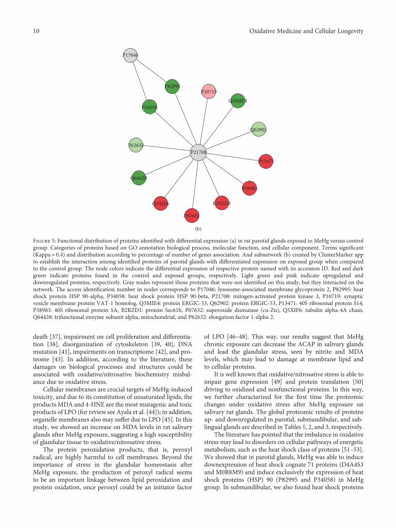
The ppi network of parotid gland (Figure 5(b)) showed acentral protein (mitogen-activated protein kinase 3; P21708),which interacts with proteins related to stress response (heatshock protein HSP 90-alpha, P82995; heat shock proteinHSP 90-beta, P34058), molecular function proteins (synapticvesicle membrane protein VAT-1 homolog, Q3MIE4;protein ERGIC-53, Q62902; 40S ribosomal protein S14,P13471; 40S ribosomal protein AS, P38983; Sec61 beta sub-unit, B2RZD1; elongation factor 1-alpha 2, P62632), oxida-tive stress enzymes (superoxide dismutase (Cu-Zn),P07632), microtubule component (tubulin alpha-4A chain,Q5XIF6), and energetic metabolism process (trifunctionalenzyme subunit alpha, mitochondrial, Q64428; ATP syn-thase subunit beta, mitochondrial, P10719).
As for the submandibular gland, our results showed that10 categories of proteins classified by Cytoscape, according toGO, were significantly altered upon chronic exposure toMeHg (Figure 6(a)). The myelin sheath proteins correspondto 31.43%; the structural constituent of cytoskeleton and thesubstance nigra development represent 11.43%, each. More-over, the nuclear nucleosome represents 8.57% and regula-tion of sodium ion transmembrane transporter activitysymbolizes 8.57% of the proteins with alterations. Adenylate
cyclase binding, mitochondrial proton-transporting ATPsynthase complex, catalytic core F (1), phosphoserine bind-ing, bHLH transcription factor binding, and eukaryotictranslation elongation factor 1 complex corresponded to3.13% each.
Similarly to what was found for the parotid gland, mostof the proteins with altered expression in the submandibu-lar gland interacted with mitogen-activated protein kinase3 (P21708), which was found in the center of the ppi net-work. The interacting proteins were those related to stressresponse (P14659), energetic metabolic process (ATP syn-thase subunit alpha, mitochondrial, P15999; ADP/ATPtranslocase 2, Q09073; ATP synthase subunit delta, mito-chondrial, P35434 and citrate synthase, mitochondrial,Q8VHF5), microtubule constituent (tubulin alpha-1B chain,Q6P9V9), molecular functions (elongation factor 1-alpha 2,P62632; uncharacterized protein, F1M446; 14-3-3 proteintheta, P68255; elongation factor 1-alpha 2, P62632), and cel-lular component (endoplasmic reticulum resident protein29, P52555; 78 kDa glucose-regulated protein, P06761)(Figure 6(b)).
Sublingual glands presented the lower number of proteingroups identified on Cytoscape, following ClueGO app GOclassification (Figure 7(a)). Each of the 6 groups of proteinsis represented by myelin sheath (33.33%), unfolded protein
Control MeHg0
50Nitr
ite co
nc. —
% o
f con
trol
100
150
200
250
⁎
(a)
Control MeHg0
50Nitr
ite co
nc. —
% o
f con
trol
100
150
200
250
⁎
(b)
Control0
50Nitr
ite co
nc. —
% o
f con
trol
100
150
200
250
MeHg
⁎
(c)
Figure 3: Levels of nitrite concentration in parotid (a), submandibular (b), and sublingual salivary glands (c) of animals exposed chronicallyto methylmercury. The values are expressed as percentage of control± SEM. ∗p < 0 05 (Student’s t-test).
5Oxidative Medicine and Cellular Longevity

binding (19.05%), retina homeostasis (14.29%), structuralconstituent of cytoskeleton (14.29%), detection of chemicalstimulus involved in sensory perception of bitter taste(9.52%), and alpha-amylase activity (9.52%).
The ppi network of the sublingual gland (Figure 7(b))showed the same protein in the center of the network (mito-gen-activated protein kinase 3; P21708), which the proteinswith altered expression interacted. The interaction proteinswere related mainly to biological processes, such as mitosisand cytoskeletal rearrangements. In addition, proteins asso-ciated to stress response (heat shock cognate 71 kDa protein,P63018 and heat shock-related 70 kDa protein 2, P14659)and microtubule constituent (tubulin alpha-1B chain,Q6P9V9) were clustered by ClusterMarker app.
4. Discussion
In this study, we showed for the first time that MeHg, in achronic period, causes damage to the salivary gland proteo-mic profile and oxidative stress. A significant total Hg depositin the gland tissue structure was verified, as well as a mem-brane cell damage by higher LPO levels, nitrogen species for-mation due to oxidative stress, and low antioxidant capacityagainst peroxyl radicals. In addition, the novelty of the toxi-cological evaluation on the proteomic profile of rat glands
after MeHg exposure brings the association with differencesin expression at important proteins of metabolic pathways,oxidative biochemistry, and mainly in the composition ofcytoarchitecture.
Most of the studies about MeHg toxicity are under acuteexposures and with high MeHg concentrations, even thoughthe reality faced by the population of endemic regions of thismetal intoxication is different. Recent studies showed thatlong-term intoxication includes other effects such as cardio-vascular diseases and genotoxicity [24–27]. This latter oneincludes oxidative stress and alterations of cytoskeleton pro-teins as main causes for these effects. The known effects ofMeHg are mainly given by chronic exposures, thus, themodel proposed by Kong et al. [17], in which we based ourstudy, mimics a daily and continuous exposure in low doses.The exposure time of our study brings the possibility to findinteresting results on an oxidative biochemistry and proteo-mic profiles at rat glands, showing that even at low doses, achronic MeHg exposure is able to induce molecular andcellular damages in living organisms.
For understanding, the nomination of low dose usedin this researcher is important to consider other ana-tomic regions and the toxic effects. The lowest observedadverse effect level (LOAEL) for neurotoxic effects suchas paresthesia is 50 ppm of mercury in hair [28]. This
MeHgControl
⁎
300
200
100
0
MD
A co
nc. —
% o
f con
trol
(a)
MeHgControl
⁎
300
200
100
0
MD
A co
nc. —
% o
f con
trol
(b)
MeHgControl
300
⁎
200
100
0
MD
A co
nc. —
% o
f con
trol
(c)
Figure 4: Levels of lipid peroxidation in parotid (a), submandibular (b), and sublingual (c) glands of animals exposed chronically tomethylmercury. The values are expressed as percentage of control± SEM. ∗p < 0 05 (Student’s t-test).
6 Oxidative Medicine and Cellular Longevity

level in hair corresponds approximately to 1 ppm ofmercury in brain, according to the proportion of250 : 50 : 1 for hair : brain :whole blood contents (reviewedby Branco et al. [29]).
The mercury content in brains of rats exposed to the doseused in our work (0.04mg/kg per day for 8 weeks) is about ahalf of that level, that is, about 0.5 ppm [17, 30], characteriz-ing a low exposure. Moreover, animals exposed to this lowlevel of methylmercury did not develop neurotoxic effects
such as alterations of forelimb grip strength, running wheelperformance, or hind limb cross [30], supporting that thismercury level in the brain is not sufficient to cause deleteriousneurobehavioral consequences.
Looking at Figure 1, an interesting fact is that salivaryglands accumulated about 10 times less mercury than0.5 ppm. However, our results show for the first time that thislow dose is sufficient to cause significant oxidative stress andalterations of protein regulation in salivary glands, revealing
Table 1: Identified proteins with expression significantly altered in rat parotid glands of group exposed to Mehg (Hg).
aAccess number Protein name description PLGS scoreFold change
Hg
P68136 Actin, alpha skeletal muscle 1873.1 −0.9P62738 Actin, aortic smooth muscle 1713.4 −0.9P63269 Actin, gamma-enteric smooth muscle 1713.4 −0.9F1LP05 ATP synthase subunit alpha 548.16 −0.8P10719 ATP synthase subunit beta, mitochondrial 353.85 −0.8P21704 Deoxyribonuclease-1 268.66 −0.6D3ZXS6 Elongation factor 1-alpha 327 2.4
M0R757 Elongation factor 1-alpha 343.66 2.5
P62630 Elongation factor 1-alpha 1 343.66 2.5
P62632 Elongation factor 1-alpha 2 327 2.5
D4A4S3 Heat shock cognate 71 kDa protein 151.3 −0.7M0R8M9 Heat shock cognate 71 kDa protein 739.22 −0.8P01946 Hemoglobin subunit alpha-1/2 1975.8 1.2
P02091 Hemoglobin subunit beta-1 1841.8 1.8
D3ZWE0 Histone H2A 764.11 2.0
P02262 Histone H2A type 1 764.11 1.9
P0C169 Histone H2A type 1-C 764.11 1.8
P0C170 Histone H2A type 1-E 764.11 2.0
Q64598 Histone H2A type 1-F 764.11 1.9
P0CC09 Histone H2A type 2-A 764.11 1.8
Q4FZT6 Histone H2A type 3 764.11 1.8
Q00728 Histone H2A type 4 764.11 1.7
A9UMV8 Histone H2A.J 764.11 1.9
P0C0S7 Histone H2A.Z 764.11 1.9
D3ZK97 Histone H3 258.66 1.2
P62804 Histone H4 2231.9 1.3
Q10758 Keratin, type II cytoskeletal 8 863.19 −0.8D3ZUQ1 Lipase 396.87 1.3
G3V812 Prolactin induced protein, isoform CRA_d 7686.8 −0.9O70417 Prolactin-inducible protein homolog 7084.7 −0.9F1LRA1 Protein ERGIC-53 400.1 1.2
F1M6C2 Protein LOC103691939 343.66 2.5
F1LZI1 Protein LOC680121 538.95 −0.7P02770 Serum albumin 489.45 1.3
B4F7C2 Tubulin beta-4 455.5 −0.7Q6P9T8 Tubulin beta-4B chain 455.5 −0.7M0RCB1 Uncharacterized protein 769.68 −0.8The identified proteins are organized according to the alphabetical order. Relative differential expression is indicated by positive value, when the protein isupregulated, and by negative values (−), when the protein is downregulated in the comparison between groups. aIdentification is based on protein ID fromUniProt protein database (http://www.uniprot.org/).
7Oxidative Medicine and Cellular Longevity

the high susceptibility of this tissue to the toxic effects ofmethylmercury.
The main form of MeHg intoxication is the oral throughfood [6], so the preferential site of absorption is in the intes-tinal lumen by enterocytes. The experimental model of intra-gastric gavage, used in this work, illustrates the MeHg intakeby food consumption. MeHg is formed by a methyl groupattached to the Hg atom (CH3Hg), and this allows a greateraffinity with thiol groups, mainly of protein components [31].
The MeHg gains systemic circulation from the effluxthrough the basolateral membrane of enterocytes and on thispart of membrane surface. There are multidrug resistance-associated proteins (MRPs), neutral amino acid transporters,as LAT2, and organic anions transporter (OAT) [32]; then, itis suggested that these proteins’ action is related to MeHgefflux. Moreover, some MRPs need GSH as cosubstrate for
their effectiveness, so MeHg may also undergo efflux to sys-temic blood circulation. According to Martinez-Madrigaland Micheau [33], blood vessels as external carotid, sublin-gual, submental, and facial supply all glands, suggesting thatvascularization is the main path by which Hg forms depositsby intragastric gavage intoxication.
In this work, we found significant Hg concentration(ppm) in the three main salivary glands in comparison tothe control group. Studies about the proteomic profile ofsalivary glands have suggested that parotid is responsible toproduce the major part of the protein components of saliva[34–36], and this could be the reason to a higher absorptionto this gland showed by our group, once this metal has anaffinity to structures present in many proteins.
Studies appoint that MeHg induces multiple effectswithin cell homeostasis. MeHg is able to induce cell
Table 2: Identified proteins with expression significantly altered in rats’ submandibular glands of group exposed to Mehg (Hg).
aAccess number Protein name description PLGS scoreFold change
Hg
Q08163 Adenylyl cyclase-associated protein 1 148.55 −0.2P15999 ATP synthase subunit alpha, mitochondrial 560.23 −0.6M0R757 Elongation factor 1-alpha 225.49 1.6
P62630 Elongation factor 1-alpha 1 225.49 1.6
P62632 Elongation factor 1-alpha 2 188.54 1.8
O88752 Epsilon 1 globin 2060.91 −0.3P11517 Hemoglobin subunit beta-2 2099.64 −0.3D3ZNH4 Histone H2B 3909.79 −0.7F1LQ08 Protein Car6 439.43 1.4
F1M6C2 Protein LOC103691939 193.93 1.7
E9PTY1 Protein Prol1 325.88 2.0
P08462 Submandibular gland secretory Glx-rich protein CB 944.25 1.3
D4A5P1 Uncharacterized protein 364.33 −0.6The identified proteins are organized according to the alphabetical order. Relative differential expression is indicated by positive value, when the protein isupregulated, and by negative values (−), when the protein is downregulated in the comparison between groups. aIdentification is based on protein ID fromUniProt protein database (http://www.uniprot.org/).
Table 3: Identified proteins with expression significantly altered in the rats’ sublingual glands of group exposed to MeHg (Hg).
aAccess number Protein name description PLGS scoreFold change
Hg
E9PSQ1 Alpha-amylase 2696.09 −0.3F1LPK5 Acidic mammalian chitinase 1384.72 −0.2G3V812 Prolactin induced protein, isoform CRA_d 6144.39 −0.2G3V844 Alpha-amylase 344.21 −0.3M0R7K9 Protein Csap1 1852.75 −0.3O70417 Prolactin-inducible protein homolog 5900.39 −0.2P00689 Pancreatic alpha-amylase 344.21 1.7
P02770 Serum albumin 600.4 −0.6P12020 Cysteine-rich secretory protein 1 221.34 2.5
The identified proteins are organized according to the alphabetical order. Relative differential expression is indicated by positive value, when the protein isupregulated, and by negative values (−), when the protein is downregulated in the comparison between groups. aIdentification is based on protein ID fromUniProt protein database (http://www.uniprot.org/).
8 Oxidative Medicine and Cellular Longevity

7.81%
Biological process - parotid gland MeHg versus control
Nucleosome
Hydrolyase activity
Aerobic respiration
Regulation of cellular respiration
Myelin sheath
Intermediate filament
20.31%
6.25%
7.81%
3.13%
4.69%
3.13%3.13%3.13%3.13%
3.13%
3.13%
3.13%
3.13%
3.13%
4.69%
3.13%
3.13%
6.65%
4.69%
Oxygen binding
Protein transmembrane transport
Melanosome
Nucleosomal DNA binding
Retina homeostasis
Benzaldehyde dehydrogenase (NAD+) activity
Pigment granule
mRNA 5’-UTR binding
Response to mercury ion
Cellular response to epidermal growth factor stimulus
Cellular response to gamma radiation
Mesenchyme migration
Eukaryotic translation elongation factor 1 complex
NAD binding
(a)
Figure 5: Continued.
9Oxidative Medicine and Cellular Longevity
death [37], impairment on cell proliferation and differentia-tion [38], disorganization of cytoskeleton [39, 40], DNAmutation [41], impairments on transcriptome [42], and pro-teome [43]. In addition, according to the literature, thesedamages on biological processes and structures could beassociated with oxidative/nitrosative biochemistry misbal-ance due to oxidative stress.
Cellular membranes are crucial targets of MeHg-inducedtoxicity, and due to its constitution of unsaturated lipids, theproducts MDA and 4-HNE are the most mutagenic and toxicproducts of LPO (for review see Ayala et al. [44]); in addition,organelle membranes also may suffer due to LPO [45]. In thisstudy, we showed an increase on MDA levels in rat salivaryglands after MeHg exposure, suggesting a high susceptibilityof glandular tissue to oxidative/nitrosative stress.
The protein peroxidation products, that is, peroxylradical, are highly harmful to cell membranes. Beyond theimportance of stress in the glandular homeostasis afterMeHg exposure, the production of peroxyl radical seemsto be an important linkage between lipid peroxidation andprotein oxidation, once peroxyl could be an initiator factor
of LPO [46–48]. This way, our results suggest that MeHgchronic exposure can decrease the ACAP in salivary glandsand lead the glandular stress, seen by nitrite and MDAlevels, which may lead to damage at membrane lipid andto cellular proteins.
It is well known that oxidative/nitrosative stress is able toimpair gene expression [49] and protein translation [50]driving to oxidized and nonfunctional proteins. In this way,we further characterized for the first time the proteomicchanges under oxidative stress after MeHg exposure onsalivary rat glands. The global proteomic results of proteinsup- and downregulated in parotid, submandibular, and sub-lingual glands are described in Tables 1, 2, and 3, respectively.
The literature has pointed that the imbalance in oxidativestress may lead to disorders on cellular pathways of energeticmetabolism, such as the heat shock class of proteins [51–53].We showed that in parotid glands, MeHg was able to inducedownexpression of heat shock cognate 71 proteins (D4A4S3and M0R8M9) and induce exclusively the expression of heatshock proteins (HSP) 90 (P82995 and P34058) in MeHggroup. In submandibular, we also found heat shock proteins
P17046
P34058
P82995P10719
Q3MIE4
Q62902
P13471
P38983
B2RZD1
P07632
Q5XIF6
P62632
Q64428
P21708
(b)
Figure 5: Functional distribution of proteins identified with differential expression (a) in rat parotid glands exposed to MeHg versus controlgroup. Categories of proteins based on GO annotation biological process, molecular function, and cellular component. Terms significant(Kappa = 0.4) and distribution according to percentage of number of genes association. And subnetwork (b) created by ClusterMarker appto establish the interaction among identified proteins of parotid glands with differentiated expression on exposed group when comparedto the control group. The node colors indicate the differential expression of respective protein named with its accession ID. Red and darkgreen indicate proteins found in the control and exposed groups, respectively. Light green and pink indicate upregulated anddownregulated proteins, respectively. Gray nodes represent those proteins that were not identified on this study, but they interacted on thenetwork. The access identification number in nodes corresponds to P17046: lysosome-associated membrane glycoprotein 2, P82995: heatshock protein HSP 90-alpha, P34058: heat shock protein HSP 90-beta, P21708: mitogen-activated protein kinase 3, P10719: synapticvesicle membrane protein VAT-1 homolog, Q3MIE4: protein ERGIC-53, Q62902: protein ERGIC-53, P13471: 40S ribosomal protein S14,P38983: 40S ribosomal protein SA, B2RZD1: protein Sec61b, P07632: superoxide dismutase (cu-Zn), Q5XIF6: tubulin alpha-4A chain,Q64428: trifunctional enzyme subunit alpha, mitochondrial, and P62632: elongation factor 1-alpha 2.
10 Oxidative Medicine and Cellular Longevity

Biological process - submandibular gland MeHg versus control
Adenylate cyclase binding
Nuclear nucleosome
Myelin sheath
Mitochondrial proton-transporting ATPsynthase complex, catalytic core (f1)
31.43%
8.57%
5.71%11.43%
8.57%
5.71%
5.71%
5.71%
5.71%
11.43%
Phosphoserine binding
bHLH transcription factor binding
Eukaryotic translation elongationfactor 1 complex
Substantia nigra development
Regulation of sodium iontransmembrane transporter activity
Structural constituent of cytoskeleton
(a)
P14659P15999
Q09073
Q6P9V9
P62630
P35434F1M446
P68255
Q8VHF5
P62632
P21708
P52555
P06761
(b)
Figure 6: Functional distribution of proteins (a) identified with differential expression in rat submandibular glands exposed to MeHg versuscontrol group. Categories of proteins based on GO annotation biological process, molecular function, and cellular component. Termssignificant (Kappa = 0.4) and distribution according to percentage of number of gene association. Subnetwork (b) created byClusterMarker app to establish the interaction among identified proteins of submandibular glands with differentiated expression onexposed group when compared to control group. The node colors indicate the differential expression of respective protein named with itsaccession ID. Red and dark green indicate proteins found in control and exposed groups, respectively. Light green and pink indicateupregulated and downregulated proteins, respectively. Gray nodes represent those proteins that were not identified on this study, but theyinteracted on the network. The access identification number in nodes corresponds to P52555: endoplasmic reticulum resident protein 29,P06761: 78 kDa glucose-regulated protein, P14659: heat shock-related 70 kDa protein 2, P15999: ATP synthase subunit alpha,mitochondrial, Q09073: ADP/ATP translocase 2, Q6P9V9: tubulin alpha-1B chain, P62632: elongation factor 1-alpha 2, P35434: ATPsynthase subunit delta, mitochondrial, F1M446 protein RGD1306148, P68255: 14-3-3 protein theta, Q8VHF5: citrate synthase,mitochondrial, and P62632: elongation factor 1-alpha 2.
11Oxidative Medicine and Cellular Longevity

33.33%
19.05%
9.52%
14.29%
14.29%9.52%
Biological functions - sublingual gland MeHg versus control
Detection of chemical stimulus involvedin sensory perception of bitter taste
Unfolded protein binding
Myelin sheath
Alpha-amylase activity
Structural constituent of cytoskeleton
Retina homeostasis
(a)
P06761
P21708
Q6P9V9
P14659
P63018
P80067
P52555
(b)
Figure 7: Functional distribution of proteins (a) identified with differential expression in rat submandibular glands exposed to MeHg versuscontrol group. Categories of proteins based on GO annotation biological process, molecular function, and cellular component. Termssignificant (Kappa = 0.4) and distribution according to percentage of number of gene association. Subnetwork (b) created byClusterMarker app to establish the interaction among identified proteins of sublingual glands with differentiated expression on exposedgroup when compared to control group. The node colors indicate the differential expression of respective protein named with its accessionID. Red and dark green indicate proteins found in control and exposed groups, respectively. Light green and pink indicate upregulatedand downregulated proteins, respectively. Gray nodes represent those proteins which were not identified on this study, but they interactedon the network. The access identification number in nodes corresponds to P80067: dipeptidyl peptidase 1, P63018: heat shock cognate71 kDa protein, P21708: mitogen-activated protein kinase 3, Q6P9V9: tubulin alpha-1B chain, P14659: heat shock-related 70 kDa protein2, P06761: 78 kDa glucose-regulated protein, P52555: endoplasmic reticulum resident protein 29.
12 Oxidative Medicine and Cellular Longevity

(M0R660 and P14659) that were unique in MeHg group,while in the sublingual gland, we found the exclusive expres-sion of heat shock cognate 71 (P63018) and heat shock-related 70 (P14659) and expression of heat shock proteinbeta-6 (P97541) only in the control group. HSPs have beenalready proposed as new therapeutic tools for some disor-ders as cancer and cardiovascular diseases, with a protectiverole especially for HSP90 [54]. The results of the presentstudy support a main role for this protein also in methyl-mercury intoxication.
In addition, glutathione S-transferase P (P04906) andcomponents of cytochrome (Q62737 and Q68FY0) wereexclusively expressed in parotid control group, suggestingan important association on protein changes and MeHg-induced oxidative stress.
The metabolic pathway class of proteins was also chan-ged after MeHg exposure. In parotid, the proteins ATPsynthase subunit alpha (F1LP05) and ATP synthase subunitbeta, mitocondrial (P10719), were downregulated andmalate dehydrogenase, cytoplasmic (O88989), and aconitatehydratase (G3V6S2) were unique in the control group. Insubmandibular, ATP synthase subunit alpha, mitochondrial(P15999), was downregulated, while ADP/ATP translocase 2(Q09073), ATP synthase subunit delta, mitochondrial(P35434), citrate synthase (G3V936), citrate synthase, andmitochondrial (Q8VHF5) were present only in the controlglands. Our results corroborate with metabolic pathway dis-orders established in others experimental models [17, 55].
MeHg treatment also impaired negatively the proteins ofcellular components, such as cytoskeleton, from the mainconstituents of salivary glands. We have showed that the pro-teins related to muscle cell contraction (actin, alpha skeletalmuscle, P68136; actin, aortic smooth muscle, P62738; andactin, gamma-enteric smooth muscle, P63269), in parotidglands were downregulated after MeHg exposure, whichmay suggest possible damages in myoepithelial cells presentin the glands. We also showed downregulation of keratin,type II cytoskeletal 8 (Q10758) that is an important compo-nent of conjunctive tissue.
Concerning to microtubule constitution, we showedhere that tubulin beta chain (B4F7C2) and tubulin beta-4Bchain (Q6P9T8) were downregulated, corroborating withthe literature, which suggests that microtubule depolymeri-zation is one of the mechanisms of Hg damage on livingcells [25, 56, 57].
A collagen chain protein (collagen alpha-1(I) chain,P02454), cytokeratins (keratin, type I cytoskeletal 10,Q6IFW6; keratin, type I cytoskeletal 14, Q6IFV1; keratin,type I cytoskeletal 15, Q6IFV3; keratin, type I cytoskeletal17, Q6IFU8; keratin, type I cytoskeletal 24, Q6IFX1; keratin,type I cytoskeletal 42, Q6IFU7; and keratin, type II cytoskel-etal 7, Q6IG12), and tubulin chains (tubulin beta chain,Q4QQV0 and tubulin alpha-4A chain, Q5XIF6) were foundexclusively in control parotid glands. In sublingual glands,we also found conjunctive constituent (collagen alpha-2(I)chain, P02466), cytoskeleton (Q5BJY9) and microtubule(tubulin alpha-1A chain, P68370; tubulin alpha-1B chain,Q6P9V9; and tubulin alpha-1C chain, Q6AYZ1) compo-nents uniquely in the nonexposed group.
As previously mentioned, salivary glands are responsiblefor salivary formation and flow; this way, the acinar andtubular constituents have great importance in physiology.In the proteomic analysis, downregulation of alpha-amylase(E9PSQ1) was observed, as well as prolactin-induced protein,isoform CRA_d (G3V812), and prolactin-inducible proteinhomolog (O70417) in the parotid. An interesting proteinrelated to biomineralization (submandibular gland secretoryGlx-rich protein CB, P08462) was upregulated in the sub-mandibular of the exposed group. Salivary flow is firstderived by ionic exchanges by sodium/potassium bombs inacini and intercalated portions of secretory duct system[58], and in this study, we noticed that sodium/potassium-transporting ATPase subunit alpha-1 (P06685) was exclusiveof control group in sublingual gland, similarly to sodiumchannel subunit beta-3 (Q9JK00) and sodium/potassium-transporting ATPase subunit alpha-2 (P06686) that wereunique of submandibular gland of animals not submitted toMeHg exposure.
The proteomic profile changes shown by this worksuggest that MeHg exposure leads to salivary gland damage,since several proteins were shown to be affected,compromising essential biological functions and compo-nents that under normal circumstances are fundamental docell and tissue viability.
5. Conclusion
The proteomic profile verified after MeHg exposure in thisstudy allowed us to see what is underlying the oxidative stresseffects in salivary glands. Our findings have showed thatsalivary glands are also targets forMeHg in systemic intoxica-tion, at low doses and chronic exposure, being able of trigger-ing imbalance in oxidative biochemistry and, consequently,the modulation of proteomic profile of the glands.
Conflicts of Interest
The authors declare that there is no conflict of interestregarding the publication of this paper.
Acknowledgments
This work was supported by the Brazilian NationalCouncil for Scientific and Technological Development(CNPq), Fundação de Amparo a Pesquisa do Estado do Pará(FAPESPA), and Pró-Reitoria de Pesquisa e Pós-Graduaçãoda UFPA (PROPESP, UFPA, Brazil). Rafael R. Lima is aninvestigator from CNPq (Edital MCTI/CNPQ/Universal14/2014). Leonardo O. Bittencourt’s scholarship is sup-ported by the Brazilian National Council for Scientific andTechnological Development (CNPq).
References
[1] J. G. Dorea and R. C. Marques, “Mercury levels and humanhealth in the Amazon Basin,” Annals of Human Biology,vol. 43, no. 4, pp. 349–359, 2016.
13Oxidative Medicine and Cellular Longevity

[2] R. Ynalvez, J. Gutierrez, and H. Gonzalez-Cantu, “Mini-review: toxicity of mercury as a consequence of enzyme alter-ation,” Biometals, vol. 29, no. 5, pp. 781–788, 2016.
[3] G. C. Compeau and R. Bartha, “Sulfate-reducing bacteria:principal methylators of mercury in anoxic estuarine sedi-ment,” Applied and Environmental Microbiology, vol. 50,no. 2, pp. 498–502, 1985.
[4] M. C. Bisinoti and W. F. Jardim, “O comportamento do metil-mercúrio (metilHg) no ambiente,” Química Nova, vol. 27,pp. 593–600, 2004.
[5] J. J. Berzas Nevado, R. C. Rodríguez Martín-Doimeadios, F. J.Guzmán Bernardo et al., “Mercury in the Tapajos River basin,Brazilian Amazon: a review,” Environment International,vol. 36, no. 6, pp. 593–608, 2010.
[6] Y. S. Hong, Y. M. Kim, and K. E. Lee, “Methylmercury expo-sure and health effects,” Journal of Preventive Medicine andPublic Health, vol. 45, no. 6, pp. 353–363, 2012.
[7] R. C. Rodríguez Martín-Doimeadios, J. J. Berzas Nevado, F. J.Guzmán Bernardo et al., “Comparative study of mercuryspeciation in commercial fishes of the Brazilian Amazon,”Environmental Science and Pollution Research, vol. 21,no. 12, pp. 7466–7479, 2014.
[8] S. Squadrone, E. Chiaravalle, S. Gavinelli, G. Monaco, M. Rizzi,and M. C. Abete, “Analysis of mercury and methylmercuryconcentrations, and selenium: mercury molar ratios for a tox-icological assessment of sperm whales (Physeter macrocepha-lus) in the most recent stranding event along the Adriaticcoast (Southern Italy, Mediterranean Sea),” Chemosphere,vol. 138, pp. 633–641, 2015.
[9] C. Feng, Z. Pedrero, S. Gentès et al., “Specific pathways ofdietary methylmercury and inorganic mercury determinedby mercury speciation and isotopic composition in zebrafish(Danio rerio),” Environmental Science & Technology, vol. 49,no. 21, pp. 12984–12993, 2015.
[10] M. S. Garcia, D. H. Constantino, A. P. Silva, and J. E. Perobelli,“Fish pollutants MeHg and Aroclor cause permanent struc-tural damage in male gonads and kidneys after prepubertalexposure,” International Journal of Experimental Pathology,vol. 97, no. 5, pp. 360–368, 2016.
[11] J. Ruelas-Inzunza, C. K. Kohen-Sandoval, M. A. Ramos-Osunaet al., “Total mercury concentrations in white and stripedmullet (Mugil curema and M. cephalus) from a coastal lagoonin the SE Gulf of California,” Journal of Environmental Scienceand Health. Part A, Toxic/Hazardous Substances & Environ-mental Engineering, vol. 52, no. 5, pp. 459–465, 2017.
[12] P. Bratt, I. Johansson, J. Linder, and T. Ericson, “Function ofthe rat salivary glands after exposure to inorganic mercury,”The Science of the Total Environment, vol. 172, no. 1, pp. 47–55, 1995.
[13] G. Warfvinge, K. Warfvinge, and A. Larsson, “Histochemicalvisualization of mercury in the oral mucosa, salivary and lacri-mal glands of BN rats with HgCl2-induced autoimmunity,”Experimental and Toxicologic Pathology, vol. 46, no. 4-5,pp. 329–334, 1994.
[14] K. Schmid, A. Sassen, R. Staudenmaier et al., “Mercuricdichloride induces DNA damage in human salivary gland tis-sue cells and lymphocytes,” Archives of Toxicology, vol. 81,no. 11, pp. 759–767, 2007.
[15] C. Peyrot des Gachons and P. A. Breslin, “Salivary amylase:digestion and metabolic syndrome,” Current Diabetes Reports,vol. 16, no. 10, p. 102, 2016.
[16] F. de Paula, T. H. N. Teshima, R. Hsieh, M. M. Souza,M. M. S. Nico, and S. V. Lourenco, “Overview of humansalivary glands: highlights of morphology and developing pro-cesses,” The Anatomical Record, vol. 300, no. 7, pp. 1180–1188,2017.
[17] H. K. Kong, M. H. Wong, H. M. Chan, and S. C. Lo, “Chronicexposure of adult rats to low doses of methylmercury induceda state of metabolic deficit in the somatosensory cortex,” Jour-nal of Proteome Research, vol. 12, no. 11, pp. 5233–5245, 2013.
[18] T. Suzuki, H. Akagi, K. Arimura et al., Mercury AnalysisManual, Ministry of the Environment, Japan, 2004.
[19] L. L. Amado, M. L. Garcia, P. B. Ramos et al., “A method tomeasure total antioxidant capacity against peroxyl radicals inaquatic organisms: application to evaluate microcystins toxic-ity,” The Science of the Total Environment, vol. 407, no. 6,pp. 2115–2123, 2009.
[20] M. Ferreira-Cravo, F. R. Piedras, T. B. Moraes et al., “Antioxi-dant responses and reactive oxygen species generation in dif-ferent body regions of the estuarine polychaeta LaeonereisAcuta (Nereididae),” Chemosphere, vol. 66, no. 7, pp. 1367–1374, 2007.
[21] N. C. Fagundes, L. M. Fernandes, R. S. Paraense et al., “Bingedrinking of ethanol during adolescence induces oxidativedamage and morphological changes in salivary glands offemale rats,” Oxidative Medicine and Cellular Longevity,vol. 2016, Article ID 7323627, 11 pages, 2016.
[22] H. Esterbauer and K. H. Cheeseman, “Determination ofaldehydic lipid peroxidation products: malonaldehyde and4-hydroxynonenal,” Methods in Enzymology, vol. 186,pp. 407–421, 1990.
[23] M. M. Bradford, “A rapid and sensitive method for the quan-titation of microgram quantities of protein utilizing the princi-ple of protein-dye binding,” Analytical Biochemistry, vol. 72,pp. 248–254, 1976.
[24] G. Genchi, M. S. Sinicropi, A. Carocci, G. Lauria, and A.Catalano, “Mercury exposure and heart diseases,” Interna-tional Journal of Environmental Research and Public Health,vol. 14, no. 1, 2017.
[25] M. E. Crespo-Lopez, G. L. Macêdo, S. I. Pereira et al., “Mercuryand human genotoxicity: critical considerations and possiblemolecular mechanisms,” Pharmacological Research, vol. 60,no. 4, pp. 212–220, 2009.
[26] M. E. Crespo-Lopez, G. L. Macêdo, G. P. Arrifano, C. PinheiroMda, J. L. do Nascimento, and A. M. Herculano, “Genotoxicityof mercury: contributing for the analysis of Amazonianpopulations,” Environment International, vol. 37, no. 1,pp. 136–141, 2011.
[27] M. E. Crespo-Lopez, A. Costa-Malaquias, E. H. Oliveira et al.,“Is low non-lethal concentration of methylmercury really safe?A report on genotoxicity with delayed cell proliferation,” PloSOne, vol. 11, no. 9, article e0162822, 2016.
[28] T. W. Clarkson and L. Magos, “The toxicology of mercury andits chemical compounds,” Critical Reviews in Toxicology,vol. 36, no. 8, pp. 609–662, 2006.
[29] V. Branco, S. Caito, M. Farina, J. Teixeira da Rocha, M.Aschner, and C. Carvalho, “Biomarkers of mercury toxicity:past, present, and future trends,” Journal of Toxicology andEnvironmental Health. Part B, Critical Reviews, vol. 20,no. 3, pp. 119–154, 2017.
[30] J. J. Day, M. N. Reed, and M. C. Newland, “Neuromotor defi-cits and mercury concentrations in rats exposed to methyl
14 Oxidative Medicine and Cellular Longevity

mercury and fish oil,” Neurotoxicology and Teratology, vol. 27,no. 4, pp. 629–641, 2005.
[31] C. C. Bridges and R. K. Zalups, “Mechanisms involved in thetransport of mercuric ions in target tissues,” Archives ofToxicology, vol. 91, no. 1, pp. 63–81, 2017.
[32] M. A. Bradley, B. D. Barst, and N. Basu, “A review of mercurybioavailability in humans and fish,” International Journal ofEnvironmental Research and Public Health, vol. 14, no. 2, 2017.
[33] F. Martinez-Madrigal and C. Micheau, “Histology of the majorsalivary glands,” The American Journal of Surgical Pathology,vol. 13, no. 10, pp. 879–899, 1989.
[34] A. Walz, K. Stühler, A. Wattenberg et al., “Proteome analysisof glandular parotid and submandibular-sublingual salivain comparison to whole human saliva by two-dimensionalgel electrophoresis,” Proteomics, vol. 6, no. 5, pp. 1631–1639, 2006.
[35] P. Denny, F. K. Hagen, M. Hardt et al., “The proteomes ofhuman parotid and submandibular/sublingual gland salivascollected as the ductal secretions,” Journal of ProteomeResearch, vol. 7, no. 5, pp. 1994–2006, 2008.
[36] N. Rosa, M. J. Correia, J. P. Arrais et al., “From the salivaryproteome to the OralOme: comprehensive molecular oral biol-ogy,”Archives of Oral Biology, vol. 57, no. 7, pp. 853–864, 2012.
[37] P. Morcillo, D. Romero, J. Meseguer, M. Á. Esteban, and A.Cuesta, “Cytotoxicity and alterations at transcriptional levelcaused by metals on fish erythrocytes in vitro,” EnvironmentalScience and Pollution Research International, vol. 23, no. 12,pp. 12312–12322, 2016.
[38] F. F. Ferreira, D. Ammar, G. F. Bourckhardt, K. Kobus-Bianchini, Y. M. Müller, and E. M. Nazari, “MeHg developingexposure causes DNA double-strand breaks and elicits cellcycle arrest in spinal cord cells,” Journal of Toxicology,vol. 2015, Article ID 532691, 10 pages, 2015.
[39] P. R. Sager, “Selectivity of methyl mercury effects on cytoskel-eton and mitotic progression in cultured cells,” Toxicology andApplied Pharmacology, vol. 94, no. 3, pp. 473–486, 1988.
[40] R. Thier, D. Bonacker, T. Stoiber et al., “Interaction of metalsalts with cytoskeletal motor protein systems,” ToxicologyLetters, vol. 140-141, pp. 75–81, 2003.
[41] X. Wang, M. Yan, L. Zhao et al., “Low-dose methylmercury-induced apoptosis and mitochondrial DNA mutation inhuman embryonic neural progenitor cells,” Oxidative Med-icine and Cellular Longevity, vol. 2016, Article ID 5137042,10 pages, 2016.
[42] T. Waldmann, M. Grinberg, A. König et al., “Stem cell tran-scriptome responses and corresponding biomarkers that indi-cate the transition from adaptive responses to cytotoxicity,”Chemical Research in Toxicology, vol. 30, no. 4, pp. 905–922,2017.
[43] V. C. de Oliveira Souza, K. C. de Marco, H. J. Laure, J. C. Rosa,and F. Barbosa Jr., “A brain proteome profile in rats exposed tomethylmercury or thimerosal (ethylmercury),” Journal ofToxicology and Environmental Health. Part A, vol. 79, no. 12,pp. 502–512, 2016.
[44] A. Ayala, M. F. Munoz, and S. Arguelles, “Lipid peroxidation:production, metabolism, and signaling mechanisms of malon-dialdehyde and 4-hydroxy-2-nonenal,” Oxidative Medicineand Cellular Longevity, vol. 2014, Article ID 360438, 31 pages,2014.
[45] E. Dare, W. Li, B. Zhivotovsky, X. Yuan, and S. Ceccatelli,“Methylmercury and H2O2 provoke lysosomal damage in
human astrocytoma D384 cells followed by apoptosis,” FreeRadical Biology and Medicine, vol. 30, no. 12, pp. 1347–1356,2001.
[46] S. Duggan, C. Rait, A. Platt, and S. Gieseg, “Protein and thioloxidation in cells exposed to peroxyl radicals is inhibited bythe macrophage synthesised pterin 7,8-dihydroneopterin,”Biochimica et Biophysica Acta (BBA) - Molecular Cell Research,vol. 1591, no. 1–3, pp. 139–145, 2002.
[47] S. P. Gieseg, J. Pearson, and C. A. Firth, “Protein hydroperox-ides are a major product of low density lipoprotein oxidationduring copper, peroxyl radical and macrophage-mediated oxi-dation,” Free Radical Research, vol. 37, no. 9, pp. 983–991,2003.
[48] S. Gieseg, S. Duggan, and J. M. Gebicki, “Peroxidation of pro-teins before lipids in U937 cells exposed to peroxyl radicals,”The Biochemical Journal, vol. 350, Part 1, pp. 215–218, 2000.
[49] E. de Nadal, G. Ammerer, and F. Posas, “Controlling geneexpression in response to stress,” Nature Reviews Genetics,vol. 12, no. 12, pp. 833–845, 2011.
[50] C. Vogel, G. M. Silva, and E. M. Marcotte, “Protein expressionregulation under oxidative stress,”Molecular & Cellular Prote-omics, vol. 10, no. 12, 2011.
[51] L. Tretter and V. Adam-Vizi, “Inhibition of Krebs cycleenzymes by hydrogen peroxide: a key role of [alpha]-ketoglu-tarate dehydrogenase in limiting NADH production underoxidative stress,” The Journal of Neuroscience, vol. 20, no. 24,pp. 8972–8979, 2000.
[52] M. Frisard and E. Ravussin, “Energy metabolism and oxidativestress: impact on the metabolic syndrome and the agingprocess,” Endocrine, vol. 29, no. 1, pp. 27–32, 2006.
[53] S. Satapati, B. Kucejova, J. A. Duarte et al., “Mitochondrialmetabolism mediates oxidative stress and inflammation infatty liver,” The Journal of Clinical Investigation, vol. 126,no. 4, p. 1605, 2016.
[54] M. B. Almeida, J. L. do Nascimento, A. M. Herculano, andM. E. Crespo-López, “Molecular chaperones: toward newtherapeutic tools,” Biomedicine & Pharmacotherapy, vol. 65,no. 4, pp. 239–243, 2011.
[55] Y. Shao, M. Yamamoto, D. Figeys, Z. Ning, and H. M. Chan,“Proteomic analysis of cerebellum in common marmosetexposed to methylmercury,” Toxicological Sciences, vol. 146,no. 1, pp. 43–51, 2015.
[56] D. G. Vogel, R. L. Margolis, and N. K. Mottet, “Analysis ofmethyl mercury binding sites on tubulin subunits andmicrotubules,” Pharmacology & Toxicology, vol. 64, no. 2,pp. 196–201, 1989.
[57] K. Miura, M. Inokawa, and N. Imura, “Effects of methylmer-cury and some metal ions on microtubule networks in mouseglioma cells and in vitro tubulin polymerization,” Toxicologyand Applied Pharmacology, vol. 73, no. 2, pp. 218–231, 1984.
[58] J. E. Melvin, D. Yule, T. Shuttleworth, and T. Begenisich,“Regulation of fluid and electrolyte secretion in salivarygland acinar cells,” Annual Review of Physiology, vol. 67,pp. 445–469, 2005.
15Oxidative Medicine and Cellular Longevity

Submit your manuscripts athttps://www.hindawi.com
Stem CellsInternational
Hindawi Publishing Corporationhttp://www.hindawi.com Volume 2014
Hindawi Publishing Corporationhttp://www.hindawi.com Volume 2014
MEDIATORSINFLAMMATION
of
Hindawi Publishing Corporationhttp://www.hindawi.com Volume 2014
Behavioural Neurology
EndocrinologyInternational Journal of
Hindawi Publishing Corporationhttp://www.hindawi.com Volume 2014
Hindawi Publishing Corporationhttp://www.hindawi.com Volume 2014
Disease Markers
Hindawi Publishing Corporationhttp://www.hindawi.com Volume 2014
BioMed Research International
OncologyJournal of
Hindawi Publishing Corporationhttp://www.hindawi.com Volume 2014
Hindawi Publishing Corporationhttp://www.hindawi.com Volume 2014
Oxidative Medicine and Cellular Longevity
Hindawi Publishing Corporationhttp://www.hindawi.com Volume 2014
PPAR Research
The Scientific World JournalHindawi Publishing Corporation http://www.hindawi.com Volume 2014
Immunology ResearchHindawi Publishing Corporationhttp://www.hindawi.com Volume 2014
Journal of
ObesityJournal of
Hindawi Publishing Corporationhttp://www.hindawi.com Volume 2014
Hindawi Publishing Corporationhttp://www.hindawi.com Volume 2014
Computational and Mathematical Methods in Medicine
OphthalmologyJournal of
Hindawi Publishing Corporationhttp://www.hindawi.com Volume 2014
Diabetes ResearchJournal of
Hindawi Publishing Corporationhttp://www.hindawi.com Volume 2014
Hindawi Publishing Corporationhttp://www.hindawi.com Volume 2014
Research and TreatmentAIDS
Hindawi Publishing Corporationhttp://www.hindawi.com Volume 2014
Gastroenterology Research and Practice
Hindawi Publishing Corporationhttp://www.hindawi.com Volume 2014
Parkinson’s Disease
Evidence-Based Complementary and Alternative Medicine
Volume 2014Hindawi Publishing Corporationhttp://www.hindawi.com