Embed Size (px)
Citation preview

Clinical StudyInfluence of Dental Restorations on Oxidative Stress in GingivalCrevicular Fluid
Ervin Taso,1 Vladimir Stefanovic,1 Ivana Stevanovic,2 Danilo Vojvodic,2
Aleksandra Topic ,3 Aleksandra Petkovic-Curcin,2 Kosovka Obradovic-Djuricic,4
Aleksa Markovic,5 Mirjana Djukic ,6 and Dragana Vujanovic6
1Clinic for Stomatology, Military Medical Academy, Belgrade, Serbia2Institute for Medical Research, Military Medical Academy, Belgrade, Serbia3Department for Biochemistry, Faculty of Pharmacy, University of Belgrade, Belgrade, Serbia4Department for Prosthetics, Faculty of Dental Medicine, University of Belgrade, Belgrade, Serbia5Department for Oral Surgery, Faculty of Dental Medicine, University of Belgrade, Belgrade, Serbia6Department for Toxicology, Faculty of Pharmacy, University of Belgrade, Belgrade, Serbia
Correspondence should be addressed to Mirjana Djukic; [email protected]
Received 1 February 2018; Accepted 25 March 2018; Published 24 July 2018
Academic Editor: Tomris Ozben
Copyright © 2018 Ervin Taso et al. This is an open access article distributed under the Creative Commons Attribution License,which permits unrestricted use, distribution, and reproduction in any medium, provided the original work is properly cited.
Biocompatibility of dental materials (DM) can be evaluated by gingival crevicular fluid (GCF) oxidative stress (OS) status. The goalof the study was to ascertain influence of dental caries degree, teeth position, and type and amount of applied DM on GCF OSprofile. For this purpose, we tested six DMs that were sealed in one session: amalgam (Amg), composites: Tetric EvoCeram andBeautifil (BF), phosphate cement—zinc phosphate and polycarboxylate cements—zinc polycarboxylate cements, and glassionomer cement (GIC). The study included 88 dental outpatients. Follow-up was scheduled at 7th and 30th day. Oxidativestress parameters (malondialdehyde (MDA) and glutathione (GSH) levels and total superoxide dismutase (tSOD) activity) weremeasured before (0th day) and after the treatment (7th and 30th day) in GCF. Control teeth were mirror-positioned healthyteeth. The DM accomplished the following effects (listed in descending order): increase of GSH in GCF was realized byZPoC>BF>GIC>Amg; tSOD activity increase by ZPoC>BF>Amg; and MDA decrease by ZPoC>ZPhC>Amg>TEC.Dental caries provokes insignificant rise of OS in GCF. ZPoC and ZPhC showed the highest antioxidant effect, contrary to GIC.Restorations with antioxidant properties may reduce gum diseases initiated by caries lesion, what is of great clinical relevancein dentistry.
1. Introduction
Convincing evidence concerning oxidative stress- (OS-)associated dental pathologies (parodontopathy, oral cavitycancer, etc.) has been reported during recent decades [1–3].Up-to-date studies on redox status in oral environment havereferred mainly to peroxidase activity in saliva [2, 4–7].Reports in 2017 turned researchers’ attention to gingivalcrevicular fluid (GCF) as a diagnostic tool for oral diseasesanalysis and treatment outcome. The impact of oral envi-ronmental stressors (hygienic food and eating habits, smok-ing, etc.) on saliva is much more intense than on GCF,
though it was reported that smoking instantly increasesGCF flow [8, 9].
Leading by the fact that GCF is a very specific oral cavityfluid (a transudate of blood plasma placed in the gingivalsulcus), less exposed to oral environmental stressors com-pared to saliva, which requires noninvasive sampling, wechose GCF as an appropriate oral matrix for this kind oftesting [8]. Herein, we tested the influence of dental caries(a bacterial disease of the dental hard tissues, also definedas a final stage of local teeth immune response to oral path-ogen invasion) as well as six dental fillings on GCF redoxhomeostasis [10].
HindawiOxidative Medicine and Cellular LongevityVolume 2018, Article ID 1823189, 17 pageshttps://doi.org/10.1155/2018/1823189

Recently, it was documented that cell redox activity, thatis, antioxidant defense against environmental stressors(including smoking, i.e., nicotine) has important implica-tions on periodontal disease and is important [9]. It is wellacknowledged that OS (or other type of stress) is an inabilityof antioxidative defense system in living organisms to copewith free radicals (FRs) overproduction that results inoxidative injury of all classes of biomolecules, includingproteins, lipids, phospholipids, and deoxyribonucleic acid.Different classes of FRs (reactive oxygen, nitrogen, sulfur,or carbon species (ROS, RNS, RSC, or RCC)) can initiatecorresponding type of stress, oxidative, nitrosative, thiyl, orcarbonyl stress (OS, NS, TS, and CS), respectively [11]. Alongwith changed cell signalization and energy breakdown, theoverall occurrences finally end up with a cell death, byapoptosis [12].
Overproduction of ROS (including superoxide anion(O2
•−), hydrogen peroxide (H2O2), hydroxyl radical (HO•),and hypochlorous acid (HOCl)) occurs in dental lesions(caries) during phagocytosis. Reactive species injure sub-cellular and/or cellular membranes of phagolysosomesand/or neutrophils during respiratory burst. Over time,oxidation products of polyunsaturated fatty acids (cell mem-brane ingredients) become converted into carbonyls, suchas malondialdehyde (MDA), a reliable marker of lipidperoxidation (LPO) [13]. Together with myeloperoxidaseand NADH-oxidase, they leak out of phagolysosomes intophagocyte cytosol and further at a site of infection orinflammation and damage phagocytes and injure tissue.Reports on exogenously present myeloperoxidase assumethat it enhances bacterial phagocytosis and intracellular kill-ing by macrophages.
Accordingly, total superoxide dismutase (SOD) (coverscytosolic and extracellular form (Cu/Zn-SOD) and mito-chondrial (Mn-SOD), as well) converts O2
•− into H2O2,which further becomes converted into H2O, by catalase. Thesebiochemical reactions can attenuate myeloperoxidase-inducedbactericidal activity within or out of phagocytes and reducemyeloperoxidase-associated lipid peroxidation (LPO) [11,14]. The role of SOD in dental pathologies has not beeninvestigated until now.
In support of the possible redox interactions of the testeddental restoratives is the fact that some xenobiotics undergoredox metabolism and contribute to O2
•− production [15].Hitherto, testing of dental materials’ pro or antioxidant activ-ity has not been implemented in biocompatibility type ofanalysis in vitro and in vivo.
By measuring GSH, MDA, and tSOD in GCF, we studiedits redox response to dental caries and six dental restorations,considering the dental lesion degree, teeth position, andplaced amount into teeth.
2. Material and Methods
The study was carried out on dental outpatient from theClinic for Stomatology at the Military Medical Academy,Belgrade, Serbia, for 30 days, in accordance with the Inter-national Ethical Guidelines and Declaration of Helsinki(1964/1975) and was approved by the Ethical Committee
of the Military Medical Academy, Ministry of Defense, Serbia(Preference number VMA/10-12/A.1). The participants wereintroduced with the essence of the study and planned proce-dures, filled out a questionnaire dental record form related togeneral and oral health, and gave written consent to partici-pate in this study.
2.1. Patients. The 88 dental outpatients, aged 18–70, wererecruited by the tabular specified criteria (Table 1).
Table 1: Patients’ recruited criteria.
The inclusioncriteria
Criteria related to teeth condition
(i) The proximal caries on anterior andposterior teeth
(ii) The existence of the same type ofantagonistic teeth (“mirror”-positionedhealthy teeth) used controls
(iii) An absence of fresh postextractional ortraumatic wounds in the restoration areaor the area of restored surfaces
(iv) An absence of infection in the area ofrestored surfaces
Other influencing criteria
(i) Patients with no bone-associated diseasesor treatments
(ii) Satisfactory oral hygiene
(iii) Reliable and cooperative patients
The exclusioncriteria
Criteria related to teeth condition
(i) Endodontic and/or periodontal infectionsin the area of cervical filling
(ii) Parodontopathy
(iii) Prominent periodontal pockets
(iv) Subgingival caries
(v) Fillings that were prominent outside thecavity
Other influencing criteria
(i) Patients on radiation immunosuppressivetherapy
(ii) Patients bone-associated diseases andmalignant diseases
(iii) Addictive patients on drug/alcohol/cigarettes(>20 cigarettes per day)
(iv) Bad oral hygiene
(v) Unreliable and uncooperative patients
Inclusion criteria: criteria related to teeth condition. The proximal caries onthe anterior and posterior teeth. The existence of the same type ofantagonistic teeth (“mirror”-positioned healthy teeth) used controls. Anabsence of fresh postextractional or traumatic wounds in the restorationarea or the area of restored surfaces. An absence of infection in the area ofrestored surfaces. Other influencing criteria. Patients with no bone-associated diseases or treatments. Satisfactory oral hygiene. Reliable andcooperative patients. Exclusion criteria: criteria related to teeth condition.Endodontic and/or periodontal infections in the area of cervical filling.Parodontopathy. Prominent periodontal pockets. Subgingival caries. Fillingsthat were prominent outside the cavity. Other influencing criteria. Patientson radiation immunosuppressive therapy. Patients’ bone-associated diseasesand malignant diseases. Addictive patients on drug/alcohol/cigarettes (>20cigarettes per day). Bad oral hygiene. Unreliable and uncooperative patients.
2 Oxidative Medicine and Cellular Longevity

In respect to Black’s Classification Criteria, patients wereclassified into four groups (K2–K5), and according to thetype of the applied dental fillings, patients were divided intosix groups: Amg, TEC, BF, ZPhC, ZPoC, and GIC. Positionof the treated teeth were also presented (Table 2) [16, 17].
2.2. GCF Sampling Procedure. The GCF sampling was per-formed by the filter paper technique. After removing supra-gingival biofilm, the sampling area (with sterile cotton rolls)was gently air dried 1min before the sampling procedure. Apaper strip (Perio-paper, Pro Flow, Amityville, NY, USA)was inserted into the gingival/periodontal sulcus/pocket untilmild resistance and left for 30 seconds [18]. Strips contami-nated with blood or saliva were discarded. The volume oftaken GCF was measured by Periotron 6000 (Interstate DrugExchange, Amityville, NY, USA), previously calibrated.The GCF sampling paper strips were placed into microcen-trifuge plastic tubes. Elution of GCF was performed with500μL phosphate-buffered saline by vortexing for 10 secondsand centrifugation at 3000 g for 5min, to remove plaque andcellular elements. The supernatants were transferred intoEppendorfs and stored at −70°C until OS analysis.
The sampling of GCF adjacent to treated teeth was per-formed three times (prior and two times after the treatment(0th, 7th, and 30th day)), while GCF sampling from the cor-responding healthy mirror-positioned, that is, antagonistichealthy teeth was done, once, at 0th day (Scheme 1).
2.3. Dental Restorations. Dental fillings (temporary and per-manent) were sealed in one session, and placed mass refersto the range 0.07–2.03 g (Table 3).
Used dental fillings referred to temporary restorations:ZPhC (Cegal NV, Galenika, R Serbia) and ZPoC (Harvard,USA); permanent restorations: Amg (Extracap D caps, Gale-nika, R Serbia); and two nanohybrid composites that require
UV light for binding in cavity: BF (the mixture of bisphenol-A-diglycidyl-dimethacrylate (BisGMA) 15–25%, triethylene-glycol-dimethacrylate (TEGDMA) 12–14%, aluminofluoro-borosilicate glass 50–60%, aluminium trioxide (Al2O3)
Table 2: Distribution of patients according to Black’s ClassificationCriteria with defined teeth position and applied type of restoratives.
Dental restorativesK2
n = 58K3
n = 10K4n = 6
K5n = 14
TEC12 (21)6/6
— —5 (36)4/1
Amg8 (14)8/0
— —6 (43)6/0
ZPoC5 (8)3/2
4 (40)4/0
5 (83)5/0
—
BF15 (26)8/7
— — —
GIC11 (19)9/2
2 (20)2/0
—1 (7)1/0
ZPhC7 (12)5/2
4 (40)3/1
1 (17)1/0
2 (14)2/0
The number of patients within the K groups were given in respect to the typeof applied restoration and in bracket (the percentage of the patients treatedwith the certain restoration in respect to all patients within the appropriateK group). Also, the number of patients with posterior/anterior positionedteeth was indicated (P/A).
Bicuspid/premolar(Single/multirooted)
Molar(Multirooted)
Right Left
Control
Control
Caries
Caries
Bicuspid/premolar(Single/multirooted)
Anterior(Single rooted)
Molar(Multirooted)
Anterior(Single rooted)
1
2
3
45
67 8 9 10
111213
14
15
16
32
31
30
292827
2625 242322
2120
19
18
17
Scheme 1: Teeth diagram. Scheme taken form https://www.pinterest.com/pin/290763719669177256/ is modified by theillustration of teeth with caries and corresponding controls.
Table 3: The amount of sealed restoratives in regard to teethposition.
Type of restoration Posterior Anterior
Number of patients (P/A)(Number ofpatients)
(Number ofpatients)
Amg, n = 14 (P/A 14/0)1.32± 0.71
/n = 14
ZPoC, n = 14 (P/A 12/2)0.23± 0.167 0.04± 0.025
n = 12 n = 2
TEC, n = 17 (P/A 10/7)0.15± 0.159 0.03± 0.023
n = 10 n = 7
BF, n = 15 (P/A 8/7)0.06± 0.042 0.03± 0.014
n = 8 n = 7
GIC, n = 14 (P/A 12/2)0.17± 0.153 0.04± 0.015
n = 12 n = 2
ZPhC, n = 14 (P/A 11/3)0.24± 0.145 0.08± 0.081
n = 11 n = 3
3Oxidative Medicine and Cellular Longevity

1-2%, and DL-Camphorquinone) (Shofu, Japan)) and TEC(the mixture of 2.5–10% of BisGMA and 2.5–10% ofurethane-dimethacrylate (UEDMA) and nonhazardousadditions (Ivoclar Vivadent, USA)); GIC (GC Fuji PLUS®,Green Circle, USA) was used for both settings, stand-alone restorations and the base for nanohybrid composites(BF and TEC).
2.4. Measurement of Oxidative Stress Markers in GCF
2.4.1. Malondialdehyde Measurements. Malondialdehyde,LPO biomarker was measured spectrophotometrically bythiobarbituric acid reactive substances (TBARS) produc-tion method. In brief, MDA forms red-colored com-pound with TBA reagent (15% TCA and 0.375% TBA,water solution, pH3.5) during the incubation at 95°C, mea-sured at 532nm. Data were expressed as nmol MDA/mg pro-teins [19].
2.4.2. Superoxide Dismutase Measurements. Superoxide dis-mutase (EC 1.15.1.1.; SOD) activity was measured spectro-photometrically, as an inhibition of epinephrine oxidationto colored product adrenochrome by O2
•−. Kinetics of SODactivity was measured at 480nm after the addition of10mmol epinephrine into samples prepared in carbonatebuffer (50mmol, pH10.2), containing 0.1mmol EDTA[20]. Data were expressed as U SOD/mg proteins.
2.4.3. Glutathione Measurements. The reduced form of gluta-thione (GSH) reduces Elman’s reagent [5,5′-dithiobis (2-nitrobenzoic acid), DTNB] (36.9mg DTNB in 10ml ofmethanol) in TRIS-HCl buffer (0.4M, pH −8.9) into yellowcolored 5-thio-2-nitrobenzoic acid (TNB) [21]. ProducedTNB is proportional to the amount of depleted GSH (onthe account of its oxidation) and was determined spectro-photometrically (at 412 nm), by the enzymatic recyclingassay. The results were expressed as nmol TNB/mg proteins.
2.5. Protein Measurements. Total protein concentrationswere estimated in supernatants of GCF samples accordingto Lowry et al. method [22].
2.6. Statistical Analysis. The appropriate statistical analysisfor this type of results after determining the normality ofdata distribution is the analysis of covariance ANCOVA,since we compare teeth with the corresponding control.The ANOVA test is inappropriate since it excludes the indi-viduality (the corresponding matches for single patient) andimplies overall values.
In more details, the one-sample Kolmogorov-Smirnovnormality test followed by nonparametric Wilcoxon signed-rank test for two related samples and two-tailed independentt-test were used to analyze the differences between OSparameters in GCF adjacent to control healthy teeth (healthyteeth mirror positioned) and untreated teeth with caries (K2–K5, 0th day). The impact of six applied restorations on theOS parameters was tested when data were analyzed in respectto both independent variables, degree of caries (K groups)and/or type of applied restorations, 2× 2 between-group
analysis of covariance (ANCOVA), and post hoc compari-sons (least-significant difference (LSD)) were used.
The influence of filling mass on OS parameter was esti-mated by nonparametric Spearman’s correlation analysis,while association between teeth position and filling masswas analyzed by Pearson correlation 2-tailed test.
In all performed analyses, dependent variables were OSparameters in GCF from 7th to 30th day, while those on0th day were used as a covariate to control individual differ-ences in therapy outcome (A-set of analyses). Value p ≤ 0 05was considered statistically significant.
Two statistical programs SPSS 17.0 were used for theabove analyses and Excel Microsoft program, version 2016,for graphical data presentation.
3. Results
Since we did not have enough patients within some ofthe formed groups (referring to the degree of dental car-ies—groups K2-K5, and the applied restorations—6 groups:Amg, TEC, BF, ZPhC, ZPoC, and GIC), we cross-examinedGCF OS status before and after the applied treatments.
The number of patients treated with certain dental fillingswithin the K groups and opposite were presented in Figure 1.Percentages of that distribution (extracted from Table 2)
1816141210
86420
Num
ber o
f pat
ient
sN
umbe
r of p
atie
nts
TEC Amg ZPoC BF GIC ZPhC
K2pK2a
K3pK3a
K4pK4a
K5pK5a
K2p K3pK2a K3a K4pK4a K5pK5a
AmgTEC ZPoC
BFGICZPhC
40353025201510
50
Distrubition of restoration groups within the K groups
Distrubition of K groups within the restoration groups
Figure 1: Distribution of patients across the groups obtained on thebasis of two criteria: K2–K5 groups and six restoration groups. Therepresentation of certain groups within the groups obtained on thebasis of the other criteria. Suffixes a and p in the K groups’nomenclature indicate teeth position, anterior and posterior,respectively. Data corresponds to the data given in Table 2.
4 Oxidative Medicine and Cellular Longevity

were mentioned in descending order, where is appropriate,within this section.
Multiple estimation approaches were performed to testthe influence of caries (four K categories) and restorations(six types of dental fillings) on OS status (tSOD, GSH, andTBARS) in GCF.
Initially, we determined differences of OS markers withinthe healthy controls (to reveal if teeth position affects GCFOS status) and then compared pretreated teeth (0th day) withcorresponding health control teeth (to test if caries by itselfaffects redox status in tooth decay degree dependent manner)(Figure 2(a)). No significance was observed, except that GSHand tSOD activities were lower (p = 0 043, in both cases)within K4 group, compared to control teeth. Data were pre-sented as histograms in Figure 2(b).
GCF OS status of pre- (0th day) and posttreatmentperiod (7th and 30th day) within K2–K5 groups was pre-sented in Figures 3 and 4(a)–4(f). The highest GSH andtSOD activities were documented in the K3 group, at 30thday: (K3: ZPhC 40%, ZPoC 40%, and GIC 20%); GSH wassignificantly higher in K3 than in K2 (∗∗p = 0 001) and K5(∗∗p = 0 001), at 30th day) (K2: BF 26%, TEC 21%, GIC19%, Amg 14%, ZPhC 12%, and ZPoC 8% and K5: Amg43%, TEC 36%, ZPhC 14%, and GIC 7%). The lowestMDA was obtained in K4 group (K4 group: ZPoC 83%and ZPhC 17%) on 30th day, and it was significantly lowercompared to K2 (p = 0 026), at 30th day for MDA (Figure 3).
Data were presented as histograms in Figures 4(a)–4(f).Significant beneficial influence of the applied restorations
on the certain OS markers in GCF mainly occurred at 30th
Pairedsamples
tGSH(nM/mg proteins) p
C / K20th 21.6 ± 1.7/20.6 ± 1.6 0.386
C / K30th 24.7 ± 5.3/17.9 ± 4.7 0.285
C / K40th 30.3 ± 5.0/23.4 ± 4.2 0.043
C / K50th 10.8 ± 1.4/11.6 ± 1.5 0.646
C / All0th 21.1 ± 1.5/18.9 ± 1.3 0.161−8 −6 −4 −2 0 2
All
K5
K4
K3
K2
tGSH
Pairedsamples
tSOD(U/mg proteins) p
C / K20th 797.3 ± 65.1/750.4 ± 65.4 0.373
C / K30th 502.2 ± 96.1/417.0 ± 97.9 0.386
C / K40th 712.6 ± 82.1/544.0 ± 76.1 0.043
C / K50th 640.0 ± 78.6/628.8 ± 67.6 0.799
C / All0th 733.5 ± 48.4/678.9 ± 48.4 0.188−200 −150 −100 −50 0
All
K5
K4
K3
K2
tSOD
Pairedsamples
TBARS(nM/mg proteins) p
C / K20th 21.9 ± 3.1/19.1 ± 2.7 0.707
C / K30th 17.9 ± 10.7/16.4 ± 8.1 0.678
C / K40th 14.4 ± 7.3/19.0 ± 6.9 0.345
C / K50th 6.8 ± 1.5/10.2 ± 2.5 0.074
C / All0th 21.1 ± 3.1/17.6 ± 2.1 0.715−4 −2 0 2 4 6
All
K5
K4
K3
K2
TBARS
(a)
GSH in GCF of anterior and posterior controls and untreatedteeth within the K2−K5 categories
50
nM T
NB/
mg
prot
eins 40
30
20
10
0Anterior
(19) (39)
K2
Posterior Anterior(1) (9)
K3
Posterior Anterior(0) (6)
K4
Posterior Anterior(1) (13)
K5
Posterior
TBARS in GCF of anterior and posterior controls and untreatedteeth within the K2–K5 categories
8070
50
30nmol
MD
A/m
g pr
otei
ns
60
40
2010
0Anterior
(19) (39)K2
Posterior Anterior(1) (9)
K3
Posterior Anterior(0) (6)
K4
Posterior Anterior(1) (13)
K5
Posterior
SOD in GCF of anterior and posterior controls and untreatedteeth within the K2–K5 categories
2000
U S
OD
/mg
prot
eins
1500
1000
500
0Anterior
(19) (39)
K2
Posterior Anterior(1) (9)
K3
SOD—controlsSOD—0 day
Posterior Anterior(0) (6)
K4
Posterior Anterior(1) (13)
K5
Posterior
(b)
Figure 2: (a) The impact of caries degree on OS parameters. Differences in OS markers in GCF between controls and untreated teethwith caries (0th day) in respect to Black’s classification (K2–K5): GSH was expressed as nmol TNB/mg proteins; LPO, that is, TBARS asnmol MDA/mg proteins and tSOD activity as U SOD/mg proteins. Zero line represents mean of the controls. Tables on the right showmean± standard error of OS parameters obtained in two related samples and differences between them (p) from all patients. Thenumber of patients within the K groups (0th day) was as follows: K2–58, K3–10, K4–6, and K5–14 (in Table 2). Nonparametric Wilcoxonsigned-rank test for two related samples was used. p ≤ 0 05 value was considered statistically significant. (b) GCF redox status inanterior and posterior controls and pretreated teeth within the K2–K5 categories. Groups K2–K follow the Black’s Classification. Teethposition: separated posterior and anterior teeth. GSH was expressed as nmol TNB/mg proteins; LPO, that is, TBARS as nmol MDA/mgproteins and tSOD activity as U SOD/mg proteins. Controls: corresponding antagonistic “mirror”- positioned teeth; 0th day: pretreatedteeth with caries.
5Oxidative Medicine and Cellular Longevity

day and are listed in descending order: elevated GSH wasobtained by ZPoC>BF>GIC>Amg and tSOD activity byZPoC>BF>Amg; while decreased MDA was gained byZPoC>ZPhC>Amg>TEC (Figure 5).
Higher tSOD activity was accomplished in anterior, com-pared with posterior teeth, on 30th day (p = 0 018).
No association was confirmed for filling mass and OSparameters. Significant correlation was obtained between fill-ing mass and teeth position (Table 3) (Pearson correlation:0.307, p = 0 004).
4. Discussion
Current reports on OS-associated dental/periodontalpathologies have mainly been related to peroxidase activityin saliva. Redox profile differs across oral environmentalcompartments including hard dental tissue, saliva, and GCF[1, 23]. Herein, we tested the influence of dental caries and
six dental fillings on GCF OS homeostasis, which recentlyhas been recognized as reliable diagnostic fluids for peri-odontal diseases and drug analysis [8].
Hence, physiology of GCF depends on teeth position(anterior includes incisors and canines versus posteriorincludes premolars and molars), size, shape, root charac-teristics, function related to pressure at bite, and so on;herein, we compared OS status of GCF across controlsand teeth with caries, before (0th day) and after the treat-ments (7th and 30th day) individually, for each patients,by using ANCOVA statistics [24, 25]. Adhering to theinclusion criteria (that also cover smokers that smoke lessthan one pack of cigarettes/day) (Table 1) and comparingindividually the obtained results for the treated teeth with thecontrol teeth (for each patient), the study was carefullydesigned to minimize bias.
We ascertained that OS status of GCF is not associatedwith teeth position, except that GSH was insignificantly
7th day
60
45
TGSH 30
15
0K5 K2 K4 K3
30th day
(a)
TSO
D
950
800
650
0K5 K2 K3 K4
7th day
30th day
(b)
TBA
RS
25
20
15
10
5K4 K3 K5 K2
7th day
30th day
(c)
Figure 3: GCF redox status of pre- and posttreatment period within the K2–K5 groups. Estimated marginal means for OS parameters at 7thand 30th day were evaluated with 0th day, in regard to Black’s classification (K2–K5): (a) GSH covariates at the 0th day was 18.4 nmolTNB/mg proteins; significant difference was found in 30th day between K2-K3 (p = 0 001) and K3–K5 (p = 0 001); (b) tSOD covariate at the0th day was 675.8 U SOD/mg protein; (c) TBARS covariate at the 0th day was 18.1 nmol MDA/mg proteins; significant difference wasfound in 30th day between K2 and K4 (p = 0 026). The patients’ distribution across the 4 K groups is tabulated (Table 2). 7th and30th days were presented with a dash and solid line, respectively. 2× 2 between-group analysis of covariance (ANCOVA) and post hoccomparisons (least-significant difference (LSD)) was used. p ≤ 0 05 value was considered statistically significant.
6 Oxidative Medicine and Cellular Longevity

�e influence of Amg on SOD2000
U S
OD
/mg
prot
eins
1500
1000
500
0GSH—Control
Posterior (n = 14)
GSH—0th day GSH—7th day GSH—30th day
�e influence of Amg on GSH50
nM T
NB/
mg
prot
eins
40
30
20
10
0GSH—Control
Posterior (n = 14)
GSH—0th day GSH—7th day GSH—30th day
�e influence of Amg on TBARS
nmol
MD
A/m
g pr
otei
ns
TBARS—Control
Posterior (n = 14)
TBARS—0th day TBARS—7th day TBARS—30th day
80
70
50
30
60
40
20
10
0
(a)
Figure 4: Continued.
7Oxidative Medicine and Cellular Longevity

50
nM T
NB/
mg
prot
eins 40
30
20
10
0GSH—Control
Posterior (n = 12)
Anterior (n = 2)
GSH—0th day
80
70
nmol
MD
A/m
g pr
otei
ns 60
50
3040
20
100
TBARS—Control TBARS—0th day TBARS—7th day TBARS—30th day
Posterior (n = 12)Anterior (n = 2)
2000
U S
OD
/mg
prot
eins 1500
1000
500
0SOD—Control
Posterior (n = 12)Anterior (n = 2)
SOD—0th day
GSH—7th day GSH—30th day
SOD—7th day SOD—30th day
�e influence of ZPoC on GSH
�e influence of ZPoC on TBARS
�e influence of ZPoC on SOD
(b)
Figure 4: Continued.
8 Oxidative Medicine and Cellular Longevity
50
40
30
20
10
0
80
70
60
50
40
30
20
10
0
GSH—Control
Anterior (n = 7)
Posterior (n = 10)
Anterior (n = 7)
TBARS—Control
Posterior (n = 10)
�e influence of TEC on SOD
GSH—0th day GSH—7th day GSH—30th day
nM T
NB/
mg
prot
eins
nmol
MD
A/m
g pr
otei
nsU
SO
D/m
g pr
otei
ns
Anterior (n = 7)
SOD—Control
Posterior (n = 10)
2000
1500
1000
500
0
�e influence of TEC on TBARS
�e influence of TEC on GSH
TBARS—0th day TBARS—7th day TBARS—30th day
SOD—0th day SOD—7th day SOD—30th day
(c)
Figure 4: Continued.
9Oxidative Medicine and Cellular Longevity

GSH—Control GSH—30th day
nM T
NB/
mg
prot
eins
50
40
30
20
10
0
Anterior (n = 7)
Anterior (n = 8)
TBARS—Control TBARS—0th day TBARS—7th day TBARS—30th day
nmol
MD
A/m
g pr
otei
ns
80
70
60
50
40
30
20
10
0
Anterior (n = 7)
Prosterior (n = 8)
Anterior (n = 7)
Prosterior (n = 8)
U S
OD
/mg
prot
eins
2000
1500
1000
500
0SOD—Control SOD—0th day SOD—7th day SOD—30th day
�e influence of BF on GSH
�e influence of BF on TBARS
�e influence of BF on SOD
GSH—0th day GSH—7th day
(d)
Figure 4: Continued.
10 Oxidative Medicine and Cellular Longevity
�e influence of GIC on GSH
50
40
30
20
10
0
nM T
NB/
mg
prot
eins
nmol
MD
A/m
g pr
otei
ns
GSH—Control GSH—0th day GSH—30th dayGSH—7th day
Anterior (n = 2)
Prosterior (n = 12)
Anterior (n = 2)
Prosterior (n = 12)
Anterior (n = 2)
Prosterior (n = 12)
�e influence of GIC on TBARS
TBARS—Control TBARS—0th day TBARS—7th day TBARS—30th day
80
70
60
50
40
30
20
10
0
2000
1500
1000
500
0
U S
OD
/mg
prot
eins
SOD—Ccontrol SOD—0th day SOD—7th day SOD—30th day
�e influence of GIC on SOD
(e)
Figure 4: Continued.
11Oxidative Medicine and Cellular Longevity

elevated in posterior teeth, though we should recall that theposterior teeth prevailed over the anterior in our patients(Table 2, Figures 2(a) and (b)). Contrary to the reports ofDavis et al., we showed insignificant OS development withdental degree, from K2–K4, but accordingly, we obtainedslightly lower OS in K5 group, what was probably a conse-quence of reduced central blood supply and teeth metabolicprocesses, thus diminished local antioxidant defense [26].According to the literature, we showed that the lowestGSH and tSOD activities were in K4 group (∗p = 0 043)
[17, 19, 27]. Slightly higher GSH level in K5 group may beexplained by reduced metabolic activities, due to insufficientblood supply (Figure 2(a)).
The reason of reduced tSOD activity in K4 group (∗p =0 043) (Figure 2(a) and 2(b)) may be prescribed to the lackof the substrate, O2
•−. Also, O2•− reacts easily with nitrogen
monoxide to form harmful peroxynitrite anion (this reactionis three times faster than dismutation catalyzed by SOD).This last mentioned reaction is involved in the acetylationof amino acids, accomplished by gram-negative anaerobes
�e influence of ZPhC on GSH
GSH—Control
GSH—0th day
GSH—7th day
GSH—30th day
Anterior (n = 3)Posterior (n = 11)
nM T
NB/
mg
prot
eins
nmol
MD
A/m
g pr
otei
ns
50
40
30
20
10
0
�e influence of ZPhC on TBARS
TBARS—Control
TBARS—0th day
TBARS—7th day
TBARS—30th day
Anterior (n = 3)Posterior (n = 11)
7080
6050403020
010
�e influence of ZPhC on SOD
U S
OD
/mg
prot
eins
2000
1500
1000
500
0SOD—Control
SOD—0th day
SOD—7th day
SOD—30th day
Anterior (n = 3)Prosterior (n = 11)
(f)
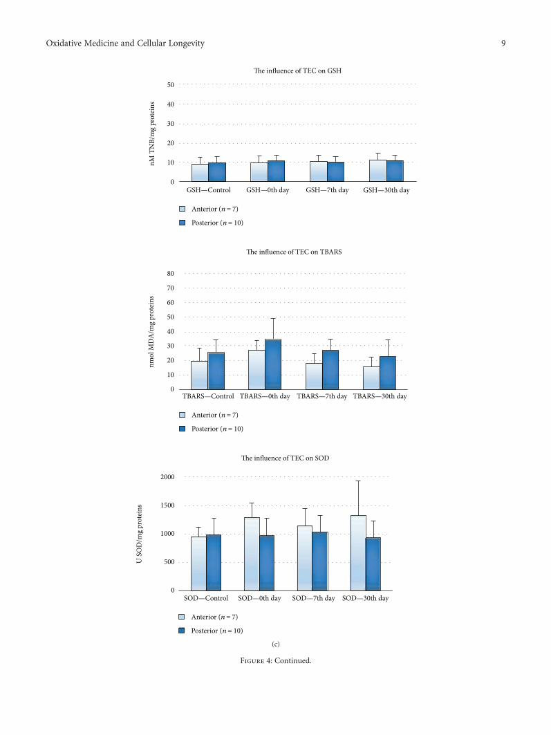
Figure 4: (a–f) The influence of tested restorations on GCF redox status of dental patients. OS parameters (GSH, MDA, and SOD) in GCFwere presented in respect to teeth position (anterior and posterior) with given number of patients per posterior and anterior treated teeth. (a)The influence of Amg on GCF redox status of dental patients. Amg group: patients with posterior treated teeth only (n = 14 patients): 8 werefrom K2 and 6 from K5 group. The amount of sealed Amg: 1.318571± 0.71267 g. (b) The influence of ZPoC on GCF redox status ofdental patients. Patients with posterior and anterior treated teeth (n = 14 patients): 12 patients with posterior treated teeth (3 from K2,4 from K3, and 5 form K4 group) and 2 patients with anterior treated teeth (2 from K2 group). The amount of sealed ZPoC foranterior was 0.035± 0.025 g and posterior was 0.229± 0.167 g. (c) The influence of Tetric EvoCeram on GCF redox status of dentalpatients. Patients with posterior and anterior treated teeth (n = 17 patients): 10 patients with posterior treated teeth (6 from K2 and 4from K5 group) and 7 patients with anterior treated teeth (6 from K2 and 1 from K5 group). The amount of sealed TEC for anteriorwas 0.029± 0.02253 g and posterior was 0.152± 0.159 g. (d) The influence of BF on GCF redox status of dental patients. Patients withposterior and anterior treated teeth (n = 15 patients): 8 patients with posterior and 7 with anterior treated teeth (all from K2 group). Theamount of sealed BF for anterior: 0.029± 0.014 g and posterior 0.064± 0.042 g. (e) The influence of GIC on GCF redox status of dentalpatients. Patients with posterior and anterior treated teeth (n = 14 patients): 12 patients with posterior treated teeth (9 from K2, 2from K3, and 1 from K5 group) and 2 patients with anterior treated teeth, from K2 group. The amount of sealed GIC for anterior was0.035± 0.015 g and posterior was 0.168± 0.153 g. (f) The influence of ZPhC on GCF redox status of dental patients. Patients withposterior and anterior treated teeth (n = 14 patients): 11 patients with posterior treated teeth (5 from K2, 3 from K3, 1from K4, and 2from K5 group) and 3 patients with anterior treated teeth (2 from K2 and 1 from K3 group). The amount of sealed ZPhC for anterior was0.08± 0.081 g and posterior was 0.235± 0.145 g.
12 Oxidative Medicine and Cellular Longevity

7th day 30th dayTEC versus Amg, GIC,BF, ZPoC
TEC versus GIC, BF,ZPoC
ZPhC versus Amg, GIC, BF, ZPoC
ZPhC versus GIC, BF,ZPoC
Amg versus BF Amg versus ZPoCGIC versus BF −F(5,72) = 4.80p = 0.001pes = 0.250
F(5,72) = 4.45 p = 0.002
pes = 0.268
45
tGSH
30
20
10TEC ZPhC GICAmg ZPoCBF
7th day
30th day
(a)
7th day 30th day
GIC versus Amg, BF GIC versus Amg, BF, ZPoC
ZPhC versus Amg, BF ZPhC versus BF, ZPoCTEC versus BF TEC versus BF, ZPoCBF versus ZPoC Amg versus ZPoCF (5.72) = 4.07p = 0.003pes = 0.225
F (5.72) = 4.90 p = 0.001
pes = 0.277
7th day
30th day
TEC ZPhC GICAmg ZPoCBF
tSO
D
1100
950
650
500
800
0
(b)
7th day 30th dayZPoC versus Amg ZPoC versus BF, GICZPhC versus BF, GIC ZPhC versus BF, GICAmg versusBF, GIC Amg versus BF, GIC− TEC versus GIC− BF versus GICF (5.72) = 2,46p = 0.041pes = 0.151
F (5.72) = 16.34 p < 0.001
pes = 0.581
ZPoC ZPhC TECAmg GICBF
TBA
RS
60
40
20
0
7th day
30th day
(c)
Figure 5: The influence of the restorations on OS parameters before and after the treatments. Estimated marginal means for OS parameters inGCF at 7th and 30th day were evaluated with 0th day (horizontal line: long dash dot dot). In regard to the applied restorative, (a) GSHcovariate at the 0th day was 19.3 nmol TNB/mg proteins; (b) tSOD covariate at the 0th day was 665.6U/mg proteins; (c) TBARS covariateat the 0th day was 17.8 nmol MDA/mg proteins. The patients’ distribution across the K groups and restorative groups is tabulated(Table 2). 7th and 30th days were presented with a dash and solid line, respectively. Tables on the right show differences (p values) in OSparameters between restoratives’ treatment groups. pes: partial eta squared. 2× 2 between-group analysis of covariance (ANCOVA) andpost hoc comparisons (least-significant difference, (LSD)) was used. p ≤ 0 05 value was considered statistically significant.
13Oxidative Medicine and Cellular Longevity

(Porphyromonas gingivalis, Prevotella nigrescens, etc.) [28].In accordance with the literature, we showed slightly increaseof LPO in advanced dental lesion, confirming OS develop-ment with caries progression (Figure 2(a)). This notion issupported by ROS overproduction via NADPH oxidase andmyeloperoxidase during phagocytosis of bacterial pathogensand their interactions with two main targets in membranephospholipids, double bond between C-atoms and theester linkage between glycerol and fatty acids [1, 29, 30].Stick to dental caries is a bacterial inflammation accompa-nying with local immune response [10]. Placed withinlysosomes (the azurophilic granules of phagocytes) of neu-trophils, NADPH oxidase and myeloperoxidase produceROS during so-called “respiratory burst.” NADPH oxidasecatalyzes superoxide anion (O2
•−) production through a largeoxygen (O2) consumption (when >80–90% of O2 becomesconverted into O2
•−), while myeloperoxidase catalyzes pro-duction of several reactive species, such as hypohaloge-nated acids (including hypochlorous acid (HOCl)) inreactions of hydrogen peroxide (H2O2) and halide ions(Cl−, Br−, and I−); hypothiocyanous acid (HOSCN) fromH2O2 and halide and pseudohalide ions; hydroxyl radical(HO•), via non-Fenton reaction between O2
•− and HOCl;and nitrating intermediates, in vivo [31–34]. After beingfused with lysosomes, phagosome (a vesicle formed aroundengulfed bacteria) matures into phagolysosomes, within theneutrophils. That is the point when intracellular killingof pathogens starts by ROS. Although ROS effects occurintracellularly, within phagolysosomes, they are diffusibleand can react outside of phagolysosomes, within the neu-trophils and surrounding tissues (for instance with GCF, incase of dental caries) [19]. The reactive species produced bymyeloperoxidase are responsible for the oxidation, chlorina-tion, and nitration of cytosolic proteins, glycoproteins, andlipoproteins in neutrophils or in nearby tissues (i.e., HOClchlorinates amines and produces toxic chloramine, orHOSCN inhibits glycolysis and energy supply, etc.) and areresponsible for the side effect of inflammation (death ofphagocytes and tissue damage) [34–36].
Development of OS in GCF of teeth with caries was antic-ipated since immunoinflammatory-associated occurrences,such as caries, are characterized by ROS overproduction,depletion of reducing equivalent sources, such as NAD (P)H and GSH, and oxidative injure of biomolecules, includinglipids (Figures 2(b)).
As to the effect of the restorations on OS profile ofGCF, ANCOVA analysis of the data sorted by the Black’sClassification Criteria (Figures 1 and 3) showed that thehighest GSH and tSOD activities were documented inthe K3 group (ZPhC=ZPoC>GIC), at 30th day, what wassignificant for GSH compared to K2 (BF>TEC>GIC>Amg>ZPhC>ZPoC) (∗∗p = 0 001) and K5 (Amg>TEC>ZPhC>GIC) (∗∗p = 0 001) and reduced LPO in K4 group(ZPoC>>ZPhC), what was significantly lower compared toK2 (∗p = 0 026), at 30th day for MDA. From this, we con-cluded that ZPoC and ZPhC, within the K3 group, have more(and equal) supportive role in increasing tSOD activity andGSH. To emphasize that, ZPoC notably reduced LPO withinthe K4 group.
Accordingly, ANCOVA analysis of the data arranged inrespect to the applied restorations showed significant GSHincrease by the following restorations listed in descendingorder: ZPoC>BF>GIC>Amg; and tSOD activity increaseby ZPoC>BF>Amg; while MDA decrease was gained byZPoC>ZPhC>Amg>TEC (Figures 4(a)–4(f) and 5). Con-sistent with the literature, we confirmed that ZPoC and ZPhCdemonstrated profound antioxidant effect in comparison tothe other used dental fillings, in terms of suppressed LPOand GSH regaining, contrary to GIC which demonstratedcompletely opposite, prooxidant effect, while composites,BF and TEC did not show noticeable effects on GCF OSstatus [37, 38].
According to the literature, the most profound antiox-idant effect of ZPoC and ZPhC can be prescribed tohydrolysis of their acid components (itaconic and maleicacids versus phosphoric acid, resp.) [37, 39]. Dicarboxylicacids, such as itaconic and maleic acids, are used as mono-mers for biopolymers (resins or synthetic fibers). Lampro-poulou et al. acclaimed itaconate as a major physiologicalregulator of the global metabolic rewiring and effectorfunctions of inflammatory macrophages. It regulates succi-nate levels and function, mitochondrial respiration, andinflammatory cytokine production during macrophage acti-vation [39]. Adhering to this, accomplished antioxidant roleof ZPoC (especially in the suppression of LPO within GCF)probably comes from itaconic acid. On the other hand, phos-phoric acid binds many divalent cations, including transientmetals (iron, cooper, etc.). It is well known that transientmetals (in low valent states) participate in Fenton reactionto produce the most potent ROS, HO• (no enzymatic systemexists in living organisms to scavenge it) [40]. It is used indentistry as an etching, that is, corrosive solution. Corrosiveskill pathogens and prevent locally bacterial diseases, includ-ing dental caries. The antioxidant effect of ZPhC was con-firmed by all three OS markers.
From all applied restorations, only GIC accomplishedprooxidant property (suppressed tSOD activity and ele-vated LPO). According to the literature, the explanationfor such occurrences lies in fluoride anion (released fromGIC) interactions with metal cations embedded in antiox-idant metalloenzymes, such as SOD, catalase, and peroxi-dase. The obtained results are consistent with Yamagutiet al.’s study in which it was shown that low-dose fluo-ride treatment affects antioxidant enzymes, includingSOD and catalase (CAT), and rises LPO in parotid andsubmandibular salivary glands of rats. Explicitly, theydemonstrated that fluoride intoxication caused more pro-nounced OS in submandibular than in parotid salivaryglands [38].
It is well known that prolonged leaching of small amountof unbound monomers (1.5–5%), such as TEGDMA forinstance, is blamed for cytotoxic and other systemic effectsof composites. The leaching of methacrylate monomersoccurs because of the incomplete UV polymerizations ofcomposites during sealing process [41]. Herein, the amountof the TEGDMA, present in the sealed composites (BF andTEC), was almost >300 times lower than its subtoxic dose(<4mM), reported by Gul et al., thus adverse/toxic effects
14 Oxidative Medicine and Cellular Longevity

(including disruption of redox homeostasis in GCF) werecompletely avoided [41–43]. Individual sensitivity of thepatients with polymorphism of GSH to TEGDMA moleculewas reported [44]. Additionally, low GSH levels in GCF ofdental patients treated with TEC contrary to BF may relateto monomer UEDMA [45].
The low levels of GSH, tSOD activity, and MDA mea-sured in K5 group before and after the dental restorationstrengthening depraved influence of insufficient blood supplyand metabolism on GCF profile.
Positive correlation between filling mass (0.07–2.03 g)and teeth position (Pearson correlation: 0.307, p = 0 004)was anticipated concerning the size of the anterior and theposterior teeth.
5. Conclusion
Taking into consideration the influential factors such as den-tal lesion degree, type of applied dental fillings, and teethposition, we made the following conclusions: (i) GCF OS sta-tus does not depend on teeth position and does not differbetween healthy teeth; (ii) untreated teeth with caries donot differ significantly from corresponding controls (exclu-sion: elevated GSH in posterior teeth); (iii) reduced GSHand MDA were recognized as a more reliable and sensitiveOS marker than tSOD; (iv) ZPoC and ZPhC achieved pro-found antioxidant effect; (v) none of the applied restorationsaccomplished complete antioxidant effect, while GIC realizedprooxidant effect; and (vi) restorations with antioxidantproperties may reduce gum diseases initiated by caries lesion.
To our knowledge, this is the first paper on this topic andperformed with dental patient. Restorations with antioxidantproperties may reduce gum diseases initiated by caries lesion,what is of great clinical relevance in dentistry. We showedand recognized that redox interactions may influence dentalmaterial biocompatibility; thus, evaluation of GCF OS statusmay be considered as a useful tool in biocompatibility testingof dental fillings.
Abbreviations
Al2O3: Aluminium trioxideAmg: AmalgamBF: BeautifilBisGMA: Bisphenol-A-diglycidyl-dimethacrylateDTNB – 5: 5-Dithiobis (2-nitrobenzoic acid)GCF: Gingival crevicular fluidGIC: Glass ionomer cementH2O2: Hydrogen peroxideLPO: Lipid peroxidationMDA: MalondialdehydeNS: Nitrosative stressO2
•−: Superoxide anionOS: Oxidative nitrosative stressTBARS: Thiobarbituric acid reactive substancesTEC: Tetric EvoCeramTEGDMA: Triethylene-glycol-dimethacrylateTNB: 5-Thio-2-nitrobenzoic acidGSH: Glutathione
tSOD: Total superoxide dismutaseUEDMA: Urethane-dimethacrylateZPhC: Zinc phosphate cementZPoC: Zinc polycarboxylate cement.
Data Availability
The data used to support the findings of this study are avail-able from the corresponding author upon request.
Conflicts of Interest
The authors declare that they have no conflicts of interest.
Acknowledgments
This work was supported by grants from the Ministry ofEducation, Science and Technological Development of theRepublic of Serbia (Project no. III 41018). The authors arealso grateful to our reviewers for the contributive criticismsand suggestions.
References
[1] D. Uğar-Çankal and N. Ozmeric, “A multifaceted molecule,nitric oxide in oral and periodontal diseases,” Clinica ChimicaActa, vol. 366, no. 1-2, pp. 90–100, 2006.
[2] R. Khanna, P. Thapa, H. Khanna, S. Khanna, A. Khanna, andH. Shukla, “Lipid peroxidation and antioxidant enzyme statusin oral carcinoma patients,” Kathmandu University MedicalJournal, vol. 3, no. 4, pp. 334–339, 2005.
[3] J. Kundalić, University of Niš, Faculty of Medicine, PhDstudent, Niš, Serbia, D. Pavlović et al., “Oxidative stress inthe pathogenesis of periodontal disease,” Acta Medica Media-nae, vol. 55, pp. 66–72, 2016.
[4] B. Mandić and T. Todorović, “Antioxidant status in oral can-cer patients,” Oral Oncology, vol. 8, pp. 38–42, 2002.
[5] D. Pavlica and T. Todorović, “Saliva–diagnostic fluid,” StomGlas S, vol. 48, pp. 137–141, 2001.
[6] M. Takane, N. Sugano, T. Ezawa, T. Uchiyama, and K. Ito, “Amarker of oxidative stress in saliva: association withperiodontally-involved teeth of a hopeless prognosis,” Journalof Oral Science, vol. 47, no. 1, pp. 53–57, 2005.
[7] M. Khorsavi Samani, A. Poorsattar Bejeh Mir, M. Kashiri, andD. Gujeq, “Introducing cut-points for salivary nitric oxide todistinguish periodontitis from the normal periodontium,”Minerva Stomatologica, vol. 61, no. 10, pp. 443–448, 2012.
[8] Z. Khurshid, M. Mali, M. Naseem, S. Najeeb, and M. Zafar,“Human gingival crevicular fluids (GCF) proteomics: an over-view,” Dentistry Journal, vol. 5, no. 1, p. 12, 2017.
[9] F. Tinti and M. Soory, “Mechanisms for redox actions of nico-tine and glutathione in cell culture, relevant to periodontitis,”Scientific Reports, vol. 2, no. 1, 2012.
[10] T. Todorovic, I. Dozic, M. Vicente-Barrero et al., “Salivaryenzymes and periodontal disease,” Medicina Oral, PatologíaOral y Cirugía Bucal, vol. 11, pp. E115–E119, 2006.
[11] M. M. Djukic, M. D. Jovanovic, M. Ninkovic et al., “Protectiverole of glutathione reductase in paraquat induced neurotoxic-ity,” Chemico-Biological Interactions, vol. 199, no. 2, pp. 74–86,2012.
15Oxidative Medicine and Cellular Longevity

[12] M. Đukić, M. Ninković, and M. Jovanović, “Oxidative stress-clinical diagnostic significance,” Journal of Medical Biochemis-try, vol. 27, no. 4, 2008.
[13] H. Bayir and V. E. Kagan, “Bench-to-bedside review: mito-chondrial injury, oxidative stress and apoptosis–there is noth-ing more practical than a good theory,” Critical Care, vol. 12,no. 1, p. 206, 2008.
[14] J. A. Lincoln, D. L. Lefkowitz, T. Cain et al., “Exogenous mye-loperoxidase enhances bacterial phagocytosis and intracellularkilling by macrophages,” Infection and Immunity, vol. 63,no. 8, pp. 3042–3047, 1995.
[15] B. Testa and S. D. Krämer, “The biochemistry of drug metab-olism–an introduction,” Chemistry & Biodiversity, vol. 4, no. 3,pp. 257–405, 2007.
[16] J. Sumitt, J. Robbins, R. Schwartz, and J. Santos, Fundamentalsof Operative Dentistry, Quintessence Publishing Co, 2001.
[17] G. J. Mount and W. R. H. BDS, “A new cavity classification,”Australian Dental Journal, vol. 43, no. 3, pp. 153–159,1998.
[18] A. Guentsch, M. Kramesberger, A. Sroka, W. Pfister,J. Potempa, and S. Eick, “Comparison of gingival crevicularfluid sampling methods in patients with severe chronic peri-odontitis,” Journal of Periodontology, vol. 82, no. 7, pp. 1051–1060, 2011.
[19] M. J. Girotti, N. Khan, and B. A. Mclellan, “Early measurementof systemic lipid peroxidation products in the plasma of majorblunt trauma patients,” Journal of Trauma and Acute CareSurgery, vol. 31, no. 1, pp. 32–35, 1991.
[20] M. Sun and S. Zigman, “An improved spectrophotometricassay for superoxide dismutase based on epinephrine autoxi-dation,” Analytical Biochemistry, vol. 90, no. 1, pp. 81–89,1978.
[21] M. Anderson, “The DTNB-GSSG reductase recycling assayfor total glutathione (GSH+ 1/2GSSG),” CRC Handbook ofMethods for Oxygen Radical Research, pp. 319–323, 1986.
[22] O. Lowry, N. Rosebrough, A. Farr, and R. Randall, “Proteinmeasurement with the Folin phenol reagent,” The Journal ofBiological Chemistry, vol. 193, no. 1, pp. 265–275, 1951.
[23] A. P. Bejeh-Mir, H. Parsian, M. A. Khoram, N. Ghasemi,A. Bijani, and M. Khosravi-Samani, “Diagnostic role of sali-vary and GCF nitrite, nitrate and nitric oxide to distinguishhealthy periodontium from gingivitis and periodontitis,”International Journal of Molecular and Cellular Medicine,vol. 3, no. 3, pp. 138–145, 2014.
[24] A. B. Petković, S. M. Matić, N. V. Stamatović et al., “Proinflam-matory cytokines (IL-1β and TNF-α) and chemokines (IL-8and MIP-1α) as markers of peri-implant tissue condition,”International Journal of Oral and Maxillofacial Surgery,vol. 39, no. 5, pp. 478–485, 2010.
[25] V. Stefanovic, E. Taso, A. Petkovic-Curcin et al., “Influence ofdental filling material type on the concentration of interleukin9 in the samples of gingival crevicular fluid,” VojnosanitetskiPregled, vol. 73, no. 8, pp. 728–734, 2016.
[26] M. S. Davis, S. W. Joseph, and J. F. Bucher, “Periapical andintracanal healing following incomplete root canal fillings indogs,” Oral Surgery, Oral Medicine, Oral Pathology, vol. 31,no. 5, pp. 662–675, 1971.
[27] C. F. Williams, N. Yarlett, M. A. Aon, and D. Lloyd, “Antiox-idant defences of Spironucleus vortens: glutathione is themajor non-protein thiol,” Molecular and Biochemical Parasi-tology, vol. 196, no. 1, pp. 45–52, 2014.
[28] C. A. Butler, P. D. Veith, M. F. Nieto, S. G. Dashper, and E. C.Reynolds, “Lysine acetylation is a common post-translationalmodification of key metabolic pathway enzymes of the anaer-obe Porphyromonas gingivalis,” Journal of Proteomics,vol. 128, pp. 352–364, 2015.
[29] Y. Huang, M. Zhu, Z. Li et al., “Mass spectrometry-basedmetabolomic profiling identifies alterations in salivary redoxstatus and fatty acid metabolism in response to inflammationand oxidative stress in periodontal disease,” Free Radical Biol-ogy and Medicine, vol. 70, pp. 223–232, 2014.
[30] K. Das and A. Roychoudhury, “Reactive oxygen species(ROS) and response of antioxidants as ROS-scavengers duringenvironmental stress in plants,” Frontiers in EnvironmentalScience, vol. 2, 2014.
[31] A. Sigel and H. Sigel, “Metal ions in biological systems,” inInterrelations between Free Radicals and Metal Ions in LifeProcesses, vol. 36, CRC Press, 1999.
[32] J. P. Gaut, J. Byun, H. D. Tran et al., “Myeloperoxidase pro-duces nitrating oxidants in vivo,” The Journal of ClinicalInvestigation, vol. 109, no. 10, pp. 1311–1319, 2002.
[33] R. I. Handin, S. E. Lux, and T. P. Stossel, Blood: Principles andPractice of Hematology, Lippincott Williams & Wilkins, 2003.
[34] D. T. Love, T. J. Barrett,M. Y.White, S. J. Cordwell, M. J. Davies,and C. L. Hawkins, “Cellular targets of the myeloperoxidase-derived oxidant hypothiocyanous acid (HOSCN) and its rolein the inhibition of glycolysis in macrophages,” Free RadicalBiology and Medicine, vol. 94, pp. 88–98, 2016.
[35] R. P. Wilkie-Grantham, N. J. Magon, D. T. Harwood et al.,“Myeloperoxidase-dependent lipid peroxidation promotesthe oxidative modification of cytosolic proteins in phagocyticneutrophils,” Journal of Biological Chemistry, vol. 290, no. 15,pp. 9896–9905, 2015.
[36] M. C. M. Vissers and C. C. Winterbourn, “Oxidative damageto fibronectin: I. The effects of the neutrophil myeloperoxidasesystem and HOCl,” Archives of Biochemistry and Biophysics,vol. 285, no. 1, pp. 53–59, 1991.
[37] A. McNaught and A. Wilkinson, Compendium of ChemicalTerminology, Blackwell Science, Oxford, 1997.
[38] P. M. Yamaguti, A. Simões, E. Ganzerla, D. N. Souza, F. N.Nogueira, and J. Nicolau, “Effects of single exposure of sodiumfluoride on lipid peroxidation and antioxidant enzymes in sal-ivary glands of rats,” Oxidative Medicine and Cellular Longev-ity, vol. 2013, Article ID 674593, 7 pages, 2013.
[39] V. Lampropoulou, A. Sergushichev, M. Bambouskova et al.,“Itaconate links inhibition of succinate dehydrogenase withmacrophage metabolic remodeling and regulation ofinflammation,” Cell Metabolism, vol. 24, no. 1, pp. 158–166,2016.
[40] E. Pinto, T. C. S. Sigaud-kutner, M. A. S. Leitao, O. K. Okamoto,D. Morse, and P. Colepicolo, “Heavy metal-induced oxidativestress in algae1,” Journal of Phycology, vol. 39, no. 6, pp. 1008–1018, 2003.
[41] S. Bouillaguet, “Biologicalrisks ofresin-basedmaterials to the-dentin-pulpcomplex,” Critical Reviews in Oral Biology &Medicine, vol. 15, no. 1, pp. 47–60, 2004.
[42] P. Gul, N. Akgul, H. H. Alp, and A. Kiziltunc, “Effects of com-posite restorations on oxidative stress in saliva: an in vivostudy,” Journal of Dental Sciences, vol. 10, no. 4, pp. 394–400,2015.
[43] J. Volk, J. Engelmann, G. Leyhausen, andW. Geurtsen, “Effectsof three resin monomers on the cellular glutathione
16 Oxidative Medicine and Cellular Longevity

concentration of cultured human gingival fibroblasts,” DentalMaterials, vol. 22, no. 6, pp. 499–505, 2006.
[44] M. Goldberg, “In vitro and in vivo studies on the toxicity ofdental resin components: a review,” Clinical Oral Investiga-tions, vol. 12, no. 1, pp. 1–8, 2008.
[45] M. Noda, J. Wataha, J. Lewis et al., “Dental adhesive com-pounds alter glutathione levels but not glutathione redoxbalance in human THP-1 monocytic cells,” Journal of Bio-medical Materials Research Part B: Applied Biomaterials,vol. 73, no. 2, pp. 308–314, 2005.
17Oxidative Medicine and Cellular Longevity

Stem Cells International
Hindawiwww.hindawi.com Volume 2018
Hindawiwww.hindawi.com Volume 2018
MEDIATORSINFLAMMATION
of
EndocrinologyInternational Journal of
Hindawiwww.hindawi.com Volume 2018
Hindawiwww.hindawi.com Volume 2018
Disease Markers
Hindawiwww.hindawi.com Volume 2018
BioMed Research International
OncologyJournal of
Hindawiwww.hindawi.com Volume 2013
Hindawiwww.hindawi.com Volume 2018
Oxidative Medicine and Cellular Longevity
Hindawiwww.hindawi.com Volume 2018
PPAR Research
Hindawi Publishing Corporation http://www.hindawi.com Volume 2013Hindawiwww.hindawi.com
The Scientific World Journal
Volume 2018
Immunology ResearchHindawiwww.hindawi.com Volume 2018
Journal of
ObesityJournal of
Hindawiwww.hindawi.com Volume 2018
Hindawiwww.hindawi.com Volume 2018
Computational and Mathematical Methods in Medicine
Hindawiwww.hindawi.com Volume 2018
Behavioural Neurology
OphthalmologyJournal of
Hindawiwww.hindawi.com Volume 2018
Diabetes ResearchJournal of
Hindawiwww.hindawi.com Volume 2018
Hindawiwww.hindawi.com Volume 2018
Research and TreatmentAIDS
Hindawiwww.hindawi.com Volume 2018
Gastroenterology Research and Practice
Hindawiwww.hindawi.com Volume 2018
Parkinson’s Disease
Evidence-Based Complementary andAlternative Medicine
Volume 2018Hindawiwww.hindawi.com
Submit your manuscripts atwww.hindawi.com