Embed Size (px)
Citation preview

i
OXIDATION OF DEOXYAMINOSUGARS BY A NEW FLAVIN-DEPENDENT
MONOOXYGENASE
By
Ahmad H. Al-Mestarihi
Dissertation
Submitted to the Faculty of the
Graduate School of Vanderbilt University
in partial fulfillment of the requirements
for the degree of
DOCTOR OF PHILOSOPHY
in
Chemistry
December, 2012
Nashville, Tennessee
Approved by:
Professor Brian O. Bachmann
Professor Tina M. Iverson
Professor Michael P. Stone
Professor Richard N. Armstrong
ii
To my late parents Se’da and Hasan
I can’t be thankful enough for all the love and support you have given me

iii
TABLE OF CONTENTS
PAGE
DEDICATION………………………………………………………………………………………………………….……….ii
ACKNOWLEDGMENTS…………………………………………………………………………………………..........vii
LIST OF TABLES…………….…………………………………………………………………………………………………x
LIST OF FIGURES…………………………………………………………………………………………………………….xi
LIST OF SCHEMES…………………………………………………………………………………………………………xiv
LIST OF ABBREVIATIONS .................................................................................................... xv
LIST OF PUBLICATIONS .................................................................................................... xviii
CHAPTER
I. INTRODUCTION ...................................................................................................... 1
Biosynthesis of N-oxygenated deoxysugars ............................................... 2
Significance of deoxysugars in natural products ............................ 2
Nitrosugar-containing natural products ......................................... 5
Biosynthesis of deoxyaminosugars ............................................... 11
Comparative genomics of the nitro sugar functionality ............... 14
Proposed deoxysugar N-oxidation pathway ................................. 18
Anthracyclines and deoxyaminosugar oxidation ...................................... 19
A brief history of anthracycline drugs........................................... 19
Biosynthesis of baumycin ............................................................. 21
Deoxysugar genes in anthracyclines ............................................. 23
The putative role of the flavoenzyme DnmZ ................................ 25
Flavoprotein monooxygenases ................................................................. 27
Biochemistry of flavoproteins ....................................................... 28
The monooxygenase family .......................................................... 30
Flavoprotein monooxygenase catalysis ........................................ 31

iv
Classifications of Flavoprotein monooxygenases ......................... 35
Dissertation Statement ............................................................................. 44
References ................................................................................................ 46
II. BIOCHEMICAL CHARACTERIZATION OF A NEW FLAVIN-DEPENDENT
NITROSOSYNTHASE IN THE BIOSYNTHETIC PATHWAYS OF EVERNINOMICIN AND
RUBRADIRIN .......................................................................................................... 61
Introduction .............................................................................................. 61
Results and discussion .............................................................................. 65
Comparative genomics ................................................................. 65
Preparation of the putative oxidases and their aminosugar
substrate ....................................................................................... 68
Enzymatic activities of ORF36 and RubN8 .................................... 70
Proposed mechanism of nitrososynthase .................................... 73
Conclusion ................................................................................................. 75
Materials and methods ............................................................................. 76
Bacterial strains, Plasmids and Material ....................................... 76
Preparation of the azido sugar substrate ..................................... 77
Cloning and overexpression of ORF36 .......................................... 78
Cloning and overexpression of rubN8 .......................................... 79
Preparation of L-TDP-epi-vancosamine ......................................... 80
ORF36/RubN8 enzymatic reactions .............................................. 81
HPLC-MS assay of L-TDP-epi-vancosamine reaction ..................... 82
References ................................................................................................ 83
III. STRUCTURAL AND MECHANISTIC STUDIES OF THE NITROSOSYNTHASE ORF36
FROM MICROMONOSPORA CARBONACEA VAR. AFRICANA ................................ 87
Introduction .............................................................................................. 87
Results ....................................................................................................... 90
Substrate preference of ORF36 .................................................... 90

v
18O2 incorporation in the ORF36 oxidation reaction. ................... 94
X-ray crystal structure of ORF36 ................................................... 97
Modeling of FAD and TDP-L-epi-vancosamine in the active site .. 99
Description of the active site ...................................................... 101
Discussion................................................................................................ 103
Substrate preference of ORF36 .................................................. 103
Structural comparison of ORF36 and KijD3 ................................ 104
Proposed minimal mechanism of ORF36 .................................... 106
Conclusions ............................................................................................. 107
Materials and methods ........................................................................... 108
Overexpression and purification of enzymes. ............................ 108
Preparation of other TDP-sugars substrates and intermediates.109
Purification of TDP-sugars. .......................................................... 109
ORF36 enzymatic reactions with the aminosugar substrates. ... 110
18O2-incorporation assays. .......................................................... 110
LC-ESI/MS method for ORF36 Substrates. .................................. 111
Crystallization of ORF36. ............................................................. 112
Structure determination and refinement. .................................. 113
Modeling of substrate and cofactor. .......................................... 114
References .............................................................................................. 115
IV. RETRO-ALDOL ACTIVITY OF THE NITROSOSYNTHASE DNMZ.............................. 121
Introduction ............................................................................................ 121
Results and discussion ............................................................................ 125
Proposed activity of DnmZ .......................................................... 125
Activity of DnmZ as a nitrososynthase ........................................ 127
Hydrazone derivatization of the DnmZ oxidation product ......... 129
Tandem MS analysis of the hydrazone derivatives. ................... 131

vi
Acid hydrolysis and HPLC-MS analysis of the
(Carboxymethyl)trimethylammonium hydrazone derivative ..... 133
High resolution MS measurements of the hydrazone
derivatives ................................................................................... 136
Materials and methods ........................................................................... 138
Bacterial strains, Plasmids and Materials. .................................. 138
Cloning and overexpression of dnmZ. ........................................ 138
Preparation of TDP-L-epi-vancosamine. ...................................... 139
DnmZ assay with TDP-L-epi-vancosamine. .................................. 140
Derivatization of DnmZ products with hydrazines. .................... 141
LC-ESI-MS Method for DnmZ assays. .......................................... 141
References .............................................................................................. 142
V. SUMMARY AND FUTURE DIRECTIONS ................................................................ 147
Synopsis................................................................................................... 147
Future directions ..................................................................................... 149
Mechanism of the nitro group formation ................................... 149
Structural studies ........................................................................ 152
Catalytic competence and kinetic studies .................................. 153
The interplay between the nitrososynthases and
glycosyltransferases .................................................................... 154
References .............................................................................................. 155
Appendix
A. SUPPLEMENTARY FIGURES FROM CHAPTER III ............................................ 157
B. SUPPLEMENTARY FIGURES FROM CHAPTER III ............................................ 176
C. MS AND MS/MS SPECTRA FROM CHAPTER IV.............................................. 180
vii
ACKNOWLEDGMENTS
This work wouldn’t have been possible without the guidance of my advisor and
research committee, help from my lab colleagues and staff at Vanderbilt University, and
support from my family and friends.
I would like to express my deepest gratitude to my advisor, Prof. Brian
Bachmann, for his outstanding guidance, support, and patience, providing me with an
excellent atmosphere for conducting research. Prof. Bachmann’s support and
encouragement exceeded my expectation even with matters outside of research. I
would also like to acknowledge the rest of my research committee members, Prof. Tina
Iverson, Prof. Michael Stone, and Prof. Richard Armstrong for their excellent support
and guidance toward the completion of my PhD at Vanderbilt University.
I have been fortunate to work with several collaborators throughout my
research projects, which enriched my experience and knowledge. I would like to thank
Prof. Tina Iverson (Vanderbilt University, Nashville, TN) for her willingness to collaborate
with our lab on solving the X-ray crystal structure of the nitrososynthase ORF36.
Particularly, I would like to thank Prof. Jessica Vey from Prof. Iverson’s research group
for conducting the crystallography experiments which resulted in a valuable publication.
I would also like to thank the research labs of Prof. Michael Burkart (University of
California, San Diego, La Jolla, CA) and Prof. Hung-wen Liu (The University of Texas,
Austin, TX) for generously providing EvaA-E and RfbB expression constructs,
respectively. Many thanks to Prof. Daniel Kahne’s research group (Harvard University,
viii
Boston, MA) for supplying the essential synthetic substrate for the nitrososynthase,
TDP-L-epi-vancosamine, which facilitated our initial biochemical characterization of
RubN8 and ORF36.
I would also like to thank all past and present members of the Bachmann lab
who I got to know over the last several years. It has been very enjoyable working with all
of you and many thanks for your help, support and friendship. Especially I would like to
acknowledge a former member of the lab, Dr. Yunfeng Hu, who prepared the ORF36
protein and participated in the initial biochemical characterization of the enzyme.
I am grateful for the funding we received for the research work described in this
dissertation from the Office of Naval Research. Their continuous support over the
course of several years made the completion of this work possible. The graduate school
at Vanderbilt University also provided me with financial support in the form of teaching
assistantships and for that I am also grateful.
To all my friends at Vanderbilt University, thank you very much for your
friendship and support. You made my life at Vanderbilt a truly memorable experience
and your friendships are invaluable to me. To my best friend Prof. Harold Moser, who
became like a father to me, following my progress closely providing support and
encouragements, I am truly grateful.
Finally, I am very grateful to my big extended family in Jordan, brothers, sisters,
nephews and nieces, thank you for your love and support. To my beloved late parents,

ix
who have raised me to be the person I am today, thank you for all the unconditional
love, guidance, and support that you have always given me.

x
LIST OF TABLES
Table Page
I-1. Possible biosynthetic genes for L-evernitrose. ...................................................... 16
I-2. Classification of external flavoprotein monooxygenases. .................................... 36
xi
LIST OF FIGURES
Figure Page
I-1. Examples of some deoxysugars linked to natural product scaffolds … ................... 3
I-2. Chemical structure of everninomicin. ..................................................................... 6
I-3. Chemical structure of rubradirin. ............................................................................ 8
I-4. Chemical structure of kijanimicin. ......................................................................... 10
I-5. Chemical structures of everninomicin and avilamycin. ......................................... 15
I-6. Chemical structure of some anthracyclines. .......................................................... 20
I-7. Chemical structures of several flavin species. ....................................................... 29
I-8.. Structures of several prototype flavoprotein monooxygenases ........................... 37
II-1. Chemical structures of everninomicin, rubradirin, and kijanimicin. ..................... 63
II-2. Chemical structures of avilamicin and everninomicin. .......................................... 66
II-3. Chemical structures of L-epi-vancosamine and L-evernitrose. ............................... 67
II-4. SDS-PAGE gel of Ni-affinity purified RubN8. .......................................................... 69
II-5. LC-ESI-MS analysis of the staudinger reduction of the synthetic (5:1 β/α)-azido
congener of TDP-L-epi-vancosamine by TCEP. ...................................................... 70
II-6. HPLC-ESI-MS chromatograms of the time course of RubN8 oxidation reaction. . 72
II-7. Tandem MS spectra of the nucleotide-hydroxyaminosugar intermediate (above)
and the nucleotide-nitrososugar product (below) from RubN8 oxidation reaction.
............................................................................................................................... 73
xii
III-1. Pathway utilized for biochemical synthesis of TDP-L-epi-vancosamine and the
TDP-4-keto-3-amino sugar precursors. ................................................................. 91
III-2. HPLC-ESI-MS traces of orf36 oxidation reactions with three potential amino
sugar substrates. ................................................................................................... 92
III-3. The 18O2 incorporation studies of orf36 oxidation reaction. ............................... 95
III-4. Structure of orf36. ................................................................................................ 98
III-5. Active site loops of orf36 .................................................................................... 101
IV-1. Chemical structure of known anthracyclines .................................................... 122
IV-2. Carbon-carbon bond cleavage reaction of 2-exo-nitroso-3-exo- hydroxy
norbornane. ........................................................................................................ 126
IV-3. Proposed pathway for the DnmZ monooxygenation activity and the subsequent
cleavge and glycoslation activities. ................................................................... 127
IV-4. SDS-PAGE gel of Ni-affinity purified DnmZ ........................................................ 128
IV-5. HPLC-MS Analysis of the dnmz reaction and the hydrozone conjugates of the
oxidation product. ............................................................................................. 131
IV-6. Tandem LC-ESI-MS of the 2-bromophenyl hydrozone derivative ..................... 132
IV-7. Tandem LC-ESI-MS OF the (Carboxymethyl)trimethylammonium hydrozone
derivative ................................................................................................................. 133
IV-8. Acid hydrolysis of the carboxymethyl-trimethylammonium hydrazone derivative
(above) and LC-ESI-MS chromatograms of the hydrolyzed species (below).. .. 134

xiii
IV-9. Tandem LC-ESI-MS spectra of the hydrolysis products of the acid treated GirT
hydrazone derivative. Fragmentation of the hydrazone at m/z 188 (above), and
the hydrazone at m/z 215 (below) .................................................................... 136
IV-10. High resolution mass measurements of the hydrozone derivative….. ............137
V-1. LC-ESI/MS chromatograms of the preparation of TDP-L-evernosamine via the
methyltransferase Rubn7 from the rubradirin pathway ......................................... 150
V-2. LC-ESI-MS chromatograms of the monooxygenation of TDP-L-evernosamine by
ORF36. ................................................................................................................. 151
xiv
LIST OF SCHEMES
Scheme Page
I-1. Entry point into TDP-deoxysugar secondary metabolism in bacteria. ................... 11
I-2. Biosynthesis of TDP-L-epi-vancosamine. ................................................................. 14
I-3. Oxidation of dibenzothiophene by DszC. ................................................................ 19
I-4. Biosynthesis of TDP-L-daunosamine. ...................................................................... 24
I-5. Proposed pathway of late-step baumycin biosynthesis including the role of
DnmZ .. ................................................................................................................. 27
I-6. General mechanism of oxygenation reactions catalyzed by external flavoprotein
monooxygenases. ................................................................................................. 30
I-7 . Reaction mechanism of p-hydroxybenzoate monooxygenase. ............................ 34
II-1. Proposed pathway for the nitrososynthase oxygenation reaction. ..................... 75
IV-2. Hydrozone derivatization of the proposed oxime-eldehyde product. ............... 129
V-1. LC-ESI/MS chromatograms of the preparation of TDP-L-evernosamine via the
methyltransferase Rubn7 from the rubradirin pathway .................................... 150
V-2. LC-ESI-MS chromatograms of the monooxygenation of TDP-L-evernosamine by
ORF36. ................................................................................................................. 151

xv
LIST OF ABBREVIATIONS
ACP acyl carrier protein
AMC rubransarol, 3-amino-4-hydroxy-7-methoxycoumarin
APS advanced photon source
BVMOs Baeyer-Villiger monooxygenases
CCK-B cholecystokinin B
CHF congestive heart failure
CoA Coenzyme A
DBT dibenzothiophene
DHDP 3,4-dihydroxydipicolinate
DMSO dimethylsulfoxide
DNA Deoxyribonucleic acid
dTDP deoxythymidine diphosphate
DTT dithiothreitol
e.e enantiomeric excess
EDTA ethylenediaminetetraacetic acid
ESI electrospray ionization
FAD flavin adenine dinucleotide
FMN flavin mononucleotide
FMOs flavin-containing monooxygenases
FNOs microbial N-hydroxylating monooxygenases
FPLC fast protein liquid chromatography
GirT Girard’s T reagent [(Carboxymethyl)trimethylammonium hydrazine]

xvi
GT glycosyltransferase
HIV human immunodeficiency virus
HPLC high performance liquid chromatography
IC50 half maximal inhibitory concentration
IPTG β–D-1-thiogalactopyranoside
kD kilo Dalton
KS ketosynthase
kV kilovolt
LB luria broth
LC liquid chromatography
LC-CAT life Sciences collaborative access team
MAT malonyl-CoA:ACP acyltransferase
Mg milligram
MHz megahertz
µL microliter
µM micromolar
mL milliliter
mm millimeter
mM millimolar
MOE molecular operating environment
MS mass spectrometry
MWCO molecular weight cut-off
NADP nicotinamide adenine dinucleotide phosphate
NCS non-crystallographic symmetry

xvii
NDP nucleotide diphosphate
Nm nanometer
NMR nuclear magnetic resonance
OD optical density
PCR polymerase chain reaction
PDB protein data bank
PKS polyketide synthase
PLP pyridoxal phosphate
PMP pyridoxamine phosphate
PPi pyrophosphate
Ppm parts per million
Psi pounds per square inch
RNA ribonucleic acid
ROS reactive oxygen species
Rpm rotations per minute
SAM S-Adenosyl methionine
SAX strong anion exchange
SDS-PAGE sodium dodecylsulfide polyacrylamide gel electrophoresis
TCEP tris(2-carboxyethyl)phosphine
TDP thymidine diphosphate
TMP thymidine monophosphate
Tris tris(hydroxymethyl)aminomethane
UV ultra violet
V volt

xviii
LIST OF PUBLICATIONS
1. Vey, J.L., Al-Mestarihi, A., Funk, M.A., Bachmann, B.O., and Iverson, T.M. (2010) Structure and mechanism of ORF36, an Amino Sugar Oxidizing Enzyme in Everninomicin Biosynthesis. Biochemistry, 49(43): 9306-9317.
2. Yunfeng Hu, Ahmad Al-Mestarihi, Catherine L. Grimes, Daniel Kahne and
Brian O. Bachmann, A unifying nitrososynthase involved in nitrosugar biosynthesis, Journal of the American Chemical Society, 130, 15756-15757 (2008)
3. Alexander N.; Bortolus M.; Al-Mestarihi A.; McHaourab H.; Meiler J.
(2008) De novo high-resolution protein structure determination from sparse spin-labeling EPR data. Structure 16, 181–195.
4. Prusakiewicz, JJ, Turman, MV, Vila, A, Ball, HL, Al-Mestarihi, AH, Di Marzo,
V, Marnett, LJ Oxidative metabolism of lipoamino acids and vanilloids by lipoxygenases and cyclooxygenases. Archives of Biochemistry and Biophysi, 464(2), 260-8, 2007.

1
CHAPTER I
INTRODUCTION
Many organisms, such as bacteria, plants and fungi are capable of synthesizing
structurally diverse bioactive natural products. The diversity of structure and bioactivity
of many of these compounds led to their use in pharmaceutical and agricultural
applications.1 Bacterial natural products often contain sugar moieties attached to their
core scaffolds and play significant roles in conferring biological activity. Many of these
bioactive sugars are derived from deoxyaminosugar oxidations, the most common of
which are deoxynitrosugars.2
The recent advances in gene cluster elucidations of several natural products
containing these N-oxidized sugars have enabled the proposal of biosynthetic pathways
of these important moieties and set the stage for biochemical characterization of the
enzymes involved in their biosynthesis. The biosynthetic pathways of deoxyaminosugar
moieties in several natural products have been well studied; however, enzymes
responsible for the amine oxidation in many deoxynitrosugar-containing and related
natural products have not been characterized previous to our investigations.
Given the biological importance of deoxysugar attachments, advances in
studying new biosynthetic pathways will provide opportunities for in vivo pathway
engineering leading to the production of new glycoconjugates with varied biological
activities. Particularly, the characterization of the biosynthetic gene(s) for the key
oxidation of aminosugars, clearly present the opportunity to increase the sugar

2
structural diversity of a vast array of glycosides via in vivo or in vitro pathway
manipulation.2-3 Additionally, discovering new biocatalysts able to perform synthetically
difficult enantioselective amine oxidations will have significant implications in the
development of alternative synthetic methods.
Biosynthesis of N-oxygenated deoxysugars
Significance of deoxysugars in natural products
Deoxysugars are common structural appendages of many bioactive natural
products and are commonly found attached to polyketide scaffolds. Several
therapeutically important drugs such as the antibiotic vancomycin and the anticancer
agent doxorubicin contain sugars attached to their aglycone cores.4-5 These sugars play
important roles in the biological activity by participating in the interaction between the
drug and the cellular target. In some cases, sugars participate in the mode of action of
many drugs as they contribute to a variety of processes, including active
transmembrane transport, stabilization of protein folding and enzyme inhibition.4-8
The most prevalent sugars among sugar-containing bacterial secondary
metabolites are deoxysugars, often deoxyaminosugars, the biosyntheses of which have
been recently reviewed.3, 9 Structures of some common deoxysugars are shown in figure
I-1.

3
Figure I-1. Examples of some deoxysugars found linked to several natural product scaffolds
Deoxygenation of these sugars proceeds via nucleotide diphosphate (NDP)
activation of 6-deoxyhexoses, usually D-glucose. The most common NDP activation is
performed by attaching thymidine diphosphate (TDP) at the anomeric carbon of D-
glucose-1-phosphate.3 TDP-activated sugars are the most structurally diverse class of
nucleotide sugars found in nature. After NDP-activation, deoxygenation proceeds via a
4-keto-6-deoxy intermediate which is shared among all known deoxysugar pathways.
A less common but very important sugar modification, deoxyaminosugar
oxidation to hydroxylamino-, nitroso-, and nitrosugars uniquely extends nature’s
glycochemical diversity. Natural products containing N-oxidized deoxysugars exhibit a
broad range of biological activities including antibacterial, antitumor, antimalarial,
anticholesteromic, antiviral, and antidiabetic activities.2, 10-12 The oxidized congeners of
deoxyaminosugars exist in many natural products such as everninomicin, rubradirin and
kijanimicin.13-15 The hydroxylamino-, nitroso-, and nitrosugar variants have been isolated
from the fermentation broth of the everninomicin producer Micromonospora
4
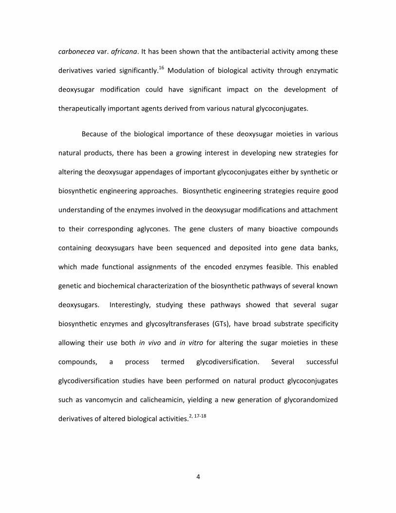
carbonecea var. africana. It has been shown that the antibacterial activity among these
derivatives varied significantly.16 Modulation of biological activity through enzymatic
deoxysugar modification could have significant impact on the development of
therapeutically important agents derived from various natural glycoconjugates.
Because of the biological importance of these deoxysugar moieties in various
natural products, there has been a growing interest in developing new strategies for
altering the deoxysugar appendages of important glycoconjugates either by synthetic or
biosynthetic engineering approaches. Biosynthetic engineering strategies require good
understanding of the enzymes involved in the deoxysugar modifications and attachment
to their corresponding aglycones. The gene clusters of many bioactive compounds
containing deoxysugars have been sequenced and deposited into gene data banks,
which made functional assignments of the encoded enzymes feasible. This enabled
genetic and biochemical characterization of the biosynthetic pathways of several known
deoxysugars. Interestingly, studying these pathways showed that several sugar
biosynthetic enzymes and glycosyltransferases (GTs), have broad substrate specificity
allowing their use both in vivo and in vitro for altering the sugar moieties in these
compounds, a process termed glycodiversification. Several successful
glycodiversification studies have been performed on natural product glycoconjugates
such as vancomycin and calicheamicin, yielding a new generation of glycorandomized
derivatives of altered biological activities.2, 17-18

5
These glycodiversification startegies can be applied for many other natural-
product scaffolds utilizing substrate-flexible enzymes to generate libraries of substances
for in vitro or in vivo glycoslation. Enzymes in the deoxysugar pathways can be further
manipulated through protein engineering which can result in an expanded pool of
glycorandomized derivatives. This new generation of compounds of varied biological
activities could lead to the discovery of potentially important drugs to mitigate the
daunting threats of human diseases.
Nitrosugar-containing natural products
Deoxynitrosugars are found in many isolated secondary metabolites with diverse
scaffolds including spirotetronate antibiotics, ansamycins, and orthosomycins.13-15 One
of the first reported deoxynitrosugar-containing natural products is the orthosomycin
antibiotic everninomicin from Micromonospora carbonacea var. africana which includes
a deoxynitrosugar moiety, evernitrose.15 This nitrosugar is called D-rubranitrose in the
polyketide rubradirin isolated from Streptomyces achromogenes and is structurally
related to D-kijanose from the spirotetronate polyketide antibiotic kijanimycin produced
by the Actinomycete Actinomadura kijaniata.14, 19 These compounds possess potent
antibacterial activity among other important biological activities. Below is a brief
description of these three important nitrosugar-containing natural products with
emphasis on the significance of their attached nitrosugar moiety.

6
Everninomicin
Everninomicin, producd by Micromonospora carbonacea var. africana, is an
oligosaccharide which belongs to the orthosomycin class of antibiotics and possesses
potent activity against Gram-positive and Gram-negative bacteria including vancomycin
resistant enterococci, methicillin resistant staphylococci, and penicillin-resistant
streptococci.20 The structure of everninomicin is composed of eight deoxysugars
including a terminal nitrosugar (L-evernitrose), and acelytated with orsellinic and
dichloroisoeverninic acid moieties (Figure I-2).
Figure I-2. Chemical structure of everninomicin.
The mechanism of action of everninomicin involves inhibition of protein
biosynthesis by binding to the ribosomal protein L16 which affects the function of the
50S ribosomal subunit.21 Everninomicin was developed through phase III clinical studies
when its further development was discontinued in May of 2000 for the stated reason:
“the balance between efficacy and safety did not justify further development of the
product”.22 However, because of its potent antibacterial activity, researchers have been

7
interested in its structure diversification to create a number of everninomicin
derivatives for structure-activity studies. Only limited chemical derivatization
experiments of everninomicin were performed for that purpose, which proved
challenging perhaps because of the complexity of the sugar linkages in orthosomycins.23
This gave rise to the interest of studying the biosynthesis of everninomicin which could
potentially lead to the utilization of the natural biocatalysts in its biosynthetic pathway
to compliment the chemical synthetic methods in the rational drug design process.
The deoxynitrosugar moiety in everninomicin was shown to be important for the
antibacterial activity. For instance, antibacterial activity of the nitrosugar congener
against Staphylococcus aureus was shown to be 125 fold higher compared to that of the
amino-sugar congener.16 This highlights the significance of studying the enzymes
responsible for the N-oxidation of this important deoxysugar moiety. The knowledge
that can be gained from understanding this important biochemical transformation could
greatly impact current efforts towards everninomicin structure diversification.
Rubradirin
Rubradirin, produced by Streptomyces achromogenes var. rubradiris, is an
ansamycin antibiotic that possesses significant activity against Gram-positive bacteria
including multidrug-resistance strains of Staphylococcus aureus.19 The structure of
rubradirin is comprised of four distinct moieties; the polyketide scaffold rubransarol, 3-
amino-4-hydroxy-7-methoxycoumarin (AMC), the aromatic bridge 3,4-
8
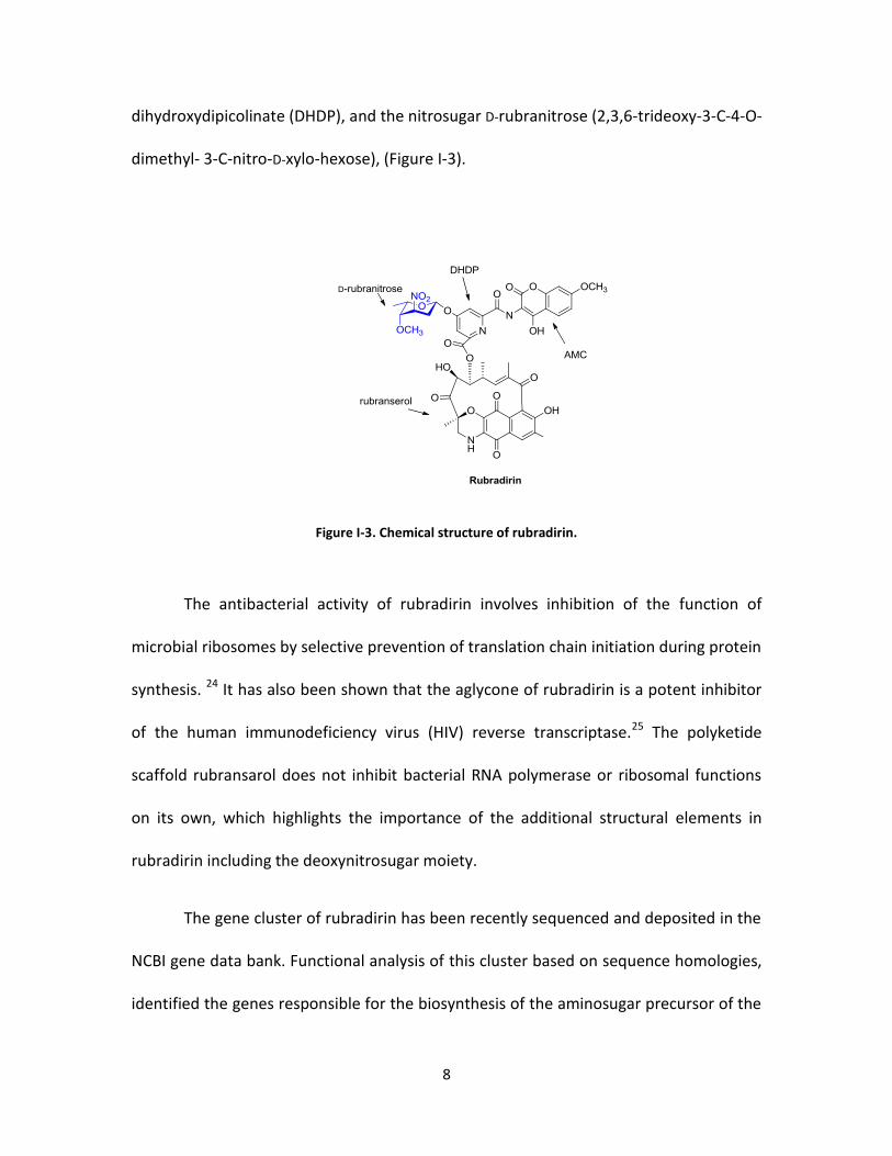
dihydroxydipicolinate (DHDP), and the nitrosugar D-rubranitrose (2,3,6-trideoxy-3-C-4-O-
dimethyl- 3-C-nitro-D-xylo-hexose), (Figure I-3).
Figure I-3. Chemical structure of rubradirin.
The antibacterial activity of rubradirin involves inhibition of the function of
microbial ribosomes by selective prevention of translation chain initiation during protein
synthesis. 24 It has also been shown that the aglycone of rubradirin is a potent inhibitor
of the human immunodeficiency virus (HIV) reverse transcriptase.25 The polyketide
scaffold rubransarol does not inhibit bacterial RNA polymerase or ribosomal functions
on its own, which highlights the importance of the additional structural elements in
rubradirin including the deoxynitrosugar moiety.
The gene cluster of rubradirin has been recently sequenced and deposited in the
NCBI gene data bank. Functional analysis of this cluster based on sequence homologies,
identified the genes responsible for the biosynthesis of the aminosugar precursor of the
9
nitrosugar D-rubranitrose.13 The lack of a gene that encodes a C-5 epimerase in the
rubradirin gene cluster is consistent with the D-configuration of the nitrosugar moiety.
Additionally, a putative oxidase gene, rubN8, was also found in the rubradirin gene
cluster and proposed to perform N-oxidation of an aminosugar precursor in the
biosynthesis of D-rubranitrose.
It is worth noting that only the nitroso congener of rubradrin (protorubradirin)
was isolated from the fermentation of the producer Streptomyces achromogenes var.
rubradiris when it was grown in complete darkness. After isolation, protorubradirin
readily converted to rubradin upon exposure to ambient light. Based on this
observation, it was proposed that the nitroso congener of rubradirin is the true
secondary metabolite and that the nitro group is formed through photooxidation.26
Kijanimicin
Kijanimicin, produced by Actinomadura kijaniata, is a spirotetronate antibiotic
and exhibits a broad range of antibacterial activity against Gram-positive bacteria,
anaerobes, and the malaria parasite Plasmodium falciparum.14 It has also been shown
that derivatives of kijanimicin possess potent activity against human liver and breast
cancer cell lines.27 The structure of kijanimicin includes a pentacyclic polyketide core,
linked to four L-digitoxose units and the nitrosugar, 2,3,4,6-tetradeoxy-4-
(methylcarbamyl)-3-C-methyl-3-nitro-D-xylo hexopyranose known as D-kijanose (Figure I-
4). Kijanimicin derivatives are also known to be produced by other high-GC Gram-

10
positive bacterial strains (actinomycetes) such as Streptomyces, Micromonospora,
Actinomadura, Saccharothrix, and Verrucosispora.14 Most members of this class of
compounds exhibit antibacterial and antitumor activities and many possess other
biological activities. Examples of these compounds include tetrocarcins and arisostatins,
which have been shown to be inducers of apoptosis;28 chlorothricins, as
anticholesterolemic agents;29 tetronothiodin, a cholecystokinin B (CCK-B) inhibitor;30
MM46115, an antiviral drug active against parainfluenzae virus 1 and virus 2.31
Figure I-4. Chemical structure of kijanimicin.
The deoxysugar moieties that decorate the polyketide core of kijanimicin play an
important role in the biological activity of this compound. Although no structure-activity
studies yet assed the importance of the D-kijanose sugar, it is likely that this unusually
modified deoxynitrosugar is important for biological activity.

11
Biosynthesis of deoxyaminosugars
Deoxyaminosuagrs are an important class of deoxysugar moieties biosynthesized
by a variety of organisms such as plants, fungi and bacteria.32 Based on gene functional
analysis of several natural products possessing N-oxidized deoxysugars, it is largely
thought that the precursors of these important moieties are deoxyaminosugars.
Therefore, understanding the biosynthetic pathways of these sugar precursors is very
helpful in the elucidation of the N-oxidation pathway. Before the introduction of the
amine group, the precursor sugar must be in its deoxygenated form. Enzymatic
deoxysugar modifications are always carried out on nucleotide activated sugars such as
thymidine diphosphate (TDP)-sugars. All known TDP-sugars are derived from glucose-1-
phosphate which is converted to TDP-glucose by a thymidilyl transferase and then to
TDP-4-keto-6-deoxy-D-glucose by TDP-D-glucose 4,6-dehydratase (Scheme I-1). This
provides the entry point for further deoxygenation and other enzymatic modification
steps including transamination.3
Scheme I-1. Entry point into TDP-deoxysugar secondary metabolism in bacteria.
Deoxyaminosugars are produced from their deoxygenated ketosugar precursors
via a transamination reaction carried out by a transaminase or an aminotransferase
12
which substitutes the keto group with an amino group. These enzymes are usually
dependent on pyridoxal phosphate (PLP), a cofactor that reacts with glutamate, which
transfers its α-amino group to PLP to make pyridoxamine phosphate (PMP). PMP then
transfers its nitrogen to the sugar, forming an aminosugar.33 Aminotransferase catalysis
proceeds via a highly conserved mechanism typically yielding regio- and
enantioselective amine installation.34
The structure of DesI, an aminotransferase involved in D-desosamine
biosynthesis in Streptomyces venezuelae, in the presence of PLP and the aminosugar
product revealed an external aldimine intermediate in which a lysine residue is in close
proximity to both C-4’ of PLP and the C-4 atom of the sugar substrate.35 This residue
likely plays a role in mediating the proton transfers that occurs during the
transamination yielding a C-4 equatorial amine installation. Unlike DesI, PseC another C-
4 aminotransferase from Helicobacter pylori, introduces a C-4 axial amino group into a
4-ketosugar.36 Interestingly, the hexose moiety was found to be rotated by 180° in PseC
compared to DesI resulting in this interesting opposite stereochemistry of the amino
group.
Biosynthesis of TDP-L-epi-vancosamine
The biosynthesis of TDP-L-epivancosamine (Scheme I-2) is a good example to
discuss because this unusual moiety is one of the most modified deoxyaminosgurs
found in natural products. Eight enzymatic steps are required for its biosynthesis

13
starting from glucose. There are two L-epivancosamine sugars linked to the aglycone
scaffold in chloroeremomycin, a member of the vancomycin family of glycopeptide
antibiotics produced by Amycolatopsis orientalis.37 The biosynthetic gene cluster of
chloroeremomycin has been sequenced which facilitated the in vitro studies of TDP-L-
epivancosamine bisosynthesis.38 In these studies, five enzymes from the
chloroeremomycin pathway, EvaA-E, have been shown to be involved in its biosynthesis
starting from TDP-6-deoxy-4-keto-D-glucose.39 These five enzymes were shown to
perform C-2 deoxygenation by EvaA, C-3 amination and methylation by EvaB and EvaC
respectively, C-5 epimerization by EvaD, and C-4 ketoreduction by EvaE (Scheme I-2).
To reconstitute the biosynthesis of TDP-L-epivancosamine in vitro, the TDP-
glucose 4,6-dehydratse (RfbB) from the rhamnose biosynthetic pathway, was used to
generate the entry sugar TDP-4-keto-6-deoxy-α-D-glucose.40 The RfbB activity requires
nicotinamide adenine dinucleotide phosphate (NADP+) which is bound to the enzyme. It
was shown that the dehydration by EvaA, 2,3-dehydratase, produces the unstable TDP-
linked 3,4-dioxo-6-deoxy sugar which is susceptible to TDP elimination with 1,2-olefin
formation.39 The 3-amino group is formed upon the activity of the aminotransferase,
EvaB, a PLP dependent enzyme. It was also shown that the activity of this enzyme is
enhanced by the inclusion of 1 mM glutamine in the assay. The C-3 methylation step is
carried out by the activity of EvaC, a SAM dependent methyltransferase. This
methylation is followed by the activity of the C-5 epimerase EvaD which results in a
change of the sugar configuration from D to L. The final step of TDP-L-epivancosamine
biosynthesis is the activity of the NADPH-dependent C-4 ketoreductase, EvaE, which

14
reduces the keto group on C-4 to a hydroxyl group. TDP-L-epivancosamine is then
attached to the aglycone scaffold of chloroeromomycin by a glycosyltransferase.
Scheme I-2. Biosynthesis of TDP-L-epi-vancosamine.
Comparative genomics of the nitro sugar functionality
The availability of gene cluster data of many isolated natural products provided
the opportunity to analyze genes based on sequence and propose functions prior to
carrying out biochemical characterization of the encoded enzymes. Sequence based
analysis alone is insufficient for identifying/discovering a precise biosynthetic gene
especially for large gene clusters that may contain up to 100 genes. However, multiple
gene clusters of related compounds can be analyzed via a comparative genomic
approach which can substantially simplify identifying genes of interest. Comparative
genomic analysis41 was performed on the related orthosomycins everninomicin and
avilamycin which is described below.42
15
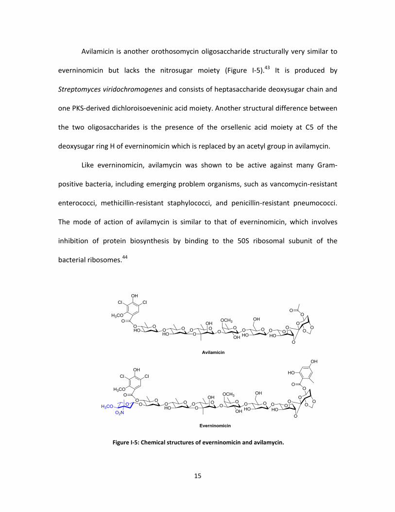
Avilamicin is another orothosomycin oligosaccharide structurally very similar to
everninomicin but lacks the nitrosugar moiety (Figure I-5).43 It is produced by
Streptomyces viridochromogenes and consists of heptasaccharide deoxysugar chain and
one PKS-derived dichloroisoeveninic acid moiety. Another structural difference between
the two oligosaccharides is the presence of the orsellenic acid moiety at C5 of the
deoxysugar ring H of everninomicin which is replaced by an acetyl group in avilamycin.
Like everninomicin, avilamycin was shown to be active against many Gram-
positive bacteria, including emerging problem organisms, such as vancomycin-resistant
enterococci, methicillin-resistant staphylococci, and penicillin-resistant pneumococci.
The mode of action of avilamycin is similar to that of everninomicin, which involves
inhibition of protein biosynthesis by binding to the 50S ribosomal subunit of the
bacterial ribosomes.44
Figure I-5: Chemical structures of everninomicin and avilamycin.
16
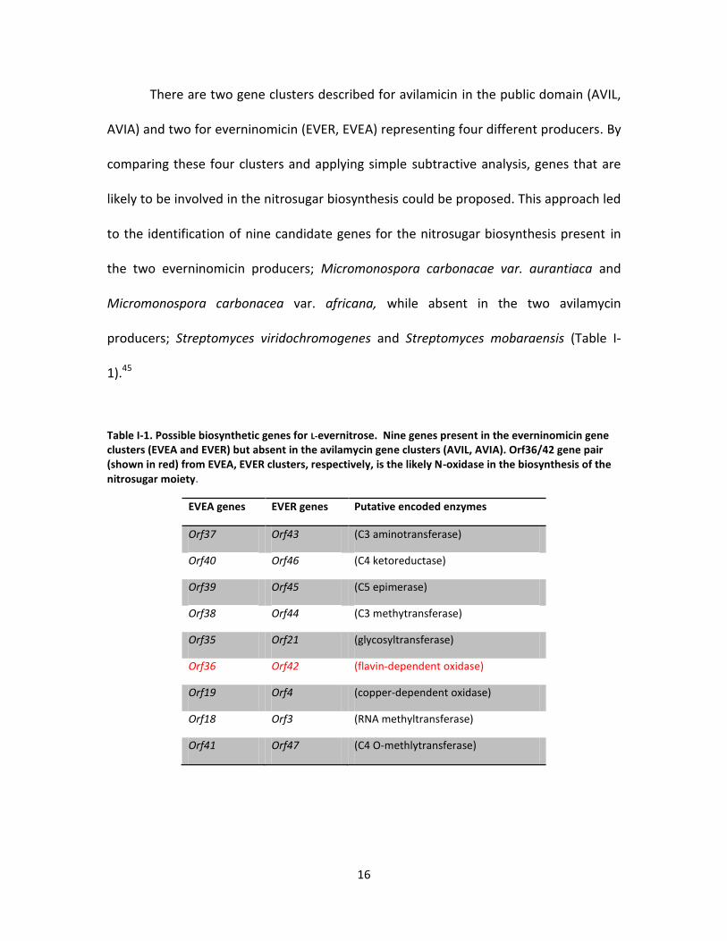
There are two gene clusters described for avilamicin in the public domain (AVIL,
AVIA) and two for everninomicin (EVER, EVEA) representing four different producers. By
comparing these four clusters and applying simple subtractive analysis, genes that are
likely to be involved in the nitrosugar biosynthesis could be proposed. This approach led
to the identification of nine candidate genes for the nitrosugar biosynthesis present in
the two everninomicin producers; Micromonospora carbonacae var. aurantiaca and
Micromonospora carbonacea var. africana, while absent in the two avilamycin
producers; Streptomyces viridochromogenes and Streptomyces mobaraensis (Table I-
1).45
Table I-1. Possible biosynthetic genes for L-evernitrose. Nine genes present in the everninomicin gene clusters (EVEA and EVER) but absent in the avilamycin gene clusters (AVIL, AVIA). Orf36/42 gene pair (shown in red) from EVEA, EVER clusters, respectively, is the likely N-oxidase in the biosynthesis of the nitrosugar moiety.
EVEA genes EVER genes Putative encoded enzymes
Orf37 Orf43 (C3 aminotransferase)
Orf40 Orf46 (C4 ketoreductase)
Orf39 Orf45 (C5 epimerase)
Orf38 Orf44 (C3 methytransferase)
Orf35 Orf21 (glycosyltransferase)
Orf36 Orf42 (flavin-dependent oxidase)
Orf19 Orf4 (copper-dependent oxidase)
Orf18 Orf3 (RNA methyltransferase)
Orf41 Orf47 (C4 O-methlytransferase)

17
Sequence homology analysis of these genes, allowed the identification of five
encoded enzymes involved in the biosynthesis of the aminosugar precursor that shared
high sequence similarity and identity with their counterparts in the well characterized
TDP-L-epivancosamine’s biosynthetic pathway.39 The nitrosugar L-evernitrose is
structurally very similar to TDP-L-epivancosamine with the difference being the N-
oxidation on C-3 and the C4 O-methylation in L-evernitrose. Among the genes shared
between the two pathways were genes that encode a C-3-aminotransferase, C-3
methyltransferase, C-5-epimerase and C-4-ketoreductase. The orf35/21 gene pair was
found to share sequence homology with genes encoding glycosyltransferases and hence
is possibly responsible for the glycoslation of L-evernitrose. The orf41/47 gene pair
shares sequence homology with C4 O-methyltransferases likely responsible for
introducing the methoxy group at C4 of the nitrosugar, a functionality that is replaced
with a hydroxyl in TDP-L-epivancosamine. The sequence homology analyses excluded
two of these nine genes as candidates for the biosynthesis of evernitrose. Namely, the
orf18/3 gene pair which has reasonable homology to RNA-methyltransferase genes and
the Orf19/4 gene pair homologous to copper-dependent oxidase genes involved in
primary metabolism. The only remaining and likely oxidase responsible for the oxidation
of the aminosugar among these nine gene pairs is orf36/42. The encoded proteins of
these genes have moderate sequence homology with the flavin dependent
monooxygenase dibenzothiophene oxidase DszC which has been shown to oxidize a
sulfide group to a sulfone via a sulfoxide intermediate.46 ORF36 also shares moderate
sequence similarity (~ 25%) with the acyl-CoA dehydrogenase family of enzymes. Acyl-

18
CoA dehydrogenases are flavin-dependent enzymes and are used as a structural model
for class-D flavin-dependent monooxygenases.
Proposed deoxysugar N-oxidation pathway
As mentioned above, the oxidase proposed to form the nitrosugar congener
from the deoxyaminosugar precursor in the everninomicin pathway is ORF36 from
Micromonospora carbonacea var. africana. This putative flavin-dependent enzyme
shares high sequence identity and similarity with other homologues in the biosynthetic
pathways of nitrosugar-containing natural products such as RubN8 from the rubradirin
pathway and KijD3 from the kijanimicin pathway. All of these putative N-oxidases share
moderate homology with the flavin-dependent dibenzothiophene oxidase DszC which
oxidizes dibenzothiophene (DBT) to DBT sulfone (DBTO2) as shown in scheme I-3. This
two-step oxidation requires the flavin mononucleotide (FMN) and the NADPH cofactors
as well as an external flavin reductase to provide reduced flavin which mediates the
monooxygenation catalysis. Reduced flavin is known to react with molecular oxygen to
form the C4a-hydroperoxyflavin, a reactive oxygen species that is responsible for the
electrophilic oxidation of the substrate.47 The oxygenation step results in the
incorporation of one oxygen atom into the substrate, usually as a hydroxyl group, and
the formation of the C4a-hydroxyflavin. The hydroxyflavin readily loses a water
molecule and is recycled to participate in further rounds of oxidations.

19
Schem I-3. Oxidation of dibenzothiophene by DszC.
The oxidation mechanism for the aminosugars by the putative flavin-dependent
oxidases could be envisioned in a similar manner whereby the reduced flavin is provided
by the function of an external flavin reductase. Monooxygenation mediated by C4a-
hydroperoxyflavin results in the formation of a hydroxylamine intermediate and
consequently, similar flavin-dependent oxidation steps result in the production of
further oxidized intermediates perhaps via the nitroso and ultimately the nitro
functionality.
Anthracyclines and deoxyaminosugar oxidation
A brief history of anthracycline drugs
Anthracyclines are another class of natural products with deoxysugar moieties
attached to their tetracyclic polyketide cores. Some anthracycline compounds are
considered the most effective anticancer drugs and possess a wide range of activity
against several types of human cancers.48-50 The first discovered anthracyclines were
doxorubicin and daunorubicin when they were isolated from Streptomyces peucetius in
early 1960s.5, 51 The tetracyclic structure of doxorubicin and daunorubicin includes
quinone-hydroquinone groups represented by rings C-B (Figure I-6). The sugar moiety in

20
these two compounds is 3-amino-2,3,6-trideoxy-L-fucose, also known as daunosamine.
Doxorubicin and daunorubicin share the same tetracyclic core with the difference of a
hydroxyl group on C-13 in doxorubicin. This small structural difference has significant
effect on the spectrum of the anticancer activity of doxorubicin vs. daunarubicin. For
example doxorubicin and some of its analogs were found to have less acute toxicity,
cause less cardiomyopathy, and in general more potent anticancer activity.52
Additionally, daunorubicin shows activity in acute lymphoblastic or myeloblastic
leukemias53 whereas doxorubicin is more active against breast cancer, childhood solid
tumors, soft tissue sarcomas, and aggressive lymphomas.54
Figure I-6. Chemical structure of some anthracyclines.
The use of doxorubicin and daunorubicin in clinic is facing some challenges such
as the development of resistance in tumor cells or toxicity in healthy tissues, resulting in

21
some cases in chronic cardiomyopathy and congestive heart failure (CHF) among other
side effects. Therefore, there is a need for improved anthracycline analogs with varied
pharmacokinetics for the treatment of different types of cancer. Hundreds of new
doxorubicin and daunorubicin analogs were synthesized but only few of them made it to
clinical development and approval.52 Among the best approved analogues were
epirubicin55-58 and idarubicin59-61, alternatives to doxorubicin and daunorubicin,
respectively. The only difference between epirubicin and doxorubicin is the
epimerization at C-4’ of daunosamine changing the orientation of the hydroxyl from the
axial in doxorubicin to the equatorial in epirubicin. Interestingly, this minor structural
difference resulted in significant changes in the pharmacokinetic properties which led to
improvements in distribution volume and total body clearance.53 The idarubicin
analogue of daunorubicin is made from the removal of the 4-methoxy group in ring-D
and was shown to possess broader spectrum of anticancer activity. It was speculated
that this improved activity may be attributed to increased lipophilicty and stabilization
of the drug-topoisomerase-DNA ternary complex.62 Other approved anthracyclin
analogues include pirarubicin, aclacinomycin A (aclarubicin), 63-64 and mitoxantrone65-67
(a substituted aglyconic anthraquinone).
Biosynthesis of baumycin
Baumycin is an another anthracycline compound derived from daunorubicin and
has been shown to exhibit high efficacy against gram-positive bacteria and certain
cancer cell lines such as leukemia cells (L1210). The structure of baumycin includes a

22
non-sugar acetal moiety attached at C-4’ position of the daunosamine sugar (Figure I-
6).68-71
In Streptomyces peucetius, the polyketide core in baumycin is produced by a
Type II polyketide synthase (PKS). The first 3-carbons of the 21-carbon decaketide chain
are synthesized from the incorporation of a single propinyl starter unit from propinyl-
CoA followed be 9 iterative condensations of malonyl extender units.72 The minimal PKS
proteins that catalyze the formation of this long chain polyketide are an acyl carrier
protein (ACP), ketosynthase (KS) and a malonyl-CoA:ACP acyltransferase (MAT).73 The
polyketide chain is converted to 12-deoxyalkalonic acid catalyzed by the dps gene
cluster which includes DpsE, an NADPH dependent 9-ketoreductase, and cyclases DpsF
and DpsY that catalyze the formation of ring D and C, respectively. The subsequent
polyketide modifications are catalyzed by the dnr gene cluster which includes C-12
oxygenase, a SAM dependent alkalonic acid methyltransferase, ring A cyclase, C-7
ketoreductase, and a C-11 hydroxylase.74 This series of polyketide modifications yields ɛ-
rhodomycinone (Figure I-6), the precursor for doxorubicin and daunorubicin. The
daunosamine sugar is then attached by DnrS, a glycosyltransferase followed by
additional modifications on the tetracyclic core to yield daunorubicin75, which is a
precursor for doxorubicin and baumycin. The biosynthesis of the actal moiety at C-4’
position of the daunosamine sugar is unclear; however, based on gene cluster analysis;
a possible pathway is proposed and discussed below.

23
Deoxysugar genes in anthracyclines
The biosynthetic genes of the deoxysugar L-danosamine in doxorubicin,
daunorubicin and baumycin were assigned based on sequence homologies with other
sugar modifying enzymes and further studied by gene manipulation. It was shown that
the minimal enzymes required for L-daunosamine biosynthesis and attachment are
encoded by the dnmLMJVUTS genes.76 These genes were cloned into the heterologous
host Streptomyces lividans and the resultant recombinant strain was fed with the
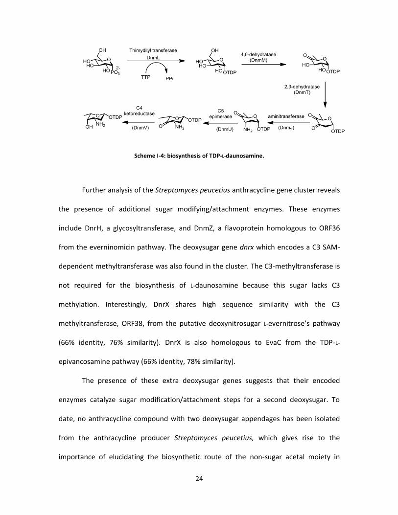
aglycone ɛ-rhodomycinone. Scheme I-4 illustrates the enzymatic steps required for the
biosynthesis of TDP-L-daunosamine starting from D-glucose-1-phosphate. The DnmL
enzyme is highly homologous to the well-known glucose-1-phosphate
thymidylyltransferases which catalyze the conversion of D-glucose-1-phosphate into
TDP-D-glucose.77 This sugar is converted to TDP-6-deoxy-4-keto-D-glucose catalyzed by
DnmM, a TDP-D-glucose 4,6-dehydratase. The 4-keto sugar is then further dehydrated
by DnmT, a 2,3-dehydratase to yield the 3,4-dioxo-6-deoxysugar nucleotide which can
be transaminated by DnmJ, a C-3 aminotransferase. At this stage, the sugar can undergo
epimerization by DnmU, a C-5 epimerase resulting in configuration change from D- to L-
deoxysugar. The last sugar modification before glycoslation appears to be catalyzed by
DnmV, a 4-ketoreductase, which yields the TDP-L-daunosamine, the sugar donor
substrate of DnmS, a glycosyltransferase. It was also shown that the aglycon of DnmS is
ɛ-rhodomycinone, a precursor for the anthracyclines doxorubicin and daunorubicin.
24
Scheme I-4: biosynthesis of TDP-L-daunosamine.
Further analysis of the Streptomyces peucetius anthracycline gene cluster reveals
the presence of additional sugar modifying/attachment enzymes. These enzymes
include DnrH, a glycosyltransferase, and DnmZ, a flavoprotein homologous to ORF36
from the everninomicin pathway. The deoxysugar gene dnrx which encodes a C3 SAM-
dependent methyltransferase was also found in the cluster. The C3-methyltransferase is
not required for the biosynthesis of L-daunosamine because this sugar lacks C3
methylation. Interestingly, DnrX shares high sequence similarity with the C3
methyltransferase, ORF38, from the putative deoxynitrosugar L-evernitrose’s pathway
(66% identity, 76% similarity). DnrX is also homologous to EvaC from the TDP-L-
epivancosamine pathway (66% identity, 78% similarity).
The presence of these extra deoxysugar genes suggests that their encoded
enzymes catalyze sugar modification/attachment steps for a second deoxysugar. To
date, no anthracycline compound with two deoxysugar appendages has been isolated
from the anthracycline producer Streptomyces peucetius, which gives rise to the
importance of elucidating the biosynthetic route of the non-sugar acetal moiety in

25
baumycin. One possibility could be that the formation of this unusual moiety is a result
of terminal deoxysugar degradation whereby the sugar undergoes a C-C bond cleavage,
perhaps combined with other chemical/enzymatic steps. To resolve this intriguing
mystery, it is essential to biochemically confirm the putative roles of these additional
deoxysugar enzymes. The glycosyltransferase DnrH and the C-4 ketoreductase DnrX
have well known and characterized homologues in other deoxysugar pathways hence,
their characterization seems less attractive. Elucidation of the function of the putative
flavin-dependent enzyme DnmZ however, seems more interesting considering its
homology to ORF36, RubN8, and KijD3 proposed to be involved in deoxyaminosugar
oxidation.
The putative role of the flavoenzyme DnmZ
As mentioned above, DnmZ shares high sequence homology with the proposed
flavin-dependent enzymes ORF36, RubN8 and KijD3 proposed to be involved in
deoxyaminosugar oxidation. For example, DnmZ shares 70% sequence similarity with
ORF36 (59% Identity). This suggests that all of these enzymes likely catalyze a similar
deoxyaminosugar oxidation reaction. Given the fact that there are no deoxysugar
moieties with N-oxidation in any of the anthracycline derivatives produced by
Streptomyces peucetius isolated to date, proposing a role for DnmZ seems difficult;
however, one scenario can be envisioned whereby DnmZ catalyzes a deoxyaminosugar
oxidation similar to that proposed for its homologues, whereby the resulting sugar
26
moiety undergoes further enzymatic or chemical transformations either before or after
glycoslation.
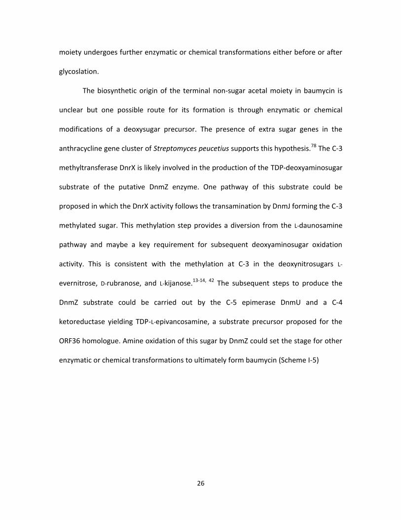
The biosynthetic origin of the terminal non-sugar acetal moiety in baumycin is
unclear but one possible route for its formation is through enzymatic or chemical
modifications of a deoxysugar precursor. The presence of extra sugar genes in the
anthracycline gene cluster of Streptomyces peucetius supports this hypothesis.78 The C-3
methyltransferase DnrX is likely involved in the production of the TDP-deoxyaminosugar
substrate of the putative DnmZ enzyme. One pathway of this substrate could be
proposed in which the DnrX activity follows the transamination by DnmJ forming the C-3
methylated sugar. This methylation step provides a diversion from the L-daunosamine
pathway and maybe a key requirement for subsequent deoxyaminosugar oxidation
activity. This is consistent with the methylation at C-3 in the deoxynitrosugars L-
evernitrose, D-rubranose, and L-kijanose.13-14, 42 The subsequent steps to produce the
DnmZ substrate could be carried out by the C-5 epimerase DnmU and a C-4
ketoreductase yielding TDP-L-epivancosamine, a substrate precursor proposed for the
ORF36 homologue. Amine oxidation of this sugar by DnmZ could set the stage for other
enzymatic or chemical transformations to ultimately form baumycin (Scheme I-5)

27
SchemI-5. Proposed pathway of late-step baumycin biosynthesis including the role of DnmZ.
Flavoprotein monooxygenases
As discussed earlier and based on sequence homologies, the enzyme responsible
for the formation of the nitro functionality in nitrosugar-containing natural products is
likely a flavin-dependent monooxygenase. Enzymes of this family are capable of efficient
and specific insertion of one or more oxygen atoms into an organic substrate, a reaction
not easy to perform via traditional chemical syntheses.79 Although many chemical
catalysts have been designed to address the difficulty of oxygenation reactions, the
exquisite specificity and efficiency of monooxygenases remain unmatched. The
discovery and characterization of a new flavin-dependent monooxygenase, will extend
our knowledge of important biosynthetic pathways, support the current efforts of
natural product structure diversification, as well as introduce new efficient biocatalysts

28
to perform difficult oxygenation reactions. Below is a brief background on flavoproteins
with emphasis on flavin-dependent monooxygenases.
Biochemistry of flavoproteins
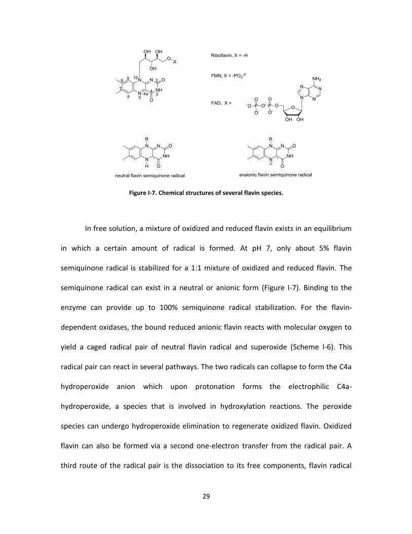
Flavin exists in nature in three principle forms; riboflavin (vitamin B2), flavin
mononucleotide (FMN), and flavin adenine dinucleotide (FAD) as shown in figure I-7.80
The FMN and FAD forms are the prosthetic groups for flavoproteins, and there are well
characterized mechanisms for their interconversions. Flavins have bright yellow color,
like most flavoproteins, and a characteristic UV-absorption that changes significantly
depending on the oxidation state of the flavin. This unique spectrophotpmetric property
allowed the study of the catalysis of many flavoproteins. Flavins undergo one electron
reduction to give stable semiquinone radicals allowing them to mediate between the
common two electron oxidations (e.g NAD(P)+/NAD(P)H) and one electron oxidations
carried out by heme or iron-sulfur cluster proteins.
29
Figure I-7. Chemical structures of several flavin species.
In free solution, a mixture of oxidized and reduced flavin exists in an equilibrium
in which a certain amount of radical is formed. At pH 7, only about 5% flavin
semiquinone radical is stabilized for a 1:1 mixture of oxidized and reduced flavin. The
semiquinone radical can exist in a neutral or anionic form (Figure I-7). Binding to the
enzyme can provide up to 100% semiquinone radical stabilization. For the flavin-
dependent oxidases, the bound reduced anionic flavin reacts with molecular oxygen to
yield a caged radical pair of neutral flavin radical and superoxide (Scheme I-6). This
radical pair can react in several pathways. The two radicals can collapse to form the C4a
hydroperoxide anion which upon protonation forms the electrophilic C4a-
hydroperoxide, a species that is involved in hydroxylation reactions. The peroxide
species can undergo hydroperoxide elimination to regenerate oxidized flavin. Oxidized
flavin can also be formed via a second one-electron transfer from the radical pair. A
third route of the radical pair is the dissociation to its free components, flavin radical
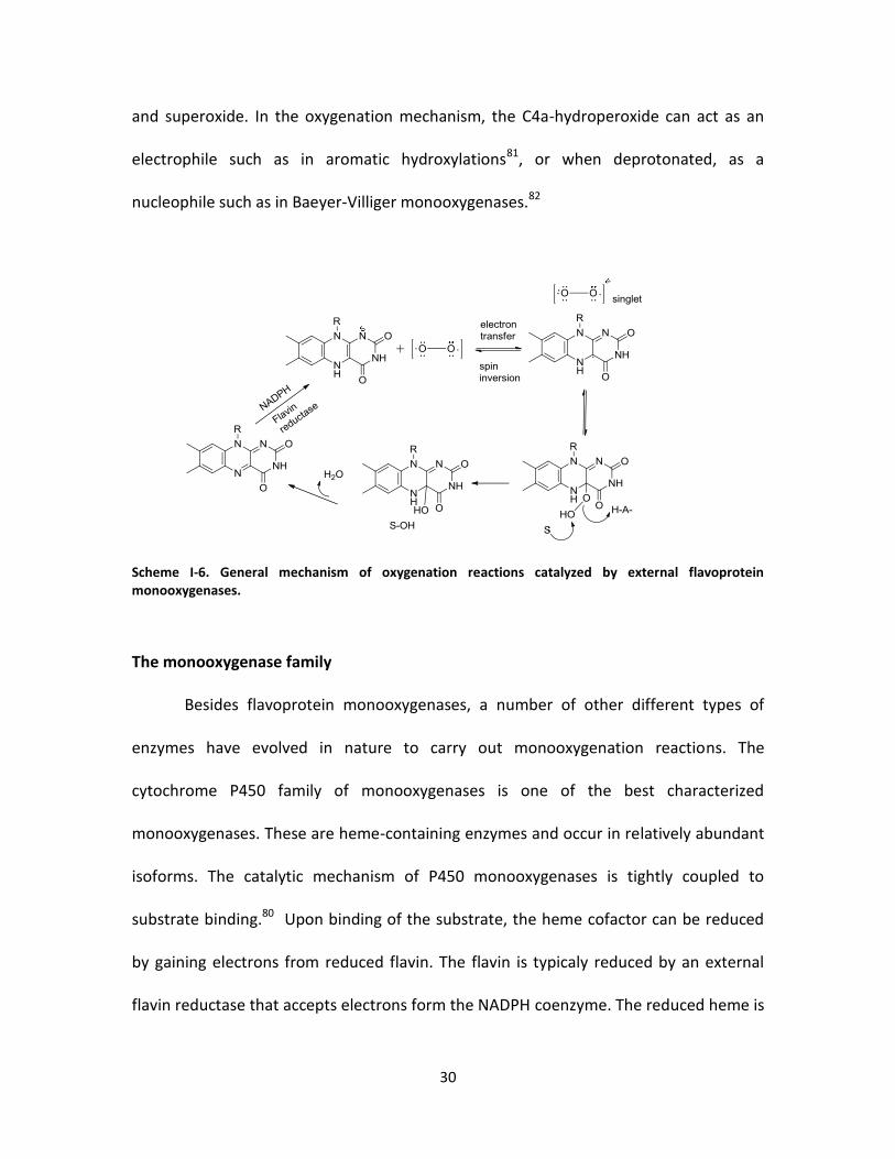
30
and superoxide. In the oxygenation mechanism, the C4a-hydroperoxide can act as an
electrophile such as in aromatic hydroxylations81, or when deprotonated, as a
nucleophile such as in Baeyer-Villiger monooxygenases.82
Scheme I-6. General mechanism of oxygenation reactions catalyzed by external flavoprotein monooxygenases.
The monooxygenase family
Besides flavoprotein monooxygenases, a number of other different types of
enzymes have evolved in nature to carry out monooxygenation reactions. The
cytochrome P450 family of monooxygenases is one of the best characterized
monooxygenases. These are heme-containing enzymes and occur in relatively abundant
isoforms. The catalytic mechanism of P450 monooxygenases is tightly coupled to
substrate binding.80 Upon binding of the substrate, the heme cofactor can be reduced
by gaining electrons from reduced flavin. The flavin is typicaly reduced by an external
flavin reductase that accepts electrons form the NADPH coenzyme. The reduced heme is
31
then used to complete the monooxygenation of the substrate. Cytochrome P450
monooxygenases are cabaple of hydroxylating carbon atoms regioselectively, which has
been shown to be of great value to modify sterols and steroids.
Other classes of monooxygenases include non-heme monooxygenases83 and
copper-dependent monooxygenases.84 A few new types of monooxygenases have been
discovered in recent years that do not contain the aforementioned cofactors including
the polyketide monooxygenase ActVA-Orf6, involved in actinorhodin biosynthesis in
Streptomyces coelicolor.81 Another example is the quinol monooxygenase YgiN, from
Escherichia coli which oxidizes multiringed aromatic substrates without the participation
of any cofactor.85 Aclacinomycin-10-hydroxylase, involved in anthracycline biosynthesis
in Streptomyces purpurascens, is another rare type of monooxygenases as it depends on
S-adenosyl-l-methionine as a cofactor.86 Flavin-dependent monooxygenases are perhaps
the most ubiquitous of monooxygenases. They perform a wide range of
monooxygenation reactions with high regio- and/or enantioselectivity. A brief
background on flavin-dependent monooxygenases is disussed below.
Flavoprotein monooxygenase catalysis
Flavoprotein monooxygenases are a broad group of enzymes which share similar
properties and mechanistic features.87 One common property of these enzymes is the
use of NADH or NADPH to supply the reduced flavin which reacts with molecular oxygen
to form C4a-peroxide required for oxygenation of the substrate. When protonated, the
C4a-peroxide is a potent electrophile and can participate in hydroxylation reactions such

32
as aromatic hydroxylations.81 The flavin peroxide anion can also react as a nucleophile
and is employed by a second subgroup of monooxygenases an example of which is
cyclohexanone monooxygenase.88 These two subgroups of monooxygenases share
common but distinctive properties. In most electrophilic monooxygenases, there is an
exquisite control mechanism to ensure that the NAD(P)H cofactor is only used when the
substrate is bound to the enzyme for oxygenation.80 Substrate binding is required for
rapid reduction of flavin by NAD(P)H. For the nucleophilic monooxygenases, substrate
binding is usually not required for flavin reduction but the C4a-peroxide formed upon
reaction with O2 stabilized by the protein is only reactive in the presence of the
substrate for oxygen transfer.89 In contrary, the C4a-hydroperoxide in electrophilic
monooxygenases is very unstable in the absence of the substrate.
One of the most well studied electrophilic monoxygenases is p-hydroxybenzoate
hydroxylase and its basic mechanism appears to be followed by most members of this
class.90 These enzymes can be studied spectrophotometrically exploiting the flavin
absorbance and fluorescence properties.91 The mechanism of p-hydroxybenzoate
hydroxylase monooxygenation starts with forming the flavin-NADPH-substrate ternary
complex resulting in a long-wavelength-absorbing charge transfer between the NADPH
as a donor and oxidized flavin as an acceptor (Scheme I-7). The next step is the
reduction of the flavin cofactor which is also accompanied by significant change in
absorbance and fluorescence causing another long wavelength charge transfer in which
reduced flavin is the donor and NADP+ is the acceptor. The NADP+ species is then
released forming the enzyme-reduced flavin-substrate complex. The resulting anionic
33
reduced flavin can then react with molecular oxygen forming the enzyme-C4a-
hydroperoxide-substrate intermediate complex. This intermediate has distinctive
spectrum with a wavelength maximum at 380 nm which is formed with a pseudo first
order constant directly proportional to the oxygen concentration. Oxygen transfer from
the hydroperoxyflavin can then take place resulting in a complex of enzyme C4a-
hydroxyflavin and the non-aromatic metastable dienone (intermediate III). The following
step is the re-aromatization to form intermediate III that includes the more stable
hydroxylated product, a step that occurs at sufficiently slow rate that allows the
determination of K6. The final step is the dehydration of the C4a-hydroxyflavin to recycle
the oxidized enzyme ready for the next round of catalysis.

34
Scheme I-7 . Reaction mechanism of p-hydroxybenzoate monooxygenase.
For p-hydroxybenzoate hydroxylase, it has been found that the flavin exisits in
two different conformations: one is mostly buried in the protein core and ideally
positioned for hydroxylation and the other is partially solvent exposed and suitable for
reduction by NADPH.92 The mobility of the flavin cofactor has been also confirmed in the
structural studies of the related phenol hydroxylase.93
In nucleophilic monooxygenation, O2 reacts with reduced flavin to form the
flavin peroxide. This is the nucleophilic flavin-oxygen intermediate that participates in
the oxygen transfer to the substrate as in Baeyer-Villiger monooxygenases.82, 94 In
cyclohexanone monooxygenase, the flavin C4a-peroxide formed after the reaction of
reduced flavin with molecular oxygen can be slowly protonated to yield the C4a-
hydroperoxyflavin, or it can react with cyclohexanone as a nucleophile to form the

35
Criege intermediate which rearranges to form ɛ-carbolactam.95 The resulting
hydroxyflavin undergoes elimination of water regenerating the oxidized flavin for next
cycle of catalysis.96-97 The NADP+ species must be bound during the reaction to protect
the reactive C4a-hydroperoxide.
Classifications of Flavoprotein monooxygenases
Several hundred flavoenzymes have been characterized to date.98-99 Most of
these enzymes contain a non-covalently bound flavin in the form of FMN or FAD but
there are some enzymes that bind the flavin cofactor covalently. Examples of flavin
covalently bound enzymes include vanillyl-alcohol oxidase which contains FAD bound to
a histidine residue.100 In p-cresol methyl hydroxylase, the FAD is linked to a tyrosine
residue. It has been shown that this covalent linkage is beneficial for catalysis in the case
of vanillyl-alcohol oxidase.101 For all internal or external flavoprotein monooxygnases
however, the flavin cofactor is not covalently linked to the enzyme.
Classification of the big family of flavoenzymes has been established based on
different criteria such as the types of reactions they catalyze, cofactor requirements,
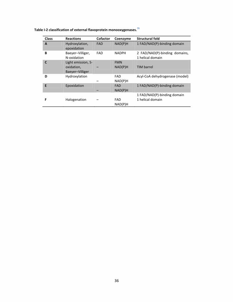
sequence homology and structural folds. More recently, external flavoprotein
monooxygenases have been classified by following similar criteria mentioned above.
They were divided into six different subclasses A-F as shown in table I-2. These
subclasses were discriminated based on sequence similarty, specific structural motifs,
types of reactions and cofactor requirements. Structures of some prototype flavin-
dependent monoxygenases are shown in figure I-8.79
36
Table I-2 classification of external flavoprotein monooxygenases.79
Class Reactions Cofactor Coenzyme Structural fold
A Hydroxylation, epoxidation
FAD NAD(P)H 1 FAD/NAD(P)-binding domain
B Baeyer–Villiger, N-oxidation
FAD NADPH 2 FAD/NAD(P)-binding domains, 1 helical domain
C Light emission, S-oxidation, Baeyer–Villiger
–
FMN NAD(P)H
TIM barrel
D Hydroxylation –
FAD NAD(P)H
Acyl-CoA dehydrogenase (model)
E Epoxidation –
FAD NAD(P)H
1 FAD/NAD(P)-binding domain
F
Halogenation
–
FAD NAD(P)H
1 FAD/NAD(P)-binding domain 1 helical domain
37
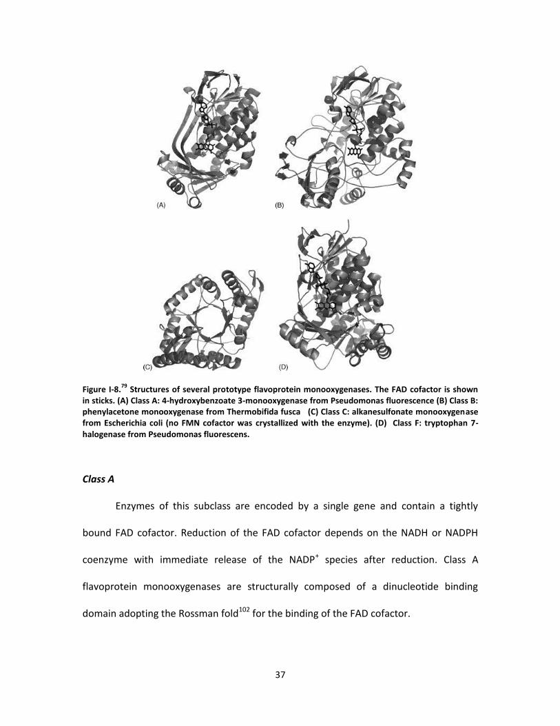
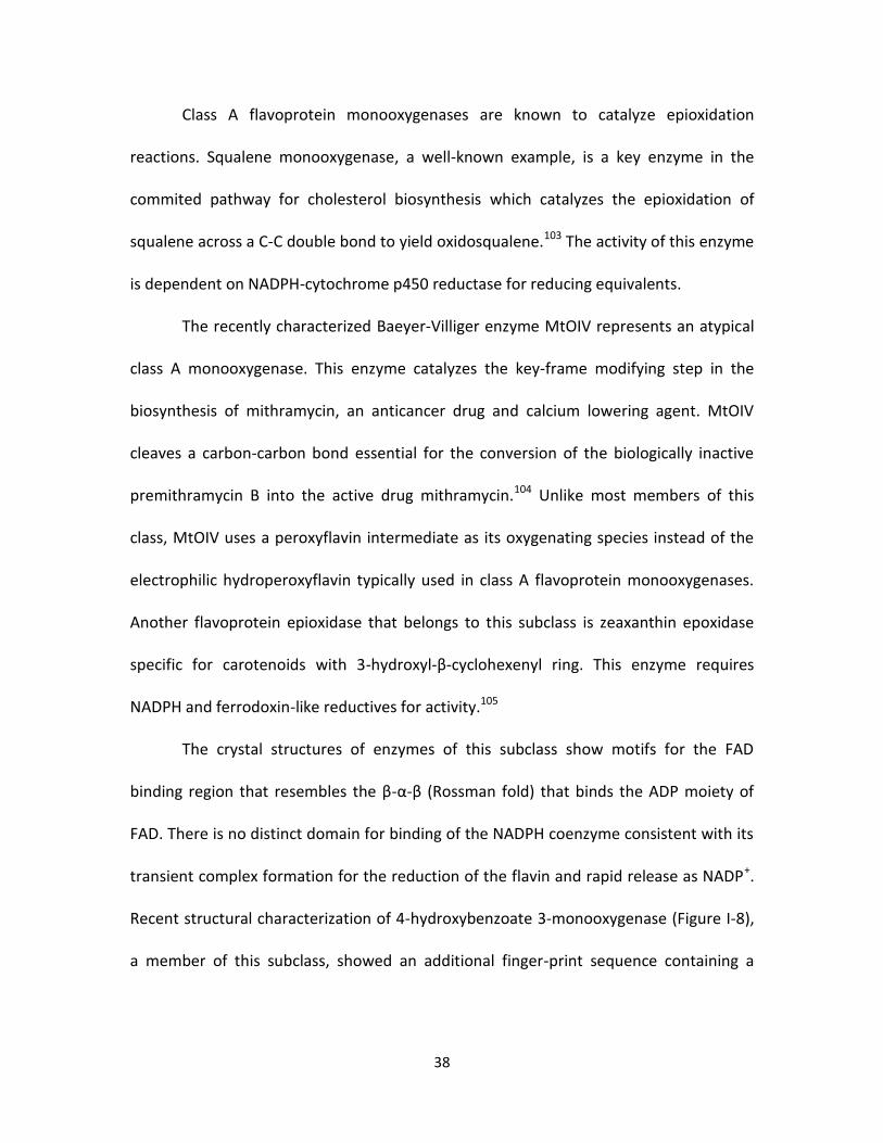
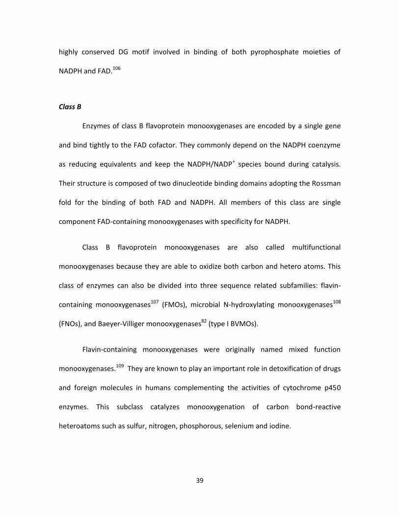
Figure I-8.79
Structures of several prototype flavoprotein monooxygenases. The FAD cofactor is shown in sticks. (A) Class A: 4-hydroxybenzoate 3-monooxygenase from Pseudomonas fluorescence (B) Class B: phenylacetone monooxygenase from Thermobifida fusca (C) Class C: alkanesulfonate monooxygenase from Escherichia coli (no FMN cofactor was crystallized with the enzyme). (D) Class F: tryptophan 7-halogenase from Pseudomonas fluorescens.
Class A
Enzymes of this subclass are encoded by a single gene and contain a tightly
bound FAD cofactor. Reduction of the FAD cofactor depends on the NADH or NADPH
coenzyme with immediate release of the NADP+ species after reduction. Class A
flavoprotein monooxygenases are structurally composed of a dinucleotide binding
domain adopting the Rossman fold102 for the binding of the FAD cofactor.
38
Class A flavoprotein monooxygenases are known to catalyze epioxidation
reactions. Squalene monooxygenase, a well-known example, is a key enzyme in the
commited pathway for cholesterol biosynthesis which catalyzes the epioxidation of
squalene across a C-C double bond to yield oxidosqualene.103 The activity of this enzyme
is dependent on NADPH-cytochrome p450 reductase for reducing equivalents.
The recently characterized Baeyer-Villiger enzyme MtOIV represents an atypical
class A monooxygenase. This enzyme catalyzes the key-frame modifying step in the
biosynthesis of mithramycin, an anticancer drug and calcium lowering agent. MtOIV
cleaves a carbon-carbon bond essential for the conversion of the biologically inactive
premithramycin B into the active drug mithramycin.104 Unlike most members of this
class, MtOIV uses a peroxyflavin intermediate as its oxygenating species instead of the
electrophilic hydroperoxyflavin typically used in class A flavoprotein monooxygenases.
Another flavoprotein epioxidase that belongs to this subclass is zeaxanthin epoxidase
specific for carotenoids with 3-hydroxyl-β-cyclohexenyl ring. This enzyme requires
NADPH and ferrodoxin-like reductives for activity.105
The crystal structures of enzymes of this subclass show motifs for the FAD
binding region that resembles the β-α-β (Rossman fold) that binds the ADP moiety of
FAD. There is no distinct domain for binding of the NADPH coenzyme consistent with its
transient complex formation for the reduction of the flavin and rapid release as NADP+.
Recent structural characterization of 4-hydroxybenzoate 3-monooxygenase (Figure I-8),
a member of this subclass, showed an additional finger-print sequence containing a
39
highly conserved DG motif involved in binding of both pyrophosphate moieties of
NADPH and FAD.106
Class B
Enzymes of class B flavoprotein monooxygenases are encoded by a single gene
and bind tightly to the FAD cofactor. They commonly depend on the NADPH coenzyme
as reducing equivalents and keep the NADPH/NADP+ species bound during catalysis.
Their structure is composed of two dinucleotide binding domains adopting the Rossman
fold for the binding of both FAD and NADPH. All members of this class are single
component FAD-containing monooxygenases with specificity for NADPH.
Class B flavoprotein monooxygenases are also called multifunctional
monooxygenases because they are able to oxidize both carbon and hetero atoms. This
class of enzymes can also be divided into three sequence related subfamilies: flavin-
containing monooxygenases107 (FMOs), microbial N-hydroxylating monooxygenases108
(FNOs), and Baeyer-Villiger monooxygenases82 (type I BVMOs).
Flavin-containing monooxygenases were originally named mixed function
monooxygenases.109 They are known to play an important role in detoxification of drugs
and foreign molecules in humans complementing the activities of cytochrome p450
enzymes. This subclass catalyzes monooxygenation of carbon bond-reactive
heteroatoms such as sulfur, nitrogen, phosphorous, selenium and iodine.

40
N-hydroxylating monooxygenases catalyze the N-hydroxylation of primary
amines and hence, play an important role in the biosynthesis of bacterial siderophores.
They have sequence homology with flavin-containing monooxygenases and require
NADPH and FAD for activity. Unlike flavin-containing monooxygenases, N-hydroxylating
monooxygenases show lower affinity for FAD which hindered mechanistic studies of
their catalysis. Ornithine hydroxylase (PvdA) catalyzes the hydroxylation of the side
chain primary amine of ornithine in the initial step of the biosynthesis of the
Pseudomonas aeruginosa siderophore pyoverdin.108 Kinetic studies of this enzyme
showed that binding of the substrate target is not required to trigger reduction of the
flavin by NADPH.110
Class B Bayer-Villiger monooxygenases (BVMOs), catalyze an atypical
oxygenation reaction converting a ketone or an aldehyde to ester or lactone.111 One of
the first studied enzymes of this family is cyclohexanone monooxygenase, from
actinobacter sp. NCBBI 9871.112 This enzyme performs Baeyer-Villiger oxidations on a
wide variety of cyclic ketones with exquisite regio- and enantioselectivity. Bayer-Villiger
monooxygenases can be identified based on the presence of two Rossman fold domains
for FAD and NADPH binding. It was shown that phenylacetone monooxygenase, a well-
characterized Baeyer-Villiger monooxygenase and member of this subfamily, has
dinucleotide binding domains flanked by two helical domains which are unique for this
type of monooxygenases.113
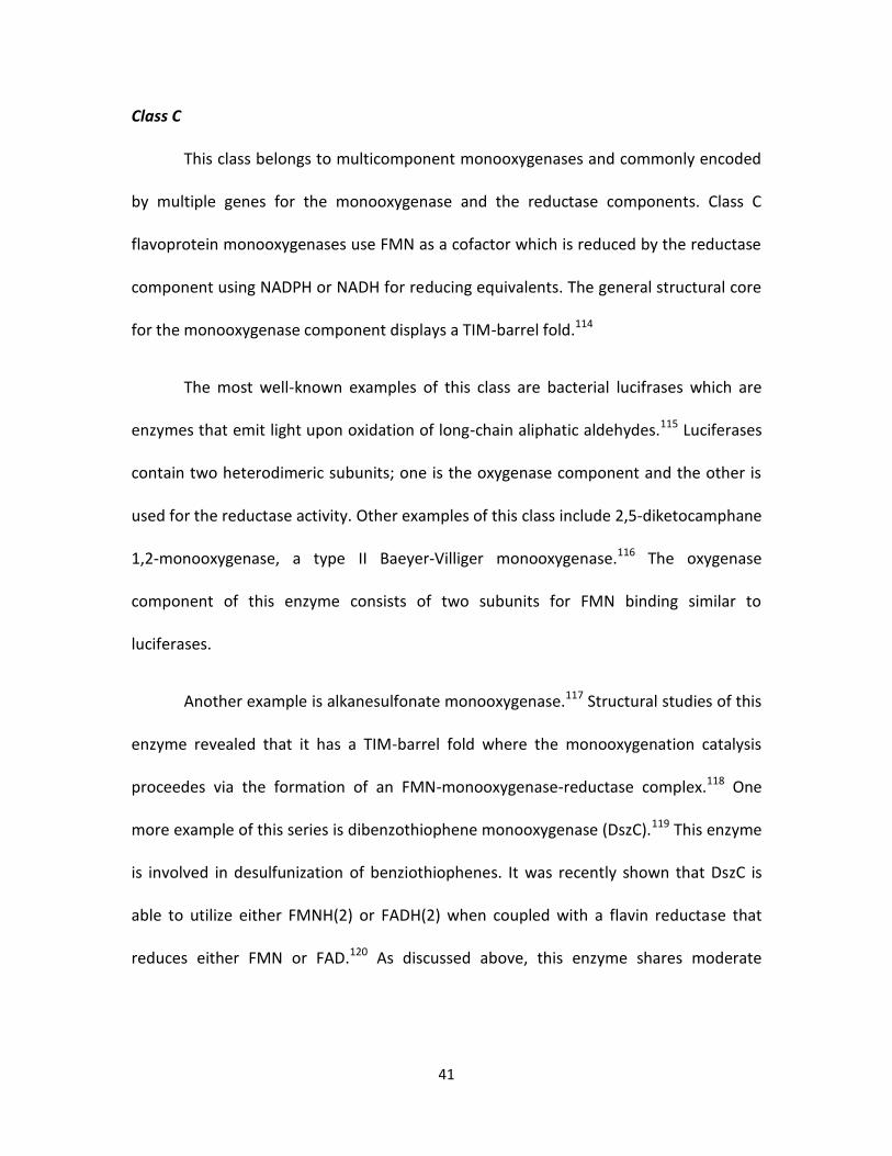
41
Class C
This class belongs to multicomponent monooxygenases and commonly encoded
by multiple genes for the monooxygenase and the reductase components. Class C
flavoprotein monooxygenases use FMN as a cofactor which is reduced by the reductase
component using NADPH or NADH for reducing equivalents. The general structural core
for the monooxygenase component displays a TIM-barrel fold.114
The most well-known examples of this class are bacterial lucifrases which are
enzymes that emit light upon oxidation of long-chain aliphatic aldehydes.115 Luciferases
contain two heterodimeric subunits; one is the oxygenase component and the other is
used for the reductase activity. Other examples of this class include 2,5-diketocamphane
1,2-monooxygenase, a type II Baeyer-Villiger monooxygenase.116 The oxygenase
component of this enzyme consists of two subunits for FMN binding similar to
luciferases.
Another example is alkanesulfonate monooxygenase.117 Structural studies of this
enzyme revealed that it has a TIM-barrel fold where the monooxygenation catalysis
proceedes via the formation of an FMN-monooxygenase-reductase complex.118 One
more example of this series is dibenzothiophene monooxygenase (DszC).119 This enzyme
is involved in desulfunization of benziothiophenes. It was recently shown that DszC is
able to utilize either FMNH(2) or FADH(2) when coupled with a flavin reductase that
reduces either FMN or FAD.120 As discussed above, this enzyme shares moderate

42
homology with the flavin-dependent monooxygenase proposed to catalyze the
oxidation of deoxyaminosugars.
Class D
This class of flavoprotein monooxygenases is typically encoded by two genes;
one is for the monooxygenase and the other is for the flavin reductase component. The
reductase component uses FAD as a cofactor and NADH or NADPH as a coenzyme.
These enzymes are structurally homologous to acyl-CoA dehydrogenases and are mostly
α-helical proteins.
Members of this class are typically active on regioselective hydroxylation of
aromatic substrates. The prototype is 4-hydroxyphenylacetate 3-monooxygenase
(Figure I-8) which catalyzes the conversion of 4-hydroxyphenylacetate to 3,4-
dihydroxyphenylacetate.121 The oxygenase component (HpaB) introduces a hydroxyl
group into the benzene ring of 4-hydroxyphenylacetate using molecular oxygen and
reduced flavin, while the reductase component (HpaC) provides free reduced flavins for
HpaB.122-123 Another example is 2,4,6-trichlorophenol monooxygenase which catalyzes
sequential dechlorinations by oxidative and hydrolytic reactions.124 The acyl-CoA
dehydrogenase is a sequence related model for class D flavoprotein monooxygenases
which catalyzes fatty acid β-oxidation in the mitochondria of cells.125

43
Class E
Class E flavoprotein monooxygenases are also encoded by two genes; the first
gene encodes the monooxygenase component, and the second encodes the reductase
component. They use reduced FAD cofactor generated from the reductase activity to
mediate the monooxygenation catalysis. The reductase component can use either NADH
or NADPH as reducing equivalents. No structures are available for enzymes of this
subclass however, sequenece analysis indicate the presence of Rossman fold for
dinucleotide binding and suggest that they evolved from class A flavoprotein
monooxygenases.
Class E flavoprotein monooxygenases are relatively rare. Styrene
monooxygenase from Pseudomonas putida is one of the few known enzymes of this
class.126 This enzyme oxidizes styrene in an enantioselective manner to form (S)-styrene
epoxide with an e.e. of 99%. Mechanistic studies of this enzyme suggested that the
reduced flavin does not have to be actively delivered by the reductase to the
monooxygenase and the monooxygenase component can stabilize the peroxyflavin after
binding reduced FAD and reacting with molecular oxygen.127
Class F
Class F flavoprotein monooxygenases are also encoded by two genes one for the
monooxygenese and one for the reductase components. This class of enzymes uses
reduced FAD generated by the reductase component which uses NADPH or NADH as

44
coenzyme for reducing equivalents. The general structural fold for FAD binding in this
enzyme class is the Rossman fold.
The prototype for class F flavoprotein monooxygenases is tryptophan 7-
halogenase from Pseudomonas fluorescens.128 Monooxygenation by this enzyme, and
related halogenases, proceed via the formation of the C4a-hydroperoxyflavin
intermediate which reacts with the chloride ion to form HOCl. In the catalysis of
tryptophan 7-halogenase, this highly reactive nucleophile will travel through a 10 Å
tunnel to reach and halogenate the bound tryptophan substrate regioselectively.129 In
this respect, the chloride ion and not the substrate to be halogenated, is regarded as the
substrate for monooxygenation.
Dissertation Statement
Deoxynitrosugar moieties are included in many isolated natural products and
known to play significant roles in conferring biological activity. These unusual sugars are
distributed among various scaffolds including spirotetronate antibiotics, ansamycins,
and orthosomycins. Examples of natural products containing deoxynitrosugars include
everninomicin, rubradirin, and kijanimicin for which the biosynthetic gene clusters have
been recently sequenced. These recent advances in gene cluster data have enabled the
proposal of biosynthetic pathways for the aminosugar N-oxidation to generate
hydroxylamino-, nitroso- and nitro-sugars. Prior to our investigations, there was no
biochemical data available for the enzymes responsible for this key oxidation to yield
45
these important deoxysugar modifications. We have targeted the producers of
everninomicin, rubradirin and baumycin to identify and biochemically characterize the
enzyme(s) responsible for their deoxyaminosugar oxidations.
Chapter II describes the initial biochemical characterization of the new
nitrososynthase ORF36, a flavin-dependent monooxygenase, from the everninomicin
biosynthetic pathway and its homologue RubN8, from the rubradirin biosynthetic
pathway which reveals their roles as flavin-dependent monooxygenases in the
deoxysugar N-oxidation pathway. Chapter III details additional biochemical
characterization of ORF36 including solving its 3-D structure by X-ray crystallography
and studying its substrate specificity towards several deoxyaminosugar intermediates.
This chapter also describes 18O-incorporation experiments aimed at the investigation of
the mechanistic details of the nitrososynthase catalysis. Chapter IV discusses the role of
the DnmZ, a nitrososynthase homologue, from the baumycin biosynthetic pathway. The
surprising role of this flavin-dependent nitrososynthase in a deoxysugar C-C bond
cleavage and its confirmed role of deoxyaminosugar oxidation are also discussed.
In summary, this work details the biochemical characterization of three flavin-
dependent nitrososynthase homologues revealing a new and important deoxysugar N-
oxidation pathway for a vast array of deoxysugar-containing natural products. The
surprising retro-aldol activity of the nitrososynthase enzyme explains a once not
understood pathway in the biosynthesis of baumycin-like compounds and provides new
opportunities for utilizing this important biocatalyist in organic chemical synthesis.

46
References
1. Méndez, C.; Salas, J. A., Altering the glycosylation pattern of bioactive compounds. Trends in Biotechnology 2001, 19 (11), 449-456.
2. Timmons, S. C.; Thorson, J. S., Increasing carbohydrate diversity via amine oxidation: aminosugar, hydroxyaminosugar, nitrososugar, and nitrosugar biosynthesis in bacteria. Current Opinion in Chemical Biology 2008, 12 (3), 297-305.
3. Thibodeaux, C. J.; Melancon, C. E., 3rd; Liu, H. W., Natural-product sugar biosynthesis and enzymatic glycodiversification. Angewandte Chemie 2008, 47 (51), 9814-59.
4. Printsevskaya, S. S.; Solovieva, S. E.; Olsufyeva, E. N.; Mirchink, E. P.; Isakova, E. B.; De Clercq, E.; Balzarini, J.; Preobrazhenskaya, M. N., Structure-activity relationship studies of a series of antiviral and antibacterial aglycon derivatives of the glycopeptide antibiotics vancomycin, eremomycin, and dechloroeremomycin. Journal of Medicinal Chemistry 2005, 48 (11), 3885-3890.
5. Aubel-Sadron, G.; Londos-Gagliardi, D., Daunorubicin and doxorubicin, anthracycline antibiotics, a physicochemical and biological review. Biochimie 1984, 66 (5), 333-52.
6. Frederick, C. A.; Williams, L. D.; Ughetto, G.; van der Marel, G. A.; van Boom, J. H.; Rich, A.; Wang, A. H., Structural comparison of anticancer drug-DNA complexes: adriamycin and daunomycin. Biochemistry 1990, 29 (10), 2538-49.
7. Kaplan, J.; Korty, B. D.; Axelsen, P. H.; Loll, P. J., The role of sugar residues in molecular recognition by vancomycin. Journal of Medicinal Chemistry 2001, 44 (11), 1837-40.
8. Weymouth-Wilson, A. C., The role of carbohydrates in biologically active natural products. Natural Product Reports 1997, 14 (2), 99-110.
9. Thibodeaux, C. J.; Melancon, C. E.; Liu, H. W., Unusual sugar biosynthesis and natural product glycodiversification. Nature 2007, 446 (7139), 1008-16.

47
10. Shimosaka, A.; Kawai, H.; Hayakawa, Y.; Komeshima, N.; Nakagawa, M.; Seto, H.; Otake, N., Arugomycin, a new anthracycline antibiotic. III. Biological activities of arugomycin and its analogues obtained by chemical degradation and modification. The Journal of Antibiotics 1987, 40 (9), 1283-91.
11. Zein, N.; Sinha, A. M.; McGahren, W. J.; Ellestad, G. A., Calicheamicin gamma 1I: an antitumor antibiotic that cleaves double-stranded DNA site specifically. Science 1988, 240 (4856), 1198-201.
12. Kind, R.; Hutter, K.; Zeeck, A.; Schmidt-Base, K.; Egert, E., Viriplanin A, a new anthracycline antibiotic of the nogalamycin group. II. The structure of a novel hydroxyamino sugar from reduced viriplanin A. The Journal of Antibiotics 1989, 42 (1), 7-13.
13. Kim, C. G.; Lamichhane, J.; Song, K. I.; Nguyen, V. D.; Kim, D. H.; Jeong, T. S.; Kang, S. H.; Kim, K. W.; Maharjan, J.; Hong, Y. S.; Kang, J. S.; Yoo, J. C.; Lee, J. J.; Oh, T. J.; Liou, K.; Sohng, J. K., Biosynthesis of rubradirin as an ansamycin antibiotic from Streptomyces achromogenes var. rubradiris NRRL3061. Archives of Microbiology 2008, 189 (5), 463-73.
14. Zhang, H.; White-Phillip, J. A.; Melancon, C. E., 3rd; Kwon, H. J.; Yu, W. L.; Liu, H. W., Elucidation of the kijanimicin gene cluster: insights into the biosynthesis of spirotetronate antibiotics and nitrosugars. Journal of the American Chemical Society 2007, 129 (47), 14670-83.
15. Wagman, G. H.; Luedemann, G. M.; Weinstein, M. J., Fermentation and Isolation of Everninomicin. Antimicrobial Agents Chemotherapy (Bethesda) 1964, 10, 33-7.
16. Marshall, S. A.; Jones, R. N.; Erwin, M. E., Antimicrobial activity of SCH27899 (Ziracin), a novel everninomicin derivative, tested against Streptococcus spp.: disk diffusion/etest method evaluations and quality control guidelines. The Quality Control Study Group. Diagnostic Microbiology and Infectious Disease 1999, 33 (1), 19-25.
17. Oh, T. J.; Kim, D. H.; Kang, S. Y.; Yamaguchi, T.; Sohng, J. K., Enzymatic synthesis of vancomycin derivatives using galactosyltransferase and sialyltransferase. The Journal of Antibiotics 2011, 64 (1), 103-9.

48
18. Zhang, C.; Griffith, B. R.; Fu, Q.; Albermann, C.; Fu, X.; Lee, I. K.; Li, L.; Thorson, J. S., Exploiting the reversibility of natural product glycosyltransferase-catalyzed reactions. Science 2006, 313 (5791), 1291-4.
19. Bhuyan, B. K.; Owen, S. P.; Dietz, A., Rubradirin, a New Antibiotic. I. Fermentation and Biological Properties. Antimicrobial Agents Chemotherapy (Bethesda) 1964, 10, 91-6.
20. Chu, M.; Mierzwa, R.; Jenkins, J.; Chan, T. M.; Das, P.; Pramanik, B.; Patel, M.; Gullo, V., Isolation and characterization of novel oligosaccharides related to Ziracin. Journal of Natural Products 2002, 65 (11), 1588-93.
21. McNicholas, P. M.; Najarian, D. J.; Mann, P. A.; Hesk, D.; Hare, R. S.; Shaw, K. J.; Black, T. A., Evernimicin binds exclusively to the 50S ribosomal subunit and inhibits translation in cell-free systems derived from both gram-positive and gram-negative bacteria. Antimicrobial Agents and Chemotherapy 2000, 44 (5), 1121-6.
22. Poulet, F. M.; Veneziale, R.; Vancutsem, P. M.; Losco, P.; Treinen, K.; Morrissey, R. E., Ziracin-induced congenital urogenital malformations in female rats. Toxicologic Pathology 2005, 33 (3), 320-8.
23. Ganguly, A. K.; Girijavallabhan, V. M.; Miller, G. H.; Sarre, O. Z., Chemical modification of everninomicins. The Journal of Antibiotics 1982, 35 (5), 561-70.
24. Reusser, F., Rubradirin, an inhibitor of ribosomal polypeptide biosynthesis. Biochemistry 1973, 12 (6), 1136-42.
25. Russer F, B. B., Tarpley WG, Althaus IW, Zapotocky BA, Rubradirin derivatives for treatment of HIV infection. Patent Cooperation Treaty W08808707 1988.
26. Bannister, B.; Zapotocky, B. A., Protorubradirin, an antibiotic containing a C-nitroso-sugar fragment, is the true secondary metabolite produced by Streptomyces achromogenes var. rubradiris. Rubradirin, described earlier, is its photo-oxidation product. The Journal of Antibiotics 1992, 45 (8), 1313-24.
27. Wei, R. B.; Xi, T.; Li, J.; Wang, P.; Li, F. C.; Lin, Y. C.; Qin, S., Lobophorin C and D, new kijanimicin derivatives from a marine sponge-associated actinomycetal strain AZS17. Marine Drugs 2011, 9 (3), 359-68.

49
28. Hara, T.; Omura-Minamisawa, M.; Chao, C.; Nakagami, Y.; Ito, M.; Inoue, T., Bcl-2 inhibitors potentiate the cytotoxic effects of radiation in Bcl-2 overexpressing radioresistant tumor cells. International Journal of Radiation Oncology, Biology, Physics 2005, 61 (2), 517-28.
29. Kawashima, A.; Nakamura, Y.; Ohta, Y.; Akama, T.; Yamagishi, M.; Hanada, K., New cholesterol biosynthesis inhibitors MC-031 (O-demethylchlorothricin), -032 (O-demethylhydroxychlorothricin), -033 and -034. The Journal of Antibiotics 1992, 45 (2), 207-12.
30. Ohtsuka, T.; Kotaki, H.; Nakayama, N.; Itezono, Y.; Shimma, N.; Kudoh, T.; Kuwahara, T.; Arisawa, M.; Yokose, K.; Seto, H., Tetronothiodin, a novel cholecystokinin type-B receptor antagonist produced by Streptomyces sp. NR0489. II. Isolation, characterization and biological activities. The Journal of Antibiotics 1993, 46 (1), 11-7.
31. Ashton, R. J.; Kenig, M. D.; Luk, K.; Planterose, D. N.; Scott-Wood, G., MM 46115, a new antiviral antibiotic from Actinomadura pelletieri. Characteristics of the producing cultures, fermentation, isolation, physico-chemical and biological properties. The Journal of Antibiotics 1990, 43 (11), 1387-93.
32. White-Phillip, J.; Thibodeaux, C. J.; Liu, H., Enzymatic synthesis of TDP-deoxysugars. Methods in Enzymology 2009, 459, 521-544.
33. Lehninger, A. L.; Nelson, D. L.; Cox, M. M., Lehninger Principles of Biochemistry. WH Freeman & Co: 2005; Vol. 1.
34. Iwasaki, A.; Matsumoto, K.; Hasegawa, J.; Yasohara, Y., A novel transaminase, (R)-amine:pyruvate aminotransferase, from Arthrobacter sp. KNK168 (FERM BP-5228): purification, characterization, and gene cloning. Applied Microbiology and Biotechnology 2012, 93 (4), 1563-73.
35. Burgie, E. S.; Holden, H. M., Molecular architecture of DesI: a key enzyme in the biosynthesis of desosamine. Biochemistry 2007, 46 (31), 8999-9006.
36. Schoenhofen, I. C.; Lunin, V. V.; Julien, J. P.; Li, Y.; Ajamian, E.; Matte, A.; Cygler, M.; Brisson, J. R.; Aubry, A.; Logan, S. M.; Bhatia, S.; Wakarchuk, W. W.; Young, N. M., Structural and functional characterization of PseC, an aminotransferase involved in the biosynthesis of pseudaminic acid, an essential flagellar
50
modification in Helicobacter pylori. The Journal of Biological Chemistry 2006, 281 (13), 8907-16.
37. Schafer, M.; Schneider, T. R.; Sheldrick, G. M., Crystal structure of vancomycin. Structure 1996, 4 (12), 1509-15.
38. van Wageningen, A. M.; Kirkpatrick, P. N.; Williams, D. H.; Harris, B. R.; Kershaw, J. K.; Lennard, N. J.; Jones, M.; Jones, S. J.; Solenberg, P. J., Sequencing and analysis of genes involved in the biosynthesis of a vancomycin group antibiotic. Chemistry & Biology 1998, 5 (3), 155-62.
39. Chen, H.; Thomas, M. G.; Hubbard, B. K.; Losey, H. C.; Walsh, C. T.; Burkart, M. D., Deoxysugars in glycopeptide antibiotics: enzymatic synthesis of TDP-L-epivancosamine in chloroeremomycin biosynthesis. Proceedings of the National Academy of Sciences of the United States of America 2000, 97 (22), 11942-7.
40. Marolda, C. L.; Valvano, M. A., Genetic analysis of the dTDP-rhamnose biosynthesis region of the Escherichia coli VW187 (O7:K1) rfb gene cluster: identification of functional homologs of rfbB and rfbA in the rff cluster and correct location of the rffE gene. Journal of Bacteriology 1995, 177 (19), 5539-46.
41. Zhu, H.; Choi, H. K.; Cook, D. R.; Shoemaker, R. C., Bridging model and crop legumes through comparative genomics. Plant Physiology 2005, 137 (4), 1189-1196.
42. Hosted, T.; Horan, A.; Wang, T., Everninomicin biosynthetic genes. WO Patent WO/2001/051,639: 2001.
43. Weitnauer, G.; Muhlenweg, A.; Trefzer, A.; Hoffmeister, D.; Sussmuth, R. D.; Jung, G.; Welzel, K.; Vente, A.; Girreser, U.; Bechthold, A., Biosynthesis of the orthosomycin antibiotic avilamycin A: deductions from the molecular analysis of the avi biosynthetic gene cluster of Streptomyces viridochromogenes Tu57 and production of new antibiotics. Chemistry & Biology 2001, 8 (6), 569-81.
44. Gaisser, S.; Trefzer, A.; Stockert, S.; Kirschning, A.; Bechthold, A., Cloning of an avilamycin biosynthetic gene cluster from Streptomyces viridochromogenes Tu57. Journal of Bacteriology 1997, 179 (20), 6271-8.

51
45. Farnet, C.; Zazopoulos, E.; Staffa, A., Compositions and methods for identifying and distinguishing orthosomycin biosynthetic loci. WO Patent WO/2002/079,505: 2002.
46. da Silva Madeira, L.; Ferreira-Leitao, V. S.; da Silva Bon, E. P., Dibenzothiophene oxidation by horseradish peroxidase in organic media: effect of the DBT:H2O2 molar ratio and H2O2 addition mode. Chemosphere 2008, 71 (1), 189-94.
47. Lo, H.; Reeves, R. E., Purification and properties of NADPH:flavin oxidoreductase from Entamoeba histolytica. Molecular and Biochemical Parasitology 1980, 2 (1), 23-30.
48. Mazue, G.; Iatropoulos, M.; Imondi, A.; Castellino, S.; Brughera, M.; Podesta, A.; Dellatorre, P.; Moneta, D., Anthracyclines - a review of general and special toxicity studies. International Journal of Oncology 1995, 7 (4), 713-26.
49. Bryant, J.; Picot, J.; Levitt, G.; Sullivan, I.; Baxter, L.; Clegg, A., Cardioprotection against the toxic effects of anthracyclines given to children with cancer: a systematic review. Health Technology Assessment 2007, 11 (27), iii, ix-x, 1-84.
50. Kaye, S.; Merry, S., Tumour cell resistance to anthracyclines--a review. Cancer Chemotherapy and Pharmacology 1985, 14 (2), 96-103.
51. Trouet, A.; Deprez-De Campeneere, D., Daunorubicin-DNA and doxorubicin-DNA. A review of experimental and clinical data. Cancer Chemotherapy and Pharmacology 1979, 2 (1), 77-9.
52. Weiss, R. B., The anthracyclines: will we ever find a better doxorubicin?, 1992; p 670.
53. Minotti, G.; Menna, P.; Salvatorelli, E.; Cairo, G.; Gianni, L., Anthracyclines: molecular advances and pharmacologic developments in antitumor activity and cardiotoxicity. Pharmacological Reviews 2004, 56 (2), 185-229.
54. Lopes, M. A.; Meisel, A.; Dirnagl, U.; Carvalho, F. D.; Bastos Mde, L., Doxorubicin induces biphasic neurotoxicity to rat cortical neurons. Neurotoxicology 2008, 29 (2), 286-93.

52
55. Coukell, A. J.; Faulds, D., Epirubicin. An updated review of its pharmacodynamic and pharmacokinetic properties and therapeutic efficacy in the management of breast cancer. Drugs 1997, 53 (3), 453-82.
56. Ormrod, D.; Holm, K.; Goa, K.; Spencer, C., Epirubicin: a review of its efficacy as adjuvant therapy and in the treatment of metastatic disease in breast cancer. Drugs & Aging 1999, 15 (5), 389-416.
57. Onrust, S. V.; Wiseman, L. R.; Goa, K. L., Epirubicin: a review of its intravesical use in superficial bladder cancer. Drugs & Aging 1999, 15 (4), 307-33.
58. Khasraw, M.; Bell, R.; Dang, C., Epirubicin: is it like doxorubicin in breast cancer? A clinical review. Breast 2012, 21 (2), 142-9.
59. Hollingshead, L. M.; Faulds, D., Idarubicin. A review of its pharmacodynamic and pharmacokinetic properties, and therapeutic potential in the chemotherapy of cancer. Drugs 1991, 42 (4), 690-719.
60. Crivellari, D.; Lombardi, D.; Spazzapan, S.; Veronesi, A.; Toffoli, G., New oral drugs in older patients: a review of idarubicin in elderly patients. Critical Reviews in Oncology/Hematology 2004, 49 (2), 153-63.
61. Buckley, M. M.; Lamb, H. M., Oral idarubicin. A review of its pharmacological properties and clinical efficacy in the treatment of haematological malignancies and advanced breast cancer. Drugs & Aging 1997, 11 (1), 61-86.
62. Binaschi, M.; Farinosi, R.; Borgnetto, M. E.; Capranico, G., In vivo site specificity and human isoenzyme selectivity of two topoisomerase II-poisoning anthracyclines. Cancer Research 2000, 60 (14), 3770-6.
63. Ota, K., Clinical review of aclacinomycin A in Japan. Drugs under Experimental and Clinical Research 1985, 11 (1), 17-21.
64. Oka, S., A review of clinical studies on aclacinomycin A--phase I and preliminary phase II evaluation of ACM. The Science Reports of the Research Institutes, Tohoku University. Ser. C, Medicine. Tohoku Daigaku 1978, 25 (3-4), 37-49.
53
65. Smith, I. E., Mitoxantrone (novantrone): a review of experimental and early clinical studies. Cancer Treatment Reviews 1983, 10 (2), 103-15.
66. Bowles, D. W.; Flaig, T. W., Mitoxantrone-associated acute myelogenous leukemia in a patient with high-risk adenocarcinoma of the prostate: a case report and brief review. Cancer Investigation 2006, 24 (5), 517-20.
67. Ehninger, G.; Schuler, U.; Proksch, B.; Zeller, K. P.; Blanz, J., Pharmacokinetics and metabolism of mitoxantrone. A review. Clinical Pharmacokinetics 1990, 18 (5), 365-80.
68. Yoshimoto, A.; Johdo, O.; Fujii, S.; Kubo, K.; Nishida, H.; Okamoto, R.; Takeuchi, T., Anthracycline metabolites from baumycin-producing Streptomyces sp. D788. I. Isolation of antibiotic-blocked mutants and their characterization. The Journal of Antibiotics 1992, 45 (8), 1255-67.
69. Yoshimoto, A.; Fujii, S.; Johdo, O.; Kubo, K.; Nishida, H.; Okamoto, R.; Takeuchi, T., Anthracycline metabolites from baumycin-producing Streptomyces sp. D788. II. New anthracycline metabolites produced by a blocked mutant strain RPM-5. The Journal of Antibiotics 1993, 46 (1), 56-64.
70. Yoshimoto, A.; Johdo, O.; Nishida, H.; Okamoto, R.; Takeuchi, T., Anthracycline metabolites from baumycin-producing Streptomyces sp. D788. III. New anthracycline metabolites produced by blocked mutants 4L-660 and YDK-18. The Journal of Antibiotics 1993, 46 (11), 1758-61.
71. Matsuzawa, Y.; Yoshimoto, A.; Kouno, K.; Oki, T., Baumycin analogs isolated from Actinomadura sp. The Journal of Antibiotics 1981, 34 (6), 774-6.
72. Hutchinson, C. R., Biosynthetic Studies of Daunorubicin and Tetracenomycin C. Chemical Reviews 1997, 97 (7), 2525-2536.
73. Meurer, G.; Gerlitz, M.; Wendt-Pienkowski, E.; Vining, L. C.; Rohr, J.; Hutchinson, C. R., Iterative type II polyketide synthases, cyclases and ketoreductases exhibit context-dependent behavior in the biosynthesis of linear and angular decapolyketides. Chemistry & Biology 1997, 4 (6), 433-43.
54
74. Cragg, G. M.; Kingston, D. G. I.; Newman, D. J., Anticancer Agents from Natural Products. CRC Press: 2011.
75. Otten, S. L.; Liu, X.; Ferguson, J.; Hutchinson, C. R., Cloning and characterization of the Streptomyces peucetius dnrQS genes encoding a daunosamine biosynthesis enzyme and a glycosyl transferase involved in daunorubicin biosynthesis. Journal of Bacteriology 1995, 177 (22), 6688-6692.
76. Olano, C.; Lomovskaya, N.; Fonstein, L.; Roll, J. T.; Hutchinson, C. R., A two-plasmid system for the glycosylation of polyketide antibiotics: bioconversion of epsilon-rhodomycinone to rhodomycin D. Chemistry & Biology 1999, 6 (12), 845-55.
77. LINDQUIST, L.; KAISER, R.; REEVES, P. R.; LINDBERG, A. A., Purification, characterization and HPLC assay of Salmonella glucose‐1‐phosphate thymidylyltransferase from the cloned rfbA gene. European Journal of Biochemistry 1993, 211 (3), 763-770.
78. Lomovskaya, N.; Doi-Katayama, Y.; Filippini, S.; Nastro, C.; Fonstein, L.; Gallo, M.; Colombo, A. L.; Hutchinson, C. R., The Streptomyces peucetius dpsY and dnrX genes govern early and late steps of daunorubicin and doxorubicin biosynthesis. Journal of Bacteriology 1998, 180 (9), 2379-86.
79. van Berkel, W. J.; Kamerbeek, N. M.; Fraaije, M. W., Flavoprotein monooxygenases, a diverse class of oxidative biocatalysts. Journal of Biotechnology 2006, 124 (4), 670-89.
80. Massey, V., The chemical and biological versatility of riboflavin. Biochemical Society Transactions 2000, 28 (4), 283-96.
81. Valton, J.; Fontecave, M.; Douki, T.; Kendrew, S. G.; Niviere, V., An aromatic hydroxylation reaction catalyzed by a two-component FMN-dependent Monooxygenase. The ActVA-ActVB system from Streptomyces coelicolor. The Journal of Biological Chemistry 2006, 281 (1), 27-35.
82. Torres Pazmino, D. E.; Dudek, H. M.; Fraaije, M. W., Baeyer-Villiger monooxygenases: recent advances and future challenges. Current Opinion in Chemical Biology 2010, 14 (2), 138-44.

55
83. Seo, M. J.; Zhu, D.; Endo, S.; Ikeda, H.; Cane, D. E., Genome mining in Streptomyces. Elucidation of the role of Baeyer-Villiger monooxygenases and non-heme iron-dependent dehydrogenase/oxygenases in the final steps of the biosynthesis of pentalenolactone and neopentalenolactone. Biochemistry 2011, 50 (10), 1739-54.
84. Balasubramanian, R.; Smith, S. M.; Rawat, S.; Yatsunyk, L. A.; Stemmler, T. L.; Rosenzweig, A. C., Oxidation of methane by a biological dicopper centre. Nature 2010, 465 (7294), 115-9.
85. Adams, M. A.; Jia, Z., Structural and biochemical evidence for an enzymatic quinone redox cycle in Escherichia coli: identification of a novel quinol monooxygenase. The Journal of Biological Chemistry 2005, 280 (9), 8358-63.
86. Jansson, A.; Koskiniemi, H.; Erola, A.; Wang, J.; Mantsala, P.; Schneider, G.; Niemi, J., Aclacinomycin 10-hydroxylase is a novel substrate-assisted hydroxylase requiring S-adenosyl-L-methionine as cofactor. The Journal of Biological Chemistry 2005, 280 (5), 3636-44.
87. Joosten, V.; van Berkel, W. J. H., Flavoenzymes. Current Opinion in Chemical Biology 2007, 11 (2), 195-202.
88. Opperman, D. J.; Reetz, M. T., Towards practical Baeyer-Villiger-monooxygenases: design of cyclohexanone monooxygenase mutants with enhanced oxidative stability. Chembiochem : a European Journal of Chemical Biology 2010, 11 (18), 2589-96.
89. Hastings, J. W.; Balny, C.; Peuch, C. L.; Douzou, P., Spectral properties of an oxygenated luciferase-flavin intermediate isolated by low-temperature chromatography. Proceedings of the National Academy of Sciences of the United States of America 1973, 70 (12 Pt 1-2), 3468-72.
90. Frederick, K. K.; Palfey, B. A., Kinetics of proton-linked flavin conformational changes in p-hydroxybenzoate hydroxylase. Biochemistry 2005, 44 (40), 13304-14.
91. Macheroux, P., UV-visible spectroscopy as a tool to study flavoproteins. METHODS IN MOLECULAR BIOLOGY-CLIFTON THEN TOTOWA- 1999, 131, 1-8.

56
92. Ortiz-Maldonado, M.; Ballou, D. P.; Massey, V., A rate-limiting conformational change of the flavin in p-hydroxybenzoate hydroxylase is necessary for ligand exchange and catalysis: studies with 8-mercapto- and 8-hydroxy-flavins. Biochemistry 2001, 40 (4), 1091-101.
93. Enroth, C., High-resolution structure of phenol hydroxylase and correction of sequence errors. Acta crystallographica. Section D, Biological Crystallography 2003, 59 (Pt 9), 1597-602.
94. Torres Pazmino, D. E.; Riebel, A.; de Lange, J.; Rudroff, F.; Mihovilovic, M. D.; Fraaije, M. W., Efficient biooxidations catalyzed by a new generation of self-sufficient Baeyer-Villiger monooxygenases. Chembiochem : a European Journal of Chemical Biology 2009, 10 (16), 2595-8.
95. Cheesman, M. J.; Kneller, M. B.; Kelly, E. J.; Thompson, S. J.; Yeung, C. K.; Eaton, D. L.; Rettie, A. E., Purification and characterization of hexahistidine-tagged cyclohexanone monooxygenase expressed in Saccharomyces cerevisiae and Escherichia coli. Protein Expression and Purification 2001, 21 (1), 81-6.
96. Mirza, I. A.; Yachnin, B. J.; Wang, S.; Grosse, S.; Bergeron, H.; Imura, A.; Iwaki, H.; Hasegawa, Y.; Lau, P. C.; Berghuis, A. M., Crystal structures of cyclohexanone monooxygenase reveal complex domain movements and a sliding cofactor. Journal of the American Chemical Society 2009, 131 (25), 8848-54.
97. Mihovilovic, M. D.; Rudroff, F.; Winninger, A.; Schneider, T.; Schulz, F.; Reetz, M. T., Microbial Baeyer-Villiger oxidation: stereopreference and substrate acceptance of cyclohexanone monooxygenase mutants prepared by directed evolution. Organic Letters 2006, 8 (6), 1221-4.
98. Joosten, V.; van Berkel, W. J., Flavoenzymes. Current Opinion in Chemical Biology 2007, 11 (2), 195-202.
99. Mattevi, A., To be or not to be an oxidase: challenging the oxygen reactivity of flavoenzymes. Trends in Biochemical Sciences 2006, 31 (5), 276-83.
100. Furukawa, H.; Wieser, M.; Morita, H.; Sugio, T.; Nagasawa, T., Purification and characterization of vanillyl-alcohol oxidase from Byssochlamys fulva V107. Journal of Bioscience and Bioengineering 1999, 87 (3), 285-90.

57
101. Southan, C.; DeWolf, W. E., Jr.; Kruse, L. I., Inactivation of dopamine beta-hydroxylase by p-cresol: evidence for a second, minor site of covalent modification at tyrosine 357. Biochimica et Biophysica Acta 1990, 1037 (2), 256-8.
102. Petsko, G. A.; Ringe, D., Protein Structure and Function. Sinauer Associates Inc: 2004.
103. Laden, B. P.; Tang, Y.; Porter, T. D., Cloning, heterologous expression, and enzymological characterization of human squalene monooxygenase. Archives of Biochemistry and Biophysics 2000, 374 (2), 381-8.
104. Beam, M. P.; Bosserman, M. A.; Noinaj, N.; Wehenkel, M.; Rohr, J., Crystal Structure of Baeyer− Villiger Monooxygenase MtmOIV, the Key Enzyme of the Mithramycin Biosynthetic Pathway. Biochemistry 2009, 48 (21), 4476-4487.
105. Hieber, A. D.; Bugos, R. C.; Yamamoto, H. Y., Plant lipocalins: violaxanthin de-epoxidase and zeaxanthin epoxidase. Biochimica et Biophysica acta 2000, 1482 (1-2), 84-91.
106. Eppink, M. H.; Schreuder, H. A.; Van Berkel, W. J., Identification of a novel conserved sequence motif in flavoprotein hydroxylases with a putative dual function in FAD/NAD(P)H binding. Protein Science : a Publication of the Protein Society 1997, 6 (11), 2454-8.
107. Phillips, I. R.; Shephard, E. A., Flavin-containing monooxygenases: mutations, disease and drug response. Trends in Pharmacological Sciences 2008, 29 (6), 294-301.
108. Olucha, J.; Lamb, A. L., Mechanistic and structural studies of the N-hydroxylating flavoprotein monooxygenases. Bioorganic Chemistry 2011, 39 (5-6), 171-7.
109. Ziegler, D. M., An overview of the mechanism, substrate specificities, and structure of FMOs. Drug Metabolism Reviews 2002, 34 (3), 503-11.
110. Meneely, K. M.; Barr, E. W.; Bollinger, J. M., Jr.; Lamb, A. L., Kinetic mechanism of ornithine hydroxylase (PvdA) from Pseudomonas aeruginosa: substrate
58
triggering of O2 addition but not flavin reduction. Biochemistry 2009, 48 (20), 4371-6.
111. Fraaije, M. W.; Kamerbeek, N. M.; van Berkel, W. J.; Janssen, D. B., Identification of a Baeyer-Villiger monooxygenase sequence motif. FEBS Lett 2002, 518 (1-3), 43-7.
112. Donoghue, N. A.; Norris, D. B.; Trudgill, P. W., The purification and properties of cyclohexanone oxygenase from Nocardia globerula CL1 and Acinetobacter NCIB 9871. European Journal of Biochemistry / FEBS 1976, 63 (1), 175-92.
113. Torres Pazmino, D. E.; Baas, B. J.; Janssen, D. B.; Fraaije, M. W., Kinetic mechanism of phenylacetone monooxygenase from Thermobifida fusca. Biochemistry 2008, 47 (13), 4082-93.
114. Wierenga, R., The TIM-barrel fold: a versatile framework for efficient enzymes. FEBS Letters 2001, 492 (3), 193-198.
115. Villa, R.; Willetts, A., Oxidations by microbial NADH plus FMN-dependent luciferases from< i> Photobacterium phosphoreum</i> and< i> Vibrio fischeri</i>. Journal of Molecular Catalysis B: Enzymatic 1997, 2 (4), 193-197.
116. McGhie, E. J.; Littlechild, J. A., The purification and crystallisation of 2,5-diketocamphane 1,2-monooxygenase and 3,6-diketocamphane 1,6-monooxygenase from Pseudomonas putida NCIMB 10007. Biochemical Society Transactions 1996, 24 (1), 29S.
117. Eichhorn, E.; van der Ploeg, J. R.; Leisinger, T., Characterization of a two-component alkanesulfonate monooxygenase from Escherichia coli. The Journal of Biological Chemistry 1999, 274 (38), 26639-46.
118. Eichhorn, E.; Davey, C. A.; Sargent, D. F.; Leisinger, T.; Richmond, T. J., Crystal structure of Escherichia coli alkanesulfonate monooxygenase SsuD. Journal of Molecular Biology 2002, 324 (3), 457-68.
119. Ohshiro, T.; Izumi, Y., Purification, characterization and crystallization of enzymes for dibenzothiophene desulfurization. Bioseparation 2000, 9 (3), 185-8.

59
120. Li, J.; Feng, J.; Li, Q.; Ma, C.; Yu, B.; Gao, C.; Wu, G.; Xu, P., Both FMNH2 and FADH2 can be utilized by the dibenzothiophene monooxygenase from a desulfurizing bacterium Mycobacterium goodii X7B. Bioresource Technology 2009, 100 (9), 2594-2599.
121. Galan, B.; Diaz, E.; Prieto, M. A.; Garcia, J. L., Functional analysis of the small component of the 4-hydroxyphenylacetate 3-monooxygenase of Escherichia coli W: a prototype of a new Flavin:NAD(P)H reductase subfamily. Journal of Bacteriology 2000, 182 (3), 627-36.
122. Kim, S. H.; Hisano, T.; Takeda, K.; Iwasaki, W.; Ebihara, A.; Miki, K., Crystal structure of the oxygenase component (HpaB) of the 4-hydroxyphenylacetate 3-monooxygenase from Thermus thermophilus HB8. The Journal of Biological Chemistry 2007, 282 (45), 33107-17.
123. Buttner, D.; Lorenz, C.; Weber, E.; Bonas, U., Targeting of two effector protein classes to the type III secretion system by a HpaC- and HpaB-dependent protein complex from Xanthomonas campestris pv. vesicatoria. Molecular Microbiology 2006, 59 (2), 513-27.
124. Xun, L.; Webster, C. M., A monooxygenase catalyzes sequential dechlorinations of 2, 4, 6-trichlorophenol by oxidative and hydrolytic reactions. Journal of Biological Chemistry 2004, 279 (8), 6696-6700.
125. Pace, C. P.; Stankovich, M. T., Oxidation-reduction properties of short-chain acyl-CoA dehydrogenase: effects of substrate analogs. Archives of Biochemistry and Biophysics 1994, 313 (2), 261-6.
126. Di Gennaro, P.; Colmegna, A.; Galli, E.; Sello, G.; Pelizzoni, F.; Bestetti, G., A new biocatalyst for production of optically pure aryl epoxides by styrene monooxygenase from Pseudomonas fluorescens ST. Applied and Environmental Microbiology 1999, 65 (6), 2794-7.
127. Schaus, S. E.; Brandes, B. D.; Larrow, J. F.; Tokunaga, M.; Hansen, K. B.; Gould, A. E.; Michael, E.; Jacobsen, E. N., Highly selective hydrolytic kinetic resolution of terminal epoxides catalyzed by chiral (salen) CoIII complexes. Practical synthesis of enantioenriched terminal epoxides and 1, 2-diols. Journal of the American Chemical Society 2002, 124 (7), 1307-1315.

60
128. Keller, S.; Wage, T.; Hohaus, K.; Holzer, M.; Eichhorn, E.; van Pee, K. H., Purification and Partial Characterization of Tryptophan 7-Halogenase (PrnA) from Pseudomonas fluorescens Angewandte Chemie 2000, 39 (13), 2300-2302.
129. Dong, C.; Flecks, S.; Unversucht, S.; Haupt, C.; Van Pee, K. H.; Naismith, J. H., Tryptophan 7-halogenase (PrnA) structure suggests a mechanism for regioselective chlorination. Science 2005, 309 (5744), 2216-2219.

61
CHAPTER II
BIOCHEMICAL CHARACTERIZATION OF A NEW FLAVIN-DEPENDENT NITROSOSYNTHASE
IN THE BIOSYNTHETIC PATHWAYS OF EVERNINOMICIN AND RUBRADIRIN
Introduction
Many bioactive bacterial natural products are appended to highly modified
deoxysugar moities that are often important for their biological activities. These
deoxysugar appendages are typically biosynthesized in the form of nucleotide
diphosphate (NDP) sugars before the attachment to their corresponding aglycones by
the activity of glycosyltransferases. Thymidine diphosphate (TDP)-activated sugars are
the most structurally diverse class of nucleotide sugars found in nature.1 Deoxygenation
of these sugars proceeds via a 4-keto-6-deoxy intermediate, catalyzed by a 4,6-
dehydratase, which provides the entry point into TDP-deoxysugar secondary
metabolism in bacteria.2 Deoxyaminosugars are among the most prevalent sugars in
sugar-containing bacterial secondary metabolites.
The recent advances in gene cluster elucidations of glycoslated natural products
facilitated the studies of several deoxyaminosugar pathways. Biosynthesis of
deoxyaminosugars includes genes encoding dehydratases, isomerases,
aminotransferases, methyltransferases, and glycosyltransferases.3 The biosynthesis of
TDP-L-epivancosamine (Scheme I-2) is one of the well-studied deoxyaminosugar
pathways. Five enzymes from the biosynthetic pathway of chloroeremomycin produced
62
by Amycolatopsis orientalis, have been shown to be involved in its biosynthesis starting
from TDP-6-deoxy-4-keto-D-glucose. These enzymes were shown to perform C-2
deoxygenation by EvaA, C-3 amination and methylation by EvaB and EvaC respectively,
C-5 epimerization by EvaD, and C-4 ketoreduction by EvaE.4
Oxidized congeners of deoxyaminosugars also exist in many natural products
such as everninomicin from Micromonospora carbonacea var. Africana and var.
aurantiaca, rubradirin from Streptomyces achromogenes var. rubradiris, and kijanimycin
from Actinomycete Actinomadura kijaniata (Figure II-1).5-7 Hydroxyamino-, nitroso-, and
nitrosugar derivatives of everninomicin have been isolated from the fermentation of
some of its producers.8 It was shown that the N-oxidation of deoxyaminosugars leads to
significant change in the antibacterial activity of the parent molecule. For example, the
antibacterial activity against Staphylococcus aureus was 125 fold greater for the nitro-
vs. the amino-sugar variant of everninomicin.9 This underlines the significance of
studying the biosynthetic pathway of this important deoxsugar modification. The
knowledge that will be gained from understanding this important N-oxidation reaction
will impact current efforts towards natural product structure diversification, as well as
introduce new biosynthetic enzymes to be utilized for otherwise difficult to perform
oxidation reactions.
The oligosaccharide everninomicin belongs to the orthosomycin class of
antibiotics and possesses potent activity against Gram-positive and Gram-negative
bacteria including vancomycin resistant enterococci, methicillin resistant staphylococci,

63
and penicillin-resistant streptococci.10 The structure of everninomicin is composed of
eight deoxysugars including a terminal nitrosugar, orsellinic and dichloroisoeverninic
acid moieties. The nitrosugar in everninomicin is called 2,3,6-trideoxy-3-C-methyl-4-O-
methyl-3-nitro-L-ribo-aldohexo-pyranose or evernitrose which is structurally related to
D-rubranitrose present in the polyketide rubradirin and to D-kijanose in the
spirotetronate polyketide antibiotic kijanimicin shown in figure II-1.
Figure II-1. Chemical structures of everninomicin, rubradirin, and kijanimicin.
Structurally related to everninomicins are the heptasaccharides avilamycins,11
which lack the nitrosugar moiety and hence, were good candidates for performing
comparative genomic analysis to identify the the nitrosugar biosynthetic gene in

64
everninomicins. These analyses identified a cassette of genes most of them having
homologues in the biochemically characterized L-epivancosamine pathway in the
biosynthesis of chloroeremomycin. The orf36 gene from M. carbonacea var. Africana
(orf42 from M. Var. aurantiaca) was proposed to encode a flavin-dependent oxidase
responsible for the N-oxidation of a TDP-aminosugar precursor to form the C-3 nitro
group present in the nitrosugar moiety in everninomicin.12
Homologues of the putative ORF36 enzyme were also found in the recently
sequenced gene clusters of the nitrosugar-containing compounds rubradirin and
kijanimicin. ORF36 is 63% identical to RubN8 from the rubradirin biosynthetic pathway
and 65% identical to KijD3 from the kijanimicin pathway. The high sequence similarities
among these enzymes and their moderate homology to flavin-dependent enzymes,
suggests that they play roles as N-oxidases in the biosynthesis of the nitrosugar moiety
in their corresponding natural product pathways.
Despite this striking sequence-based analysis, biochemical characterization of
these putative flavin-dependent enzymes in the N-oxidation of aminosugars has not
been previously performed. In this study, we targeted the producers of everninomicin
and rubradirin and amplified the two putative flavin dependent oxidase genes orf36
from M.carbonacea var. africana and rubn8 from S. achromogenes var. rubradiris.
Genes were amplified from their corresponding genomic DNA by the polymerase chain
reaction (PCR). The PCR products were subcloned into a pET28a expression plasmid and
transfomed to E.coli BL21(DE3) via electroporation. The proteins were overexpressed

65
and purified to homogeneity by Ni+2-affinity chromatography. A close analog to the
authentic substrate evernosamine (TDP-L-epivancosamine), lacking the C4 O-
methylation, was prepared in situ by a Staudinger reduction13 of a synthetic azide
congener with tris(2-carboxyethyl)phosphine. Next, we developed an LC-ESI-MS method
to assay the activity of the putative oxidases with the synthetically prepared TDP-L-
epivancosamine. Besides the putative oxidase and the substrate TDP-L-epivancosamine,
the assay included FAD, NADPH and an external flavin reductase from Photobacterium
fischeri14 to generate reduced flavin required for catalysis.
Results and discussion
Comparative genomics
Avilamycins are heptasaccharides with structures very similar to everninomicins
but lacking the nitrosugar moiety (Figure II-2). Another main structural difference is the
lack of the terminal orsellinic acid moiety present in everninomicins which is replaced by
an acetyl group in avilamycins. The availability of gene cluster data for these
oligosaccharides from different bacterial producers facilitated a comparative genomic
analysis to identify the genes responsible for the biosynthesis of the nitrosugar moiety
in everninomicins. These analyses were performed on two everninomicin producers:
Micromonospora carbonacae var. aurantiaca and Micromonospora carbonacea var.
africana, and two avilamycin producers; Streptomyces viridochromogenes and
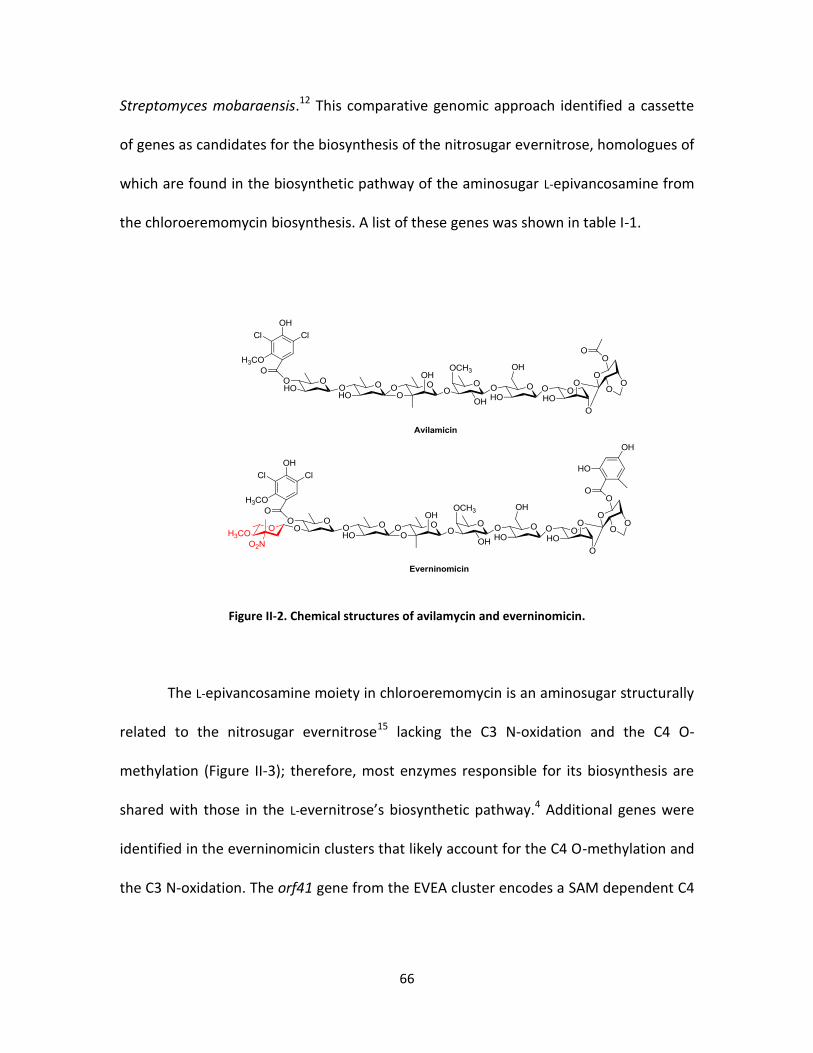
66
Streptomyces mobaraensis.12 This comparative genomic approach identified a cassette
of genes as candidates for the biosynthesis of the nitrosugar evernitrose, homologues of
which are found in the biosynthetic pathway of the aminosugar L-epivancosamine from
the chloroeremomycin biosynthesis. A list of these genes was shown in table I-1.
Figure II-2. Chemical structures of avilamycin and everninomicin.
The L-epivancosamine moiety in chloroeremomycin is an aminosugar structurally
related to the nitrosugar evernitrose15 lacking the C3 N-oxidation and the C4 O-
methylation (Figure II-3); therefore, most enzymes responsible for its biosynthesis are
shared with those in the L-evernitrose’s biosynthetic pathway.4 Additional genes were
identified in the everninomicin clusters that likely account for the C4 O-methylation and
the C3 N-oxidation. The orf41 gene from the EVEA cluster encodes a SAM dependent C4
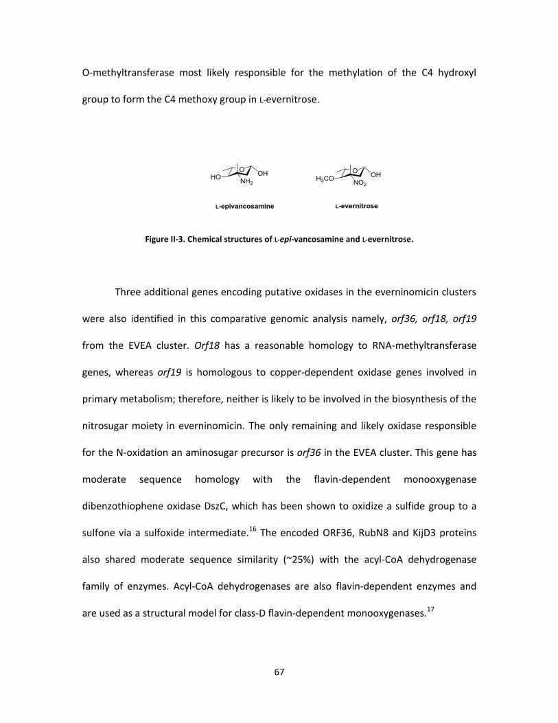
67
O-methyltransferase most likely responsible for the methylation of the C4 hydroxyl
group to form the C4 methoxy group in L-evernitrose.
Figure II-3. Chemical structures of L-epi-vancosamine and L-evernitrose.
Three additional genes encoding putative oxidases in the everninomicin clusters
were also identified in this comparative genomic analysis namely, orf36, orf18, orf19
from the EVEA cluster. Orf18 has a reasonable homology to RNA-methyltransferase
genes, whereas orf19 is homologous to copper-dependent oxidase genes involved in
primary metabolism; therefore, neither is likely to be involved in the biosynthesis of the
nitrosugar moiety in everninomicin. The only remaining and likely oxidase responsible
for the N-oxidation an aminosugar precursor is orf36 in the EVEA cluster. This gene has
moderate sequence homology with the flavin-dependent monooxygenase
dibenzothiophene oxidase DszC, which has been shown to oxidize a sulfide group to a
sulfone via a sulfoxide intermediate.16 The encoded ORF36, RubN8 and KijD3 proteins
also shared moderate sequence similarity (~25%) with the acyl-CoA dehydrogenase
family of enzymes. Acyl-CoA dehydrogenases are also flavin-dependent enzymes and
are used as a structural model for class-D flavin-dependent monooxygenases.17

68
Preparation of the putative oxidases and their aminosugar substrate
We targeted the producers of everninomicin18 and rubradirin19 to prepare the
putative enzymes responsible for the aminosugar oxidation to form the nitro group in
their deoxynitrosugar moieties. The genes orf36 from Micromonospora var. africana and
rubn8 from Streptomyces achromogenes var. rubradiris were proposed to encode a
flavin-dependent monooxygenase responsible for the N-oxidation of a TDP-activated
deoxyaminosugar precursor.20-21 We amplified these genes using the genomic DNA of
their producers via the polymerase chain reaction (PCR), and cloned the resulting gene
products into the NdeI/HindIII site of the expression plasmid pET28.22 The gene products
were designed to encode an N-terminal hexahistidine tag to facilitate the protein
purification using a Ni2+-affinity column. The recombinant plasmids were transformed to
the E.coli strain BL21 (DE3) for overexpression. Both proteins were found to require 0.1
mM β–D-1-thiogalactopyranoside (IPTG) and the best expression temperature was
found to be 28 °C. Analysis of the purified proteins by SDS-PAGE showed protein bands
consistent with their calculated molecular weight (~44 kD) as shown in figure II-4 for the
Ni2+-purified RubN8.

69
Figure II-4. SDS-PAGE gel of Ni2+-affinity purified RubN8.
A close substrate analogue, (5:1 β/α )-TDP-L-epivancosamine, in which the C-4
methoxy group is replaced by a hydroxyl group, was prepared by in situ Staudinger
reaction of its synthetic azide congener with tris(2-carboxyethyl)phosphine (TCEP)
(Figure II-5). The azide congener was kindly provided to us by Prof. Daniel Kahne’s
research group at Harvard University.23 We expected TDP-L-epivancosomine to be a
candidate substrate for the amine oxidation by the proposed oxidases although it lacked
the C4 O-methylation because an everninomicin variant, in which the methoxy group at
C-4 of the nitrosugar moiety is replaced by a hydroxyl group, was also isolated from the
fermentation of M. micromonospora var. africana.8 HPLC-MS analysis of the above
mentioned Staudinger reaction showed that 95% of the (5:1)-β/α mixture of TDP-L-
epivancosamine azide could be reduced to L-TDP-epi-vancosamine providing the
aminosugar substrate for ORF36 and RubN8 biochemical characterization.

70
Figure II-5. LC-ESI-MS analysis of the Staudinger reduction of the synthetic (5:1 β/α)-azido congener of TDP-L-epi-vancosamine by TCEP.
Enzymatic activities of ORF36 and RubN8
As discussed in the introduction, ORF36 and RubN8 from the everninomycin and
rubradirin biosynthetic pathways, respectively, are proposed to be flavin-dependent
enzymes responsible for the N-oxidation of TDP-activated aminosugar, which ultimately
produces its nitrosugar congener. These putative oxidases share moderate homology
with the flavin-dependent enzyme DszC, which oxidizes dibenzothiophene (DBT) to DBT
sulfone (DBTO2).24 This two-step oxidation requires flavin mononucleotide (FMN),
NADPH, and an external flavin reductase to provide reduced flavin which, in turn,
provides reducing equivalents and mediates the monooxygenation. Reduced flavin
reacts with molecular oxygen to form the C4a-hydroperoxyflavin, a reactive oxygen
species that is responsible for the electrophilic oxidation of the substrate.25 The
oxygenation results in the incorporation of one oxygen, usually as a hydroxyl group, and
71
the formation of reduced C4a-hydroxy flavin. The hydroxyflavin readily loses a water
molecule to generate the oxidized flavin required for a subsequent round of catalysis.
With flavin-dependent enzyme cofactor requirements taken into consideration,26
we designed an enzymatic assay for both ORF36 and RubN8 to reconstitute their
activities in vitro. In addition to the putative oxidase, the assay included FAD,
Photobacterium fischeri flavin reductase, and NADPH. Considering the possibility that
reactive oxygen species (ROS) may oxidize TDP-L-epi-vancosamine, all enzymatic
reactions were also performed in the presence of a catalytic excess of catalase and
superoxide dismutase. The catalase catalyzes the decomposition of hydrogen peroxide
to oxygen and water while the superoxide dismutase catalyzes the dismutation of the
superoxide anion into oxygen and hydrogen peroxide.27
Product formation was assayed by electrospray HPLC-MS using a graphitic
Hypercarb column in the negative ion mode, which effectively separated all TDP
products.28 As shown in Figure II-6, time course assays revealed rapid disappearance of
TDP-L-epivancosamine (m/z) 544), transient appearance of intermediary hydroxylamine
(m/z) 560), and ultimately the formation of the nitroso compound TDP-epi-
vancosonitrose (m/z) 558 and a species of +18 mass units at (m/z) 576. Of note, only
the β anomer of TDP-L-epivancosamine was oxidized, consistent with the catalytic
competence known for the TDP-sugar-modifying enzymes. The formation of the TDP-
nitrosugar congener was only observed at slow rates ambiguous to be counted as an
enzymatic conversion. Control reactions omitting ORF36/RubN8, FAD, or NADPH

72
produced no detectable oxidation products, which validated the authenticity of the
RubN8’s and ORF36’s activities.
Figure II-6. HPLC-ESI-MS chromatograms of the time course of RubN8 oxidation reaction.
Tandem MS fragmentation of selected product ions (Figure II-7) showed diagnostic
fragmentation patterns of the hydroxylamino intermediate and the nitroso product
including the loss of TDP anion (m/z 401) and it’s dehydrate (m/z 383). Another
fragment ion with low intensity representing the loss of the hydroxylamino group from
the TDP-3-hydroxyaminosugar intermediate was observed at m/z 527.

73
Figure II-7. Tandem MS spectra of the nucleotide-hydroxyaminosugar intermediate (above) and the nucleotide-nitrososugar product (below) from RubN8 oxidation reaction.
Proposed mechanism of nitrososynthase
Based on our biochemical data obtained in this work, a minimal mechanism can
be proposed for the catalysis of this new nitrososynthase enzyme (Scheme II-1). The
four-electron flavin-mediated oxidation of an amine to a nitroso functional group likely
involves two monooxygenation steps. The first step involvs the oxidation of the
aminosugar to the corresponding hydroxylamine via a flavin monooxygenase
mechanism similar to that of L-ornithine N5-oxygenase29 from pyoverdin biosynthesis
and p-hydroxybenzoate hydroxylase from Acinetobacter calcoaceticus .30 In this

74
mechanism, we propose that bound reduced FAD, generated by flavin reductase, reacts
with molecular oxygen to form flavin C4a-hydroperoxide, which acts as the electrophilic
oxidizing species in the formation of the hydroxylamino sugar. The resulting flavin-C4a-
hydroxide loses a water molecule recycling the oxidized FAD cofactor. In the second
step, the oxidation of the hydroxylamino sugar intermediate to the nitroso sugar may
proceed via an iterative oxidative process in which the bound C4a-hydroperoxy flavin
mediates another monooxygenation to ultimately form the nitroso sugar. This requires
the regeneration of reduced flavin by the flavin reductase/NADPH for the second round
of oxidation. To confirm FAD recycling, reactions were performed by stoichiometrically
limiting NADPH. The formation of the hydroxylamine was observed, but no further
oxidation products appeared until additional NADPH was added to the reaction,
suggesting further reducing equivalents are required for the second monooxygenation
step via the supplied NADPH/flavin reductase enzyme.

75
Scheme II-1. Proposed pathway for the nitrososynthase oxygenation reaction. Pathway B was proposed to be not likely to occur, because limiting the NADPH coenzyme resulted in the formation of only the hydroxylamino intermediate.
Conclusion
In this study, the proposed flavin-dependent enzymes RubN8 and ORF36 were
shown to be capable of oxidizing a synthetic aminosugar substrate analogue (TDP-L-
epivancosamine), lacking the C4 O-methylation, to its corresponding nitroso congener.
The oxidation appears to proceed via a hydroxylamino intermediate and requires
reduced FAD generated by an external flavin reductase with NADPH as reducing
equivelent. Although the C4 O-methylation in the authentic aminosugar substrate may
affect subsequent oxidation reactions, our data are consistent with previous studies
showing that the nitroso variant of rubradirin is the direct oxidation product when

76
fermentations were performed in the absence of ambient light. Oxidation to the nitro
group remains to be further investigated and possible pathways may include latent
(photo)chemical process or endogenous reactive oxygen species (ROS).31
Taken together, the biochemical data presented in this work provide the first
evidence for a new class of flavin-dependent monooxygenase we termed a
nitrososynthase responsible for the N-oxidation of deoxyaminosugars in vast array of
nitrosugar-containing natural product pathways. The observed monooxygenase activity
of RubN8 and ORF36 is direct biochemical evidence for the deoxyaminosugar N-
oxidation in the rubradirn and the everninomicin pathways, respectively, and can
explain the roles of nitrososynthase homologues in related biosynthetic pathways. This
study provided the basis for more detailed biochemical characterization of this
important class of enzymes and their roles in several biosynthetic pathways.
Materials and methods
Bacterial strains, Plasmids and Material
All reagents were obtained from Sigma-Aldrich Corporation and used without
further purification unless otherwise noted. E. Coli TOP10 and BL21(DE3) competent
cells were obtained from Invitrogen Inc. (Carlsbad, CA) and Novagen (Madison, WI),
respectively. Restriction endonucleases and T4 DNA ligase were obtained from New
England Biolabs (Ipswich, MA). The pET28a expression vector was purchased from

77
Novagen Inc. Taqplus DNA polymerase was purchased from Stratagene Inc. (La Jolla,
CA). DNA primers were obtained from Operon Biotechnologies (Huntsville, AL).
Preparation of the azido sugar substrate
The azido congener of TDP-L-epivancosamine shown in figure II-5, was prepared
by Prof. Daniel Kahne’s research group at the Department of Chemistry and Chemical
Biology, Harvard University. The following procedure was provided: Solvents were
reagent grade and were further dried when necessary. Analytical thin-layer
chromatography was performed on glass plates precoated with silica gel (250 μm,
Sorbent Technologies), with detection by UV and/or spraying with H2SO4 (50%). Flash
chromatography was carried out on silica gel (60 Å, 32-63 μm), purchased from Sorbent
Technologies. Analytical HPLC of synthetic reaction mixtures was performed on a
Hewlett-Packard 1100 series instrument using a Phenomenex Luna 5 μm C18 column
(250 mm x 4.6 mm). Compounds bearing a thymidine chromophore were monitored at
an absorbance of 270 nm. Synthetic reactions were monitored by HPLC using gradient A
(linear gradient from H2O/0.1% NH4HCO3 to 100% MeOH/0.1% NH4HCO3 over the
course of 25 min). Preparative HPLC was performed on a Varian ProStar instrument
using a Phenomenex Luna 10 μm C18 column (250 mm x 50 mm). NMR spectra were
recorded on Varian Inova 400 or 500 MHz spectrometers. Mass spectra (ESI) for
synthetic compounds were obtained using an Agilent Technologies LC/MSD instrument
(Model #G1956B).
78
Thymidine 5'-(3-amino-2,3,6-trideoxy-3-C-methyl-a,b-l-arabino hexopyranosyl
diphosphate), TDP a,b-epi-L-vancosamine azide (5). The protected TDP epi-vancosamine
(reference: Org. Lett., Vol 6, 2004) (38 mg, 60 μmol) was dissolved in MeOH/H2O/Et3N
(2:2:1, 4 mL) and stirred at room temperature for 16 h. Following evaporation, the
residue was redissolved in MeOH/H2O (2:1, 2 mL) and purified by reversed-phase HPLC.
(18 mg, 92%) was obtained as its ammonium salt, tR = 9.0 min, method A. 1H NMR (400
MHz, D2O): 2.25b: δ = 7.62 (s, 1 H, 6-H), 6.24 – 6.21 (m, 1 H, 1'-H), 5.22 – 5.17 (dt, J1,2b
= J1,P = 9.0, J1,2a = 2.1 Hz, 1 H, 1"-H), 4.47 (m, 1 H, 3'-H), 4.02 (m, 3 H, 4'-H, 5'-H2),
3.52–3.47 (m, 1 H, 5"-H), 3.16 (d, J4,5 = 10 Hz, 1 H, 4"-H), 2.22 - 2.08 (m, 2 H, 2'-H2) 2.07
(d, ,J2a,2b = 12.6 Hz, 1 H, 2"-Ha ), 1.71 (s, 3 H, thymidine CH3), 1.56-1.51 (dd, J1,2b =
9.0, J2a,2b = 12.6 Hz, 1 H, 2"-Hb), 1.25 (s, 3 H, 3"-CH3), 1.13 (d, J5,6 = 6.4 Hz, 3 H, 6"-H3);
31P NMR (162 MHz, CD3OD): δ = –10.61, –12.82. LRMS (ESI) for C17H27N5O13P2
(570.12): 571 [M–1H]–.
Cloning and overexpression of ORF36
This protein was cloned, expressed and purified by Dr. Yunfeng Hu. The gene
encoding ORF36 was amplified from genomic DNA of Micromonospora carbonacea var.
africana (NRRL 15099) using the following primers: 5'-
GCACATATGGCGGCGGATCTTCGCGC3' and 5'-TTGAAGCTTTTATTACGCCGA
GGTCCGGGAGC-3' (NdeI and HindIII restriction enzyme sites underlined). PCR reactions
were carried out using Taqplus DNA polymerase according to the manufacture protocol.

79
Subcloning of orf36 into the NdeI/HindIII sites of pET28a yielded the recombinant
plasmid pET28-36N for expression as an N-terminal hexahistidine fusion protein.
Plasmid pET28-36N was transformed into E. Coli BL21(DE3) for heterologous expression
of ORF36. Cultures of E. Coli BL21(DE3)/pET28-36N was grown at 37°C to an OD600 of
0.6, at which point the culture was induced with 0.1mM isopropyl-beta-D-
thiogalactopyranoside (IPTG) and grown an additional 6 hours at 28°C. Cells were
harvested by centrifugation and stored at -80°C until needed. IPTG induced E. Coli
BL21(DE3)/pET28-36N cells were resuspended in buffer A (20 mM Imidazole, 0.5M NaCl,
20mM Tris-Cl, pH 7.5) and lysed by sonication. The lyate was loaded onto a charged 5-ml
Histrap crude column (Amersham Biosciences) and purified by FPLC at a flow rate of
5mL/min. The column was washed with buffer A (20 mM Imidazole, 0.5M NaCl, 20 mM
Tris-Cl, pH 7.5) and buffer B (500 mM Imidazole, 0.5M NaCl, 20 mM Tris-Cl, pH 7.5) using
a step gradient. Fractions containing ORF36 were analyzed by SDS-page. The protein
was desalted via a desalting column (HisTrap) using buffer C (20 mM Tris-Cl, 1mM
dithiothreitol and 5% glycerol, pH 7.5) and stored at -80 °C until assayed.
Cloning and overexpression of rubN8
The gene encoding RubN8 was amplified from Streptomyces achromogenese var.
rubradiris (NRRL3061) genomic DNA. The following PCR primer pairs were used to clone
the gene into the NdeI/HindIII site of pET28a (restriction sites underlined): 5’-
TCCATATGA-TGGAGACGGAACAGGCCCC-3’ and 5’-TCAAGCTTTCACCGGCTGTCCACCGGC-

80
3’. Restriction-digested PCR products were subcloned into the digested pET28a vector to
yield pET28a-his6-RubN8. Cultures of E.coli BL21(DE3) transformants (1 L Luria broth
(LB) media with 50 µg/mL kanamycin) were grown at 37 °C to an OD600 of 0.6 at which
point the cultures were induced with 0.25 mM isopropyl-1-thio-β-D-galactoside (IPTG)
and grown for additional 16 h. Cells were harvested by centrifugation and frozen at -
80°C until needed. Frozen cells were resuspended in binding buffer [20 mM imidazole,
0.5 M NaCl, 20 mM sodium phosphate (pH 7.5)] and lysed by sonication. After
clarification by centrifugation at 10000g, the lysates were loaded onto Ni2+-charged 5-
mL HisTrap column and purified using FPLC (Amersham Biosciences) with a flow rate of
5 mL/min. The column was washed with 50 mL of binding buffer and proteins were
eluted with a gradient of imidazole (20-500 mM imidazole). Fractions containing the
proteins were analyzed by SDS-PAGE, pooled, concentrated to 5 mL and desalted by
FPLC using a 5 mL size exclusion desalting column (HiTrap) from Amersham Biosciences.
The column was equilibrated with 50 mL desalting buffer [ 20 mM Tris.HCl, 5%glycerol, 1
mM DTT, pH 7.5], the proteins were then loaded onto the column (maximum of 1.5 mL)
and eluted at a flow rate of 5 mL/min. Fractions containing the protein were analyzed
again by SDS-PAGE and stored at -80 °C until assayed.
Preparation of L-TDP-epi-vancosamine
L-TDP-epi-vancosamine (4-O-desmethyl L-TDP-evernosamine) was prepared by
reduction of a synthetic azide congener (0.5mM) with 1mM tris(2-
81
carboxyethyl)phosphine (TCEP) in 20 mM Tris-Cl (pH 7.5) at 22°C for 24 hours. Amino
sugar was stored in small aliquots at -80 °C for assays. HPLC/MS, performed for L-TDP-
epi-vancosamine enzymatic reaction described below, indicated that the extent of
reaction was > 96%.
ORF36/RubN8 enzymatic reactions
ORF36 activity was assayed in 20 mM Tris-HCl buffer (pH 7.5) in the presence of
0.4 – 2 mM NADPH, 30 µM FAD, 250 µM L-TDP-epi-vancosamine, 0.001 mg/ml of the
NAD(P)H oxidoreductase of Photobacterium fischeri (Boehringer Mannheim, Mannheim,
Germany) and 0.5mg/ml of ORF36 per 50 µl. Reactions were maintained at 30 °C.
Solutions of the substrate L-TDP-epi-vancosamine, flavin adenine dinucleotide
(FAD) and nicotinamide adenine dinucleotide phosphate (NADPH), were made in 20 mM
Tris-HCl pH 7.5. RubN8 was aliquoted in storage buffer [20 mM Tris.HCl, 5% glycerol, 1
mM DTT, pH 7.5]. Flavin reductase was stored in 40% glycerol, 1 mM EDTA, 0.1 mM DTT
and 50 mM potassium phosphate, pH 7.0. The catalase and superoxide dismutase
enzymes were made in [20 mM Tris.HCl 40% glycerol, pH 7.5]. In a total volume of 50
µL, 250 µM of the substrate, L-TDP-epi-vancosamine, was incubated with 30 µM FAD, 1
U/mL catalase, 1 U/mL superoxide dismutase, 0.001 mg/mL flavin reductase, and 2 mM
NADPH. Control assays lacking RubN8, FAD or NADPH were performed. The reaction
was initiated by the addition of 0.5 mg/mL RubN8 or 0.5 mg/mL ORF36. The time course
of the reaction was followed by HPLC/MS at 30 °C.
82
HPLC/MS assay for L-TDP-epi-vancosamine reaction
The oxidation of L-TDP-epi-vancosamine to corresponding products was carried
out using an Agilent 1100 HPLC system (Agilent, Palo Alto, CA), comprising a binary
pump and refrigerated autosampler. Mass spectrometry was performed by using a
ThermoFinnigan (San Jose, CA) LCQ Quantum Deca XP three-dimensional Ion Trap Mass
Spectrometer equipped with an API electrospray ionization source outfitted with a 50
μm I.D. deactivated fused silica capillary. The injection volume was 5 μl. Products were
detected using a Thermo Hypercarb column (3mm × 5cm). Mobile phases were: (A) H2O
with 50 mM ammonium acetate and 0.1% (v/v) diethylamine and (B) H2O/acetonitrile
(5:95) with 50 mM ammonium acetate and 0.1% (v/v) diethylamine. Gradient conditions
were as follows: 0-5 min, B = 15%; 5-15 min, linear gradient to 35% B; 15 to 16 min,
linear gradient to 100%B; 16 to 21 min, hold B = 100%; 21 to 22 min, linear gradient to
15% B; 22-30 min, B = 15%. The flow rate was maintained 0.3 ml/min. The mass
spectrometer was operated in the negative ion mode, and the electrospray needle was
maintained at 3,400 V. The ion transfer tube was operated at -47.50 V and 275 °C. The
tube lens voltage was set to -46 V. The mass spectrometer was operated in full scan
mode. Full scan spectra were acquired from m/z 50.0 to 1500.0 full and product ion
scans as follows: 544→155-548 with collision energy of 30%; 558→155-560, with
collision energy of 30%; 560→155-562 with collision energy of 30%; 544→155-548 with
collision energy of 30%; 570→155-575 with collision energy of 30%. Data were acquired
in profile mode. The following optimized parameters were used for the detection: N2
sheath gas 46 psi; N2 auxiliary gas 13 psi; spray voltage 3.4 kV.

83
References
1. Thibodeaux, C. J.; Melancon, C. E., 3rd; Liu, H. W., Natural-product sugar biosynthesis and enzymatic glycodiversification. Angewandte Chemie 2008, 47 (51), 9814-59.
2. Allard, S. T.; Giraud, M. F.; Whitfield, C.; Messner, P.; Naismith, J. H., The purification, crystallization and structural elucidation of dTDP-D-glucose 4,6-dehydratase (RmlB), the second enzyme of the dTDP-L-rhamnose synthesis pathway from Salmonella enterica serovar typhimurium. Acta Crystallographica. Section D, Biological Crystallography 2000, 56 (Pt 2), 222-5.
3. Nedal, A.; Zotchev, S., Biosynthesis of deoxyaminosugars in antibiotic-producing bacteria. Applied Microbiology and Biotechnology 2004, 64 (1), 7-15.
4. Chen, H.; Thomas, M. G.; Hubbard, B. K.; Losey, H. C.; Walsh, C. T.; Burkart, M. D., Deoxysugars in glycopeptide antibiotics: enzymatic synthesis of TDP-L-
epivancosamine in chloroeremomycin biosynthesis. Proceedings of the National Academy of Sciences of the United States of America 2000, 97 (22), 11942-7.
5. Meyer, C. E., Rubradirin, a New Antibiotic. Ii. Isolation and Characterization. Antimicrobial Agents and Chemotherapy 1964, 10, 97-9.
6. Nakashio, S.; Iwasawa, H.; Dun, F. Y.; Kanemitsu, K.; Shimada, J., Everninomicin, a new oligosaccharide antibiotic: its antimicrobial activity, post-antibiotic effect and synergistic bactericidal activity. Drugs under Experimental and Clinical Research 1995, 21 (1), 7-16.
7. Zhang, H.; White-Phillip, J. A.; Melancon, C. E., 3rd; Kwon, H. J.; Yu, W. L.; Liu, H. W., Elucidation of the kijanimicin gene cluster: insights into the biosynthesis of spirotetronate antibiotics and nitrosugars. Journal of the American Chemical Society 2007, 129 (47), 14670-83.
8. Chu, M.; Mierzwa, R.; Jenkins, J.; Chan, T. M.; Das, P.; Pramanik, B.; Patel, M.; Gullo, V., Isolation and characterization of novel oligosaccharides related to Ziracin. Journal of Natural Products 2002, 65 (11), 1588-93.

84
9. Marshall, S. A.; Jones, R. N.; Erwin, M. E., Antimicrobial activity of SCH27899 (Ziracin), a novel everninomicin derivative, tested against Streptococcus spp.: disk diffusion/etest method evaluations and quality control guidelines. The Quality Control Study Group. Diagnostic microbiology and Infectious Disease 1999, 33 (1), 19-25.
10. Jones, R. N.; Marshall, S. A.; Erwin, M. E., Antimicrobial activity and spectrum of SCH27899 (Ziracin) tested against gram-positive species including recommendations for routine susceptibility testing methods and quality control. Quality Control Study Group. Diagnostic Microbiology and Infectious Disease 1999, 34 (2), 103-10.
11. Mertz, J. L.; Peloso, J. S.; Barker, B. J.; Babbitt, G. E.; Occolowitz, J. L.; Simson, V. L.; Kline, R. M., Isolation and structural identification of nine avilamycins. The Journal of Antibiotics 1986, 39 (7), 877-87.
12. Farnet, C. M.; Zazopoulos, E.; Staffa, A., Compositions and methods for identifying and distinguishing orthosomycin biosynthetic loci. Google Patents: 2002.
13. Gololobov, Y. G.; Zhmurova, I.; Kasukhin, L., Sixty years of Staudinger reaction. Tetrahedron 1981, 37 (3), 437-472.
14. Vetrova, E. V.; Kudryasheva, N. S.; Visser, A. J.; van Hoek, A., Characteristics of endogenous flavin fluorescence of Photobacterium leiognathi luciferase and Vibrio fischeri NAD(P)H:FMN-oxidoreductase. Luminescence : the Journal of Biological and Chemical Luminescence 2005, 20 (3), 205-9.
15. Ganguly, A. K.; Sarre, O. Z.; Reimann, H., Evernitrose, a naturally occurring nitro sugar from everninomicins. Journal of the American Chemical Society 1968, 90 (25), 7129-30.
16. Ohshiro, T.; Aoi, Y.; Torii, K.; Izumi, Y., Flavin reductase coupling with two monooxygenases involved in dibenzothiophene desulfurization: purification and characterization from a non-desulfurizing bacterium, Paenibacillus polymyxa A-1. Applied Microbiololgy and Biotechnology 2002, 59 (6), 649-57.
17. Ghisla, S.; Thorpe, C., Acyl‐CoA dehydrogenases. European Journal of Biochemistry 2004, 271 (3), 494-508.

85
18. Wagman, G. H.; Luedemann, G. M.; Weinstein, M. J., Fermentation and Isolation of Everninomicin. Antimicrobial Agents Chemotherapy (Bethesda) 1964, 10, 33-7.
19. Bhuyan, B. K.; Owen, S. P.; Dietz, A., Rubradirin, a New Antibiotic. I. Fermentation and Biological Properties. Antimicrobial Agents Chemotherapy (Bethesda) 1964, 10, 91-6.
20. Kim, C. G.; Lamichhane, J.; Song, K. I.; Nguyen, V. D.; Kim, D. H.; Jeong, T. S.; Kang, S. H.; Kim, K. W.; Maharjan, J.; Hong, Y. S.; Kang, J. S.; Yoo, J. C.; Lee, J. J.; Oh, T. J.; Liou, K.; Sohng, J. K., Biosynthesis of rubradirin as an ansamycin antibiotic from Streptomyces achromogenes var. rubradiris NRRL3061. Archives of Microbiology 2008, 189 (5), 463-73.
21. Hosted, T.; Horan, A.; Wang, T., Everninomicin biosynthetic genes. WO Patent WO/2001/051,639: 2001.
22. Hu, Y.; Al-Mestarihi, A.; Grimes, C. L.; Kahne, D.; Bachmann, B. O., A unifying nitrososynthase involved in nitrosugar biosynthesis. Journal of the American Chemical Society 2008, 130 (47), 15756-7.
23. Oberthuer, M.; Leimkuhler, C.; Kahne, D., A Practical Method for the Stereoselective Generation of β-2-Deoxy Glycosyl Phosphates. Organic Letters 2004, 6 (17), 2873-2876.
24. Ohshiro, T.; Kojima, T.; Torii, K.; Kawasoe, H.; Izumi, Y., Purification and characterization of dibenzothiophene (DBT) sulfone monooxygenase, an enzyme involved in DBT desulfurization, from Rhodococcus erythropolis D-1. Journal of Bioscience and Bioengineering 1999, 88 (6), 610-6.
25. Massey, V., The chemical and biological versatility of riboflavin. Biochemical Society Transactions 2000, 28 (4), 283-96.
26. Joosten, V.; van Berkel, W. J. H., Flavoenzymes. Current Opinion in Chemical Biology 2007, 11 (2), 195-202.
27. Loprasert, S.; Vattanaviboon, P.; Praituan, W.; Chamnongpol, S.; Mongkolsuk, S., Regulation of the oxidative stress protective enzymes, catalase and superoxide dismutase in Xanthomonas--a review. Gene 1996, 179 (1), 33-7.
86
28. Xing, J.; Apedo, A.; Tymiak, A.; Zhao, N., Liquid chromatographic analysis of nucleosides and their mono‐, di‐and triphosphates using porous graphitic carbon stationary phase coupled with electrospray mass spectrometry. Rapid Communications in Mass Spectrometry 2004, 18 (14), 1599-1606.
29. Meneely, K. M.; Barr, E. W.; Bollinger, J. M., Jr.; Lamb, A. L., Kinetic mechanism of ornithine hydroxylase (PvdA) from Pseudomonas aeruginosa: substrate triggering of O2 addition but not flavin reduction. Biochemistry 2009, 48 (20), 4371-6.
30. Clarkson, J.; Palfey, B. A.; Carey, P. R., Probing the chemistries of the substrate and flavin ring system of p-hydroxybenzoate hydroxylase by raman difference spectroscopy. Biochemistry 1997, 36 (41), 12560-6.
31. Filip, M.; Paduraru, I.; Jerca, L.; Filip, F.; Saramet, A., [Oxygen biochemistry. I. Reactive species of reduced oxygen and endogenous sources]. Revista Medico-Chirurgicala a Societatii de Medici si Naturalisti din Iasi 1992, 96 (3-4), 289-92.

87
Chapter III
STRUCTURAL AND MECHANISTIC STUDIES OF THE NITROSOSYNTHASE ORF36 FROM
MICROMONOSPORA CARBONACEA VAR. AFRICANA
Introduction
The experiments outlined in Chapter II describing the activity of the
nitrososynthases ORF36 from Micromonospora carbonacea var. africana, and RubN8
from Streptomyces achromogenes var. rubradiris, formed the basis for further
biochemical characterization of this new class of flavin-dependent monooxygenases.
ORF36 and RubN8 were shown to catalyze the 2-step N-oxidation of a close substrate
analog, TDP-L-epivancosmine, resulting in the formation of its nitroso congener via a
hydroxylamino intermediate.1 Although no enzymatic oxidation to the nitro group was
observed, the nitroso sugar product is consistent with previous studies with rubradirin,
which suggested that the nitro oxidation state is a result of photo oxidation of the
nitroso group.2
Several enzymes in nature catalize the N-oxidation of amines. For example, the
Rieske, aminopyrrolnitrin N-oxygenase (PrnD) catalyzes the unusual flavin-dependent
oxidation of an arylamine to an arylnitro group.3 Another example is the non-heme di-
iron monooxygenase (AurF) from Streptomyces thioluteus which catalyzes the formation
of unusual polyketide synthase starter unit p-nitrobenzoic acid from p-aminobenzoic
acid in the biosynthesis of the antibiotic aureothin.4 N-Oxidation can also be catalyzed
by P450 enzymes5 and several single-component flavin monooxygenases.6

88
Our preliminary biochemical characterization of ORF36 and RubN8 showed that
the nitrososynthase is a flavin-dependent monooxygenase.1 Flavin-dependent
monooxygenases are a diverse class of enzymes capable of efficient and specific
insertion of one or more oxygen atoms into an organic substrate, a reaction that is
difficult to achieve via standard chemical synthesis. Oxidation reactions catalyzed by
flavin-dependent monooxygenases include hydroxylation, halogenation, sulfoxidation,
and Baeyer-Villiger oxidation.7 The double N-oxidation of aminosugars by the
nitrososynthase represents a new addition to this set of reactivities. This new enzyme
may be best classified as Class D flavin-containing monooxygenase based on its
sequence homology with acyl-CoA dehydrogenases, which are sequence models for this
class. Members of class D flavin-containing monooxygenases typically hydroxylate
aromatic substrates.8-11 Interestingly, nitrososynthases have more sequence similarities
with acyl-CoA dehydrogenases (~25%) than with flavin-containing monooxygenases
(~15%) contrary to their biochemical evidence suggesting a monooxygenase activity.
Class D flavoprotein monooxygenases are typically encoded by two genes; one
encodes the monooxygenase while the other encodes the flavin reductase component.
The reductase component uses FAD as a cofactor and NADH or NADPH as a coenzyme.
These enzymes are structurally homologous to acyl-CoA dehydrogenases and are mostly
α-helical proteins. In our previous biochemical characterization of the nitrososynthase
ORF36, it was shown that its activity requires flavin and NADPH with significant
enhancement reached upon the inclusion of an external flavin reductase.1 Furthermore,
ORF36 was shown to display weak, reversible binding for oxidized flavin. ORF36 was also
89
shown to utilize both FAD and FMN as its flavin cofactors with preference to FAD. This
cofactor promiscuity was observed in the class D flavoprotein monooxygenase 4-
hydroxyphenylacetate monooxygenase from Acinetobacter baumannii.12
Our recent biochemical characterization of the nitrososynthase ORF36 was
impeded by several challenges including the lengthy synthesis of the TDP-sugar
substrate, its storage instability, and the low affinity of the flavin cofactor which
typically provides a convenient spectrophotometric monitoring of the flavin cofactor’s
redox states. To address these challenges, we developed a modified in vitro procedure
utilizing six purified enzymes to generate the close substrate analog TDP-L-
epivancosamine, lacking the C4 O-methylation, in sufficient quantities to study enzyme
turnover of this substrate and its TDP-aminosugar precursors in its biosynthetic
pathway.13 Additionally, we studied the nitrososynthase oxidation reaction with the
biogenic TDP-L-epivancosamine under 18O2 and observed oxygen incorporation into the
oxidation products and intermediates which provided more evidence for a
monooxygenase mechanism. Finally, in collaboration with Prof. Tina Iverson’s research
group, Dr. Jessica Vey solved the 3-D structure of ORF36 by X-ray crystallography, which
revealed important chemical and physical constraints of the protein scaffold that houses
the flavin-dependent double N-oxidation of the TDP-aminosugar.14 Taken together
these studies provided important biochemical details of the nitrososynthase responsible
for the N-oxidation of deoxyaminosugars in everninomicin and related nitro-sugar
containing bioactive natural products.

90
Results
Substrate preference of ORF36
An ever-increasing number of nitrososynthase homologs have been deposited to
the GenBank corresponding to their oxidative role as flavin-dependent enzymes in the
biosynthesis of many nitrosugar-containing natural products.15-17 The structural
modifications of the nitrosugar moiety in these compounds are mostly conserved with
notable functional variability at C-4 position which can be found as hydroxyl, O-methyl
or N-carbamoyl group. Functional variability also includes the C-5 position leading to
possibilities between the sugar’s D and L configurations.15-17 The diversity of structures
of the aminosugar precursor substrates is in contrast with the high sequence identity
between all the nitrososynthase homologues in several nitrosugar pathways18, which
prompted us to investigate whether the reported nitrososynthases act on a common
intermediate biochemically upstream of the last biosynthesized aminosugar substrate in
the corresponding pathways. 19
To address this possibility and further investigate the timing of aminosugar
oxidation to ultimately yield the nitroso functionality, we reconstituted the biosynthetic
pathway for the aminosugar TDP-L-epi-vancosamine using enzymes obtained by the
Walsh group from the chloroereomycin pathway13, and studied the activity of ORF36
with two additional upstream TDP linked C4-keto-3-aminosugar intermediates in this
pathway (Figure III-1). We were unable to isolate enough quantities from this multistep
enzymatic synthesis of these TDP-sugar intermediates to perform Michaelis-Menten

91
kinetics due to their low storage stability; however, we were able to compare turnover
of substrates at identical, titrated substrate concentrations (30 µM).
Figure III-1. Pathway utilized for biochemical synthesis of TDP-L-epi-vancosamine and the TDP-4-keto-3-amino sugar precursors. This figure is taken from reference 14 in which the author of this document is a coauthor.
Analysis of the enzyme assays by HPLC-MS showed that all potential aminosugar
substrates displayed a degree of chemical competence for single oxidation to
hydroxylamine, but only TDP-L-epivancosamine underwent double oxidation to form
both the hydroxylamine and subsequently the nitrososugar product (Figure III-2).

92
Figure Figure III-2. HPLC-ESI-MS traces of ORF36 oxidation reactions with three potential amino sugar substrates. (A) TDP-L-epivancosamine (m/z 544) at 0 min (blue) and 100 min (green), hydroxylamine intermediate (m/z 560) at 15 min (red), and nitroso product (m/z 558) at 100 min (purple). (B) TDP-L-3-amino-4-keto sugar substrate (m/z 542) at 0 min (blue) and 100 min (green) and hydroxylamine intermediate (m/z 558) at 100 min (purple). (C) TDP-D-3-amino-4-keto sugar substrate (m/z 542) at 0 min (blue) and 100 min (green) and hydroxylamine intermediate (m/z 558) at 100 min (purple). This figure is taken from reference 14 in which the author of this document is a coauthor.
The two 3-amino-4-keto sugar precursors of TDP-L-epivancosamine (Figure III-
2B,C) differ in the configuration at C-5 with the first biosynthesized ketosugar having a D
configuration and upon the activity of EvaD, its L congener is formed. The TDP-L-3-
amino-4-keto sugar (Figure III-2B) was assayed by including a catalytic excess of the C-5
epimerase (EvaD) in the nitrososynthase oxidation reaction. This TDP-L-aminosugar
substrate was rapidly oxidized to its hydroxylamino congener but did not further oxidize
to other species under the ORF36 assay conditions (Figure III-2B). The TDP-D-3-amino-4-
93
keto sugar precursor was also oxidized to its hydroxylamino oxidation state, but at a
substantially slower apparent rate than when the epimerase was added to reactions
(Figure III-2C). This is in agreement with its recently reported partial oxidation by KijD3,
which likewise only evidenced the formation of a hydroxylamine.18 Unlike these studies,
however, we did not observe additional peaks at m/z = 527 rationalized by the authors
as a fragment of the hydroxylamine. This mass corresponds to a reduction product, the
identity of which remains undetermined. In our ORF36 assays, the two 4-keto D- and L-
aminosugar substrates were oxidized to their corresponding hydroxylamino compounds
and no other detectable TDP-sugar decomposition products were apparent by MS or
MS/MS analysis.
The oxidation of TDP-L-epivancosamine generated from the reconstitution of its
natural biosynthetic pathway confirmed our previous results using the chemically
synthesized compound1 and importantly, validated the successful biosynthetic
production of its two TDP-4-keto-3-amino sugar precursors. In summary, these data
indicate that the double N-oxidation catalyzed by the nitrososynthase ORF36 improves
with substrates more closely related to TDP-L-epivancosamine and only a single
oxidation step to a hydroxylamine occurs for the TDP-3-amino-4-keto sugar precursors.
The oxidation of the C-4 O-methylated congener of TDP-L-epivancosamine remains to be
investigated to determine whether this additional methyl group has any effect on the
oxidation activity of ORF36. However, the isolation of everninomicin variants from M.
carbonacea var. africana with both O-methyl and hydroxyl functionality in their
corresponding nitrosugar moieties20, suggests that O-methyl substitution at this position
94
is not essential for nitrososynthase reactivity and may occur prior or subsequent to
amine oxidation reactions.20
18O2 incorporation in the ORF36 oxidation reaction.
To determine whether the incorporation of the oxygen atom into the amino
sugar substrate upon ORF36 activity with TDP-L-epivancosamine comes from molecular
oxygen as opposed to oxygen from H2O, we performed the ORF36 oxidation reaction
under 18O2. The incorporation of molecular oxygen into the substrate would also
support the flavin-dependent monooxygenase catalysis. To provide a control reaction,
the same previous ORF36 assay with TDP-L-epivancosamine under 16O2 was repeated.
Reactions contained the same concentrations of NADPH, FAD, flavin reductase, and
ORF36 were incubated with catalytic excess of catalase21 under atmosphere of either
18O2 or 16O2. As shown in figure III-3, HPLC-MS analysis showed single oxygen atom
incorporation corresponding to the hydroxylamine intermediate (m/z = 560) and the
nitroso product (m/z = 558), which demonstrated enrichment of the +2 m/z shift at 86%
and 87% incorporation, respectively.

95
Figure III-3. The 18
O2 incorporation studies of ORF36 oxidation reaction. (A) Summary of proposed species observed in the
18O2 incorporation experiments. (B-D)
18O2 incorporation with ORF36 and TDP-L-
epivancosamine incubated with 16
O2 (green) and 18
O2 (red). Masses shown under structures correspond to unlabeled species. HPLC-MS and MS/MS data were collected at a 15 min reaction time, when the hydroxylamine intermediate was most abundant. (B) TDP-L-epivancosamine (m/z 544 and tR = 4.5 min). (C) Hydroxylamine (m/z 560, and tR = 5.3 min). (D) Peaks at tR values of 7-9 min are proposed to correspond to the nitroso compound (tR = 8 min) and an additional oxidation product at m/z 576 (tR = 7.8 min). This new mass has a retention time distinct from that of the nitroso sugar, and on the basis of MS/MS analysis, the additional mass of the m/z 576 ion is constrained to the pyranose ring. One possible structure is the 3-hydroxylamino-4-keto sugar, which is detected as a hydrate under ESI conditions. MS/MS analysis supports this hypothesis in that the labeled ion at m/z 578 readily fragments to an ion at m/z 560.09, and this fragmentation pattern is identical to the reaction product of the TDP-L-3-amino-4-keto sugar shifted by 2 mass units. This figure is taken from reference 14 in which the author of this document is a coauthor.
In addition to confirming the incorporation of molecular oxygen into the
expected oxidation products, these experiments also showed oxygen incorporation for
additional reaction products. We observed a +2 m/z shift in what we have previously
assigned to be the electrospray induced hydrate of the TDP-nitroso sugar (M + H2O, m/z
= 576 ).1 However, the single incorporation of oxygen into this species and the lack of
demonstrable back-exchange of labeled nitroso with H2O rule out the proposed

96
dihydroxylamino species. Closer examination of the HPLC/MS chromatograms indicated
that within the broad peak encompassing this region, the nitrososugar (m/z 558), and
the compound at m/z = 576 possess different retention times (8.8 and 7.8 min
respectively). Both compounds contain fragmentation peaks diagnostic of a TDP-sugar
including the loss of TDP (m/z = 401) and TMP (m/z = 321) but the unknown m/z 576
compound demonstrated an additional major fragment at 558, indicating loss of water.
Further characterization of the compound at m/z 576 was impeded by the lack of the
availability of enough quantities and the inherent storage stability. However, The
MS/MS analysis demonstrated that the oxidation is constrained to the pyranose ring,
and considering the likelihood that the oxidation is occurring on the C-3/4 heteroatoms,
we proposed that the m/z 576 peak corresponds to a product oxidized at C-4, likely the
C-4 ketone. Consistent with that, this putative structure shares similar retention time
and fragmentation pattern with the hydroxylamino product generated from the
oxidation of the substrate analog TDP-L-3-amino-4-keto sugar (Figure III-2). It should be
noted however, that other structures are possible, including a possible electrocyclic
rearrangement of the hydroxyketone (not shown).22 Further structural elucidation of
these oxidation products via chemical derivatization is discussed in chapter IV which
revealed a surprising outcome.

97
X-ray crystal structure of ORF36
In collaboration with Prof. Tina Iverson’s research group, Dr. Jessica Vey was able
to crystallize and determine the structure of the ORF36 enzyme.14 The polyalanine
model of human short-branched chain acyl-CoA dehydrogenase (PDB 2JIF)23 was used as
the search model to determine the structure to a 3.15 Å resolution by molecular
replacement (Figure III-4). ORF36 formed a tetramer with 3,500 Å2 of surface area
buried per monomer. Size exclusion chromatography on a Superdex S-200 column
confirmed the tetrameric structure of ORF36 in solution. The monomers of ORF36 adopt
a three-domain fold with the N- and C-terminal helical domains separated by a β-sheet
domain (Figure III-4B). The oligomerization interface of ORF36 is predominated by
interactions between the adjacent C-terminal helical domains.

98
Figure III-4. Structure of ORF36. (A) The ORF36 tetramer is shown as a cartoon representation with a transparent surface, colored by domain.The N-terminal domains (residues 1-129) are colored gray, the central β-sheet domains (residues 130-235) green, and the C-terminal domains (residues 235-398) purple. (B) ORF36 monomer shown in cartoon representation with modeled FAD and TDP-L-evernosamine shown as transparent sticks. Colors are as follows: yellow for FAD carbons, magenta for TDP-L-evernosamine carbons, red for oxygen, blue for nitrogen, and orange for phosphate. The view in panel B is rotated 45° about a vertical axis from the bottom monomer shown boxed in panel A. This figure is taken from reference 14 in which the author of this document is a coauthor.
As this work was being prepared for publication early 2010, the structure of the
nitrososynthase KijD3 was reported18 and appeared in the EMBL Dali search24 as the
closest structural homolog. Both KijD3 and ORF36 share structural similarity with
members of the acyl-CoA dehydrogenase superfamily25 which includes oxidases,
dehydrogenases and flavin-containing monooxygenases. The structural folds of ORF36
and KijD3 are surprisingly closer to those of the dehydrogenases than the
monooxygenase functional homologs which is in contrast with their functions as
monooxygenases. One explanation for this could be that the monooxygenase activity of
the nitrososynthases evolved independently from acyl-CoA dehydrogenases, while other
class D monooxygenases and the acyl-CoA dehydrogenases underwent a divergent
evolutionary route.

99
Modeling of FAD and TDP-L-epi-vancosamine in the active site
Attempts to co-crystallize ORF36 with the flavin cofactors FAD and FMN each in
their oxidized and reduced forms, as well as with the substrate TDP-L-epivancosamine
were unsuccessful. None of these experiments resulted in the appearance of new
electron density for the substrate or the flavin cofactor. Therefore, the FAD cofactor and
the substrate TDP-L-epivancosamine were modeled into the active site of ORF36 using a
combination of manual and automated methods. Ideal cofactor-substrate distances and
geometry were predicted based on comparison with the co-structures of flaving-
containing monooxygenases in complex with their substrates.26-29
The resulting predicted model suggests that the pre-formed pockets in the active
site of ORF36 bind the isoalloxazine ring of the flavin cofactor and the TDP moiety of
TDP-L-epivancosamine. Residues in the vicinity of the predicted isoalloxazine ring
pocket, particularly at loops L3, L5 and L9, are conserved in nitrosynthases and are
similar to those in acyl-CoA dehydrogenases and flavin-containing monooxygenases. In
contrast, the adenine nucleotide moiety of the flavin cofactor in this predicted model is
in a region in the active site where the loops are in an open conformation. Loop 7 in
both dehydrogenases and flavin-containing monooxygenases, which forms specific
contacts with flavin, is shorter in enzymes that preferentially bind FAD and longer in
enzymes that preferentially bind FMN, and is known to undergo a conformational
change upon flavin binding to facilitate contacts with the nucleotide moiety. This loop is
similar to those found in acyl-CoA dehydrogenase superfamily that utilizes FAD as their
flavin cofactor. This is supported by the observation that the activity of ORF36 is
100
significantly improved with the FAD cofactor as compared to that with FMN. In this
ORF36 structure, loop L7 adopts a conformation that likely will undergo adjustment
upon flavin binding.

101
Figure III-5. Active site loops. The loops that form the active site are as follows: the loops between α3 and α4 (loop L1; residues 106 – 109), β1 and β2 (loop L2; residues 134-142), β3 and β4 (loop L3, residues 158-164), β4 and β5 (loop L4; residues 178-180), β6 and β7 (loop L5; residues 201-213), β9 and α6 (loop L6; residues 248-253), α6’ and α7’ (where the ’ indicates this is from an adjacent monomer; loop L7; residues 272-280), α7 and α8 (loop L8; residues 310-317), α8’ and α9’ (loop L9, residues 351-353), α9 and α10, (loop L10; residues 375-379) and at the C-terminus (loop L11; residues 390-412). (A) A ribbon diagram of ORF36 shown with modeled flavin (yellow carbons) and TDP-L-evernosamine (magenta carbons) as sticks. Active site loops L3, L5, L7, and L9, are predicted to interact with flavin and are colored orange, active site loops L1, L4, L6, L8, and L11 are predicted to interact with substrate and are colored purple, and active site loops L2 and L10 are predicted to interact with both substrate and cofactor and are colored teal. (B) A ribbon diagram of human short branched-chain acyl Co-A dehydrogenase (PDB entry 2JIF)
30, highlighting loops L1 to L11 colored are colored as in (A). (C) A
ribbon diagram of A. baumannii 4-hydroxyphenylacetate monooxygenase (PDB entry 2JBT) 31
highlighting loops L1 to L11 colored as in (A). (D) Stereoview of the ORF36 loop L10 containing a tandem cis-peptide. Loop L10 is shown in sticks with teal carbons. The Q376-P377 and P377-Y378 bonds both adopt a cis conformation. Modeled flavin and substrate are displayed as in (A). This figure is taken from reference 14 in which the author of this document is a coauthor.
Description of the active site
The predicted active site of ORF36 contains amino acid residues mainly from the
central β-sheet domain and the N-terminal helical domain, but also includes residues
from the C-terminal domain of an adjacent monomer.25 The base of the active site cleft
is formed along helix α4. Flavin binding is predicted to be mediated by interactions with
residues in α4, β1, α6 and α8, whereas substrate binding presumably occurs via

102
interactions with residues in α4 and α6-α8 helices. Additionally, loops L1-L11
surrounding the active site likely play a role in substrate binding and catalysis. Loops L1,
L2, L4, L5, L6, L7, L8 and L11 differ in structure compared to their counterparts in the
aforementioned structural homologs of nitrososynthases, suggesting that they likely
play an important role in conferring substrate and cofactor specificity.
The active sites of both ORF36 and KijD3 measure 30 Å wide by 20 Å deep and
appear to be solvent exposed. This is in contrast to the active sites in flavin-containing
monooxygenases and acyl-CoA dehydrogenases. This unusually large pocket results
from the open conformations of loops L1, L2, L4, L6, L7, and L11 in ORF36 and KijD3,
which is unlike the more compact conformations of their counterparts in the acyl-CoA
dehydrogenases and flavin-containing monooxygenases. Most of these loops are known
to have interactions with the substrate in the acyl-CoA dehydrogenase superfamily,
suggesting that significant main chain conformational changes may occur upon
substrate binding. Of note, loop L10 located on the floor of the active site cleft in ORF36
likely contains tandem cis-peptide bonds between Gln 376 and Pro 377 and between
Pro377 and Tyr 378. Although the assignment of cis-peptide bond is speculative at this
resolution, modeling the loop with trans-peptide bonds or with only a single cis-proline
resulted in disallowed Ramachandran angles for Tyr 378 and poor agreement of the
main chain with the electron density. This assignment is also supported with the
observation that KijD3, with its L10 sequence identical to that of ORF36, contains
tandem cis-peptide bonds in this location.18
103
The crystallographic temperature factors of residues in the loops surrounding
the active site in both ORF36 and KijD3 are significantly higher when compared to
residues in the remainder of the protein. Temperature factors, also known as atomic
displacement parameters or B-factors, can reflect the thermal motion of atoms and hint
at regions of increased relative mobility in a given structure.
Discussion
Substrate preference of ORF36
The nitrososynthase activity of ORF36 with TDP-L-epi-vancosamine confirms that
this aminosugar is preferred over its 4-ketosugar upstream congeners. Moreover, the O-
methylation of the C4 hydroxyl group of TDP-L-epivancosamine was shown not to be
necessary for the nitrososynthase activity. This was an expected outcome, because a
variant of everninomicin lacking the C4-O methylation in its nitrosugar moiety was also
isolated from M. carbonacea var. africana.20 The single oxidation of the TDP 4-keto
sugar substrates suggests that nitrososynthases in the biosynthesis of other nitrosugar-
containing natural products may act on closely related precursors not necessarily
identical to TDP-L-eivancosamine. The retention times of the oxidation products from
the 4-keto aminosugar substrates matches that of the nitroso sugar produced from the
activity of ORF36 with TDP-L-epivancosamine. This led us to propose a shunt reaction
product, the C4 ketone, in which the 4-hydroxy position of TDP-L-epivancosamine is
oxidized perhaps via dehydrogenation or hydroperoxylation of the C4 hydroxyl to the

104
ketone oxidation state. Other oxidation possibilities may also exist. Preliminary
experiments of the ORF36 activity with the authentic substrate TDP-evernosamine,
which is protected by O-methylation at C4, showed the production of only the nitroso
sugar with no additional oxidation products. The proposed oxidation of the C4 position
of TDP-L-epivancosamine upon ORF36 activity is intriguing and further experimentation
should be performed to validate the identity of the proposed structures. Chapter IV
details some chemical derivatization experiments and HPLC/MS analysis of the
derivatized oxidation products of the nitrososynthase activity with TDP-L-
epivancosamine which reveals a surprising outcome.
Structural comparison of ORF36 and KijD3
The overall structure of ORF3614 matches that of its KijD318 homolog from the
kijanimicin biosynthetic pathway with a root-mean-square deviation of 0.76 Å between
the Cα atoms of the tetramers of the two proteins. The main differences in both
sequence and structure between these two homologs are found in the regions that map
to the active site cleft, and may be related to differences in function and substrate
binding. Of note, variations in the interface of the predicted isoalloxazine and the TDP
binding sites may account for substrate preference. The TDP and the isoalloxazine
binding pockets themselves are almost completely conserved in both enzymes. The
major structural variations between ORF36 and KijD3 are apparent in the active site
loops L2, L4 and L6 located within the central β-sheet domain, and L7 and L8 of the C-

105
terminal domain. The high crystallographic temperature factors of these regions in both
enzymes may reflect flexibility that facilitates movements of the substrate or the
cofactor upon catalysis.
The active site of both nitrosoynthases ORF36 and KijD3 are relatively large and
highly solvent accessible with their loops adopting open conformations. In comparison
with structures of the acyl-coA dehydrogenases and class D flavin-containing
monooxygenases, exclusion of water from the active site is presumably facilitated by
conformational changes of loops L2, L4, L6, L7, L8 and L11. The active site residues have
elevated temperature factors compared to the remainder of the protein likely
accounting for substrate and cofactor mobility. Additionally, both enzymes possess a
tandem cis-peptide bond within the active site loop L10 which likely undergoes
isomerization initiating a conformational change to facilitate catalysis. These
observations suggest that the active site loops may undergo significant conformational
changes following cofactor and substrate binding. Conformational adjustments upon
substrate binding are known for many enzymes30-31, which suggest that the open active
site architecture of both ORF3614 and KijD318 maybe be physiologically relevant. The
open loops in these enzymes may facilitate entry of the relatively large TDP sugar
substrates, and the FAD cofactor. While further experiments are required to examine
this possibility, this conformational variability in the active site may be necessary to
prevent unwanted side reactions or nonproductive FAD cycling in the absence of
substrate.
106
Proposed minimal mechanism of ORF36
Our biochemical characterization experiments outlined in this work confirmed
the previously proposed monooxygenase activity of the nitrososynthase ORF36. The
HPLC-MS analysis of ORF36 activity suggests that the enzyme first oxidizes TDP-L-
epivancosamine to the hydroxylamine intermediate which undergoes a second flavin-
dependent N-oxidation step to yield the nitroso sugar. These two oxidation steps result
in single incorporation of oxygen into in the nitrososugar product evidenced by the
results from performing the ORF36 oxidation reaction under 18O2. Based on these
observations, and with typical falvin-dependent monooxygenase mechanisms taken into
consideration, we propose that ORF36 oxidation activity is mediated by flavin-C4a-
hydroperoxide acting as an electrophile in a nucleophilic substitution reaction.32-33
Under this monooxygenation scenario, ORF36 likely first binds reduced flavin,
generated by an external flavin reductase which readily reacts with molecular oxygen to
form the oxidative electrophile flavin-C4a-hydroperoxide. This series of transformations
are typical for flavin-dependent monooxygenases.34 Subsequently, the aminosugar
substrate binds and undergoes oxidation to form the hydroxylamino intermediate while
the flavin cofactor is transformed to the C4a-hydroxyflavin which upon dehydration
regenerates the diffusible oxidized flavin to be released along with the hydroxylamine. A
similar second N-oxidation step followed by dehydration generates the nitroso sugar
product. Alternatively, the nitroso sugar could be generated via a dehydrogenase
mechanism similar to that catalyzed by the homologous acyl-CoA dehydrogenase. We
performed the ORF36 oxidation reaction with TDP-L-epivancosamine under limited

107
concentration of NADPH (0.4 mM) and observed the formation of only the
hydroxylamine intermediate suggesting that the second oxidation step requires reduced
flavin. Therefore, we favor a monooxygenase activity that involves iterative electrophilic
oxygenation followed by dehydration over the dehydrogenase mechanism. In support of
molecular oxygen participation in catalysis, incorporation of single 18O atom into the
final product could be only observed when the oxidation reaction was performed under
18O2, but not H218O.
Conclusions
This work provided important biochemical characterization of the new flavin-
dependent nitrososynthase ORF36 which catalyzes the N-oxidation of the aminosugar
TDP-L-epivancosamine to its corresponding nitrososugar. We propose that the
tetrameric enzyme undergoes conformational changes upon binding of the flavin
cofactor and the substrate to facilitate the monooxygenation catalysis. The
nitrososynthase enzyme represents a new class of flavin-dependent monooxygenases
with an acyl-CoA dehydrogenase fold. The surprising contrast in the function of the
nitrososynthase compared to its structural closest homologs suggests that
nitrososynthases have functionally evolved divergently from acyl-CoA dehydrogenases.
This could have implications on the growing number of enzymes annotated as acyl-CoA
dehydrogenase-like enzymes, while they possibly perform different oxidation reactions.
Future investigations of the activity of ORF36 with other aminosugar substrates such as

108
the authentic C4 O-methylated TDP-L-evernosamine will lead to a better understanding
of the molecular determinants controlling selectivity and catalysis by this new class of
enzymes. The biochemical characterization experiments provided in this work allowed
the proposal of reasonable minimal mechanism which sets the stage for future
mechanistic and structural studies.
Materials and methods
Overexpression and purification of enzymes. ORF36 from M. carbonacea var.
africana14, EvaA–E from Amycolatopsis orientalis13, and RfbB from Salmonella enterica
(strain LT2)35-36 were purified from freshly transformed E. coli BL21(DE3) by nickel
affinity chromatography as previously described with the exception of EvaE, which was
isolated as an insoluble preparation. EvaE preparations were generated from 3 L
induced cultures, which were disrupted via a French pressure cell. The insoluble fraction
was isolated by centrifugation and washed with 20 mM Tris-HCl, pH 7.5, resuspended in
10 mL, 20 mM Tris-HCl, pH 7.5, 5% glycerol, and stored at -80 °C prior to use.
Preparation of TDP-6-deoxy-4-keto-D-glucose. Reactions (1 mL) containing 8
mM thymidine 5’-diphosphate-glucose disodium salt (4.5 mg), 50 mM Tris-HCl (pH 8.0),
4 mM NADP+, and 160 µM RfbB were incubated at 24 °C for two hours. Proteins were
removed by 10K molecular weight cutoff filtration (Centricon, Millipore Corp.) and the
product was purified via SAX-HPLC (see below) followed by lyophilization. The final yield
109
was 4.0 mg of TDP-6-deoxy-4-keto-D-glucose (88% yield). TDP-6-deoxy-4-keto-D-glucose
was analyzed by ESI-LC/MS, which determined the m/z to be 563.20 ([M-H + H2O]-,
calculated for C16H25N2O16P2 is 563.07).
Preparation of other TDP-sugars substrates and intermediates. Other TDP-
sugars were biochemically synthesized from TDP-6-deoxy-4-keto-D-glucose using
purified enzyme preparations and the general procedures outlined by Chen et. al.13 with
the following modifications. Briefly, thimidine-5’-diphosphate-3-amino-2,3,6-trideoxy-3-
C-methyl-D-erythrohexopyranos-4-ulose (the TDP-4-keto-3-amino D-sugar), was
synthesized via tandem reaction with EvaA, EvaB and EvaC and purified as described
below in a 36% overall yield for three steps. ESI-LC mass spectra yielded the m/z of
560.27 ([M-H + H2O]-, calculated for C17H28N3O14P2 is 560.10). The substrate thymidine-
5-diphospho-3-amino-2,3,6-trideoxy-3-C-methyl-L-arabinohexose (TDP-epivancosamine)
was generated from the TDP-4-keto-3-amino D-sugar (1 mM) in a 1-mL reaction
containing 100 mM Tris-HCl (pH 7.5), 5 mM NADPH, 20 µM EvaD and 50 µL of the
resuspended EvaE pellet. The reaction was incubated at 24 °C for 2 h, proteins and cell
debris were removed using a 10K centrifugal filter (Centricon), and the product was
purified by SAX-HPLC (described below) followed by lyophilization which resulted in 300
µg (55% yield) of TDP-L-epivancosamine. ESI-LC MS confirmed the mass of TDP-L-
epivancosamine ([M-H]- calculated for C17H28N3O13P2 is 544.11, found 544.27).
Purification of TDP-sugars. Preparative enzymatic reaction progress was
followed via HPLC using a series 600 Waters HPLC system with a Waters 2996 diode
110
array detector. Analytical separations were performed on an Adsorbosphere strong
anion exchange (SAX) column (5 mm, 4.6 x 20 mm, Alltech Associates) and a linear
gradient of 50 to 500 mM NH4HCO2, pH 3.5, at 1 mL/min for 45 min. Preparative HPLC
separations were performed using a semi-preparative Adsorbosphere SAX column (5
µm, 22 x 250 mm) using a similar protocol, but at 10 mL/min. The pH of the fractions
containing TDP-chromophores (267 nm) were immediately adjusted to ~7 with 1 M
NH4OH and fractions containing reaction product were pooled, lyophilized and stored at
-80 °C until assayed. Resuspended compound concentrations were determined by
measuring the absorbance at 267 nm using a Nanodrop spectrophotometer (Thermo,
Inc.) and comparison to a standard curve of dTDP.
ORF36 enzymatic reactions with the aminosugar substrates. Reactions of
ORF36 with the TDP-4-keto-3-amino D-sugar and TDP-L-epivancosamine were performed
in a total volume of 50 μL containing 30 μM of the corresponding substrates with 30 μM
FAD, 1 U/mL catalase, 0.05 U flavin reductase, and 2.0 mM NADPH. The TDP-4-keto-3-
amino L-sugar was generated in situ by including 20 µM EvaD in the reaction with its D-
sugar precursor. All reactions were initiated by addition of 12 µM ORF36 and 10 μL
samples were withdrawn at increasing time points (0 - 120 min) and were quenched
with 10 μL of acetone and stored at -80 °C until analyzed by LC-ESI/MS. Control assays
were performed omitting ORF36, FAD, or NADPH.
18O2-incorporation assays. Reactions with 18O2 gas were performed in a 5 mL
round bottom flask fitted with a rubber septum and a balloon (~5 mL, connected via a

111
syringe needle). The reaction vessel at 4 °C was connected via stainless steel cannula to
a closed gas cylinder (Cambridge Isotope Laboratories) containing 1 L, 22 psi of 18O2 that
was also fitted with a rubber septum. A freshly prepared solution (200 µL) containing 20
mM Tris-HCl, pH 7.5, 30 μM FAD, 1 U/mL catalase, 0.05 U/mL flavin reductase, 2.0 mM
NADPH, and 0.5 mg/mL ORF36 was introduced into the flask and the entire reaction
system was degassed in vacuo (25 - 50 mm Hg) for 20 minutes. The 18O2 cylinder was
opened to release sufficient 18O2 to fill the flask and the small balloon with ~5-10 mL of
gas. Reactions were initiated by the addition of 30 µM of vacuum degassed TDP-L-epi-
vancosamine via a gas-tight syringe. Reactions were followed for 2 h at 30 °C by
withdrawing 25 µL aliquots via syringe at increasing time points, quenching via addition
of 25 µL acetone and storing samples at -80 °C until analyzed by LC-ESI/MS as described
below.
LC-ESI/MS method for ORF36 Substrates.The oxidation of ORF36 substrates was
analyzed using Agilent 1100 HPLC system (Agilent, Palo Alto, CA) with a LCQ Quantum
Decca XP ion trap mass analyzer (ThermoFinnigan, San Jose, CA) equipped with an API
electrospray ionization source fitted with a 50 μm I.D. deactivated fused silica capillary.
Injections of 10 μl were separated using a Hypercarb column (3 × 50 mm, Thermo Inc).
Mobile phases were: (A) H2O with 50 mM NH4CH3COO- and 0.1% (v/v) diethylamine and
(B) H2O/acetonitrile (5:95) with 50 mM NH4CH3COO- and 0.1% (v/v) diethylamine.
Gradient conditions were as follows: 0-5 min, B = 15%; 5-15 min, linear gradient to 35%

112
B; 15 to 16 min, linear gradient to 100% B; 16 to 21 min, B = 100%; 21 to 22 min, linear
gradient to 15% B; 22-30 min, B = 15%. The flow rate was maintained at 0.3 ml/min. The
mass spectrometer was operated in both the negative ion and full scan profile modes
and the electrospray needle was maintained at 3,400 V. The ion transfer tube was
operated at -47.50 V and 275°C. The tube lens voltage was set to -46 V. The collision
energy for all product ion scans was set at 30%. For TDP-L-epi-vancosamine 10, full
product and ion scans were set as follows: 544→155-548; 558→155-560; 560→155-562.
For the assays with the 3-amin-4-keto sugars, the scans were set as follows: 542→155-
548; 560→155-562; 558→155-560. Data were acquired in profile mode. The following
optimized parameters were used for the detection: N2 sheath gas 46 psi; N2 auxiliary gas
13 psi; spray voltage 3.4 kV.
Crystallization of ORF36. Initial crystallization conditions for ORF36 were
identified by sparse matrix screening and were improved using diffraction-based
feedback on the Life Sciences Collaborative Access Team (LS-CAT) beamlines 21-ID-D,
21-ID-F, and 21-ID-G at the Advanced Photon Source (APS), and beamline 9-1 at
Stanford Synchrotron Radiation Lightsource (SSRL). Optimized crystals of ORF36 were
grown using the hanging drop vapor diffusion technique at 277 K from drops containing
1 µL protein solution (7 mg/mL ORF36 in 20 mM Tris.HCl, 5% glycerol, 1 mM DTT, pH
7.5) and 1 µL crystallization solution (0.1 M Tris-HCl pH 8.5, 0.2 M MgCl2, 10%
polyethylene glycol 4000), equilibrated against 1 mL crystallization solution. Crystals
belonging to the hexagonal space group P65 reached a maximal size of 0.1 mm x 0.1 mm
x 0.4 mm in 5-6 days. The volume of the unit cell was consistent with a tetramer in the

113
asymmetric unit and 53% solvent content. Crystals were cryoprotected by equilibration
in crystallization solution supplemented with 20% glycerol or 20% ethylene glycol and
cryo-cooled in liquid nitrogen prior to data collection.
Native data set 1 was collected at 100 K at the beamline 21-ID-D of the Advanced
Photon Source using a Rayonix MX 225 detector. Native data set 2 was collected at 100
K on beamline 9-2 at SSRL using a MarUSA MarMosaic-325 CCD detector. Data
integration and scaling were performed with the HKL2000 suite of programs.
Structure determination and refinement. The structure of ORF36 was
determined by molecular replacement in Phaser37 using a polyalanine version of the
human short-branched chain acyl-CoA dehydrogenase tetramer as the search model
(PDB entry 2JIF23 (35), 25% sequence identity). Molecular replacement was only
successful with the Native data set 1. The molecular replacement solution was
transferred into the higher-resolution Native data set 2 and refined using alternating
rounds of manual model building in XtalView38 and refinement in CNS39 with strict non-
crystallographic symmetry (NCS) constraints. Once the R¬cryst reached 26%, each chain
was examined for main chain differences, which were observed within the central β-
domain between the ORF36 monomers. As a result, during the final four cycles of
refinement, each monomer was refined with NCS restraints applied to the N- and C-
terminal domains, but deviations from NCS were allowed in the central β-domain.
The final model of ORF36 comprises residues 1 to 395 (of 412 total residues),
with several breaks observed in chains B, C, and D. In chain B, electron density for
114
residues 143-151, 177-184, and 229-235 is not observed; in chain C, electron density for
residues 144-156, 176-182, 218-221 and 235 is not observed; in chain D electron density
for residues 226-227 and 316 is not observed. The test set of reflections for the Rfree
consisted of 9.1% of the data, totaling 2816 reflections. The final model has an Rcryst of
24.2%, an Rfree of 28.1%, 88.5% of residues in the most favored regions of the
Ramachandran diagram, 10.9% in the additionally allowed regions, 0.6% in the
generously allowed regions, and 0.0% in the disallowed regions. Figures III-4 was
prepared with PyMOL.40 Figure III-5 was prepared using MOLSCRIPT41 and Raster3D.42
Modeling of substrate and cofactor. FAD was modeled into the active site by
superpositioning the ORF36 structure with that of human short-branched chain acyl-CoA
dehydrogenase (PDB entry 2JIF23) and using the position of bound FAD from 2JIF as a
starting point. Attempts to improve the position of FAD using the molecular docking
program Molecular Operating Environment (MOE, Chemical Computing Group,
Montreal, Canada) did not result in a solution that both alleviated steric clashes
between the FAD and protein and retained a reasonable position for the isoalloxazine
ring.
A model for TDP-L-evernosamine was constructed by manual combination of
deoxythymidine diphosphate (ligand name TYD) and the epi-vancosaminyl derivative of
vancomycin (ligand name VAX), both available via the HIC-Up server.43 The TDP-L-
evernosamine was manually positioned within the active site such that the amino sugar
was within 5.5 Å of the expected position of the C4a of the isoalloxazine ring, but the

115
location of the TDP varied. The docking algorithm in MOE was used to improve each
manual starting point. Docking was performed using a radius of 10 Å around the initial
position of the ligand, the triangle matcher algorithm for placement, London dG
rescoring, and force field refinement. Between 30 and 100 positions were retained from
each trial and validated manually by evaluating the orientation of the sugar moiety in
the active site, and the distance of the amino group from the expected position of C4a
of the isoalloxazine ring. The optimized position for TDP-L-evernosamine had a location
of the TDP similar to that experimentally observed for the ordered dTDP moiety of
dTDP-phenol in complex with the nitrososynthase KijD3.18
References
1. Hu, Y.; Al-Mestarihi, A.; Grimes, C. L.; Kahne, D.; Bachmann, B. O., A unifying nitrososynthase involved in nitrosugar biosynthesis. Journal of the American Chemical Society 2008, 130 (47), 15756-7.
2. Bannister, B.; Zapotocky, B. A., Protorubradirin, an antibiotic containing a C-nitroso-sugar fragment, is the true secondary metabolite produced by Streptomyces achromogenes var. rubradiris. Rubradirin, described earlier, is its photo-oxidation product. The Journal of Antibiotics 1992, 45 (8), 1313-24.
3. Lee, J.; Zhao, H., Mechanistic studies on the conversion of arylamines into arylnitro compounds by aminopyrrolnitrin oxygenase: Identification of intermediates and kinetic studies. Angewandte Chemie 2006, 118 (4), 638-641.
4. Simurdiak, M.; Lee, J.; Zhao, H., A New Class of Arylamine Oxygenases: Evidence that p‐Aminobenzoate N‐Oxygenase (AurF) is a Di‐iron Enzyme and Further Mechanistic Studies. ChemBioChem 2006, 7 (8), 1169-1172.

116
5. Seto, Y.; Guengerich, F., Partitioning between N-dealkylation and N-oxygenation in the oxidation of N, N-dialkylarylamines catalyzed by cytochrome P450 2B1. Journal of Biological Chemistry 1993, 268 (14), 9986-9997.
6. Joosten, V.; van Berkel, W. J. H., Flavoenzymes. Current Opinion in Chemical Biology 2007, 11 (2), 195-202.
7. Van Berkel, W.; Kamerbeek, N.; Fraaije, M., Flavoprotein monooxygenases, a diverse class of oxidative biocatalysts. Journal of Biotechnology 2006, 124 (4), 670-689.
8. Duffner, F. M.; Kirchner, U.; Bauer, M. P.; Müller, R., Phenol/cresol degradation by the thermophilic Bacillus thermoglucosidasius A7: cloning and sequence analysis of five genes involved in the pathway. Gene 2000, 256 (1), 215-221.
9. Kadiyala, V.; Spain, J. C., A Two-Component Monooxygenase Catalyzes Both the Hydroxylation of p-Nitrophenol and the Oxidative Release of Nitrite from 4-Nitrocatechol in Bacillus sphaericusJS905. Applied and Environmental Microbiology 1998, 64 (7), 2479-2484.
10. Kirchner, U.; Westphal, A. H.; Müller, R.; van Berkel, W. J. H., Phenol hydroxylase from Bacillus thermoglucosidasius A7, a two-protein component monooxygenase with a dual role for FAD. Journal of Biological Chemistry 2003, 278 (48), 47545.
11. van den Heuvel, R. H. H.; Fraaije, M. W.; van Berkel, W. J. H., Direction of the reactivity of vanillyl-alcohol oxidase with 4-alkylphenols. FEBS Letters 2000, 481 (2), 109-112.
12. Xun, L.; Sandvik, E. R., Characterization of 4-hydroxyphenylacetate 3-hydroxylase (HpaB) of Escherichia coli as a reduced flavin adenine dinucleotide-utilizing monooxygenase. Applied and Environmental Microbiology 2000, 66 (2), 481-486.
13. Chen, H.; Thomas, M. G.; Hubbard, B. K.; Losey, H. C.; Walsh, C. T.; Burkart, M. D., Deoxysugars in glycopeptide antibiotics: enzymatic synthesis of TDP-L-
epivancosamine in chloroeremomycin biosynthesis. Proceedings of the National Academy of Sciences 2000, 97 (22), 11942.

117
14. Vey, J. L.; Al-Mestarihi, A.; Hu, Y.; Funk, M. A.; Bachmann, B. O.; Iverson, T. M., Structure and mechanism of ORF36, an amino sugar oxidizing enzyme in everninomicin biosynthesis. Biochemistry 2010, 49 (43), 9306-17.
15. Hosted, T.; Horan, A.; Wang, T., Everninomicin biosynthetic genes. WO Patent WO/2001/051,639: 2001.
16. Kim, C. G.; Lamichhane, J.; Song, K. I.; Nguyen, V. D.; Kim, D. H.; Jeong, T. S.; Kang, S. H.; Kim, K. W.; Maharjan, J.; Hong, Y. S.; Kang, J. S.; Yoo, J. C.; Lee, J. J.; Oh, T. J.; Liou, K.; Sohng, J. K., Biosynthesis of rubradirin as an ansamycin antibiotic from Streptomyces achromogenes var. rubradiris NRRL3061. Archives of Microbiology 2008, 189 (5), 463-73.
17. Zhang, H.; White-Phillip, J. A.; Melancon, C. E., 3rd; Kwon, H. J.; Yu, W. L.; Liu, H. W., Elucidation of the kijanimicin gene cluster: insights into the biosynthesis of spirotetronate antibiotics and nitrosugars. Journal of the American Chemical Society 2007, 129 (47), 14670-83.
18. Bruender, N. A.; Thoden, J. B.; Holden, H. M., X-ray structure of kijd3, a key enzyme involved in the biosynthesis of D-kijanose. Biochemistry 2010, 49 (17), 3517-24.
19. Timmons, S. C.; Thorson, J. S., Increasing carbohydrate diversity via amine oxidation: aminosugar, hydroxyaminosugar, nitrososugar, and nitrosugar biosynthesis in bacteria. Current Opinion in Chemical Biology 2008, 12 (3), 297-305.
20. Chu, M.; Mierzwa, R.; Jenkins, J.; Chan, T. M.; Das, P.; Pramanik, B.; Patel, M.; Gullo, V., Isolation and characterization of novel oligosaccharides related to Ziracin. Journal of Natural Products 2002, 65 (11), 1588-93.
21. Greenwald, R. A., Superoxide dismutase and catalase as therapeutic agents for human diseases. A critical review. Free Radical Biology & Medicine 1990, 8 (2), 201-9.
22. Chow, Y. L.; Chen, S. C.; Mojelsky, T., Nitrosamine photoaddition to norbornene and the mechanism of nitrosoalkane cleavage. J. Chem. Soc., Chem. Commun. 1973, (21), 827-828.

118
23. Pike, A. C. W., Hozjan, V., Smee, C., Niesen, F. H., Kavanagh,K. L.,; Umeano, C., Turnbull, A. P., Von Delft, F., Weigelt, J., Edwards, A.,; Arrowsmith, C. H., Sundstrom, M., and Oppermann, U., Crystal Structure of Human Short-Branched Chain Acyl-Coa Dehydrogenase. 2010, Manuscript being prepared for publication.
24. Holm, L.; Kaariainen, S.; Rosenstrom, P.; Schenkel, A., Searching protein structure databases with DaliLite v.3. Bioinformatics 2008, 24 (23), 2780-1.
25. Thorpe, C.; Kim, J. J., Structure and mechanism of action of the acyl-CoA dehydrogenases. FASEB journal: official publication of the Federation of American Societies for Experimental Biology 1995, 9 (9), 718-25.
26. McCulloch, K. M.; Mukherjee, T.; Begley, T. P.; Ealick, S. E., Structure of the PLP degradative enzyme 2-methyl-3-hydroxypyridine-5-carboxylic acid oxygenase from Mesorhizobium loti MAFF303099 and its mechanistic implications. Biochemistry 2009, 48 (19), 4139-49.
27. Alfieri, A.; Fersini, F.; Ruangchan, N.; Prongjit, M.; Chaiyen, P.; Mattevi, A., Structure of the monooxygenase component of a two-component flavoprotein monooxygenase. Proceedings of the National Academy of Sciences of the United States of America 2007, 104 (4), 1177-82.
28. Lindqvist, Y.; Koskiniemi, H.; Jansson, A.; Sandalova, T.; Schnell, R.; Liu, Z.; Mantsala, P.; Niemi, J.; Schneider, G., Structural basis for substrate recognition and specificity in aklavinone-11-hydroxylase from rhodomycin biosynthesis. Journal of Molecular Biology 2009, 393 (4), 966-77.
29. Gatti, D. L.; Entsch, B.; Ballou, D. P.; Ludwig, M. L., pH-dependent structural changes in the active site of p-hydroxybenzoate hydroxylase point to the importance of proton and water movements during catalysis. Biochemistry 1996, 35 (2), 567-78.
30. Weikl, T. R.; von Deuster, C., Selected-fit versus induced-fit protein binding: kinetic differences and mutational analysis. Proteins 2009, 75 (1), 104-10.
31. Johnson, K. A., Role of induced fit in enzyme specificity: a molecular forward/reverse switch. The Journal of Biological Chemistry 2008, 283 (39), 26297-301.

119
32. Massey, V., Activation of molecular oxygen by flavins and flavoproteins. The Journal of Biological Chemistry 1994, 269 (36), 22459-62.
33. Palfey, B. A.; McDonald, C. A., Control of catalysis in flavin-dependent monooxygenases. Archives of Biochemistry and Biophysics 2010, 493 (1), 26-36.
34. Massey, V., The chemical and biological versatility of riboflavin. Biochemical Society Transactions 2000, 28 (4), 283-96.
35. Romana, L. K.; Santiago, F. S.; Reeves, P. R., High level expression and purification of dthymidine diphospho-D-glucose 4,6-dehydratase (rfbB) from Salmonella serovar typhimurium LT2. Biochemical and Biophysical Research Communications 1991, 174 (2), 846-52.
36. Takahashi, H.; Liu, Y. N.; Liu, H. W., A two-stage one-pot enzymatic synthesis of TDP-L-mycarose from thymidine and glucose-1-phosphate. Journal of the American Chemical Society 2006, 128 (5), 1432-3.
37. McCoy, A. J.; Grosse-Kunstleve, R. W.; Adams, P. D.; Winn, M. D.; Storoni, L. C.; Read, R. J., Phaser crystallographic software. Journal of Applied Crystallography 2007, 40 (Pt 4), 658-674.
38. McRee, D. E., XtalView/Xfit--A versatile program for manipulating atomic coordinates and electron density. Journal of Structural Biology 1999, 125 (2-3), 156-165.
39. Brunger, A. T.; Adams, P. D.; Clore, G. M.; DeLano, W. L.; Gros, P.; Grosse-Kunstleve, R. W.; Jiang, J. S.; Kuszewski, J.; Nilges, M.; Pannu, N. S., Crystallography & NMR system: A new software suite for macromolecular structure determination. Acta Crystallographica Section D: Biological Crystallography 1998, 54 (5), 905-921.
40. DeLano, W., The PyMOL Molecular Graphics System, DeLano Scientific, San Carlos, CA, USA, 2002. Number of salt bridge-forming Arg Residues Higher in A. Tropicalis Higher in A. pasteurianus No difference 2002.

120
41. Kraulis, P. J., MOLSCRIPT: a program to produce both detailed and schematic plots of protein structures. Journal of Applied Crystallography 1991, 24 (5), 946-950.
42. Merritt, E.; Murphy, M. E. P., Raster3D Version 2.0. A program for photorealistic molecular graphics. Acta Crystallographica Section D: Biological Crystallography 1994, 50 (6), 869-873.
43. Kleywegt, G. J., Crystallographic refinement of ligand complexes. Acta Crystallographica Section D: Biological Crystallography 2006, 63 (1), 94-100.
121
Chapter IV
RETRO-ALDOL ACTIVITY OF THE NITROSOSYNTHASE DNMZ
Introduction
The anthracycline antibiotics and anticancer agents are very important class of
aromatic microbial polyketides.1-3 These red compounds are produced by some strains
of Streptomyces, and about one quarter of them was found in so-called rare
actinomycetes.4 Anthracyclines are diversified structurally through modifications in their
tetracyclic polyketide core and a wide array of attached deoxysugar appendages. The
high interest in studying anthracyclines arose from their remarkable pharmaceutical
properties especially their potent antitumor activities. Consequently, they have been
some of the most intensively studied natural products over the past 50 years.5
Daunorubicin and doxorubicin produced by Streptomyces peucetius are among
the first isolated anthracyclines (Figure IV-1).6-7 Clinically, they are useful against several
types of cancer especially solid tumors that are normally resistant to most types of
chemotherapy. However, the use of anthracyclines for cancer treatment is usually
accompanied by undesired side effects such as dose-related cardiotoxicity.8 Therefore,
researchers have been interested in developing synthetic and other biogenic
anthracycline derivatives to achieve improved pharmacological properties. To that end,

122
many efforts have been made to diversify the structures of anthracyclines with the aim
of creating a wider spectrum of activities and reduced toxicity.9-14
Figure IV-1. Chemical structure of known anthracyclines
The tetracyclic polyketide core in anthracyclines is typically assembled from
acetyl units in the form of malonyl- CoA by an iterative, type II polyketide synthase
followed by aromatase, cyclase and tailoring enzymes.15-17 This yields the basic
tetracyclic structure which undergoes subsequent enzymatic modification steps. The
biosynthesis of most anthracyclines starts with the incorporation of a propinyl starter
unit from propinyl-CoA, catalyzed by a ketoreductase and a thioesterase. In
daunorubicin, a precursor for baumycin, the minimal PKS proteins that catalyze the
formation of the long chain polyketide, are an acyl carrier protein (ACP), ketosynthase
(KS) and a malonyl-CoA:ACP acyltransferase (MAT).18 The polyketide chain is converted
to 12-deoxyalkalonic acid catalyzed by the dps gene cluster which includes DpsE, an

123
NADPH dependent 9-ketoreductase, and cyclases DpsF and DpsY that catalyze the
formation of ring D and C, respectively.19 The subsequent polyketide modifications are
catalyzed by the dnr gene cluster which includes C-12 oxygenase, a SAM dependent
alkalonic acid methyltransferase, ring A cyclase, C-7 ketoreductase, and a C-11
hydroxylase.20 This series of polyketide modifications yields ɛ-rhodomycinone (Figure IV-
1), the precursor for doxorubicin and daunorubicin.
Perhaps the most important chemical modification of the anthracycline aglycone
core is glycosylation at one or several positions. It has been shown that the deoxysugar
moieties are required for the antimicrobial and antitumor activities.21 The deoxysugar
moieties are usually attached at position C-7 and/or C-10 of the aglycone core by a
variety of glycosyltransferases.22-24 In daunorubicin, the deoxysugar L-daunosamine is
attached at C-7 of the tetracyclic polyketide. The biosynthesis of the L-daunosamine
sugar has been studied via molecular genetics, which identified six biosynthetic genes
responsible for the formation of its TDP-activated derivative starting from glucose-1-
phosphate.25 The presence of additional deoxysugar genes in the daunorubicin gene
cluster such as dnrh which encodes a glycosyltransferase, indicated that daunorubicin is
a precursor for further glycoslated downstream derivatives. Daunorubicin was shown to
be a precursor of baumycin, which includes a non-sugar acetal moity attached at C-4
hydroxyl of the daunosamine sugar.26 Other baumycin-like compounds such as the
angiostatic agent TAN-1120 (Figure IV-1) have also been isolated from Streptomyces
peucetius.27 Besides dnrh, two more deoxysugar genes were found in the daunorubicin
gene cluster: dnrx which encodes a SAM dependent C-3-methyltransferase, and dnmz
124
which encodes a flavin-dependent nitrososynthase homologous to ORF36 described in
chapters II and III.28
The DnmZ protein shares high sequence homology with the nitrososynthases
ORF36, RubN8 and KijD3.29-30 For example, DnmZ25 shares 70% sequence similarity with
ORF36 (59% Identity). This strongly suggests that DnmZ catalyzes a flavin-dependent
deoxyaminosugar oxidation. Given the fact that there are no N-oxidized deoxysugar
moieties in any of the anthracycline derivatives produced by Streptomyces peucetius
isolated to date, the role of DnmZ in the biosynthesis of these compounds is seemingly
puzzling. However, one scenario can be envisioned in which DnmZ catalyzes a
deoxyaminosugar oxidation similar to that carried out by its nitrososynthase
homologues, whereby the resulting sugar undergoes further enzymatic and/or chemical
transformations either before or after glycoslation.
Our investigations to elucidate the role of DnmZ in the biosynthesis of baumycin-
like anthracyclines from Streptomyces peucetius, aimed at providing biochemical
evidence of its activity as a flavin-dependent nitrososynthase. Additionally, we were
interested in investigating the fate of the N-oxidation product of DnmZ and the possible
pathways for its further enzymatic or chemical modifications.

125
Results and discussion
Proposed activity of DnmZ
The biosynthesis of the non-sugar acetal moiety attached at the C4 hydroxyl of L-
daunosamine in baumycin (Figure IV-1) is not yet studied. Synthesis of its carbon
skeleton and functionalities are hard to assign to known enzymatic transformations.
One possibility is that this 7-carbon moiety originates from a deoxysugar precursor
which undergoes enzymatic and/or chemical modifications involving a carbon-carbon
bond cleavage. The presence of additional genes that encode for deoxysugar modifying
enzymes in the anthracycline gene cluster of Streptomyces peucetius, is in support of
this hypothesis.31 We proposed that the DnmZ enzyme could catalyze a flavin-
dependent N-oxidation of a TDP-aminosugar precursor to yield its nitroso congener
given its high similarity with the nitrososynthases ORF36 and RubN8. The C-3
methyltransferase DnrX28 is likely involved in the production of the TDP activated
deoxyaminosugar substrate of the DnmZ enzyme. One biosynthetic pathway of this
substrate could be proposed in which the SAM dependent methyltransferase DnrX
activity follows the transamination by DnmJ forming the C-3 methylated TDP-sugar. This
methylation step is not required for the biosynthesis of L-daunosamine25 and maybe a
key requirement for subsequent deoxyaminosugar oxidation activity. This is consistent
with the methylation at C-3 in the deoxynitrosugars L-evrnitrose, D-rubranose, and L-
kijanose biosynthesized via a nitrososynthase activity.32-34 The subsequent step to
produce the DnmZ substrate could be carried out by a C-4 ketoreductase (possibly
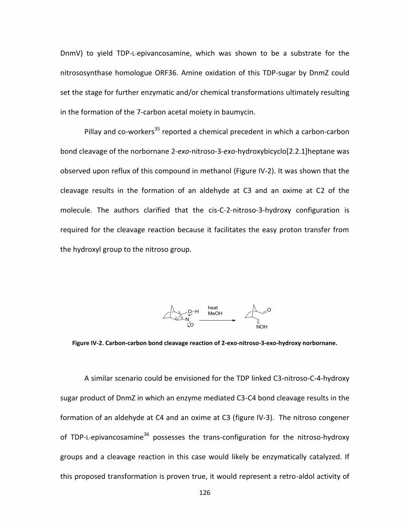
126
DnmV) to yield TDP-L-epivancosamine, which was shown to be a substrate for the
nitrososynthase homologue ORF36. Amine oxidation of this TDP-sugar by DnmZ could
set the stage for further enzymatic and/or chemical transformations ultimately resulting
in the formation of the 7-carbon acetal moiety in baumycin.
Pillay and co-workers35 reported a chemical precedent in which a carbon-carbon
bond cleavage of the norbornane 2-exo-nitroso-3-exo-hydroxybicyclo[2.2.1]heptane was
observed upon reflux of this compound in methanol (Figure IV-2). It was shown that the
cleavage results in the formation of an aldehyde at C3 and an oxime at C2 of the
molecule. The authors clarified that the cis-C-2-nitroso-3-hydroxy configuration is
required for the cleavage reaction because it facilitates the easy proton transfer from
the hydroxyl group to the nitroso group.
Figure IV-2. Carbon-carbon bond cleavage reaction of 2-exo-nitroso-3-exo-hydroxy norbornane.
A similar scenario could be envisioned for the TDP linked C3-nitroso-C-4-hydroxy
sugar product of DnmZ in which an enzyme mediated C3-C4 bond cleavage results in the
formation of an aldehyde at C4 and an oxime at C3 (figure IV-3). The nitroso congener
of TDP-L-epivancosamine36 possesses the trans-configuration for the nitroso-hydroxy
groups and a cleavage reaction in this case would likely be enzymatically catalyzed. If
this proposed transformation is proven true, it would represent a retro-aldol activity of

127
the nitrososynthase DnmZ, a function not known for any falvin dependent
monooxygenase characterized to date.
Figure IV-3. Proposed pathway for the DnmZ monooxygenation activity and the subsequent cleavge and glycoslation activities.
Activity of DnmZ as a nitrososynthase
Given the high sequence homology of DnmZ with the characterized flavin-
dependent nitrososynthase ORF36, it is expected that both enzymes catalyze similar
deoxysugar N-oxidation reaction. To confirm this, we amplified the dnmz gene from
Streptomyces peucetius via the polymerase-chain reaction (PCR), cloned it into the
NdeI/HindIII sites of pET28a expression vector, and transformed the resulting plasmid

128
into the BL21 DE3 E.coli bacterial strain via electroporation. Expression conditions for
the protein were found to be similar to those previously described for its ORF36 and
RubN8 homologues. The protein was purified by Ni-affinity chromatography and
analyzed by SDS-PAGE (Figure IV-4).
Figure IV-4. SDS-PAGE gel of Ni-affinity purified DnmZ
We used the biogenic aminosugar TDP-L-epivancosamine as a substrate to assay
the nitrososynthase activity of DnmZ and followed the same assay conditions performed
for ORF36 oxidation reaction. The HPLC-MS analysis of the DnmZ assay showed similar
reaction profile as that of ORF36 ultimately yielding products with m/z 558 and 576
previously identified as the nitrososugar congener and a shunt oxidation product
respectively.
According to our proposed retro-aldol activity of Dnmz, the C3-C4 bond cleavage
would result in an aldehyde at C4 and an oxime at C3 of the TDP-3-nitro-4-hydroxy sugar
(Figure IV-3). This TDP linked compound with terminal oxime and aldehyde groups has
an m/z of 558 in the negative mode. Aldehydes are known to exist in their hydrated
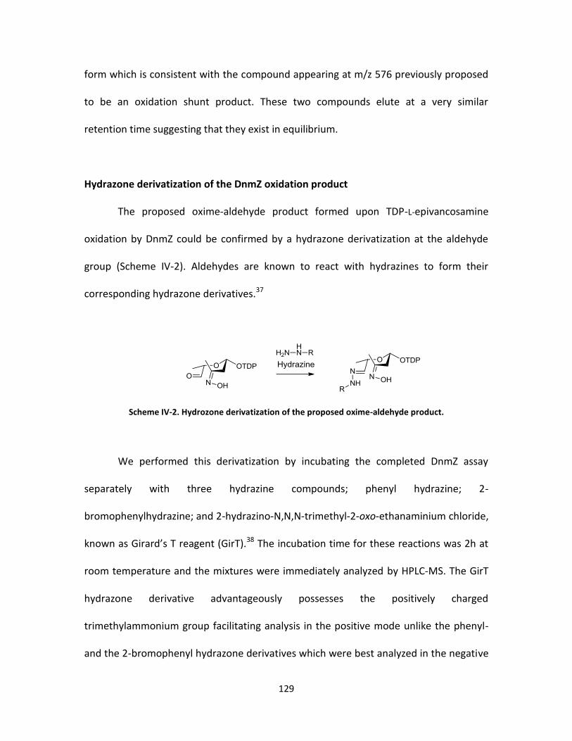
129
form which is consistent with the compound appearing at m/z 576 previously proposed
to be an oxidation shunt product. These two compounds elute at a very similar
retention time suggesting that they exist in equilibrium.
Hydrazone derivatization of the DnmZ oxidation product
The proposed oxime-aldehyde product formed upon TDP-L-epivancosamine
oxidation by DnmZ could be confirmed by a hydrazone derivatization at the aldehyde
group (Scheme IV-2). Aldehydes are known to react with hydrazines to form their
corresponding hydrazone derivatives.37
Scheme IV-2. Hydrozone derivatization of the proposed oxime-aldehyde product.
We performed this derivatization by incubating the completed DnmZ assay
separately with three hydrazine compounds; phenyl hydrazine; 2-
bromophenylhydrazine; and 2-hydrazino-N,N,N-trimethyl-2-oxo-ethanaminium chloride,
known as Girard’s T reagent (GirT).38 The incubation time for these reactions was 2h at
room temperature and the mixtures were immediately analyzed by HPLC-MS. The GirT
hydrazone derivative advantageously possesses the positively charged
trimethylammonium group facilitating analysis in the positive mode unlike the phenyl-
and the 2-bromophenyl hydrazone derivatives which were best analyzed in the negative

130
mode aided by deprotonation of their attached nucleotide phosphates. As shown in
figure IV-5, HPLC-MS peaks at m/z’s corresponding to all of the expected three
hydrazone derivatives were observed at different retention times. The
phenylhydrazone derivative was observed at 31.1 minutes matching the expected m/z
of 648. The 2-bromophynlhydrazone derivative appeared at 34.4 minutes with m/z’s of
726 and 728 at similar abundance corresponding to the bromine isotopic distribution.39
Finally, the GirT hydrazone derivative was observed at 26.4 minutes with m/z of 673 in
the positive mode. The observation of m/z’s matching these hydrozone conjugates is
consistent with the proposed TDP-linked oxime-aldehyde product; however, further
structure characterization is required to validate the identity of these conjugates.
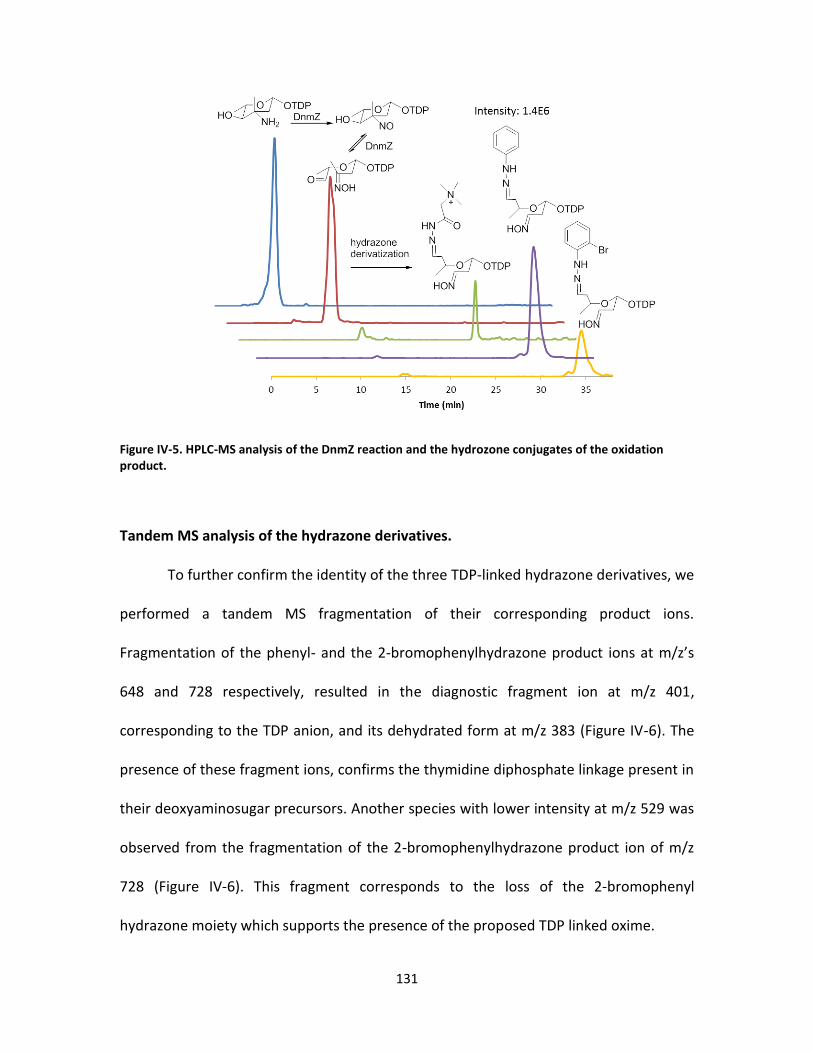
131
Figure IV-5. HPLC-MS analysis of the DnmZ reaction and the hydrozone conjugates of the oxidation product.
Tandem MS analysis of the hydrazone derivatives.
To further confirm the identity of the three TDP-linked hydrazone derivatives, we
performed a tandem MS fragmentation of their corresponding product ions.
Fragmentation of the phenyl- and the 2-bromophenylhydrazone product ions at m/z’s
648 and 728 respectively, resulted in the diagnostic fragment ion at m/z 401,
corresponding to the TDP anion, and its dehydrated form at m/z 383 (Figure IV-6). The
presence of these fragment ions, confirms the thymidine diphosphate linkage present in
their deoxyaminosugar precursors. Another species with lower intensity at m/z 529 was
observed from the fragmentation of the 2-bromophenylhydrazone product ion of m/z
728 (Figure IV-6). This fragment corresponds to the loss of the 2-bromophenyl
hydrazone moiety which supports the presence of the proposed TDP linked oxime.

132
Figure IV-6. Tandem LC-ESI-MS of the 2-bromophenyl hydrozone derivative
Changing the fragmentation conditions for these two hydrazones in both the
negative and the positive modes did not result in any additional observable fragment
ions diagnostic of the hydrazone side of these derivatives. Therefore, we performed
tandem MS fragmentation of the GirT hydrazone product ion of m/z 673 in the positive
mode. The hydrazone side of this molecular ion is positively charged, and as expected its
fragmentation produced several fragment ions that could be easily assigned to relevant
structures. Of note were the fragments that represented to the loss of thymidine
monophosphate (TMP) at m/z 351, and the loss of TDP at m/z 271 (Figure IV-7).

133
Another fragment was observed at m/z 170 which is a result of the cleavage of the
acetal’s C-O bond as shown in Figure IV-7.
Figure IV-7. Tandem LC-ESI-MS of the GirT hydrozone derivative
Acid hydrolysis and HPLC-MS analysis of the Girard’s hydrazone derivative
We aimed to obtain more structural evidence of the hydrazone conjugates by
hydrolyzing these derivatives with 1 M HCl and analyzing the resulting mixture by HPLC-
MS. The acid hydrolysis of these acetal molecules would result in the formation of an
alcohol on the hydrazone side and an aldehyde on the oxime side of the molecule
(Figure IV-8). The TDP moiety is readily hydrolyzed upon acid treatment. Since the

134
hydrazine is present in excess, the newly formed aldehyde species is expected to form a
second hydrazone conjugate at the anomeric C1 of the original sugar. HPLC-MS analysis
both in the negative and positive modes identified no molecular ions corresponding to
the expected hydrolyzed fragments of the acid-treated phenyl- and 2-bromophenyl
hydrazone derivatives. This is perhaps due to the lack of ionization of the small
molecular weight hydrolysis products possessing neutral charge. However, the acid
treatment of the positively charged GirT hydrazone derivative produced two species at
m/z 188 and 215 corresponding to both the oxime and the alcohol hydrazone
conjugates while the original GirT hydrozone disappeared (Figure IV-8).
Figure IV-8. Acid hydrolysis of the (carboxymethyl)trimethylammonium hydrazone derivative (above), and LC-ESI-MS chromatograms of the hydrolyzed species (below).
Further MS/MS analysis of these species showed fragment ions consistent with
their structures (Figure IV-9). Of note, fragmentation of the hydrazone derivative at m/z

135
188 resulted in species corresponding to the loss of the trimethylammonium group at
m/z 129; dehydration of the secondary alcohol at m/z 170, and the cleavage of the
amide bond to yield a fragment ion with m/z 87. The oxime hydrazone derivative at m/z
215 also showed a fragment corresponding to the loss of the trimethylammonium
group at m/z 156 as well as a fragment corresponding to the formation of the
protonated trimethylammonium species at m/z 60. These tandem MS data strongly
support the structure assignment of the original GirT hydrazone derivative and provide
more evidence for the carbon-carbon bond cleavage of the TDP-attached nitrososugar
product of DnmZ oxidation.
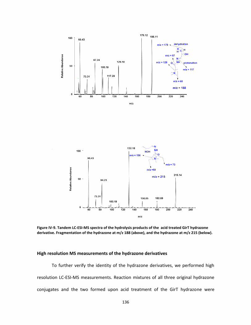
136
Figure IV-9. Tandem LC-ESI-MS spectra of the hydrolysis products of the acid treated GirT hydrazone derivative. Fragmentation of the hydrazone at m/z 188 (above), and the hydrazone at m/z 215 (below).
High resolution MS measurements of the hydrazone derivatives
To further verify the identity of the hydrazone derivatives, we performed high
resolution LC-ESI-MS measurements. Reaction mixtures of all three original hydrazone
conjugates and the two formed upon acid treatment of the GirT hydrazone were

137
measured. As shown in Figure IV-10, mass accuracies of less than 2.70 ppm were
confirmed for all of the hydrazone conjugates measured. Taken together, these high
resolution mass measurements are consistent with the assigned structures of the TDP
linked hydraznes and are in support of the proposed deoxysugar C-C bond cleavge
mediated by DnmZ activity.
Figure IV-10. High resolution mass measurements of the hydrazone derivatives.
Conclusion
In this work, we used the nitrososynthase Dnmz from Streptomyces peucetius to
show that the formation of the previously uncharecterized acetal moiety in baumycin is
derived from the cleavage of a carbon-carbon bond mediated by DnmZ N-oxidation of a
TDP-deoxyaminosugar. This retro-aldol activity of DmnZ represents a new flavin-

138
dependent monooxygenase function which is of great significance for elucidating the
biosynthetic pathways of various natural products such as baumycin-like anthracyclines.
This additional monooxygenase reactivity is also helpful in chemical synthesis and
provides green biocatalysis for some difficult to perform retro-aldol reactions. Future
investigations of this pathway should focus on elucidating the glycosylation
requirements as well as the reduction steps of the oxime and aldehyde groups to
ultimately form the alcohol functionalities.
Materials and methods
Bacterial strains, Plasmids and Materials.
All reagents were obtained from Sigma-Aldrich Corporation and used without
further purification unless otherwise noted. E. coli TOP10 and BL21 (DE3) competent
cells were obtained from Invitrogen Inc. (Carlsbad, CA) and Novagen (Madison, WI),
respectively. Restriction endonucleases and T4 DNA ligase were obtained from New
England Biolabs (Ipswich, MA). The pET28a expression vector was purchased from
Novagen Inc. Taqplus DNA polymerase was purchased from Stratagene Inc. (La Jolla,
CA).
Cloning and overexpression of dnmZ.
The gene encoding DnmZ was amplified from the genomic DNA of Streptomyces
peucetius17 using the following primers 5’- CATATGACAAAGCCATCTGTGCACG-3’ and 5’-

139
AAGCTTTCATCGGCCCAACGCC- ACTT-3’ (NdeI and HindIII restriction enzyme sites
underlined). PCR reactions were carried out using Taqplus DNA polymerase according to
the manufacture’s protocol. Subcloning of dnmZ into the NdeI/HindIII sites of pET28a
yielded recombinant plasmid pET28-ZN for expression as an N-terminal hexahistidine
fusion protein. Plasmid pET28-ZN was transformed by electroporation into E. coli
BL21(DE3) for heterologous expression of DnmZ. Cultures of E. coli BL21(DE3)/pET28-ZN
were grown at 37°C to an OD600 of 0.6, at which point the culture was induced with 0.1
mM isopropyl-beta-D-thiogalactopyranoside (IPTG) and grown an additional 6 hours at
28°C. Cells were harvested by centrifugation and stored at -80°C until needed. IPTG
induced E. coli BL21(DE3)/pET28-ZN cells were resuspended in buffer A (20 mM
Imidazole, 0.5M NaCl, 20mM Tris-Cl, pH 7.5) and lysed by sonication. The lysate was
loaded onto a charged 5-mL Histrap crude column (Amersham Biosciences) and purified
by FPLC at a flow rate of 5mL/min. The column was washed with buffer A (20 mM
Imidazole, 0.5 M NaCl, 20 mM Tris-Cl, pH 7.5) and buffer B (500 mM Imidazole, 0.5 M
NaCl, 20 mM Tris-Cl, pH 7.5) using a step gradient. Fractions containing DnmZ were
analyzed by SDS-page and buffer was exchanged via a desalting column (HisTrap) using
buffer C (20 mM Tris-Cl, 1 mM dithiothreitol and 5% glycerol, pH 7.5) and stored at -80
°C until assayed.
Preparation of TDP-L-epi-vancosamine.
TDP-L-epivancosamine was biochemically synthesized from Thymidine-5’-
diphospho-α-D-glucose using the general procedures outlined by Chen et al. The
140
following enzymes were used to biochemically transform Thymidine-5’-diphospho-α-D-
glucose to TDP-L-epivancosamine: RfbB from the rhamnose biosynthetic pathway40and,
EvaA-E from chloroeremomycin biosynthetic pathway. Preparative enzymatic reaction
progress was followed via HPLC using a series 600 Waters HPLC system with a Waters
2996 diode array detector. Analytical separations were performed on an Adsorbosphere
strong anion exchange (SAX) column (5 µm, 4.6 mm 20 mm, Alltech Associates) and a
linear gradient from 50 to 500 mM NH4HCO2 (pH 3.5) at 1 mL/min for 45 min.
Preparative HPLC separations were performed using a semipreparative Adsorbosphere
SAX column (5 μm, 22 mm 250 mm) using a similar protocol, but at 10 mL/min. The pH
values of the fractions containing TDP chromophores (267 nm) were immediately
adjusted to ∼7 with 1 M NH4OH, and fractions containing reaction product were pooled,
lyophilized, and stored at -80 °C until they were assayed. Resuspended compound
concentrations were determined by measurement of the absorbance at 267 nm using a
Nanodrop spectrophotometer (Thermo, Inc.) and comparison to a standard curve of
dTDP.
DnmZ assay with TDP-L-epi-vancosamine.
Reactions of DnmZ with TDP-L-epivancosamine were performed in a total volume
of 50 μL containing 30 μM substrate, 30 μM FAD, 1 unit/mL catalase, 1 U/mL
superoxidedismutase, 0.4 U/mL flavin reductase, and 2.0 mM NADPH. All reactions were
initiated by addition of 15 μM DnmZ, reactions were quenched with equal volume of
141
acetone, and mixtures stored at -80 °C until they were analyzed by LC-ESI-MS. Control
assays were performed without DnmZ, FAD, or NADPH.
Derivatization of DnmZ products with hydrazines.
Upon completion of the DnmZ reaction with TDP-L-epivancosamine, the reaction
mixture (50 uL) was incubated with the corresponding hydrazine (1 mM) for 2 h at room
temperature. The mixture was kept at - 80 °C until analyzed by HPLC-MS. The following
hydrazines were used for the derivatization experiments: phenylhydrazine, 2-
bromophenylhydrazine, and Girard’s reagent (Carboxymethyl)trimethylammonium
chloride hydrazide; 2-Hydrazino-N,N,N-trimethyl-2-oxo-ethanaminium chloride).
LC-ESI-MS Method for DnmZ assays.
DnmZ assays were analyzed using Thermo Finnigan (San Jose, CA) TSQ®
Quantum triple quadrupole mass spectrometer equipped with a standard electrospray
ionization source outfitted with a 50 μm inside diameter deactivated fused silica
capillary. Injections of 10 μL were separated using a Hypercarb column (3mm x 50mm,
Thermo Inc.). Mobile phases were (A) H2O with 50 mM NH4CH3COO and 0.1% (v/v)
diethylamine and (B) a H2O/acetonitrile mixture (5:95) with 50 mM NH4CH3COO and
0.1% (v/v) diethylamine. Gradient conditions were as follows: linear gradient from 0 to
100% B from for 30 min, 100% B from 30 to 40 min, 100% A from 40 to 50 min. The flow
rate was maintained at 0.2 mL/min. The mass spectrometer was operated in both the
negative ion and full scan profile modes, and the electrospray needle was maintained at
142
3100 V. The ion transfer tube was operated at -47.50 V and 275 °C. The tube lens
voltage was set to -46 V. The collision energy for all product ion scans was set at 25%.
References
1. du Bois, A.; Luck, H. J.; Pfisterer, J.; Meier, W.; Bauknecht, T., [Anthracyclines in therapy of ovarian carcinoma: a systematic review of primary and 2nd-line therapy after platinum]. Zentralblatt fur Gynakologie 2000, 122 (5), 255-67.
2. Garcia, A.; Muggia, F. M., Activity of anthracyclines in refractory ovarian cancer: recent experience and review. Cancer Investigation 1997, 15 (4), 329-34.
3. Mazue, G.; Iatropoulos, M.; Imondi, A.; Castellino, S.; Brughera, M.; Podesta, A.; Dellatorre, P.; Moneta, D., Anthracyclines - a review of general and special toxicity studies. International Journal of Oncology 1995, 7 (4), 713-26.
4. Laatsch, H.; Fotso, S., Naturally occurring anthracyclines. Anthracycline Chemistry and Biology I 2008, 3-74.
5. Krohn, K., Anthracycline Chemistry and Biology II: Mode of Action, Clinical Aspects and New Drugs. Springer Verlag: 2008; Vol. 2.
6. Aubel-Sadron, G.; Londos-Gagliardi, D., Daunorubicin and doxorubicin, anthracycline antibiotics, a physicochemical and biological review. Biochimie 1984, 66 (5), 333-52.
7. Trouet, A.; Deprez-De Campeneere, D., Daunorubicin-DNA and doxorubicin-DNA. A review of experimental and clinical data. Cancer Chemotherapy and Pharmacology 1979, 2 (1), 77-9.
8. Hortobagyi, G., Anthracyclines in the treatment of cancer. An overview. Drugs 1997, 54, 1.
143
9. Chhikara, B. S.; St Jean, N.; Mandal, D.; Kumar, A.; Parang, K., Fatty acyl amide derivatives of doxorubicin: synthesis and in vitro anticancer activities. European Journal of Medicinal Chemistry 2011, 46 (6), 2037-42.
10. Altreuter, D. H.; Dordick, J. S.; Clark, D. S., Nonaqueous biocatalytic synthesis of new cytotoxic doxorubicin derivatives: exploiting unexpected differences in the regioselectivity of salt-activated and solubilized subtilisin. Journal of the American Chemical Society 2002, 124 (9), 1871-6.
11. Vey, J. L.; Al-Mestarihi, A.; Hu, Y.; Funk, M. A.; Bachmann, B. O.; Iverson, T. M., Structure and mechanism of ORF36, an amino sugar oxidizing enzyme in everninomicin biosynthesis. Biochemistry 2010, 49 (43), 9306-17.
12. Stefanska, B.; Dzieduszycka, M.; Bontemps-Gracz, M.; Borowski, E.; Martelli, S., Synthesis and antileukemic activity of N-enamine derivatives of daunorubicin, 5-iminodaunorubicin, and doxorubicin. Journal of Antibiotics (Tokyo) 1988, 41 (2), 193-8.
13. Ghirmai, S.; Mume, E.; Tolmachev, V.; Sjoberg, S., Synthesis and radioiodination of some daunorubicin and doxorubicin derivatives. Carbohydrate Research 2005, 340 (1), 15-24.
14. Chhikara, B. S.; Mandal, D.; Parang, K., Synthesis, anticancer activities, and cellular uptake studies of lipophilic derivatives of doxorubicin succinate. Journal of Medicinal Chemistry 2012, 55 (4), 1500-10.
15. Madduri, K.; Hutchinson, C. R., Functional characterization and transcriptional analysis of the dnrR1 locus, which controls daunorubicin biosynthesis in Streptomyces peucetius. Journal of Bacteriology 1995, 177 (5), 1208-15.
16. McGuire, J. C.; Thomas, M. C.; Stroshane, R. M.; Hamilton, B. K.; White, R. J., Biosynthesis of daunorubicin glycosides: role of epsilon-rhodomycinone. Antimicrobial Agents and Chemotherapy 1980, 18 (3), 454-64.
17. Otten, S. L.; Stutzman-Engwall, K. J.; Hutchinson, C. R., Cloning and expression of daunorubicin biosynthesis genes from Streptomyces peucetius and S. peucetius subsp. caesius. Journal of Bacteriology 1990, 172 (6), 3427-34.
144
18. Hutchinson, C. R., Biosynthetic Studies of Daunorubicin and Tetracenomycin C. Chemical Reviews 1997, 97 (7), 2525-2536.
19. Krohn, K., Anthracycline Chemistry and Biology I: Biological Occurence and Biosynthesis, Synthesis and Chemistry. Springer Verlag: 2008; Vol. 1.
20. Liu, E. H.; Qi, L. W.; Wu, Q.; Peng, Y. B.; Li, P., Anticancer agents derived from natural products. Mini Reviews in Medicinal Chemistry 2009, 9 (13), 1547-55.
21. Fujii, I.; Ebizuka, Y., Anthracycline Biosynthesis in Streptomyces galilaeus. Chemical Reviews 1997, 97 (7), 2511-2524.
22. Miyamoto, Y.; Johdo, O.; Nagamatsu, Y.; Yoshimoto, A., Cloning and characterization of a glycosyltransferase gene involved in the biosynthesis of anthracycline antibiotic beta-rhodomycin from Streptomyces violaceus. FEMS Microbiology Letters 2002, 206 (2), 163-8.
23. Sianidis, G.; Wohlert, S. E.; Pozidis, C.; Karamanou, S.; Luzhetskyy, A.; Vente, A.; Economou, A., Cloning, purification and characterization of a functional anthracycline glycosyltransferase. Journal of Biotechnology 2006, 125 (3), 425-33.
24. Garrido, L. M.; Lombo, F.; Baig, I.; Nur, E. A. M.; Furlan, R. L.; Borda, C. C.; Brana, A.; Mendez, C.; Salas, J. A.; Rohr, J.; Padilla, G., Insights in the glycosylation steps during biosynthesis of the antitumor anthracycline cosmomycin: characterization of two glycosyltransferase genes. Applied Microbiology and Biotechnology 2006, 73 (1), 122-31.
25. Otten, S. L.; Gallo, M. A.; Madduri, K.; Liu, X.; Hutchinson, C. R., Cloning and characterization of the Streptomyces peucetius dnmZUV genes encoding three enzymes required for biosynthesis of the daunorubicin precursor thymidine diphospho-L-daunosamine. Journal of Bacteriology 1997, 179 (13), 4446-50.
26. Yoshimoto, A.; Fujii, S.; Johdo, O.; Kubo, K.; Nishida, H.; Okamoto, R.; Takeuchi, T., Anthracycline metabolites from baumycin-producing Streptomyces sp. D788. II. New anthracycline metabolites produced by a blocked mutant strain RPM-5. The Journal of Antibiotics 1993, 46 (1), 56.
145
27. Nozaki, Y.; Hida, T.; Iinuma, S.; Ishii, T.; Sudo, K.; Muroi, M.; Kanamaru, T., TAN-1120, a new anthracycline with potent angiostatic activity. The Journal of Antibiotics 1993, 46 (4), 569.
28. Lomovskaya, N.; Doi-Katayama, Y.; Filippini, S.; Nastro, C.; Fonstein, L.; Gallo, M.; Colombo, A. L.; Hutchinson, C. R., The Streptomyces peucetius dpsY and dnrX genes govern early and late steps of daunorubicin and doxorubicin biosynthesis. Journal of Bacteriology 1998, 180 (9), 2379-86.
29. Hu, Y.; Al-Mestarihi, A.; Grimes, C. L.; Kahne, D.; Bachmann, B. O., A unifying nitrososynthase involved in nitrosugar biosynthesis. Journal of the American Chemical Society 2008, 130 (47), 15756-7.
30. Bruender, N. A.; Thoden, J. B.; Holden, H. M., X-ray structure of kijd3, a key enzyme involved in the biosynthesis of D-kijanose. Biochemistry 2010, 49 (17), 3517-24.
31. Hutchinson, C.; Colombo, A., Genetic engineering of doxorubicin production in Streptomyces peucetius: a review. Journal of Industrial Microbiology & Biotechnology 1999, 23 (1), 647-652.
32. Ganguly, A. K.; Sarre, O. Z.; Reimann, H., Evernitrose, a naturally occurring nitro sugar from everninomicins. Journal of the American Chemical Society 1968, 90 (25), 7129-30.
33. Zhang, H.; White-Phillip, J. A.; Melancon, C. E., 3rd; Kwon, H. J.; Yu, W. L.; Liu, H. W., Elucidation of the kijanimicin gene cluster: insights into the biosynthesis of spirotetronate antibiotics and nitrosugars. Journal of the American Chemical Society 2007, 129 (47), 14670-83.
34. Mizsak, S. A.; Hoeksema, H.; Pschigoda, L. M., The chemistry of rubradirin. II. Rubranitrose. J Antibiot (Tokyo) 1979, 32 (7), 771-2.
35. K. Somosekharen Pillay, S. C. C., Thomas Mojelsky, Yuan L. Chow Photoreaction of Nitroso Compounds in Solution. Part XXXI. Mechanisms of Intramolecular Acid Catalyzed Cleavage Reactions and a Hydride Initiated Rearrangement of C-Nitroso Alkanes. Canadian Journal of Chemistry 1975, 53 (20), 3014-3021.

146
36. Chen, H.; Thomas, M. G.; Hubbard, B. K.; Losey, H. C.; Walsh, C. T.; Burkart, M. D., Deoxysugars in glycopeptide antibiotics: enzymatic synthesis of TDP-L-
epivancosamine in chloroeremomycin biosynthesis. Proceedings of the National Academy of Sciences of the United States of America 2000, 97 (22), 11942-7.
37. Shriner, R. L.; HERMANN, C. K., The Systematic Identification or Organic compounds, WIE. John Wiley & Sons (New York, 1980): 2003.
38. Johnson, D. W., A modified Girard derivatizing reagent for universal profiling and trace analysis of aldehydes and ketones by electrospray ionization tandem mass spectrometry. Rapid Communications in Mass Spectrometry : RCM 2007, 21 (18), 2926-32.
39. Cameron, A. E.; Lippert, E. L., Jr., Isotopic Composition of Bromine in Nature. Science 1955, 121 (3135), 136-7.
40. Marolda, C. L.; Valvano, M. A., Genetic analysis of the dTDP-rhamnose biosynthesis region of the Escherichia coli VW187 (O7:K1) rfb gene cluster: identification of functional homologs of rfbB and rfbA in the rff cluster and correct location of the rffE gene. Journal of Bacteriology 1995, 177 (19), 5539-46.

147
CHAPTER V
SUMMARY AND FUTURE DIRECTIONS
Synopsis
Flavin-dependent monooxygenases are a diverse class of oxidative biocatalysts
many of which have been extensively studied over the past few decades.1-3 They have
been shown to catalyze a remarkable wide variety of reactions such as regioselective
hydroxylations, epioxidations, Bayer-Villiger oxidations and enantioselective
sulfoxidations. The exquisite specificity and efficiency of these enzymes are often
unparalleled compared to chemical catalysts. Recent advances in genome sequence
data suggested that many more flavin-dependent monooxygenases exist and await
discovery and characterization. The work presented in this dissertation describes the
biochemical characterization of a new flavin-dependent monooxygnase, which catalyzes
the N-oxidation of deoxyaminosugars in vast array of glycosylated natural products.
The nitrososynthase homologues ORF36 and RubN8 from the everninomicn and
the rubradirin biosynthetic pathways, respectively, were shown to catalyze the amine to
nitroso oxidation of the deoxyaminosugar TDP-L-epivancosamine.4-5 Biochemical
evidence provided in this work, indicates that the nitrososynthase catalysis involves two
FAD-dependent monooxygenation steps in which the aminosugar is first oxidized to the
corresponding hydroxylamine, followed by a second iterative N-oxygenation to
ultimately form the nitrososugar. The nitrososynthase activity is significantly enhanced

148
by the inclusion of an external flavin reductase which generates the reduced flavin
cofactor required for mediating the oxygenation catalysis. The co-enzyme used for
reducing equivalents in this interesting flavin-dependen monooxygenation catalysis is
NADPH.
Several TDP-aminosugar intermediates in the biosynthetic pathway of TDP-L-
epivancosamine6 were examined as candidate substrates for the N-oxidation by the
nitrososynthases ORF36 and RubN8. The aminosugar substrates upstream of TDP-L-
epivancosamine underwent only single-oxidation reactions terminating in the
hydroxylamine oxidation state pointing to the substrate catalytic competence of the
nitrososynthase enzyme. Performing the nitrososynthase catalysis under 18O2
demonstrated that the source of oxygen in the oxidation intermediates and products
stems from molecular oxygen rather than from water, which is consistent with flavin-
dependent monooxygenase catalysis. The X-ray crystal structure of the nitrososynthase
ORF364 revealed a tetrameric enzyme that shares a fold with acyl-CoA dehydrogenases
but has an unusually large open active site.
Surprisingly, the nitrososynthase DnmZ from Streptomyces peucetius was shown
to mediate a carbon-carbon bond cleavage of the β-hydroxy-nitrososugar produced
from the N-oxidation of TDP-L-epivancosamine, catalyzed by the same enzyme. This
retro-aldol activity represents a new and important addition to the functions known for
flavin-dependent monooxygenases. Furthermore, it explains the biosynthetic origin of
the once not understood non-sugar acetal moiety in baumycin-like natural products.

149
Future directions
Mechanism of the nitro group formation
The pathway for the conversion of nitrososugars to their nitrosugar congeners
remains to be elucidated. To that end, fermentations of S. achromogenes in complete
darkness produced predominantly protorubradirin, the nitroso congener of rubradirin,
which reportedly converted to rubradirin (i.e the nitrososugar moiety was oxidized to its
nitrosugar congener) upon exposure to ambient light.7 This suggested that the oxidation
to the nitro group is induced photochemically and that the nitrososugar natural product
congeners are the original biosynthetic metabolites. Seemingly at odds with this, the
nitrososugar analog of everninomicin was also produced from the fermentation of M.
carbonacae var. africana along with other derivatives including the nitrosugar analog
under ambient light.8 Other possibilities for the nitro group formation include oxidation
via endogenous reactive oxygen species9 or by flavin-dependent monooxygenation
catalyzed by the nitrososynthase. In our HPLC-MS analysis of the nitrososynthase assay,
we observed a mass peak corresponding to the TDP-nitrosugar, but it was forming at
low rates, ambiguous to be counted as an enzymatic conversion.5 Since the TDP-L-
epivancosmine substrate lacks C4 O-methylation and was shown to undergo enzymatic
carbon-carbon bond cleavage upon the nitrososynthase catalysis, the authentic C4 O-
methylated TDP-aminosugar substrtae (TDP-L-evernosamine) is perhaps more suitable
for studying the nitro group formation mechanism. We performed preliminary
nitrososynthase assays with this substrate, which was prepared enzymatically from TDP-

150
L-epivancosaime via the C4-methyltransferase RubN7 from the rubradirin pathway
(Figure V-1), and found that both ORF36 and RubN8 were able to catalyze conversions
to the nitrososugar (Figure V-II). Additionally, and as expected, there was no hydrate
observed for this nitrososugar product, consistent with the protection of the C-4
position. Surprisingly, DnmZ did not oxidize TDP-L-evernosamine suggesting diversity in
catalytic competence among nitrososynthases.
Figure V-1. LC-ESI/MS chromatograms of the preparation of TDP-L-evernosamine via the
methyltransferase Rubn7 from the rubradirin pathway.

151
Figure V-2. LC-ESI-MS chromatograms of the monooxygenation of TDP-L-evernosamine by ORF36.
A good way to study whether the nitro group formation is catalyzed by a
nitrososynthase activity is to isolate the C4 O-methylated TDP-nitrososugar and perform
enzymatic assays of this substrate with the nitrososynthase ORF36. Analysis by HPLC-
MS using our established methods would show whether this transformation is carried
out enzymatically. The major challenge that faces this approach is that these TDP-sugars
are produced in low quantities and are susceptible to hydrolysis of their attached TDP
moiety. Chemical synthesis of these deoxysugar nucleotides is an alternative route to
obtain sufficient quantities for enzymatic assays.10
152
Structural studies
Cocrystallization of the nitrososynthases with FAD or FMN cofactors and/or an
aminosugar substrate remains an important experiment to perform. Preliminary
cocrystallization attempts have been so far unsuccessful perhaps due to the complexity
of substrate and cofactor binding requirements. The nitrososynthase KijD3 from
Kijaniata actinomadura was successfully cocrystallized with the nucleotide thymidine
diphosphate phenol (dTDP-phenol).11 While the resulting structure provided only little
details about the molecular determinates of catalysis, it gave hope for finding
appropriate cocrystallization conditions for the enzyme-substrate or enzyme-substrate-
cofactor complexes.
The nitrososynthase co-structures would reveal important amino acid residues
required for catalysis, while site directed mutagenesis of these residues would validate
their roles in the monooxygenation mechanism. We performed site directed
mutagenesis experiments on selected residues chosen based on modeling TDP-L-
epivancosamine and FAD into the active site of ORF36; however, none of the mutations
abolished the nitrososynthase activity. As discussed in chapter III, the nitrososynthase
likely undergoes significant conformational adjustment upon substrate binding and
catalysis thus, identifying the active site residues would require obtaining enzyme-
substrate-FAD co-structures.
As mentioned above, preliminary experiments suggested that the catalytic
competence vary among nitrososynthases. For example, unlike ORF36, DnmZ showed

153
no monooxygenase activity with the C4 O-methylated substrate (TDP-L-evernosamine).
It is intriguing to investigate the structural differences among nitrososynthases that
allows this interesting selectivity. The recent success in crystallizing the DnmZ enzyme
by Dr. Jessica Vey’s research group at California State University, Northridge, is a step
towards the completion of these structural investigations.
Catalytic competence and kinetic studies
Biochemical characterization of the nitrososynthase enzymes included in this
work revealed important substrate preference data and set the stage for further
catalytic competence studies. Our successful enzymatic production of the aminosugar
substrate TDP-L-evernosamine and the preliminary data of its oxidation by the
nitrososynthases studied in this work is an effort towards the completion of this goal.
Michaelis-Menten kinetic measurements12 of the available aminosugar substrates will
aid in the elucidation of the nirososynthase’s monooxygenation mechanism. Challenges
that may render kinetic studies problematic via our HPLC-MS methods include the
availability of the substrates in sufficient quantities, and the susceptibility to hydrolysis
of their attached TDP moieties. Chemical synthesis of the TDP-sugar substrates10 and
the use of internal standards are possible solutions for these issues.

154
The interplay between the nitrososynthases and glycosyltransferases
The remarkable nitrososynthase catalysis described in this work, results in the
formation of TDP-linked sugar donor substrates of unusual modifications. The
subsequent sugar cleavage upon the DnmZ activity raises the question as to how this
cleaved sugar gets attached to its corresponding anthracycline aglycone to ultimately
form baumycin-like glycosides. One possible scenario for the glycoslation is that the
resulting cleaved sugar exists in equilibrium with its non-cleaved form which can
facilitate its glycoslation by the glycosyltransferase DnrH.13 This equilibrium could
further exist after glycoslation and combined with subsequent chemical or enzymatic
reduction steps may result in the formation of several baumycin-like compounds.14-15
Performing glycoslation experiments using the glycosyltransferase DnrH identified in the
anthracycline biosynthetic pathway in Streptomyces peucetius would test this proposed
pathway. Some natural product glycosyltransferases require a partner for activity and it
has been shown that co-expression of the GT with its partner greatly improves the
solubility of GTs. Gene disruption studies showed that the DnrS glycosyltransferese in
the daunorubicin biosynthesis requires a protein partner, DnrQ, for its activity.13 These
considerations should be taken into account when designing the glycoslation
experiments. The commercial availability of the aglycone substrate daunorubicin is
advantageously suited for conducting these studies.
It is also intriguing to determine the preferred substrate for the GT responsible
for glycoslating of the N-oxidized sugar in everninomicin biosynthesis. The identity of
the TDP-sugar donor substrate of glycoslation would reveal important clues into the

155
interplay between the nitrososynthase ORF36 and the subsequent activity of the GT.
The aglycone substrate can be obtained via a mild hydrolysis of everninomicin described
by Ganguly, Sarre, and Szmulewicz in U.S. Patent 3,920,629. Our successful production
of two biogenic sugar donor substrate candidates will aid in the completion of these
studies.
References
1. Massey, V., The chemical and biological versatility of riboflavin. Biochemical Society Transactions 2000, 28 (4), 283-96.
2. Joosten, V.; van Berkel, W. J. H., Flavoenzymes. Current Opinion in Chemical biology 2007, 11 (2), 195-202.
3. van Berkel, W. J.; Kamerbeek, N. M.; Fraaije, M. W., Flavoprotein monooxygenases, a diverse class of oxidative biocatalysts. Journal of Biotechnology 2006, 124 (4), 670-89.
4. Vey, J. L.; Al-Mestarihi, A.; Hu, Y.; Funk, M. A.; Bachmann, B. O.; Iverson, T. M., Structure and mechanism of ORF36, an amino sugar oxidizing enzyme in everninomicin biosynthesis. Biochemistry 2010, 49 (43), 9306-17.
5. Hu, Y.; Al-Mestarihi, A.; Grimes, C. L.; Kahne, D.; Bachmann, B. O., A unifying nitrososynthase involved in nitrosugar biosynthesis. Journal of the American Chemical Society 2008, 130 (47), 15756-7.
6. Chen, H.; Thomas, M. G.; Hubbard, B. K.; Losey, H. C.; Walsh, C. T.; Burkart, M. D., Deoxysugars in glycopeptide antibiotics: enzymatic synthesis of TDP-L-epivancosamine in chloroeremomycin biosynthesis. Proceedings of the National Academy of Sciences of the United States of America 2000, 97 (22), 11942-7.
156
7. Bannister, B.; Zapotocky, B. A., Protorubradirin, an antibiotic containing a C-nitroso-sugar fragment, is the true secondary metabolite produced by Streptomyces achromogenes var. rubradiris. Rubradirin, described earlier, is its photo-oxidation product. The Journal of Antibiotics 1992, 45 (8), 1313-24.
8. Chu, M.; Mierzwa, R.; Jenkins, J.; Chan, T. M.; Das, P.; Pramanik, B.; Patel, M.; Gullo, V., Isolation and characterization of novel oligosaccharides related to Ziracin. Journal of Natural Products 2002, 65 (11), 1588-93.
9. Filip, M.; Paduraru, I.; Jerca, L.; Filip, F.; Saramet, A., [Oxygen biochemistry. I. Reactive species of reduced oxygen and endogenous sources]. Revista medico-chirurgicala a Societatii de Medici si Naturalisti din Iasi 1992, 96 (3-4), 289-92.
10. Oberthuer, M.; Leimkuhler, C.; Kahne, D., A Practical Method for the Stereoselective Generation of β-2-Deoxy Glycosyl Phosphates. Organic Letters 2004, 6 (17), 2873-2876.
11. Bruender, N. A.; Thoden, J. B.; Holden, H. M., X-ray structure of kijd3, a key enzyme involved in the biosynthesis of D-kijanose. Biochemistry 2010, 49 (17), 3517-24.
12. Matthews, J. N.; Allcock, G. C., Optimal designs for Michaelis-Menten kinetic studies. Statistics in Medicine 2004, 23 (3), 477-91.
13. Scotti, C.; Hutchinson, C. R., Enhanced antibiotic production by manipulation of the Streptomyces peucetius dnrH and dnmT genes involved in doxorubicin (adriamycin) biosynthesis. Journal of Bacteriology 1996, 178 (24), 7316-21.
14. Matsuzawa, Y.; Yoshimoto, A.; Kouno, K.; Oki, T., Baumycin analogs isolated from Actinomadura sp. The Journal of Antibiotics 1981, 34 (6), 774-6.
15. Nozaki, Y.; Hida, T.; Iinuma, S.; Ishii, T.; Sudo, K.; Muroi, M.; Kanamaru, T., TAN-1120, a new anthracycline with potent angiostatic activity. The Journal of Antibiotics 1993, 46 (4), 569-79.

157
APPENDIX A
SUPPLEMENTARY FIGURES FROM CHAPTER II

158
Figure A1. SDS-PAGE analysis of the overproduction of RubN8. From left to right, Lane 1: Molecular weight markers; Lane 2: Uninduced total protein extract; Lane 3 Induced total protein; Lane 4: Flow-through of Ni
2+ column; Lane 5: Pooled fractions from Ni
2+ column; Lane 6: Desalted fraction.
Figure A2. DSD-PAGE analysis of the overproduction of RubN8. From left to right, Lane 1: Molecular weight markers; Lane 2: Uninduced cell-free extract; Lane 3 Induced cell-free extract; Lane 4: Flow-through of Ni
2+ column; Lane 5: Pooled fractions from Ni
2+ column; Lane 6: Desalted fraction.

159

160
Figure A3. HPLC/MS analysis of the reduction of TDP-L-epi-vancosamine with TCEP.
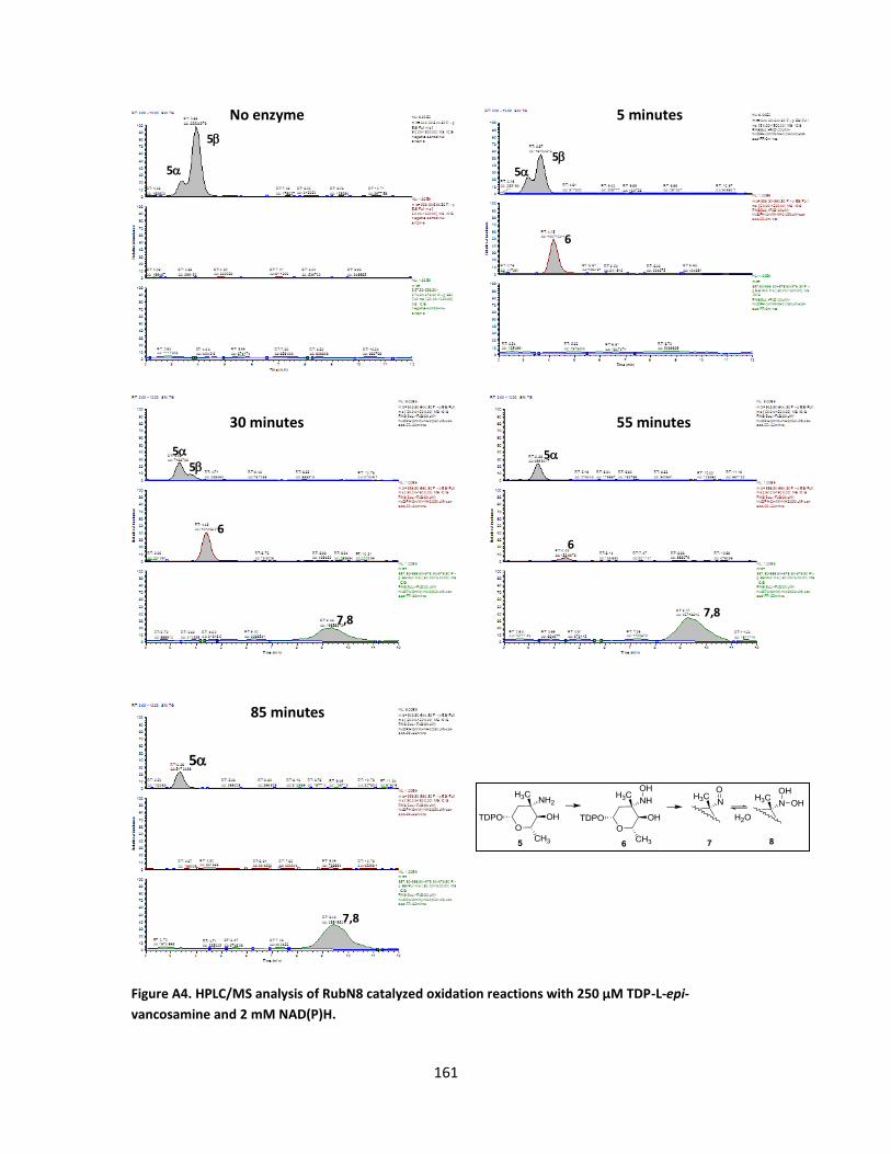
161
No enzyme 5 minutes
30 minutes 55 minutes
85 minutes
Figure A4. HPLC/MS analysis of RubN8 catalyzed oxidation reactions with 250 µM TDP-L-epi-
vancosamine and 2 mM NAD(P)H.
5
5
5
5
5
5 5
5
6
6 6
7,8 7,8
7,8

162
No enzyme 5 minutes
30 minutes 55 minutes
85 minutes
Figure A5. HPLC/MS analysis of RubN8 catalyzed oxidation reactions with TDP-L-epi-vancosamine and
400 µM NAD(P)H.
5
5
6
6
6
5
5
6
5
5
5
5

163
Figure A6. MS and Tandem MS of product ions for RubN8 catalyzed reactions. Shown spectra are MS (above) and MS/MS (below) of the substrate (starting material) TDP-L-epi- vancosamine.
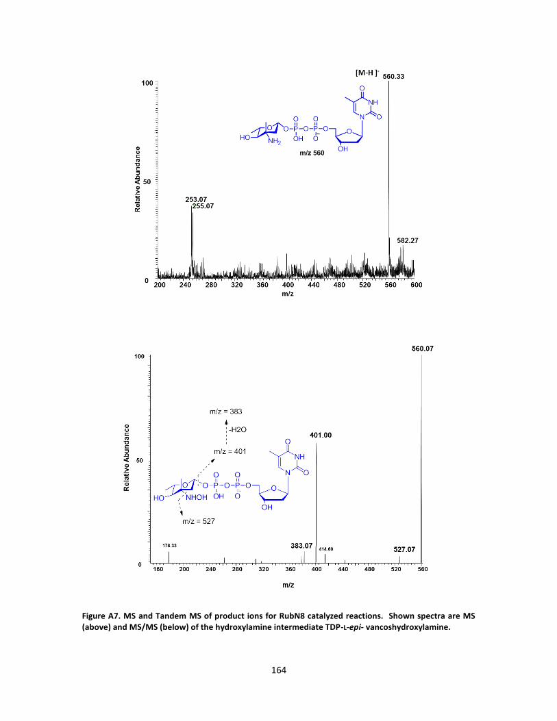
164
Figure A7. MS and Tandem MS of product ions for RubN8 catalyzed reactions. Shown spectra are MS (above) and MS/MS (below) of the hydroxylamine intermediate TDP-L-epi- vancoshydroxylamine.

165
Figure A8. MS and Tandem MS of product ions for RubN8 catalyzed reactions. Shown spectra are MS (above) and MS/MS (below) of TDP-L-epi-vanconitrose oxidation product (m/z 558).
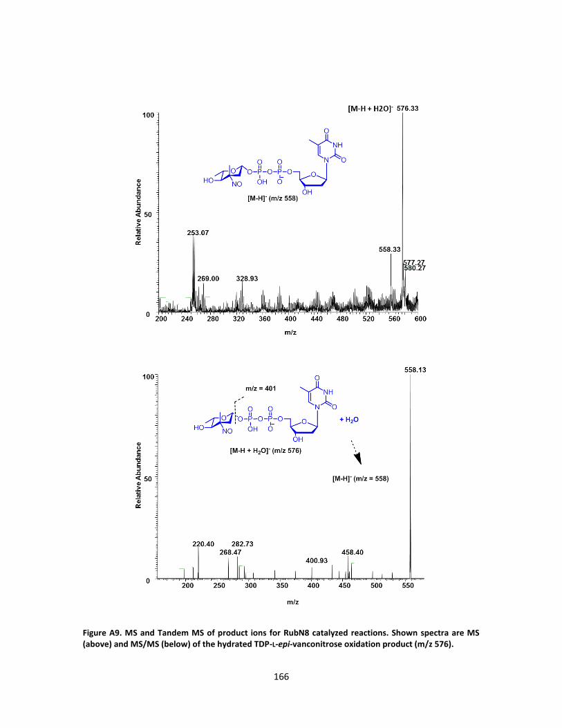
166
Figure A9. MS and Tandem MS of product ions for RubN8 catalyzed reactions. Shown spectra are MS (above) and MS/MS (below) of the hydrated TDP-L-epi-vanconitrose oxidation product (m/z 576).
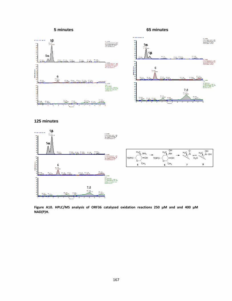
167
5 minutes 65 minutes 125 minutes
Figure A10. HPLC/MS analysis of ORF36 catalyzed oxidation reactions 250 µM and and 400 µM NAD(P)H.

168
Figure A11. MS and Tandem MS of product ions for ORF36 catalyzed reactions. Shown spectra are MS (above) and MS/MS (below) of the substrate (starting material) TDP-L-epi- vancosamine.

169
Figure A12. MS and Tandem MS of product ions for ORF36 catalyzed reactions. Shown spectra are MS (above) and MS/MS (below) of the hydroxylamine intermediate TDP-L-epi- vancoshydroxylamine.
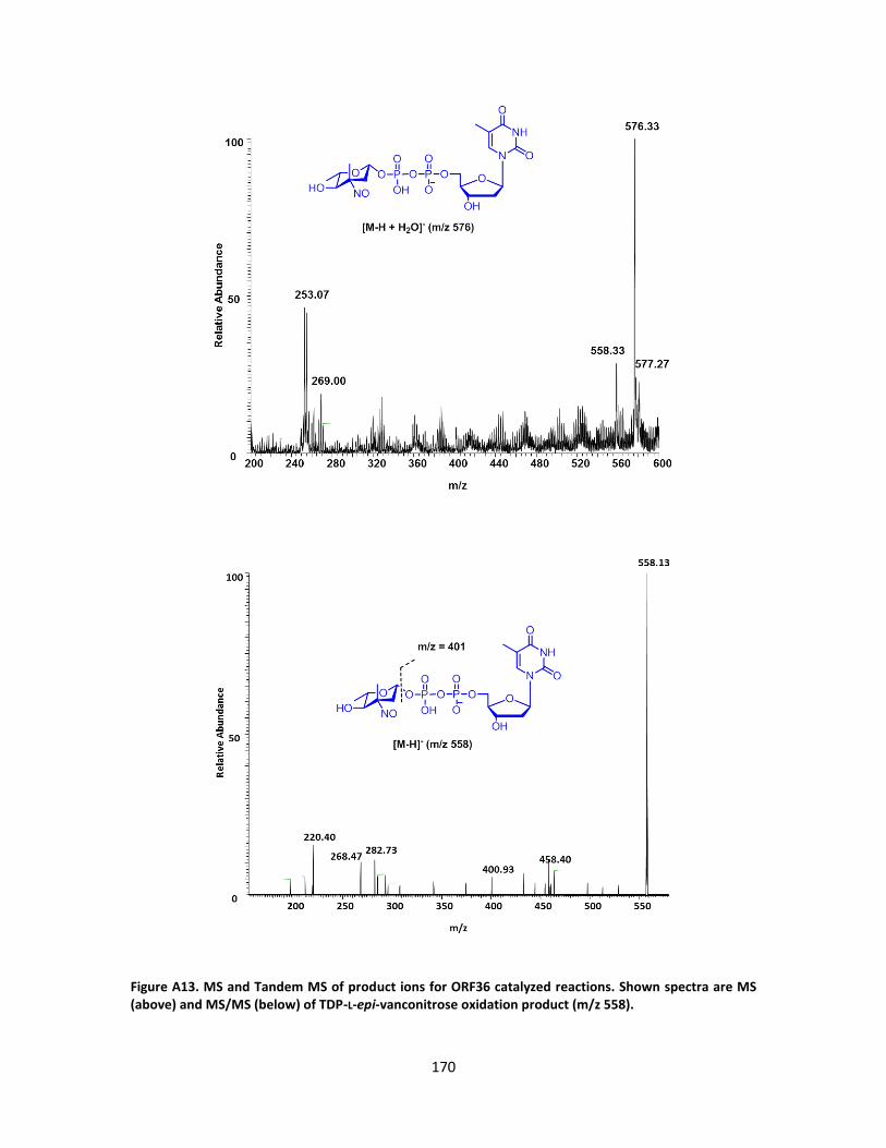
170
Figure A13. MS and Tandem MS of product ions for ORF36 catalyzed reactions. Shown spectra are MS (above) and MS/MS (below) of TDP-L-epi-vanconitrose oxidation product (m/z 558).
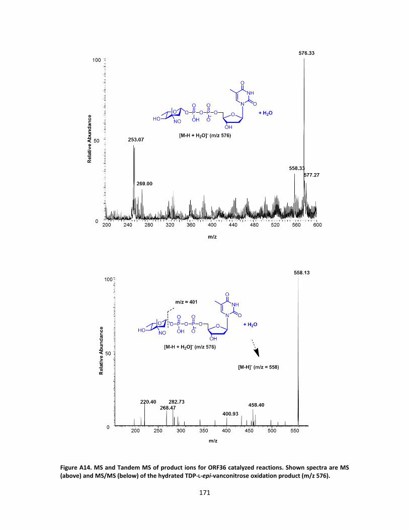
171
Figure A14. MS and Tandem MS of product ions for ORF36 catalyzed reactions. Shown spectra are MS (above) and MS/MS (below) of the hydrated TDP-L-epi-vanconitrose oxidation product (m/z 576).

172
GTGGCGGCGGATCTTCGCGCGCCGCTCACGCCGGCGGGGCGCACGGTGGTCG
ACCTGCTTGCCGGCGTGATCCCGAGAATCAGTGCGGAGGCCGCCGACCGGGA
CCGCACCGGCACCTTCCCGGTGGAGGCGTTCGAGCAGTTCGCGAAGCTCGGGT
TGATGGGCGCCACCGTTCCCGCCGAGCTGGGCGGCCTGGGGTTGACCCGGCT
GTACGACGTGGCGACCGCGCTGATGCGGCTGGCCGAGGCCGACGCGTCCACC
GCGCTGGCCTGGCACGTGCAGCTCAGCCGGGGCCTCACCCTCACCTACGAGTG
GCAGCACGGCACGCCGCCGGTGCGCGCGATGGCGGAGCGGCTGCTGCGGGC
GATGGCGGAGGGCGAGGCCGCCGTCTGCGGCGCGCTCAAGGACGCCCCCGG
CGTGGTCACCGAGCTGCATTCCGACGGCGCCGGCGGCTGGCTGCTGTCGGGC
CGCAAGGTGCTGGTCAGCATGGCGCCCATCGCGACCCACTTCTTCGTGCACGC
CCAGCGGCGCGACGACGACGGCTCGGTGTTCCTCGCCGTGCCGGTCGTGCAC
CGCGACGCCCCCGGGCTCACGGTGCTGGACAACTGGGACGGCCTGGGAATGC
GTGCCTCGGGGACGCTGGAGGTGGTCTTCGACCGGTGCCCGGTCCGGGCCGA
CGAGCTGCTGGAGCGCGGCCCGGTCGGGGCCCGGCGGGACGCCGTGCTGGC
CGGGCAGACGGTCAGCTCGATCACCATGCTCGGCATCTACGCCGGCATCGCCC
AGGCGGCCCGGGACATCGCGGTCGGTTTCTGCGCGGGGCGCGGCGGCGAGC
CACGGGCCGGTGCCCGGGCGCTGGTCGCCGGGCTGGACACCCGGCTCTACGC
GCTACGCACCACGGTCGGCGCGGCGTTGACCAACGCCGACGCGGCGTCGGTC
GACCTGTCCGGCGACCCGGACGAACGCGGCCGACGGATGATGACCCCGTTCCA
GTACGCGAAGATGACCGTCAACGAGCTGGCCCCGGCGGTGGTGGACGACTGC
CTCAGCCTGGTCGGCGGCCTCGCCTACACGGCCGGGCACCCACTCTCCCGGCT
CTATCGCGACGTGCGGGCCGGCGGGTTCATGCAGCCCTACAGCTACGTGGACG
CCGTCGACTACCTGAGCGGTCAGGCACTTGGACTCGACCGGGACAACGACTAC
ATGAGCGTGCGCGCGCTCCGCTCCCGGACCTCGGCGTAG
Figure A15. Orf36 DNA sequence from Micromonospora carbonacea var africana (NRRL 15099) (Genbank locus accession: AX574199).
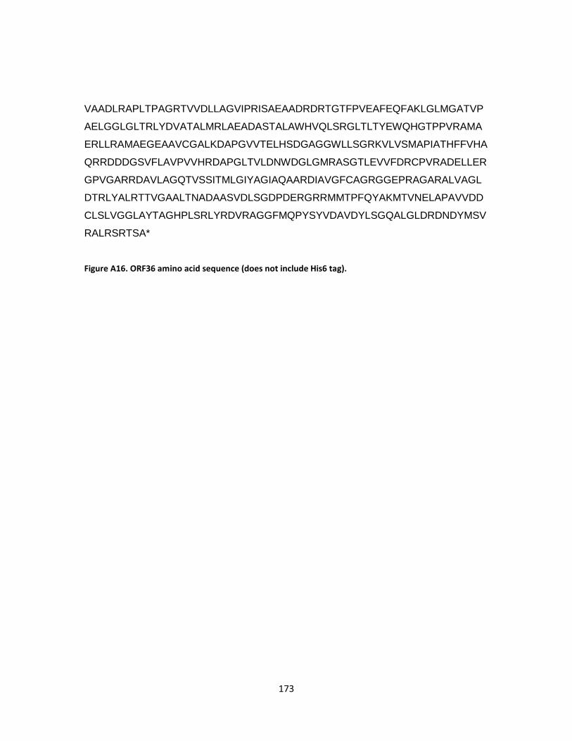
173
VAADLRAPLTPAGRTVVDLLAGVIPRISAEAADRDRTGTFPVEAFEQFAKLGLMGATVP
AELGGLGLTRLYDVATALMRLAEADASTALAWHVQLSRGLTLTYEWQHGTPPVRAMA
ERLLRAMAEGEAAVCGALKDAPGVVTELHSDGAGGWLLSGRKVLVSMAPIATHFFVHA
QRRDDDGSVFLAVPVVHRDAPGLTVLDNWDGLGMRASGTLEVVFDRCPVRADELLER
GPVGARRDAVLAGQTVSSITMLGIYAGIAQAARDIAVGFCAGRGGEPRAGARALVAGL
DTRLYALRTTVGAALTNADAASVDLSGDPDERGRRMMTPFQYAKMTVNELAPAVVDD
CLSLVGGLAYTAGHPLSRLYRDVRAGGFMQPYSYVDAVDYLSGQALGLDRDNDYMSV
RALRSRTSA*
Figure A16. ORF36 amino acid sequence (does not include His6 tag).
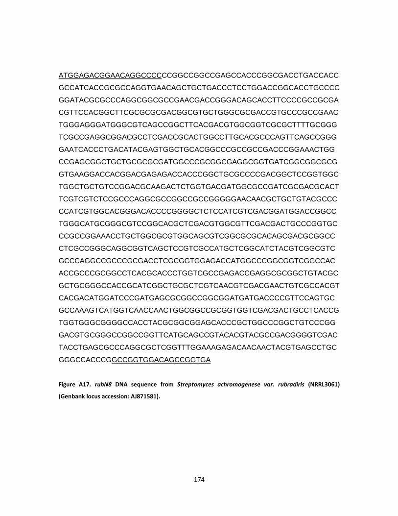
174
ATGGAGACGGAACAGGCCCCCCGGCCGGCCGAGCCACCCGGCGACCTGACCACC
GCCATCACCGCGCCAGGTGAACAGCTGCTGACCCTCCTGGACCGGCACCTGCCCC
GGATACGCGCCCAGGCGGCGCCGAACGACCGGGACAGCACCTTCCCCGCCGCGA
CGTTCCACGGCTTCGCGCGCGACGGCGTGCTGGGCGCGACCGTGCCCGCCGAAC
TGGGAGGGATGGGCGTCAGCCGGCTTCACGACGTGGCGGTCGCGCTTTTGCGGG
TCGCCGAGGCGGACGCCTCGACCGCACTGGCCTTGCACGCCCAGTTCAGCCGGG
GAATCACCCTGACATACGAGTGGCTGCACGGCCCGCCGCCGACCCGGAAACTGG
CCGAGCGGCTGCTGCGCGCGATGGCCCGCGGCGAGGCGGTGATCGGCGGCGCG
GTGAAGGACCACGGACGAGAGACCACCCGGCTGCGCCCCGACGGCTCCGGTGGC
TGGCTGCTGTCCGGACGCAAGACTCTGGTGACGATGGCGCCGATCGCGACGCACT
TCGTCGTCTCCGCCCAGGCGCCGGCCGCCGGGGGAACAACGCTGCTGTACGCCC
CCATCGTGGCACGGGACACCCCGGGGCTCTCCATCGTCGACGGATGGACCGGCC
TGGGCATGCGGGCGTCCGGCACGCTCGACGTGGCGTTCGACGACTGCCCGGTGC
CCGCCGGAAACCTGCTGGCGCGTGGCAGCGTCGGCGCGCACAGCGACGCGGCC
CTCGCCGGGCAGGCGGTCAGCTCCGTCGCCATGCTCGGCATCTACGTCGGCGTC
GCCCAGGCCGCCCGCGACCTCGCGGTGGAGACCATGGCCCGGCGGTCGGCCAC
ACCGCCCGCGGCCTCACGCACCCTGGTCGCCGAGACCGAGGCGCGGCTGTACGC
GCTGCGGGCCACCGCATCGGCTGCGCTCGTCAACGTCGACGAACTGTCGCCACGT
CACGACATGGATCCCGATGAGCGCGGCCGGCGGATGATGACCCCGTTCCAGTGC
GCCAAAGTCATGGTCAACCAACTGGCGGCCGCGGTGGTCGACGACTGCCTCACCG
TGGTGGGCGGGGCCACCTACGCGGCGGAGCACCCGCTGGCCCGGCTGTCCCGG
GACGTGCGGGCCGGCCGGTTCATGCAGCCGTACACGTACGCCGACGGGGTCGAC
TACCTGAGCGCCCAGGCGCTCGGTTTGGAAAGAGACAACAACTACGTGAGCCTGC
GGGCCACCCGGCCGGTGGACAGCCGGTGA
Figure A17. rubN8 DNA sequence from Streptomyces achromogenese var. rubradiris (NRRL3061)
(Genbank locus accession: AJ871581).
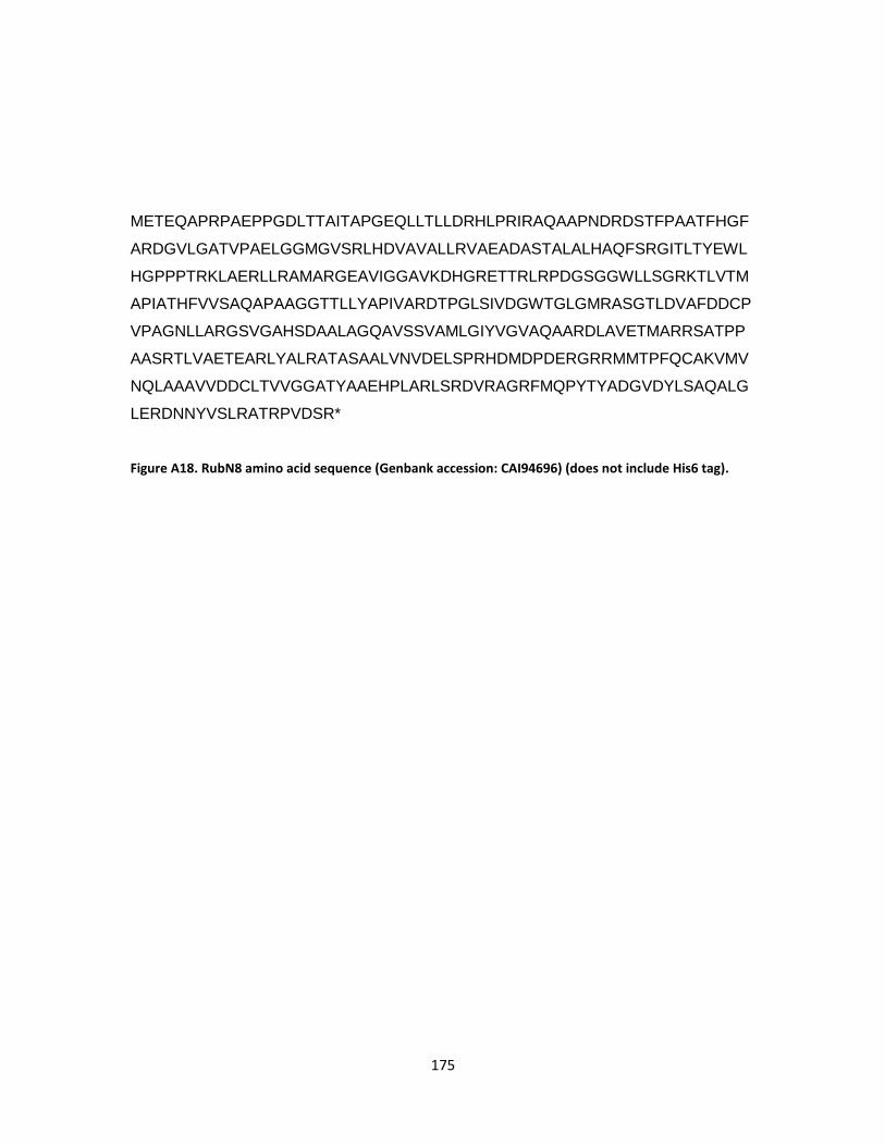
175
METEQAPRPAEPPGDLTTAITAPGEQLLTLLDRHLPRIRAQAAPNDRDSTFPAATFHGF
ARDGVLGATVPAELGGMGVSRLHDVAVALLRVAEADASTALALHAQFSRGITLTYEWL
HGPPPTRKLAERLLRAMARGEAVIGGAVKDHGRETTRLRPDGSGGWLLSGRKTLVTM
APIATHFVVSAQAPAAGGTTLLYAPIVARDTPGLSIVDGWTGLGMRASGTLDVAFDDCP
VPAGNLLARGSVGAHSDAALAGQAVSSVAMLGIYVGVAQAARDLAVETMARRSATPP
AASRTLVAETEARLYALRATASAALVNVDELSPRHDMDPDERGRRMMTPFQCAKVMV
NQLAAAVVDDCLTVVGGATYAAEHPLARLSRDVRAGRFMQPYTYADGVDYLSAQALG
LERDNNYVSLRATRPVDSR*
Figure A18. RubN8 amino acid sequence (Genbank accession: CAI94696) (does not include His6 tag).

176
APPENDIX B
SUPPLEMENTARY FIGURES FROM CHAPTER III

177
Figure B1. Evolutionary relationships of 13 taxa including nitrososynthases (ORF36 and KijD3), acyl-CoA dehydrogenases (ACADS), and flavincontaining monooxygenases. The evolutionary history was inferred using the Neighbor-Joining method.
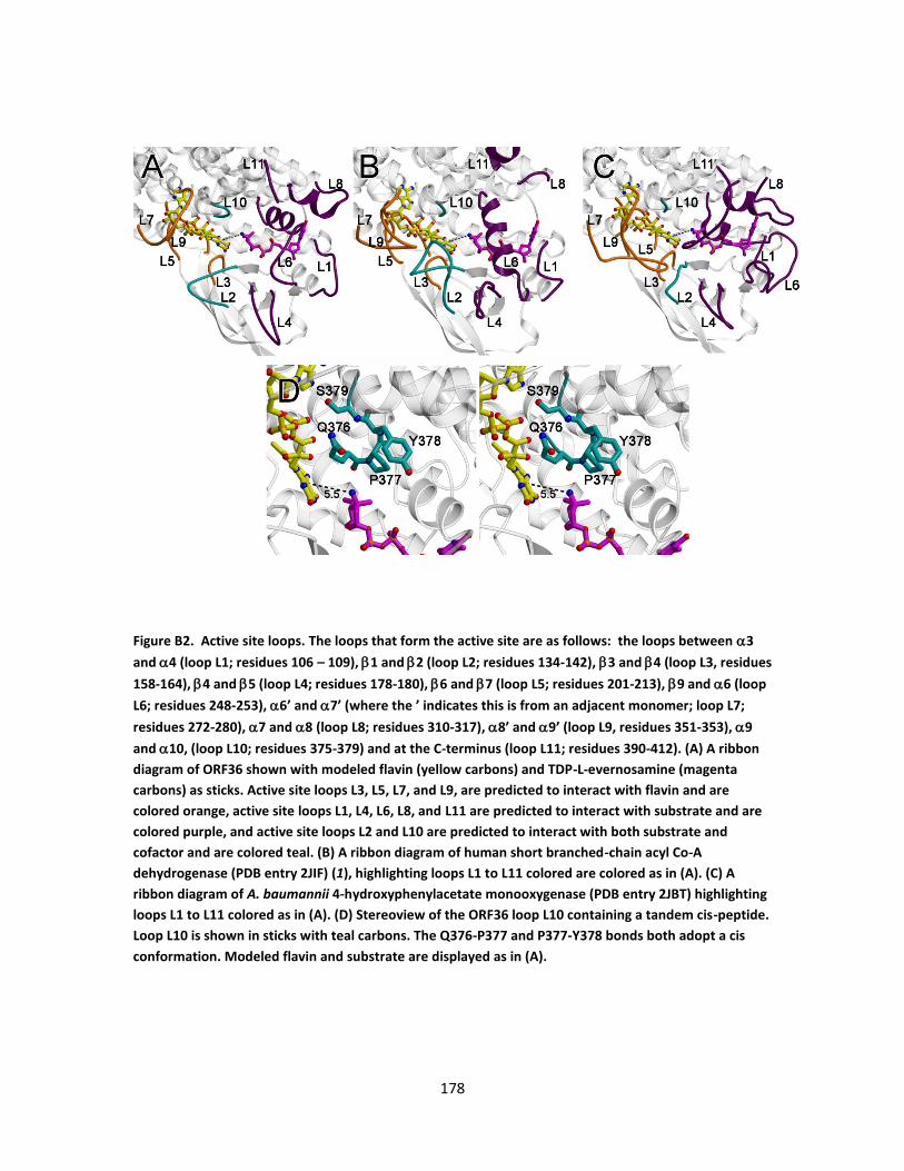
178
Figure B2. Active site loops. The loops that form the active site are as follows: the loops between 3
and 4 (loop L1; residues 106 – 109), 1 and 2 (loop L2; residues 134-142),3 and 4 (loop L3, residues
158-164), 4 and 5 (loop L4; residues 178-180), 6 and 7 (loop L5; residues 201-213), 9 and 6 (loop
L6; residues 248-253), 6’ and 7’ (where the ’ indicates this is from an adjacent monomer; loop L7;
residues 272-280), 7 and 8 (loop L8; residues 310-317), 8’ and 9’ (loop L9, residues 351-353), 9
and 10, (loop L10; residues 375-379) and at the C-terminus (loop L11; residues 390-412). (A) A ribbon
diagram of ORF36 shown with modeled flavin (yellow carbons) and TDP-L-evernosamine (magenta
carbons) as sticks. Active site loops L3, L5, L7, and L9, are predicted to interact with flavin and are
colored orange, active site loops L1, L4, L6, L8, and L11 are predicted to interact with substrate and are
colored purple, and active site loops L2 and L10 are predicted to interact with both substrate and
cofactor and are colored teal. (B) A ribbon diagram of human short branched-chain acyl Co-A
dehydrogenase (PDB entry 2JIF) (1), highlighting loops L1 to L11 colored are colored as in (A). (C) A
ribbon diagram of A. baumannii 4-hydroxyphenylacetate monooxygenase (PDB entry 2JBT) highlighting
loops L1 to L11 colored as in (A). (D) Stereoview of the ORF36 loop L10 containing a tandem cis-peptide.
Loop L10 is shown in sticks with teal carbons. The Q376-P377 and P377-Y378 bonds both adopt a cis
conformation. Modeled flavin and substrate are displayed as in (A).
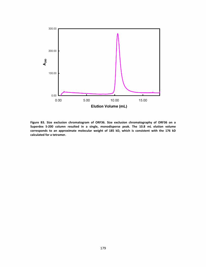
179
Figure B3. Size exclusion chromatogram of ORF36. Size exclusion chromatography of ORF36 on a Superdex S-200 column resulted in a single, monodisperse peak. The 10.8 mL elution volume corresponds to an approximate molecular weight of 185 kD, which is consistent with the 176 kD calculated for a tetramer.
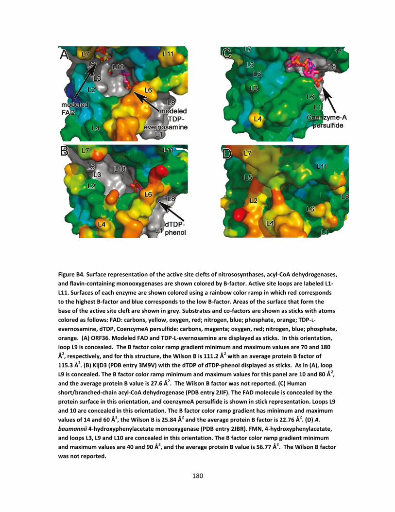
180
Figure B4. Surface representation of the active site clefts of nitrososynthases, acyl-CoA dehydrogenases,
and flavin-containing monooxygenases are shown colored by B-factor. Active site loops are labeled L1-
L11. Surfaces of each enzyme are shown colored using a rainbow color ramp in which red corresponds
to the highest B-factor and blue corresponds to the low B-factor. Areas of the surface that form the
base of the active site cleft are shown in grey. Substrates and co-factors are shown as sticks with atoms
colored as follows: FAD: carbons, yellow, oxygen, red; nitrogen, blue; phosphate, orange; TDP-L-
evernosamine, dTDP, CoenzymeA persulfide: carbons, magenta; oxygen, red; nitrogen, blue; phosphate,
orange. (A) ORF36. Modeled FAD and TDP-L-evernosamine are displayed as sticks. In this orientation,
loop L9 is concealed. The B factor color ramp gradient minimum and maximum values are 70 and 180
Å2, respectively, and for this structure, the Wilson B is 111.2 Å
2 with an average protein B factor of
115.3 Å2. (B) KijD3 (PDB entry 3M9V) with the dTDP of dTDP-phenol displayed as sticks. As in (A), loop
L9 is concealed. The B factor color ramp minimum and maximum values for this panel are 10 and 80 Å2,
and the average protein B value is 27.6 Å2. The Wilson B factor was not reported. (C) Human
short/branched-chain acyl-CoA dehydrogenase (PDB entry 2JIF). The FAD molecule is concealed by the
protein surface in this orientation, and coenzymeA persulfide is shown in stick representation. Loops L9
and 10 are concealed in this orientation. The B factor color ramp gradient has minimum and maximum
values of 14 and 60 Å2, the Wilson B is 25.84 Å
2 and the average protein B factor is 22.76 Å
2. (D) A.
baumannii 4-hydroxyphenylacetate monooxygenase (PDB entry 2JBR). FMN, 4-hydroxyphenylacetate,
and loops L3, L9 and L10 are concealed in this orientation. The B factor color ramp gradient minimum
and maximum values are 40 and 90 Å2, and the average protein B value is 56.77 Å
2. The Wilson B factor
was not reported.

181
Figure B5. Location of the FAD and TDP-L-evernosamine molecules modeled within ORF36 active site. Chain A of ORF36 is shown colored by domain as in Figure III-2. Chains B, C and D are colored white, black and grey, respectively. The modeled small molecules (shown as transparent sticks) are colored as follows: FAD, yellow carbons, TDP-L-evernosamine, magenta carbons, oxygen, red; nitrogen, blue; sulfur, gold; phosphorus, orange. (A) The physiologically relevant tetramer, with modeled ligands shown at one active site. (B) A closer view of the labeled ORF36 monomer, shown in stereo. The monomer is shown in the same orientation as in Figure 5b, with the other three chains of the tetramer colored as in (A). Selected secondary structural elements from chain C are labeled in white text.
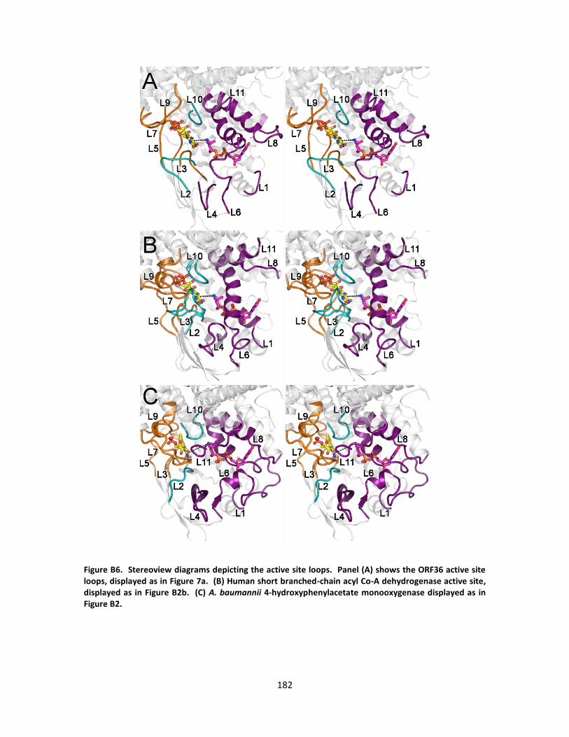
182
Figure B6. Stereoview diagrams depicting the active site loops. Panel (A) shows the ORF36 active site loops, displayed as in Figure 7a. (B) Human short branched-chain acyl Co-A dehydrogenase active site, displayed as in Figure B2b. (C) A. baumannii 4-hydroxyphenylacetate monooxygenase displayed as in Figure B2.
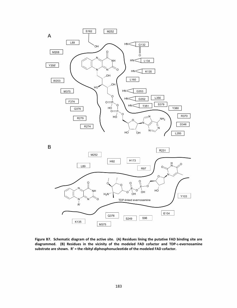
183
Figure B7. Schematic diagram of the active site. (A) Residues lining the putative FAD binding site are diagrammed. (B) Residues in the vicinity of the modeled FAD cofactor and TDP-L-evernosamine substrate are shown. R’ = the ribityl diphosphonucleotide of the modeled FAD cofactor.
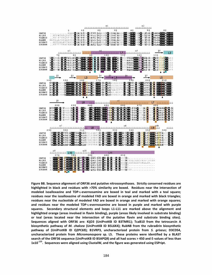
184
Figure B8. Sequence alignment of ORF36 and putative nitrososynthases. Strictly conserved residues are highlighted in black and residues with >70% similarity are boxed. Residues near the intersection of modeled isoalloxazine and TDP-L-evernosamine are boxed in teal and marked with a teal square; residues near the isoalloxazine of modeled FAD are boxed in orange and marked with black triangles; residues near the nucleotide of modeled FAD are boxed in orange and marked with orange squares; and residues near the modeled TDP-L-evernosamine are boxed in purple and marked with purple squares. Secondary structural elements and loops L1-L11 are marked above the alignment and highlighted orange (areas involved in flavin binding), purple (areas likely involved in substrate binding) or teal (areas located near the intersection of the putative flavin and substrate binding sites). Sequences aligned with ORF36 are: KijD3 (UniProtKB ID B3TMR1); TcaB10 from the tetrocarcin A biosynthetic pathway of M. chalcea (UniProtKB ID B5L6K4); RubN8 from the rubradirin biosynthetic pathway of (UniProtKB ID Q2PC69); B1VRP3, uncharacterized protein from S. griseus; D3CDS4, uncharacterized protein from Micromonospora sp. L5. These proteins were identified by a BLAST search of the ORF36 sequence (UniProtKB ID B5APQ9) and all had scores > 450 and E-values of less than 1x10
-125. Sequences were aligned using ClustalW, and the figure was generated using ESPript.

185
Figure B9. Sequence alignment of ORF36, KijD3, selected acyl-coA dehydrogenases, and selected Class D
flavin monooxygenases. Loops and residues at the active site are highlighted as in Supplemental Figure
S4. Of particular interest are the residues involved in the isoalloxazine binding site. For clarity, select
divergent portions of individual sequences were truncated and are denoted with red lettering.
Sequences aligned with ORF36 are: KijD3 (UniProtKB ID B3TMR1); ACAD1, short branched-chain acyl-
coA dehydrogenase (H. sapiens), 2JIF; ACAD2, medium-chain acyl-coA dehydrogenase (S. scrofa), PDB
entry 3MDE; HpaB1, 4-hydroxyphenylacetate monooxygenase (A. baumannii), PDB entry 2JBT;
monoox1, 3-hydroxy-9,10-seco-nandrost-1,3,5(10)-triene-9,17-dione hydroxylase (Rhodococcus sp
Rha1), PDB entry 2RFQ.

186
Figure B10. Stereoview of electron density for the tandem cis-peptide in L10 of ORF36. The protein chain is depicted as light grey cartoons. 2Fo-Fc electron density is shown in blue for residues 372-389, which encompasses the two sequential cis-peptides between Gln376-Pro377 and Pro377-Tyr378. Those residues in loop L10 are shown in stick representation, and the main chain carbon atoms of the cis-peptides are highlighted in magenta.
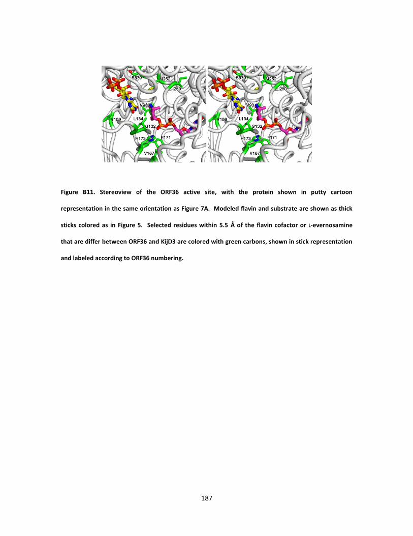
187
Figure B11. Stereoview of the ORF36 active site, with the protein shown in putty cartoon
representation in the same orientation as Figure 7A. Modeled flavin and substrate are shown as thick
sticks colored as in Figure 5. Selected residues within 5.5 Å of the flavin cofactor or L-evernosamine
that are differ between ORF36 and KijD3 are colored with green carbons, shown in stick representation
and labeled according to ORF36 numbering.
188
APPENDIX C
MS AND MS/MS SPECTRA FROM CHAPTER IV

189
Figure C1. MS (above) and MS/MS (below) of the substrate (starting material) TDP-L-epi- vancosamine.

190
Figure C2. MS of the oxime-aldehyde species (m/z 558) and its hydrate (m/z 576) produced from the
monooxygenation/retro-aldol activity of DnmZ with TDP-L-epi- vancosamine (above), and MS/MS of the
oxime-aldehyde species (m/z 558) (below).
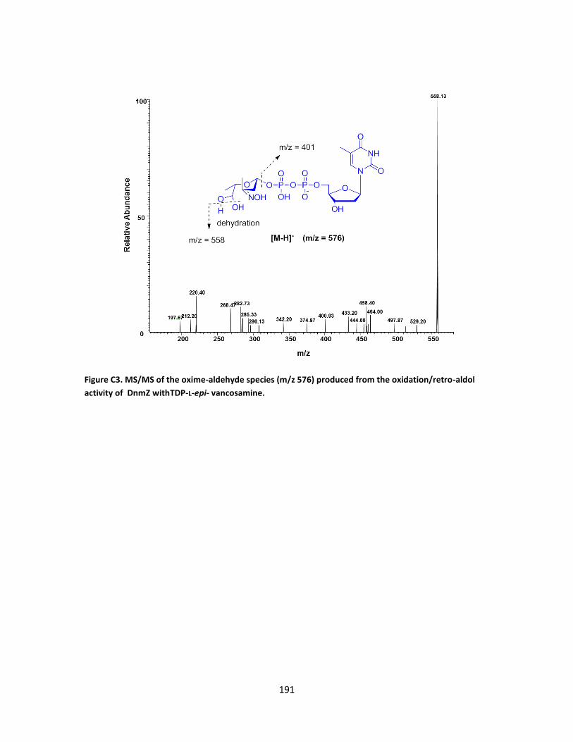
191
Figure C3. MS/MS of the oxime-aldehyde species (m/z 576) produced from the oxidation/retro-aldol
activity of DnmZ withTDP-L-epi- vancosamine.

192
Figure C4. MS of the phenylhydrazone derivative (m/z 648) produced from the reaction of the
corresponding oxime-aldehyde with phenylhydrazine.
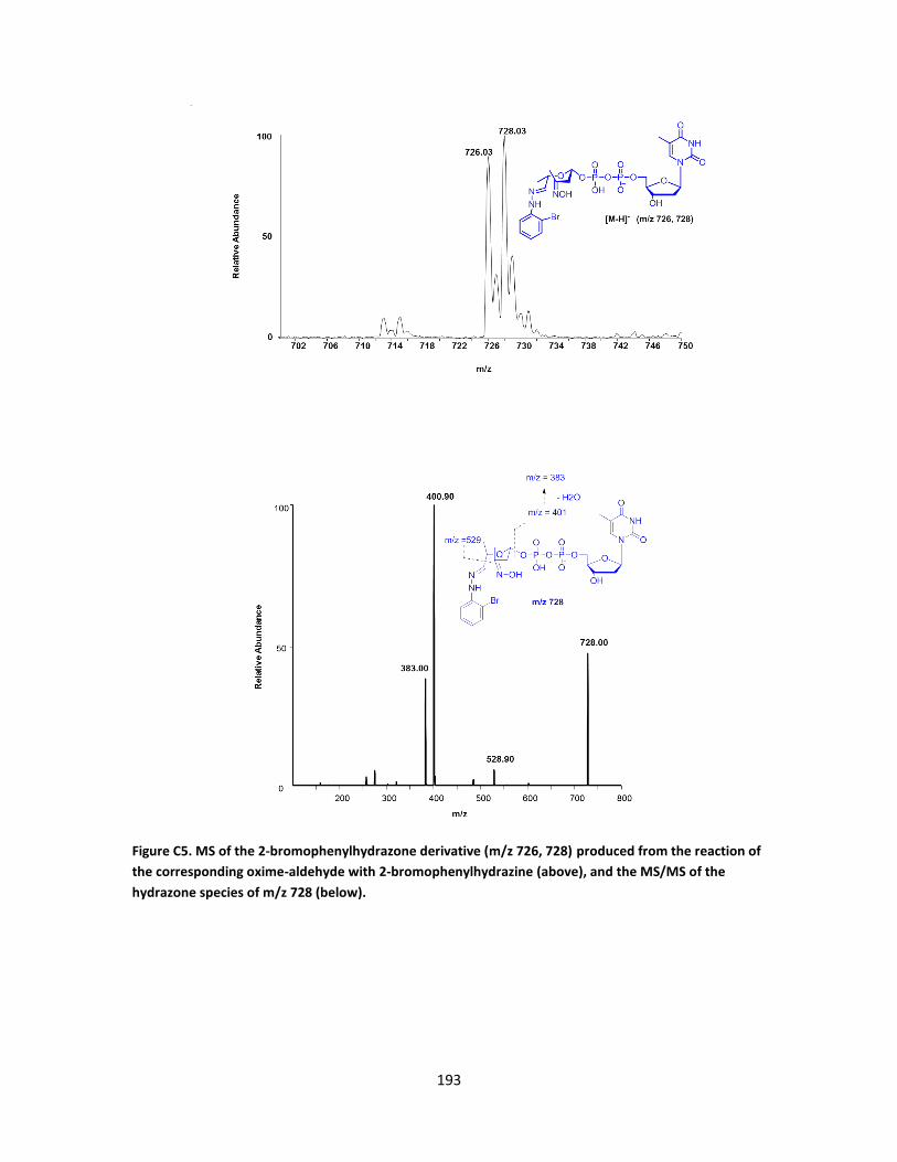
193
Figure C5. MS of the 2-bromophenylhydrazone derivative (m/z 726, 728) produced from the reaction of
the corresponding oxime-aldehyde with 2-bromophenylhydrazine (above), and the MS/MS of the
hydrazone species of m/z 728 (below).
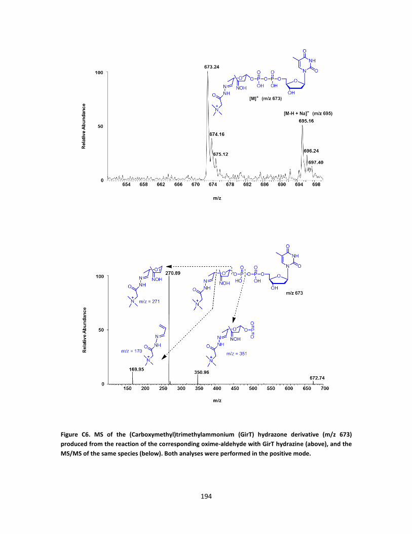
194
Figure C6. MS of the (Carboxymethyl)trimethylammonium (GirT) hydrazone derivative (m/z 673)
produced from the reaction of the corresponding oxime-aldehyde with GirT hydrazine (above), and the
MS/MS of the same species (below). Both analyses were performed in the positive mode.

195
Figure C7. MS/MS of the acid hydrolysis fragments of m/z 188 (above), and m/z 215 (below) produced
from the treatment of the (Carboxymethyl)trimethylammonium (GirT) hydrazone with HCl (1 M).