Embed Size (px)
Citation preview
JASs Invited ReviewsJournal of Anthropological Sciences
Vol. 92 (2014), pp. 119-146
the JASs is published by the Istituto Italiano di Antropologia www.isita-org.com
Osteoporosis and paleopathology: a review
Francisco Curate
Research Centre for Anthropology and Health, University of Coimbra, Rua Arco da Traição, 7, 3000 Coimbra, Portugale-mail: [email protected]
Summary - Osteoporosis is a complex and heterogeneous disorder, of multi-factor aetiology. It is the most frequent metabolic bone disorder, affecting an increasing number of post-menopausal women and aging individuals from both sexes. Although first recognized more than 250 years ago, the clinical and epidemiological knowledge about osteoporosis is largely limited to the last 70 years. Within the conceptual frames of paleopathology, disease is necessarily perceived in a space without depth (the skeleton) and of coincidence without development (the crucial moment of death) – but is also interpreted in a time interval which adds an historical gaze to its “biography”. The study of osteoporosis in past populations (which faced sociocultural conditions utterly different from the genus vitae experienced by modern communities) supplements diachronic depth to the knowledge about bone modifications related to age, menopausal status or lifestyle. This article aims to provide a comprehensive record on the history of osteoporosis and fragility fractures as perceived by the biomedical, historical and, particularly, paleopathological sciences. As such, the main focus of this review is to present an exhaustive and historical-framed exposition of the studies of osteoporosis, bone loss and associated fractures within the field of paleopathology and, to a lesser extent, in the history of medicine. A biomedical-oriented synopsis of the main operational definitions, etiological agents and epidemiological features of osteoporosis and osteoporotic fractures is also provided
Keywords - Osteoporosis, Bone loss, Fractures, History of medicine, Paleopathology.
Introduction
Osteoporosis (OP) – the «silent thief» – is a metabolic disorder characterized by the reduction in bone mass, impaired bone quality and subsequent increase in the risk of fracture (Consensus Development Conference, 1993; NIH Consensus Development Panel, 2001). The loss of mineral content per unit of volume, alongside trabecular deterioration, concurs to bone fragility and increased propensity to frac-ture (Strømsøe, 2004). The classical hip, distal radius and vertebral compression fractures are the main clinical complications associated with osteoporosis (Johnell & Kanis, 2006).
Joseph Guichard Duverney (1648-1730), professor of anatomy and surgery at the Jardin du Roi (medical school established by Louis XIV
of France), first described osteoporosis (without christening it) more than 250 years ago in his post-humous «Traité des Maladies des Os» (Duverney, 1751). In the beginning of the 18th century, Jean-Louis Petit (1674-1750) already had docu-mented the inherent fragility of bones (Petit, 1705). A century later – and after – the readily apparent and recognizable propensity of bones to break due to «fragility» was well acknowledged in the medical literature (e.g., Paiva, 1804; Cooper, 1822). The term «osteoporosis» (from the Greek ostéon-oûn: bone, and póros: porous) was coined by the French pathologist Johann Lobstein, the Younger (1777-1835), in a text entitled De l’osteoporose (Lobstein, 1820). Lobstein charac-terized it as a disease that causes an increase in the size of bones and a rarefication of their internal tissue. Notwithstanding, the disease described by
doi 10.4436/JASS.92003
120 Osteoporosis and paleopathology
Johann Lobstein was probably osteogenesis imper-fecta (Schapira & Schapira, 1992). A few years later, OP was histologically distinguished from osteomalacia (Pommer, 1885).
Unlike other medical concepts, OP defini-tion has changed substantively, reflecting the state of knowledge about the disease (Schapira & Schapira, 1992; Wylie, 2010). With the incep-tion of clinical radiology, OP was defined as a noticeable loss of bone mass, or as a condition in which bone resorption exceeds bone forma-tion (Nordin, 1987). However, generalized loss of bone should be termed osteopenia (Agarwal, 2008; Frost, 2003). The American endocri-nologist Fuller Albright (1900-1969) described osteoporosis as a vertebral fracture syndrome in postmenopausal women, defining it as “too lit-tle calcified bone” (Albright et al., 1941). Later, Albright & Riefenstein (1948) proposed two primary categories of osteoporosis: postmeno-pausal osteoporosis and senile osteoporosis. Riggs & Melton III (1986) refined this scheme with the analogous designations, Types I and II. Although heuristically valuable, the model does not account for the intricate etiopathogenesis of the disease (Marcus & Bouxsein, 2008).
Until the 1990’s, different definitions of osteoporosis emerged in the medical literature (Bijlsma et al., 2012). The introduction of dual-energy X-ray absorciometry (DXA) scanners in the late 1980’s inspired a raging discussion about the definition of osteoporosis (e.g., Mazess, 1987; Melton III & Wahner, 1989; Nordin, 1987) until the consensus conference promoted by the WHO. In 1992, an experts study group, led by the English clinician and researcher John Kanis, met in Rome and proposed a densito-metric definition of osteoporosis: a reduction of bone mineral density (BMD) by 2.5 standard deviations or more from the peak bone mass in early adulthood, taking into account gender and ethnicity (WHO, 1994).
Though it has been suggested since the 19th century that OP is a disease that typically affects older women (Bruns, 1882), it was Albright et al. (1941) who first highlighted the role of estro-gen depletion in postmenopausal osteoporosis,
noticing that vertebral fractures occurred more often in women who were subjected to an early oophorectomy. The relevance of sex steroids in the etiopathogeny of the disease has been com-prehensively established by subsequent studies (Almeida, 2010; Lindsay, 2010). However, OP is a complex pathological condition with multiple etiological drives, including senescence, genetics, physical activity, reproductive history, and die-tary status (Burnham & Leonard, 2008; Curate et al., 2012; Heaney, 2008; Livshits et al., 2004; Møller et al., 2012; Recker et al., 2004; Zhang et al., 2009).
The demographic profile of the world pop-ulation changed dramatically in the past few years, with a remarkable increase in the total and relative numbers of elderly individuals. As OP affects a large proportion of the aged population, resulting in fractures that have costly human and economic consequences, it is now recognized as one of the major public health concerns affecting the geriatric community (Becker et al., 2010). Although typically acknowledged as a “mod-ern disease”, OP has a vast diachronic depth (Agarwal, 2008; Brickley, 2002; Turner-Walker et al., 2001). The prevalence of the disease has oscillated with the historical changes in its etio-logical agents, like longevity or nutrition. The study of osteoporosis epidemiology in the past is, therefore, crucial to the scientific perception of the disease. Paleopathology – the study of dis-eases, human or nonhuman, in the past using a plethora of different sources (Ortner, 2003) – has been focused in age-related bone loss since the late 1960’s, with the pivotal papers by Dewey et al. (1969) and Armelagos et al. (1972), on three Sudanese Nubia samples, and the studies of van Gerven et al. (1969) or Perzigian (1973) with pre-historic Native-American materials. The body of knowledge in the paleopathology of osteoporosis has developed since, recounting the long history of bone involution and fragility fractures in past communities all over the world (e.g., Agarwal & Grynpas, 2009; Agnew & Stout, 2012; Curate et al., 2009; Curate et al., 2013b; Lees et al., 1993; Mafart et al., 2008; Mays et al., 1998; Mays et al., 2006; Cho & Stout, 2011; Zaki et al., 2009).
www.isita-org.com
121F. Curate
Etiopathogenesis of osteoporosis
Bone tissue constitutes the fundamental template of the skeleton, a complex multifunc-tional system comprising three key functions: mechanical / structural, protection, and meta-bolical. Bone is a mineralized connective tissue, composed by an extracellular matrix (organic and inorganic) and a distinctive group of cells (Nolla & Rozadilla, 2004). The organic matrix, which constitutes approximately 25% of the dry bone weight, is largely composed of collagen (Boyd, 2009; Fleisch, 2000; Nolla & Rozadilla, 2004). The inorganic bone phase is formed by hydroxyapatite. Bone cells occur in four funda-mental types: osteoblasts, osteoclasts, osteocytes and bone lining cells (Boyd, 2009). Mature bone is macroscopically dissociated in two compart-ments: trabecular bone, prevalent in the vertebral bodies, pelvis and long bones epiphyses; and cor-tical bone, which predominates in the diaphysis of the long bones (Fleisch, 2000).
Once formed, bone is exposed to a con-tinuous process of renovation and modifica-tion trough modeling and remodeling (Fleisch, 2000; Gosman & Stout, 2010). Bone modeling is a mechanically mediated adaptive process for modifying bone size, shape or position. Bone remodeling is the continuously renewal of bone in the adult skeleton, involving the elimination of mineralized bone by osteoclasts from the sur-faces of trabecular and cortical bone. Osteoblasts subsequently lay down new bone matrix that becomes mineralized (Boyce & Xing, 2008; Roberts et al., 2004). Initial skeletal formation depends on the direct apposition of bone but bone remodeling becomes the prevailing skel-etal metabolical activity at the end of puberty (Prestwood & Raisz, 2000). Bone remodeling occurs in temporary anatomical structures, first identified by Harold Frost (1969), termed Basic Multicellular Units (BMU). These functional units operate in a cycle of five phases: activa-tion, bone resorption, reversal in the cellular proliferation, bone formation and, at last, bone mineralization (Frost, 2003). Bone remodeling is a dynamic combination of bone formation and
resorption. As such, any imbalance in the pro-cess favoring bone resorption results in bone loss (Nolla & Rozadilla, 2004).
Almost none of the most common diseases of mankind can be attributed to only one cause; the majority stems from multiple causes, better described as risk factors. In the case of osteopo-rosis, the risk factors arise at different levels but they are not mutually exclusive (Nordin, 2008). OP is a gargantuan landscape difficult to classify by its pathogenesis: the question remains whether it should be viewed as a unique disease or a group of syndromes of skeletal fragility resulting from a stochastic process (Heaney, 2008; Marcus & Bouxsein, 2008).
Peak bone massPeak bone mass (PBM) is defined as the
maximum quantity of bone mass acquired dur-ing growth (National Osteoporosis Foundation, 2010). Adult bone mass is usually determined by the PBM attained at the third decade of life at which is subtracted the bone mass lost throughout the period of physiological aging (Gilsanz, 1999). Stochastic models by Horsman & Burkinshaw (1989) suggest that two thirds of fracturary risk in women can be predicted on the basis of individual PBM. Peak bone mass is classically influenced by a multiplicity of factors, including genetics (accounting for up to 85% of the variation in bone mass) and ethnical affili-ation, sex, nutrition, mechanical loads exerted over the skeleton, parity, and alcohol or tobacco consumption (Burnham & Leonard, 2008; Rizzoli & Bonjour, 2010).
AgeOP prevalence increases with age, fitting a
Gompertzian pattern common to other chronic diseases, like atherosclerosis or adenocarcinoma (Melton III, 1990). Age is a risk factor for osteo-porosis with direct and indirect effects on bone mass. Osteoblastic activity decreases during the process of senescence; hence, bone formation decelerates (Recker et al., 2004; Riggs & Melton III, 1986). Moreover, intestinal absorption of calcium declines, triggering a state of secondary

122 Osteoporosis and paleopathology
hyperparathiroydism and, indirectly, an increase in bone resorption (Halloran & Bikle, 1999; Riggs, 2003). There is also a decrease in the intestinal production of 1,25(OH)2D (vitamin D metabolite) which plays a distinctive role in the etiopathogenesis of osteoporosis. Aging also accounts for the accumulation of damage in osse-ous tissue and the reduction of viable osteocytes (Vashishth et al., 2003)
Menopause and estrogensNatural menopause is physiologically defined
as the last spontaneous episode of menstrual flow, defined retrospectively a year after (Nelson, 2008). All women experience menopause around the average age of 50 years and age at menopause seems to have remained rather stable trough the last 2000 years (Pavelka & Fedigan, 1991). Fuller Albright first acknowledged estrogen influence on the skeleton in the 1940’s but bone regula-tion mechanisms by estrogens are still poorly documented (Komm et al., 2008). Normal pre-menopausal levels of estradiol shield the skeleton against the increase of bone turnover. As such, early menopause constitutes a major risk factor for OP. Estrogen depletion is the main cause of postmenopausal bone decline and bone archi-tecture disruption in women, also contribut-ing for age-related bone loss in men (Almeida, 2010; Lindsay, 2010). The decline of estrogen levels increases bone resorption, boosting bone sensitivity to parathyroid hormone (PTH), and reducing the intestinal absorption and renal reabsorption of calcium. Bone formation also decreases (Almeida, 2010; Komm et al., 2008; Nordin, 2008). Estrogen actions are mediated mostly through estrogen receptor α (ERα) and also ERβ. Estrogen induces osteoclast apoptosis, wielding an opposite effect on osteoblasts and osteocytes. The beneficial effects of estrogen are due in part to the ability of estrogen to suppress osteoclastogenic cytokine production in T-cells and osteoblasts (Khosla, 2010).
Genetics and ancestryOsteoporosis and related phenotypes are
highly influenced by genetic factors, which exert
significant effects in peak bone mass and age-related bone loss (Williams & Spector, 2007). Bone mineral density is highly heritable, as are other risk factors for osteoporotic fracture, such as proximal femur geometry, bone turnover and bone quality. Most likely, multiple genes medi-ate susceptibility to osteoporosis, each under-taking a small effect in the osteoporotic pheno-types (Williams & Spector, 2007; Zhang et al., 2009). Several studies have shown an association between candidate genes (e.g., COL1AI, VDR or LRP5) and BMD (Ferrari, 2008). The major-ity of twin and familial studies suggest that 50 to 80% of BMD variance is genetically determined (Williams & Spector, 2007). BMD and trabecu-lar thickness are probably influenced by genetic differences between ethnical groups (Mitchell et al., 2003). Nevertheless, there is a great varia-tion in the prevalence of osteoporosis and fragil-ity fractures within and among different ethnic groups (Williams & Spector, 2007).
NutritionBone physiology is the result of multiple cel-
lular processes. Obviously, the cells responsible for bone deposition, maintenance or reparation are as dependent of nutrients as any other cell in the body. For example, the production of bone matrix relies on collagen synthesis and modifi-cation. The nutrients involved in this process include proteins, vitamins C, D and K, and sev-eral minerals. Furthermore, the skeleton stores vast quantities of Ca and P, and the extent of the reserve complies with the daily equilibrium between absorption and excretion of the two ele-ments (Heaney, 2008). Daily Ca requirements are reasonably high, but the absorption efficiency is low and further declines between 40 and 60 years of age, remarkably in women (Fishbein, 2004). When dietary calcium absorption is insufficient to counteract urinary and fecal losses, calcium is resorbed from the skeleton – which contains 99% of the body’s calcium stores – to uphold serum Ca at a stable level (National Osteoporosis Foundation, 2010). Protein intake is probably related with calcium phosphate metabolism, bone mass and even osteoporotic fracture risk, but any

www.isita-org.com
123F. Curate
enduring impact of dietary protein on bone min-eral metabolism and bone mass so far has been problematic to detect (Rizzoli & Bonjour, 2010). Alcohol and coffee consumption probably influ-ence bone metabolism but their effect on bone mass is contentious (Nordin, 2008).
Physical activitySkeletal response to physical activity is seem-
ingly mediated by genetic and hormonal fac-tors (Uusi-Rasi et al., 2008). In accordance with Carter (1984), the mechanical forces applied to the bone stimulate both osteoclastic and osteo-blastic activity. Physical activity during growth, especially strenuous activities, excite osteogenic processes influencing peak bone mass (Burnham & Leonard, 2008). Physical exercise also ben-efits bone health in postmenopausal women and aging individuals from both sexes (Kaptoge et al., 2003). The impact of the loading exter-nal environment on bone structure and biology is termed mechanobiology (Gosman & Stout, 2010). Julius Wolff (1892) recognized that the structural and geometrical properties of the bone could be described under a general prin-ciple, Wollf ’s law, in which healthy bone adapts to the loads that impact it. Also, Wilhelm Roux suggested that functional adaptation of trabecu-lar bone is autoregulated and that bone cells respond to local mechanical stimuli (Gosman & Stout, 2010). Drawing on Roux’s theory, Harold Frost proposed that bone architecture is under the control of a biomechanical cybernetic sys-tem, the mechanostat (Frost, 2003). This system directs bone modeling, and as a result, directs its spatial organization, load capacity and force translation proficiency. The pressure wielded by external interference factors, like physical activ-ity, triggers a feedback control loop and bone adapts its biomechanical properties according to the mechanical function, i.e., bone mass, bone geometry and consequently bone strength.
Reproductive factorsDuring gestation and breastfeeding, sub-
stantial changes take place in the maternal bone mineral metabolism and calcium homeostasis to
fulfill the calcium requirements of the fetus and the neonate (Møller et al., 2012). The mater-nal skeleton strives to adjust to the demand of Ca and other minerals throughout pregnancy (especially during the last trimester), which are relocated through the placenta to mineralize the developing fetal skeleton. Similarly, the increas-ing needs of calcium during breastfeeding also press for an adjustment of the bone mineral homeostasis in the lactating women (Agarwal, 2008; Møller et al., 2012). Although bone mineral density declines during pregnancy and breastfeeding, the decline is transient (Karlsson et al., 2005; Møller et al., 2012) and BMD can be maintained in a context of increased repro-ductive stress (Henderson et al., 2000). Later in life, parity (number of births) appears to protect bone health (Streeten et al., 2005). Early age at menarche (first menstrual cycle) is also related with higher bone mineral density (Ito et al., 1995).
Secondary osteoporosisSecondary osteoporosis is more common in
males, stirring fractures at an earlier age. The development of secondary OP is influenced by several factors, including prolonged immobil-ity, hypogonadism, inadequate nutrition, and a panoply of pathological conditions (Nolla & Rozadilla, 2004; Painter et al., 2006).
Osteoporosis in paleopathology
Bone loss in the pastAlthough described during the 18th cen-
tury (Duverney, 1751), clinical awareness about osteoporosis was essentially nonexistent before the mid-19th century. As such, paleopathologi-cal investigations of osteoporosis and its sequels (the fractures) can provide a relevant insight into the diachronic evolution of a seemingly modern nosological entity. Thus, the studies of the galaxy of osteoporosis in the past developed noticeably in the last decades, with additional and impor-tant references of cultural and social experiences from past lives.
124 Osteoporosis and paleopathology
Even though paleopathological studies do not reveal uniform patterns of bone loss in the past, a growing body of osteological data proves beyond doubt that OP has occurred through-out human history (Agarwal & Grynpas, 1996; Agarwal, 2008; Brickley & Ives, 2008). A report by Dewey and colleagues (1969) probably fea-tures the first advance of paleopathology into the vast landscape of osteoporosis. The study was of major importance since it established osteoporo-tic bone loss as an age-related degenerative pro-cess with historical depth. The analysis included skeletal Nubian samples from the Meroitic (350 BC – 350 AD), X-Group (350 – 550 AD) and Christian (550 – 1400 AD) periods. The authors were able to demonstrate a significant decrease in the femoral cortical thickness with age in Nubian women. Also, the loss of cortical bone in Nubian females appears to have begun earlier than in modern counterparts. In the same year, van Gerven et al. (1969) studied femoral corti-cal bone in a sample of prehistoric Mississipians (1540 – 1700 AD), suggesting that the reduc-tion in cortical thickness with age (in both sexes) was comparable with the loss in modern popula-tions. The authors also found that the cortical bone decline occurred earlier and was steeper in females.
Early paleopathological studies documented a similar pattern of bone loss in different past communities, with age-related bone loss and greater loss in females (Carlson et al., 1976; Ericksen, 1976; Laughlin et al., 1979; Martin & Armelagos, 1979; Martin et al., 1985; Ruff & Hayes, 1982; Thompson & Guness-Hey, 1981). Some of the more recent studies have also found age-related bone loss in past popula-tions, remarkably pungent in post-menopausal women, suggesting that the general patterns and prevalence of osteoporosis were essentially the same as in modern populations (Cho & Stout, 2011; Curate et al., 2009; Curate et al., 2013b; Fulpin et al., 2001; Glencross & Agarwal, 2011; Hammerl et al., 1990; Kneissel et al., 1997; Mafart et al., 2002; Mafart et al., 2008; Mays, 1996; Mays et al., 1998; McEwan et al., 2004; Zaki et al., 2009). Nonetheless, other studies have
found different patterns of bone loss – unlike those of modern westernized populations – with less bone loss than modern populations (Drusini et al., 2002; Lees et al., 1993; Mays, 2000; Mays, 2001; Rewekant, 1994), trivial or no loss with age in one or both sexes (Agarwal et al., 2004; Brickley & Waldron, 1998; Ekenman et al., 1997; Lynnerup & von Wowern, 1997; Poulsen et al., 2001), precocious bone loss in females (Armelagos et al., 1972; Holck, 2007; Mays, 2006a; Mays et al., 2006; Poulsen et al., 2001; Rewekant, 2001), and/or irrelevant differences between sexes (Beauchesne & Agarwal, 2011).
Chronological and geographical differences in risk factors, like genetics, ages at menarche and menopause, physical activity, reproductive status or diet, certainly accounted, at least partially, for the different patterns observed It is unclear whether these distinct bone loss patterns are also due to the nature of mortality sample demo-graphics such as the heterogeneity of older age groups, methodological difficulties with age at death determination and sex diagnosis, bone loss in young-age women reflecting transient repro-ductive stress, or differing bone loss assessment methods and skeletal sites of analysis (Agarwal, 2008; Agarwal & Grynpas, 1996; Brickley & Agarwal, 2003). As an example, physical exercise shows a differential effect on cortical and tra-becular bone density, increasing the latter while leaving the former mostly unaltered (Hagihara et al., 2009).
Since the papers by Dewey et al. (1969) and van Gerven et al. (1969), several paleopatho-logical studies have focused on the association between bone mass and nutrition (Agarwal, 2008; Brickley & Ives, 2008). The appar-ent poorer nutrition of some past populations probably played a role in the acquisition of bone during growth (Mays, 2008b), influenc-ing peak bone mass and bone mass later in life (Rizzoli & Bonjour, 2010). Calcium intake has been extensively discussed in the anthropologi-cal literature, although recent clinical and epi-demiological studies have raised doubts over the effects of calcium on bone loss (Agarwal, 2008). Nutritional change during the Neolithic

www.isita-org.com
125F. Curate
Revolution is associated with lower bone mass in the first agricultural populations (Nelson et al., 2002) – a substantial modification in the sources and quantities of Ca certainly occurred during the transition to agriculture (Agarwal, 2008; Brickley & Ives, 2008; Smith et al., 1984).
The data obtained in the Nubian samples have been classically interpreted as a reflex of chronic malnutrition (Armelagos et al., 1972; Dewey et al., 1969). Nutritional stress has also been related with bone loss in several Native-American and Arctic communities (Cassidy, 1984; Ericksen, 1976; Ericksen, 1980; Nelson, 1984; Pfeiffer & King, 1983; Richman et al., 1979; Thompson & Gunness-Hey, 1981). Ericksen (1980) linked the high-protein diet of the Eskimo and the low-protein diet of the Arikara to the differ-ences in the bone remodeling parameters of both groups. Protein intake probably influences cal-cium phosphate metabolism and bone mass but clinical research failed to observe any long-term impact of dietary protein on bone metabolism (Rizzoli & Bonjour, 2010). Contra mundum, Anthony Perzigian (1973) suggested that dietary sufficiency did not contribute substantially to the maintenance of both cortical and trabecular bone during aging in two prehistoric Native-American populations. More recently, the high prevalence of osteopenia in prehistoric collective burials from Gran Canaria (Spain) was justified by episodes of food shortage and dietetic defi-cits (González-Reimers et al., 1998; González-Reimers et al., 2007). Notwithstanding, the estimation of age at death could not be accom-plished (and, sometimes, also sex diagnosis) in most of the samples; as such, any interpretation that links nutrition with bone loss in these popu-lations is seriously flawed (Agarwal, 2008).
Another research pathway has emphasized the importance of mechanical loading and phys-ical activity in the maintenance of bone mass and structure (Lees et al., 1993; Mulhern & van Gerven, 1997; Peck & Stout, 2007; Pfeiffer & Lazenby, 1994). The increased physical load-ing impacts both bone geometry (distribution) and mass (Hagihara et al., 2009). During the Neolithic revolution, the subsistence shift was
accompanied by an increase in sedentarism. Some indicators of activity seemingly suggest a decline in workload with the adoption of agri-culture. As a rule, bone geometry parameters also reveal a decline in bone strength associated with increased sedentarism following agriculture and animal domestication (Larsen, 2003; Ruff et al., 2006). The archaeological data indicates that the overall decline in physical activity can be a contributing factor to the rise in the incidence of osteoporosis in modern populations (Lees et al., 1993). It is important to remind that work-load was very flexible in hunter-gatherers and horticulturalist groups, and also in more recent populations (Larsen, 2003). Also, other factors beyond physical activity influence the structural behavior of bones, including age, sex and disease (Cole & van der Meulen, 2010).
Ruff et al. (1984) evaluated changes in fem-oral cross-sectional geometry in a diachronic Native-American sample (Pecos Pueblo) com-prising both hunter-gatherer and horticulturalist groups. The authors observed a decline in cross-sectional area, which resulted from a decrease in mechanical loading following a reduction of activity levels and an increase of sedentarism with the adoption of agriculture. Although bone mass declines with age, Ruff & Hayes (1983) found that the matching increase in external dimen-sions caused by continuing subperiosteal expan-sion offsets biomechanically the loss of bone, resulting in the maintenance of bone strength in the elderly individuals from Pecos Pueblo. The role of mechanical loading in bone health is deeply discussed in several paleopathological populations, but it is obvious that the effects of physical activity cannot be interpreted secluded from other factors, like nutrition (Agarwal, 2008; Pfeiffer & Lazenby, 1994). For example, Ericksen (1980) examined the patterns of bone loss in three Native-American and Arctic popula-tions and suggested that the differences observed were due to dietary and physical activity dif-ferences between the groups. Finally, variation in the size of structures within mature cortical bone, like osteons, does not appear to be con-nected to physical activity (Pfeiffer et al., 2006).
126 Osteoporosis and paleopathology
Reproductive factors have also been pon-dered in the explanation of bone loss in histori-cal populations, particularly in females. Bone mass related to reproductive behavior has been considered in several archeological populations, with decreased bone mass in young adult females interpreted as the result of temporary reproduc-tive stress (Agarwal, 2008; Agarwal & Stuart-Macadam, 2003; Brickley & Ives, 2008). Dewey et al. (1969) detected precocious cortical thin-ning in Nubian females, caused by a combination of poor calcium intake and extended breastfeed-ing. Armelagos et al. (1972) also proposed that the early cortical bone loss observed in Nubian women echoes the physiological stress connected to prolonged breastfeeding and deficient calcium consumption. Likewise, other authors have sug-gested that bone loss in young adult females from European medieval samples could be related to the hazards of pregnancy and breastfeeding (Agarwal et al., 2004; Mays et al., 2006; Poulsen et al., 2001; Turner-Walker et al., 2001). On the contrary, a radiogrammetric study in a young females’ sample from pre-industrial Coimbra Identified Skeletal Collection did not found sig-nificant differences in the cortical parameters of the second metacarpal between those that died during or shortly after birth («maternal deaths») and those that died from other causes (Curate et al., 2012). Most epidemiological studies have found that bone mineral density decreases dur-ing pregnancy and breastfeeding, resuming shortly after weaning. Nevertheless, BMD can be preserved in a setting of increased reproduc-tive stress. Parity also appears to have a protec-tive effect on bone mass later in life (Henderson et al., 2000; Karlsson et al., 2005; Møller et al., 2012; Streeten et al., 2005). The relationship between reproductive factors and bone loss is, at best, inconsistent and bone mass during preg-nancy and breastfeeding is also influenced by diet, physical activity or body weight (Karlsson et al., 2005; To & Wong, 2012).
The numerous studies of bone loss in histori-cal populations have relied on diverse analytical approaches, without the standardization of inves-tigation methodologies, enfeebling the classical
anthropological comparative research (Agarwal, 2008; Brickley & Ives, 2008). Nonetheless, sev-eral archaeological samples clearly show patterns of bone loss that emulate those of modern, west-ernized, populations – far from being a «disease of civilization», osteoporosis apparently has a his-tory with deep roots in the past (Curate et al., 2013b; Mays, 2008b). Evidently, the etiology of bone loss in historical populations can never be conclusively established – the causes of bone loss and OP are multiple, not unequivocal or undis-puted – but the primary causes of osteoporosis in modern populations, such as estrogen with-drawal, nutrition or senescence, were already affecting bone health in the past. While some paleopathological studies have insisted in allocat-ing definite causes for bone loss in the past, like diet or physical activity, others emphasized com-plex and holistic approaches (Agarwal, 2008). The latter research approach, which integrates anthropological and clinical knowledge about bone loss, is certainly the superlative way to gain diachronic insight about osteoporosis.
Osteoporotic fractures in the pastOsteoporosis can be crippling but is ‘silent’
(symptomless) prior to bone fracture (Wylie, 2010). The term fracture designates a complete or partial break in the continuity of a bone (Müller, 1990). The general fracture pattern in the population has peaks in the younger and elderly groups. The fractures affecting the latter group are usually perceived as osteoporotic or fragility fractures, and they are often related with moderate trauma at trabecular-rich skeletal sites. Their incidence increases with age, being higher in females (Strømsøe, 2004). Fragility fractures are commonly associated with a fall to the floor from an orthostatic position (Kannus et al., 1996). Low BMD is related with an increased fracturary risk at the population level (Strømsøe, 2004). The spatial distribution of bone (i.e., bone geometry), bone quality and propensity for falling also stand as chief risk factors for fragility fractures in the elderly (Pietschmann et al., 2009). Osteoporotic fractures typically occur in the ver-tebral body, the distal radius and the proximal
www.isita-org.com
127F. Curate
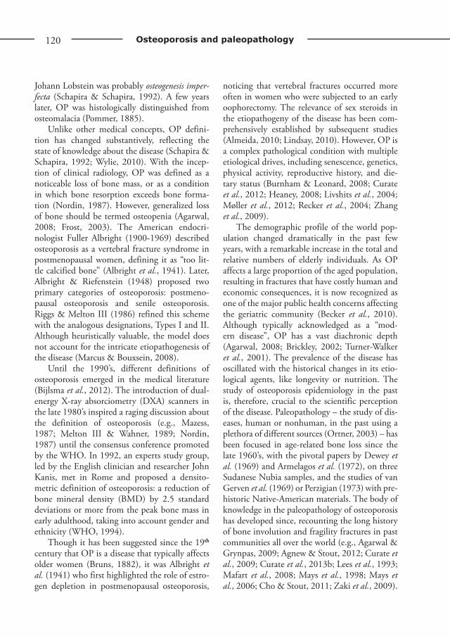
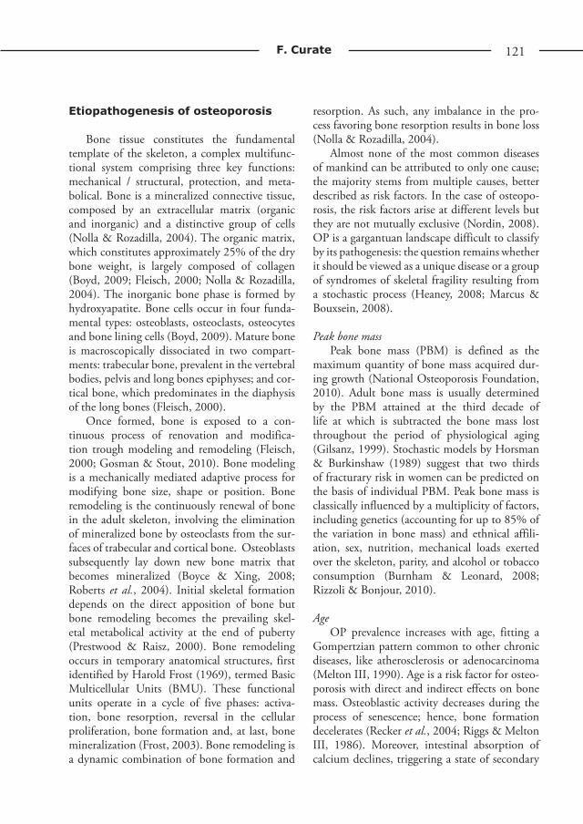
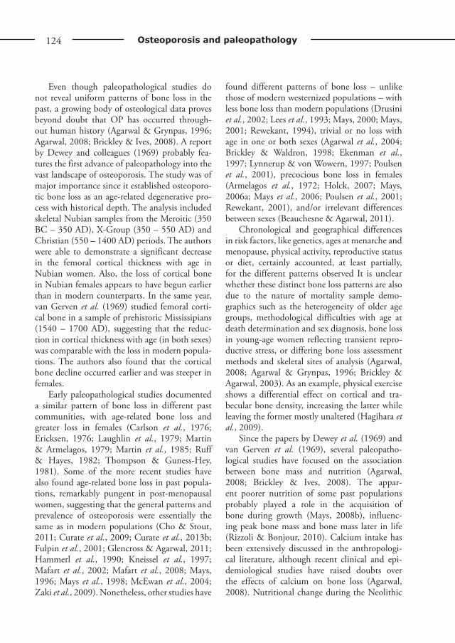
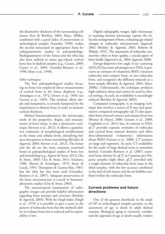
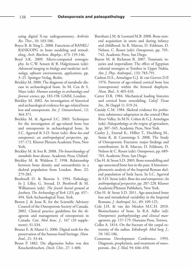
femur (Johnell & Kanis, 2006). Likewise, frac-tures of the proximal humerus (Fig. 1) are often related to an osteoporotic disorder (Reitman et al., 2008).
The anatomical relevance and the social and cultural repercussions of trauma in past commu-nities are categorical (Lovell, 1997) and a multi-plicity of publications have made consequential contributions to the way in which trauma has been used to recognize and interpret acciden-tal injury or interpersonal violence throughout history (e.g., Domett & Tayles, 2006; Djurić et al., 2006; Mitchell, 2006). Although ubiquitous in the archeological record, fractures are, most of the times, related to traumatic events and not with bone intrinsic frailty (Dequeker et al., 1997). Fragility fractures – especially fractures of the proximal femur – were deemed infrequent in archaeological samples (Agarwal et al., 2004; Brickley, 2002; Ortner, 2003) but evidences of osteoporotic fractures in the past are growing steadily (Curate et al., 2011).
The assumed low frequency of fragility frac-tures in the past is commonly explained as the result of selective mortality and low life expec-tancy at birth in historical populations. As such, the lower prevalence of these fractures in archeo-logical skeletal samples suggests that the older cohorts in the past were resilient to the action of natural selection, being genetically more adapted to adverse environmental circumstances (Agarwal et al., 2004; Agarwal, 2008). One of the themes of the well-known osteological paradox (Wood et al., 1992) conveys the notion that individuals differ considerably in the susceptibility to illness, and that the factors that subsidize this discrep-ancy are usually not identifiable – while genet-ics can surely prompt frailty; other factors will also be involved in the predisposition for disease (Wright & Yoder, 2003). In short, any unidi-mensional hypothesis on the causes of OP and associated fractures tends to overlook the hybrid nature of the human body, simultaneously bio-logical and cultural (Sofaer, 2004).
Also, the notion that few individuals reached a sufficiently advanced age to sustain an osteo-porotic fracture is somewhat flawed. The low
life expectancy at birth in the past is closely con-nected to an exceptionally high infant mortality, and the individuals that surpassed the critical stage of infancy had good chances of living into old age, being more prone to chronic diseases, like OP and attendant sequels (Brickley & Ives, 2008). Also, paleodemographic profiles are more influenced by fertility than mortality (Wright & Yoder, 2003), which does have major implica-tions in the absolute and relative number of old adults in any given sample.
Factors beyond bone mass, like bone quality and geometry, environmental hazards or propen-sity to falls, can explain the low prevalence of fra-gility fractures in most past populations (Agarwal & Grynpas, 1996; Agarwal et al., 2004; Mays, 2008b) – but fracture patterns can only be fully perceived within a biocultural, context-specific, framework. In fact, the prevalence of osteoporo-tic fractures in past communities seems to display geographical and chronological variations instead of uniform patterns of low frequency. This should
Fig. 1 - Fracture of the cirurgical neck of the left humerus, with pronounced angulation and bone repair; male, 83 years (Identified Skeletal Collection of Coimbra). The colour version of this figure is available at the JASs website.
128 Osteoporosis and paleopathology
be at least considered since nowadays the severity and frequency of osteoporosis and related frac-tures varies considerably among different popula-tions (Johnell & Kanis, 2006). To maintain that osteoporotic fractures «in the past» were not fre-quent is an essentializing statement that embraces a view of historical populations as uniform and archetypal entities (Sofaer, 2004).
The comparison of osteoporotic fractures’ fre-quency between archeological and living samples is constrained by the nature of the epidemiologi-cal data (usually presented as incidence rates) ver-sus the paleoepidemiological data (only the preva-lence rates can be calculated). As such, similarities or dissimilarities in fracture frequency between skeletal and in vivo populations are restricted to those few studies that tabulate fracture prevalence according to age and sex classes (e.g., Kwok et al., 2013; van der Voort et al., 2001).
Alternatively, only the general pattern of frac-ture occurrence should be compared. The study of fractures in skeletal assemblages from archeo-logical sites is also limited by poor bone pres-ervation, unsatisfactory age at death estimation in adults and disparate scoring methods (Judd & Roberts, 1998). Moreover, older individuals are more likely to present bone fractures simply because they lived longer and the probability that they sustained a fracture is higher (Glencross & Sawchuk, 2003; Mays, 2008b). Finally, most of the times it is impossible to establish the exact
individual age at which the fracture occurred (Domett & Tayles, 2006).
A small fraction of paleopathological stud-ies on fractures have addressed the association of bone mass with fragility fractures (Curate et al., 2009; Curate et al., 2013b; Foldes et al., 1995; Frigo & Lang, 1995; Kilgore et al., 1997; Mays, 2000; Strouhal et al., 2003; Mays, 2006a, Mays et al., 2006b; Domett & Tayles, 2006). In these studies, osteoporotic fractures are usually corre-lated with low bone mass. For example, Curate et al. (2013b) found that women with osteoporosis had a much higher probability of showing a fra-gility fracture than women of the same age diag-nosed with normal or osteopenic values of BMD. There is a possibility that bone loss occurred after fracture and not before (Brickley & Ives, 2008) but evidences that non-fragility fractures are not associated with low bone mass argue against that hypothesis (Mays et al., 2006).
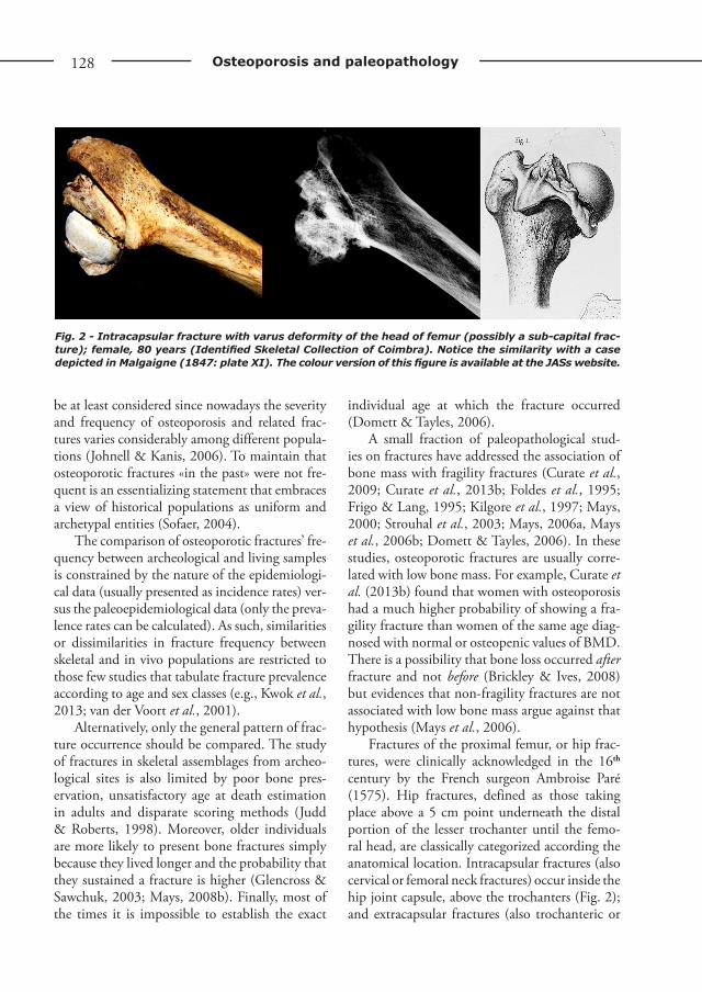
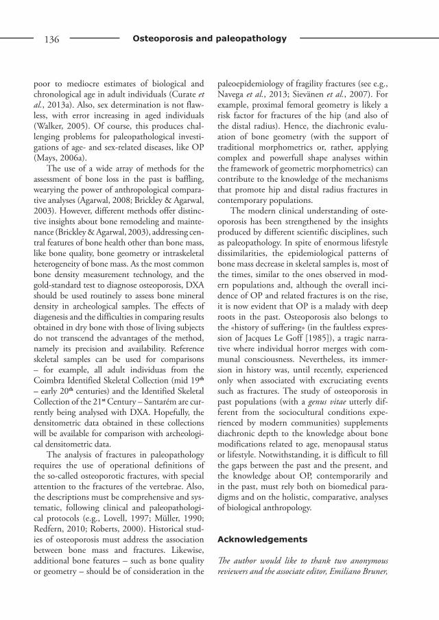
Fractures of the proximal femur, or hip frac-tures, were clinically acknowledged in the 16th century by the French surgeon Ambroise Paré (1575). Hip fractures, defined as those taking place above a 5 cm point underneath the distal portion of the lesser trochanter until the femo-ral head, are classically categorized according the anatomical location. Intracapsular fractures (also cervical or femoral neck fractures) occur inside the hip joint capsule, above the trochanters (Fig. 2); and extracapsular fractures (also trochanteric or
Fig. 2 - Intracapsular fracture with varus deformity of the head of femur (possibly a sub-capital frac-ture); female, 80 years (Identified Skeletal Collection of Coimbra). Notice the similarity with a case depicted in Malgaigne (1847: plate XI). The colour version of this figure is available at the JASs website.
www.isita-org.com
129F. Curate
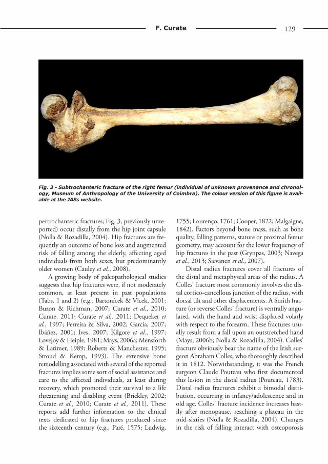
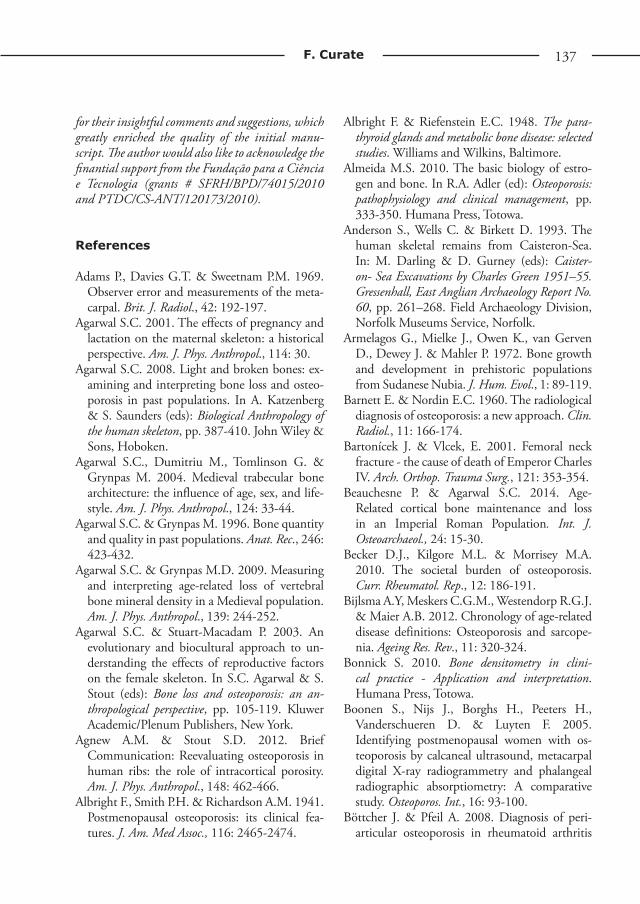
pertrochanteric fractures; Fig. 3, previously unre-ported) occur distally from the hip joint capsule (Nolla & Rozadilla, 2004). Hip fractures are fre-quently an outcome of bone loss and augmented risk of falling among the elderly, affecting aged individuals from both sexes, but predominantly older women (Cauley et al., 2008).
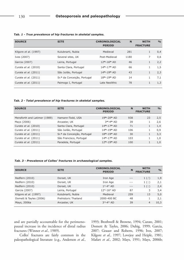
A growing body of paleopathological studies suggests that hip fractures were, if not moderately common, at least present in past populations (Tabs. 1 and 2) (e.g., Bartonícek & Vlcek, 2001; Buzon & Richman, 2007; Curate et al., 2010; Curate, 2011; Curate et al., 2011; Dequeker et al., 1997; Ferreira & Silva, 2002; Garcia, 2007; Ibáñez, 2001; Ives, 2007; Kilgore et al., 1997; Lovejoy & Heiple, 1981; Mays, 2006a; Mensforth & Latimer, 1989; Roberts & Manchester, 1995; Stroud & Kemp, 1993). The extensive bone remodelling associated with several of the reported fractures implies some sort of social assistance and care to the affected individuals, at least during recovery, which promoted their survival to a life threatening and disabling event (Brickley, 2002; Curate et al., 2010; Curate et al., 2011). These reports add further information to the clinical texts dedicated to hip fractures produced since the sixteenth century (e.g., Paré, 1575; Ludwig,
1755; Lourenço, 1761; Cooper, 1822; Malgaigne, 1842). Factors beyond bone mass, such as bone quality, falling patterns, stature or proximal femur geometry, may account for the lower frequency of hip fractures in the past (Grynpas, 2003; Navega et al., 2013; Sievänen et al., 2007).
Distal radius fractures cover all fractures of the distal and metaphyseal areas of the radius. A Colles’ fracture most commonly involves the dis-tal cortico-cancellous junction of the radius, with dorsal tilt and other displacements. A Smith frac-ture (or reverse Colles’ fracture) is ventrally angu-lated, with the hand and wrist displaced volarly with respect to the forearm. These fractures usu-ally result from a fall upon an outstretched hand (Mays, 2006b; Nolla & Rozadilla, 2004). Colles’ fracture obviously bear the name of the Irish sur-geon Abraham Colles, who thoroughly described it in 1812. Notwithstanding, it was the French surgeon Claude Pouteau who first documented this lesion in the distal radius (Pouteau, 1783). Distal radius fractures exhibit a bimodal distri-bution, occurring in infancy/adolescence and in old age. Colles’ fracture incidence increases hast-ily after menopause, reaching a plateau in the mid-sixties (Nolla & Rozadilla, 2004). Changes in the risk of falling interact with osteoporosis
Fig. 3 - Subtrochanteric fracture of the right femur (individual of unknown provenance and chronol-ogy, Museum of Anthropology of the University of Coimbra). The colour version of this figure is avail-able at the JASs website.
130 Osteoporosis and paleopathology
SOuRCE SITE CHRONOLOGICAL PERIOD
N WITH FRACTuRE
%
Kilgore et al. (1997) Kulubnarti, Nubia Medieval 281 1 0,4
Ives (2007) Several sites, UK Post-Medieval 1180 7 0,6
Garcia (2007) Leiria, Portugal 12th-16th AD 46 1 2,2
Curate et al. (2010) Santa Clara, Portugal 14th-17th AD 66 1 1,5
Curate et al. (2011) São Julião, Portugal 14th-19th AD 43 1 2,3
Curate et al. (2011) Sr.ª da Conceição, Portugal 18th-19th AD 14 1 7,1
Curate et al. (2011) Paimogo I, Portugal Late Neolithic 78 1 1,3
Tab. 2 - Total prevalence of hip fractures in skeletal samples.
SOuRCE SITE CHRONOLOGICAL PERIOD
N WITH FRACTuRE
%
Mensforth and Latimer (1989) Hamann-Todd, USA 19th-20th AD 938 23 2,5Mays (2006) Ancaster, UK 3rd-4th AD 39 1 2,6Curate et al. (2010) Santa Clara, Portugal 14th-17th AD 71 1 1,4Curate et al. (2011) São Julião, Portugal 14th-19th AD 106 1 0,9Curate et al. (2011) Sr.ª da Conceição, Portugal 18th-19th AD 30 1 3,3Curate et al. (2011) São Francisco, Portugal 14th-17th AD 103 1 1,0Curate et al. (2011) Paradela, Portugal 12th-19th AD 100 1 1,0
Tab. 3 - Prevalence of Colles’ fractures in archaeological samples.
SOuRCE SITECHRONOLOGICAL
PERIODN
WITH FRACTuRE
%
Redfern (2010) Dorset, UK Iron Age --- 1 (♂) 1,9Redfern (2010) Dorset, UK Iron Age --- 1 (♀) 2,1Redfern (2010) Dorset, UK 1st-4th AD --- 1 (♀) 2,4Garcia (2007) Leiria, Portugal 12th-16th AD 87 3 3,4Kilgore et al. (1997) Kulubnarti, Nubia Medieval 259 13 5,0Domett & Tayles (2006) Prehistoric Thailand 2000-400 BC 48 1 2,1Mays, 2006a Ancaster, UK 3rd-4th AD 39 4 10,3
and are partially accountable for the perimeno-pausal increase in the incidence of distal radius fractures (Winner et al., 1989).
Colles’ fractures are fairly common in the paleopathological literature (e.g., Anderson et al.,
1993; Brothwell & Browne, 1994; Curate, 2001; Domett & Tayles, 2006; Duhig, 1999; Garcia, 2007; Grauer and Roberts, 1996; Ives, 2007; Kilgore et al., 1997; Lovejoy and Heiple, 1981; Mafart et al., 2002; Mays, 1991; Mays, 2006b;
Tab. 1 - True prevalence of hip fractures in skeletal samples.

www.isita-org.com
131F. Curate
Miles, 1989; Redfern, 2010; Reis et al., 2003; Roberts & Wakely, 1992; Stroud & Kemp, 1993; Wells, 1982) and although their prevalence is gen-erally low (Tab. 3), it is not lower than other types of fracture (e.g., Garcia, 2007; Redfern, 2010). Medical authors, like Astley Paston Cooper (1822) or Guillaume Dupuytren (1847), suggested that distal radius’ fractures were very common in the first quarter of the 19th century. For example, dur-ing the 1829/1830 biennium, Dupuytren recorded 45 (out of a total of 206 fractures) fractures of the distal radius in the Hotel de Dîeu, Paris. This frequency is similar to the prevalence observed in modern Trauma and Orthopedic Services (Nolla & Rozadilla, 2004). Morbidity associated to Colles’ fractures is reduced but, occasionally, some residual deficit in the affected forearm persists over time. One paleopathological study suggested that distal radius fractures seldom healed without deformity (Grauer & Roberts, 1996).
Vertebral compression fractures are the hallmark of the «silent thief», being the most prevalent fracture in postmenopausal women (Johnell & Kanis, 2006; Nolla & Rozadilla, 2004). Notwithstanding, vertebral fractures are inadequately defined (there is not a consensual definition) and frequently asymptomatic, which induces an underestimation of their true inci-dence in the clinical practice (Grados et al., 2009).
Paleopathological descriptions of vertebral compression fractures are common but they usu-ally refer to anecdotal cases (e.g., Foldes et al., 1995; Ortner, 2003; Reis et al., 2003; Sambrook et al., 1988; Strouhal et al., 2003) or to poorly defined methods for the identification of vertebral fractures (e.g., Domett & Tayles, 2006; Hirata & Morimoto, 1994; Ives, 2007; Mays, 1996, 2006a; Mays et al., 2006; Mensforth & Latimer, 1989; Snow, 1948). The «Spine Score» (Barnett & Nordin, 1960) has been used for the assess-ment of vertebral fractures in archaeological sam-ples (González-Reimers et al., 2004). In a small number of paleopathological studies (e.g., Curate et al., 2009; Garcia, 2007), Genant’s semi-quan-titative method (Genant et al., 1993) was applied for the assessment of vertebral fractures. The «International Society for Clinical Densitometry»
endorses Genant’s method to diagnose verte-bral fractures in the clinical setting (Schousboe et al., 2008). The method is easy to apply, suc-cessful in ruling out vertebral deformities due
Fig. 4 - «Arrival of the English Ambassadors» (detail), Vittore Carpaccio, 1495-1500, tempera on canvas (Gallerie dell’Accademia, Venice). The old woman on the footstep of the stairway most likely suffered from spinal osteoporosis (Dequeker, 1994). The colour version of this fig-ure is available at the JASs website.
132 Osteoporosis and paleopathology
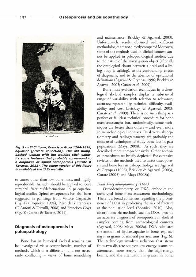
to causes other than low bone mass, and highly reproducible. As such, should be applied to score vertebral fractures/deformations in paleopatho-logical studies. Spinal osteoporosis has also been suggested in paintings from Vittore Carpaccio (Fig. 4) (Dequeker, 1994), Piero della Francesca (D’Antoni & Terzulli, 2008) and Francisco Goya (Fig. 5) (Curate & Tavares, 2011).
Diagnosis of osteoporosis in paleopathology
Bone loss in historical skeletal remains can be investigated via a comprehensive number of methods, which offer different – and not neces-sarily conflicting – views of bone remodeling
and maintenance (Brickley & Agarwal, 2003). Unfortunately, results obtained with different methodologies are not directly compared Moreover, some of the methods used in clinical context can-not be applied in paleopathological studies, due to the nature of the investigation object (after all, the ontological chasm between a dead and a liv-ing body is striking), to the confounding effects of diagenesis, and to the absence of operational definitions (Agarwal & Grynpas, 1996; Brickley & Agarwal, 2003; Curate et al., 2009).
Bone mass evaluation techniques in archeo-logical skeletal samples display a substantial range of variability with relation to relevance, accuracy, repeatability, technical difficulty, avail-ability and cost (Brickley & Agarwal, 2003; Curate et al., 2009). There is no such thing as a perfect or faultless technical procedure for bone mass assessment but, undoubtedly, some tech-niques are better than others – and even more so in archeological contexts. Dual x-ray absorp-tiometry and radiogrammetry are probably the most used techniques to study bone loss in past populations (Mays, 2008b). As such, they are described more comprehensively. Other techni-cal procedures are briefly depicted. For extensive reviews of the methods used to assess osteoporo-sis and bone loss in paleopathology see Agarwal & Grynpas (1996), Brickley & Agarwal (2003), Curate (2005) and Mays (2008a).
Dual X-ray absorptiometry (DXA)Osteodensitometry, or DXA, embodies the
archetypal bone mass assessment methodology. There is a broad consensus regarding the promi-nence of DXA in predicting the risk of fracture at the population level (Bonnick, 2010). Also, absorptiometric methods, such as DXA, provide an accurate diagnosis of osteoporosis in skeletal samples coming from archaeological contexts (Agarwal, 2008; Mays, 2008a). DXA calculates the amount of hydroxyapatite in bone, express-ing it in grams of mineral per area unit (Fig. 6). The technology involves radiation that stems from two discrete sources: low energy beams are attenuated more steeply than the high-energy beams, and the attenuation is greater in bone.
Fig. 5 - «El Chiton», Francisco Goya 1764-1824, aquatint (private collection). The old hump-backed woman with the walking stick exhib-its some features that probably correspond to a diagnosis of spinal osteoporosis (Curate & Tavares, 2011). The colour version of this figure is available at the JASs website.

www.isita-org.com
133F. Curate
The radiation source is collimated into a «pencil beam» and pointed to a radiation detector posi-tioned away from the place of measurement. The bone mineral content (BMC) affects the attenu-ation of the radioactive beam. The bone area is determined by specific software and bone mineral density is calculated as the ratio of measured min-eral content per area (Bonnick, 2010). DXA does not measure bone volumetric density and BMD results are not entirely standardized for bone size (Bonick, 2010; Lees et al., 1993).
Theoretically, densitometry can be per-formed in any part of the skeleton, but conven-tional clinical practice established that bone min-eral density assessment should be accomplished in the proximal femur or the lumbar column (the axial skeleton in the realm of densitometry), and for the diagnosis of OP should be considered the lowest T-score of the lumbar column, the neck of the femur or the total hip (Lewiecki et al., 2004). Bone density in the forearm, calcaneus and total body can also be measured with DXA. Peripheral measurements can be good predictors of BMD but it seems prudent not to assume that they can diagnose osteoporosis as good as measurements in the axial skeleton (Bonnick, 2010).
Studies of BMD in the lumbar spine are prob-ably the most common in clinical context but the proximal femur has received the preference in anthropological studies (e.g., Lees et al., 1993; Curate et al., 2013a; Mafart et al., 2008; Mays et al., 2006; Zaki et al., 2009). Precision error is reduced, both in the lumbar column (~1%) and the proximal femur (1–3%). Nevertheless, the femur preserves generally better than the lumbar spine in archaeological contexts and its positioning in the densitometer is much simpler. The radius has also been used to assess bone loss in paleopathological studies (e.g., McEwan et al., 2004; Zaki et al., 2009) but BMD assessment at the forearm in archeological samples is shown to be highly problematic due to the frequent inability of the densitometer to detect bone mass at this location (Ferreira et al., 2012). Although precise and reproducible, DXA measurements in archeological skeletal material can be distorted by taphonomic processes (Agarwal, 2008). Notwithstanding, there is a body of evidence (both direct and indirect) suggesting that diagen-esis affects bone mineral content only marginally (Mays et al., 1998; Mays et al., 2006; Mays, 2008a; Turner-Walker & Syversen, 2002). The
Fig. 6 - DXA report in a 58-year-old woman from the Coimbra Identified Skeletal Collection, per-formed in the left femur. Basic results and the WHO classification for this individual are presented. The colour version of this figure is available at the JASs website.
134 Osteoporosis and paleopathology
lack of soft tissues and bone marrow in histori-cal skeletal remains also hinders DXA measure-ments (Brickley, 2000; Mays, 2008a) – a water bath or rice can be used as surrogates of soft tis-sue (Brickley & Agarwal, 2003; McEwan et al., 2004) but comparisons with living individuals are thorny, and should be performed judiciously or even avoided
RadiogrammetryRadiogrammetry quantifies the amplitude or
geometry of cortical bone in tubular bones (usu-ally computes the ratio between the medullary cavity thickness and the total width of diaphy-sis), through direct measurements in a plain radi-ograph (Ives & Brickley, 2004). Although inef-fective to diagnose OP and assess fracture risk in individual patients, radiogrammetry is a valuable method to assess cortical bone loss in epidemio-logical settings (Boonen et al., 2005; Yasaku et al., 2009), and it is still widely used in studies directed to certain pathological conditions, like rheumatoid arthritis (Böttcher & Pfeil, 2008) or lupus erythematosus (Kalla et al., 1992). This technique was introduced in the clinical litera-ture in 1960, by different researchers (Barnett & Nordin,1960; Virtamä & Mahonen, 1960), also holding a long history in paleopathology (Ives & Brickley, 2004).
Conventional radiogrammetry only reveals modifications occurring in the cortical bone, i.e.,
periosteal apposition and, particularly, endosteal resorption (Adams et al., 1969); being insensi-tive to early bone loss (Steiner et al., 1996). The mineralized bone volume decay results in the reduction of calcium and radiographic absorp-tion (Grampp et al., 1997). The sites of resorp-tion (endosteal, intracortical and periosteal sur-faces) can react contradictorily to the different metabolical stimuli, and the subtle alterations in the endosteal envelope usually elicit chal-lenging interpretations of cortical bone loss at this location. As such, while the measurement of total with is precise and reproducible, the direct measurement of the medullary width is less accurate (Bonnick, 2010). The precision of the method has been variously reported between 5 to 10%, depending on the measurement site, but in expert hands precision is greatly enhanced (Adams et al., 1969; Ives & Brickley, 2004). The repeatability of radiogrammetric measurements in paleopathological studies is purportedly good (Mays, 2008a). Digital x-ray radiogrammetry (DXR) is more accurate and suitable for epide-miological studies than traditional radiogram-metry (Boonen et al., 2005; Bötcher et al., 2005), with results roughly comparable to DXA (Brown & Josse, 2002), but has not been used in paleopathological studies.
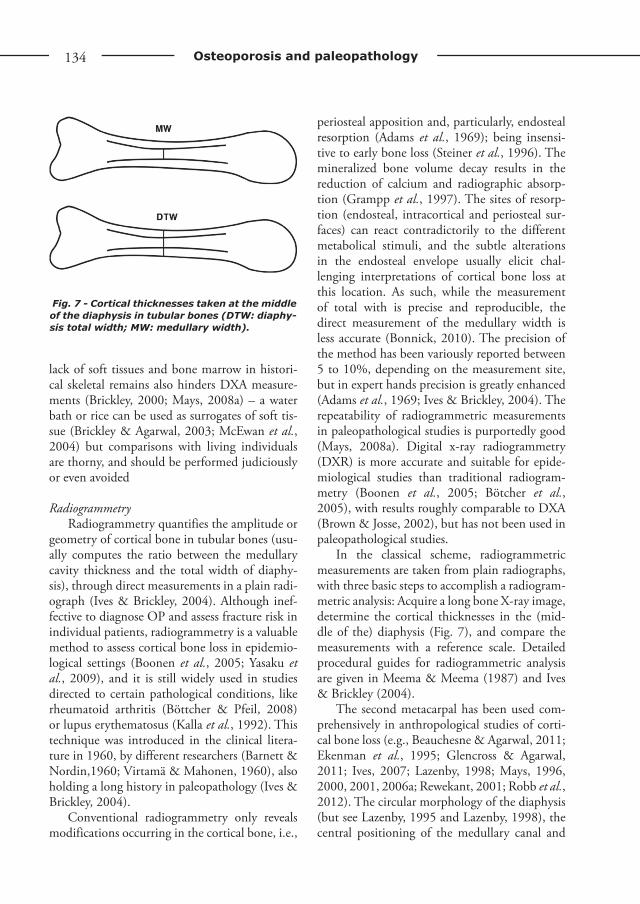
In the classical scheme, radiogrammetric measurements are taken from plain radiographs, with three basic steps to accomplish a radiogram-metric analysis: Acquire a long bone X-ray image, determine the cortical thicknesses in the (mid-dle of the) diaphysis (Fig. 7), and compare the measurements with a reference scale. Detailed procedural guides for radiogrammetric analysis are given in Meema & Meema (1987) and Ives & Brickley (2004).
The second metacarpal has been used com-prehensively in anthropological studies of corti-cal bone loss (e.g., Beauchesne & Agarwal, 2011; Ekenman et al., 1995; Glencross & Agarwal, 2011; Ives, 2007; Lazenby, 1998; Mays, 1996, 2000, 2001, 2006a; Rewekant, 2001; Robb et al., 2012). The circular morphology of the diaphysis (but see Lazenby, 1995 and Lazenby, 1998), the central positioning of the medullary canal and
Fig. 7 - Cortical thicknesses taken at the middle of the diaphysis in tubular bones (DTW: diaphy-sis total width; MW: medullary width).
www.isita-org.com
135F. Curate
the diminutive thickness of the surrounding soft tissues (Ives & Brickley, 2004; Mays, 2008a), combined with a good index of preservation in archeological samples (Lazenby, 1998), makes the second metacarpal an appropriate bone for radiogrammetric studies in paleopathology. Radiogrammetry of the femur and the tibia has also been utilized to assess age-related cortical bone loss in skeletal samples (e.g., Curate, 2009; Curate et al., 2009; González-Reimers et al., 1998; Mays et al., 1998).
Other techniquesThe first paleopathological studies focus-
ing on bone loss employed direct measurements of cortical bone in the femur diaphysis (e.g., Armelagos et al., 1972; Dewey et al., 1969; van Gerven et al., 1969). This method, albeit sim-ple and inexpensive, is severely hampered by the requirement to destroy bone in order to measure cortical thickness.
Skeletal histomorphometry, the microscopic study of the properties, shapes, and measure-ments of bone tissue, is also a destructive tech-nique (Stewart et al., 2012). It allows a quantita-tive evaluation of morphological modifications at the tissue and cellular levels, identifying bal-ance disruptions in bone remodeling (Brickley & Agarwal, 2003; Stewart et al., 2012). The femur and the rib are the most common examined bones in paleopathological studies of bone loss and remodeling (e.g., Agnew & Stout, 2012; Cho & Stout, 2003; Cho & Stout, 2011; Ericksen, 1980; Martin & Armelagos, 1979; Stout & Lueck, 1995; Thompson & Guness-Hey, 1981) but the tibia has also been used (González-Reimers et al., 2007). Adequate preservation of the bone microstructure is crucial in histomor-phometric studies (Cho & Stout, 2003).
The macroscopical examination of radio-graphic images can provide helpful information regarding bone amount and structure (Brickley & Agarwal, 2003). With the Singh Index (Singh et al., 1970) it is possible to give a score to the pattern of trabecular bone but the method’s abil-ity to evaluate bone loss is reduced and its repeat-ability is low.
Digital radiographic images, light microscopy or scanning electron microscopy capture the tra-becular arrangement of bone, evaluating age-related changes in trabecular microstructure (Agarwal, 2001; Brickley & Agarwal, 2003; Roberts & Wakely, 1992). The assessment of trabecular con-nectivity refers to bone quality, a crucial aspect of bone health (Agarwal et al., 2004; Agarwal, 2008).
Energy-dispersive low angle X-ray scattering (EDLAX) has some advantages over DXA: it gen-erates an estimate of volumetric BMD, measures trabecular and compact bone, or just trabecular bone, and recognizes the different minerals in a bone sample (Brickley & Agarwal, 2003; Mays, 2008a). Unfortunately, the technique produces high radiation doses and cannot be used in clini-cal settings. As such, its availability is exceedingly reduced (Brickley & Agarwal, 2003).
Computed tomography is an imaging tech-nique that involves a source of X-rays and quan-titative computed tomography (qCT) also quan-tifies bone mineral content and assesses bone loss (Bruner & Manzi, 2006; Genant et al., 2008; Guglielmi et al., 2011) but, in contrast to DXA, qCT provides separate estimates of trabecular and cortical bone mineral densities and offers three-dimensional (volumetric) information about BMD (Genant et al., 2008). CT scanners are large and expensive. As such, CT availability for the study of large skeletal series is somewhat limited. González-Reimers et al. (2007) exam-ined bone density by qCT in Canarian pre-His-panic samples (right tibia). qCT provided only a rough estimate of trabecular bone mass in the tibial samples, with the low accuracy attributed to the lack of soft tissues and the air bubbles con-fined within the trabecular bone.
Current problems and future directions
One of the greatest drawbacks in the study of OP in archeological samples pertains to the assessment of age at death in adult skeletal remains. Biological aging is extremely variable, and the appraisal of age at death usually renders
136 Osteoporosis and paleopathology
poor to mediocre estimates of biological and chronological age in adult individuals (Curate et al., 2013a). Also, sex determination is not flaw-less, with error increasing in aged individuals (Walker, 2005). Of course, this produces chal-lenging problems for paleopathological investi-gations of age- and sex-related diseases, like OP (Mays, 2006a).
The use of a wide array of methods for the assessment of bone loss in the past is baffling, wearying the power of anthropological compara-tive analyses (Agarwal, 2008; Brickley & Agarwal, 2003). However, different methods offer distinc-tive insights about bone remodeling and mainte-nance (Brickley & Agarwal, 2003), addressing cen-tral features of bone health other than bone mass, like bone quality, bone geometry or intraskeletal heterogeneity of bone mass. As the most common bone density measurement technology, and the gold-standard test to diagnose osteoporosis, DXA should be used routinely to assess bone mineral density in archeological samples. The effects of diagenesis and the difficulties in comparing results obtained in dry bone with those of living subjects do not transcend the advantages of the method, namely its precision and availability. Reference skeletal samples can be used for comparisons – for example, all adult individuas from the Coimbra Identified Skeletal Collection (mid 19th – early 20th centuries) and the Identified Skeletal Collection of the 21st Century – Santarém are cur-rently being analysed with DXA. Hopefully, the densitometric data obtained in these collections will be available for comparison with archeologi-cal densitometric data.
The analysis of fractures in paleopathology requires the use of operational definitions of the so-called osteoporotic fractures, with special attention to the fractures of the vertebrae. Also, the descriptions must be comprehensive and sys-tematic, following clinical and paleopathologi-cal protocols (e.g., Lovell, 1997; Müller, 1990; Redfern, 2010; Roberts, 2000). Historical stud-ies of osteoporosis must address the association between bone mass and fractures. Likewise, additional bone features – such as bone quality or geometry – should be of consideration in the
paleoepidemiology of fragility fractures (see e.g., Navega et al., 2013; Sievänen et al., 2007). For example, proximal femoral geometry is likely a risk factor for fractures of the hip (and also of the distal radius). Hence, the diachronic evalu-ation of bone geometry (with the support of traditional morphometrics or, rather, applying complex and powerfull shape analyses within the framework of geometric morphometrics) can contribute to the knowledge of the mechanisms that promote hip and distal radius fractures in contemporary populations.
The modern clinical understanding of oste-oporosis has been strengthened by the insights produced by different scientific disciplines, such as paleopathology. In spite of enormous lifestyle dissimilarities, the epidemiological patterns of bone mass decrease in skeletal samples is, most of the times, similar to the ones observed in mod-ern populations and, although the overall inci-dence of OP and related fractures is on the rise, it is now evident that OP is a malady with deep roots in the past. Osteoporosis also belongs to the «history of suffering» (in the faultless expres-sion of Jacques Le Goff [1985]), a tragic narra-tive where individual horror merges with com-munal consciousness. Nevertheless, its immer-sion in history was, until recently, experienced only when associated with excruciating events such as fractures. The study of osteoporosis in past populations (with a genus vitae utterly dif-ferent from the sociocultural conditions expe-rienced by modern communities) supplements diachronic depth to the knowledge about bone modifications related to age, menopausal status or lifestyle. Notwithstanding, it is difficult to fill the gaps between the past and the present, and the knowledge about OP, contemporarily and in the past, must rely both on biomedical para-digms and on the holistic, comparative, analyses of biological anthropology.
Acknowledgements
The author would like to thank two anonymous reviewers and the associate editor, Emiliano Bruner,
www.isita-org.com
137F. Curate
for their insightful comments and suggestions, which greatly enriched the quality of the initial manu-script. The author would also like to acknowledge the finantial support from the Fundação para a Ciência e Tecnologia (grants # SFRH/BPD/74015/2010 and PTDC/CS-ANT/120173/2010).
References
Adams P., Davies G.T. & Sweetnam P.M. 1969. Observer error and measurements of the meta-carpal. Brit. J. Radiol., 42: 192-197.
Agarwal S.C. 2001. The effects of pregnancy and lactation on the maternal skeleton: a historical perspective. Am. J. Phys. Anthropol., 114: 30.
Agarwal S.C. 2008. Light and broken bones: ex-amining and interpreting bone loss and osteo-porosis in past populations. In A. Katzenberg & S. Saunders (eds): Biological Anthropology of the human skeleton, pp. 387-410. John Wiley & Sons, Hoboken.
Agarwal S.C., Dumitriu M., Tomlinson G. & Grynpas M. 2004. Medieval trabecular bone architecture: the influence of age, sex, and life-style. Am. J. Phys. Anthropol., 124: 33-44.
Agarwal S.C. & Grynpas M. 1996. Bone quantity and quality in past populations. Anat. Rec., 246: 423-432.
Agarwal S.C. & Grynpas M.D. 2009. Measuring and interpreting age-related loss of vertebral bone mineral density in a Medieval population. Am. J. Phys. Anthropol., 139: 244-252.
Agarwal S.C. & Stuart-Macadam P. 2003. An evolutionary and biocultural approach to un-derstanding the effects of reproductive factors on the female skeleton. In S.C. Agarwal & S. Stout (eds): Bone loss and osteoporosis: an an-thropological perspective, pp. 105-119. Kluwer Academic/Plenum Publishers, New York.
Agnew A.M. & Stout S.D. 2012. Brief Communication: Reevaluating osteoporosis in human ribs: the role of intracortical porosity. Am. J. Phys. Anthropol., 148: 462-466.
Albright F., Smith P.H. & Richardson A.M. 1941. Postmenopausal osteoporosis: its clinical fea-tures. J. Am. Med Assoc., 116: 2465-2474.
Albright F. & Riefenstein E.C. 1948. The para-thyroid glands and metabolic bone disease: selected studies. Williams and Wilkins, Baltimore.
Almeida M.S. 2010. The basic biology of estro-gen and bone. In R.A. Adler (ed): Osteoporosis: pathophysiology and clinical management, pp. 333-350. Humana Press, Totowa.
Anderson S., Wells C. & Birkett D. 1993. The human skeletal remains from Caisteron-Sea. In: M. Darling & D. Gurney (eds): Caister-on- Sea Excavations by Charles Green 1951–55. Gressenhall, East Anglian Archaeology Report No. 60, pp. 261–268. Field Archaeology Division, Norfolk Museums Service, Norfolk.
Armelagos G., Mielke J., Owen K., van Gerven D., Dewey J. & Mahler P. 1972. Bone growth and development in prehistoric populations from Sudanese Nubia. J. Hum. Evol., 1: 89-119.
Barnett E. & Nordin E.C. 1960. The radiological diagnosis of osteoporosis: a new approach. Clin. Radiol., 11: 166-174.
Bartonícek J. & Vlcek, E. 2001. Femoral neck fracture - the cause of death of Emperor Charles IV. Arch. Orthop. Trauma Surg., 121: 353-354.
Beauchesne P. & Agarwal S.C. 2014. Age-Related cortical bone maintenance and loss in an Imperial Roman Population. Int. J. Osteoarchaeol., 24: 15-30.
Becker D.J., Kilgore M.L. & Morrisey M.A. 2010. The societal burden of osteoporosis. Curr. Rheumatol. Rep., 12: 186-191.
Bijlsma A.Y, Meskers C.G.M., Westendorp R.G.J. & Maier A.B. 2012. Chronology of age-related disease definitions: Osteoporosis and sarcope-nia. Ageing Res. Rev., 11: 320-324.
Bonnick S. 2010. Bone densitometry in clini-cal practice - Application and interpretation. Humana Press, Totowa.
Boonen S., Nijs J., Borghs H., Peeters H., Vanderschueren D. & Luyten F. 2005. Identifying postmenopausal women with os-teoporosis by calcaneal ultrasound, metacarpal digital X-ray radiogrammetry and phalangeal radiographic absorptiometry: A comparative study. Osteoporos. Int., 16: 93-100.
Böttcher J. & Pfeil A. 2008. Diagnosis of peri-articular osteoporosis in rheumatoid arthritis
138 Osteoporosis and paleopathology
using digital X-ray radiogrammetry. Arthritis Res. Ther., 10: 103-106.
Boyce B. & Xing L. 2008. Functions of RANKL/RANK/OPG in bone modeling and remod-eling. Arch. Biochem. Biophys., 473: 139-146.
Boyd S.K. 2009. Micro-computed tomogra-phy. In C.W. Sensen & B. Halgrímsson (eds): Advanced imaging in biology and medicine: tech-nology, software environments, applications, pp. 3–25. Springer-Verlag, Berlin.
Brickley M. 2000. The diagnosis of metabolic dis-ease in archaeological bone. In M. Cox & S. Mays (eds): Human osteology in archaeology and forensic science, pp. 183-198. GMM, London.
Brickley M. 2002. An investigation of historical and archaeological evidence for age-related bone loss and osteoporosis. Int. J. Osteoarchaeol., 12: 364-371.
Brickley M. & Agarwal S.C. 2003. Techniques for the investigation of age-related bone loss and osteoporosis in archaeological bone. In S.C. Agarwal & S.D. Stout (eds): Bone loss and osteoporosis: an anthropological perspective, pp. 157-172. Kluwer Plenum Academic Press, New York.
Brickley M. & Ives R. 2008. The bioarchaeology of metabolic bone disease. Academic Press, Oxford.
Brickley M. & Waldron T. 1998. Relationship between bone density and osteoarthritis in a skeletal population from London. Bone, 22: 279-283.
Brothwell D. & Browne S. 1994. Pathology. In J. Lilley, G. Stroud, D. Brothwell & M. Williamson (eds): The Jewish burial ground at Jewbury. The Archaeology of York 12/3, pp. 457–494. York Archaeological Trust, York.
Brown J. & Josse R. for the Scientific Advisory Council of the Osteoporosis Society of Canada. 2002. Clinical practice guidelines for the di-agnosis and management of osteoporosis in Canada. Can. Med Assoc. J., 167 (10 supple-ment): S1-S34.
Bruner E. & Manzi G. 2006. Digital tools for the preservation of the human fossil heritage. Hum. Evol., 21: 33-44.
Bruns P. 1882. Die allgemeine hehre von den Knockenbruchen. Dtsch. Chir., 27: 1-400.
Burnham J.M. & Leonard M.B. 2008. Bone min-eral acquisition in utero and during infancy and childhood. In R. Marcus, D. Feldman, D. Nelson, C. Rosen (eds): Osteoporosis, pp. 705-742. Academic Press, San Diego.
Buzon M. & Richman R. 2007. Traumatic in-juries and imperialism: The effect of Egyptian colonial strategies at Tombos in Upper Nubia. Am. J. Phys. Anthropol., 133: 783-791.
Carlson D.S., Armelagos G.J. & van Gerven D.P. 1976. Patterns of age-related cortical bone loss (osteoporosis) within the femoral diaphysis. Hum. Biol., 3: 405-410.
Carter D.R. 1984. Mechanical loading histories and cortical bone remodeling. Calcif. Tissue Int., 36 (Suppl 1): S19-24.
Cassidy C.M. 1984. Skeletal evidence for prehis-toric subsistence adaptation in the central Ohio River Valley. In M.N. Cohen & G.J. Armelagos (eds): Paleopathology at the origins of agriculture, pp. 307–345. Academic Press, New York.
Cauley J., Ensrud K., Hillier T., Hochberg M., Stone K. & Cummings S. 2008. The Study of Osteoporotic Fractures: major findings and contributions. In R. Marcus, D. Feldman, D. Nelson & C. Rosen (eds): Osteoporosis, pp. 689-703. Academic Press, San Diego.
Cho H. & Stout S.D. 2003. Bone remodelling and age-associated bone loss in the past: A histomor-phometric analysis of the Imperial Roman skel-etal population of Isola Sacra. In S.C. Agarwal & S.D. Stout (eds): Bone loss and osteoporosis: An anthropological perspective, pp. 207-228. Kluwer Academic/Plenum Publishers, New York.
Cho H. & Stout S.D. 2011. Age-associated bone loss and intraskeletal variability in the Imperial Romans. J. Anthropol. Sci., 89: 109-125.
Cole J.H. & van der Meulen M.C.H. 2010. Biomechanics of bone. In R.A. Adler (ed): Osteoporosis: pathophysiology and clinical man-agement, pp. 157-179. Humana Press, Totowa.
Colles A. 1814. On the fracture of the carpal ex-tremity of the radius. Edinburgh. Med Surg. J., 10: 182-186.
Consensus Development Conference. 1993. Diagnosis, prophylaxis, and treatment of osteo-porosis. Am. J. Med, 94: 646–650.

www.isita-org.com
139F. Curate
Cooper A.P. 1822. A treatise on dislocations and fractures of the joints. Bransby B. Cooper, London.
Curate F. 2009. Pierda de hueso cortical relacio-nada con la edad y fracturas osteoporóticas en la Colección de Esqueletos Identificados del Museo Antropológico de la Universidad de Coimbra. In M. Polo Cerdà & E. Garcia-Prósper (eds): Investigaciones histórico-médicas sobre salud y enfermedad en el pasado, pp. 421-434. Grupo Paleolab & Sociedad Española de Paleopatología, Valencia.
Curate F., Albuquerque A. & Cunha E.M. 2013a. Age at death estimation using bone densitom-etry: Testing the Fernández Castillo and López Ruiz method in two documented skeletal samples from Portugal. Forensic Sci. Int., 226: 296e1-296e6.
Curate F., Albuquerque A., Correia J., Ferreira I., Pedroso de Lima, J. & Cunha E.M. 2013b. A glimpse from the past: osteoporosis and os-teoporotic fractures in a Portuguese identified skeletal sample. Acta Reumatol. Port., 38: 20-27.
Curate F., Assis S., Lopes C. & Silva A.M. 2011. Hip fractures in the Portuguese archaeological record. Anthropol. Sci., 119: 87-93.
Curate F., Lopes C. & Cunha E.M. 2010. A 14th-17th century osteoporotic hip fracture from the Santa Clara-a-Velha Convent in Coimbra (Portugal). Int. J. Osteoarchaeol., 20: 591-596.
Curate F., Pedroso de Lima J., Albuquerque A., Ferreira I., Correia J. & Cunha E.M. 2012. Parto, morte e massa óssea na Colecção de Esqueletos Identificados do Museu Antropológico da Universidade de Coimbra (Portugal): alguns avanços preliminares. Cad. GEEvH, 1: 57-65.
Curate F., Piombino-Mascali D., Tavares A. & Cunha E.M. 2009. Assottigliamento corticale del femore e fratture da fragilità ossea: uno stu-dio della Collezione Scheletrica Identificata di Coimbra (Portogallo). Arch. Antr. Etn., 139: 129-146.
Curate F. & Tavares A. 2011. Cifosis vertebral en la pintura de Francisco Goya (1764-1824): un ejercício de diagnóstico diferencial. In A. González Martín, O. Cambra-Moo, J. Rascón
Pérez, M. Campo Martín, M. Robledo Ancinas, E. Labajo González & J. Sánchez Sánchez (eds): Paleopatología: ciencia multidisciplinar, pp. 611-616. Sociedad de Ciencias Aranzadi, Donostia-San Sébastian.
D’Antoni A. & Terzulli S. 2008. Federico di Montefeltro’s hyperkyphosis: a visual-historical case. J. Med Case Reports, 2: 11-14.
Dequeker J. 1994. Vertebral osteoporosis as paint-ed by Vittore Carpaccio (1465): reflections on paleopathology of osteoporosis in pictorial art. Calcif. Tissue Int., 55: 321-323.
Dequeker J., Ortner D., Stix A., Cheng X., Brys P. & Boonen, S. 1997. Hip fracture and os-teoporosis in a XIIth Dynasty female skeleton from Lisht, Upper Egypt. J. Bone Miner. Res., 12: 881-888.
Dewey J., Armelagos G. & Bartley M. 1969. Femoral cortical involution in three Nubian ar-chaeological populations. Hum. Biol., 41: 13-28.
Djurić M., Roberts C., Rakočević Z., Djonić D. & Lešić A. 2006. Fractures in Late Medieval skeletal populations from Serbia. Am. J. Phys. Anthropol., 130: 167-178.
Domett K. & Tayles N. 2006. Adult fracture pat-terns in prehistoric Thailand: A biocultural in-terpretation. Int. J. Osteoarchaeol., 16: 185-199.
Drusini A., Bredariol S., Carrara N. & Bonati M. 2000. Cortical bone dynamics and age-related osteopenia in a Longobard archaeological sam-ple from three graveyards of the Veneto Region (Northeast Italy). Int. J. Osteoarchaeol., 10: 268-279.
Duhig C. 1999. The human bone. In T. Malim & J. Hines (eds): The Anglo-Saxon cem-etery at Edix Hill (Barrington A), pp. 154–196. Cambridgeshire, Council for British Archaeology Research Report 112, Council for British Archaeology, York.
Dupuytren G. 1847. On the injuries and diseases of bones. Collected edition of the clinical lectures of Baron Dupuytren. C. and J. Adlard, London.
Duverney J.G. 1751. Traité des maladies des os. De Bure, Paris.
Ekenman I., Eriksson S. & Lindgren J. 1995. Bone density in medieval skeletons. Calcif. Tissue Int., 56: 355-358.

140 Osteoporosis and paleopathology
Ericksen M.F. 1976. Cortical bone loss with age in three Native American populations. Am. J. Phys. Anthropol., 45: 443-452.
Ericksen M.F. 1980. Patterns of microscopic bone remodeling in three aboriginal American popu-lations. In D.L. Brownman (ed): Early Native Americans: Prehistoric demography, economy, and technology, pp. 239-270. Houton, The Hague.
Ferrari S. Human genetics of osteoporosis. 2008. Best. Pract. Res. Clin. Endocrinol. Metab., 22: 723-735.
Ferreira M. & Silva, A.M. 2002. A case of osteo-myelitis in the hip of a Medieval Portuguese male skeleton. Antropol. Port., 19: 65-70.
Ferreira T., Albuquerque A., Ferreira I., Cunha E.M. & Curate F. 2012. Utilização do rádio no diagnóstico da osteoporose em paleopato-logia: complementaridade ou inadequação? In C. Marques, C. Lopes, C.B. Cruz, F.C. Silva, F. Curate, S. Assis & V. Matos (eds): III Jornadas Portuguesas de Paleopatologia: a saúde e a doença no passado. Programa-resumos, p. 31. Centro de Investigação em Antropologia e Saúde, Coimbra.
Fishbein L. 2004. Multiple sources of dietary calcium-some aspects of its essentiality. Regul. Toxicol. Pharmacol., 39: 67-80.
Fleisch H. 2000. Bisphosphonates in bone diseases: from the laboratory to the patient. Academic Press, San Diego.
Foldes A., Moscovici A., Popovtzer M., Mogle P., Urman D. & Zias J. 1995. Extreme osteopo-rosis in a Sixth Century skeleton from Negev Desert. Int. J. Osteoarchaeol., 5: 157-162.
Frigo P. & Lang C. 1995. Osteoporosis in a woman of the Early Bronze Age. N. Engl. J. Med, 333: 1468.
Frost H. 1969. Tetracycline-based histological analysis of bone remodelling. Calcif. Tissue Res., 3: 211-237.
Frost H. 2003. Bone’s mechanostat: A 2003 up-date. Anat. Rec., 275A: 1081–1101.
Fulpin J., Mafart B., Chouc P. & D’archimbaud G. 2001. Étude par absorptiométrie de la den-sité minérale osseuse dans une population mé-diévale (Nécropole de Notre Dame du Bourg, Digne, Alpes de Haute-Provence France, XIe-XIIIe et XVIe-XVIIIe s.). Bull. Mém. Soc. Anthropol. Paris, 13: 337-341.
Garcia S. 2007. Maleitas do corpo em tempos me-dievais. PhD Thesis, University of Coimbra, Coimbra.
Genant H.K., Engelke K. & Prevrhal S. 2008. Advanced CT bone imaging in osteoporosis. Rheumatology (Oxford), 47(suppl 4): iv9–iv16.
Genant H.K., Wu C., Vankuijk C. & Nevitt M. 1993. Vertebral fracture assessment using a semi-quantitative technique. J. Bone Miner. Res., 8: 1137-1148.
Gilsanz V. 1999. Accumulation of bone mass dur-ing childhood and adolescence. In E. Orwoll (ed): Osteoporosis in men. pp. 65-85, Academic Press, San Diego.
Glencross B. & Agarwal S.C. 2011. An investiga-tion of cortical bone loss and fracture patterns in the Neolithic community of Catalhöyük, Turkey using metacarpal radiogrammetry. J. Archaeol. Sci., 38: 513-521.
Glencross B. & Sawchuk L. 2003. The person-years construct: ageing and the prevalence of health related phenomena from skeletal sam-ples. Int. J. Osteoarchaeol., 13: 369-374.
González-Reimers E., Velasco-Vázquez J., Arnay-de-la-Rosa M., Santolaria-Fernández F., Gómez-Rodríguez M. & Machado-Calvo M. 2002. Double-energy X-ray absorptiometry in the diagnosis of osteopenia in ancient skeletal remains. Am. J. Phys. Anthropol., 118: 134-145.
González-Reimers E., Velasco-Vázquez J., Arnay-de-la-Rosa M. & Machado-Calvo M. 2007. Quantitative computerized tomography for the diagnosis of osteopenia in prehistoric skeletal remains. J. Archaeol. Sci., 34: 554-561.
González-Reimers E., Velasco-Vazquez J., Barros-Lopez N., Arnay-de-la-Rosa M., Santolaria-Fernandez F. & Castilla-Garcia A. 1998. Corticomedular index of the right tibia in the diagnosis of osteopenia in prehistoric skeletal remains. Am. J. Hum. Biol., 10: 37-44.
Grados F., Fechtenbaum J., Flipon E., Kolta S., Roux C. & Fardellone P. 2009. Radiographic methods for evaluating osteoporotic vertebral fractures. Joint Bone Spine, 76: 241-247.
Grauer A. & Roberts C. 1996. Paleoepidemiology, healing, and possible treatment of trauma in the medieval cemetery population of St.

www.isita-org.com
141F. Curate
Helen-on-the-Walls, York, England. Am. J. Phys. Anthropol., 100: 531-544.
Grampp S., Steiner E. & Imhof H. 1997. Radiological diagnosis of osteoporosis. Eur. Radiol., 7 (Suppl. 2): S11-S19.
Grynpas M. 2003. The role of bone quality on bone loss and bone fragility. In S.C. Agarwal & S.D. Stout (eds): Bone loss and osteoporosis – An anthropological perspective, pp. 33-44. Kluver Academic/Plenum Publishers, New York.
Gosman J.H. & Stout S.D. 2010. Current con-cepts in bone biology. In C.S. Larsen (ed): A companion to Biological Anthropology, pp. 465-484. Wiley-Blackwell, Chichester.
Guglielmi G., Muscarella S., Bazzocchi A. 2011. Integrated imaging approach to os-teoporosis: state-of-the-art review and update. RadioGraphics, 31: 1343-1364.
Hagihara Y., Nakajima A., Fukuda S., Goto S., Lida H. & Yamazaki M. 2009. Running exer-cise for short duration increases bone mineral density of loaded long bones in young growing rats. Tohoku J. Exp. Med., 219: 139-143.
Halloran B. & Bikle, D. 1999. Age-related changes in mineral metabolism. In E. Orwoll (ed): Osteoporosis in men, pp. 179-195. Academic Press, San Diego.
Hammerl J., Protsch R. & Happ J. 1990. Osteodensitometrie des femurhalses an his-torichen skeletten. In R. Werner & H. Matthiass (eds): Osteologieinterdisziplinar, pp. 139-142. Springer, Berlin.
Heaney R. 2008. Nutrition and risk for osteopo-rosis. In R. Marcus, D. Feldman, D. Nelson & C. Rosen (eds): Osteoporosis, pp. 799-836. Academic Press, San Diego.
Henderson P., Sowers M., Kutzko K. & Jannausch M. 2000. Bone mineral density in grand mul-tiparous women with extended lactation. Am. J. Obst. Gynecol., 182: 1371-1377.
Hirata K. & Morimoto I. 1994. Vertebral osteo-porosis in Late Edo Japanese. Anthropol. Sci., 102: 345-361.
Holck P. 2007. Bone mineral densities in the Prehistoric, Viking-Age and Medieval populations of Norway. Int. J. Osteoarchaeol., 17: 199-206.
Horsman A. & Burkinshaw L. 1989. Stochastic models of femoral bone loss and hip fracture
risk. In M. Kleerekoper & S. Krane (eds): Clinical disorders of bone and mineral metabo-lism, pp. 253-263. Mary Ann Liebert, New York.
Ibáñez M. 2001. Aspectos antropológicos y paleopa-tológicos de las inhumaciones prehistóricas del Tabaya (Aspe, Alicante). In M. Martín & F. Rodríguez (eds): Dónde estamos? Pasado, pre-sente y futuro de la paleopatología, pp. 263-268. Universidad Autónoma de Madrid, Madrid.
Ives R. 2007. An investigation of vitamin d defi-ciency osteomalacia and age-related osteoporosis in six post-medieval urban collections. PhD Thesis, University of Birmingham, Birmingham.
Ives R. & Brickley M. 2004. A procedural guide to metacarpal radiogrammetry inarchaeology. Int. J. Osteoarchaeol., 14: 7-17.
Ito M., Yamada M., Hayashi K., Ohki M., Uetani M. & Nakamura T. 1995. Relation of early menarche to high bone mineral density. Calcif. Tissue Int., 57: 11-14.
Johnell O. & Kanis J. 2006. An estimate of the worldwide prevalence and disability associated with osteoporotic fractures. Osteoporos. Int., 17: 1726-1733.
Judd M. & Roberts C. 1998. Fracture patterns at the Medieval Leper hospital in Chichester. Am. J. Phys. Anthropol., 105: 43–55.
Kalla A., Kotze T. & Meyers O. 1992. Metacarpal bone mass in lupus systemic erythematosus. Clin. Rheumatol., 11: 475-482.
Kannus P., Parkkari J., Sievänen H., Heinoen A., Vuori I. & Järvinen M. 1996. Epidemiology of hip fractures. Bone, 18: 57-63.
Kaptoge S.K., Dalzell N., Jakes R.W., Wareham N., Day N.E. & Khaw K.T. 2003. Hip section modulus, a measure of bending resistance, is more strongly related to reported physical activ-ity than BMD. Osteoporos. Int., 14: 941–949.
Karlsson M.K., Ahlborg H.G. & Karlsson C. 2005. Maternity and bone mineral density. Acta Orthop., 76: 2-13.
Khosla S. 2010. Update on estrogens and the skeleton. J. Clin. Endocrinol. Metab., 95: 3569-3577.
Kilgore L., Jurmain R. & van Gerven D. 1997. Palaeoepidemiological patterns of trauma in

142 Osteoporosis and paleopathology
a Medieval Nubian skeletal population Int. J. Osteoarchaeol., 7: 103-114.
Kneissel M., Boyde A., Hahn M., Teschler-Nicola M., Kalchhauser G. & Plenk H. 1997. Age- and sex-dependent cancellous bone changes in a 4000y BP population. Bone, 15: 539-545.
Komm B, Cheskis B, Bodine P. 2008. Regulation of bone cell function by estrogens. In R. Marcus, D. Feldman, D. Nelson & C. Rosen (eds): Osteoporosis, pp. 383-423. Academic Press, San Diego.
Kwok A.W.L., Gong J-S., Wang Y-X.J., Leung J.C.S., Kwok T., Griffith J.F. & Leung P.C. 2013. Prevalence and risk factors of radiograph-ic vertebral fractures in elderly Chinese men and women: results of Mr. OS (Hong Kong) and Ms. OS (Hong Kong) studies. Osteoporos. Int., 24: 877-885.
Larsen C.S. 2003. Animal source foods and hu-man health during evolution. J. Nutr., 133: 3893S-3897S.
Laughlin W.S., Harper A.B. & Thompson D.D. 1979. New approaches to the pre- and post-contact history of Arctic peoples. Am. J. Phys. Anthropol., 51: 579-587.
Lazenby R.A. 1995. Brief communication: non-circular geometry and radiogrammetry of the second metacarpal. Am. J. Phys. Anthropol., 97: 323-327.
Lazenby R.A. 1998. Second metacarpal midshaft geometry in an historic cemetery sample. Am. J. Phys. Anthropol., 106: 157-167.
Lees B., Molleson T., Arnett T.R. & Stevenson J.C. 1993. Differences in proximal femur bone den-sity over two centuries. Lancet, 341: 673–675.
Le Goff J. 1985. As doenças tem história. Terramar, Lisboa.
Lewiecki E., Watts N., Mcclung M., Petak S., Bachrach L., Shepherd J. & Downs Jr. R. for the International Society for Clinical Densitometry. 2004. Official positions of the International Society for Clinical Densitometry. J. Clin. Endocrinol. Metab., 89: 3651-3655.
Lindsay R. 2010. Estrogens and estrogen agonists/antagonists. In R.A. Adler (ed): Osteoporosis: pathophysiology and clinical management, pp. 351-358. Humana Press, Totowa.
Lobstein J. 1820. Traité d’anatomie pathologique. Livre II. F.G. Levrault, Strasbourg.
Lourenço A.G. 1761. Cirurgia classica, lusitana, anatomica, farmaceutica, medica, a mais mod-erna. Officina de Antonio Gomes Galhardo, Lisboa.
Lovejoy C. & Heiple, K. 1981. The analysis of fractures in skeletal populations with an ex-ample from the Libben Site, Ottowa County Ohio. Am. J. Phys. Anthropol., 55: 529-541.
Lovell N. 1997. Trauma analysis in paleopathol-ogy. Yearb. Phys. Anthropol., 40: 139-170.
Ludwig C. 1755. De collo femoris ejusque fractu-ra. In A. Haller (ed): Disputationes chirurgicae selecta, pp. 365-375. Marci-Michael Bousquet & Socir, Lausanne.
Lynnerup N. & von Wowern N. 1997. Bone min-eral content in Medieval Greenland Norse. Int. J. Osteoarchaeol., 7: 235-240 .
Mafart B., Chouc P., Fulpin J. & Boutin J. 2002. Etude par absorptiométrie du contenu minéral osseux dans une population de moniales mé-diévales cisterciennes (Abbaye Saint- Pierre de l’Almanarre, Hyères, Var, 12ème–14ème siècle). Bull. Soc. Biométrie Hum., 20: 181-185.
Mafart B., Fulpin J. & Chouc P. 2008. Postmenopausal bone loss in human skeletal re-mains of a historical population of southeastern France. Osteoporos. Int., 19: 381-382.
Malgaigne J-F. 1847. Traité des fractures et des luxa-tions. Baillière, Paris.
Marcus R. & Bouxsein M. 2008. The nature of osteoporosis. In R. Marcus, D. Feldman, D. Nelson & C. Rosen (eds): Osteoporosis, pp. 27-36. Academic Press, San Diego.
Martin D. & Armelagos GJ. 1979. Morphometrics of compact bone: an example from Sudanese Nubia. Am. J. Phys. Anthropol., 51: 571-577.
Martin D., Goodman A. & Armelagos G. 1985. Skeletal pathologies as indicators of quality and quantity of diet. In R. Gilbert & J. Mielke (eds): The analysis of prehistoric diets, pp. 227-279. Academic Press, New York.
Mays SA. 1991. The medieval burials from the Blackfriars Friary, School Street, Ipswich, Suffolk (excavated 1983-5). Ancient Monuments Laboratory Report 16/91, London.
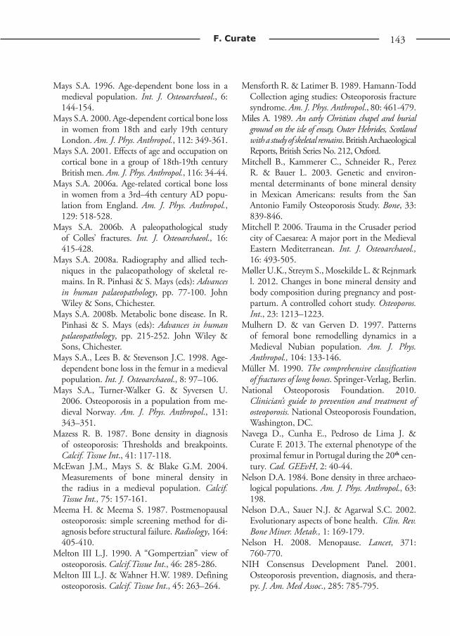
www.isita-org.com
143F. Curate
Mays S.A. 1996. Age-dependent bone loss in a medieval population. Int. J. Osteoarchaeol., 6: 144-154.
Mays S.A. 2000. Age-dependent cortical bone loss in women from 18th and early 19th century London. Am. J. Phys. Anthropol., 112: 349-361.
Mays S.A. 2001. Effects of age and occupation on cortical bone in a group of 18th-19th century British men. Am. J. Phys. Anthropol., 116: 34-44.
Mays S.A. 2006a. Age-related cortical bone loss in women from a 3rd–4th century AD popu-lation from England. Am. J. Phys. Anthropol., 129: 518-528.
Mays S.A. 2006b. A paleopathological study of Colles’ fractures. Int. J. Osteoarchaeol., 16: 415-428.
Mays S.A. 2008a. Radiography and allied tech-niques in the palaeopathology of skeletal re-mains. In R. Pinhasi & S. Mays (eds): Advances in human palaeopathology, pp. 77-100. John Wiley & Sons, Chichester.
Mays S.A. 2008b. Metabolic bone disease. In R. Pinhasi & S. Mays (eds): Advances in human palaeopathology, pp. 215-252. John Wiley & Sons, Chichester.
Mays S.A., Lees B. & Stevenson J.C. 1998. Age-dependent bone loss in the femur in a medieval population. Int. J. Osteoarchaeol., 8: 97–106.
Mays S.A., Turner-Walker G. & Syversen U. 2006. Osteoporosis in a population from me-dieval Norway. Am. J. Phys. Anthropol., 131: 343–351.
Mazess R. B. 1987. Bone density in diagnosis of osteoporosis: Thresholds and breakpoints. Calcif. Tissue Int., 41: 117-118.
McEwan J.M., Mays S. & Blake G.M. 2004. Measurements of bone mineral density in the radius in a medieval population. Calcif. Tissue Int., 75: 157-161.
Meema H. & Meema S. 1987. Postmenopausal osteoporosis: simple screening method for di-agnosis before structural failure. Radiology, 164: 405-410.
Melton III L.J. 1990. A “Gompertzian” view of osteoporosis. Calcif.Tissue Int., 46: 285-286.
Melton III L.J. & Wahner H.W. 1989. Defining osteoporosis. Calcif. Tissue Int., 45: 263–264.
Mensforth R. & Latimer B. 1989. Hamann-Todd Collection aging studies: Osteoporosis fracture syndrome. Am. J. Phys. Anthropol., 80: 461-479.
Miles A. 1989. An early Christian chapel and burial ground on the isle of ensay, Outer Hebrides, Scotland with a study of skeletal remains. British Archaeological Reports, British Series No. 212, Oxford.
Mitchell B., Kammerer C., Schneider R., Perez R. & Bauer L. 2003. Genetic and environ-mental determinants of bone mineral density in Mexican Americans: results from the San Antonio Family Osteoporosis Study. Bone, 33: 839-846.
Mitchell P. 2006. Trauma in the Crusader period city of Caesarea: A major port in the Medieval Eastern Mediterranean. Int. J. Osteoarchaeol., 16: 493-505.
Møller U.K., Streym S., Mosekilde L. & Rejnmark l. 2012. Changes in bone mineral density and body composition during pregnancy and post-partum. A controlled cohort study. Osteoporos. Int., 23: 1213–1223.
Mulhern D. & van Gerven D. 1997. Patterns of femoral bone remodelling dynamics in a Medieval Nubian population. Am. J. Phys. Anthropol., 104: 133-146.
Müller M. 1990. The comprehensive classification of fractures of long bones. Springer-Verlag, Berlin.
National Osteoporosis Foundation. 2010. Clinician’s guide to prevention and treatment of osteoporosis. National Osteoporosis Foundation, Washington, DC.
Navega D., Cunha E., Pedroso de Lima J. & Curate F. 2013. The external phenotype of the proximal femur in Portugal during the 20th cen-tury. Cad. GEEvH, 2: 40-44.
Nelson D.A. 1984. Bone density in three archaeo-logical populations. Am. J. Phys. Anthropol., 63: 198.
Nelson D.A., Sauer N.J. & Agarwal S.C. 2002. Evolutionary aspects of bone health. Clin. Rev. Bone Miner. Metab., 1: 169-179.
Nelson H. 2008. Menopause. Lancet, 371: 760-770.
NIH Consensus Development Panel. 2001. Osteoporosis prevention, diagnosis, and thera-py. J. Am. Med Assoc., 285: 785-795.
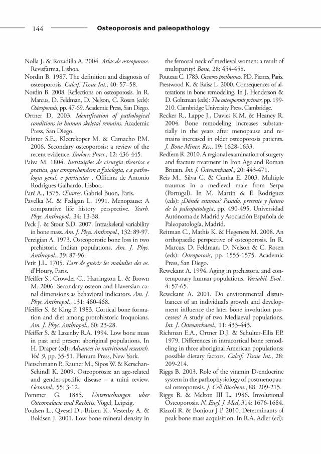
144 Osteoporosis and paleopathology
Nolla J. & Rozadilla A. 2004. Atlas de osteoporose. Revisfarma, Lisboa.
Nordin B. 1987. The definition and diagnosis of osteoporosis. Calcif. Tissue Int., 40: 57–58.
Nordin B. 2008. Reflections on osteoporosis. In R. Marcus, D. Feldman, D. Nelson, C. Rosen (eds): Osteoporosis, pp. 47-69. Academic Press, San Diego.
Ortner D. 2003. Identification of pathological conditions in human skeletal remains. Academic Press, San Diego.
Painter S.E., Kleerekoper M. & Camacho P.M. 2006. Secondary osteoporosis: a review of the recent evidence. Endocr. Pract., 12: 436-445.
Paiva M. 1804. Instituições de cirurgia theorica e pratica, que comprehendem a fysiologia, e a patho-logia geral, e particular . Officina de Antonio Rodrigues Galhardo, Lisboa.
Paré A., 1575. Œuvres. Gabriel Buon, Paris.Pavelka M. & Fedigan L. 1991. Menopause: A
comparative life history perspective. Yearb. Phys. Anthropol., 34: 13-38.
Peck J. & Stout S.D. 2007. Intraskeletal variability in bone mass. Am. J. Phys. Anthropol., 132: 89-97.
Perzigian A. 1973. Osteoporotic bone loss in two prehistoric Indian populations. Am. J. Phys. Anthropol., 39: 87-96.
Petit J.L. 1705. L’art de guérir les maladies des os. d’Houry, Paris.
Pfeiffer S., Crowder C., Harrington L. & Brown M. 2006. Secondary osteon and Haversian ca-nal dimensions as behavioral indicators. Am. J. Phys. Anthropol., 131: 460-468.
Pfeiffer S. & King P. 1983. Cortical bone forma-tion and diet among protohistoric Iroquoians. Am. J. Phys. Anthropol., 60: 23-28.
Pfeiffer S. & Lazenby R.A. 1994. Low bone mass in past and present aboriginal populations. In H. Draper (ed): Advances in nutritional research. Vol. 9, pp. 35-51. Plenum Press, New York.
Pietschmann P., Rauner M., Sipos W. & Kerschan-Schindl K. 2009. Osteoporosis: an age-related and gender-specific disease – a mini review. Gerontol., 55: 3-12.
Pommer G. 1885. Untersuchungen uber Osteomalacie und Rachitis. Vogel, Leipzig.
Poulsen L., Qvesel D., Brixen K., Vesterby A. & Boldsen J. 2001. Low bone mineral density in
the femoral neck of medieval women: a result of multiparity? Bone, 28: 454-458.
Pouteau C. 1783. Oeuvres posthumes. P.D. Pierres, Paris.Prestwood K. & Raisz L. 2000. Consequences of al-
terations in bone remodeling. In J. Henderson & D. Goltzman (eds): The osteoporosis primer, pp. 199-210. Cambridge University Press, Cambridge.
Recker R., Lappe J., Davies K.M. & Heaney R. 2004. Bone remodeling increases substan-tially in the years after menopause and re-mains increased in older osteoporosis patients. J. Bone Miner. Res., 19: 1628-1633.
Redfern R. 2010. A regional examination of surgery and fracture treatment in Iron Age and Roman Britain. Int. J. Osteoarchaeol., 20: 443-471.
Reis M., Silva C. & Cunha E. 2003. Multiple traumas in a medieval male from Serpa (Portugal). In M. Martín & F. Rodríguez (eds): ¿Dónde estamos? Pasado, presente y futuro de la paleopatología, pp. 490-495. Universidad Autónoma de Madrid y Asociación Española de Paleopatología, Madrid.
Reitman C., Mathis K. & Hegeness M. 2008. An orthopaedic perspective of osteoporosis. In R. Marcus, D. Feldman, D. Nelson & C. Rosen (eds): Osteoporosis, pp. 1555-1575. Academic Press, San Diego.
Rewekant A. 1994. Aging in prehistoric and con-temporary human populations. Variabil. Evol., 4: 57-65.
Rewekant A. 2001. Do environmental distur-bances of an individual’s growth and develop-ment influence the later bone involution pro-cesses? A study of two Mediaeval populations. Int. J. Osteoarchaeol., 11: 433-443.
Richman E.A., Ortner D.J. & Schulter-Ellis F.P. 1979. Differences in intracortical bone remod-eling in three aboriginal American populations: possible dietary factors. Calcif. Tissue Int., 28: 209-214.
Riggs B. 2003. Role of the vitamin D-endocrine system in the pathophysiology of postmenopau-sal osteoporosis. J. Cell Biochem., 88: 209-215.
Riggs B. & Melton III L. 1986. Involutional Osteoporosis. N. Engl. J. Med, 314: 1676-1684.
Rizzoli R. & Bonjour J-P. 2010. Determinants of peak bone mass acquisition. In R.A. Adler (ed):
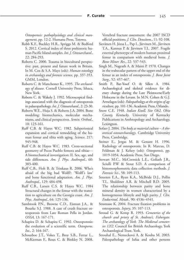
www.isita-org.com
145F. Curate
Osteoporosis: pathophysiology and clinical man-agement, pp. 12-2. Humana Press, Totowa.
Robb K.F., Buckley H.R., Spriggs M. & Bedford S. 2012. Cortical index of three prehistoric hu-man Pacific Island samples. Int. J. Osteoarchaeol., 22: 284-293.
Roberts C. 2000. Trauma in biocultural perspec-tive: past, present and future work in Britain. In M. Cox & S.A. Mays (eds): Human osteology in archaeology and forensic science, pp. 337-353. GMM, London.
Roberts C. & Manchester K., 1995. The archaeol-ogy of disease. Cornell University Press, Ithaca, New York.
Roberts C. & Wakely J. 1992. Microscopical find-ings associated with the diagnosis of osteoporosis in palaeopathology. Int. J. Osteoarchaeol., 2: 23-30.
Roberts W.E., Huja S. & Roberts J.A. 2004. Bone modeling: biomechanics, molecular mecha-nisms, and clinical perspectives. Semin. Orthod., 10: 123-161.
Ruff C.B. & Hayes W.C. 1982. Subperiosteal expansion and cortical remodeling of the hu-man femur and tibia with aging. Science, 217: 945-948.
Ruff C.B. & Hayes W.C. 1983. Cross-sectional geometry of Pecos Pueblo femora and tibiae—A biomechanical investigation: II. Sex, age, and side differences. Am. J. Phys. Anthropol., 60: 383-400.
Ruff C.B., Holt B. & Trinkaus R. 2006. Who’s afraid of the big bad Wolff?: ‘Wolff ’s law’ and bone functional adaptation. Am. J. Phys. Anthropol., 129: 484-498.
Ruff C.B., Larsen C.S. & Hayes W.C. 1984 Structural changes in the femur with the transi-tion to agriculture on the Georgia coast. Am. J. Phys. Anthropol., 64: 125-136.
Sambrook P.N., Browne C.D., Eisman J.A., & Bourke S.J. 1988. A case of crush fracture os-teoporosis from Late Roman Pella in Jordan. OSSA, 13: 167-171.
Schapira D. & Schapira C. 1992. Osteoporosis: the evolution of a scientific term. Osteoporos.Int., 2: 164-167.
Schousboe J.T., Vokes T., Broy S.B., Ferrar L., McKiernan F., Roux C. & Binkley N. 2008.
Vertebral fracture assessment: the 2007 ISCD official positions. J. Clin. Densitom., 11: 92-108.
Sievänen H. Józsa L., Pap I., Järvinen M., Järvinen T.A., Kannus P. & Järvinen T.L. 2007. Fragile external phenotype of modern human proximal femur in comparison with medieval bone. J. Bone Miner. Res., 22: 537-543.
Singh M., Nagrath A. & Maini P. 1970. Changes in the trabecular pattern of the upper end of the femur as an index of osteoporosis. J. Bone Joint Surg., 52: 457-467.
Smith P., Bar-Yosef O. & Sillen A. 1984. Archaeological and skeletal evidence for di-etary change during the Late Pleistocene/Early Holocene in the Levant. In M.N. Cohen & G.J. Armelagos (eds): Paleopathology at the origins of ag-riculture, pp. 101-136. Academic Press, Orlando.
Snow C.E. 1948. Indian Knoll, Site Oh2, Ohio County, Kentucky. University of Kentucky Publications in Anthropology and Archaeology, Lexington.
Sofaer J. 2004. The body as material culture – A the-oretical osteoarchaeology. Cambridge University Press, Cambridge.
Steiner E., Jergas M. & Genant H. 1996. Radiology of osteoporosis. In R. Marcus, D. Feldman & J. Kelsey (eds): Osteoporosis, pp. 1019-1054. Academic Press, San Diego.
Stewart M.C., McCormick L.E., Goliath J.R., Sciulli P.W & Stout S.D. A comparison of histomorphometric data collection methods. J. Forensic Sci., 58: 109-113.
Streeten E.A., Ryan K.A., McBride D.J., Pollin T.I., Shuldiner A.R. & Mitchell B.D. 2005.The relationship between parity and bone mineral density in women characterized by a homogeneous lifestyle and high parity. J. Clin. Endocrinol. Metab., 90: 4536–4541.
Strømsøe K. 2004. Fracture fixation problems in osteoporosis. Injury, 35: 107-113.
Stroud G. & Kemp R. 1993. Cemeteries of the church and priory of St. Andrew’s, Fishergate. The archaeology of York. The Medieval cemeter-ies 12/2. Council for British Archaeology, York Archaeological Trust, York.
Strouhal E., Nemecková A. & Kouba M. 2003. Paleopathology of Iufaa and other persons
146 Osteoporosis and paleopathology
found beside his shaft tomb at Abusir (Egypt). Int. J. Osteoarchaeol., 13: 331-338 .
Stout S.D. & Lueck R. 1995. Bone remodeling rates and skeletal maturation in three ar-chaeological skeletal populations. Am. J. Phys. Anthropol., 93: 123-129.
Thompson D. & Guness-Hey M. 1981. Bone mineral-osteon analysis of Yupik-Inupiaq skel-etons. Am. J. Phys. Anthropol., 55: 1-7.
To W.W. & Wong M.W. 2012. Bone mineral density during pregnancy in actively exercising women as measured by quantitative ultrasound. Arch. Gynecol. Obstet., 286: 357-363.
Turner-Walker G. & Syversen U. 2002. Quantifying histological changes in archaeo-logical bones using BSE-SEM image analysis. Archaeometry, 44: 461-468.
Turner-Walker G., Syversen U. & Mays S. 2001. The archaeology of osteoporosis. Eur. J. Archaeol., 4: 263-269.
Uusi-Rasi K., Kannus P. & Sievänen, H. 2008. Physical activity in prevention of osteoporosis and associated fractures. In R. Marcus, D. Feldman, D. Nelson & C. Rosen (eds): Osteoporosis, pp. 837-859. Academic Press, San Diego.
van der Voort D.J.M., Geusens P.P. & Dinant G.J. 2001. Risk factors for osteoporosis related to their outcome: fractures. Osteoporos. Int., 12: 630-638.
van Gerven D., Armelagos G. & Bartley M. 1969. Roentgenographic and direct measure-ment of femoral cortical involution in a pre-historic Mississippian population. Am. J. Phys. Anthropol., 31: 23-38.
Vashishth D., Tanner K. & Bonfield W. 2003. Experimental validation of a microcracking-based toughening mechanism for cortical bone. J. Biomech., 36: 121-124.
Virtamä P. & Mahonen H. 1960. Thickness of the cortical layer as an estimate of mineral content of human finger bones. Brit. J. Radiol., 33: 60-62.
Vogel M., Hahn M., Caselitz P., Woggan J., Pompesius-Kempa M. & Delling G. 1990. Comparison of trabecular bone structure in man today and an ancient population in Western Germany. In H.E. Takahashi (ed): Bone morpho-metry, pp. 220–223. Nishimura Co.,Tokyo.
Walker P.L. 2005. Greater sciatic notch morphol-ogy: Sex, age and population differences. Am. J. Phys. Anthropol., 127: 385-391.
Wells C. 1982. The human burials. In A. McWhirr, L. Viner & C. Wells (eds): Romano-British cemeteries at Cirencester, Cirencester exca-vations II, pp. 135–202. Cirencester Excavation Committee, Cirencester.
Williams F.M.K. & Spector T.D. 2007. The genetics of osteoporosis. Acta Reumatol. Port., 32: 231-240.
Winner S.J., Morgan C.A. & Grimley Evans J. 1989. Perimenopausal risk of falling and in-cidence of distal forearm fracture. BMJ, 298: 1486-1488.
Wolff J. 1892. Das Gesetz der Transformation der Knochen. August Hirschwald, Berlin.
Wood J.W., Milner G.R., Harpending H.C., Weiss, K.M. 1992. The osteological paradox: Problems of inferring prehistoric health from skeletal samples. Curr. Anthropol., 33: 343-370.
World Health Organization. 1994. Assessment of fracture risk and its application to screening for postmenopausal osteoporosis. Technical Report Series, WHO, Geneva.
Wright L.E., Yoder C.J. 2003. Recent progress in bioarchaeology: approaches to the Osteological Paradox. J. Archaeol. Res., 11: 43-70.
Wylie C.D. 2010. Setting a standard for a ‘‘silent’’ dis-ease: defining osteoporosis in the 1980s and 1990s. Stud. Hist. Philos. Biol. Biomed Sci., 41: 376-385.
Yasaku K., Ishikawa-Takata K., Koitaya N., Yoshimoto K. & Ohta T. 2009. One-year change in the second metacarpal bone mass as-sociated with menopause nutrition and physical activity. J. Nutr. Health Aging, 13: 545-549.
Zaki M., Hussien F. & El Banna R. 2009. Osteoporosis among ancient Egyptians. Int. J. Osteoarchaeol., 19: 78-89.
Zhang H., Sol-Church K., Rydbeck H., Stabley D., Spotila LD. & Devoto M. 2009. High-resolution linkage and linkage disequilibrium analyses of chromosome 1p36 SNPs identify new positional candidate genes for low bone mineral density. Osteoporos. Int., 20: 341-346.
Associate Editor, Emiliano Bruner