Embed Size (px)
Citation preview
University of Pisa Division of Paleopathology,
History of Medicine and Bioethics
Gino Fornaciari
The challenging case of Cardinal Carlo de’ Medici
The First Clinical Challenge: Eminence versus Evidence in Spondyloarthritis
June 22-23, 2012 – Pisa
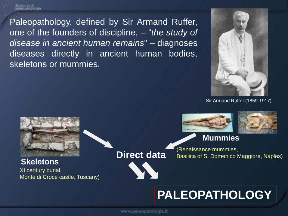
Skeletons Direct data
Paleopathology, defined by Sir Armand Ruffer, one of the founders of discipline, – “the study of disease in ancient human remains” – diagnoses diseases directly in ancient human bodies, skeletons or mummies.
PALEOPATHOLOGY
(Renaissance mummies, Basilica of S. Domenico Maggiore, Naples)
Mummies
XI century burial, Monte di Croce castle, Tuscany)
Sir Armand Ruffer (1859-1917)

PALEOPATHOLOGY
INTEREST
Origin and evolution of diseases
Medicine
Life style of ancient populations
(infectious diseases, cancer..) (trauma, arthritis..)
Anthropology
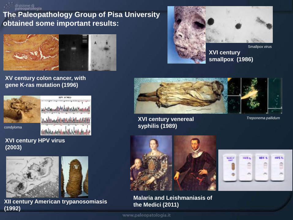
XII century American trypanosomiasis (1992)
Treponema pallidum XVI century venereal syphilis (1989)
XVI century smallpox (1986)
Smallpox virus
XV century colon cancer, with gene K-ras mutation (1996)
XVI century HPV virus (2003)
The Paleopathology Group of Pisa University obtained some important results:
condyloma
Malaria and Leishmaniasis of the Medici (2011)
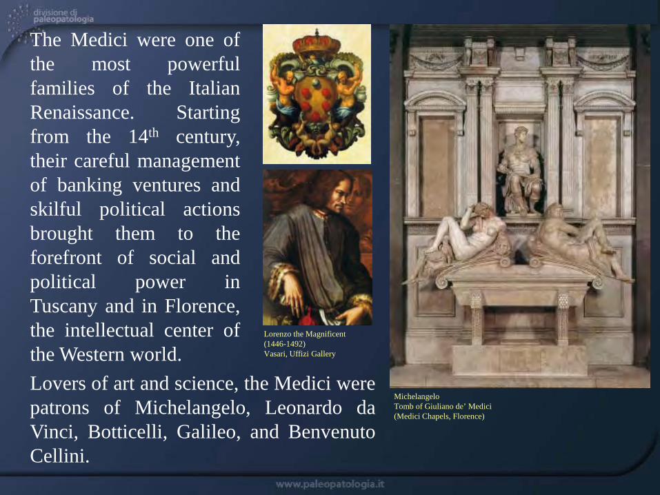
The Medici were one of the most powerful families of the Italian Renaissance. Starting from the 14th century, their careful management of banking ventures and skilful political actions brought them to the forefront of social and political power in Tuscany and in Florence, the intellectual center of the Western world. Lovers of art and science, the Medici were patrons of Michelangelo, Leonardo da Vinci, Botticelli, Galileo, and Benvenuto Cellini.
Lorenzo the Magnificent (1446-1492) Vasari, Uffizi Gallery
Michelangelo Tomb of Giuliano de’ Medici (Medici Chapels, Florence)
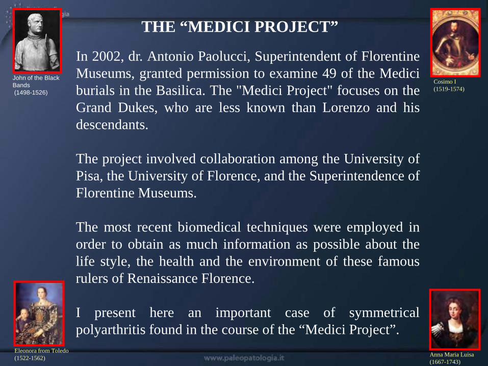
In 2002, dr. Antonio Paolucci, Superintendent of Florentine Museums, granted permission to examine 49 of the Medici burials in the Basilica. The "Medici Project" focuses on the Grand Dukes, who are less known than Lorenzo and his descendants. The project involved collaboration among the University of Pisa, the University of Florence, and the Superintendence of Florentine Museums. The most recent biomedical techniques were employed in order to obtain as much information as possible about the life style, the health and the environment of these famous rulers of Renaissance Florence. I present here an important case of symmetrical polyarthritis found in the course of the “Medici Project”.
Anna Maria Luisa (1667-1743)
Cosimo I (1519-1574)
Eleonora from Toledo (1522-1562)
John of the Black Bands (1498-1526)
THE “MEDICI PROJECT”
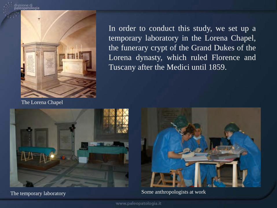
In order to conduct this study, we set up a temporary laboratory in the Lorena Chapel, the funerary crypt of the Grand Dukes of the Lorena dynasty, which ruled Florence and Tuscany after the Medici until 1859.
The Lorena Chapel
The temporary laboratory Some anthropologists at work

THE CRYPT OF SAN LORENZO
Map of the crypt, with the Medici tombs already explored in yellow.
The crypt of the Basilica of San Lorenzo in Florence, Mausoleum of the Grand Dukes of the Medici family.
INFECTIOUS AND PARASITIC DISEASES
smallpox tuberculosis malaria syphilis
METABOLIC DISEASES
obesity anemia urinary stones
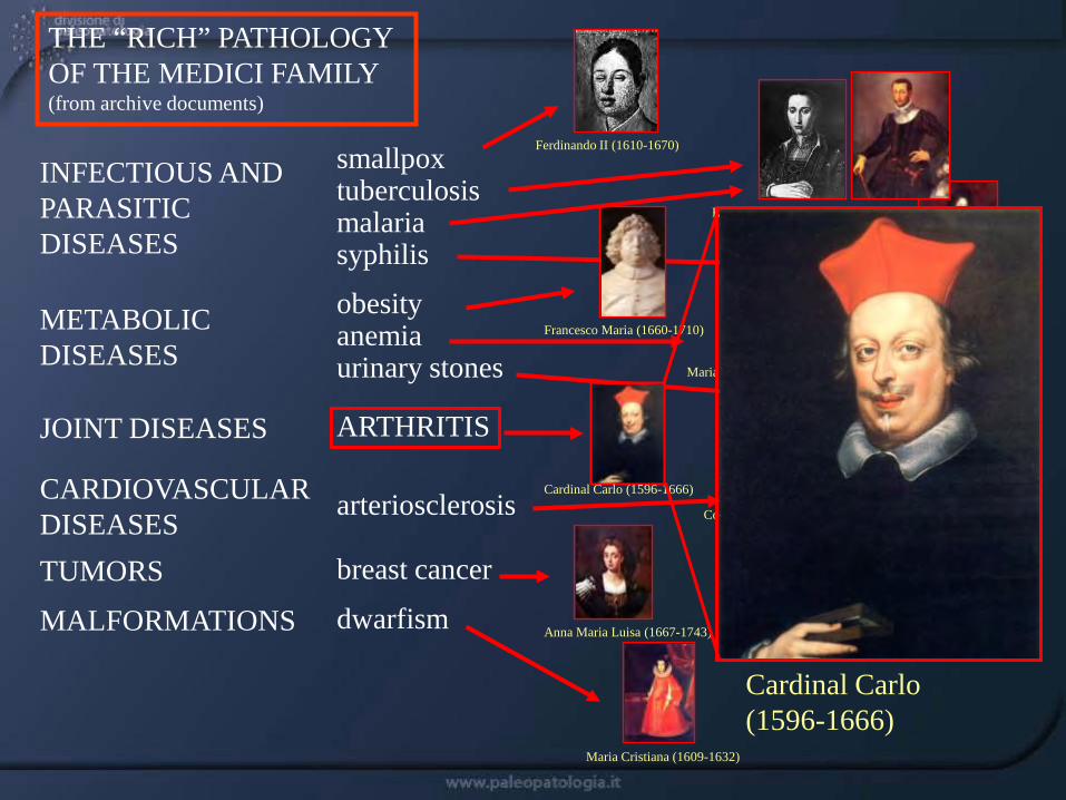
JOINT DISEASES ARTHRITIS
CARDIOVASCULAR DISEASES
arteriosclerosis
TUMORS breast cancer
MALFORMATIONS dwarfism
Francesco I (1541-1587)
Maria Cristiana (1609-1632)
Anna Maria Luisa (1667-1743)
Cosimo I (1519-1574)
Cosimo III (1642-1723)
Maria Salviati (1499-1543)
Ferdinando II (1610-1670)
Eleonora from Toledo (1522-1562)
Ferdinando (1663-1713)
Francesco Maria (1660-1710)
THE “RICH” PATHOLOGY OF THE MEDICI FAMILY (from archive documents)
Cardinal Carlo (1596-1666)
Cardinal Carlo (1596-1666)
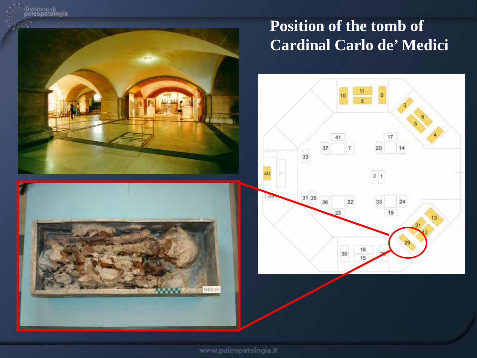
Position of the tomb of Cardinal Carlo de’ Medici

Cardinal Carlo (1596-1666)
Sustermans (c.1640), Florence
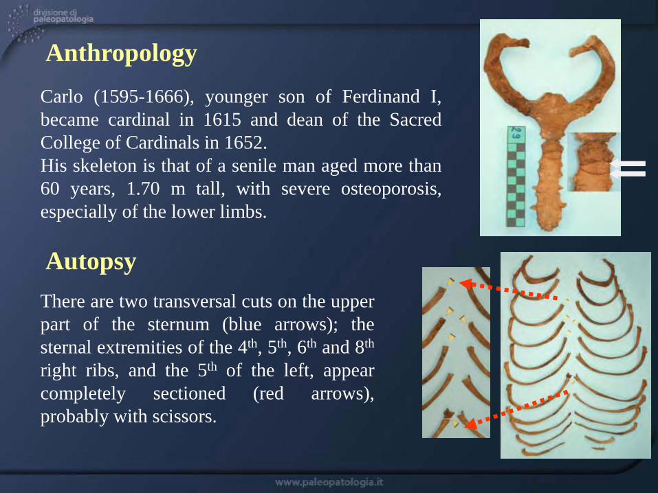
Carlo (1595-1666), younger son of Ferdinand I, became cardinal in 1615 and dean of the Sacred College of Cardinals in 1652. His skeleton is that of a senile man aged more than 60 years, 1.70 m tall, with severe osteoporosis, especially of the lower limbs.
Anthropology
Autopsy There are two transversal cuts on the upper part of the sternum (blue arrows); the sternal extremities of the 4th, 5th, 6th and 8th right ribs, and the 5th of the left, appear completely sectioned (red arrows), probably with scissors.
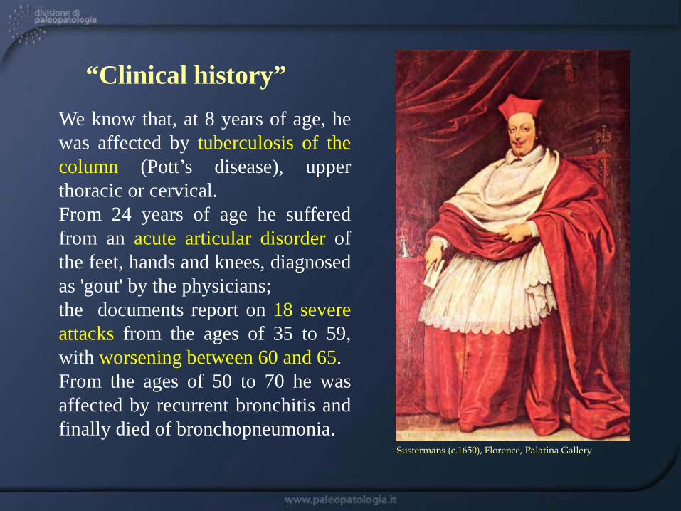
We know that, at 8 years of age, he was affected by tuberculosis of the column (Pott’s disease), upper thoracic or cervical. From 24 years of age he suffered from an acute articular disorder of the feet, hands and knees, diagnosed as 'gout' by the physicians; the documents report on 18 severe attacks from the ages of 35 to 59, with worsening between 60 and 65. From the ages of 50 to 70 he was affected by recurrent bronchitis and finally died of bronchopneumonia.
“Clinical history”
Sustermans (c.1650), Florence, Palatina Gallery

The facial skeleton shows a marked hypoplasia of the right mandibular corpus, with right deviation of the face and probable chronic torticollis, as appears in a rare engraving (white arrow).
Paleopathology
Haelvegh, engraving X-ray Prof. N. Villari (University of Florence)
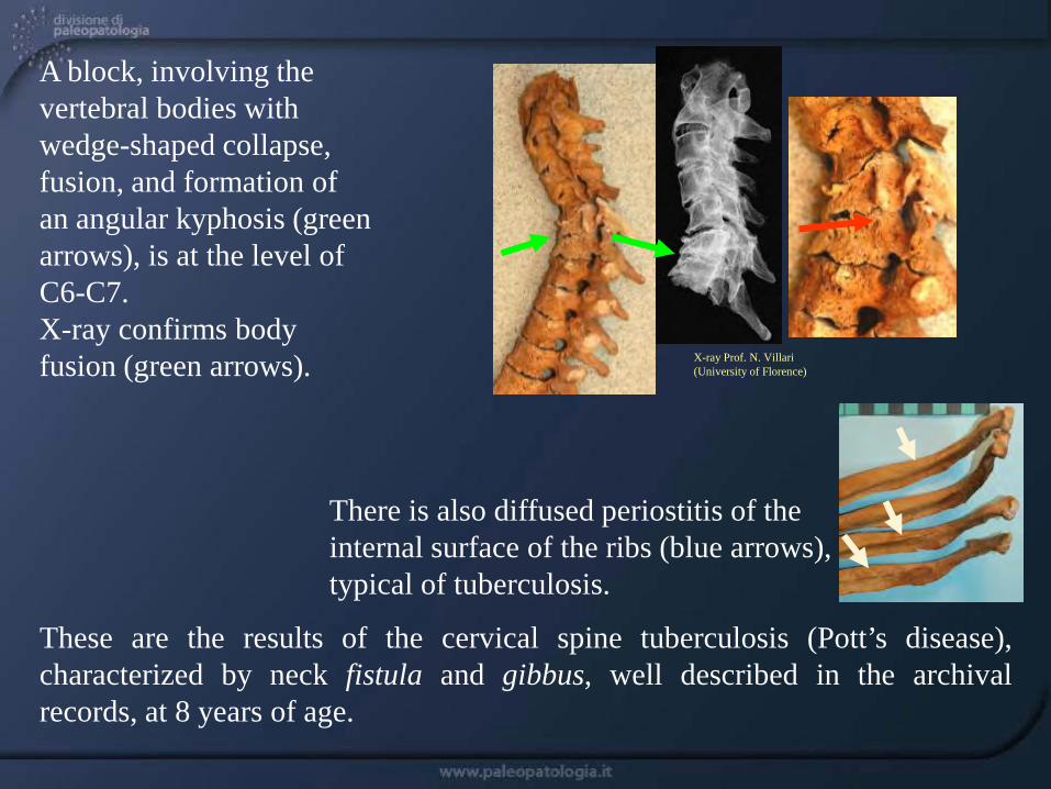
A block, involving the vertebral bodies with wedge-shaped collapse, fusion, and formation of an angular kyphosis (green arrows), is at the level of C6-C7. X-ray confirms body fusion (green arrows).
These are the results of the cervical spine tuberculosis (Pott’s disease), characterized by neck fistula and gibbus, well described in the archival records, at 8 years of age.
There is also diffused periostitis of the internal surface of the ribs (blue arrows), typical of tuberculosis.
X-ray Prof. N. Villari (University of Florence)
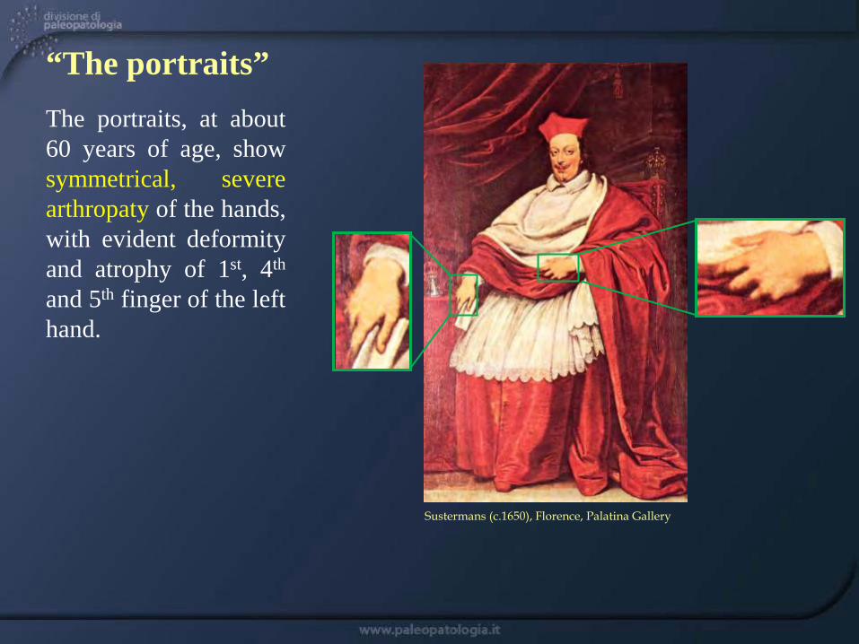
The portraits, at about 60 years of age, show symmetrical, severe arthropaty of the hands, with evident deformity and atrophy of 1st, 4th and 5th finger of the left hand.
“The portraits”
Sustermans (c.1650), Florence, Palatina Gallery
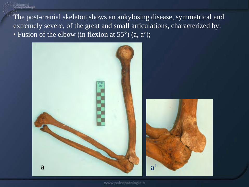
The post-cranial skeleton shows an ankylosing disease, symmetrical and extremely severe, of the great and small articulations, characterized by: • Fusion of the elbow (in flexion at 55°) (a, a’);
a a’
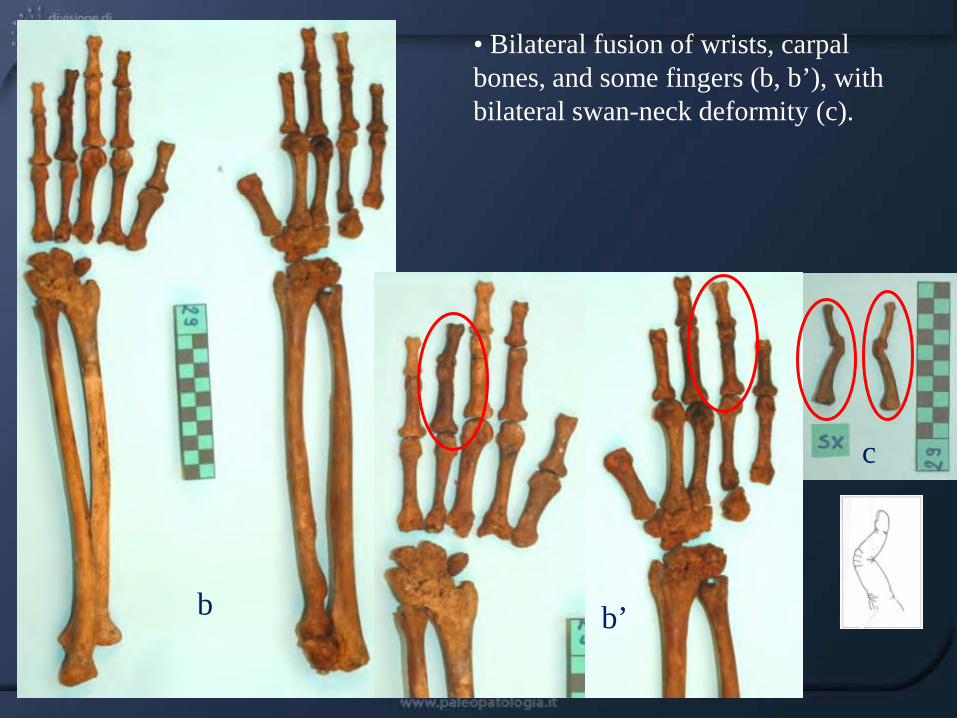
• Bilateral fusion of wrists, carpal bones, and some fingers (b, b’), with bilateral swan-neck deformity (c).
b b’
c

•Fusion of the right sacroiliac joint (d) (red arrow)
•Fusion of knees and rotulae (in flexion at 90°) (e)
d
e X-ray Prof. N. Villari (University of Florence)
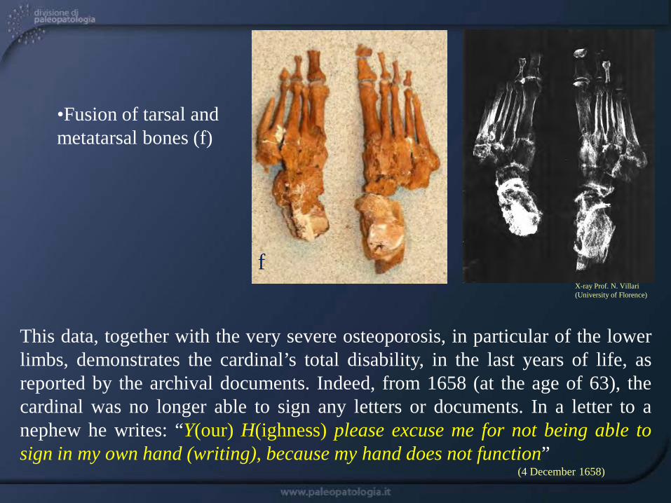
This data, together with the very severe osteoporosis, in particular of the lower limbs, demonstrates the cardinal’s total disability, in the last years of life, as reported by the archival documents. Indeed, from 1658 (at the age of 63), the cardinal was no longer able to sign any letters or documents. In a letter to a nephew he writes: “Y(our) H(ighness) please excuse me for not being able to sign in my own hand (writing), because my hand does not function” (4 December 1658)
•Fusion of tarsal and metatarsal bones (f)
f X-ray Prof. N. Villari (University of Florence)

Results of the PCR-SSP genotyping test for HLA-DRB locus of Cardinal Carlo: The band positions of PCR products at lines 8 and 23 correspond to DRB1*04 DRB4 alleles (Line 1 represents the negative control). The remaining positive bands define the second alleles of Cardinal Carlo, exactly DRB1*11 (serotype DR11, lines 13 and 16) always co-expressed in conjunction with DRB3 (serotype DR52, line 22).
Dr. G. Fontecchio, Regional Centre for Immunohematology and Tissue Typing, San Salvatore Hospital, L’Aquila
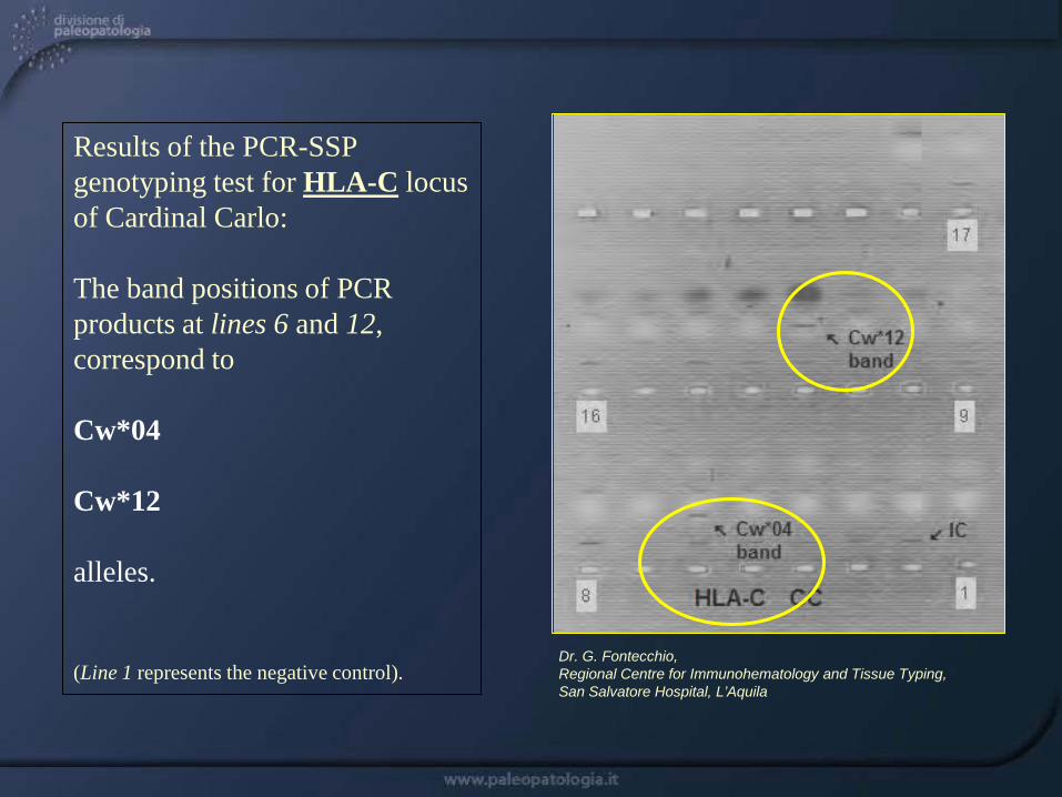
Results of the PCR-SSP genotyping test for HLA-C locus of Cardinal Carlo: The band positions of PCR products at lines 6 and 12, correspond to Cw*04 Cw*12 alleles. (Line 1 represents the negative control).
Dr. G. Fontecchio, Regional Centre for Immunohematology and Tissue Typing, San Salvatore Hospital, L’Aquila
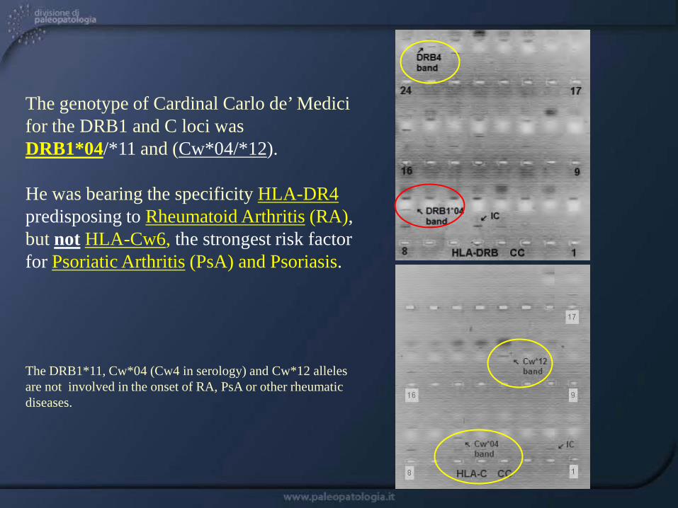
The genotype of Cardinal Carlo de’ Medici for the DRB1 and C loci was DRB1*04/*11 and (Cw*04/*12). He was bearing the specificity HLA-DR4 predisposing to Rheumatoid Arthritis (RA), but not HLA-Cw6, the strongest risk factor for Psoriatic Arthritis (PsA) and Psoriasis. The DRB1*11, Cw*04 (Cw4 in serology) and Cw*12 alleles are not involved in the onset of RA, PsA or other rheumatic diseases.
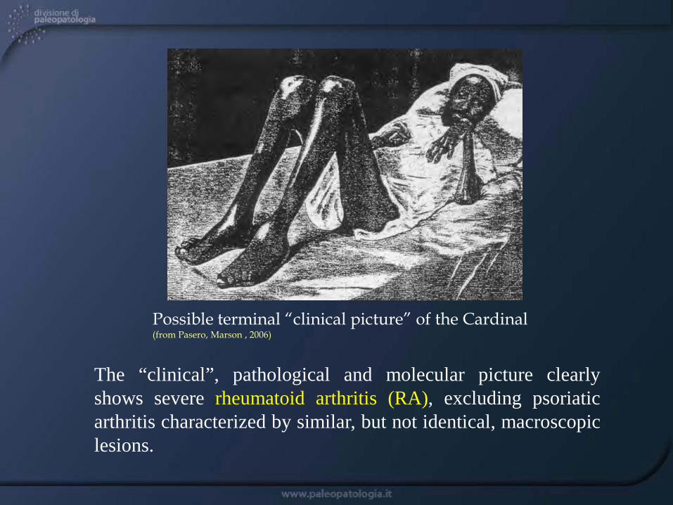
The “clinical”, pathological and molecular picture clearly shows severe rheumatoid arthritis (RA), excluding psoriatic arthritis characterized by similar, but not identical, macroscopic lesions.
Possible terminal “clinical picture” of the Cardinal (from Pasero, Marson , 2006)

Institutions involved
University of Pisa Superintendence of Florentine Museums University of Florence Opificio delle Pietre Dure Opera Mediceo-Laurenziana University of Long Island University of Minnesota MGM Biotechnology, Pisa