Embed Size (px)
Citation preview
HistoricalPerspectives
M.R. Zimmerman, MD, PhD
The Paleopathology of
the Cardiovascular System
Paleopathology, the study of disease in ancient remains, adds the dimension of time toour study of health and disease. The oldest preserved heart is from a mummified rabbitof the Pleistocene epoch, over 20, 000 years old. Cardiovascular disease has been iden-tified in human mummies from Alaska and Egypt, covering a time span ranging fromapproximately 3,000 to 300 years ago. An experimental study suggests that the poten-tial exists for identifying a wide range of cardiovascular pathologic conditions in mum-mified remains. The antiquity and ubiquity of arteriosclerotic heart disease is consideredin terms of pathogenesis. (Texas Heart Institute Journal 1993;20:252-7)
P aleopathology, the study of the evidence of disease in ancient human andanimal remains, adds the dimension of time to our study of health anddisease. Pathogenic organisms and mechanisms-and the patterns of dis-
ease they cause-evolve just as do larger organisms, including the hosts and vec-tors of disease. Paleopathologic studies have given us insight into the antiquityand evolution of congenital, traumatic, nutritional, degenerative, and infectiousdisease,' while suggesting that cancer may be a relatively recent disease.2 Mum-mies, defined as bodies preserved either naturally or artificially, hold a great poten-tial for paleopathologic examination. Postmortem examinations can be performedon mummies, and the diagnoses of many disorders can be made with relative ac-curacy and confidence.
Rehydration of desiccated tissues is accomplished by overnight immersion ofsmall samples in a solution of water, alcohol, and sodium carbonate, a techniquedeveloped in Egypt in 1921 by the father of modern paleopathology, Sir MarcArmand Ruffer.4 The rehydrated tissue is processed for microscopic examinationin the same fashion as fresh tissue. Nuclear detail is usually minimal or lacking,but, in general, skin and connective tissues are reasonably well preserved. Thestate of preservation of internal organs over long periods of time is somewhat vari-able, depending on mummification rites, entombment, and environmental condi-tions.' A number of mummies have shown preservation of the heart and majorblood vessels, which has enabled paleopathologists to gain insight into the antiqu-ity of disorders affecting the cardiovascular system.
Key words: Cardiovas-cular diseases; mummies;paleopathology
From: The Department ofPathology and LaboratoryMedicine, HahnemannUniversity; and the Depart-ment of Anthropology,University of Pennsylvania;Philadelphia, Pennsylvania
Address for reprints:Michael R. Zimmerman, MD,PhD, M.S. 113,Hahnemann University,Broad and Vine,Philadelphia, PA 19102
Alaskan MummiesThe oldest preserved bodies from Alaska are 20,000-year-old mammals of the latePleistocene epoch. Among several specimens that I examined in 1975, from theAmerican Museum of Natural History in New York City, was a rabbit in which theviscera were easily identifiable and grossly appeared to be well preserved, andwhich showed preservation of the general architecture of the liver but no trace ofhistologic structure in the other organs, including the heart.'The preservative effect of freezing and subsequent mummification lasts much
longer than previously suspected. However, the tissue destruction observed doessuggest that a significant period of time elapsed between the death of the animalsand their entombment in the permafrost, countering a popular notion that Arcticmammals were killed and preserved instantaneously by a catastrophic climaticchange.
Naturally frozen bodies of ancient human beings have also been found inAlaska. The oldest one, dating to about 400 AD), was that of a 53-year-old Eskimowoman found on St. Lawrence Island in the Bering Sea.-8 In October of 1972, afrozen body washed out of a low beach cliff on the island, which is about 130miles from mainland Alaska. The body was transported to Fairbanks, where it was
252 Paleopathology of the Cardiovascular System V'olume 20, vitinber 4. 19(.')'
thawed, and George Smith, of the National Park Ser-vice and University of Alaska, and I performed acomplete autopsy.The internal organs were somewhat desiccated, a
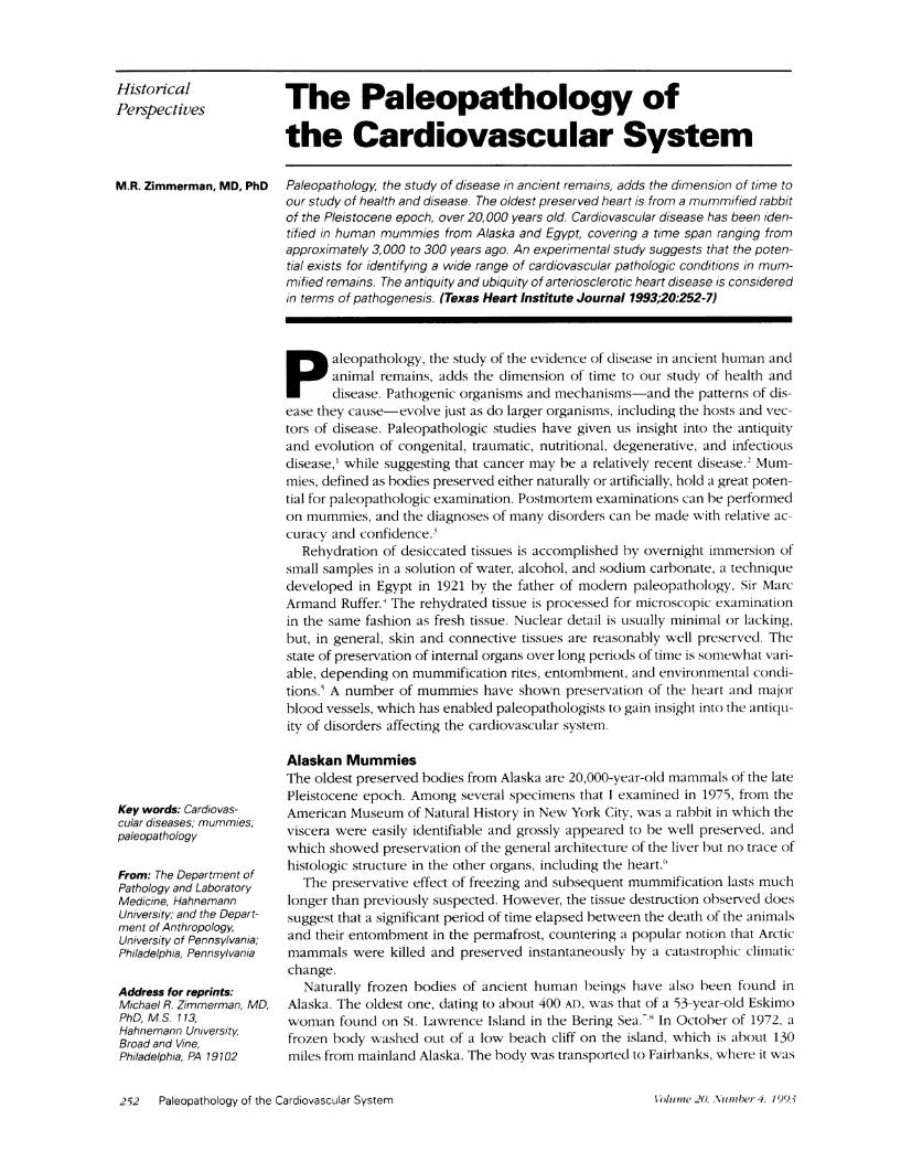
process that continues even in the frozen state. Therewas a moderate degree of aortic and coronary ath-erosclerosis, visible grossly as yellow streaking in thebrown vessels, and confirmed on microscopy (Fig.1). There was no evidence of myocardial infarction,acute or healed. The well-preserved valves andchambers were normal.
Death was determined to have been traumatic; inaddition to gross and microscopic evidence of skullfractures, the smaller bronchi of both lungs werepacked with moss that formed casts of the bronchi.The histologic finding of aspirated moss associatedwith hemorrhage suggested that accidental burialand suffocation played a significant role in thiswoman's death. Our conclusion was that she hadbeen buried alive in a landslide or earthquake, andasphyxiated. Aspiration of foreign material into thebronchi associated with hemorrhage is known tooccur in accidental inhumation, and has been dem-onstrated in persons who fall into or are buried incoal heaps. Nor is it unusual for red blood cells tobe preserved for extended periods; preserved eryth-rocytes have been reported in the tissues of Peruvianand North American Indian mummies, and in 6,000-year-old Egyptian mummies.'
In 1980, several other frozen Eskimo bodies wererecovered from a site in Barrow, the northernmostcommunity in Alaska."' 'An entire family had beentrapped while asleep, crushed and frozen in theirhouse on a hluff overlooking the Arctic Ocean.Spring storms can break up the ice and force it onto
Fig. 1 A coronary artery of a 53-year-old Eskimo woman whodied 1, 600 years ago, showing atherosclerosis. (H + E orig.x95)
(From: Zimmerman MR, Smith GS,' courtesy of the New YorkAcademy of Medicine.)
the shore with tremendous destructive force, a phe-nomenon known by the Eskimos as ivu.
Dr. Arthur Aufderheide of the University of Min-nesota-Duluth and I were confronted with a mass ofsleeping robes, bodies, and bones. Radiographs andcomputed-tomographic scans allowed us to sort out5 bodies. These radiologic studies also demonstratedthe difficulties of interpretation in ancient bodies;frozen pleural fluid was at first interpreted as aeratedlung tissue. Three of the bodies were sub-adults re-duced to skeletons. The other 2 bodies were intactand extraordinarily well preserved. On the basis ofwhere they were found in the house, the intact bod-ies were named the Northern Body (NB) and theSouthern Body (SB). A radiocarbon date on the NBwas 1520 AD ± 70, well before white contact in thearea (and in most of the New World).The NB, a 25- to 30-year-old female, was found
on the sleeping platform in the house, wrapped inher sleeping robes. The entire right hemithorax wascrushed, indicated in part by multiple rib fractures.Although the computed-tomographic scan seemedto indicate that the lungs were inflated, autopsy re-vealed collapsed lungs and bilateral frozen pleuraleffusions containing many bubbles. Analysis of thefluid revealed traces of hemoglobin, confirming ahemopneumothorax.The heart showed a slight dilatation of the right
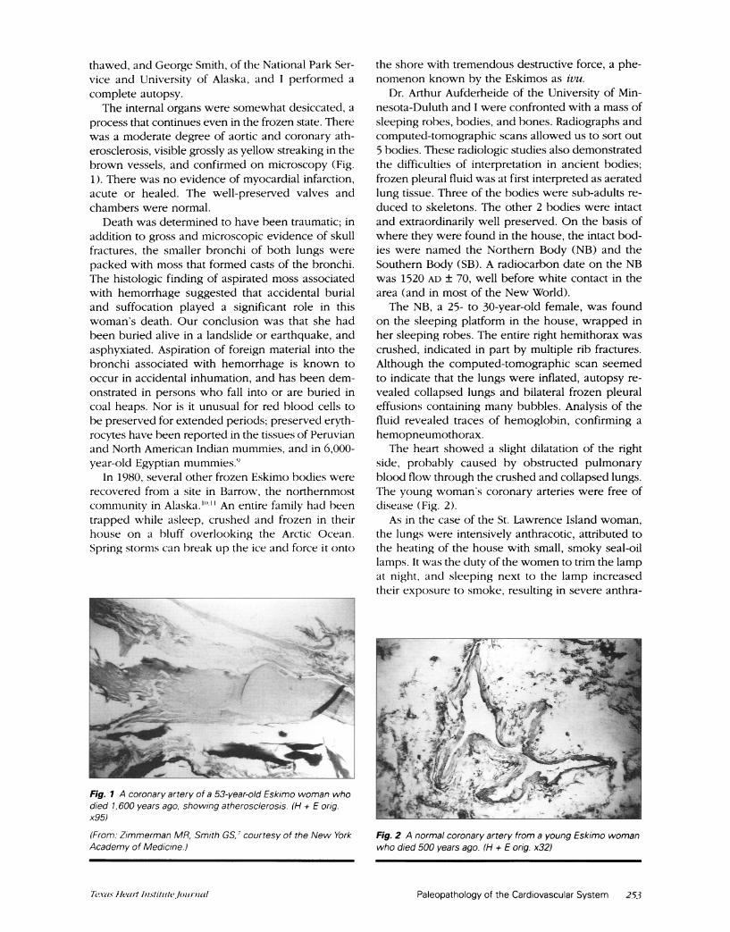
side, probably caused by obstructed pulmonaryblood flow through the crushed and collapsed lungs.The young woman's coronary arteries were free ofdisease (Fig. 2).
As in the case of the St. Lawrence Island woman,the lungs were intensively anthracotic, attributed tothe heating of the house with small, smoky seal-oillamps. It was the duty of the women to trim the lampat night, and sleeping next to the lamp increasedtheir exposure to smoke, resulting in severe anthra-
Fig. 2 A normal coronary artery from a young Eskimo womanwho died 500 years ago. (H + E orig. x32)
Paleopathology of the Cardiovascular System 253Texas Head Instititte.lournal
cosis at an early age. Although anthracotic pigmentis relatively innocuous, the introduction of cigarettesmoking to Alaska during World War II has created asynergistic effect, and lung cancer is a major healthproblem for modern Eskimo women.The bladder was markedly dilated and the stom-
ach empty. Similar findings in the other adult, the SB,led to the conclusion that the catastrophe occurredearly in the morning, trapping the sleeping family.The other woman, did, however, try to escape.
Her body was found in the doorway, with her bootsin one hand and a roof beam across her chest. Therewere multiple fractures of both right and left ribs andboth lungs were collapsed.The SB was 42 to 45 years of age, so she had more
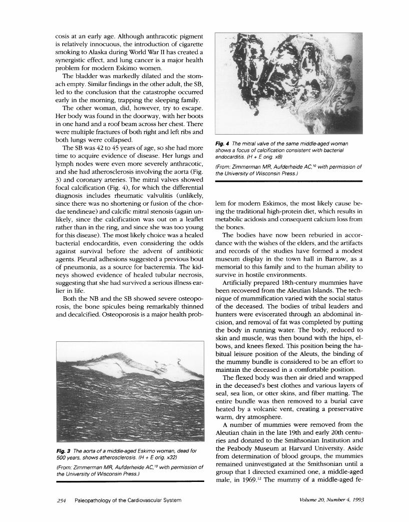
time to acquire evidence of disease. Her lungs andlymph nodes were even more severely anthracotic,and she had atherosclerosis involving the aorta (Fig.3) and coronary arteries. The mitral valves showedfocal calcification (Fig. 4), for which the differentialdiagnosis includes rheumatic valvulitis (unlikely,since there was no shortening or fusion of the chor-dae tendineae) and calcific mitral stenosis (again un-likely, since the calcification was out on a leafletrather than in the ring, and since she was too youngfor this disease). The most likely choice was a healedbacterial endocarditis, even considering the oddsagainst survival before the advent of antibioticagents. Pleural adhesions suggested a previous boutof pneumonia, as a source for bacteremia. The kid-neys showed evidence of healed tubular necrosis,suggesting that she had survived a serious illness ear-lier in life.
Both the NB and the SB showed severe osteopo-rosis, the bone spicules being remarkably thinnedand decalcified. Osteoporosis is a major health prob-
Fig. 3 The aorta of a middle-aged Eskimo woman, dead for500 years, shows atherosclerosis. (H + E orig. x32)
(From: Zimmerman MR, Aufderheide AC, 10 with permission ofthe University of Wisconsin Press.)
Fig. 4 The mitral valve of the same middle-aged womanshows a focus of calcification consistent with bacterialendocarditis. (H + E orig. x8)
(From: Zimmerman MR, Aufderheide AC, 10 with permission ofthe University of Wisconsin Press.)
lem for modern Eskimos, the most likely cause be-ing the traditional high-protein diet, which results inmetabolic acidosis and consequent calcium loss fromthe bones.
The bodies have now been reburied in accor-dance with the wishes of the elders, and the artifactsand records of the studies have formed a modestmuseum display in the town hall in Barrow, as amemorial to this family and to the human ability tosurvive in hostile environments.
Artificially prepared 18th-century mummies havebeen recovered from the Aleutian Islands. The tech-nique of mummification varied with the social statusof the deceased. The bodies of tribal leaders andhunters were eviscerated through an abdominal in-cision, and removal of fat was completed by puttingthe body in running water. The body, reduced toskin and muscle, was then bound with the hips, el-bows, and knees flexed. This position being the ha-bitual leisure position of the Aleuts, the binding ofthe mummy bundle is considered to be an effort tomaintain the deceased in a comfortable position.The flexed body was then air dried and wrapped
in the deceased's best clothes and various layers ofseal, sea lion, or otter skins, and fiber matting. Theentire bundle was then removed to a burial caveheated by a volcanic vent, creating a preservativewarm, dry atmosphere.A number of mummies were removed from the
Aleutian chain in the late 19th and early 20th centu-ries and donated to the Smithsonian Institution andthe Peabody Museum at Harvard University. Asidefrom determination of blood groups, the mummiesremained uninvestigated at the Smithsonian until agroup that I directed examined one, a middle-agedmale, in 1969.12 The mummy of a middle-aged fe-
254 Paleopathology of the Cardiovascular System Volume 20, Number 4, 199-3
male from the Peabody'" has also been examined.Both appear to have been commoners, as neitherhad been eviscerated.
Both mummies were examined initially by roent-gen ray, and then unwrapped. The anterior thoracicand abdominal walls were removed and the internalorgans identified and removed.The various tissues were sampled and rehydrated
for histologic examination. In the Smithsonian mum-my, the general fascicular architecture of the heartwas preserved, and seen scattered throughout thetissue were small aggregates of crystalline materialranging from 50 to 200 microns in diameter. Thesecontained numerous gram-negative bacilli and wereconsidered to be abscesses.
The pulmonary architecture was generally wellpreserved, showing a moderate amount of anthra-cosis and some fibrosis, coalescence of alveoli, andan increase in elastic tissue. A number of abscesses,again containing gram-negative bacilli, were seen,particularly in the pleural areas, as well as in thekidneys. A section of the right lower lobe showedcomplete loss of the normal architecture, the tissueconsisting of amorphous material containing freegram-negative bacilli and scattered small abscesses.Examination of the aorta and other arteries showedatherosclerotic plaques.
Death was concluded to be due to a right-lower-lobe pneumonia, probably caused by Klebsiellapneumoniae, with bacteremic spread to the heart,other lobes of the lungs, and kidneys. Other findingsincluded anthracosis, again due to open cooking andheating fires in the home.The Aleut mummy from Harvard University's
Peabody Museum was a middle-aged woman, whoalso showed evidence of atherosclerosis, as well asprevious pneumonia and anthracosis. The kidneys,like those of the SB from Barrow, showed evidenceof healed acute tubular necrosis, probably related toan episode of shock in the course of her pneumo-nia. The cause of death could not be determined.
Egyptian MummiesThe heart is usually present in Egyptian mummies,because part of the funerary mythology was theweighing of the heart against the feather of truth bythe god Maat. Failure of the heart, literally, to "mea-sure up" (a virtuous heart would prove lighter thanthe feather) condemned the deceased to an unhappyafterlife in a part of the underworld inhabited bysnakes, 3-headed monsters, and other evil crea-tures. '4
Previous archeologic and paleopathologic studieshave documented the antiquity of cardiovasculardisease in Egypt. Sudden deaths depicted in reliefsor described in inscriptions in Egyptian tombs havebeen diagnosed as evidence of coronary artery dis-
ease and myocardial infarction'5 and cerebrovascu-lar accident.'6 This historical evidence is well sub-stantiated by the finding of atherosclerosis in manyEgyptian mummies.'_ Severe aortic atherosclerosiswas seen in the Pharaoh Merneptah,' 8'9 and Ruffer4found involvement of all arteries, large and small, tobe very common among the hundreds of mummieshe examined. Long20 demonstrated coronary arterydisease, myocardial fibrosis, and arteriolar neph-rosclerosis in a 3,000-year-old Egyptian mummy.Shaw2' noted involvement of the superior mesentericartery in the mummy of Har-mose, a singer of the18th Dynasty. A recent autopsy of a mummy fromthe collection of the University of Pennsylvania Mu-seum revealed severe atherosclerosis of the aortaand diffuse arteriolar sclerosis on microscopic ex-amination,3 but my examination of the remains of 50mummies in a tomb in Upper Egypt revealed athero-sclerosis in only one.22
Radiologic studies have detected calcification ofvessels in Amenhotep and Ramses II,23 but a radio-logic survey of Egyptian mummies in European andBritish museums detected such changes in only 4 of88 adult mummies.2- This low figure probably re-flects the inadequacy of radiologic evaluation for thisdisorder.The diagnosis of the sequelae of atherosclerosis,
e.g., thrombosis, embolization, and myocardial orpulmonary infarction, has not been made in anymummy. This is surprising in view of the historic andanatomic evidence of atherosclerosis but may be theresult of a problem in preservation, as discussedbelow.
An ExperimentalStudy of MummificationThe difficulties of diagnosis of pathologic conditionsin modern tissue specimens are immensely magni-fied in studying ancient remains. An approach to thismatter was undertaken by an experimental study, inwhich small tissue specimens from cadavers under-going postmortem examination were desiccated,rehydrated, and examined histologically. 25 Thespecimens were dried in an oven, rehydrated, andprocessed. Fresh tissues were used as controls. Avariety of tissues (including cardiac tissues) and ofdisease processes were studied by this technique.
In normal myocardium, the outlines of the fiberswere preserved, although cross striations were notwell seen. Examination of a severely atheroscleroticand thrombosed coronary artery showed excellentpreservation of the atherosclerosis and calcificationafter mummification and rehydration. The thrombuswas reduced to eosinophilic material indistinguish-able with hematoxylin and eosin from a mummifiedpostmortem blood clot. Phosphotungstic acid hema-toxylin diffusely stained the material within the lu-
Paleopathology of the Cardiovascular System 255Texas Hean Institutejournal
men: the red blood cells and fine detail of the fibrinwere no longer seen, but the mesh-like fibrin pat-tern remained.An acute myocardial infarction was essentially
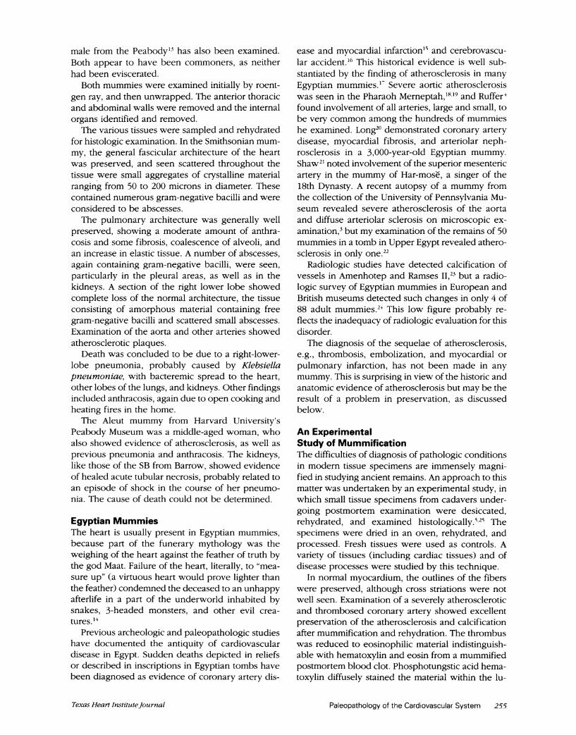
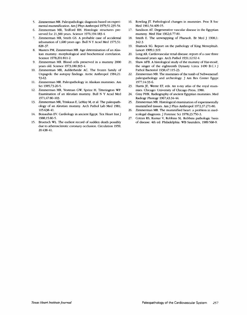
undiagnosable, the mummified necrotic tissue beingdistinguished from the adjacent autolyzed myocar-dium only by a few fragmented neutrophils (Fig. 5).The necrosis of muscle in an acute infarct is in es-sence a process of in situ autolysis, and diagnosis ofan acute infarct in a mummified heart would have tobe based on the finding of the remains of a neutro-philic infiltrate-a difficult matter at best in the ex-perimental setting, and probably impossible in anaturally or artificially preserved mummy.The fibrosis of a inummified healing myocardial
infarct was seen as a lighter-staining eosinophilicarea with a decreased number of nuclei. Trichromestaining showed the fibrosis and peripheral organi-zation well. Chronic passive congestion of the lungs,liver, and spleen was well displayed, particularly thehemosiderin pigment present, which retained amarked avidity for the iion stain.
This study indicated that the diagnosis of an acutemyocardial infarction is probably not possible in amummified body. Strong presumptive evidence, in-cluding coronary artery disease or thrombosis, myo-cardial scarring, and chronic visceral congestion,should be preserved and allow for the assignmentof atherosclerotic heart disease as the cause of deathin mummified bodies, in either the archeological orforensic setting.")
Conclusions
Natural or artificial mummies have proved to be ex-cellent subjects for paleopathologic examination.Conditions in the Arctic would seem to provide forexcellent preservation of soft tissuLes, bUt bodies are
o
W -A.
Fig. 5 An acute myocardial infarct, experimentally mummi-fied, is essentially indistinguishable from autolyzed myocar-dium. (H + E orig. x80)
in fact preserved there only under extraordinary cir-cumstances. The frozen ground makes winter buri-als impossible, and the permafrost layer, being onlya few centimeters below the surface, discouragesdeep burials even in summer. Cycles of freezing andthawing tend to bring summer burials to the surface,exposing remains to the ravages of climate and ani-mals.
Studies of Arctic mummies do point out a majorfocus of paleopathology, the reconstruLction of an-cient disease patterns. Rare finds such as those de-scribed above give us a glimpse into the prehistoricArctic and show health hazards shared by past andpresent inhabitants of a once-remote area. Some ofthe cardiovascular diseases discovered in thesemummies, such as bacterial endocarditis and myo-cardial abscesses, have well-known natural historiesand are relatively easily explained in the context ofthe Arctic ecosystem.We have also seen that ancient Eskimos, far re-
moved from the stresses of modern technologicalsociety, suffered from coronary artery disease, a pro-cess that has also been well documented as far backas dynastic Egypt, by both historical and anatomicevidence. This anatomic evidence in Alaska not onlyconfirms the antiquity of arteriosclerotic heart dis-ease but also its occurrence in a preliterate societythat lacks the historical evidence seen in Egypt.
Various factors have been implicated in the patho-genesis of atherosclerosis, incluiding diets high incholesterol and saturated fat, hypercholesterolemia,hypertension, diabetes, salt intake, lack of physicalactivity, and cigarette smoking.' Cigarette smokinguIsuLally does not apply in the consideration of an-cient populations, and most of these other factors arenot amenable to documentation in paleopathologicspecimens, although hypertension is suLggested bythe occasional finding of arteriolar nephrosclerosis.2'Heavy physical activity was most often a major fea-tuLre of life in antiquity. The Eskimo diet was almostentirely meat, as attested to by their severe osteo-porosis, while the Egyptian diet contained meat onlyat occasional festival times; yet the finding of athero-sclerosis in both groups suLggests that diet alone maynot he a critical factor. If stress is a factor in this dis-ease, perhaps the lesson is that stress is a compo-nent of life sharecd by all huLman societies.
References1. Ziijiinieriman MR. Kelley MIA. Atlas of hlUman paleopathologv.
New- York: 13raieger. 1982.2. Zimmllerman MR. An experimental study of mummLiillificaition
pertinent to the antiqlit- of cancer. Caincer 1 9X740: 1358-62.3. Cockhurn A. CockhUrn E. eds. MuLmlllmiieS, disease aindl an-
cient CLltuLres. Canmbridge: University P'ress. 1980.-4. RuLffer MA. StuLdies in the paleopathologv of Egypt. Chicago:
University of Chicago IPress. 1921.
256 Paleopathology of the Cardiovascular System lblume2O.Numbei-4, 19913
5. Zimmerman MR. Paleopathologic diagnosis based on experi-mental mummification. AmJ Phys Anthropol 1979;51:235-54.
6. Zimmerman MR, Tedford RH. Histologic structures pre-served for 21,300 years. Science 1976;194:183-4.
7. Zimmerman MR, Smith GS. A probable case of accidentalinhumation of 1,600 years ago. Bull N Y Acad Med 1975;51:828-37.
8. Masters PM, Zimmerman MR. Age determination of an Alas-kan mummy: morphological and biochemical correlation.Science 1978;201:811-2.
9. Zimmerman MR. Blood cells preserved in a mummy 2000years old. Science 1973;180:303-4.
10. Zimmerman MR, Aufderheide AC. The frozen family ofUtqiagvik: the autopsy findings. Arctic Anthropol 1984;21:53-63.
11. Zimmerman MR. Paleopathology in Alaskan mummies. AmSci 1985;73:20-5.
12. Zimmerman MR, Yeatman GW, Sprinz H, Titterington WP.Examination of an Aleutian mummy. Bull N Y Acad Med1971;47:80-103.
13. Zimmerman MR, Trinkaus E, LeMay M, et al. The paleopath-ology of an Aleutian mummy. Arch Pathol Lab Med 1981;105:638-41.
14. Boisaubin EV. Cardiology in ancient Egypt. Tex Heart Inst J1988;15:80-5.
15. Bruetsch WL. The earliest record of sudden death possiblydue to atherosclerotic coronary occlusion. Circulation 1959;20:438-41.
16. Rowling JT. Pathological changes in mummies. Proc R SocMed 1961;54:409-15.
17. Sandison AT. Degenerative vascular disease in the Egyptianmummy. Med Hist 1962;6:77-81.
18. Smith E. The unwrapping of Pharaoh. Br Med J 1908;1:342-3.
19. Shattock SG. Report on the pathology of King Menephtah.Lancet 1909;1:319.
20. Long AR. Cardiovascular renal disease: report of a case threethousand years ago. Arch Pathol 1931;12:92-4.
21. Shaw AFB. A histological study of the mummy of Har-mose,the singer of the eighteenth Dynasty (circa 1490 B.C.) JPathol Bacteriol 1938;47:115-23.
22. Zimmerman MR. The mummies of the tomb of Nebwenenef:paleopathology and archeology. J Am Res Center Egypt1977; 14:33-6.
23. Harris JE, Wente EF, eds. An x-ray atlas of the royal mum-mies. Chicago: University of Chicago Press, 1980.
24. Gray PHK. Radiography of ancient Egyptian mummies. MedRadiogr Photogr 1967;43:34-44.
25. Zimmerman MR. Histological examination of experimentallymummified tissues. Am J Phys Anthropol 1972;37:271-80.
26. Zimmerman MR. The mummified heart: a problem in med-icolegal diagnosis. J Forensic Sci 1978;23:750-3.
27. Cotran RS, Kumar V, Robbins SL. Robbins pathologic basisof disease. 4th ed. Philadelphia: WB Saunders, 1989:568-9.
Paleopathology of the Cardiovascular System 257Texas Heart Institutejournal