Embed Size (px)
Citation preview
Osteochondrosis of the elbow: a review of the pathogenesis and a new approach to treatment
MJ THOMSON* and GM ROBINS West Chermside Veterinary Clinic, 263 Appleby Road, Stafford Heights, Queensland 4053
SUMMARY: To test the hypothesis that joint incongruity contributes to the pathogenesis of elbow osteochondrosis, the left and right radius and ulna of 20 young large breed dogs were measured to determine any variation in length and to observe any incongruity of the elbow joint. Both lame and normal dogs were included in the study. Nine of the 20 dogs had marked disparity in radial and ulnar lengths yet only one had obvious elbow joint incongruity. The use of a sliding osteotomy for the treatment of fragmented coronoid process and a lengthening osteotomy for the treatment of an ununited anconeal process is also discussed. All four dogs treated with a sliding osteotomy showed a marked clinical improvement, and two of the three dogs treated with a lengthening osteotomy showed radiographic fusion of the anconeal process. Aust Vet J 72: 375 - 378
Introduction Osteochondrosis (OC) of the elbow is a common cause of lame-
ness of the fore limb in the dog. Three well-recognised forms of elbow OC are fragmented coronoid process (FCP), ununited anconeal process (UAP) and osteochondritis dissecans (OCD) of the medial ridge of the humeral condyle.
The pathogenesis of OC is considered multifactorial, with a com- bination of general and local factors being involved (Olsson 1993). OC occurs as a result of abnormal endochondral ossification.
The normal differentiation of chondrocytes is delayed, resulting in abnormal thickening and retention of cartilage (Boudrieau et a1 1983). The general consensus of opinion is that elbow OC has an hereditary component (Grondalen and Lingaas 1991). Audell(l992) found that heritability estimates in Rottweilers were highly signifi- cant and stable in the range of 0.3 to 0.4. Elbow OC is seen most commonly in Rottweilers, German Shepherd Dogs (GSD), Labradors and Retrievers, with any large to medium sized breed at risk (Olsson 1993; Fox and Walker 1993). It is reportedly twice as common in male dogs as in females (Read et a1 1990; Grondalen and Lingaas 1991). Nutrition also plays a significant role. Hedham- mar et af (1974) and Hazewinkel (1989) found that high calcium intake directly or in combination with other nutrients plays an important role in disturbances of endochondral ossification. Local factors involve mechanical stresses and joint incongruity.
Mechanical stresses in the elbow joint occur from tension by the annular ligament, pressure on the medial coronoid process by rotation between the radius and ulna, and forces transmitted from the humeral condyle during weight bearing (Fox and Walker 1993). Olsson (1993) stated that slight joint incongruity caused by dispro- portional growth of the radius and ulna is probably the most impor- tant reason for the occurrence of OC lesions in the elbow joint. Wind (1986) suggested that joint incongruity was a common denominator in all forms of elbow OC and that this incongruity was due to an abnormality of the trochlea notch of the ulna, with the arc of the notch being too small to encompass the humeral trochlea. This abnormality would create major contact points in the areas of the anconeal and coronoid processes. FCP has frequently been reported in dogs with premature closure of the radial physis and resultant humero-radial subluxation (Barr and Denny 1985; Robins 1987; Macpherson et a1
* Present address: School of Veterinaly Studies, Murdoch University, Murdoch. Western Australia 6150
D
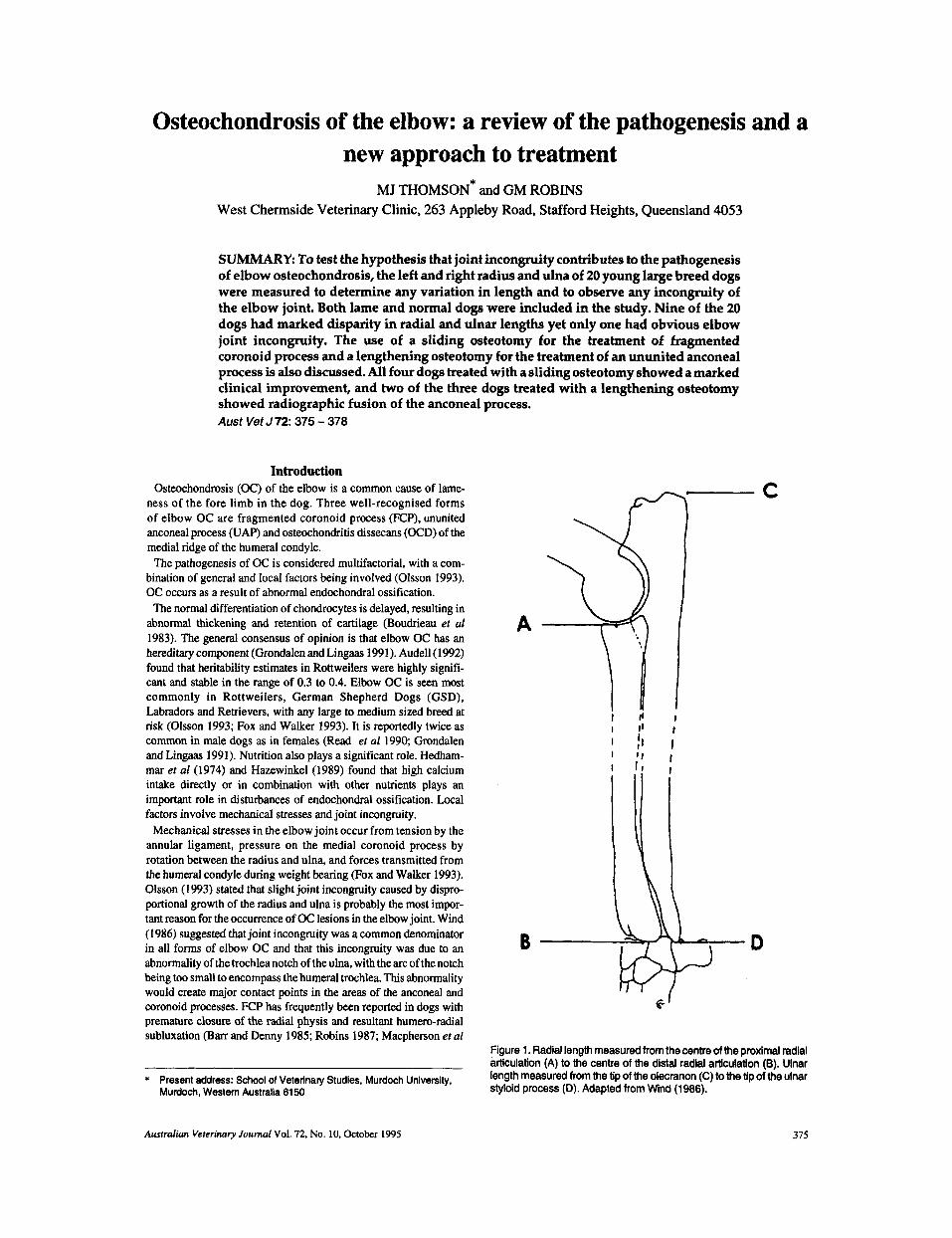
Figure 1. Radial length measured from the centre of the proximal radial articulation (A) to the centre of the distal radial articulation (9). Ulnar length measured from the tip of the olecranon (C) to the tip of the ulnar slyloid process (D). Adapted from Wind (1986).
Australian Veterinary Journal Vol. 72, No. 10, October 1995 375

TABLE 1 Comparative lengths of the radius and ulna in 20 dogs
No. sex Breed Age Radius Ulna Lame (mo) fore limb
Len RigM Len Right (mm) (mm) (mm) (mm)
1 2 3 4 5 6 7 8 9
10 1 1 12 13 14 15 16 17 18 19 20
M F M F M F F M F F M F M M F M F M M M
GSD' 7.5 GSD 10 GSD 10 GSD 14 Rottweiler 6 Rottweiler 6 Rottweiler 7 Rottweiler 7 Rottweiler 9 Rottweiler 10 Rottweiler 10 Rottweiler 12 Rottweiler 18 Rottweiler 18 Rottweiler 18 Rottweiler 19 Rottweiler 24 Dobermann 12 BemeseMtnDog 24 St Bernard 12
194 189 196 184 176 1 74 I 76 178 177 174 180 186 1 92 1 95 190 195 175 210 189 230
191 192 196 187 176 172 176 175 179 172 181 188 188 194 188 196 176 206 190 23 1
245 232 243 226 217 215 221 222 209 219 222 231 242 240 234 240 217 259 239 286
237 236 243 233 219 215 220 220 214 217 227 231 235 239 233 242 221 254 241 285
left right both nil both left both both left nil nil right both both both left nil nil both right
* German Shepherd Dog
1992). Robins (1987) stated that the collateral ligaments from the displaced radial head to the humerus draws the humerus distally, further increasing the load on the coronoid process. Evidence of FCP in association with acquired elbow incongruity suggests that abnor- mal mechanical loading of the coronoid process is involved in the pathogenesis of FCP (Macpherson er a1 1992).
The aim of this study was to measure any variation in the left and right radius and ulnar lengths in young large breed dogs, and observe any resultant elbow joint incongruity. Both lame and clinically normal dogs were included in this study to determine whether or not disparity in radial and ulnar lengths occurred as a variation of the normal growth pattern in large growing dogs. We also discuss the role of surgical correction of joint incongruity in the treatment of elbow OC.
Materials and Methods Measurements were taken from full length radiographs of the
radius and ulna in both fore limbs of 20 young large breed dogs (Table 1). Five dogs were clinically sound, 8 dogs were lame in both fore limbs, 3 were lame in the right leg and 4 were lame in the left leg. The ages ranged from 6 to 24 months, and the breeds were 13 Rottweilers (65%), 4 GSD (20%), 1 Dobermann (5%). 1 St Bernard (5%) and 1 Bernese Mountain Dog (5%). Ulnar measurements were taken from the proximal olecranon to the distal styloid process, and radial measurements were taken from the proximal to distal articular surfaces (Figure 1 ).
In 7 cases of elbow OC an ulnar osteotomy was performed to alter the congruity of the elbow joint. In 4 cases of FCP a proximal ulnar sliding osteotomy was performed (includes dogs 1.2 and 12 but the 4th dog's measurements were unavailable).
In 3 of these dogs standard surgical removal of the fragmented coronoid process via a medial arthrotomy had been performed pre- viously with no improvement. The sliding osteotomy was performed
in the proximal metaphysis of the ulna. The ulna was exposed via a caudolateral approach, elevating the extensor carpi ulnaris muscle and the flexor carpi ulnaris muscle (Piermattei 1993). An oblique osteotomy was performed in the proximal ulna with an oscillating saw. The pull of the triceps brachii muscles plus the direction of the osteotomy allows sliding and tilting of the proximal ulnar fragment, decreasing the pressure on the coronoid process (Figure 2). The osteotomy site was allowed to heal by callus formation, without internal fixation. The external fascia of the flexor and extensor carpi ulnaris muscles were sutured with 3-0 polyglyconatet in a simple continuous pattern. The subcutaneous tissues were closed with 3-0 polyglyconatein a simple continuous pattern, and the skin was closed with 3-0 nylont in a simple interrupted pattern.
The remaining three cases of elbow OC were treated with a length- ening osteotomy for correction of UAP (radius and ulnar lengths unavailable). The approach to the osteotomy was performed as described above. An oblique osteotomy was performed in the proxi- mal ulna in the opposite direction to that described above. In this situation the action of the triceps brachii muscle allows lengthening of the proximal ulna and a reduction in the pressure exerted on the anconeal process by the humerus (Figure 3). An intramedullary pin was placed proximally in the ulna to provide limited stabilisation. The external fascia, subcutaneous tissues and the skin were closed as described above.
Results Measurements of both the left and right radius and ulna in 20
dogs, 5 of which were clinically normal, revealed marked dispar- ity in length in 9 (45%). Disparity was considered significant if there was a variation of greater than 2 mm between the left and right radius
t Maon@, Davis and Geck, Baulkham Hills, NSW $ Dermalon@, Davis and Geck, Baulkham Hills, NSW
376 Australian Veterinary Journal VoI. 72, No. 10, October 1995
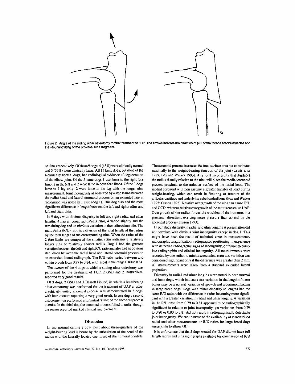
Figure 2. Angle of the sliding ulnar osteotomy for the treatment of FCP. The arrows indicate the direction of pull of the triceps brachii muscles and the resultant tilting of the proximal ulna fragment.
or ulna, respectively. Of these 9 dogs, 4 (45%) were clinically normal and 5 (55%) were clinically lame. All 15 lame dogs, but none of the 4 clinically normal dogs, had radiological evidence of degeneration of the elbow joint. Of the 5 lame dogs 1 was lame in the right fore limb, 2 in the left and 2 were lame in both fore limbs. Of the 3 dogs lame in 1 leg only, 2 were lame in the leg with the longer ulna measurement. Joint incongruity as observed by a step lesion between the radial head and lateral coronoid process on an extended lateral radiograph was noted in 1 case (dog 1). This dog also had the most significant difference in length between the left and right radius and left and right ulna.
In 9 dogs with obvious disparity in left and right radial and ulnar lengths, 4 had an equal radiudulna ratio, 4 varied slightly and the remaining dog had an obvious variation in the radiushlna ratio. The radiushlna (WV) ratio is a division of the total length of the radius by the total length of the corresponding ulna. When the ratios of the 2 fore limbs are compared the smaller ratio indicates a relatively longer ulna or relatively shorter radius. Dog 1 had the greatest variation between the left and right WV ratio and also had an obvious step lesion between the radial head and lateral coronoid process on an extended lateral radiograph. The WV ratio varied between and within breeds from 0.79 to 0.84, with most in the range 0.80 to 0.81.
The owners of the 4 dogs in which a sliding ulnar osteotomy was performed for the treatment of FCP, 2 GSD and 2 Rottweilers, reported very good results.
Of 3 dogs, 2 GSD and 1 Bassett Hound, in which a lengthening ulnar osteotomy was performed for the treatment of UAP a radio- graphically united anconeal process was demonstrated in 2 dogs, with both owners reporting a very good result. In one dog a second osteotomy was performed after initial failure of the anconeal process to unite. In the third dog the anconeal process failed to unite, though the owner reported marked clinical improvement.
The coronoid process increases the total surface area but contributes minimally to the weight-bearing function of the joint (Lewis et a1 1989; Fox and Walker 1993). Any joint incongruity that displaces the radius distally relative to the ulna will place the medial coronoid process proximal to the articular surface of the radial head. The medial coronoid will then assume a greater transfer of load during weight-bearing, which can result in fissuring or fracture of the articular cartilage and underlying subchondral bone (Fox and Walker 1993; Olsson 1993). Relative overgrowth of the ulna can cause FCP and OCD, whereas relative overgrowth of the radius can cause UAP. Overgrowth of the radius forces the trochlea of the humerus in a proximal direction, exerting more pressure than normal on the anconeal process (Olsson 1993).
In our study disparity in radial and ulnar lengths at presentation did not correlate with obvious joint incongruity except in dog 1. This might have been the result of technical error in measurements, radiographic magnification, radiographic positioning, inexperience with detecting radiographic signs of incongruity, or failure to corre- late radiographic and clinical incongruity. All measurements were recorded by one author to minimise technical error and variation was considered significant only if the difference was greater than 2 mm. All measurements were taken from a standard extended lateral projection.
Disparity in radial and ulnar lengths were noted in both normal and lame dogs, which indicates that variation in the length of these bones may be a normal variation of growth and a common finding in large breed dogs. Dogs with minor disparity in lengths had the same WV ratio, with the difference in ratios becoming more signifi- cant with a greater variation in radial and ulnar lengths. A variation in the WV ratio from 0.79 to 0.8 1 appeared to be radiographically significant in relation to joint incongruity, yet variations from 0.79 to 0.80 or 0.80 to 0.81 did not result in radiographically detectable joint incongruity. We are unaware of the availability of standardised radial and ulnar measurements or WV ratios for large breed dogs susceptible to elbow oc.
It is unfortunate that the 3 dogs treated for UAP did not have full length radius and ulna radiographs available for comparison of R/U
Discussion In the normal canine elbow joint about three-quarters of the
weight-bearing load is borne by the articulation of the head of the radius with the laterally located capitulum of the humeral condyle.
Australian Veterinary Journal Vol. 72, No. 10, October 1995 311
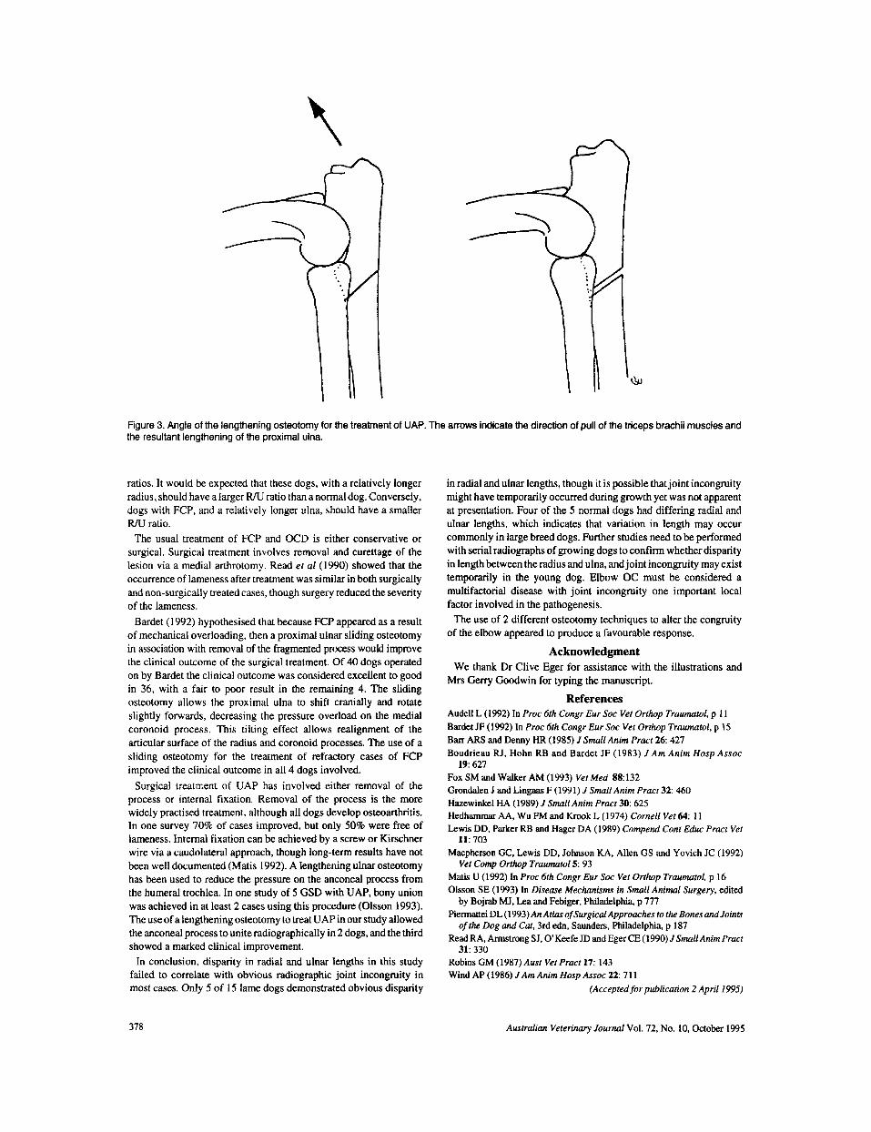
Figure 3. Angle of the lengthening osteotomy for the treatment of UAP. The arrows indicate the direction of pull of the triceps brachii muscles and the resultant lengthening of the proximal ulna.
ratios. It would be expected that these dogs, with a relatively longer radius, should have a larger R/U ratio than a normal dog. Conversely, dogs with FCP, and a relatively longer ulna, should have a smaller R/U ratio.
The usual treatment of FCP and OCD is either conservative or surgical. Surgical treatment involves removal and curettage of the lesion via a medial arthrotomy. Read et a1 (1990) showed that the occurrence of lameness after treatment was similar in both surgically and non-surgically treated cases, though surgery reduced the severity of the lameness.
Bardet (1992) hypothesised that because FCP appeared as a result of mechanical overloading, then a proximal ulnar sliding osteotomy in association with removal of the fragmented process would improve the clinical outcome of the surgical treatment. Of 40 dogs operated on by Bardet the clinical outcome was considered excellent to good in 36, with a fair to poor result in the remaining 4. The sliding osteotomy allows the proximal ulna to shift cranially and rotate slightly forwards, decreasing the pressure overload on the medial coronoid process. This tilting effect allows realignment of the articular surface of the radius and coronoid processes. The use of a sliding osteotomy for the treatment of refractory cases of FCP improved the clinical outcome in all 4 dogs involved.
Surgical treatment of UAP has involved either removal of the process or internal fixation. Removal of the process is the more widely practised treatment, although all dogs develop osteoarthritis. In one survey 70% of cases improved, but only 50% were free of lameness. Internal fixation can be achieved by a screw or Kirschner wire via a caudolateral approach, though long-term results have not been well documented (Matis 1992). A lengthening ulnar osteotomy has been used to reduce the pressure on the anconeal process from the humeral trochlea. In one study of 5 GSD with UAP, bony union was achieved in at least 2 cases using this procedure (Olsson 1993). The use of a lengthening osteotomy to treat UAP in our study allowed the anconeal process to unite radiographically in 2 dogs, and the third showed a marked clinical improvement.
In conclusion, disparity in radial and ulnar lengths in this study failed to correlate with obvious radiographic joint incongruity in most cases. Only 5 of 15 lame dogs demonstrated obvious disparity
in radial and ulnar lengths, though it is possible that joint incongruity might have temporarily occurred during growth yet was not apparent at presentation. Four of the 5 normal dogs had differing radial and ulnar lengths, which indicates that variation in length may occur commonly in large breed dogs. Further studies need to be performed with serial radiographs of growing dogs to confirm whether disparity in length between the radius and ulna, and joint incongruity may exist temporarily in the young dog. Elbow OC must be considered a multifactorial disease with joint incongruity one important local factor involved in the pathogenesis.
The use of 2 different osteotomy techniques to alter the congruity of the elbow appeared to produce a favourable response.
Acknowledgment We thank Dr Clive Eger for assistance with the illustrations and
References Mrs Gerry Goodwin for typing the manuscript.
Audell L (1992) In Proc 6th Congr Eur Soc Vet Orthop Traumarol, p I 1 Bardet JF (1992) In Proc 6th Congr Eur SOC Vet Orthop Traumaiol, p 15 Ban A R S and Denny HR (1985) J Small Anim Pract 2 6 427 Boudrieau RJ, Hohn RB and Bardet JF (1983) J Am Anim Hosp Assoc
Fox SM and Walker AM (1993) Ver Med 88132 Grondalen J and Lingaas F (1991) J Small Anim Pracr 32: 460 Hazewinkel HA (1989) J SmallAnim Pract 30: 625 Hedhammar AA, Wu FM and Krook L ( 1974) Cornell Vet 64: 1 1 Lewis DD, Parker RB and Hager DA (1989) Compend Conr Educ Pracr Ver
Macphexson GC, Lewis DD, Johnson KA, Allen GS and Yovich JC (1992)
Matis U (1992) In Proc 6th Congr Eur SOC Vet Orthop Traumatol, p 16 Olsson SE (1993) In Disease Mechanisms in Small Animal Surgery, edited
Piermattei DL (1 993) An Atlas of Surgical Approaches to the Bones and Joints
Read RA, Armstrong SJ, 0 Keefe JD and Eger CE (1990) JSmall Anim Pracr
Robins GM (1987) Ausr Ver Pract 17: 143 Wind AP ( 1986) J Am Anim Hosp Assoc 22: 7 1 1
1 9 627
11: 703
Vet Comp Orthop Traumarol 5: 93
by Bojrab MJ, Lea and Febiger, Philadelphia, p 777
ofthe Dog and Cat, 3rd edn, Saunders, Philadelphia, p 187
31: 330
(Accepted for publication 2 April 1995)
378 Australian Veterinary Journal Vol. 72, No. 10, October 1995