Embed Size (px)
Citation preview
Impaired postnatal development of hippocampal neurons
and axon projections in the Emx2–/– mutants
Nicolai E. Savaskan,*,� Gonzalo Alvarez-Bolado,�,1 Robert Glumm,* Robert Nitsch,*
Thomas Skutella§,2 and Bernd Heimrich*,2
*Department of Cell and Neurobiology, Institute of Anatomy, Berlin, Germany
�Department of Neurology, Humboldt University Medical School Charite, Berlin, Germany
�Department of Molecular Cell Biology, Max Planck Institute of Biophysical Chemistry, Gottingen, Germany
§Neuroscience Research Center, Humboldt University Medical School Charite, Berlin, Germany
Abstract
The specification and innervation of cerebral subregions is a
complex layer-specific process, primed by region-specific
transcription factor expression and axonal guidance cues. In
Emx2–/– mice, the hippocampus fails to form a normal den-
tate gyrus as well as the normal layering of principal neurons
in the hippocampus proper. Here, we analyzed the late
embryonic and postnatal development of the hippocampal
formation and its axonal projections in mice lacking Emx2
expression in vitro. As these mutants die perinatally, we used
slice cultures of Emx2 mutant hippocampus to circumvent this
problem. In late embryonic Emx2–/– cultivated hippocampi,
both the perforant path as well as the distribution of calretinin-
positive cells are affected. Traced entorhinal afferents in
co-cultures with hippocampus from embryonic Emx2–/– mice
terminate diffusely in the prospective dentate gyrus in contrast
to the layer-specific termination of co-cultures from wild-type
littermates. In addition, in brain slice cultures from null mutants
the presumptive dentate gyrus failed to develop its normal
cytoarchitecture and mature dentate granule cells, including
the lack of their mossy fiber projection. Our data indicate that
Emx2 is essential for the terminal differentiation of granular
cells and the correct formation of extrinsic and intrinsic hip-
pocampal connections.
Keywords: axon guidance, cortical maturation, dentate
gyrus, homeobox gene, perforant path, slice cultures.
J. Neurochem. (2002) 83, 1196–1207.
The hippocampus, with its intrinsic and extrinsic projections,
is a current model system to study axonal pathfinding and
layer-specificity. The Emx2 gene, which is a vertebrate
homeobox gene related to the Drosophila empty spiracles
(ems) gene (Dalton et al. 1989), is expressed in the anterior
CNS of the developing mouse embryo, including the
hippocampal primordium (Simeone et al. 1992; Gulisano
et al. 1996; Mallamaci et al. 1998). The role of Emx2 in
forebrain development can be inferred from studies carried
out in the cortex and olfactory bulb of Emx2 mutants. In the
isocortex, Emx2 is essential not only for correct area
determination, but also for proper thalamo-cortical connec-
tivity (Mallamaci et al. 2000a). Failure in axonal connectiv-
ity has also been reported in the olfactory bulb of Emx2
mutants (Yoshida et al. 1997). In both isocortex and
olfactory bulb, Emx2 deficiency is accompanied by histo-
logical disorganization, probably caused by improper neur-
onal migration due to impaired reelin signaling (Cecchi and
Boncinelli 2000; Mallamaci et al. 2000b).
Because Emx2 homozygous embryos die by the day of
birth, however, knowledge of the involvement of Emx2 in
hippocampal development (many of those key processes take
place after birth) had lagged behind. In Emx2–/– embryos,
the medial limbic cortex and the hippocampus show a
reduction in size and a missing dentate gyrus (Pellegrini
et al. 1996; Yoshida et al. 1997), although region-specific
Received April 6, 2002; revised manuscript received September 9, 2002;
accepted September 11, 2002.
Address correspondence and reprint requests to Dr Nicolai
E. Savaskan, Institute of Anatomy, Department Molecular Cell and
Neurobiology, Oskar-Hertwig House, Humboldt University Hospital
Charite, Philippstr. 12, D-10115 Berlin, Germany.
E-mail: [email protected] address: Department of Neuroembryology, Max Planck Institute
for Molecular Endocrinology, Hannover, Germany.2These investigators are joint senior authors.
Abbreviations used: DIV, days in vitro; NGS, normal goat serum; PB,
phosphate buffer.
Journal of Neurochemistry, 2002, 83, 1196–1207
1196 � 2002 International Society for Neurochemistry, Journal of Neurochemistry, 83, 1196–1207
molecular markers still show correctly positioned hippocam-
pal area identity (Tole et al. 2000). The main hippocampal
connections, however, are formed perinatally, and cannot be
studied in these mutants because they die by the day of birth
(Pellegrini et al. 1996).
The developmental processes in the hippocampus are
quite similar to those in the neocortex, i.e. they have an
�inside-out� gradient (Caviness 1973; Caviness and Rakic
1978; Marin-Padilla 1978). The hippocampal pyramidal
cells, which later on form the cornu ammonis (CA1–CA3),
migrate along radial glia and terminate in the hippocampal
plate in an inside-out manner (Angevine 1965). During
neurogenesis of granule cells in the dentate gyrus, however,
cells are laid down in an �outside-in� gradient (Stanfield and
Cowan 1979; Cowan et al. 1980). There, the dentate gyrus
is the central afferent target and thus comprises all sensory
input to the hippocampus. Major sources of principal
extrinsic projections to the hippocampus arise from ipsi-
and contralateral entorhinal cortex and hippocampus
(Blackstad et al. 1967; Raisman et al. 1966). Entorhinal
and commissural axons terminate in the dentate gyrus in a
segregated non-overlapping fashion. Interestingly, the layer-
specific termination of entorhino-hippocampal afferents is
independent of their final target neurons, i.e. dentate granule
cells. Entorhinal axons still find their termination zone after
elimination of granule cells by neonatal X-irradiation
(Frotscher et al. 2001), after altered dentate granule cell
migration in the mutant shaking rat Kawasaki (Woodhams
and Terashima 1999), and after depletion of reelin in the
reeler mutant (Deller et al. 1999) or during altered granule
cell differentiation in NeuroD mutants (Schwab et al.
2000).
The lack of available information on postnatal CNS
development in Emx2–/– mice is the result of their prenatal
death mainly due to stunted kidney development. To
overcome these difficulties we performed organotypic cul-
tures of hippocampi from Emx2–/– embryos. Our findings
include a reduced number of reelin immunoreactive Cajal–
Retzius cells in the marginal zones of the hippocampus, an
altered pattern in the neocortex, and a more diffusely
developed entorhinal projection in Emx2 mutants at late
embryonic stage. In addition, the entorhinal projection is
partly misrouted and dentate granule cells fail to differentiate
dendritic as well as axonal processes in Emx2–/– slice
cultures.
Materials and methods
Animals
Emx2 mutant mice were generated and genotyped as previously
described (Pellegrini et al. 1996). The embryonic day 18.5 was
selected as the first developmental stage of analysis (Pellegrini et al.
1996; Yoshida et al. 1997). At that time point, the hippocampus is
developed in wild-type animals. The major hippocampal fields are
readily identifiable by either cytoarchitecture or molecular markers
at E18.5. A morphological dentate gyrus is evident in wild-type
mice by E16.5, and a range of molecular markers distinguish
different hippocampal fields and subregions by E15.5 (Caviness
1973; Stanfield and Cowan 1979; Tole et al. 2000).
Histology and immunohistochemistry
Pregnant mice were killed by delivering an overdose of pentobar-
bital at gestation day E 18.5. The embryos were quickly removed by
cesarean surgery and decapitated. Brains were removed and
immersed in fixative [4% paraformaldehyde (PFA), 0.1 M phosphate
buffer (PB)] overnight. Some of the brains were transferred into
30% sucrose for cryoprotection for 24 h, frozen on dry ice and
stored at )20�C. Horizontal cryostat sections (30 lm) were cut and
sections containing the hippocampal formation were selected for
Nissl stain.
Several brains were used for the combined calretinin immuno-
cytochemistry and Nissl counterstain of the hippocampal formation.
After fixation brains were rinsed in 0.1 M PB and cut on a vibratome
into 50-lm thick horizontal sections. Prior to incubation with the
primary antibody, the endogenous peroxidase was blocked by
incubating the slices with 2% H2O2 for 30 min. After several rinses,
the sections were permeabilized in PB containing 0.2% Triton
X-100 for 30 min. This was followed by incubating the sections
with 5% normal goat serum (NGS). The primary antibody anti-
calretinin (dilution: 1 : 2500, Swant, Bellinzona, Switzerland) was
administered at 4�C overnight. As secondary layer biotinylated goat
anti-rabbit IgG (dilution 1 : 1000) was used. On the next day the
slices were washed and transferred into a ABC solution (ABC-elite,
1 : 50, Vector Laboratories, Burlingame, CA, USA) at room
temperature for 2 h and subsequently reacted with 0.07% 3,3¢-diaminobenzidine tetrahydrochloride in PB containing 0.02%
(NH4)2(NiSO4)2 and 0.024% CoCl2 for intensification. Sections
were developed by adding 0.001% H2O2. Sections that exhibited
immunopositive cells were mounted onto gelatin coated slides, Nissl
counterstained, dehydrated, through an ascending series of ethanol,
flat-embedded with Hypermount and coverslipped. Sections were
digitally photographed (Olympus BX-50).
Some hippocampal and neocortex sections were selected for
immunostaining with a mouse monoclonal antibody against the
extracellular matrix protein reelin (1 : 1000, G10, generously
provided by Dr Goffinet, Namur, Belgium), which is a marker to
label layer I Cajal–Retzius cells. Secondary antibody (Cy2-conju-
gated anti-mouse IgG; 1 : 1000) was administered for 4 h. This was
followed by nuclear staining with Hoechst 33528 (1 : 6000) for
20 min. After thorough rinsing, section were mounted onto gelatin-
coated slides embedded with Moviol (Hoechst, Frankfurt, Germany),
coverslipped, and examined under epifluorescence microscopy.
Tracing of the entorhino-hippocampal pathway in vitro
Pregnant mice were killed with an overdose of ether at embryonic
stages E18. Embryos were removed and the skull was opened.
Brains were removed and immersed in a solution containing 4%
paraformaldehyde in 0.1 M PB. A single injection of DiI (Molecular
Probes, Eugene, OR, USA) was delivered into the entorhinal cortex
under visual control via a glass micropipette. Then, brains were
stored in fixative for 2–3 weeks in the dark. Coronal vibratome
Hippocampal development in Emx2 null mutants 1197
� 2002 International Society for Neurochemistry, Journal of Neurochemistry, 83, 1196–1207
sections (80 lm) were mounted on slides and coverslipped with
Mowiol Sections displaying entorhinal projection were digitally
photographed using a TRITC filter set.
Organotypic slice cultures
Complex slice cultures of entorhinal cortex connected to the
adjacent hippocampus were prepared from E18 mice. Slices (350-lmthick) were cultivated on membranes for up to 14 days as described
(Stoppini et al. 1991; Zafirov et al. 1994; Frotscher et al. 1995). For
anterograde biocytin tracing, slice cultures of at least 10 days in vitro
(DIV) were used only. One set of experiments was performed to
label dentate granule cells with their mossy fiber axons. Under
visual control, small crystals of biocytin were placed onto the
presumptive dentate gyrus (DG) (from –/– mutant mice) or on the
delineated granule cell layer (from wild-type littermates). Cultures
were further incubated to allow for anterograde transport of the
tracer (36–48 h). They were then fixed for 2 h in a solution
containing 4% PFA, 0.1% glutaraldehyde. After several rinses,
cultures were resliced on a vibratome (50 lm), and sections were
incubated with ABC-elite complex overnight (1 : 50; Vector
Laboratories, Burlingame, CA, USA). Subsequent DAB reaction
was heavy metal-intensified as described (Schwab et al. 2000).
Sections were counterstained (cresyl violet), dehydrated, and
coverslipped. Some of the co-cultures that had been incubated for
9 days were selected to trace of an entorhino-hippocampal pathway
developed in vitro. Therefore, crystals of biocytin were placed onto
the superficial layers of the entorhinal cortex, the tracer allowed for
anterograde transport and the tissue further processed as described
above.
For silver impregnation, the method of Golgi–Collonier was
performed. For all semiquantitative analysis (reelin staining, Golgi
impregnation) neuron counts were performed by two independent
observers according to the criteria described by Clark and
Oppenheim (1995). Calbindin and calretinin immunofluorescence
were used to visualize dentate granule cells and hilar mossy cells,
respectively. Briefly, after 10–14 DIV, cultures were fixed in
4% PFA in PB (2 h), vibratome-sectioned (50 lm), incubated with
5% NGS blocking solution (30 min) and permeabilized with 0.1%
Triton-X for 30 min before applying the primary antibody
(anti-calretinin, 1 : 2500; anti-calbindin, 1 : 6000, Swant, Bellinzo-
na, Switzerland) (4�C, overnight). Secondary antibody (Alexa Fluor
488 anti-rabbit IgG; 1 : 800) was administered for 4 h. This was
followed by Hoechst nuclear stain (1 : 10 000) for 20 min. After
thoroughly rinsing for 3 h, sections were mounted and embedded on
glass slides with Vectashield mounting medium (Vector Laborator-
ies, Burlingame, CA, USA). Cultures showing immunofluorescent
neurons were digitally photographed.
Results
Cajal–Retzius cells are reduced in number but correctly
orientated in Emx2 null mutants
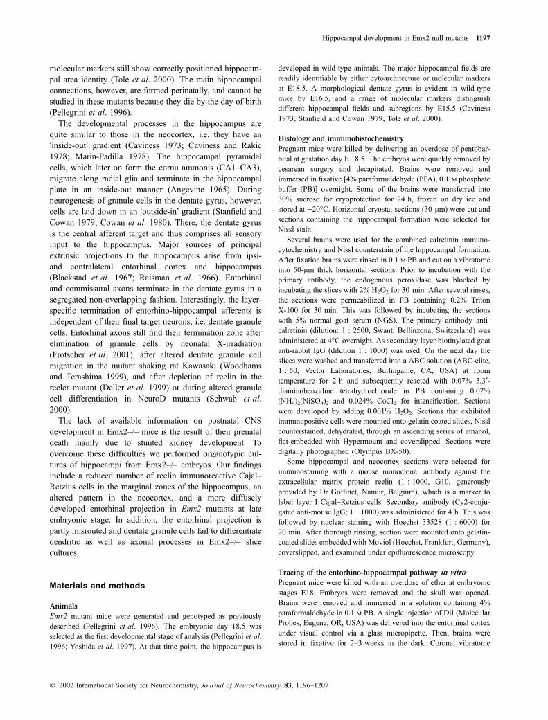
At embryonic day E18.5, Nissl staining of the hippocampal
formation revealed a timely normal development of the
principal cell layers in wild-type and heterozygous mice. In
the dentate gyrus (DG), the suprapyramidal blade of the
granule cell layer had started to form (Figs 1a and b). A
smaller CA1 and CA3 cell layer, but a demarcated hilar
region and a granule cell layer were consistently missing in
Emx2 deficient mice (Fig. 1c). Instead, a dense band of cells
persisted in vicinity to the ventricular zone. The curling of
Fig. 1 Cytoarchitectural organization of the embryonic (E 18.5) hip-
pocampal formation. Nissl-stained horizontal sections through the
hippocampus of wild-type (a), heterozygous (b), and Emx2–/– homo-
zygous (c) mutant mice. In (a) and (b), the pyramidal cell layer and the
granule cell primordium is present. (c) The hippocampus is reduced in
size in the Emx2–/–mutant. Somata of the presumed dentate gyrus
are Nissl stained, although the typical C-shaped cytoarchitecture is
missing. dg, dentate gyrus; pcl, pyramidal cell layer; pdg, presumed
dentate gyrus; EC, entorhinal cortex. Scale bars: 170 lm (a), 230 lm
(b), 300 lm (c).
1198 N. E. Savaskan et al.
� 2002 International Society for Neurochemistry, Journal of Neurochemistry, 83, 1196–1207
the developing dentate gyrus over itself to adopt its
characteristic convoluted C-shape appeared delayed in
Emx2–/– compared to wild-type littermates (Fig. 2). It has
been reported that Emx2-deficient isocortex has Cajal–
Retzius cell problems which translate into inappropriate
neuronal migration and histogenesis (Mallamaci et al.
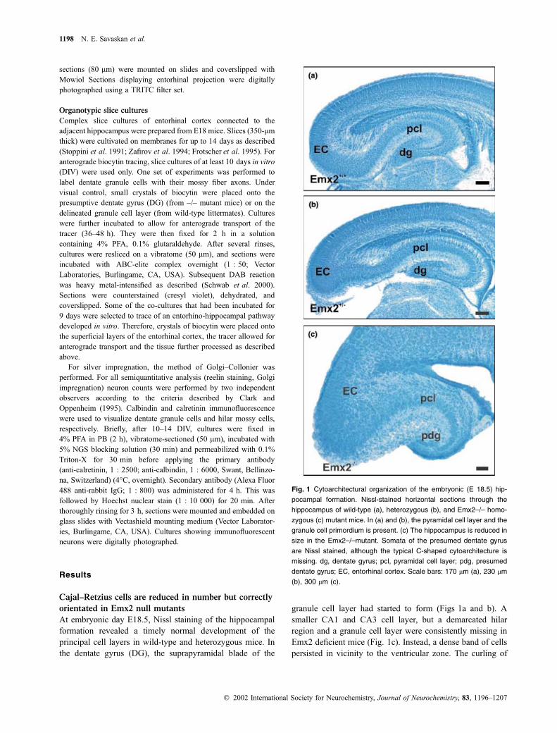
2000b). Calretinin immunocytochemistry was carried out to
examine the existence of early generated layer I Cajal–
Retzius cells. In all sections of the examined genotypes
[wild-type (n ¼ 3), heterozygous (n ¼ 5) and Emx2–/–
(n ¼ 4)] calretinin immunopositive neurons were found.
Immunolabeled cells were packed densely in the marginal
zones of the dentate gyrus and the hippocampal anlage from
Emx2+/– mice (Figs 2a and b). Only a few calretinin positive
cells were detected in these zones of Emx2–/– mutant
embryos with a loosely distribution not only restricted in the
marginal zone (Figs 2c and d). We confirmed these results
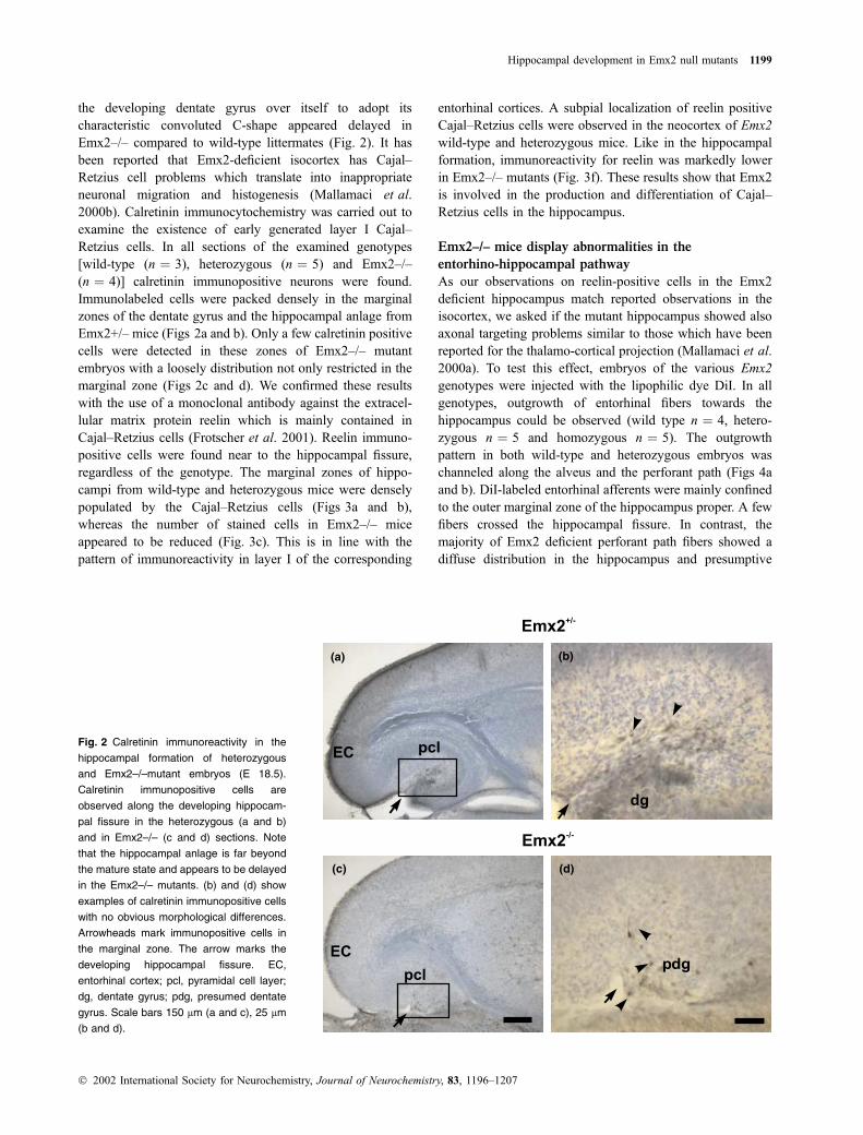
with the use of a monoclonal antibody against the extracel-
lular matrix protein reelin which is mainly contained in
Cajal–Retzius cells (Frotscher et al. 2001). Reelin immuno-
positive cells were found near to the hippocampal fissure,
regardless of the genotype. The marginal zones of hippo-
campi from wild-type and heterozygous mice were densely
populated by the Cajal–Retzius cells (Figs 3a and b),
whereas the number of stained cells in Emx2–/– mice
appeared to be reduced (Fig. 3c). This is in line with the
pattern of immunoreactivity in layer I of the corresponding
entorhinal cortices. A subpial localization of reelin positive
Cajal–Retzius cells were observed in the neocortex of Emx2
wild-type and heterozygous mice. Like in the hippocampal
formation, immunoreactivity for reelin was markedly lower
in Emx2–/– mutants (Fig. 3f). These results show that Emx2
is involved in the production and differentiation of Cajal–
Retzius cells in the hippocampus.
Emx2–/– mice display abnormalities in the
entorhino-hippocampal pathway
As our observations on reelin-positive cells in the Emx2
deficient hippocampus match reported observations in the
isocortex, we asked if the mutant hippocampus showed also
axonal targeting problems similar to those which have been
reported for the thalamo-cortical projection (Mallamaci et al.
2000a). To test this effect, embryos of the various Emx2
genotypes were injected with the lipophilic dye DiI. In all
genotypes, outgrowth of entorhinal fibers towards the
hippocampus could be observed (wild type n ¼ 4, hetero-
zygous n ¼ 5 and homozygous n ¼ 5). The outgrowth
pattern in both wild-type and heterozygous embryos was
channeled along the alveus and the perforant path (Figs 4a
and b). DiI-labeled entorhinal afferents were mainly confined
to the outer marginal zone of the hippocampus proper. A few
fibers crossed the hippocampal fissure. In contrast, the
majority of Emx2 deficient perforant path fibers showed a
diffuse distribution in the hippocampus and presumptive
(a) (b)
(c) (d)
Fig. 2 Calretinin immunoreactivity in the
hippocampal formation of heterozygous
and Emx2–/–mutant embryos (E 18.5).
Calretinin immunopositive cells are
observed along the developing hippocam-
pal fissure in the heterozygous (a and b)
and in Emx2–/– (c and d) sections. Note
that the hippocampal anlage is far beyond
the mature state and appears to be delayed
in the Emx2–/– mutants. (b) and (d) show
examples of calretinin immunopositive cells
with no obvious morphological differences.
Arrowheads mark immunopositive cells in
the marginal zone. The arrow marks the
developing hippocampal fissure. EC,
entorhinal cortex; pcl, pyramidal cell layer;
dg, dentate gyrus; pdg, presumed dentate
gyrus. Scale bars 150 lm (a and c), 25 lm
(b and d).
Hippocampal development in Emx2 null mutants 1199
� 2002 International Society for Neurochemistry, Journal of Neurochemistry, 83, 1196–1207
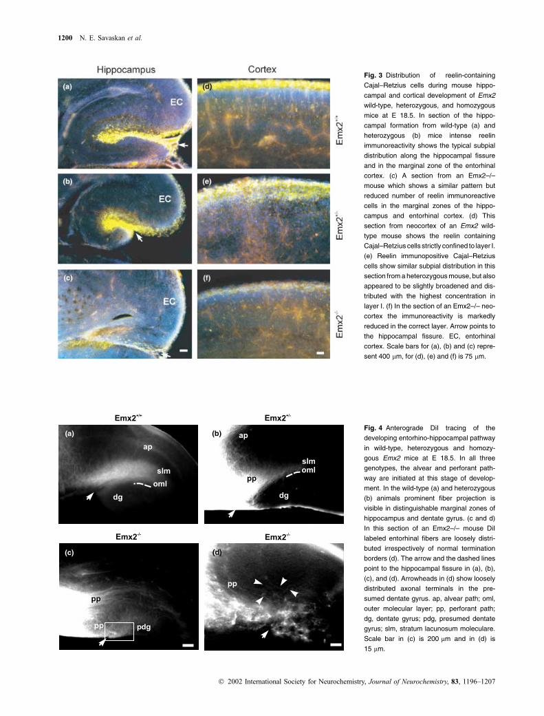
Fig. 3 Distribution of reelin-containing
Cajal–Retzius cells during mouse hippo-
campal and cortical development of Emx2
wild-type, heterozygous, and homozygous
mice at E 18.5. In section of the hippo-
campal formation from wild-type (a) and
heterozygous (b) mice intense reelin
immunoreactivity shows the typical subpial
distribution along the hippocampal fissure
and in the marginal zone of the entorhinal
cortex. (c) A section from an Emx2–/–
mouse which shows a similar pattern but
reduced number of reelin immunoreactive
cells in the marginal zones of the hippo-
campus and entorhinal cortex. (d) This
section from neocortex of an Emx2 wild-
type mouse shows the reelin containing
Cajal–Retzius cells strictly confined to layer I.
(e) Reelin immunopositive Cajal–Retzius
cells show similar subpial distribution in this
section from a heterozygous mouse, but also
appeared to be slightly broadened and dis-
tributed with the highest concentration in
layer I. (f) In the section of an Emx2–/– neo-
cortex the immunoreactivity is markedly
reduced in the correct layer. Arrow points to
the hippocampal fissure. EC, entorhinal
cortex. Scale bars for (a), (b) and (c) repre-
sent 400 lm, for (d), (e) and (f) is 75 lm.
(a)
(c) (d)
(b)Fig. 4 Anterograde DiI tracing of the
developing entorhino-hippocampal pathway
in wild-type, heterozygous and homozy-
gous Emx2 mice at E 18.5. In all three
genotypes, the alvear and perforant path-
way are initiated at this stage of develop-
ment. In the wild-type (a) and heterozygous
(b) animals prominent fiber projection is
visible in distinguishable marginal zones of
hippocampus and dentate gyrus. (c and d)
In this section of an Emx2–/– mouse DiI
labeled entorhinal fibers are loosely distri-
buted irrespectively of normal termination
borders (d). The arrow and the dashed lines
point to the hippocampal fissure in (a), (b),
(c), and (d). Arrowheads in (d) show loosely
distributed axonal terminals in the pre-
sumed dentate gyrus. ap, alvear path; oml,
outer molecular layer; pp, perforant path;
dg, dentate gyrus; pdg, presumed dentate
gyrus; slm, stratum lacunosum moleculare.
Scale bar in (c) is 200 lm and in (d) is
15 lm.
1200 N. E. Savaskan et al.
� 2002 International Society for Neurochemistry, Journal of Neurochemistry, 83, 1196–1207
dentate gyrus disregarding laminar boundaries (Figs 4c and d).
Our results show that, at least at this early phase of the
invasion of the hippocampus by perforant path axons, Emx2
is essential for the segregation of the incoming axons to their
corresponding target layers.
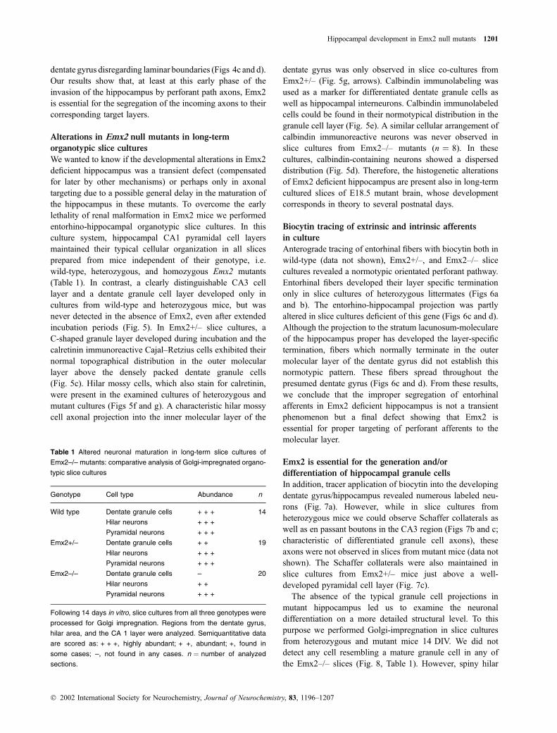
Alterations in Emx2 null mutants in long-term
organotypic slice cultures
We wanted to know if the developmental alterations in Emx2
deficient hippocampus was a transient defect (compensated
for later by other mechanisms) or perhaps only in axonal
targeting due to a possible general delay in the maturation of
the hippocampus in these mutants. To overcome the early
lethality of renal malformation in Emx2 mice we performed
entorhino-hippocampal organotypic slice cultures. In this
culture system, hippocampal CA1 pyramidal cell layers
maintained their typical cellular organization in all slices
prepared from mice independent of their genotype, i.e.
wild-type, heterozygous, and homozygous Emx2 mutants
(Table 1). In contrast, a clearly distinguishable CA3 cell
layer and a dentate granule cell layer developed only in
cultures from wild-type and heterozygous mice, but was
never detected in the absence of Emx2, even after extended
incubation periods (Fig. 5). In Emx2+/– slice cultures, a
C-shaped granule layer developed during incubation and the
calretinin immunoreactive Cajal–Retzius cells exhibited their
normal topographical distribution in the outer molecular
layer above the densely packed dentate granule cells
(Fig. 5c). Hilar mossy cells, which also stain for calretinin,
were present in the examined cultures of heterozygous and
mutant cultures (Figs 5f and g). A characteristic hilar mossy
cell axonal projection into the inner molecular layer of the
dentate gyrus was only observed in slice co-cultures from
Emx2+/– (Fig. 5g, arrows). Calbindin immunolabeling was
used as a marker for differentiated dentate granule cells as
well as hippocampal interneurons. Calbindin immunolabeled
cells could be found in their normotypical distribution in the
granule cell layer (Fig. 5e). A similar cellular arrangement of
calbindin immunoreactive neurons was never observed in
slice cultures from Emx2–/– mutants (n ¼ 8). In these
cultures, calbindin-containing neurons showed a dispersed
distribution (Fig. 5d). Therefore, the histogenetic alterations
of Emx2 deficient hippocampus are present also in long-term
cultured slices of E18.5 mutant brain, whose development
corresponds in theory to several postnatal days.
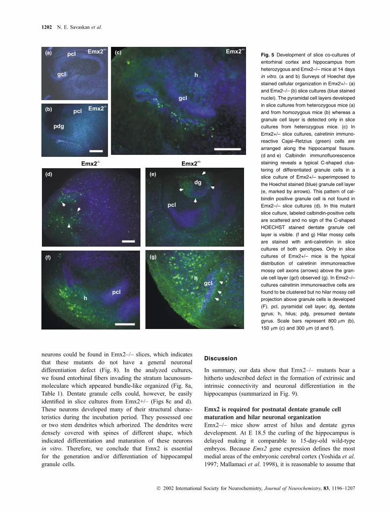
Biocytin tracing of extrinsic and intrinsic afferents
in culture
Anterograde tracing of entorhinal fibers with biocytin both in
wild-type (data not shown), Emx2+/–, and Emx2–/– slice
cultures revealed a normotypic orientated perforant pathway.
Entorhinal fibers developed their layer specific termination
only in slice cultures of heterozygous littermates (Figs 6a
and b). The entorhino-hippocampal projection was partly
altered in slice cultures deficient of this gene (Figs 6c and d).
Although the projection to the stratum lacunosum-moleculare
of the hippocampus proper has developed the layer-specific
termination, fibers which normally terminate in the outer
molecular layer of the dentate gyrus did not establish this
normotypic pattern. These fibers spread throughout the
presumed dentate gyrus (Figs 6c and d). From these results,
we conclude that the improper segregation of entorhinal
afferents in Emx2 deficient hippocampus is not a transient
phenomenon but a final defect showing that Emx2 is
essential for proper targeting of perforant afferents to the
molecular layer.
Emx2 is essential for the generation and/or
differentiation of hippocampal granule cells
In addition, tracer application of biocytin into the developing
dentate gyrus/hippocampus revealed numerous labeled neu-
rons (Fig. 7a). However, while in slice cultures from
heterozygous mice we could observe Schaffer collaterals as
well as en passant boutons in the CA3 region (Figs 7b and c;
characteristic of differentiated granule cell axons), these
axons were not observed in slices from mutant mice (data not
shown). The Schaffer collaterals were also maintained in
slice cultures from Emx2+/– mice just above a well-
developed pyramidal cell layer (Fig. 7c).
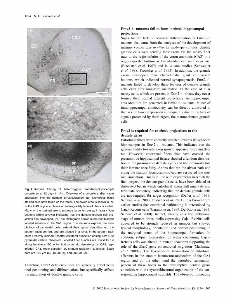
The absence of the typical granule cell projections in
mutant hippocampus led us to examine the neuronal
differentiation on a more detailed structural level. To this
purpose we performed Golgi-impregnation in slice cultures
from heterozygous and mutant mice 14 DIV. We did not
detect any cell resembling a mature granule cell in any of
the Emx2–/– slices (Fig. 8, Table 1). However, spiny hilar
Table 1 Altered neuronal maturation in long-term slice cultures of
Emx2–/– mutants: comparative analysis of Golgi-impregnated organo-
typic slice cultures
Genotype Cell type Abundance n
Wild type Dentate granule cells + + + 14
Hilar neurons + + +
Pyramidal neurons + + +
Emx2+/– Dentate granule cells + + 19
Hilar neurons + + +
Pyramidal neurons + + +
Emx2–/– Dentate granule cells – 20
Hilar neurons + +
Pyramidal neurons + + +
Following 14 days in vitro, slice cultures from all three genotypes were
processed for Golgi impregnation. Regions from the dentate gyrus,
hilar area, and the CA 1 layer were analyzed. Semiquantitative data
are scored as: + + +, highly abundant; + +, abundant; +, found in
some cases; –, not found in any cases. n ¼ number of analyzed
sections.
Hippocampal development in Emx2 null mutants 1201
� 2002 International Society for Neurochemistry, Journal of Neurochemistry, 83, 1196–1207
neurons could be found in Emx2–/– slices, which indicates
that these mutants do not have a general neuronal
differentiation defect (Fig. 8). In the analyzed cultures,
we found entorhinal fibers invading the stratum lacunosum-
moleculare which appeared bundle-like organized (Fig. 8a,
Table 1). Dentate granule cells could, however, be easily
identified in slice cultures from Emx2+/– (Figs 8c and d).
These neurons developed many of their structural charac-
teristics during the incubation period. They possessed one
or two stem dendrites which arborized. The dendrites were
densely covered with spines of different shape, which
indicated differentiation and maturation of these neurons
in vitro. Therefore, we conclude that Emx2 is essential
for the generation and/or differentiation of hippocampal
granule cells.
Discussion
In summary, our data show that Emx2–/– mutants bear a
hitherto undescribed defect in the formation of extrinsic and
intrinsic connectivity and neuronal differentiation in the
hippocampus (summarized in Fig. 9).
Emx2 is required for postnatal dentate granule cell
maturation and hilar neuronal organization
Emx2–/– mice show arrest of hilus and dentate gyrus
development. At E 18.5 the curling of the hippocampus is
delayed making it comparable to 15-day-old wild-type
embryos. Because Emx2 gene expression defines the most
medial areas of the embryonic cerebral cortex (Yoshida et al.
1997; Mallamaci et al. 1998), it is reasonable to assume that
(a)
(b)
(c)
(e)(d)
(f) (g)
Fig. 5 Development of slice co-cultures of
entorhinal cortex and hippocampus from
heterozygous and Emx2–/– mice at 14 days
in vitro. (a and b) Surveys of Hoechst dye
stained cellular organization in Emx2+/– (a)
and Emx2–/– (b) slice cultures (blue stained
nuclei). The pyramidal cell layers developed
in slice cultures from heterozygous mice (a)
and from homozygous mice (b) whereas a
granule cell layer is detected only in slice
cultures from heterozygous mice. (c) In
Emx2+/– slice cultures, calretinin immuno-
reactive Cajal–Retzius (green) cells are
arranged along the hippocampal fissure.
(d and e) Calbindin immunofluorescence
staining reveals a typical C-shaped clus-
tering of differentiated granule cells in a
slice culture of Emx2+/– superimposed to
the Hoechst stained (blue) granule cell layer
(e, marked by arrows). This pattern of cal-
bindin positive granule cell is not found in
Emx2–/– slice cultures (d). In this mutant
slice culture, labeled calbindin-positive cells
are scattered and no sign of the C-shaped
HOECHST stained dentate granule cell
layer is visible. (f and g) Hilar mossy cells
are stained with anti-calretinin in slice
cultures of both genotypes. Only in slice
cultures of Emx2+/– mice is the typical
distribution of calretinin immunoreactive
mossy cell axons (arrows) above the gran-
ule cell layer (gcl) observed (g). In Emx2–/–
cultures calretinin immunoreactive cells are
found to be clustered but no hilar mossy cell
projection above granule cells is developed
(F). pcl, pyramidal cell layer; dg, dentate
gyrus; h, hilus; pdg, presumed dentate
gyrus. Scale bars represent 800 lm (b),
150 lm (c) and 300 lm (d and f).
1202 N. E. Savaskan et al.
� 2002 International Society for Neurochemistry, Journal of Neurochemistry, 83, 1196–1207
Emx2 specifies areal identities in part at least of the dentate
gyrus, i.e. the polymorph hilar region and the principal layer
with its granule cells. Based on the role of Emx2 in area
specification Pellegrini et al. (1996) suggested a lack of area
specification in affected subregions of the hippocampus. A
recent study by Tole et al. (2000) using molecular region
markers showed that each hippocampal subfield is specified
with its typical neuronal messenger RNA. Furthermore, each
cell population develops in its correct position relative to
each other and expression analysis of Wnt5a gene indicates
the presence of dentate granule cell precursors. However,
most of the dentate granule cells are known to be generated
and migrate to their final position during the first few
postnatal weeks (Angevine 1965). In consequence, granule
cell maturation first takes place during postnatal stages.
Another function of Emx2 has to be suggested when
considering the results from the slice culture experiments in
the present study. Here, our immunocytochemistry data show
that calbindin-positive cells and hilar mossy cells are present
in Emx2–/– mutants. However, the well-defined and charac-
teristic C-shaped layering of dentate granule cells has been
found neither in Emx2–/– slice cultures nor the in the
morphology of labeled cells resembling differentiated gran-
ule cells, even after extended incubation periods. This
indicates an arrest of normal migration and differentiation
of this cell type. These developmental processes were not
impaired in cultures from wild-type and heterozygous
animals that showed a normal distribution of typical granule
cells. Moreover, the arrangement of hilar mossy cells
identified by their immunoreactivity for the calcium binding
protein calretinin was not as compact. As a result of this
cellular disorganization, they did not develop their normal
axonal projection to the proximity of granule cell dendrites in
Emx2–/– mutants. Lack of Golgi impregnated neurons with
morphological characteristics of dentate granule cells further
show that Emx2 is essential in dentate granule cell matur-
ation. Data from the Golgi technique have to take with
caution since the nature of the Golgi impregnation is still
poorly understood. However, spiny hilar neurons have been
found in the same slices which lack identifiable granule cells.
(a) (b)
(d)(c)
Fig. 6 Biocytin-traced entorhinal projection in heterozygous and
knockout co-cultures after 12 days in vitro. In heterozygous entorhino-
hippocampal slice cultures axonal projections (arrowheads) are con-
fined to the appropriate termination zone, the outer molecular layer of
the dentate gyrus (dg) (a; b is a higher magnification of boxed area in
a). Cresyl violet staining revealed that both the supra- and infra-
pyramidal blade of the dentate granular cell layer had developed. In a
slice culture from Emx2–/–, an entorhinal projection is also formed
(c and d is a higher magnification of boxed area in c). The majority of
traced entorhinal afferents is distributed throughout the presumed
dentate gyrus (pdg) and the layer specificity is only retained for those
fibers which entered the stratum lacunosum-moleculare (slm). The
arrow indicates the hippocampal fissure. Arrowheads mark axonal
profiles in the presumed dentate gyrus, and stratum lacunosum
moleculare. gcl, granule cell layer, ml, molecular layer, EC, entorhinal
cortex; CA1, regio superior. Scale bar represents for 300 lm (a) and
(c); 20 lm in (b); and 15 lm in (d).
Hippocampal development in Emx2 null mutants 1203
� 2002 International Society for Neurochemistry, Journal of Neurochemistry, 83, 1196–1207
Therefore, Emx2 deficiency does not generally affect neur-
onal positioning and differentiation, but specifically affects
the maturation of dentate granule cells.
Emx2–/– mutants fail to form intrinsic hippocampal
projections
Signs for the lack of neuronal differentiation in Emx2–/–
mutants also came from the analyses of the development of
intrinsic connections in vitro. In wild-type cultures, dentate
granule cells were sending their axons via the mossy fiber
tract to the regio inferior of the cornu ammonis (CA3) in a
region-specific fashion as has already been seen in in vivo
(Blackstad et al. 1967) and in in vitro studies (Schwegler
et al. 1988; Frotscher et al. 1995). In addition, the granule
axons developed their characteristic giant en passant
boutons, which indicated normal synaptogenesis. Emx2–/–
mutants failed to develop these features of dentate granule
cells even after long-term incubation. In the case of hilar
mossy cells, which are present in Emx2–/– slices, they never
formed their normal efferent projections. As hippocampal
area identities are generated in Emx2–/– mutants, failure of
intrahippocampal connectivity can be directly attributed to
the lack of Emx2 expression subsequently due to the lack of
signals presented by their targets, the mature dentate granule
cells.
Emx2 is required for extrinsic projections to the
dentate gyrus
Entorhinal fibers were correctly directed towards the adjacent
hippocampus in Emx2–/– mutants. This indicates that the
general ability towards axon growth appeared to be unaffec-
ted. However, entorhinal fibers that have crossed the
presumptive hippocampal fissure showed a random distribu-
tion in the presumptive dentate gyrus and had obviously lost
their laminar specificity. Axons that ran the alvear path and
along the stratum lacunosum-moleculare respected the nor-
mal lamination. This is in line with experiments in which the
final targets, the dentate granule cells, have been ablated or
dislocated but in which entorhinal axons still innervate and
terminate accurately, indicating that the dentate granule cells
are not required for target recognition (Deller et al. 1999;
Schwab et al. 2000; Frotscher et al. 2001). It is known from
earlier studies that entorhinal pathfinding is determined by
Cajal–Retzius cells (Ceranik et al. 1999; Del Rio et al. 1997;
Schwab et al. 2000). In fact, already at a late embryonic
stage of mutant brain, reelin-expressing Cajal–Retzius cells
appeared to be strongly reduced in number but showed
typical morphology, orientation, and correct positioning in
the marginal zones of the hippocampal formation. In
addition, subpial localization of reelin containing Cajal–
Retzius cells was altered in mutant neocortex supporting the
role of the Emx2 gene on neuronal migration (Mallamaci
et al. 2000a). The layer-specific termination of entorhinal
afferents in the stratum lacunosum-moleculare of the CA1
region and on the other hand the perturbed termination
pattern of these fibers in the presumptive dentate gyrus
coincides with the cytoarchitectural organization of the cor-
responding hippocampal subfields. The observed misrouting
(a)
(b)
(c)
Fig. 7 Biocytin tracing of heterozygous entorhino-hippocampal
co-cultures at 12 days in vitro. Overview of a co-culture after tracer
application into the dentate gyrus/subiculum (a). Numerous black
stained cells have taken up the tracer. The boxed area is shown in (b).
In the CA3 region a plexus of anterogradely labeled fibers is visible.
Many of the stained axons protrude large en passant mossy fiber
boutons (white arrows) indicating that the dentate granule cell pro-
jection has developed. (c) This micrograph shows numerous biocytin
labeled neurons in the CA1 region. The neurons express the mor-
phology of pyramidal cells, extend their apical dendrites into the
stratum radiatum (sr), and are aligned in a layer. In the stratum radi-
atum a heavily stained Schaffer collateral projection arising from CA3
pyramidal cells is observed. Labeled fiber bundles are found to run
along the alveus. EC, entorhinal cortex; dg, dentate gyrus; CA3, regio
inferior; CA1, regio superior; sr, stratum radiatum; a, alveus. Scale
bars are 150 lm (a), 40 lm (b), and 200 lm (c).
1204 N. E. Savaskan et al.
� 2002 International Society for Neurochemistry, Journal of Neurochemistry, 83, 1196–1207
of entorhinal fibers in the presumptive dentate gyrus may be
accordingly regarded as a secondary effect due to the Emx2
related changes in reelin signaling. In a recent study, it has
been shown that during the development of the perforant
path Cajal–Retzius cells form a pioneer projection from the
hippocampus to the entorhinal cortex and provide a template
for later outgrowing entorhinal axons (Ceranik et al. 1999).
However, most Cajal Retzius cells are confined to their
normal position, the marginal zones of the hippocampus in
Emx2 heterozygous and null mutants. Thus, it cannot be
excluded that the observed misrouted entorhinal projection is
caused by the slightly altered Cajal–Retzius cell patterning.
In this line, so far, unknown guidance cues, which are
probably regulated by Emx2, may underlie the defects in
target recognition seen in Emx2 mutants.
To overcome the neonatal lethality of Emx2 deficiencies
further studies should generate neuron-specific Emx2–/–
mutants which would offer the possibilities to analyze the
downstream targets of Emx2 regulated genes responsible for
the anatomical phenotype.
Acknowledgements
We thank Peter Gruss for providing Emx2–/– mutant mice and
Shanting Zhao for reelin immunofluorescence. Anja U. Brauer is
acknowledged for transcription analysis and in situ hybridization.
We also gratefully acknowledge the graphical art work of
Brigitte Mannsfeldt and Sabine Lewandowski. This study was
supported by the DFG (DFG He1520/2–1 to BH, SFB 515/A5 to
RN and NES, and DFG Sk49/5–1 to TS). NES was financially
(c) (d)
(a) (b)
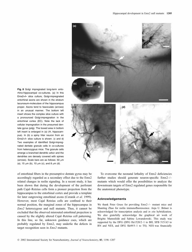
Fig. 8 Golgi impregnated long-term ento-
rhino-hippocampal co-cultures. (a) In this
Emx2–/– slice culture, Golgi-impregnated
entorhinal axons are shown in the stratum
lacunosum-moleculare of the hippocampus
proper. Axons tend to fasciculate (arrows)
in an unusual manner. The bottom left
insert shows the complex slice culture with
a pronounced Golgi-impregnation in the
entorhinal cortex (EC). Note the lack of
cellular impregnation in the presumed den-
tate gyrus (pdg). The boxed area in bottom
left insert is enlarged in (a) (H, hippocam-
pus). In (b) a spiny hilar neuron from an
Emx2–/– slice culture is shown. (c and d)
Two examples of identified Golgi-impreg-
nated dentate granule cells in co-cultures
from heterozygous mice. The granule cells
emerge a branched dendritic arbor and the
dendrites are densely covered with spines
(arrows). Scale bars are as follows: 50 lm
(a), 15 lm (b), 10 lm (c), and 8 lm (d).
Hippocampal development in Emx2 null mutants 1205
� 2002 International Society for Neurochemistry, Journal of Neurochemistry, 83, 1196–1207
supported by the GENNSA Tech. Inc. (Berlin, Germany) and
NES is an investigator of the Charite Biomedical Research
Foundation.
References
Angevine J. B. Jr (1965) Time of neuron origin in the hippocampal region.
An autoradiographic study in the mouse. Exp. Neurol. 2, 1–70.
Blackstad T.-W., Fuxe K. and Hokfelt T. (1967) Noradrenaline nerve
terminals in the hippocampal region of the rat and the guinea pig.
Z Zellforsch Mikrosk. Anat. 78, 463–473.
Caviness V.-S. (1973) Time of neuron origin in the hippocampus and
dentate gyrus of normal and reeler mutant mice: an autoradio-
graphic analysis. J. Comp. Neurol. 151, 113–120.
Caviness V.-S. and Rakic P. (1978) Mechanisms of cortical development:
a view from mutations in mice. Annu. Rev. Neurosci. 1297, 326.
Cecchi C. and Boncinelli E. (2000) Emx homeogenes and mouse brain
development. Trends Neurosci. 23, 347–352.
Ceranik K., Deng J., Heimrich B., Lubke J., Zhao S., Forster E. and
Frotscher M. (1999) Hippocampal Cajal–Retzius cells project to
the entorhinal cortex: retrograde tracing and intracellular labelling
studies. Eur. J. Neurosci. 11, 4278–4290.
Clark P. H. G. and Oppenheim R. W. (1995) Neuron death in vertebrate
development: in vivo methods. Methods Cell Biol. 46, 277–321.
Cowan W.-M., Stanfield B.-B. and Kishi K. (1980) The development of
the dentate gyrus. Curr. Top. Dev. Biol. 15, 57.
Dalton D., Chadwick R. and McGinnis W. (1989) Expression and
embryonic function of empty spiracles: a Drosophila homeo box
gene with two patterning functions on the anterior-posterior axis of
the embryo. Genes Dev. 3, 1940–1956.
Del Rio J.-A., Heimrich B., Borrell V., Forster E., Drakew A., Alcantara
S., Nakajima K., Miyata T., Ogawa M., Mikoshiba K., Derer P.,
Frotscher M. and Soriano E. (1997) A role for Cajal–Retzius cells
and reelin in the development of hippocampal connections. Nature
385, 70–74.
Deller T., Drakew A. and Frotscher M. (1999) Different primary
target cells are important for fiber lamination in the fascia
dentata: a lesson from reeler mutant mice. Exp. Neurol. 156,
239–253.
Frotscher M., Zafirov S. and Heimrich B. (1995) Development of
identified neuronal types and of specific synaptic connections in
slice cultures of rat hippocampus. Prog. Neurobiol. 45, 143–164.
Frotscher M. L., Seress and Heimrich (2001) Early generated Cajal–
Retzius cells have different functions in cortical development,
in: Brain Stem Cells (Thorndyke M., Beesley P., Miyan J. and
Bannister C., eds), pp. 43–49. BIOS Scientific Publishers Ltd,
Oxford.
Gulisano M., Broccoli V., Pardini C. and Boncinelli E. (1996) Emx1 and
Emx2 show different patterns of expression during proliferation
and differentiation of the developing cerebral cortex in the mouse.
Eur. J. Neurosci. 8, 1037–1050.
Mallamaci A., Iannone R., Briata P., Pintonello L., Mercurio S.,
Boncinelli E. and Corte G. (1998) EMX2 protein in the developing
mouse brain and olfactory area. Mech. Dev. 77, 165–172.
Mallamaci A., Muzio L., Chan C.-H., Parnavelas J. and Boncinelli E.
(2000a) Area identity shifts in the early cerebral cortex of Emx2-/-
mutant mice. Nat. Neurosci. 3, 679–686.
(b)
(d)
(a)
(c)
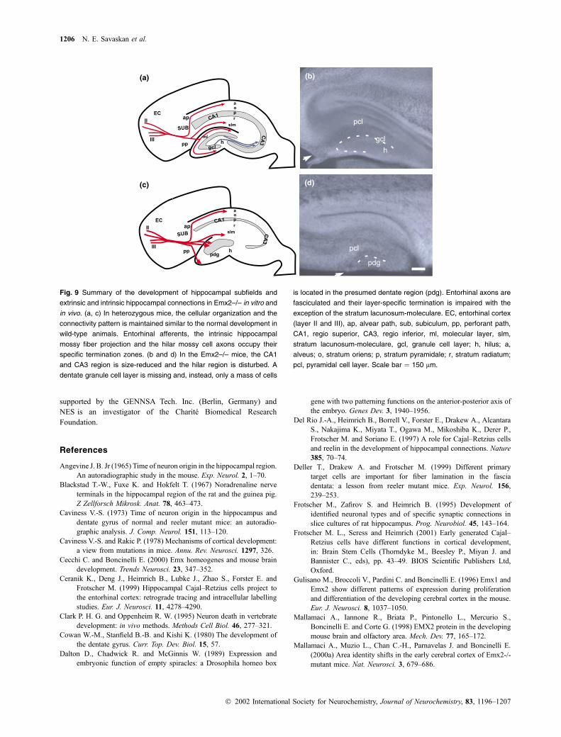
Fig. 9 Summary of the development of hippocampal subfields and
extrinsic and intrinsic hippocampal connections in Emx2–/– in vitro and
in vivo. (a, c) In heterozygous mice, the cellular organization and the
connectivity pattern is maintained similar to the normal development in
wild-type animals. Entorhinal afferents, the intrinsic hippocampal
mossy fiber projection and the hilar mossy cell axons occupy their
specific termination zones. (b and d) In the Emx2–/– mice, the CA1
and CA3 region is size-reduced and the hilar region is disturbed. A
dentate granule cell layer is missing and, instead, only a mass of cells
is located in the presumed dentate region (pdg). Entorhinal axons are
fasciculated and their layer-specific termination is impaired with the
exception of the stratum lacunosum-moleculare. EC, entorhinal cortex
(layer II and III), ap, alvear path, sub, subiculum, pp, perforant path,
CA1, regio superior, CA3, regio inferior, ml, molecular layer, slm,
stratum lacunosum-moleculare, gcl, granule cell layer; h, hilus; a,
alveus; o, stratum oriens; p, stratum pyramidale; r, stratum radiatum;
pcl, pyramidal cell layer. Scale bar ¼ 150 lm.
1206 N. E. Savaskan et al.
� 2002 International Society for Neurochemistry, Journal of Neurochemistry, 83, 1196–1207
Mallamaci A., Mercurio S., Muzio L., Cecchi C., Pardini C.-L., Gruss P.
and Boncinelli E. (2000b) The lack of Emx2 causes impairment of
Reelin signaling and defects of neuronal migration in the devel-
oping cerebral cortex. J. Neurosci. 20, 1109–1118.
Marin-Padilla M. (1978) Dual origin of the mammalian neocortex and
evolution of the cortical plate. Anat. Embryol. (Berl.) 152,
109–126.
Pellegrini M., Mansouri A., Simeone A., Boncinelli E. and Gruss P.
(1996) Dentate gyrus formation requires Emx2. Development 122,
3893–3898.
Raisman G., Cowan W.-M. and Powell T.-P. (1966) An experimental
analysis of the efferent projection of the hippocampus. Brain 89,
83–108.
Schwab M.-H., Bartholomae A., Heimrich B., Feldmeyer D., Druffel
A. S., Goebbels S., Naya F.-J., Zhao S., Frotscher M., Tsai M.-J.
and Nave K.-A. (2000) Neuronal basic helix-loop-helix pro-
teins (NEX and BETA2/Neuro D) regulate terminal granule cell
differentiation in the hippocampus. J. Neurosci. 20, 3714–
3724.
Schwegler H., Heimrich B., Keller F., Renner P. and Crusio W.-E.
(1988) Strain-specific development of the mossy fiber system in
organotypic cultures of the mouse hippocampus. Neurosci. Lett.
87, 7–10.
Simeone A., Gulisano M., Acampora D., Stornaiuolo A., Rambaldi M.
and Boncinelli E. (1992) Two vertebrate homeobox genes related
to the Drosophila empty spiracles gene are expressed in the
embryonic cerebral cortex. EMBO J. 11, 2541–2550.
Stanfield B.-B. and Cowan W.-M. (1979) The development of the hip-
pocampus and dentate gyrus in normal and reeler mice. J. Comp.
Neurol. 185, 423–459.
Stoppini L., Buchs P.-A. and Muller D. (1991) A simple method for
organotypic cultures of nervous tissue. J. Neurosci. Methods 37,
173–182.
Tole S., Goudreau G., Assimacopoulos S. and Grove E.-A. (2000) Emx2
is required for growth of the hippocampus but not for hippocampal
field specification. J. Neurosci. 20, 2618–2625.
Woodhams P.-L. and Terashima T. (1999) Laminar boundaries persist in
the hippocampal dentate molecular layer of the mutant shaking rat
Kawasaki despite aberrant granule cell migration. J. Comp. Neurol.
409, 57–70.
Yoshida M., Suda Y., Matsuo I., Miyamoto N., Takeda N., Kuratani S.
and Aizawa S. (1997) Emx1 and Emx2 functions in development
of dorsal telencephalon. Development 124, 101–111.
Zafirov S., Heimrich B. and Frotscher M. (1994) Dendritic development
of dentate granule cells in the absence of their specific extrinsic
afferents. J. Comp. Neurol. 345, 472–480.
Hippocampal development in Emx2 null mutants 1207
� 2002 International Society for Neurochemistry, Journal of Neurochemistry, 83, 1196–1207