Embed Size (px)
Citation preview
ORTHOPANTOMOGRAPH™ OP 3D ProA platform for changing needs
2 | KaVo
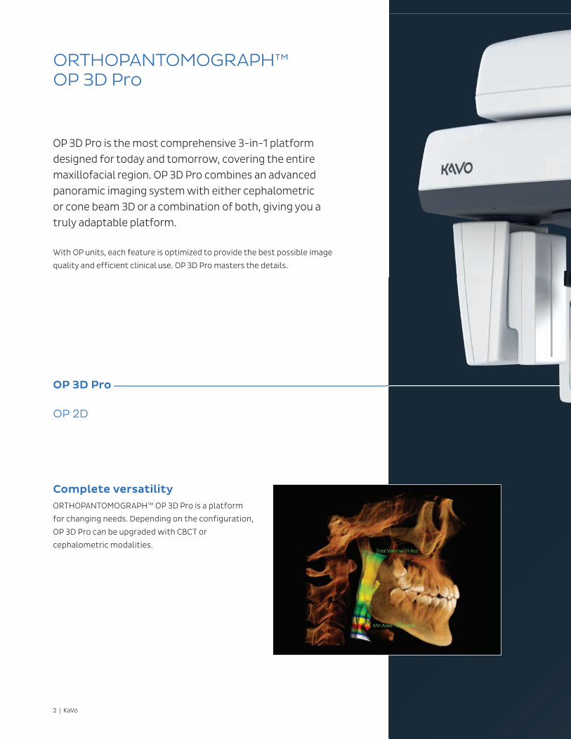
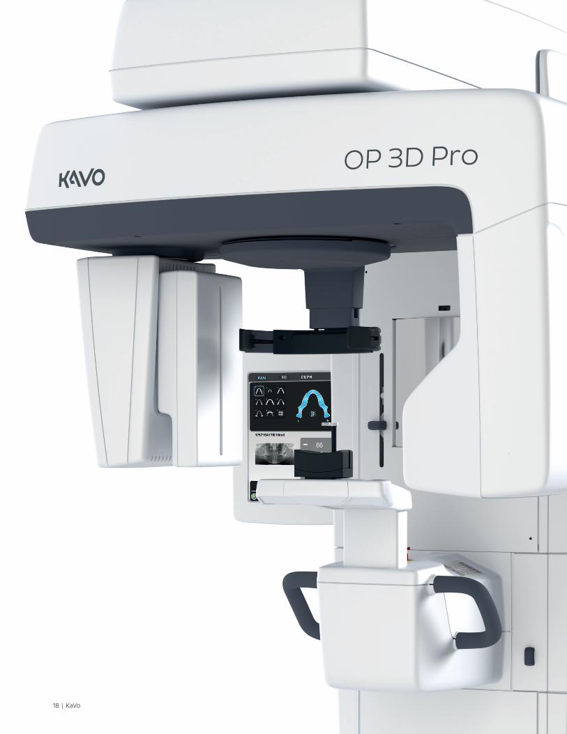
ORTHOPANTOMOGRAPH™ OP 3D Pro
OP 3D Pro is the most comprehensive 3-in-1 platform designed for today and tomorrow, covering the entire maxillofacial region. OP 3D Pro combines an advanced panoramic imaging system with either cephalometric or cone beam 3D or a combination of both, giving you a truly adaptable platform.
With OP units, each feature is optimized to provide the best possible image
quality and efficient clinical use. OP 3D Pro masters the details.
OP 3D Pro
OP 2D
Complete versatilityORTHOPANTOMOGRAPH™ OP 3D Pro is a platform
for changing needs. Depending on the configuration,
OP 3D Pro can be upgraded with CBCT or
cephalometric modalities.
KaVo | 3
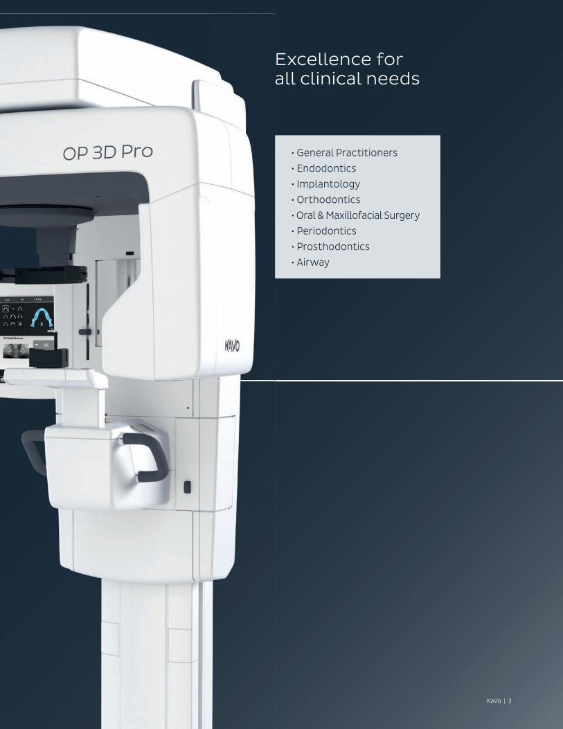
Excellence for all clinical needs
• General Practitioners
• Endodontics
• Implantology
• Orthodontics
• Oral & Maxillofacial Surgery
• Periodontics
• Prosthodontics
• Airway
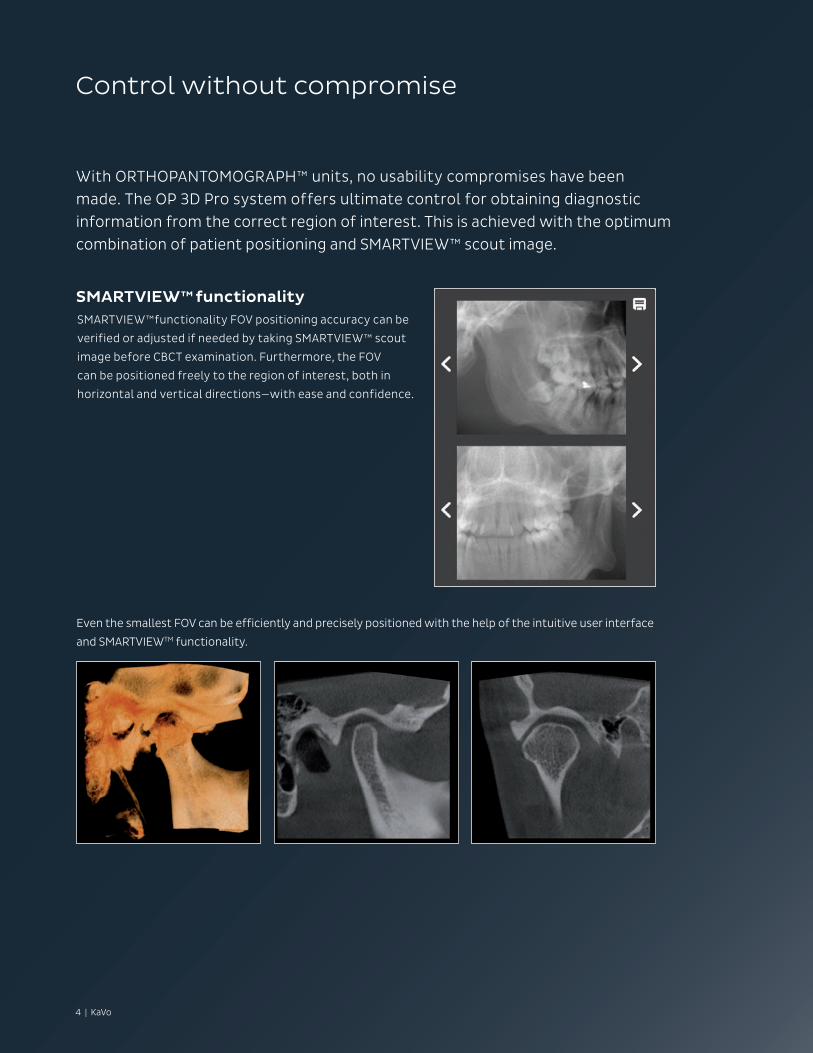
Control without compromise
With ORTHOPANTOMOGRAPH™ units, no usability compromises have been made. The OP 3D Pro system offers ultimate control for obtaining diagnostic information from the correct region of interest. This is achieved with the optimum combination of patient positioning and SMARTVIEW™ scout image.
SMARTVIEW™functionalitySMARTVIEW™functionality FOV positioning accuracy can be
verified or adjusted if needed by taking SMARTVIEW™ scout
image before CBCT examination. Furthermore, the FOV
can be positioned freely to the region of interest, both in
horizontal and vertical directions—with ease and confidence.
Even the smallest FOV can be efficiently and precisely positioned with the help of the intuitive user interface
and SMARTVIEWTM functionality.
4 | KaVo
KaVo | 5
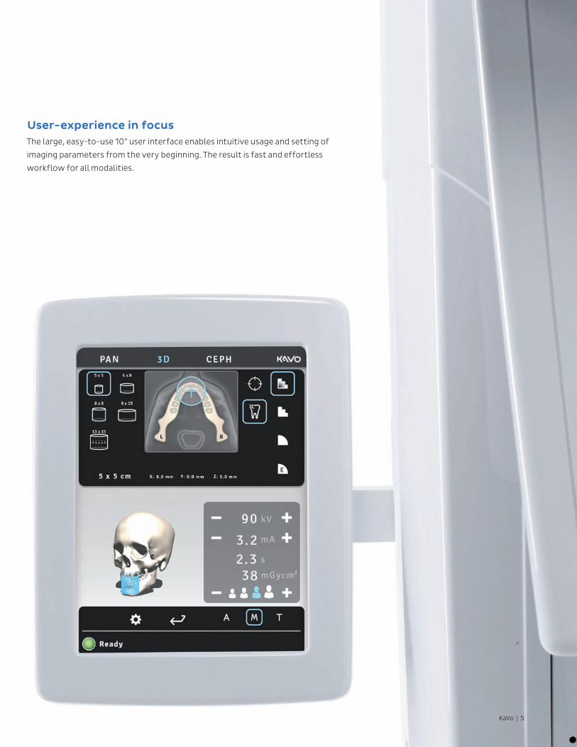
User-experience in focusThe large, easy-to-use 10” user interface enables intuitive usage and setting of
imaging parameters from the very beginning. The result is fast and effortless
workflow for all modalities.
6 | KaVo
Precision for every patient
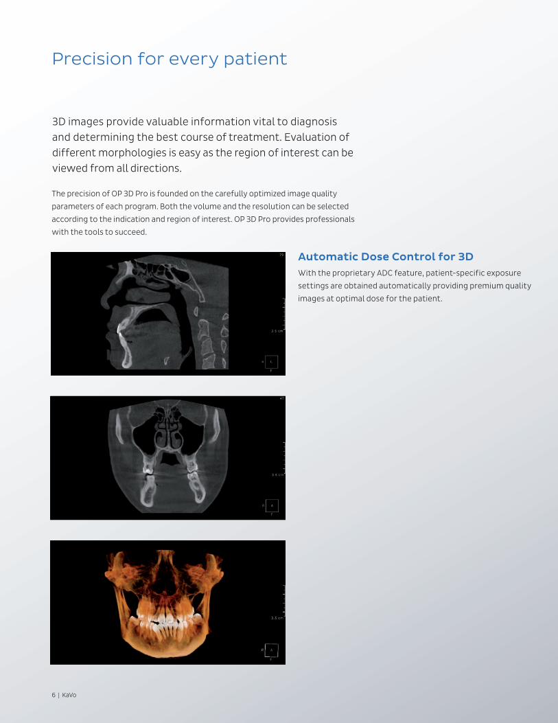
3D images provide valuable information vital to diagnosis and determining the best course of treatment. Evaluation of different morphologies is easy as the region of interest can be viewed from all directions.
Automatic Dose Control for 3DWith the proprietary ADC feature, patient-specific exposure
settings are obtained automatically providing premium quality
images at optimal dose for the patient.
The precision of OP 3D Pro is founded on the carefully optimized image quality
parameters of each program. Both the volume and the resolution can be selected
according to the indication and region of interest. OP 3D Pro provides professionals
with the tools to succeed.
KaVo | 7*OP300 Maxio (OP 3D Pro) Dosimetry report, Prof. John B. Ludlow, April 2014.
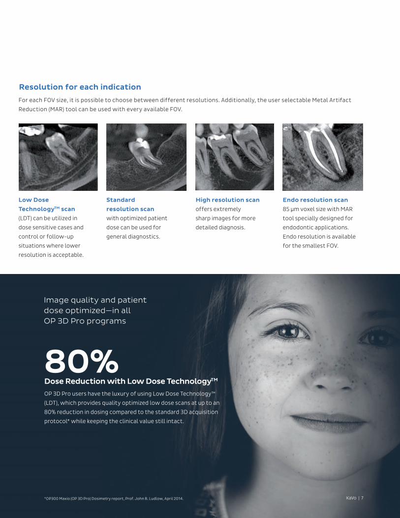
Resolution for each indication
For each FOV size, it is possible to choose between different resolutions. Additionally, the user selectable Metal Artifact
Reduction (MAR) tool can be used with every available FOV.
Low Dose
TechnologyTM scan
(LDT) can be utilized in
dose sensitive cases and
control or follow-up
situations where lower
resolution is acceptable.
Standard
resolution scan
with optimized patient
dose can be used for
general diagnostics.
High resolution scan
offers extremely
sharp images for more
detailed diagnosis.
Endo resolution scan
85 µm voxel size with MAR
tool specially designed for
endodontic applications.
Endo resolution is available
for the smallest FOV.
Dose Reduction with Low Dose TechnologyTM
OP 3D Pro users have the luxury of using Low Dose Technology™
(LDT), which provides quality optimized low dose scans at up to an
80% reduction in dosing compared to the standard 3D acquisition
protocol* while keeping the clinical value still intact.
80%
Image quality and patient dose optimized—in all OP 3D Pro programs
8 | KaVo
KaVo | 9
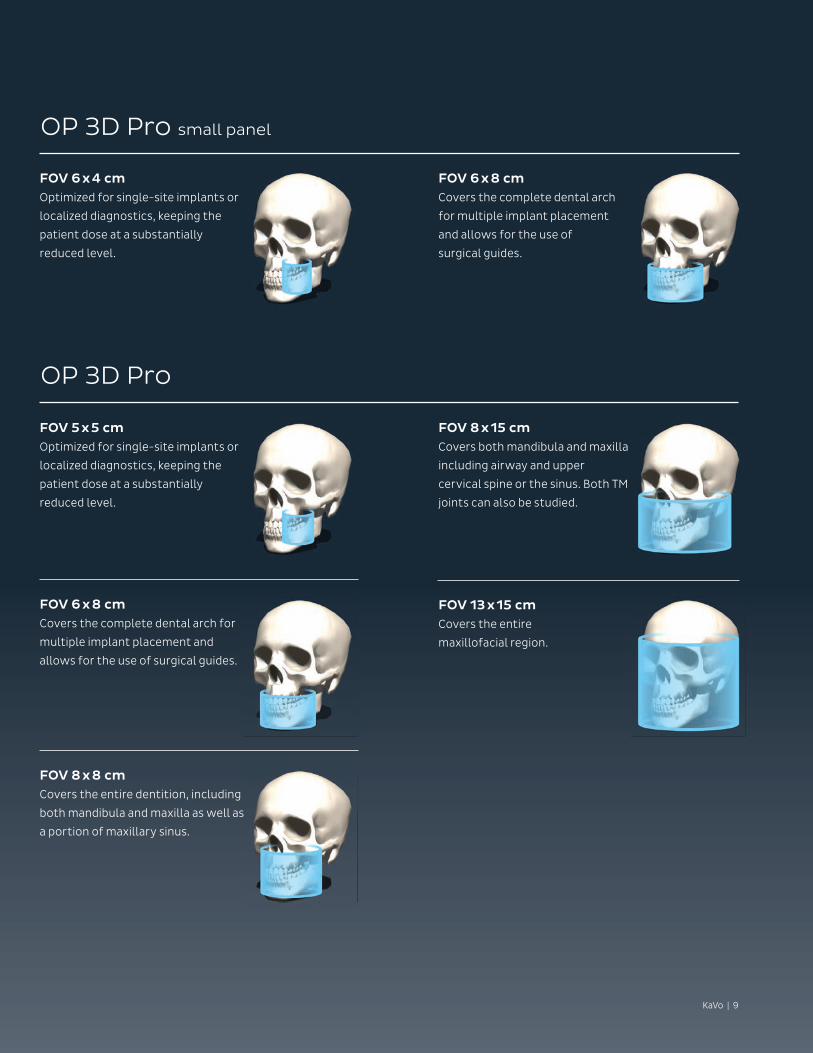
OP 3D Pro small panel
OP 3D Pro
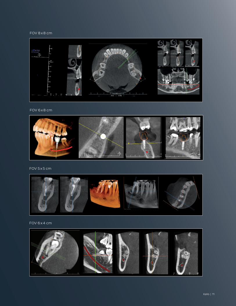
FOV 6 x 4 cmOptimized for single-site implants or
localized diagnostics, keeping the
patient dose at a substantially
reduced level.
FOV 6 x 8 cmCovers the complete dental arch
for multiple implant placement
and allows for the use of
surgical guides.
FOV 5 x 5 cmOptimized for single-site implants or
localized diagnostics, keeping the
patient dose at a substantially
reduced level.
FOV 6 x 8 cmCovers the complete dental arch for
multiple implant placement and
allows for the use of surgical guides.
FOV 8 x 8 cmCovers the entire dentition, including
both mandibula and maxilla as well as
a portion of maxillary sinus.
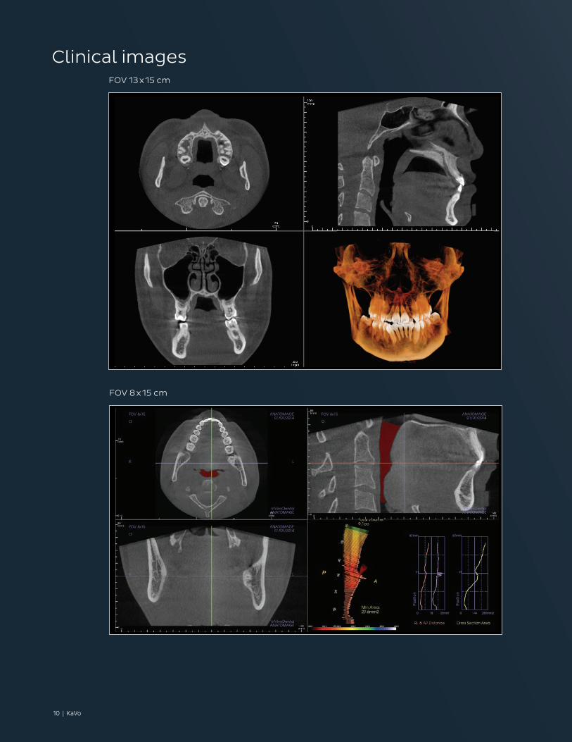
FOV 8 x 15 cmCovers both mandibula and maxilla
including airway and upper
cervical spine or the sinus. Both TM
joints can also be studied.
FOV 13 x 15 cmCovers the entire
maxillofacial region.
10 | KaVo
FOV 13 x 15 cm
FOV 8 x 15 cm
Clinical images
KaVo | 11
FOV 8 x 8 cm
FOV 6 x 8 cm
FOV 5 x 5 cm
FOV 6 x 4 cm
12 | KaVo
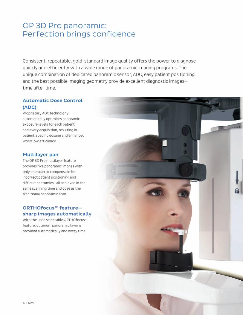
OP 3D Pro panoramic:Perfection brings confidence
Consistent, repeatable, gold-standard image quality offers the power to diagnose quickly and efficiently with a wide range of panoramic imaging programs. The unique combination of dedicated panoramic sensor, ADC, easy patient positioning and the best possible imaging geometry provide excellent diagnostic images— time after time.
Automatic Dose Control (ADC)Proprietary ADC technology
automatically optimizes panoramic
exposure levels for each patient
and every acquisition, resulting in
patient-specific dosage and enhanced
workflow efficiency.
Multilayer panThe OP 3D Pro multilayer feature
provides five panoramic images with
only one scan to compensate for
incorrect patient positioning and
difficult anatomies—all achieved in the
same scanning time and dose as the
traditional panoramic scan.
ORTHOfocus™ feature— sharp images automaticallyWith the user-selectable ORTHOfocus™
feature, optimum panoramic layer is
provided automatically and every time.
KaVo | 13
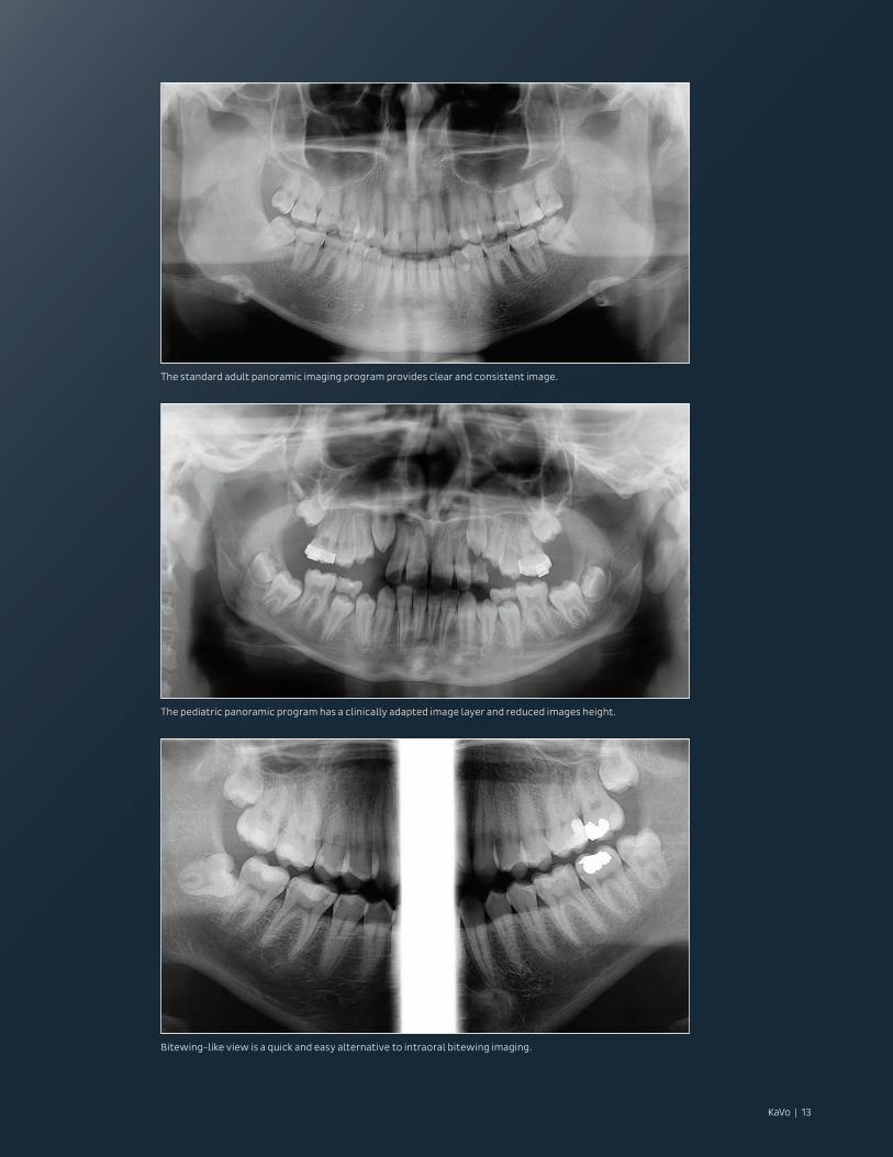
The standard adult panoramic imaging program provides clear and consistent image.
The pediatric panoramic program has a clinically adapted image layer and reduced images height.
Bitewing-like view is a quick and easy alternative to intraoral bitewing imaging.
14 | KaVo
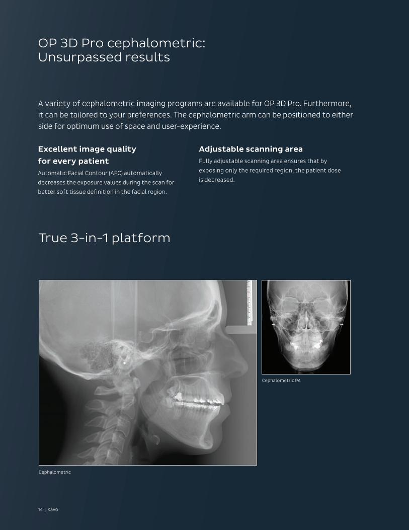
OP 3D Pro cephalometric:Unsurpassed results
True 3-in-1 platform
A variety of cephalometric imaging programs are available for OP 3D Pro. Furthermore, it can be tailored to your preferences. The cephalometric arm can be positioned to either side for optimum use of space and user-experience.
Excellent image quality
for every patientAutomatic Facial Contour (AFC) automatically
decreases the exposure values during the scan for
better soft tissue definition in the facial region.
Adjustable scanning areaFully adjustable scanning area ensures that by
exposing only the required region, the patient dose
is decreased.
Cephalometric
Cephalometric PA
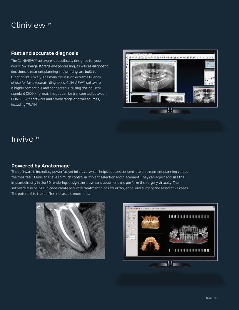
Cliniview™
Invivo™
Fast and accurate diagnosis
The CLINIVIEW™ software is specifically designed for your
workflow. Image storage and processing, as well as diagnostic
decisions, treatment planning and printing, are built to
function intuitively. The main focus is on extreme fluency
of use for fast, accurate diagnoses. CLINIVIEW™ software
is highly compatible and connected. Utilizing the industry-
standard DICOM format, images can be transported between
CLINIVIEW™ software and a wide range of other sources,
including TWAIN.
Powered by AnatomageThe software is incredibly powerful, yet intuitive, which helps doctors concentrate on treatment planning versus
the tool itself. Clinicians have so much control in implant selection and placement. They can adjust and size the
implant directly in the 3D rendering, design the crown and abutment and perform the surgery virtually. The
software also helps clinicians create accurate treatment plans for ortho, endo, oral surgery and restorative cases.
The potential to treat different cases is enormous.
KaVo | 15
16 | KaVo
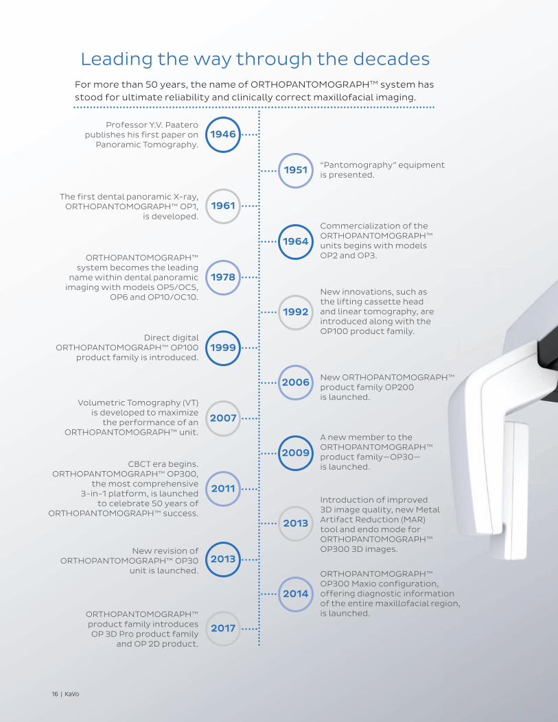
Professor Y.V. Paatero publishes his first paper on
Panoramic Tomography.
The first dental panoramic X-ray, ORTHOPANTOMOGRAPH™ OP1,
is developed.
ORTHOPANTOMOGRAPH™ system becomes the leading
name within dental panoramic imaging with models OP5/OC5,
OP6 and OP10/OC10.
Direct digital ORTHOPANTOMOGRAPH™ OP100
product family is introduced.
Volumetric Tomography (VT) is developed to maximize
the performance of an ORTHOPANTOMOGRAPH™ unit.
CBCT era begins. ORTHOPANTOMOGRAPH™ OP300,
the most comprehensive 3-in-1 platform, is launched
to celebrate 50 years of ORTHOPANTOMOGRAPH™ success.
New revision of ORTHOPANTOMOGRAPH™ OP30
unit is launched.
“Pantomography” equipment is presented.
Commercialization of the ORTHOPANTOMOGRAPH™ units begins with models OP2 and OP3.
New innovations, such as the lifting cassette head and linear tomography, are introduced along with the OP100 product family.
New ORTHOPANTOMOGRAPH™ product family OP200 is launched.
A new member to the ORTHOPANTOMOGRAPH™ product family—OP30— is launched.
Introduction of improved 3D image quality, new Metal Artifact Reduction (MAR) tool and endo mode for ORTHOPANTOMOGRAPH™ OP300 3D images.
ORTHOPANTOMOGRAPH™ OP300 Maxio configuration, offering diagnostic information of the entire maxillofacial region, is launched.
1946
1961
1978
1999
2007
2011
2013
2017
1951
1964
1992
2006
2009
2013
2014
Leading the way through the decadesFor more than 50 years, the name of ORTHOPANTOMOGRAPHTM system has stood for ultimate reliability and clinically correct maxillofacial imaging.
ORTHOPANTOMOGRAPH™ product family introduces OP 3D Pro product family
and OP 2D product.
KaVo | 17
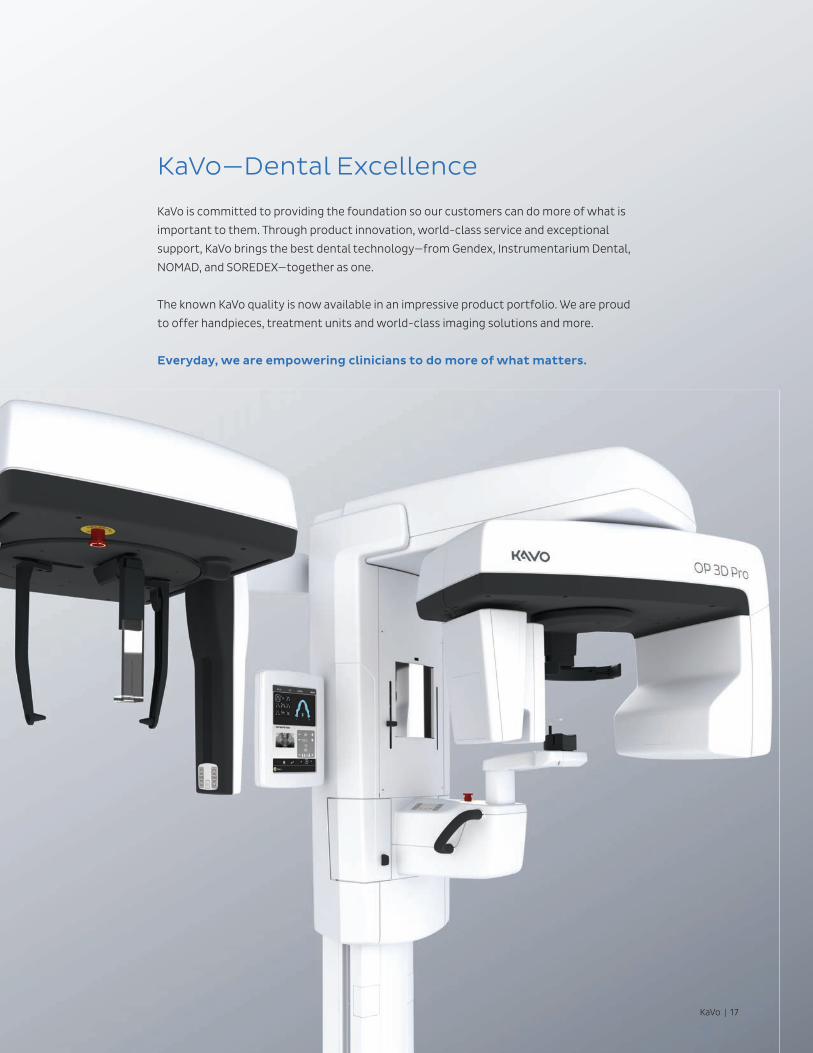
KaVo—Dental Excellence
KaVo is committed to providing the foundation so our customers can do more of what is
important to them. Through product innovation, world-class service and exceptional
support, KaVo brings the best dental technology—from Gendex, Instrumentarium Dental,
NOMAD, and SOREDEX—together as one.
The known KaVo quality is now available in an impressive product portfolio. We are proud
to offer handpieces, treatment units and world-class imaging solutions and more.
Everyday, we are empowering clinicians to do more of what matters.
18 | KaVo
KaVo | 19
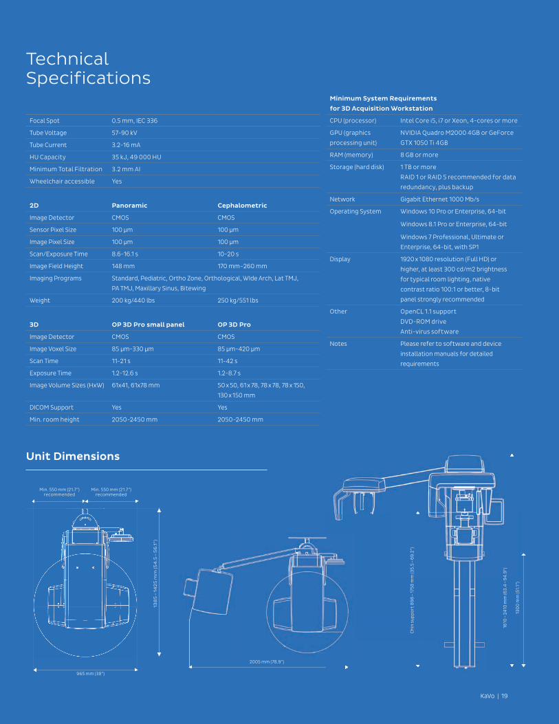
TechnicalSpecifications
Unit Dimensions
Minimum System Requirements
for 3D Acquisition Workstation
CPU (processor) Intel Core i5, i7 or Xeon, 4-cores or more
GPU (graphics
processing unit)
NVIDIA Quadro M2000 4GB or GeForce
GTX 1050 Ti 4GB
RAM (memory) 8 GB or more
Storage (hard disk) 1 TB or more
RAID 1 or RAID 5 recommended for data
redundancy, plus backup
Network Gigabit Ethernet 1000 Mb/s
Operating System Windows 10 Pro or Enterprise, 64-bit
Windows 8.1 Pro or Enterprise, 64-bit
Windows 7 Professional, Ultimate or
Enterprise, 64-bit, with SP1
Display 1920 x 1080 resolution (Full HD) or
higher, at least 300 cd/m2 brightness
for typical room lighting, native
contrast ratio 100:1 or better, 8-bit
panel strongly recommended
Other OpenCL 1.1 support
DVD-ROM drive
Anti-virus software
Notes Please refer to software and device
installation manuals for detailed
requirements
Focal Spot 0.5 mm, IEC 336
Tube Voltage 57-90 kV
Tube Current 3.2-16 mA
HU Capacity 35 kJ, 49 000 HU
Minimum Total Filtration 3.2 mm AI
Wheelchair accessible Yes
2D Panoramic Cephalometric
Image Detector CMOS CMOS
Sensor Pixel Size 100 µm 100 µm
Image Pixel Size 100 µm 100 µm
Scan/Exposure Time 8.6-16.1 s 10-20 s
Image Field Height 148 mm 170 mm–260 mm
Imaging Programs Standard, Pediatric, Ortho Zone, Orthological, WIde Arch, Lat TMJ,
PA TMJ, Maxillary Sinus, Bitewing
Weight 200 kg/440 lbs 250 kg/551 lbs
3D OP 3D Pro small panel OP 3D Pro
Image Detector CMOS CMOS
Image Voxel Size 85 µm-330 µm 85 µm-420 µm
Scan Time 11-21 s 11-42 s
Exposure Time 1.2-12.6 s 1.2-8.7 s
Image Volume Sizes (HxW) 61x41, 61x78 mm 50 x 50, 61 x 78, 78 x 78, 78 x 150,
130 x 150 mm
DICOM Support Yes Yes
Min. room height 2050-2450 mm 2050-2450 mm
2005 mm (78.9”)
1610
– 2
410
mm
(63
.4 –
94
.9”)
130
0 m
m (5
1.1”
)
Ch
in s
up
po
rt 8
98
– 17
58 m
m (3
5.5
– 6
9.2
”)
138
5 –
14
25
mm
(54
.5 –
56
.1”)
965 mm (38”)
Min. 550 mm (21.7”) recommended
Min. 550 mm (21.7”) recommended
ORTHOPANTOMOGRAPH™ / OP™ / SMARTVIEW™ / CLINIVIEW™ / Low Dose Technology™ / ORTHOfocus™are either registered trademarks or trademarks of KaVo Kerr Group Finland in the United States and/or ther countries. KaVo™ is either registered trademark or trademark of Kaltenbach & Voigt GmbH in the United States and/or other countries. All other trademarks are property of their respective owners.
KaVo | 11727 Fruehauf Dr | Charlotte NC 28273 | USA | 1-888-ASK-KAVO
www.kavo.com
KaVo Kerr Group Finland reserves the right to make changes to specifications and features shown herein, or to discontinue the Product described at any time without notice or obligation. Contact your local authorized representative for the most current information. CE marked according to Medical Device Directive (NB 0537). Electrical safety according to IEC 60601-1. Operations comply with ISO 13485:2003, ISO 9001:2008, and ISO 14001:2004. Manufactured by Palodex Group OY, Nahkelantie 160, 04300 Tuusula, Finland.
Dental Excellence from KaVo.
HandpiecesKaVo has always been the leader in creating innovative solutions
for dental practitioners. Our vast line of quality handpieces
showcase our attention to your level of care while delivering
performance that lasts.
Treatment Units Beautiful lines, patient comfort and simple operation are
just a few of the benefits to the line of KaVo treatment
units. Everything you need to perform any procedure—
all in one solution.
Imaging SolutionsDesigned with ease-of-use for all clinicians in mind, KaVo now
offers dependable and consistent imaging solutions that provide
vital information to support accurate diagnosis and predictable
treatment planning.
The products, equipment and services illustrated and described in this brochure reflect knowledge at the
time of printing. KaVo Dental accepts no liability for any deviation from the illustrations in terms of color or
shape, or any errors or print errors, and retains the right to make changes to the brochure at any time. Full or
partial reprinting is only permitted with permission from KaVo Dental. For indications for use, please visit:
www.kavo.com/us/ifu.
© 2017 KaVo Dental. All rights reserved. KV00009/B3.17