Embed Size (px)
Citation preview
1
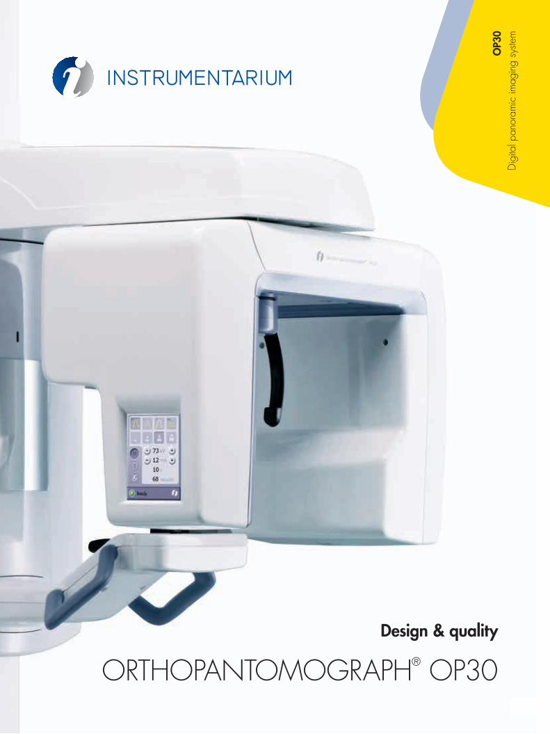
Design & quality
ORTHOPANTOMOGRAPH® OP30
OP3
0 D
igita
l pan
oram
ic im
agin
g sy
stem
2 3
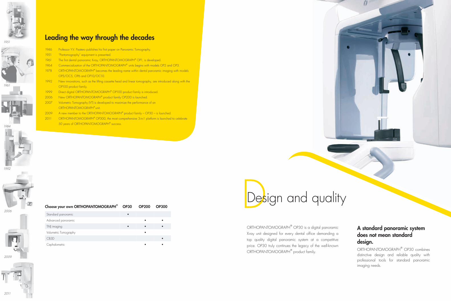
A standard panoramic system does not mean standard design. ORTHOPANTOMOGRAPH® OP30 combines distinctive design and reliable quality with professional tools for standard panoramic imaging needs.
DDesign and quality
ORTHOPANTOMOGRAPH® OP30 is a digital panoramic
X-ray unit designed for every dental office demanding a
top quality digital panoramic system at a competitive
price. OP30 truly continues the legacy of the well-known
ORTHOPANTOMOGRAPH® product family.
1946 Professor Y.V. Paatero publishes his first paper on Panoramic Tomography.
1951 “Pantomography” equipment is presented.
1961 The first dental panoramic X-ray, ORTHOPANTOMOGRAPH® OP1, is developed.
1964 Commercialization of the ORTHOPANTOMOGRAPH® units begins with models OP2 and OP3.
1978 ORTHOPANTOMOGRAPH® becomes the leading name within dental panoramic imaging with models
OP5/OC5, OP6 and OP10/OC10.
1992 New innovations, such as the lifting cassette head and linear tomography, are introduced along with the
OP100 product family.
1999 Direct digital ORTHOPANTOMOGRAPH® OP100 product family is introduced.
2006 New ORTHOPANTOMOGRAPH® product family OP200 is launched.
2007 Volumetric Tomography (VT) is developed to maximize the performance of an
ORTHOPANTOMOGRAPH® unit.
2009 A new member to the ORTHOPANTOMOGRAPH® product family – OP30 – is launched.
2011 ORTHOPANTOMOGRAPH® OP300, the most comprehensive 3-in-1 platform is launched to celebrate
50 years of ORTHOPANTOMOGRAPH® success.
Leading the way through the decades
Choose your own ORTHOPANTOMOGRAPH® OP30 OP200 OP300
Standard panoramic •
Advanced panoramic • •
TMJ imaging • • •
Volumetric Tomography •
CB3D •
Cephalometric • •
4 5
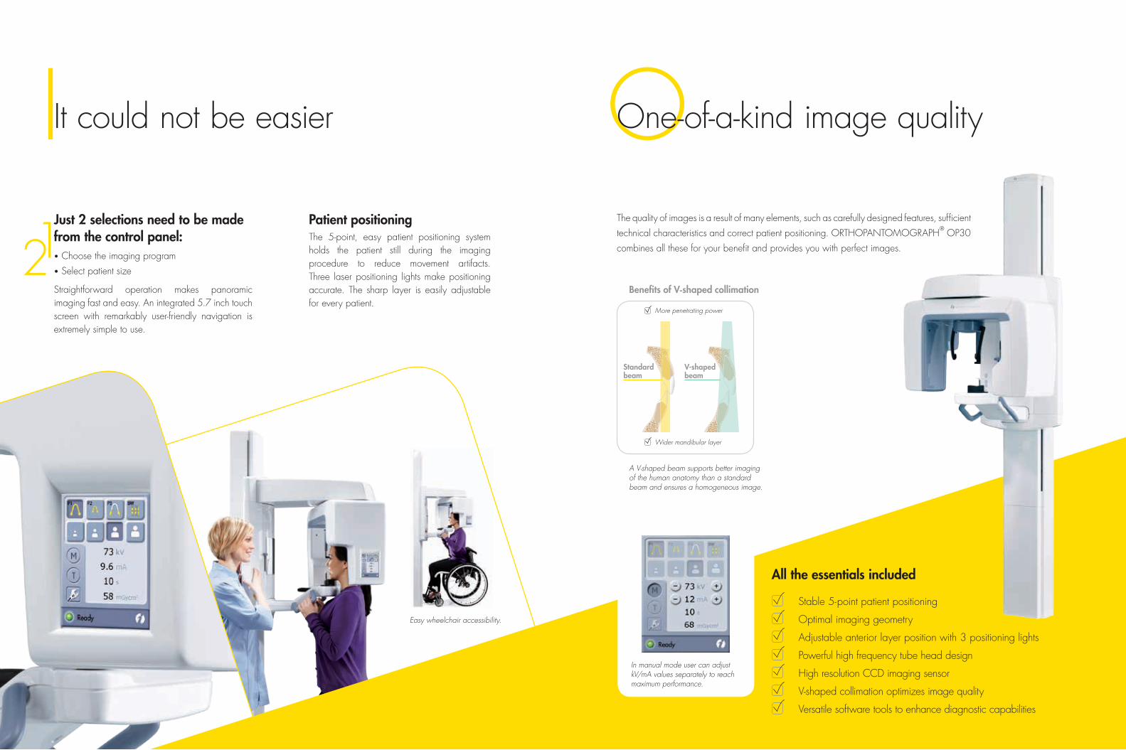
The quality of images is a result of many elements, such as carefully designed features, sufficient
technical characteristics and correct patient positioning. ORTHOPANTOMOGRAPH® OP30
combines all these for your benefit and provides you with perfect images.
Patient positioningThe 5-point, easy patient positioning system holds the patient still during the imaging procedure to reduce movement artifacts. Three laser positioning lights make positioning accurate. The sharp layer is easily adjustable for every patient.
OOne-of-a-kind image qualityIIt could not be easier
Just 2 selections need to be made from the control panel:• Choose the imaging program
• Select patient size
Straightforward operation makes panoramic imaging fast and easy. An integrated 5.7 inch touch screen with remarkably user-friendly navigation is extremely simple to use.
Stable 5- point patient positioning
Optimal imaging geometry
Adjustable anterior layer position with 3 positioning lights
Powerful high frequency tube head design
High resolution CCD imaging sensor
V-shaped collimation optimizes image quality
Versatile software tools to enhance diagnostic capabilities
All the essentials included
A V-shaped beam supports better imaging of the human anatomy than a standard beam and ensures a homogeneous image.
Benefits of V-shaped collimation
More penetrating power
Wider mandibular layer
Standardbeam
V-shapedbeam
Easy wheelchair accessibility.
In manual mode user can adjust kV/mA values separately to reach maximum performance.
6 7
I DImaging programs Dimensions & technical specifications
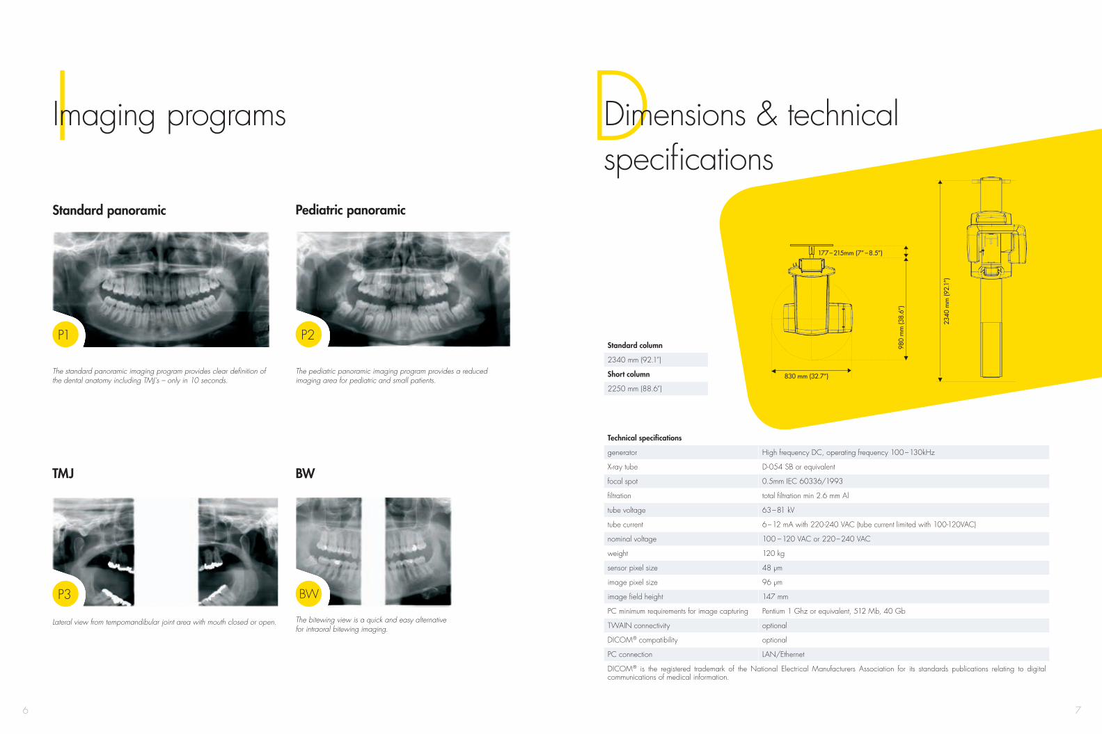
Standard panoramic
TMJ BW
The standard panoramic imaging program provides clear definition of the dental anatomy including TMJ’s – only in 10 seconds.
The pediatric panoramic imaging program provides a reduced imaging area for pediatric and small patients.
Lateral view from tempomandibular joint area with mouth closed or open. The bitewing view is a quick and easy alternative for intraoral bitewing imaging.
830 mm (32.7”)
980
mm
(38.
6”)
177 – 215mm (7” – 8.5”)
2340
mm
(92.
1”)
Pediatric panoramic
Technical specifications
generator High frequency DC, operating frequency 100 – 130kHz
X-ray tube D-054 SB or equivalent
focal spot 0.5mm IEC 60336/1993
filtration total filtration min 2.6 mm Al
tube voltage 63 – 81 kV
tube current 6 – 12 mA with 220-240 VAC (tube current limited with 100-120VAC)
nominal voltage 100 – 120 VAC or 220 – 240 VAC
weight 120 kg
sensor pixel size 48 µm
image pixel size 96 µm
image field height 147 mm
PC minimum requirements for image capturing Pentium 1 Ghz or equivalent, 512 Mb, 40 Gb
TWAIN connectivity optional
DICOM® compatibility optional
PC connection LAN/Ethernet
DICOM® is the registered trademark of the National Electrical Manufacturers Association for its standards publications relating to digital communications of medical information.
Standard column
2340 mm (92.1”)
Short column
2250 mm (88.6”)
P1
P3
P2
BW
8
© 2013 Instrumentarium Dental
205572-5 English
Instrumentarium Dental develops, manufactures and markets
high-tech systems and solutions for dental and maxillo-facial imaging. We work in close co-operation with dental
professionals, universities and other research centers in our quest
to develop solutions that will meet and exceed the expectations
of our customers. As the establisher of panoramic X-ray imaging,
we are committed to providing high clinical performance while
still maintaining simplicity, ease of use and workflow efficiency.
The Instrumentarium Dental product portfolio consists of a
full range of premium quality imaging solutions for intraoral,
extraoral and 3D imaging. For more detailed information about
our products, please visit www.instrumentariumdental.com.
Instrumentarium Dental reserves the right to make changes to specifications and features shown herein, or to discontinue the product described at any time without notice or obligation. Contact your Instrumentarium Dental representative for the most current information. CE marked according to Medical Device Directive (NB 0537). Electrical safety according to IEC 60601-1. Operations comply with ISO 13485:2003, ISO 9001:2008, and ISO 14001:2004.
ORTHOPANTOMOGRAPH® is a registered trademark of
Instrumentarium Dental, PaloDEx Group Oy.
www.instrumentariumdental.com
HeadquartersInstrumentarium DentalNahkelantie 160P.O. Box 20FI-04301 Tuusula FinlandTel. +358 10 270 2000 Fax +358 10 270 2230
USAInstrumentarium Dental Inc. 1245 W. Canal Street Milwaukee, Wisconsin 53233 U.S.ATel. +1 800 558 6120Fax +1 414 481 8665