Embed Size (px)
Citation preview
ORIGINAL RESEARCH PAPER
ORIGINAL ARTICLE ON OCULAR MYIASIS
Dr Sulin Behera Associate Professor
Dr Sharmistha Behera*
Associate Professor *Corresponding Author
Dr Pooja Dash PG Trainee
Dr Deepankar Assistant Professor
Dr Jayashree Dora Professor
ABSTRACTWe report three cases of extensive ocular myiasis : A 50 yr old female , a 70 yr old male and a 60 yr old male . In our first case reported by the lady, a ulcerative lesion of 6x8x1 cm was noted extending from medial canthus to lateral canthus of right eye not involving the lower lid giving a clinical appearance of basal cell carcinoma. In case of the man the lesion noted was 6 x10x2 cm extending from medial canthus to the right ear with preseptal edema in upper lid. In the third case the male presented with an extensive lesion in the left eye with maggots larvae crawling out of the lesion .The larvae isolated were found to be that of Chrysomyia bezziana (screwworm fly).The larva were diligiently removed , debridement of the wound done during dressing. CT scan of reports were found to be normal in all the three cases . Radical antibiotics were given and the wound healed by secondary intention. Excisional biopsy done and sent for culture
KEYWORDSophthalmomyiasis, chrysomyia bezziana
INTRODUCTION :Human ophthalmomyiasis was first reported by Keyt in 1900 and later on by Elliot, in 1910 Ocular Myiasis is the infestation of the human ocular tissue by the immature larval stage of flies called maggots
[1]feeding on the host tissue-dead or living Eye involvement in human myiasis is reported to be less than 5%. The parasites most commonly affecting the eye and orbit are the larva of Hypoderma bovis (hornet fly), Oestrus ovis (sheep botfly), and, rarely, by Chrysomya bezziana. C. bezziana, also known as Old World screwworm, is an obligate parasite and belongs to the order Diptera, family Calliphyridae, and suborder Cyclorrhpha. We report the clinical history and presentations of 3 different patients who presented with extensive ocular myiasis
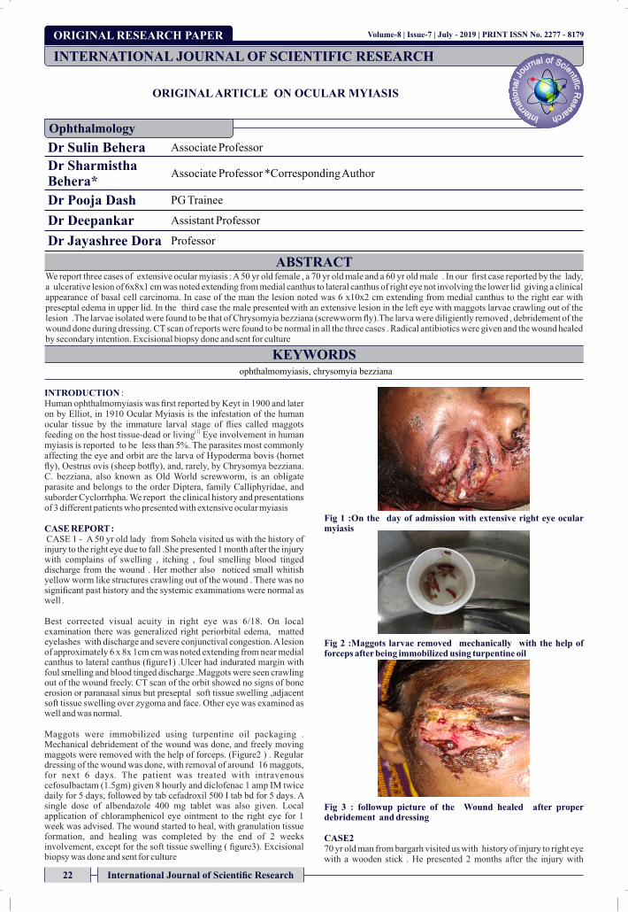
CASE REPORT : CASE 1 - A 50 yr old lady from Sohela visited us with the history of injury to the right eye due to fall .She presented 1 month after the injury with complains of swelling , itching , foul smelling blood tinged discharge from the wound . Her mother also noticed small whitish yellow worm like structures crawling out of the wound . There was no significant past history and the systemic examinations were normal as well .
Best corrected visual acuity in right eye was 6/18. On local examination there was generalized right periorbital edema, matted eyelashes with discharge and severe conjunctival congestion. A lesion of approximately 6 x 8x 1cm cm was noted extending from near medial canthus to lateral canthus (figure1) .Ulcer had indurated margin with foul smelling and blood tinged discharge .Maggots were seen crawling out of the wound freely. CT scan of the orbit showed no signs of bone erosion or paranasal sinus but preseptal soft tissue swelling ,adjacent soft tissue swelling over zygoma and face. Other eye was examined as well and was normal.
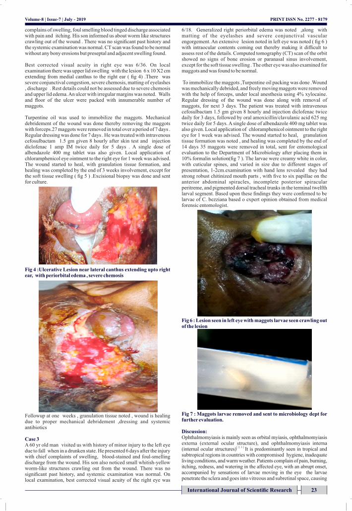
Maggots were immobilized using turpentine oil packaging . Mechanical debridement of the wound was done, and freely moving maggots were removed with the help of forceps. (Figure2 ) . Regular dressing of the wound was done, with removal of around 16 maggots, for next 6 days. The patient was treated with intravenous cefosulbactam (1.5gm) given 8 hourly and diclofenac 1 amp IM twice daily for 5 days, followed by tab cefadroxil 500 I tab bd for 5 days. A single dose of albendazole 400 mg tablet was also given. Local application of chloramphenicol eye ointment to the right eye for 1 week was advised. The wound started to heal, with granulation tissue formation, and healing was completed by the end of 2 weeks involvement, except for the soft tissue swelling ( figure3). Excisional biopsy was done and sent for culture
Fig 1 :On the day of admission with extensive right eye ocular myiasis
Fig 2 :Maggots larvae removed mechanically with the help of forceps after being immobilized using turpentine oil
Fig 3 : followup picture of the Wound healed after proper debridement and dressing
CASE2 70 yr old man from bargarh visited us with history of injury to right eye with a wooden stick . He presented 2 months after the injury with
INTERNATIONAL JOURNAL OF SCIENTIFIC RESEARCH
Ophthalmology
Volume-8 | Issue-7 | July - 2019 | PRINT ISSN No. 2277 - 8179
22 International Journal of Scientific Research
complains of swelling, foul smelling blood tinged discharge associated with pain and itching. His son informed us about worm like structures crawling out of the wound . There was no significant past history and the systemic examination was normal. CT scan was found to be normal without any bony erosions but preseptal and adjacent swelling found.
Best corrected visual acuity in right eye was 6/36. On local examination there was upper lid swelling with the lesion 6 x 10 X2 cm extending from medial canthus to the right ear ( fig 4) .There was severe conjunctival congestion, severe chemosis, matting of eyelashes , discharge . Rest details could not be assessed due to severe chemosis and upper lid edema. An ulcer with irregular margins was noted. Walls and floor of the ulcer were packed with innumerable number of maggots.
Turpentine oil was used to immobilize the maggots. Mechanical debridement of the wound was done thereby removing the maggots with forceps.27 maggots were removed in total over a period of 7 days . Regular dressing was done for 7 days . He was treated with intravenous cefosulbactam 1.5 gm given 8 hourly after skin test and injection diclofenac 1 amp IM twice daily for 5 days . A single dose of albendazole 400 mg tablet was also given. Local application of chloramphenicol eye ointment to the right eye for 1 week was advised. The wound started to heal, with granulation tissue formation, and healing was completed by the end of 3 weeks involvement, except for the soft tissue swelling ( fig 5 ) .Excisional biopsy was done and sent for culture.
Fig 4 :Ulcerative Lesion near lateral canthus extending upto right ear, with periorbital edema , severe chemosis
Followup at one weeks , granulation tissue noted , wound is healing due to proper mechanical debridement ,dressing and systemic antibiotics
Case 3 A 60 yr old man visited us with history of minor injury to the left eye due to fall when in a drunken state. He presented 6 days after the injury with chief complaints of swelling, blood-stained and foul-smelling discharge from the wound. His son also noticed small whitish-yellow worm-like structures crawling out from the wound. There was no significant past history, and systemic examination was normal. On local examination, best corrected visual acuity of the right eye was
6/18. Generalized right periorbital edema was noted ,along with matting of the eyelashes and severe conjunctival vascular engorgement. An extensive lesion noted in left eye was noted ( fig 6 ) with intraocular contents coming out thereby making it difficult to assess rest of the details. Computed tomography (CT) scan of the orbit showed no signs of bone erosion or paranasal sinus involvement, except for the soft tissue swelling . The other eye was also examined for maggots and was found to be normal.
To immobilize the maggots ,Turpentine oil packing was done .Wound was mechanically debrided, and freely moving maggots were removed with the help of forceps, under local anesthesia using 4% xylocaine. Regular dressing of the wound was done along with removal of maggots, for next 3 days. The patient was treated with intravenous cefosulbactam 1.5 gm given 8 hourly and injection diclofenac twice daily for 3 days, followed by oral amoxicillin/clavulanic acid 625 mg twice daily for 5 days. A single dose of albendazole 400 mg tablet was also given. Local application of chloramphenicol ointment to the right eye for 1 week was advised. The wound started to heal, granulation tissue formation was noted , and healing was completed by the end of 14 days 35 maggots were removed in total, sent for entomological evaluation to the Department of Microbiology after placing them in 10% formalin solution(fig 7 ). The larvae were creamy white in color, with cuticular spines, and varied in size due to different stages of presentation, 1-2cm.examination with hand lens revealed they had strong robust chitinized mouth parts , with five to six papillae on the anterior abdominal spiracles, incomplete posterior spiracular peritreme, and pigmented dorsal tracheal trunks in the terminal twelfth larval segment. Based upon these findings they were confirmed to be larvae of C. bezziana based o expert opinion obtained from medical forensic entomologist.
Fig 6 : Lesion seen in left eye with maggots larvae seen crawling out of the lesion
Fig 7 : Maggots larvae removed and sent to microbiology dept for further evaluation.
Discussion: Ophthalmomyiasis is mainly seen as orbital myiasis, ophthalmomyiasis externa (external ocular structure), and ophthalmomyiasis interna
[ 2, 3 ] (internal ocular structures) It is predominantly seen in tropical and subtropical regions in countries with compromised hygiene, inadequate living conditions, and warm weather. Patients complain of pain, burning, itching, redness, and watering in the affected eye, with an abrupt onset, accompanied by sensations of larvae moving in the eye the larvae penetrate the sclera and goes into vitreous and subretinal space, causing
PRINT ISSN No. 2277 - 8179Volume-8 | Issue-7 | July - 2019
International Journal of Scientific Research 23

ophthalmomyiasis interna as a complication if timely treatment is not started thereby manifesting as pigmented and atrophic retinal pigment epithelial tracts, in conjunction with fibrovascular proliferation, hemorrhage, and exudative detachment of the retina leading to blindness .Infiltration into the lacrimal sac and can migration through the lacrimal canal to the nasal cavity of maggots is also seen. Base of the skull may also be involved if the maggots reach into the cranial cavity .
Chronic debilitating conditions, such as leprosy, diabetes mellitus, open wounds, fungating carcinomas, psychiatric illness, intellectual disability, hemiplegia, and immunosuppressive agents are the main risk groups predisposing to myiasis Only C. bezziana and Cochliomyia hominivorax have been implicated in causing ophthalmomyiasis in
[4].living humans Peculiar characterstic of C. bezziana is it can invade tissue without preexisting necrotic tissue and cause extensive damage to living tissue if left untreated. It buries itself deep in the tissue, causes necrosis, and remains firmly attached to it with help of its cuticular spines. poverty and poor hygiene are considered as the main risk factors . Despite of improvement in health sector in our country , ocular myiasis has proved to be a debacle due to compromised hygiene and economic state of a developing country like India .
Basic method of removing the maggots is either suffocating the larvae and forcing them out or first paralyzing them followed with mechanical debridement . Systemic treatment with broad-spectrum antibiotics, such as amoxicillin with clavulanic acid, metronidazole,
[5].and cefazolin are indicated to prevent secondary bacterial infections Antiparasitic drugs such as ivermectin, a semisynthetic macrocyclic lactone, can be used in cases of advanced orbital myiasis, in a dose of
[6]200 μg/kg
REFERENCES :1. Sivaramasubramanyam P, Sadanand AV. Ophthalmomyiasis. Br J Ophthalmol.
1968;52(1):64.2. Khataminia G, Aghajanzadeh R, Vazirianzadeh B, Rahdar M. Orbital myiasis. J
Ophthalmic Vis Res. 2011;6(3):199–203. 3. Sigauke E, Beebe WE, Gander RM, Cavuoti D, Southern PM. Case report:
ophthalmomyiasis externa in Dallas County, Texas. Am J Trop Med Hyg. 2003;68(1):46–47.
4. Khurana S, Biswal M, Bhatti H. Ophthalmomyiasis: Three cases from North India. Indian J Med Microbiol. 2010;28(3):257–261
5. Asokan GS, Anand V, Balaji N, Parthiban J, Jeelani S. Maggots in the mouth–oral myiasis: a rare case report. J Indian Aca Oral Med Radiol. 2013;25(3):225–228.
6. Campbell WC. Ivermectin: an update. Parasitol Today. 1985;1(1):10–16.
PRINT ISSN No. 2277 - 8179Volume-8 | Issue-7 | July - 2019
24 International Journal of Scientific Research