Embed Size (px)
Citation preview
Int J Clin Exp Pathol 2016;9(3):3115-3124www.ijcep.com /ISSN:1936-2625/IJCEP0022178
Original ArticlePrognostic value of the density of Wilms tumour protein 1 (WT-1) positive microvessels in stage IIA colorectal cancer
Valeria Barresi1, Luca Reggiani Bonetti2, Giovanni Branca1, Enrica Vitarelli1, Antonio Ieni1, Giovanni Tuccari1
1Department of Human Pathology “G. Barresi”, AOU G. Martino, Pad D, Via Consolare Valeria, Messina 98125, Italy; 2Department of Forensic Medicine, Laboratory and Pathologic Anatomy, Polyclinic of Modena, Via del Pozzo, Modena 41124, Italy
Received December 17, 2015; Accepted February 26, 2016; Epub March 1, 2016; Published March 15, 2016
Abstract: Stage IIA colorectal cancer (CRC) displays 5-year survival rate around 85%; hence it is not currently submit-ted to any adjuvant treatment. However a percentage of stage IIA CRCs undergoes disease progression and would benefit from additional therapies. In the aim to investigate whether Wilms Tumor-1 (WT-1) expression might repre-sent a prognostic factor in stage IIA CRC, we analyzed its immunohistochemical expression in 90 stage IIA CRCs divided into two groups according to the evidence of disease progression. While WT-1 staining in the neoplastic cells was not significantly associated with any of the clinico-pathological parameters, the density of intra-tumoral microvessels positive for WT-1 (WT-1 MVD) was significantly higher in stage IIA CRCs characterized by disease progression compared to non-recurring tumours and it was significantly and independently associated with shorter disease-free survival. This study is the first to demonstrate that WT-1 MVD may be useful to discriminate high risk patients with stage IIA CRCs and who may benefit from adjuvant treatment. WT-1 expression in the tumor vessels, but not in the vessels of normal colorectal mucosa, suggests its possible role in tumor neo-angiogenesis and it may represent a target for novel anti-angiogenic therapies in stage IIA CRC at high risk of progression.
Keywords: Colorectal cancer, WT-1, microvessel density, prognosis, stage IIA, poorly differentiated clusters
Introduction
According to the current (7th) edition of the post-surgical (p) TNM (tumor, node metastasis) staging of malignant tumours [1, 2], stage II colorectal carcinomas (CRCs) are those spread-ing into the subserosa or non peritonealized pericolic or perirectal tissue (pT3), or perforat-ing visceral peritoneum and/or directly invading other organs or structures (pT4), in the absence of nodal (N0) and distant metastasis (M0). On the whole, stage II CRC has good prognosis, with 5-year overall survival rate ranging bet- ween 75% and 95% after surgery alone [3, 4]. Hence the benefit of adjuvant chemotherapy in patients affected by stage II CRC is controver-sial [5, 6] and, according to the current interna-tional guidelines, only patients at high risk of progression should receive additional treat-ments [3, 7, 8]. At present, high-risk patients are identified by reason of several pathological
parameters such as pT4 stage, lymphatic/vas-cular invasion, positive margins, poorly differen-tiated histology and a number of harvested lymph nodes lower than 12 [3, 7, 8]. Hence, among stage II CRCs, stage IIA (pT3N0M0) tumors do not receive adjuvant chemotherapy because of their significantly better prognosis compared to stage IIB (pT4aN0M0) and stage IIC (pT4bN0M0) ones [5]. However, around 10% of patients affected by stage IIA CRC undergo disease progression for causes still to be deter-mined [8]. For this reason, there is the need to identify potential immunomarkers which could allow to discriminate patients with stage IIA CRCs at high risk of progression, who could benefit from adjuvant chemotherapy.
Wilms tumour protein-1 (WT-1) is a transcription factor which controls cell growth and differenti-ation, tumorigenesis and normal and neoplas- tic angiogenesis through the transcriptional or
WT-1 MVD in CRC
3116 Int J Clin Exp Pathol 2016;9(3):3115-3124
post-transcriptional regulation of gene expres-sion [9-16]. WT-1 was firstly identified in Wilm’s tumour [17] and it was initially considered to be an oncosuppressor due to its ability to inhibit GC-rich gene promoters [16]. Recently, with the advent of commercially available antibodies (clone 6F-H2) against the N-terminus of the pro-tein, WT-1 has been identified in the cytoplasm of several human developing tissues (skeletal muscle tissues; endothelial cells, neuroblasts; radial glia) [18] as well as in the neoplastic cells of several pediatric tumors, such as infantile fibrosarcoma, ganglioneuroblastoma and rhab-domyosarcoma [19, 20]. The demonstration of WT-1 over-expression in breast, lung and thy-roid carcinomas [9, 10, 13-15] suggested that this protein may also have a pro-tumorigenic role and that it behaves as an oncosuppressor or as an oncogene depending upon the cellular context [21]. Of note, WT-1 was defined as the most important cancer antigen in a ranking based on specificity, oncogenicity, immunoge-nicity and therapeutic function [22]. In addition, it seems to be one of the most promising target antigens for tumour immunotherapy [23].
To the best of our knowledge, only few studies investigated the expression of WT-1 in CRC [14, 24-26]. According to those, WT-1 is over-expressed in CRC surgical tissue and cell lines compared to normal colonic mucosa [14, 24-26] and it is significantly associated with worse prognosis in this tumor [26]. In addition, experiments in vitro suggest that WT-1 protein may represent a potential target for antigen-specific immunotherapy in human colon cancer [26].
Given this premise, the goal of the present study was to investigate the possible prognos-tic value of WT-1 expression in the specific sub-group of pTNM stage IIA (pT3N0M0) CRCs. As WT1 expression was previously demonstrated in the vessels of CRC [25], we also assessed the density of WT-1 stained microvessels (WT-1 MVD) and its prognostic value for recurrence risk in stage IIA CRC.
Materials and methods
Study subjects and clinical data
43 pTNM Stage IIA (pT3N0M0) consecutive CRCs which had been surgically resected between 2002 and 2004 and which had under-
gone to disease progression were taken from our files and included in the present study. Then, 47 stage IIA CRCs occurred in the same years and with no evidence of disease progres-sion in a follow-up time longer than five years were selected. Finally, the cohort in the study included a total of 90 cases (age range: 42-90 years; mean age: 69 years).
All cases were anonymously collected and all procedures were performed in accordance with the Helsinki Declaration. All relevant ethical issues were identified and discussed with the local Ethical Committee. No further ethical approval was necessary to perform immunohis-tochemistry in the cases included in the study herein.
None of the patients had received chemothera-py for their cancer. In all the cases, pathological staging had been performed according to the pTNM system and at least 12 lymph nodes had been retrieved from the peri-visceral adipose tissue.
According to their location, the tumors were divided into three groups: 1) CRC located in the right colon, including caecum, ascending and transverse colon, 2) CRC located in the left colon, including descending and sigmoid colon, and 3) CRC located in the rectum.
Pathological analyses
All tumors had been fixed in neutral buffered formalin and paraffin embedded at 56°C for the histological evaluation with haematoxylin and eosin (H&E) stain. For each case, we revised the H&E-stained slides and assessed the histological grade according to the WHO cri-teria [2] as well as the tumor budding and tumor border configuration. Tumor budding was defined as isolated single cancer cells or clus-ters of cells composed of less than five ele-ments in the stroma of the actively invasive margin of the tumor. In detail, after choosing one field where the budding was the most intensive, a budding count was made under × 200 magnification, with a count of less than five foci considered as negative and a count of five or more as positive [27].
For each case we also evaluated the histologi-cal grade based on the count of poorly differen-tiated clusters (PDC) of cancer cells in tumor
WT-1 MVD in CRC
3117 Int J Clin Exp Pathol 2016;9(3):3115-3124
tissue, as previously described [28-30]. PDC grade was determined according to the original definition provided by Ueno and coll [31]. who described PDC as cancer clusters composed of ≥ 5 cancer cells lacking a gland-like structure within the tumor stroma (at the invasive front and within the tumor). The whole tumor was first scanned at a lower power magnification to identify the area with the highest number of PDC. Then the clusters were counted under the microscopic field of a × 20 objective lens (i.e., a microscopic field with a major axis of 1 mm), using a Zeiss microscope. Cancers with < 5, 5 to 9, and ≥ 10 clusters were classified as grade 1 (G1), grade 2 (G2) and grade 3 (G3), respec-tively, as previously suggested [31].
Immunohistochemistry
4 µm consecutive tissue sections were cut from one representative paraffin block of each tumor for the immunohistochemical reactions against WT-1 and CD34.
The intrinsic endogenous peroxidase activity was blocked with 0.1% H2O2 in methanol for 20 min; then, normal sheep serum was applied for 30 min. to prevent unspecific adherence of serum proteins. WT-1 antigen was unmasked by microwave oven pre-treatment at 750 W in 10 mM, pH 6.0 sodium citrate buffer for 3 cycles × 5 min. Sections were successively incubated with the primary antibodies against WT-1 (clone 6F-H2; Dako Cytomation, Glostrup, Denmark; working dilution 1:50) and CD34 (clone QBEnd; Dako Cytomation, Glostrup, Denmark; working dilution 1:50) by using Dako Autostainer. The bound primary antibodies were visualized by using the LSAB kit (Dako Cytomation, Glostrup, Denmark) according to
Nuclear counterstaining was performed by Mayer’s haemalum. Sections of Wilms’ tumour tissue were used as positive controls for WT-1 immunohistochemical reaction.
Two independent pathologists, blinded to the clinico-pathological data, examined the whole immunostained sections. In case of disagree-ment, consensus was reached by examination using a double-headed microscope.
Staining for WT-1 was evaluated in the neoplas-tic cells and in the tumor vessels in all the cases.
WT-1 immuno-expression in the tumor cells was scored semi-quantitatively by taking into account the intensity of staining (IS) and the area of staining positivity (ASP). The IS was graded as weak (1), moderate (2), strong (3), while ASP was classified as 0 (absence of stained cells), 1 (1-50% stained cells), 2 (> 50% stained cells). Then an intensity distribution (ID) score was obtained for each case by multiply-ing the value of IS and ASP. Cases showing an ID score of 0 were considered to be negative for WT-1.
The quantification of WT-1 positive microves-sels was performed as previously described [32-37]. Briefly, the three most vascularized areas detected by WT-1 staining were initially selected (so-called hot spots) under 40 × field. Then WT-1 positive microvessels were counted in each of these areas under a 400 × field. Only vessels with a clearly defined lumen were counted. The assessment was done within the cancerous mass, excluding all the necrotic and the ulcerated areas. The mean value of three × 400 field (0.30 mm2) counts was recorded as
Table 1. Clinico-pathological characteristics and WT-1 MVD in the 90 stage IIA CRCs in the studyAge (mean ± SD) 69 ± 9.9Site (n) Right colon (45) Left colon (40) Rectum (5)WHO Grade (n) Low (66) High (24)PDC grade (n) G1 (61) G2 (16) G3 (13)TB (n) Absent (57) Present (33)Tumour border configuration Expansive (42) Infiltrative (48)WT-1 expression neoplastic cells (n) Absent (71) Present (19)WT-1 MVD (mean ± SD) 33.51 ± 25.12CD34 MVD (mean ± SD) 68.83 ± 25.63Recurrences (n) Absent (47) Present (43)
the manufacturer’s in- structions. To reveal the immunostaining, the sections were in- cubated in darkness for 10 min. with 3-3’ diaminobenzidine tet- ra hydrochloride (Sig- ma Chemical Co., St. Louis, MO, USA), in the amount of 100 mg in 200 ml 0.03% hyd- rogen peroxide in ph- osphate-buffered sa- line solution (PBS).
WT-1 MVD in CRC
3118 Int J Clin Exp Pathol 2016;9(3):3115-3124
WT-1 MVD of the section. Finally the MVD value was converted into the mean number of microvessels/mm2 for the statistical analyses. The vessels were counted using a Zeiss micro-scope by two independent observers blinded to the clinico-pathological data. The same proce-dure was carried out on consecutive slides stained by anti-CD34 antibody.
Statistical analyses
The statistical association between WT-1 expression in the tumor cells and the various clinico-pathological variables was analyzed by using Chi-squared test, while Mann-Whitney and Kruskal-Wallis tests were applied to inves-tigate the correlation between WT-1 MVD and
the clinico-pathological features of the CRCs. Spearman correlation test was carried out to verify the correlation between WT-1- and CD34-MVD.
Disease free survival (DFS) was assessed by the Kaplan-Meier method, with the date of pri-mary surgery as the entry data and length of survival to the detection of a recurrent tumor as the end point. The Mantel-Cox log-rank test was applied to assess the strength of association between DFS and each of the parameters (site, WHO grade, tumor budding, PDC grade, WT-1 MVD, CD34 MVD) as a single variable. Succe- ssively, a multivariate analysis (Cox regression model) was utilized to determine the indepen-dent effect of each variable on DFS.
The median values in the cohort (WT-1-MVD median value: 26.6; CD34 median value: 64.95) were used as cut-off values for the DFS analyses according to WT-1-MVD and CD34-MVD of the tumors. A probability (P) value lower than 0.05 was considered to be statistically sig-nificant. Data were analyzed using the SPSS package version 6.1.3 (SPSS Inc., Chicago, IL, USA).
Results
The clinico-pathological characteristics and immunohistochemical data of the analyzed cases are summarized in Table 1.
In 19 cases WT-1 staining was observed in the cytoplasm of the neoplastic cells (ID score > 0). Of those cases, 10 had WT-1 ID score 1, 6 ID
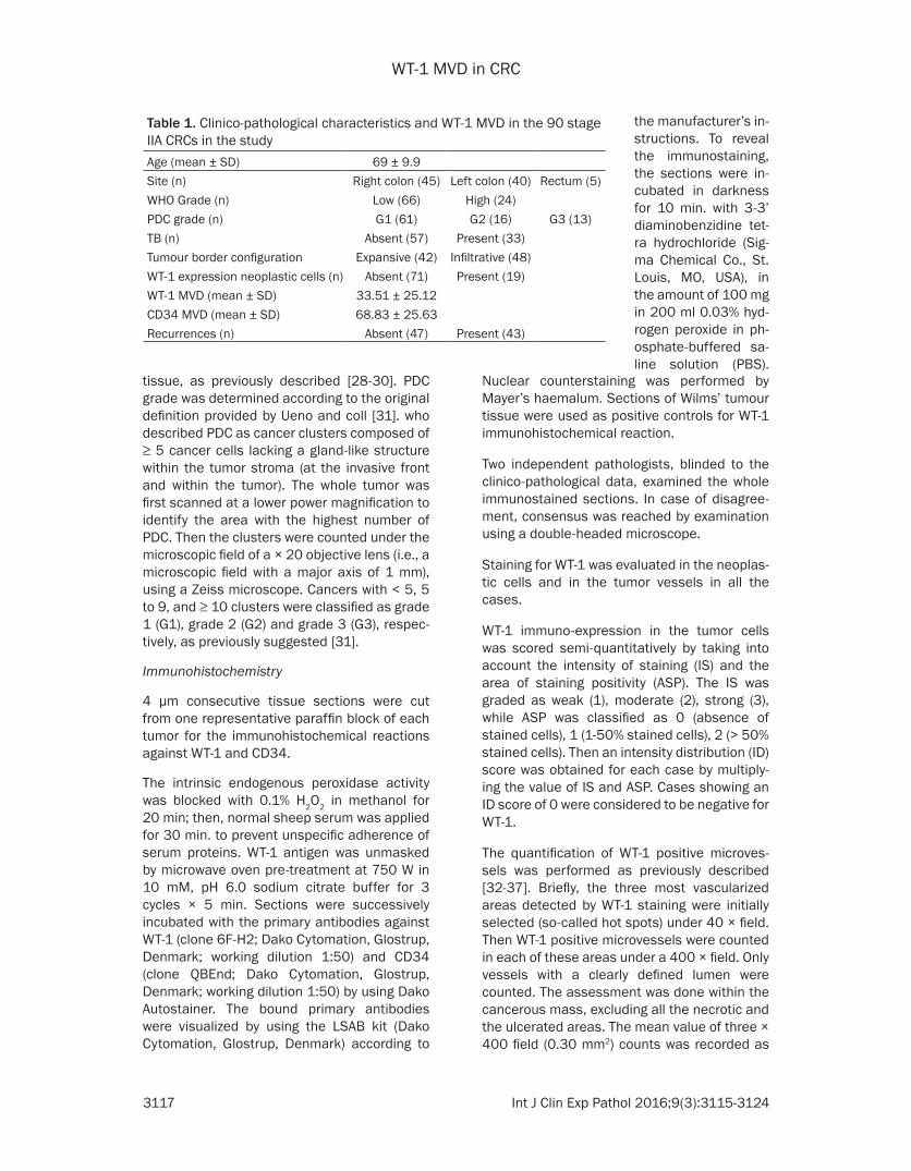
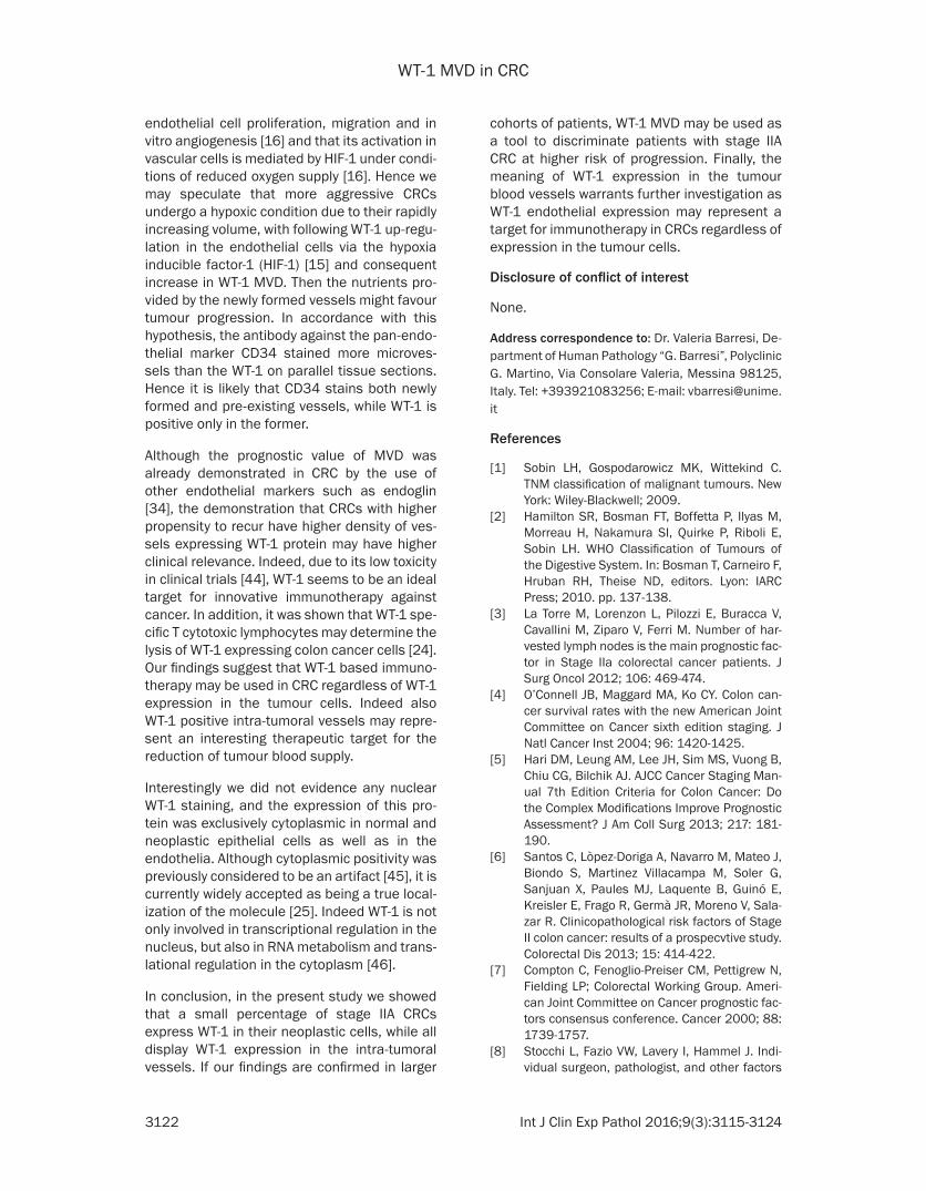
Figure 1. A. Cytoplasmic staining for WT-1 in the neoplastic cells of CRC (WT-1 stain; original magnification, × 200). B. Weak cytoplasmic staining for WT-1 in the epithelial cells of normal colonic mucosa adjacent to a CRC (WT-1 stain; original magnification, × 200).
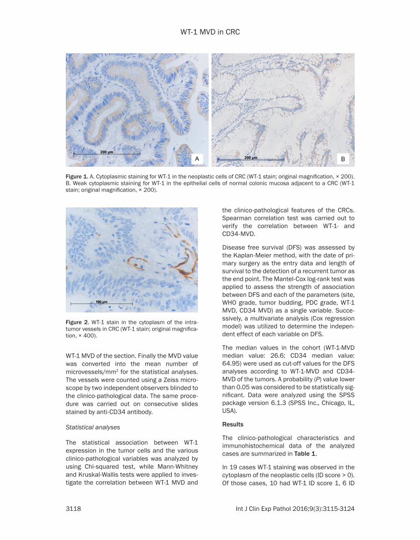
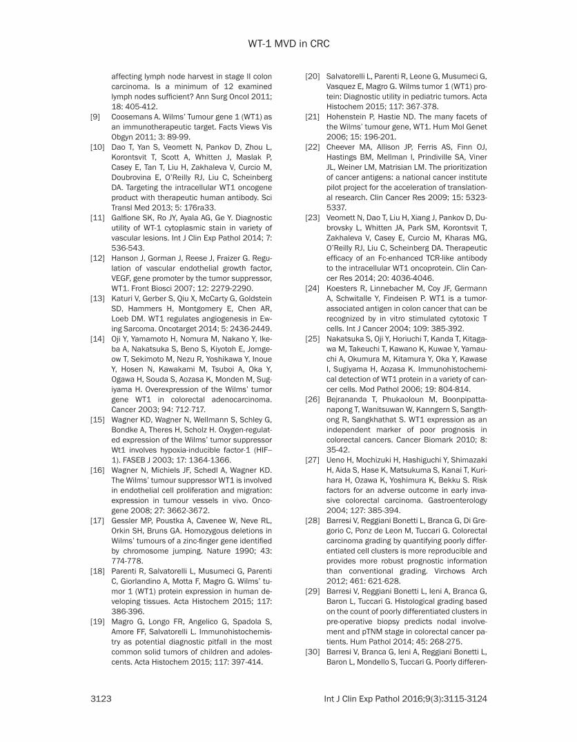
Figure 2. WT-1 stain in the cytoplasm of the intra-tumor vessels in CRC (WT-1 stain; original magnifica-tion, × 400).
WT-1 MVD in CRC
3119 Int J Clin Exp Pathol 2016;9(3):3115-3124
score 2 and 3 ID score 3 (Figure 1A). The per-centage of stained cells was always lower than 50% (ASP < 2) in all of the positive cases. WT-1 staining in the neoplastic cells was observed with higher frequency in WHO low-grade CRCs, although statistical significance was not reached (P=0.0673). No significant association was evidenced with the other clini-co-pathological features (P > 0.05). In 3 out of the 90 analyzed cases WT-1 immuno-reactivity was also appreciable in the glands of normal colonic mucosa adjacent to the tumors (Figure 1B).
In all the analyzed cases WT-1 staining was noted in the cytoplasm of the endothelial cells of tumor vessels (Figure 2), with variable num-ber of stained vessels. Specifically, WT-1 MVD ranged between 1.11 and 156.6 (mean: 33.51 ± 25.12) in the CRCs of our cohort. Statistical analyses showed that the density of WT-1 posi-tive vessels was significantly higher in CRCs showing high PDC grade (P=0.028), infiltrative tumor border configuration (P=0.038), and dis-ease progression (P=0.0017) (Table 2; Figure 3).
In 38 cases WT-1 expression was also evi-denced in the endothelia of some of the ves-sels present within the normal mucosa adja-cent to the tumors. In those cases, WT-1 MVD in normal mucosa ranged between 1.11 and 20 (mean: 7.49 ± 7.3) and it was significantly lower compared to that assessed in the neo-plastic tissue (P < 0.0001).
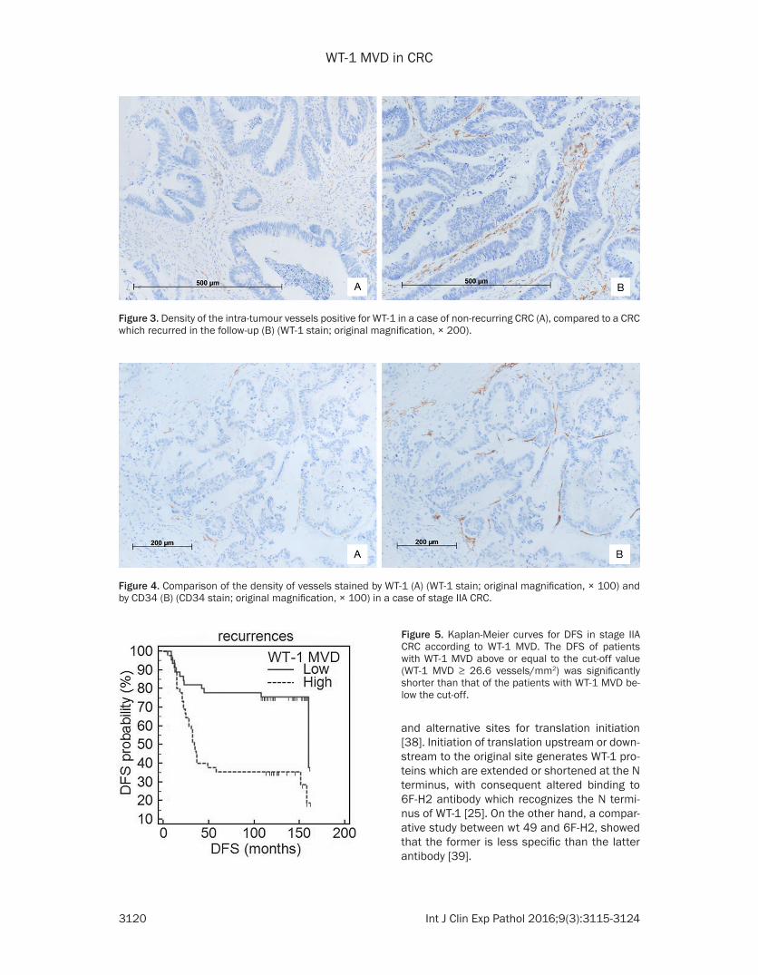
CD34 immunoreaction stained all kinds of ves-sels present in all CRCs. In the case by case analysis, the number of microvessels stained by WT-1 was lower than the one revealed by CD34 staining (27.87 ± 26.41 versus 68.83 ± 25.63) (Figure 4); by Spearman’s test, the- re was a highly significant correlation betwe- en WT-1-MVD and CD34-MVD (r=0.785, P < 0.0001).
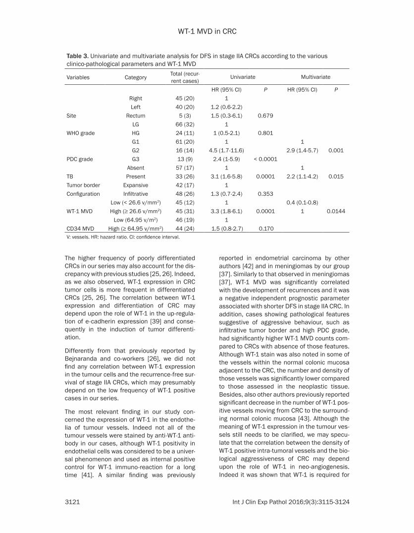
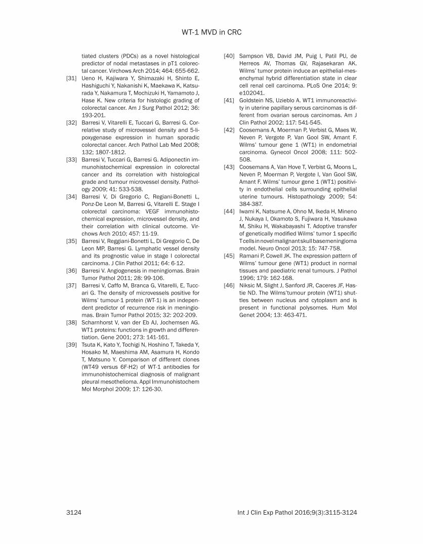
The follow-up time ranged between 9 and 162 months. Univariate analysis revealed that high PDC grade (P < 0.0001) the presence of tumor budding (P=0.0001), and high (≥ 26.6 vessels/mm2) WT-1 MVD (P=0.0001) (Figure 5; Table 3) were significant negative prognostic parame-ters associated with shorter DFS in stage IIA CRC. All of those were independent prognostic variables in multivariate analysis by using step-wise selection method (Table 3).
Discussion
In this study we investigated the immunohisto-chemical expression of WT-1 in a series of stage IIA CRCs. WT-1 expression was evidenced in the neoplastic cells of 21% CRCs. This per-centage is significantly lower compared to those (80% and 91.1%, respectively) demon-strated in previous papers [14, 26]. Disagree- ment between our data and those achieved by other authors [14, 26] may depend upon the use of different antibodies for the detection of WT-1. Indeed we used WT-1 monoclonal anti-body clone 6F-H2, while clone WT49 [26] and the polyclonal antibody C19 [14] were used in the other immunohistochemical analyses. The two monoclonal antibodies 6F-H2 and WT 49 both react with the N terminus of WT-1 protein, while polyclonal antibody C-19 binds to the C terminus [14]. It was shown that the sensitivity of C-19 and 6F-H2 antibodies differs greatly among tumours and this difference may depend upon aberrant or deregulated splicing and alterations of the WT-1 gene [25]. Indeed WT1 gene encodes for at least 24 isoforms, which originate from alternative splicing, RNA editing,
Table 2. Statistical correlation between WT-1 MVD and the various clinico-pathological pa-rameters of stage IIA CRCs assessed by Mann-Whitney and Kruskal-Wallis testsVariable n Mean rank PSite Right colon 45 42.21 Left colon 40 50.43 Rectum 5 35.7 0.241WHO Grade Low grade 66 44.7 High grade 24 47.5 0.561PDC grade G1 61 40.4 G2 16 55.2 G3 13 57.1 0.028Tumour budding Absent 57 42.3 Present 33 50.8 0.136Tumour border configuration Expansive 42 39.4 Infiltrative 48 50.8 0.038Recurrences Not 47 37.2 Yes 43 54.5 0.0017
WT-1 MVD in CRC
3120 Int J Clin Exp Pathol 2016;9(3):3115-3124
and alternative sites for translation initiation [38]. Initiation of translation upstream or down-stream to the original site generates WT-1 pro-teins which are extended or shortened at the N terminus, with consequent altered binding to 6F-H2 antibody which recognizes the N termi-nus of WT-1 [25]. On the other hand, a compar-ative study between wt 49 and 6F-H2, showed that the former is less specific than the latter antibody [39].
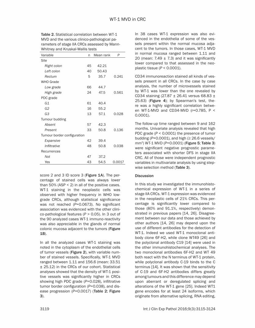
Figure 3. Density of the intra-tumour vessels positive for WT-1 in a case of non-recurring CRC (A), compared to a CRC which recurred in the follow-up (B) (WT-1 stain; original magnification, × 200).
Figure 4. Comparison of the density of vessels stained by WT-1 (A) (WT-1 stain; original magnification, × 100) and by CD34 (B) (CD34 stain; original magnification, × 100) in a case of stage IIA CRC.
Figure 5. Kaplan-Meier curves for DFS in stage IIA CRC according to WT-1 MVD. The DFS of patients with WT-1 MVD above or equal to the cut-off value (WT-1 MVD ≥ 26.6 vessels/mm2) was significantly shorter than that of the patients with WT-1 MVD be-low the cut-off.
WT-1 MVD in CRC
3121 Int J Clin Exp Pathol 2016;9(3):3115-3124
The higher frequency of poorly differentiated CRCs in our series may also account for the dis-crepancy with previous studies [25, 26]. Indeed, as we also observed, WT-1 expression in CRC tumor cells is more frequent in differentiated CRCs [25, 26]. The correlation between WT-1 expression and differentiation of CRC may depend upon the role of WT-1 in the up-regula-tion of e-cadherin expression [39] and conse-quently in the induction of tumor differenti- ation.
Differently from that previously reported by Bejnaranda and co-workers [26], we did not find any correlation between WT-1 expression in the tumour cells and the recurrence-free sur-vival of stage IIA CRCs, which may presumably depend on the low frequency of WT-1 positive cases in our series.
The most relevant finding in our study con-cerned the expression of WT-1 in the endothe-lia of tumour vessels. Indeed not all of the tumour vessels were stained by anti-WT-1 anti-body in our cases, although WT-1 positivity in endothelial cells was considered to be a univer-sal phenomenon and used as internal positive control for WT-1 immuno-reaction for a long time [41]. A similar finding was previously
reported in endometrial carcinoma by other authors [42] and in meningiomas by our group [37]. Similarly to that observed in meningiomas [37], WT-1 MVD was significantly correlated with the development of recurrences and it was a negative independent prognostic parameter associated with shorter DFS in stage IIA CRC. In addition, cases showing pathological features suggestive of aggressive behaviour, such as infiltrative tumor border and high PDC grade, had significantly higher WT-1 MVD counts com-pared to CRCs with absence of those features. Although WT-1 stain was also noted in some of the vessels within the normal colonic mucosa adjacent to the CRC, the number and density of those vessels was significantly lower compared to those assessed in the neoplastic tissue. Besides, also other authors previously reported significant decrease in the number of WT-1 pos-itive vessels moving from CRC to the surround-ing normal colonic mucosa [43]. Although the meaning of WT-1 expression in the tumour ves-sels still needs to be clarified, we may specu-late that the correlation between the density of WT-1 positive intra-tumoral vessels and the bio-logical aggressiveness of CRC may depend upon the role of WT-1 in neo-angiogenesis. Indeed it was shown that WT-1 is required for
Table 3. Univariate and multivariate analysis for DFS in stage IIA CRCs according to the various clinico-pathological parameters and WT-1 MVD
Variables Category Total (recur-rent cases)
Univariate Multivariate
HR (95% CI) P HR (95% CI) P Right 45 (20) 1
Left 40 (20) 1.2 (0.6-2.2)Site Rectum 5 (3) 1.5 (0.3-6.1) 0.679
LG 66 (32) 1WHO grade HG 24 (11) 1 (0.5-2.1) 0.801 G1 61 (20) 1 1
G2 16 (14) 4.5 (1.7-11.6) 2.9 (1.4-5.7) 0.001PDC grade G3 13 (9) 2.4 (1-5.9) < 0.0001
Absent 57 (17) 1 1TB Present 33 (26) 3.1 (1.6-5.8) 0.0001 2.2 (1.1-4.2) 0.015Tumor border Expansive 42 (17) 1Configuration Infiltrative 48 (26) 1.3 (0.7-2.4) 0.353
Low (< 26.6 v/mm2) 45 (12) 1 0.4 (0.1-0.8)WT-1 MVD High (≥ 26.6 v/mm2) 45 (31) 3.3 (1.8-6.1) 0.0001 1 0.0144
Low (64.95 v/m2) 46 (19) 1CD34 MVD High (≥ 64.95 v/mm2) 44 (24) 1.5 (0.8-2.7) 0.170 V: vessels. HR: hazard ratio. CI: confidence interval.
WT-1 MVD in CRC
3122 Int J Clin Exp Pathol 2016;9(3):3115-3124
endothelial cell proliferation, migration and in vitro angiogenesis [16] and that its activation in vascular cells is mediated by HIF-1 under condi-tions of reduced oxygen supply [16]. Hence we may speculate that more aggressive CRCs undergo a hypoxic condition due to their rapidly increasing volume, with following WT-1 up-regu-lation in the endothelial cells via the hypoxia inducible factor-1 (HIF-1) [15] and consequent increase in WT-1 MVD. Then the nutrients pro-vided by the newly formed vessels might favour tumour progression. In accordance with this hypothesis, the antibody against the pan-endo-thelial marker CD34 stained more microves-sels than the WT-1 on parallel tissue sections. Hence it is likely that CD34 stains both newly formed and pre-existing vessels, while WT-1 is positive only in the former.
Although the prognostic value of MVD was already demonstrated in CRC by the use of other endothelial markers such as endoglin [34], the demonstration that CRCs with higher propensity to recur have higher density of ves-sels expressing WT-1 protein may have higher clinical relevance. Indeed, due to its low toxicity in clinical trials [44], WT-1 seems to be an ideal target for innovative immunotherapy against cancer. In addition, it was shown that WT-1 spe-cific T cytotoxic lymphocytes may determine the lysis of WT-1 expressing colon cancer cells [24]. Our findings suggest that WT-1 based immuno-therapy may be used in CRC regardless of WT-1 expression in the tumour cells. Indeed also WT-1 positive intra-tumoral vessels may repre-sent an interesting therapeutic target for the reduction of tumour blood supply.
Interestingly we did not evidence any nuclear WT-1 staining, and the expression of this pro-tein was exclusively cytoplasmic in normal and neoplastic epithelial cells as well as in the endothelia. Although cytoplasmic positivity was previously considered to be an artifact [45], it is currently widely accepted as being a true local-ization of the molecule [25]. Indeed WT-1 is not only involved in transcriptional regulation in the nucleus, but also in RNA metabolism and trans-lational regulation in the cytoplasm [46].
In conclusion, in the present study we showed that a small percentage of stage IIA CRCs express WT-1 in their neoplastic cells, while all display WT-1 expression in the intra-tumoral vessels. If our findings are confirmed in larger
cohorts of patients, WT-1 MVD may be used as a tool to discriminate patients with stage IIA CRC at higher risk of progression. Finally, the meaning of WT-1 expression in the tumour blood vessels warrants further investigation as WT-1 endothelial expression may represent a target for immunotherapy in CRCs regardless of expression in the tumour cells.
Disclosure of conflict of interest
None.
Address correspondence to: Dr. Valeria Barresi, De- partment of Human Pathology “G. Barresi”, Polyclinic G. Martino, Via Consolare Valeria, Messina 98125, Italy. Tel: +393921083256; E-mail: [email protected]
References
[1] Sobin LH, Gospodarowicz MK, Wittekind C. TNM classification of malignant tumours. New York: Wiley-Blackwell; 2009.
[2] Hamilton SR, Bosman FT, Boffetta P, Ilyas M, Morreau H, Nakamura SI, Quirke P, Riboli E, Sobin LH. WHO Classification of Tumours of the Digestive System. In: Bosman T, Carneiro F, Hruban RH, Theise ND, editors. Lyon: IARC Press; 2010. pp. 137-138.
[3] La Torre M, Lorenzon L, Pilozzi E, Buracca V, Cavallini M, Ziparo V, Ferri M. Number of har-vested lymph nodes is the main prognostic fac-tor in Stage IIa colorectal cancer patients. J Surg Oncol 2012; 106: 469-474.
[4] O’Connell JB, Maggard MA, Ko CY. Colon can-cer survival rates with the new American Joint Committee on Cancer sixth edition staging. J Natl Cancer Inst 2004; 96: 1420-1425.
[5] Hari DM, Leung AM, Lee JH, Sim MS, Vuong B, Chiu CG, Bilchik AJ. AJCC Cancer Staging Man-ual 7th Edition Criteria for Colon Cancer: Do the Complex Modifications Improve Prognostic Assessment? J Am Coll Surg 2013; 217: 181-190.
[6] Santos C, Lòpez-Doriga A, Navarro M, Mateo J, Biondo S, Martinez Villacampa M, Soler G, Sanjuan X, Paules MJ, Laquente B, Guinó E, Kreisler E, Frago R, Germà JR, Moreno V, Sala-zar R. Clinicopathological risk factors of Stage II colon cancer: results of a prospecvtive study. Colorectal Dis 2013; 15: 414-422.
[7] Compton C, Fenoglio-Preiser CM, Pettigrew N, Fielding LP; Colorectal Working Group. Ameri-can Joint Committee on Cancer prognostic fac-tors consensus conference. Cancer 2000; 88: 1739-1757.
[8] Stocchi L, Fazio VW, Lavery I, Hammel J. Indi-vidual surgeon, pathologist, and other factors
WT-1 MVD in CRC
3123 Int J Clin Exp Pathol 2016;9(3):3115-3124
affecting lymph node harvest in stage II colon carcinoma. Is a minimum of 12 examined lymph nodes sufficient? Ann Surg Oncol 2011; 18: 405-412.
[9] Coosemans A. Wilms’ Tumour gene 1 (WT1) as an immunotherapeutic target. Facts Views Vis Obgyn 2011; 3: 89-99.
[10] Dao T, Yan S, Veomett N, Pankov D, Zhou L, Korontsvit T, Scott A, Whitten J, Maslak P, Casey E, Tan T, Liu H, Zakhaleva V, Curcio M, Doubrovina E, O’Reilly RJ, Liu C, Scheinberg DA. Targeting the intracellular WT1 oncogene product with therapeutic human antibody. Sci Transl Med 2013; 5: 176ra33.
[11] Galfione SK, Ro JY, Ayala AG, Ge Y. Diagnostic utility of WT-1 cytoplasmic stain in variety of vascular lesions. Int J Clin Exp Pathol 2014; 7: 536-543.
[12] Hanson J, Gorman J, Reese J, Fraizer G. Regu-lation of vascular endothelial growth factor, VEGF, gene promoter by the tumor suppressor, WT1. Front Biosci 2007; 12: 2279-2290.
[13] Katuri V, Gerber S, Qiu X, McCarty G, Goldstein SD, Hammers H, Montgomery E, Chen AR, Loeb DM. WT1 regulates angiogenesis in Ew-ing Sarcoma. Oncotarget 2014; 5: 2436-2449.
[14] Oji Y, Yamamoto H, Nomura M, Nakano Y, Ike-ba A, Nakatsuka S, Beno S, Kiyotoh E, Jomge-ow T, Sekimoto M, Nezu R, Yoshikawa Y, Inoue Y, Hosen N, Kawakami M, Tsuboi A, Oka Y, Ogawa H, Souda S, Aozasa K, Monden M, Sug-iyama H. Overexpression of the Wilms’ tumor gene WT1 in colorectal adenocarcinoma. Cancer 2003; 94: 712-717.
[15] Wagner KD, Wagner N, Wellmann S, Schley G, Bondke A, Theres H, Scholz H. Oxygen-regulat-ed expression of the Wilms’ tumor suppressor Wt1 involves hypoxia-inducible factor-1 (HIF--1). FASEB J 2003; 17: 1364-1366.
[16] Wagner N, Michiels JF, Schedl A, Wagner KD. The Wilms’ tumour suppressor WT1 is involved in endothelial cell proliferation and migration: expression in tumour vessels in vivo. Onco-gene 2008; 27: 3662-3672.
[17] Gessler MP, Poustka A, Cavenee W, Neve RL, Orkin SH, Bruns GA. Homozygous deletions in Wilms’ tumours of a zinc-finger gene identified by chromosome jumping. Nature 1990; 43: 774-778.
[18] Parenti R, Salvatorelli L, Musumeci G, Parenti C, Giorlandino A, Motta F, Magro G. Wilms’ tu-mor 1 (WT1) protein expression in human de-veloping tissues. Acta Histochem 2015; 117: 386-396.
[19] Magro G, Longo FR, Angelico G, Spadola S, Amore FF, Salvatorelli L. Immunohistochemis-try as potential diagnostic pitfall in the most common solid tumors of children and adoles-cents. Acta Histochem 2015; 117: 397-414.
[20] Salvatorelli L, Parenti R, Leone G, Musumeci G, Vasquez E, Magro G. Wilms tumor 1 (WT1) pro-tein: Diagnostic utility in pediatric tumors. Acta Histochem 2015; 117: 367-378.
[21] Hohenstein P, Hastie ND. The many facets of the Wilms’ tumour gene, WT1. Hum Mol Genet 2006; 15: 196-201.
[22] Cheever MA, Allison JP, Ferris AS, Finn OJ, Hastings BM, Mellman I, Prindiville SA, Viner JL, Weiner LM, Matrisian LM. The prioritization of cancer antigens: a national cancer institute pilot project for the acceleration of translation-al research. Clin Cancer Res 2009; 15: 5323-5337.
[23] Veomett N, Dao T, Liu H, Xiang J, Pankov D, Du-brovsky L, Whitten JA, Park SM, Korontsvit T, Zakhaleva V, Casey E, Curcio M, Kharas MG, O’Reilly RJ, Liu C, Scheinberg DA. Therapeutic efficacy of an Fc-enhanced TCR-like antibody to the intracellular WT1 oncoprotein. Clin Can-cer Res 2014; 20: 4036-4046.
[24] Koesters R, Linnebacher M, Coy JF, Germann A, Schwitalle Y, Findeisen P. WT1 is a tumor-associated antigen in colon cancer that can be recognized by in vitro stimulated cytotoxic T cells. Int J Cancer 2004; 109: 385-392.
[25] Nakatsuka S, Oji Y, Horiuchi T, Kanda T, Kitaga-wa M, Takeuchi T, Kawano K, Kuwae Y, Yamau-chi A, Okumura M, Kitamura Y, Oka Y, Kawase I, Sugiyama H, Aozasa K. Immunohistochemi-cal detection of WT1 protein in a variety of can-cer cells. Mod Pathol 2006; 19: 804-814.
[26] Bejrananda T, Phukaoloun M, Boonpipatta-napong T, Wanitsuwan W, Kanngern S, Sangth-ong R, Sangkhathat S. WT1 expression as an independent marker of poor prognosis in colorectal cancers. Cancer Biomark 2010; 8: 35-42.
[27] Ueno H, Mochizuki H, Hashiguchi Y, Shimazaki H, Aida S, Hase K, Matsukuma S, Kanai T, Kuri-hara H, Ozawa K, Yoshimura K, Bekku S. Risk factors for an adverse outcome in early inva-sive colorectal carcinoma. Gastroenterology 2004; 127: 385-394.
[28] Barresi V, Reggiani Bonetti L, Branca G, Di Gre-gorio C, Ponz de Leon M, Tuccari G. Colorectal carcinoma grading by quantifying poorly differ-entiated cell clusters is more reproducible and provides more robust prognostic information than conventional grading. Virchows Arch 2012; 461: 621-628.
[29] Barresi V, Reggiani Bonetti L, Ieni A, Branca G, Baron L, Tuccari G. Histological grading based on the count of poorly differentiated clusters in pre-operative biopsy predicts nodal involve-ment and pTNM stage in colorectal cancer pa-tients. Hum Pathol 2014; 45: 268-275.
[30] Barresi V, Branca G, Ieni A, Reggiani Bonetti L, Baron L, Mondello S, Tuccari G. Poorly differen-
WT-1 MVD in CRC
3124 Int J Clin Exp Pathol 2016;9(3):3115-3124
tiated clusters (PDCs) as a novel histological predictor of nodal metastases in pT1 colorec-tal cancer. Virchows Arch 2014; 464: 655-662.
[31] Ueno H, Kajiwara Y, Shimazaki H, Shinto E, Hashiguchi Y, Nakanishi K, Maekawa K, Katsu-rada Y, Nakamura T, Mochizuki H, Yamamoto J, Hase K. New criteria for histologic grading of colorectal cancer. Am J Surg Pathol 2012; 36: 193-201.
[32] Barresi V, Vitarelli E, Tuccari G, Barresi G. Cor-relative study of microvessel density and 5-li-poxygenase expression in human sporadic colorectal cancer. Arch Pathol Lab Med 2008; 132; 1807-1812.
[33] Barresi V, Tuccari G, Barresi G. Adiponectin im-munohistochemical expression in colorectal cancer and its correlation with histological grade and tumour microvessel density. Pathol-ogy 2009; 41: 533-538.
[34] Barresi V, Di Gregorio C, Regiani-Bonetti L, Ponz-De Leon M, Barresi G, Vitarelli E. Stage I colorectal carcinoma: VEGF immunohisto-chemical expression, microvessel density, and their correlation with clinical outcome. Vir-chows Arch 2010; 457: 11-19.
[35] Barresi V, Reggiani-Bonetti L, Di Gregorio C, De Leon MP, Barresi G. Lymphatic vessel density and its prognostic value in stage I colorectal carcinoma. J Clin Pathol 2011; 64: 6-12.
[36] Barresi V. Angiogenesis in meningiomas. Brain Tumor Pathol 2011; 28: 99-106.
[37] Barresi V, Caffo M, Branca G, Vitarelli, E, Tucc-ari G. The density of microvessels positive for Wilms’ tumour-1 protein (WT-1) is an indepen-dent predictor of recurrence risk in meningio-mas. Brain Tumor Pathol 2015; 32: 202-209.
[38] Scharnhorst V, van der Eb AJ, Jochemsen AG. WT1 proteins: functions in growth and differen-tiation. Gene 2001; 273: 141-161.
[39] Tsuta K, Kato Y, Tochigi N, Hoshino T, Takeda Y, Hosako M, Maeshima AM, Asamura H, Kondo T, Matsuno Y. Comparison of different clones (WT49 versus 6F-H2) of WT-1 antibodies for immunohistochemical diagnosis of malignant pleural mesothelioma. Appl Immunohistochem Mol Morphol 2009; 17: 126-30.
[40] Sampson VB, David JM, Puig I, Patil PU, de Herreos AV, Thomas GV, Rajasekaran AK. Wilms’ tumor protein induce an epithelial-mes-enchymal hybrid differentiation state in clear cell renal cell carcinoma. PLoS One 2014; 9: e102041.
[41] Goldstein NS, Uzieblo A. WT1 immunoreactivi-ty in uterine papillary serous carcinomas is dif-ferent from ovarian serous carcinomas. Am J Clin Pathol 2002; 117: 541-545.
[42] Coosemans A, Moerman P, Verbist G, Maes W, Neven P, Vergote P, Van Gool SW, Amant F. Wilms’ tumour gene 1 (WT1) in endometrial carcinoma. Gynecol Oncol 2008; 111: 502-508.
[43] Coosemans A, Van Hove T, Verbist G, Moons L, Neven P, Moerman P, Vergote I, Van Gool SW, Amant F. Wilms’ tumour gene 1 (WT1) positivi-ty in endothelial cells surrounding epithelial uterine tumours. Histopathology 2009; 54: 384-387.
[44] Iwami K, Natsume A, Ohno M, Ikeda H, Mineno J, Nukaya I, Okamoto S, Fujiwara H, Yasukawa M, Shiku H, Wakabayashi T. Adoptive transfer of genetically modified Wilms’ tumor 1 specific T cells in novel malignant skull basemeningioma model. Neuro Oncol 2013; 15: 747-758.
[45] Ramani P, Cowell JK. The expression pattern of Wilms’ tumour gene (WT1) product in normal tissues and paediatric renal tumours. J Pathol 1996; 179: 162-168.
[46] Niksic M, Slight J, Sanford JR, Caceres JF, Has-tie ND. The Wilms’tumour protein (WT1) shut-tles between nucleus and cytoplasm and is present in functional polysomes. Hum Mol Genet 2004; 13: 463-471.