Embed Size (px)
Citation preview

Int J Clin Exp Pathol 2015;8(7):8433-8437www.ijcep.com /ISSN:1936-2625/IJCEP0007490
Original Article Primary malignant mixed tumor of bone: a case report
Zhansan Su1, Zhi Li1, Baoan Liu2
1Department of Pathology, Xiangya Third Hospital, Central South University, Changsha 410013, Hunan Province, China; 2Department of Pathology, Xiangya Hospital, Central South University, Changsha 410008, Hunan Province, China
Received March 2, 2015; Accepted April 15, 2015; Epub July 1, 2015; Published July 15, 2015
Abstract: Background: An extremely rare primary mixed tumor occurring in left proximal femurs of a 47-year old female is reported. Case report: She had left hip pain for three months in April 2004. Radiological examinations revealed that a translucent expansive lesion in the left greater trochanter. She received the curettage of lesion and bone graft surgery. Curettage specimens were diagnosed as malignant mixed tumor, considered to be metastatic. Five months late the lesion recurred. She underwent obturator neurotomy plus total hip replacement of left hip. A long-term of more than ten years follow-up showed there were no evidence of disease recurrence or metastasis and no any signs of other tumor in her body. Discussion: The tumor contained myoepithelial component with positive immunostain of S-100 protein, p63, CK-pan, and vimentin, epithelial component confirmed by CK-pan, CK-LMW and cartilage, which indicated the tumor was a mixed tumor. Cellular atypia, relative high mitosis index, cartilage consistent with grade I chordrosarcoma, focal coagulative necrosis, and infiltration between trabeculae found in the tumor indicated that the tumor had a low grade malignant nature. During long-time follow-up there were no signs of any tumor found in the patient, which strongly suggested that the tumor be a primary one.
Keywords: Primary malignant mixed tumor, bone
Introduction
Mixed tumor, also known as pleomorphic ade-noma, contains two components: an epithelial cell component including myoepithelial cell and a mesenchymal component. The identification of these components is essential to the recog-nition of it. It is the most common tumor of major salivary gland [1] and it may, much less commonly, appear in lungs, breast, skin and soft tissue. When it occurs in the skin or soft tissue, it is also termed as “chondroid syringo-ma” or “myoepithelioma/parachordoma” in WHO Classification of Tumors [2, 3]. Mixed tumor occurring in bone has been described, most of which are from metastasis of malig-nant or benign mixed tumor in other sites [4, 5], primary ones are extremely rare, only five cases has been reported in the literature [6-10].
Case report
A 47-year-old woman was admitted to Xiangya Third Hospital because of her left hip pain for
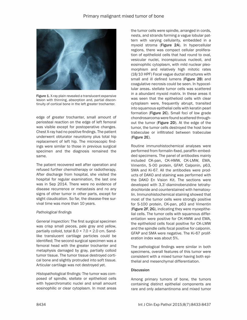
three months in April 2004. X-ray plain and CT imaging revealed that translucent expansive bone destruction in the left greater trochanter with thinning, absorption, partial discontinuity of cortical bone (Figure 1). The lesion had no local soft tissue mass and the structure of the left hip joint was normal. Chest X-ray had no abnormal findings. The lesion was considered to be benign, probable a bone cyst. The patient had a history of cholecystectomy because of gallstones 15 years ago. She received the curettage of bone lesions and bone graft sur-gery. Curettage specimens were diagnosed as malignant mixed tumor, considered to be meta-static. No primary site was found by further physical and CT examination of the chest, abdo-men-pelvis, and head and neck.
After surgery the patient gradually recovered and was able to get out of bed. However,her left hip pain recurred again in 5 months. X-ray showed that there were small dots with mixed low and high density on the outer upper edge of greater trochanter and soft tissue at outer lower
Primary malignant mixed tumor of bone
8434 Int J Clin Exp Pathol 2015;8(7):8433-8437
edge of greater trochanter, small amount of periosteal reaction on the edge of left femoral was visible except for postoperative changes. Chest X-ray had no positive findings. The patient underwent obturator neurotomy plus total hip replacement of left hip. The microscopic find-ings were similar to those in previous surgical specimen and the diagnosis remained the same.
The patient recovered well after operation and refused further chemotherapy or radiotherapy. After discharge from hospital, she visited the hospital for regular examination, the last one was in Sep 2014. There were no evidence of disease recurrence or metastasis and no any signs of other tumor in other parts, except for slight claudication. So far, the disease-free sur-vival time was more than 10 years.
Pathological findings
General inspection: The first surgical specimen was crisp small pieces, pale gray and yellow, partially colloid, total 8.0 × 7.0 × 2.0 cm. Sand-like translucent cartilage particles could be identified; The second surgical specimen was a femoral head with the greater trochanter and metaphysis damaged by gray, partially colloid tumor tissue. The tumor tissue destroyed corti-cal bone and slightly protruded into soft tissue. Articular cartilage was not destroyed yet.
Histopathological findings: The tumor was com-posed of spindle, stellate or epithelioid cells with hyperchromatic nuclei and small amount eosinophilic or clear cytoplasm. In most areas
the tumor cells were spindle, arranged in cords, nests, and strands forming a vague lobular pat-tern with varying cellularity, embedded in a myxoid stroma (Figure 2A). In hypercellular regions, there was compact cellular prolifera-tion of epithelioid cells that had round to oval, vesicular nuclei, inconspicuous nucleoli, and eosinophilic cytoplasm, with mild nuclear pleo-morphism and relatively high mitotic rates (18/10 HPF) Focal vague ductal structures with small and ill defined lumens (Figure 2B) and coagulative necrosis could be seen. In hypocel-lular areas,stellate tumor cells was scattered in a abundant myxoid matrix. In these areas it was seen that the epithelioid cells with clear cytoplasm were, frequently abrupt, transited into squamous epithelial cells with keratin pearl formation (Figure 2C). Small foci of low grade chondrosarcoma were found scattered through-out the tumor (Figure 2D). At the edge of the tumor, the tumor cells destroyed the host bone trabeculae or infiltrated between trabeculae (Figure 2E).
Routine immunohistochemical analyses were performed from formalin-fixed, paraffin-embed-ded specimens. The panel of antibodies mainly included CK-pan, CK-HMW, CK-LMW, EMA, Vimentin, S-00 protein, GFAP, Calponin, p63, SMA and Ki-67. All the antibodies were prod-ucts of DAKO and staining was performed with the DAKO En Vision Kit. The sections were developed with 3,3’-diaminobenzidine tetrahy-drochloride and counterstained with hematoxy-lin. Immunohistochemical staining showed that most of the tumor cells were strongly positive for S-100 protein, CK-pan, p63 and Vimentin (Figure 2F, 2G), indicating they were myoepithe-lial cells. The tumor cells with squamous differ-entiation were positive for CK-HMW and EMA, the epithelioid cells focal positive for CK-LMW and the spindle cells focal positive for calponin. GFAP and SMA were negative. The Ki-67 prolif-eration index was about 5%.
The pathological findings were similar in both specimens, overall features of this tumor were consistent with a mixed tumor having both epi-thelial and mesenchymal differentiation.
Discussion
Among primary tumors of bone, the tumors containing distinct epithelial components are rare and only adamantinoma and mixed tumor
Figure 1. X-ray plain revealed a translucent expansive lesion with thinning, absorption and, partial discon-tinuity of cortical bone in the left greater trochanter.

Primary malignant mixed tumor of bone
8435 Int J Clin Exp Pathol 2015;8(7):8433-8437

Primary malignant mixed tumor of bone
8436 Int J Clin Exp Pathol 2015;8(7):8433-8437
are considered as definite entities. Adaman- tinoma is a low-grade neoplasm with epithelial differentiation and a striking predilection to involve the tibia. Histologically, it resembles the adamantinoma of the jawbones, showing basa-loid pattern with peripheral palisading of cuboi-dal and columnar tumor cells or the pattern of fibrous dysplasia with scattering islands of epi-thelial cells, without myoepithelial and chon-drosarcomatous components. Such histologic features were not found in the case reported here and the diagnosis of adamantinoma could be excluded.
Mixed tumors mainly occur in major and minor salivary glands and they may occasionally appear in skin, breast, lung and soft tissue. Mixed tumors also have been reported appear-ing in bone though most of the benign or malig-nant mixed tumors found in bone war metastat-ic from other parts of the body. However, there were four case reports in the literature showed that similar tumors could occur in bone as pri-mary tumor though their histogenesis was still an enigma. Squamous and chondroid compo-nents as well as myoepithelial component which were confirmed by S-100 protein and p63 immunostain found in present case strong-ly suggested that the tumor was a mixed tumor. When the specimen of the first operation was observed, we thought that it was a metastatic mixed tumor. However, there was no primary site was found by comprehensive examination. Furthermore, during the follow-up period more than 10 years, no any tumor has been found in the patient by periodical examination at hospi-tals. It should be safe to assume that it was a primary mixed tumor of the bone. Though there were no obvious carcinomatous or sarcoma-tous components found in this case, there were cellular atypia, relative high mitosis index, carti-lage consistent with low grade chordrosarco-ma, focal coagulative necrosis, and infiltration between bone trabeculae. It should be reason-able to consider it a low-grade malignant mixed
tumor. The good outcome of the case might benefit from the low grade malignant nature of the tumor and the radical surgical treatment.
There were two interesting cases of chondro-sarcoma with distinct epithelial differentiation reported in the literature [11, 12]. In those cases, the tumor was termed as “chondrosar-coma associated with squamous cell carcino-ma” and “primary bone carcinosarcoma”. Interestingly, the histopathological findings were similar in both cases, the tumor consisted of three components: well differentiated chon-drosarcoma, undifferentiated spindle cell sar-coma with varying cellularity and squamous cell carcinoma with prominent keratin pearl forma-tion. Both patients were died of the disease at 6 months and three and a half years after diag-nosis respectively. According to the description of morphology, it was not entirely impossible that the lesions were malignant mixed tumor with carcinosarcoma transformation. Further study of immnophenotyping and molecular genetics may reveal their histogenesis.
Disclosure of conflict of interest
None.
Address correspondence to: Dr. Baoan Liu, Depart- ment of Pathology, Xiangya Hospital, Central South University, 87# Xiangya Road, Changsha 410008, Hunan Province, China. E-mail: [email protected]
References
[1] Gnepp DR, Wenig BM. Surgical Pathology of the Salivary Gland. Philadelphia; W B Saunders; 1991. pp. 350-68.
[2] Philip E, LeBoit PE, Burg G. (WHO) Pathology and Genetics of Skin Tumours. Lyon: IARC Press; 2006. pp. 147-8.
[3] Fletcher CD, Unni KK and Mertens F. (WHO) Pathology and Genetics of Tumours of Soft Tissue and Bone. Lyon: IARC Press; 2002. pp. 198-9.
Figure 2. A. The tumor cells were spindle, arranged in cords and strands forming a vague lobular pattern, embedded in a myxoid stroma. B. Epithelioid tumor cells that had round to oval, vesicular nuclei, inconspicuous nucleoli, and eosinophilic cytoplasm, with mild nuclear pleomorphism and an increased mitotic activity (Red arrows) (H&E stain). C. The cords of epithelioid cells with clear cytoplasm were scattered in a myxoid or myxochondroid stroma and tran-sited into squamous epithelial cells with keratin pearl formation (H&E stain). D. Small foci of cellular cartilage with nuclear atypia, consistent with low grade chondrosarcoma were found scattered throughout the tumor (H&E stain). E. The dense spindle tumor cells with coagulative necrosis infiltrated between bone trabeculae (H&E stain). F. The tumor cells were positive for p63 strongly and diffusely with a pattern of nuclear staining. G. The spindle tumor cells were strongly positive for S-100 protein.

Primary malignant mixed tumor of bone
8437 Int J Clin Exp Pathol 2015;8(7):8433-8437
[4] Manucha V, Loffe OB. Metastasizing pleomor-phic adenoma of the salivary gland. Arch Pathol Lab Med 2008; 132: 1445-7.
[5] Parikh HK, Parikh DM. Malignant mixed tumor of the salivary gland with bone metastasis: a case report and literature review. Indian J Cancer 1991; 28: 171-5.
[6] Barreto CA, Lipton MN, Smith HB, Potter GK. Intraosseous chondroid syringoma of the hal-lux. J Am Acad Dermatol 1994; 30: 374-378.
[7] Gadgil RK, Ranadive NU. Chondroid syringoma (mixed tumour) of radius. Indian J Cancer 1981; 18: 81-83.
[8] Hirsch P, Helwig EB. Chondroid syringoma, mixed tumor of skin, salivary gland type. Arch Dermatol 1961; 84: 835-847.
[9] de Pinieux G, Beabout JW, Unni KK, Sim FH. Primary mixed tumor of bone. Skeletal Radiol 2001; 30: 534-536.
[10] 10. Yu H, Liu X, Li H, Shi D, Wang C. Primary plemorphic adenoma of bone: report of a case and literature review. Indian J Pathol Microbiol 2012; 55: 320-322.
[11] Ling LL, Steiner GC. Primary multipotential ma-lignant neoplasm of bone: chondrosarcoma associated with squamous cell carcinoma. Hum Pathol 1986; 17: 317-320.
[12] Shiraishi J, Mukai M, Yabe H, Shibata R, Yamada T, Miura K, Anazawa U, Morioka H, Sakamoto M. Primary bone carcinosarcoma: Chondrosarcoma and squamous cell carcino-ma with keratin pearl formation. Pathol Int 2005; 55: 504-509.