Embed Size (px)
Citation preview
1
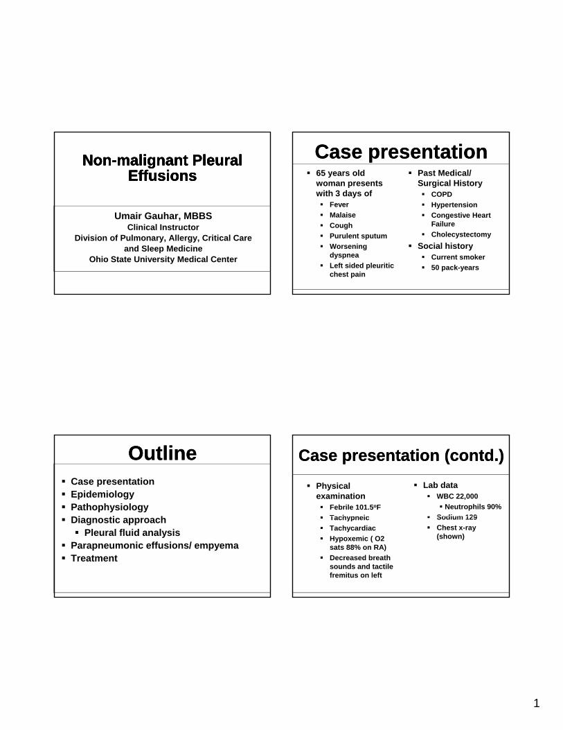
NonNon--malignant Pleural malignant Pleural EffusionsEffusions
NonNon--malignant Pleural malignant Pleural EffusionsEffusions
U i G h MBBSUmair Gauhar, MBBSClinical Instructor
Division of Pulmonary, Allergy, Critical Care and Sleep Medicine
Ohio State University Medical Center
OutlineOutline Case presentation Epidemiology Pathophysiology
Di ti h Diagnostic approach Pleural fluid analysis
Parapneumonic effusions/ empyema Treatment
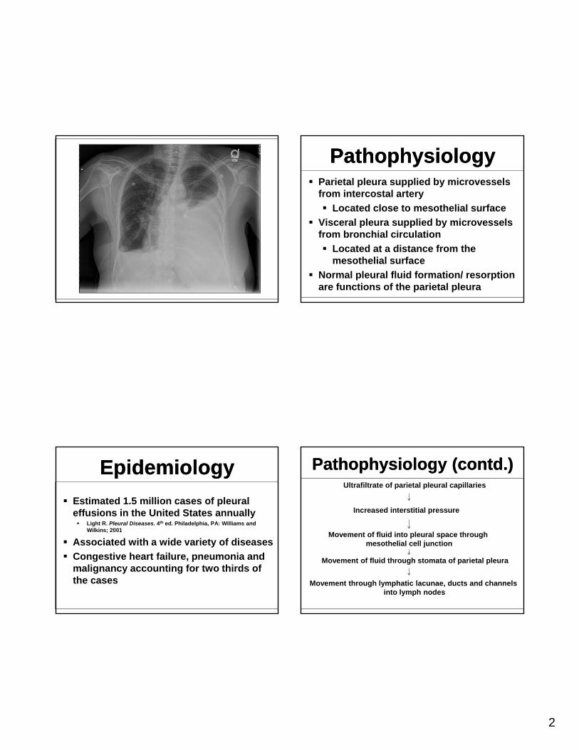
Case presentationCase presentation 65 years old
woman presents with 3 days of Fever
M l i
Past Medical/ Surgical History COPD
Hypertension
C ti H t Malaise
Cough
Purulent sputum
Worsening dyspnea
Left sided pleuritic chest pain
Congestive Heart Failure
Cholecystectomy
Social history Current smoker
50 pack-years
Case presentation (contd.)Case presentation (contd.)
Physical examination Febrile 101.5oF
Tachypneic
Lab data WBC 22,000
Neutrophils 90%
Sodium 129 Tachypneic
Tachycardiac
Hypoxemic ( O2 sats 88% on RA)
Decreased breath sounds and tactile fremitus on left
Sodium 129
Chest x-ray (shown)
2
EpidemiologyEpidemiology
Estimated 1.5 million cases of pleural effusions in the United States annually Light R. Pleural Diseases. 4th ed. Philadelphia, PA: Williams and
Wilkins; 2001Wilkins; 2001
Associated with a wide variety of diseases
Congestive heart failure, pneumonia and malignancy accounting for two thirds of the cases
PathophysiologyPathophysiology Parietal pleura supplied by microvessels
from intercostal artery
Located close to mesothelial surface
Vi l l li d b i l Visceral pleura supplied by microvessels from bronchial circulation
Located at a distance from the mesothelial surface
Normal pleural fluid formation/ resorption are functions of the parietal pleura
Pathophysiology (contd.)Pathophysiology (contd.)Ultrafiltrate of parietal pleural capillaries
Increased interstitial pressure
Movement of fluid into pleural space through mesothelial cell junction
Movement of fluid through stomata of parietal pleura
Movement through lymphatic lacunae, ducts and channels into lymph nodes
3
Pathophysiology (contd.)Pathophysiology (contd.) Normal pleural fluid
0.1 to 0.2 ml/kg
Clear
Low protein (1.0 to 1.5 g/dl)
< 1500 nucleated cells / L< 1500 nucleated cells / L
61% to 77% monocytes-macrophages
9 to 30% mesothelial cells
7% to 11% lymphocytes
2% neutrophils
0% eosinophils
pH > 7.60
Pathophysiology (contd.)Pathophysiology (contd.)
Mechanism of abnormal pleural fluid formation
Increased hydrostatic pressure (CHF)
Decreased oncotic pressure (hypoalbuminemia)
Decreased pleural pressure (trapped lung)
Increased endolethial permeability (pneumonia)
Decreased lymphatic drainage (malignancy)
Movement from peritoneal space (hepatic hydrothorax)
Movement from extra-vascular space (duropleural fistula, migrated/ misplaced CVC/ feeding tube)
Diagnostic Approach1. Clinical History
Diagnostic Approach1. Clinical History
Could be asymptomatic
Dyspnea and chest pain are the two most common presenting symptoms
Dyspnea most likely fromDyspnea most likely from
decreased chest wall compliance
depression of ipsilateral diaphragm and
increased output from neurogenic receptors
Dyspnea out of proportion to exam findings can suggest PE
Diagnostic Approach1. Clinical History
Diagnostic Approach1. Clinical History
Chest pain
Usually pleuritic
Intensity proportional to degree of pleural inflammationinflammation
May be decreased by splinting by manual pressure over the chest wall
May be localized or radiating
Central diaphragmatic inflammation causes radiating pain in the posterior neck, shoulder and trapezius area
4
Diagnostic Approach1. Clinical History
Diagnostic Approach1. Clinical History
Asymptomatic
BAPE
Rheumatoid pleural ff i
Symptomatic
Bacterial pneumonia
Lupus pleuritiseffusion
Nephrotic syndrome
Yellow nail syndrome
Trapped lung
Urinothorax
Peritoneal dialysis associated effusion
Postcardiac injury syndrome
Pulmonary embolism
Congestive heart failure
Diagnostic Approach1. Clinical History
Diagnostic Approach1. Clinical History
Useful clues Orthopnea, PND, lower extremity CHF
H/O asbestos exposure BAPE
H/O alcoholism, poor dentition, loss of consciousness aspiration/ anerobic empyema
H/O of CABG post-cardiac injury syndrome or trapped lung
H/O retching esophageal rupture
H/O SLE (or procainamide use) lupus pleuritis
Obstructive uropathy urinothorax
Spinal surgery or trauma duropleural fistula
Diagnostic Approach2. Physical Examination
Diagnostic Approach2. Physical Examination
Signs depend on volume of pleural effusion
< 300 ml not detectable on physical examination
500 ml dull percussion, decreased fremitus, decreased breath sounds
> 1000 ml bulging of ICS, decreased chest expansion, bronchovesicular sounds and egophony at upper level of effusion
Diagnostic Approach3. Chest Radiograph
Diagnostic Approach3. Chest Radiograph
Sensitivity proportional to volume of pleural fluid 5mlblunting of
posterior costophrenic angle on lateral decubitus film
50-75 ml blunting of posterior costophrenic angle on lateral view
175-200 ml blunting of costophrenic angle on PA film
> 500 ml opacification of lung base
5
Diagnostic Approach3. Chest Radiograph
Diagnostic Approach3. Chest Radiograph
Non-malignant causes of massive effusion with mediastinal shift
Tuberculosis
Empyema
Hepatic hydrothorax
Chylothorax
Hemothorax
Congestive heart failure
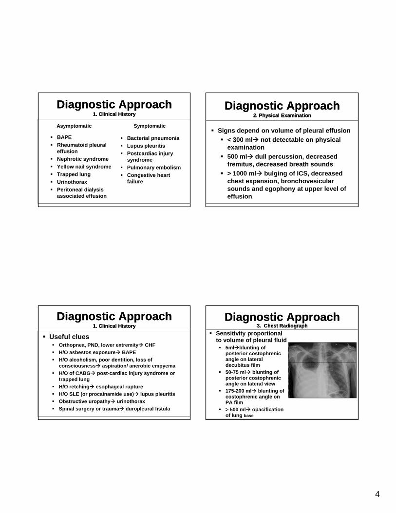
Diagnostic Approach4. Ultrasound
Diagnostic Approach4. Ultrasound
Diaphragm
Pleural effusionChest wall
Liver
1. Diagnosis and sampling ofloculated pleural effusions
Compressed lung
Liver
2. Guided sampling of smallpleural effusions
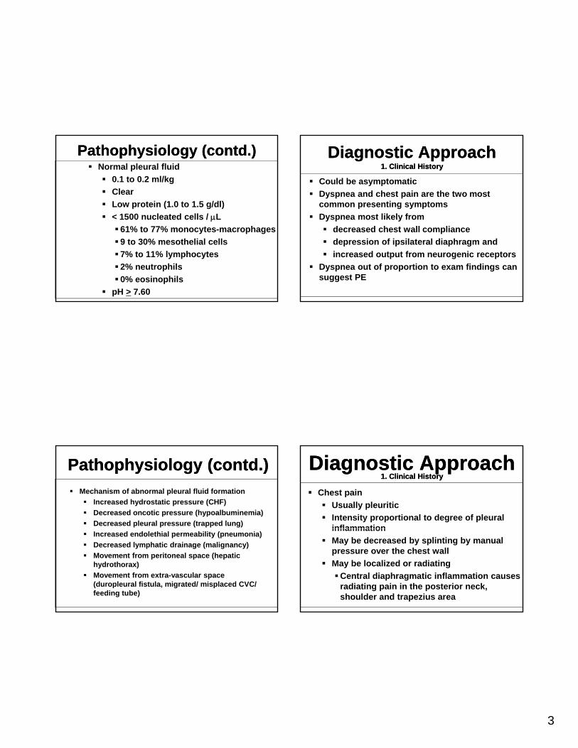
Diagnostic Approach5. Computed Tomography
Diagnostic Approach5. Computed Tomography
Most sensitive radiographic study
Useful for diff ti ti fdifferentiating free flowing effusions, loculated effusions, parenchymal lesions and extrapleural disease
Compressed lungPleural Effusion
Diagnostic Approach6. Pleural Fluid Analysis (PFA)
Diagnostic Approach6. Pleural Fluid Analysis (PFA)
Observation acceptable in
Small effusions (< 1 cm thickness on lateral decubitus films)
Patients presenting with typical symptoms of CHF and bilateral pleural effusions of similar size and absence of chest pain or fever
PFA should be performed in all new effusions
Therapeutic thoracentesis (1 to 1.5 L) in symptomatic effusions
6
Diagnostic Approach6. Pleural Fluid Analysis (PFA)
Diagnostic Approach6. Pleural Fluid Analysis (PFA)
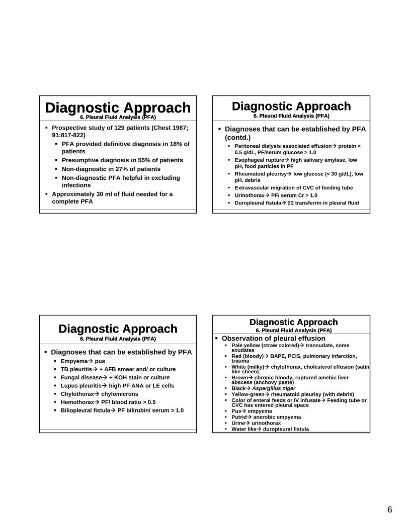
Prospective study of 129 patients (Chest 1987; 91:817-822)
PFA provided definitive diagnosis in 18% of patientsp
Presumptive diagnosis in 55% of patients
Non-diagnostic in 27% of patients
Non-diagnostic PFA helpful in excluding infections
Approximately 30 ml of fluid needed for a complete PFA
Diagnostic Approach6. Pleural Fluid Analysis (PFA)
Diagnostic Approach6. Pleural Fluid Analysis (PFA)
Diagnoses that can be established by PFA Empyema pus
TB pleuritis + AFB smear and/ or culture
Fungal disease + KOH stain or culture
Lupus pleuritis high PF ANA or LE cells
Chylothorax chylomicrons
Hemothorax PF/ blood ratio > 0.5
Biliopleural fistula PF bilirubin/ serum > 1.0
Diagnostic Approach6. Pleural Fluid Analysis (PFA)
Diagnostic Approach6. Pleural Fluid Analysis (PFA)
Diagnoses that can be established by PFA (contd.) Peritoneal dialysis associated effusion protein <
0.5 g/dL, PF/serum glucose > 1.0g , g
Esophageal rupture high salivary amylase, low pH, food particles in PF
Rheumatoid pleurisy low glucose (< 30 g/dL), low pH, debris
Extravascular migration of CVC of feeding tube
Urinothorax PF/ serum Cr > 1.0
Duropleural fistula 2 transferrin in pleural fluid
Diagnostic Approach6. Pleural Fluid Analysis (PFA)
Diagnostic Approach6. Pleural Fluid Analysis (PFA)
Observation of pleural effusion Pale yellow (straw colored) transudate, some
exudates Red (bloody) BAPE, PCIS, pulmonary infarction,
trauma White (milky) chylothorax, cholesterol effusion (satin
like sheen)) Brown chronic bloody, ruptured amebic liver
abscess (anchovy paste) Black Aspergillus niger Yellow-green rheumatoid pleurisy (with debris) Color of enteral feeds or IV infusate Feeding tube or
CVC has entered pleural space Pus empyema Putrid anerobic empyema Urine urinothorax Water like duropleural fistula
7
Diagnostic Approach6. Pleural Fluid Analysis (PFA)
Diagnostic Approach6. Pleural Fluid Analysis (PFA)
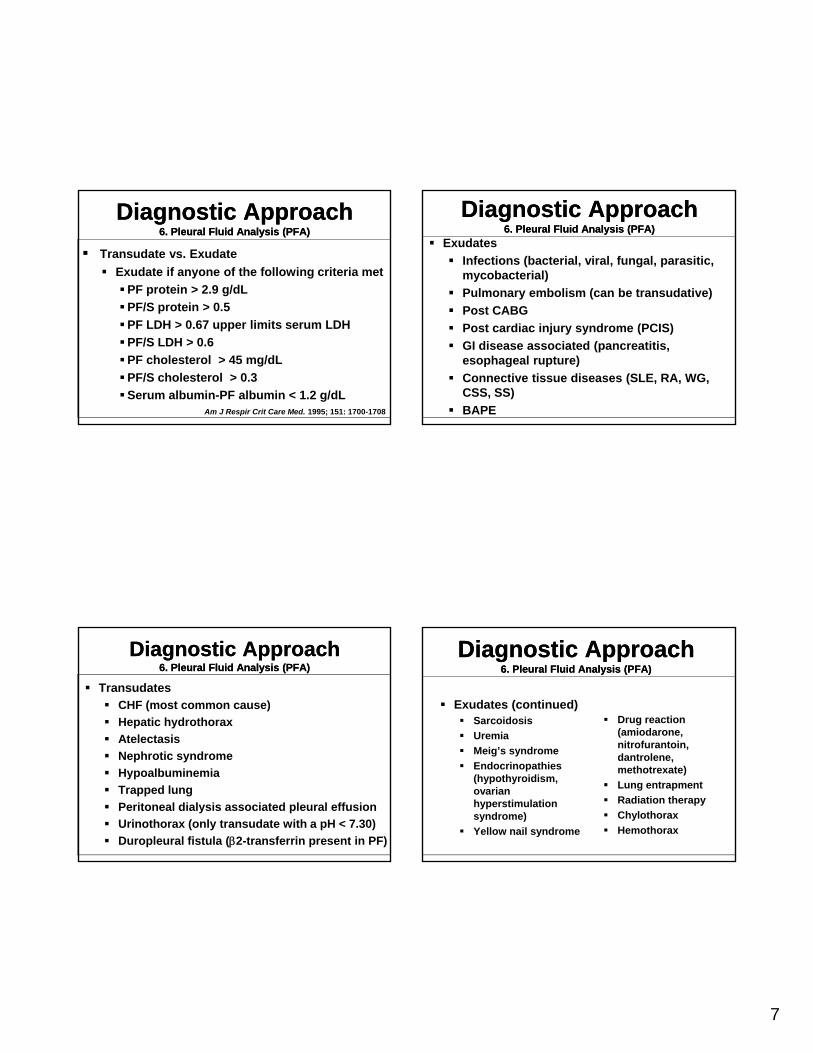
Transudate vs. Exudate
Exudate if anyone of the following criteria met
PF protein > 2.9 g/dL
PF/S protein > 0 5PF/S protein > 0.5
PF LDH > 0.67 upper limits serum LDH
PF/S LDH > 0.6
PF cholesterol > 45 mg/dL
PF/S cholesterol > 0.3
Serum albumin-PF albumin < 1.2 g/dLAm J Respir Crit Care Med. 1995; 151: 1700-1708
Diagnostic Approach6. Pleural Fluid Analysis (PFA)
Diagnostic Approach6. Pleural Fluid Analysis (PFA)
Transudates
CHF (most common cause)
Hepatic hydrothorax
Atelectasis
Nephrotic syndrome
Hypoalbuminemia
Trapped lung
Peritoneal dialysis associated pleural effusion
Urinothorax (only transudate with a pH < 7.30)
Duropleural fistula (2-transferrin present in PF)
Diagnostic Approach6. Pleural Fluid Analysis (PFA)
Diagnostic Approach6. Pleural Fluid Analysis (PFA)
Exudates
Infections (bacterial, viral, fungal, parasitic, mycobacterial)
Pulmonary embolism (can be transudative)
P t CABG Post CABG
Post cardiac injury syndrome (PCIS)
GI disease associated (pancreatitis, esophageal rupture)
Connective tissue diseases (SLE, RA, WG, CSS, SS)
BAPE
Diagnostic Approach6. Pleural Fluid Analysis (PFA)
Diagnostic Approach6. Pleural Fluid Analysis (PFA)
Exudates (continued) Sarcoidosis
Uremia
Drug reaction (amiodarone, nitrofurantoin
Meig’s syndrome
Endocrinopathies (hypothyroidism, ovarian hyperstimulation syndrome)
Yellow nail syndrome
nitrofurantoin, dantrolene, methotrexate)
Lung entrapment
Radiation therapy
Chylothorax
Hemothorax
8
Diagnostic Approach6. Pleural Fluid Analysis (PFA)
Diagnostic Approach6. Pleural Fluid Analysis (PFA)
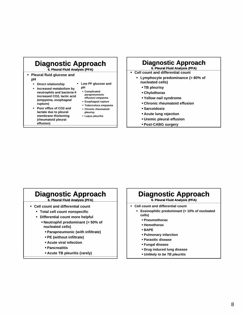
Pleural fluid glucose and pH Direct relationship
Increased metabolism by neutrophils and bacteria
Low PF glucose and pH Complicatedneutrophils and bacteria
increased CO2, lactic acid (empyema, esophageal rupture)
Poor efflux of CO2 and lactate due to pleural membrane thickening (rheumatoid pleural effusion)
Complicated parapneumonic effusion/ empyema
Esophageal rupture
Tuberculous empyema
Chronic rheumatoid pleurisy
Lupus pleuritis
Diagnostic Approach6. Pleural Fluid Analysis (PFA)
Diagnostic Approach6. Pleural Fluid Analysis (PFA)
Cell count and differential count
Total cell count nonspecific
Differential count more helpful
Neutrophil predominant (> 50% of p p ( %nucleated cells)
Parapneumonic (with infiltrate)
PE (without infiltrate)
Acute viral infection
Pancreatitis
Acute TB pleuritis (rarely)
Diagnostic Approach6. Pleural Fluid Analysis (PFA)
Diagnostic Approach6. Pleural Fluid Analysis (PFA)
Cell count and differential count
Lymphocyte predominance (> 80% of nucleated cells)
TB pleurisy
ChylothoraxChylothorax
Yellow nail syndrome
Chronic rheumatoid effusion
Sarcoidosis
Acute lung rejection
Uremic pleural effusion
Post-CABG surgery
Diagnostic Approach6. Pleural Fluid Analysis (PFA)
Diagnostic Approach6. Pleural Fluid Analysis (PFA)
Cell count and differential count
Eosinophilic predominant (> 10% of nucleated cells)
Pneumothorax
Hemothorax
BAPE
Pulmonary infarction
Parasitic disease
Fungal disease
Drug induced lung disease
Unlikely to be TB pleuritis
9
Diagnostic Approach6. Pleural Fluid Analysis (PFA)
Diagnostic Approach6. Pleural Fluid Analysis (PFA)
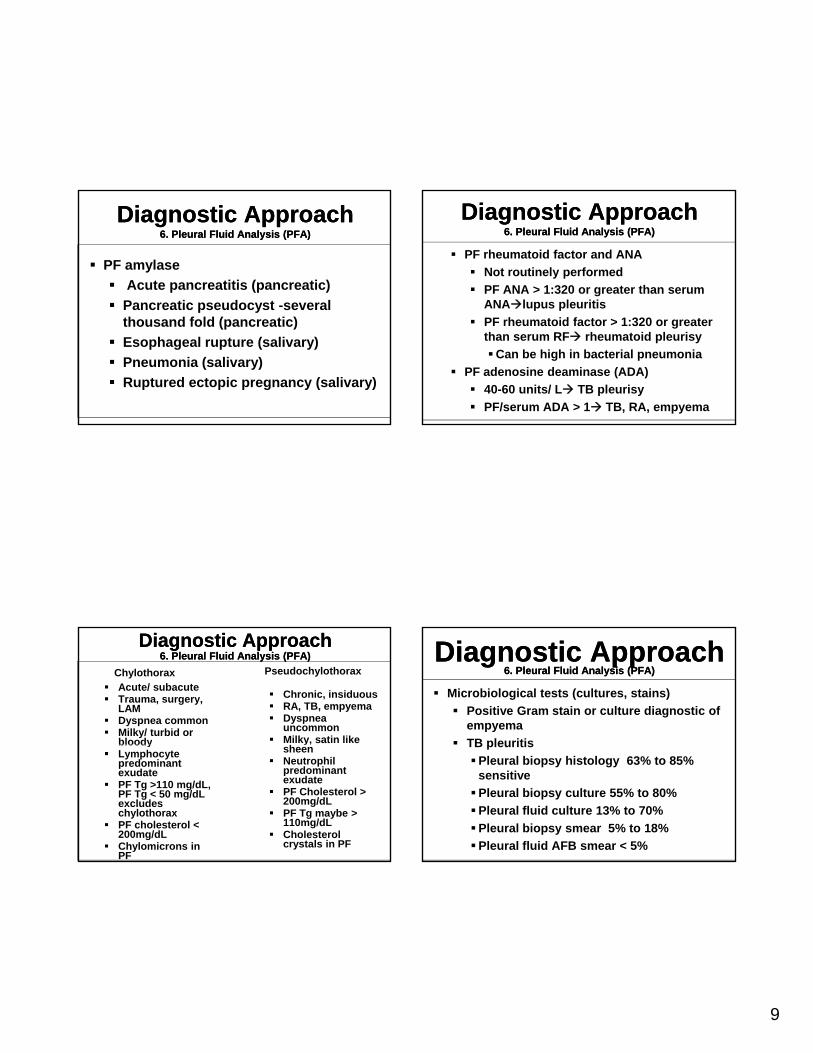
PF amylase
Acute pancreatitis (pancreatic)
Pancreatic pseudocyst -severalPancreatic pseudocyst several thousand fold (pancreatic)
Esophageal rupture (salivary)
Pneumonia (salivary)
Ruptured ectopic pregnancy (salivary)
Diagnostic Approach6. Pleural Fluid Analysis (PFA)
Diagnostic Approach6. Pleural Fluid Analysis (PFA)
Acute/ subacute Trauma, surgery,
LAM Dyspnea common Milky/ turbid or
bloody
Chronic, insiduous RA, TB, empyema Dyspnea
uncommon Milky, satin like
Chylothorax Pseudochylothorax
bloody Lymphocyte
predominant exudate
PF Tg >110 mg/dL, PF Tg < 50 mg/dL excludes chylothorax
PF cholesterol < 200mg/dL
Chylomicrons in PF
ysheen
Neutrophil predominant exudate
PF Cholesterol > 200mg/dL
PF Tg maybe > 110mg/dL
Cholesterol crystals in PF
Diagnostic Approach6. Pleural Fluid Analysis (PFA)
Diagnostic Approach6. Pleural Fluid Analysis (PFA)
PF rheumatoid factor and ANA
Not routinely performed
PF ANA > 1:320 or greater than serum ANAlupus pleuritisANAlupus pleuritis
PF rheumatoid factor > 1:320 or greater than serum RF rheumatoid pleurisy
Can be high in bacterial pneumonia
PF adenosine deaminase (ADA)
40-60 units/ L TB pleurisy
PF/serum ADA > 1 TB, RA, empyema
Diagnostic Approach6. Pleural Fluid Analysis (PFA)
Diagnostic Approach6. Pleural Fluid Analysis (PFA)
Microbiological tests (cultures, stains)
Positive Gram stain or culture diagnostic of empyema
TB pleuritisTB pleuritis
Pleural biopsy histology 63% to 85% sensitive
Pleural biopsy culture 55% to 80%
Pleural fluid culture 13% to 70%
Pleural biopsy smear 5% to 18%
Pleural fluid AFB smear < 5%
10
Parapneumonic Effusions/ Empyema
Parapneumonic Effusions/ Empyema
40% to 57% of cases of pneumonia associated with parapneumonic effusion
Complicated parapneumonic effusions in 10% toComplicated parapneumonic effusions in 10% to 15% of patients
Empyema (pus in the pleural space) in 5% of patients
Exudative phase (0–72h) fibrinopurulent phase (3-10 days) organizational phase (10–21 days)
Parapneumonic Effusions/ Empyema (contd.)
Parapneumonic Effusions/ Empyema (contd.)
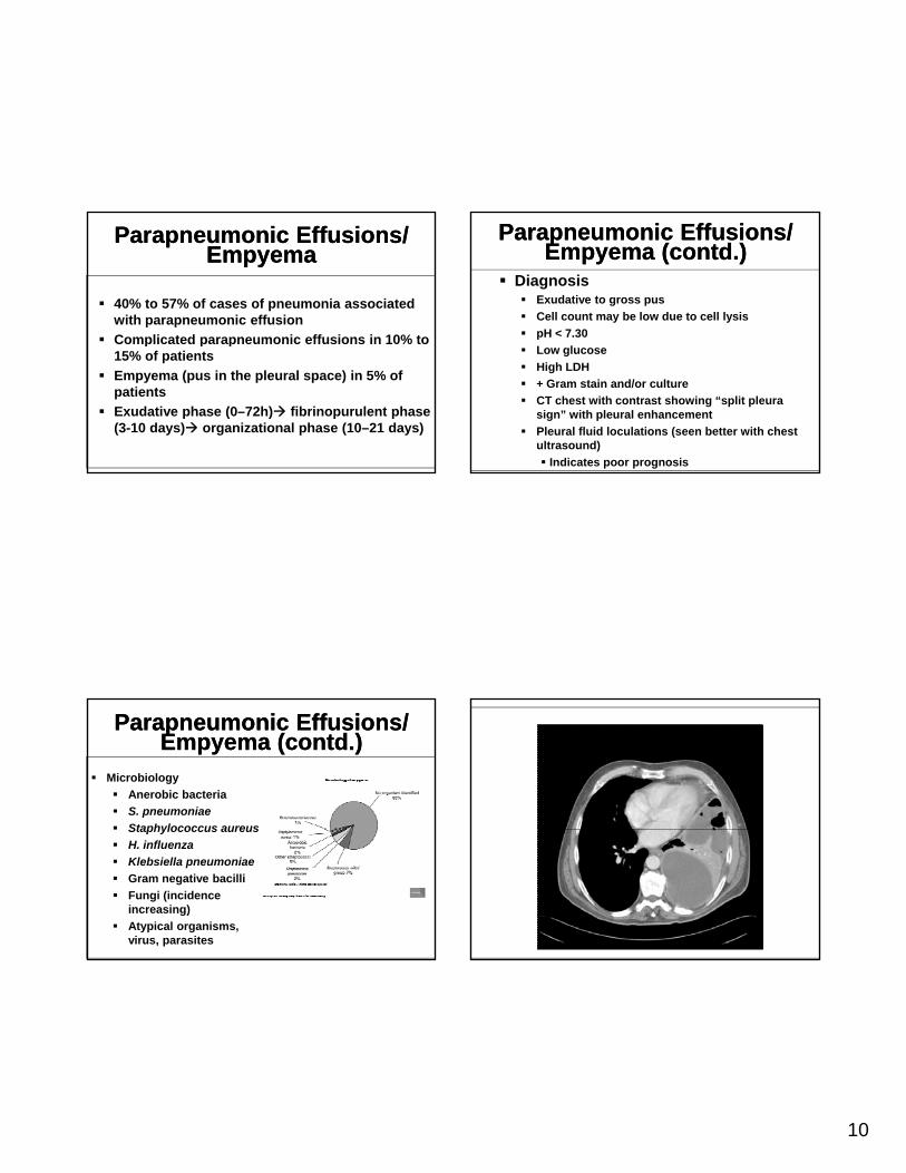
Microbiology
Anerobic bacteria
S. pneumoniae
Staphylococcus aureusStaphylococcus aureus
H. influenza
Klebsiella pneumoniae
Gram negative bacilli
Fungi (incidence increasing)
Atypical organisms, virus, parasites
Parapneumonic Effusions/ Empyema (contd.)
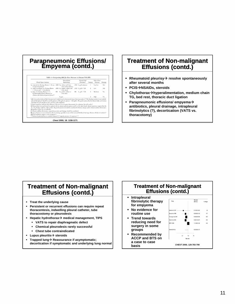
Parapneumonic Effusions/ Empyema (contd.)
Diagnosis Exudative to gross pus
Cell count may be low due to cell lysis
pH < 7.30
Low glucose
High LDH
+ Gram stain and/or culture
CT chest with contrast showing “split pleura sign” with pleural enhancement
Pleural fluid loculations (seen better with chest ultrasound)
Indicates poor prognosis
11
Parapneumonic Effusions/ Empyema (contd.)
Parapneumonic Effusions/ Empyema (contd.)
Chest 2000; 18: 1158-1171
Treatment of Non-malignant Effusions (contd.)
Treatment of Non-malignant Effusions (contd.)
Treat the underlying cause
Persistent or recurrent effusions can require repeat thoracentesis, indwelling pleural catheter, tube thoracostomy or pleurodesisy p
Hepatic hydrothorax medical management, TIPS
VATS to repair diaphragmatic defect
Chemical pleurodesis rarely successful
Chest tube contraindicated
Lupus pleuritis steroids
Trapped lung Reassurance if asymptomatic; decortication if symptomatic and underlying lung normal
Treatment of Non-malignant Effusions (contd.)
Treatment of Non-malignant Effusions (contd.)
Rheumatoid pleurisy resolve spontaneously after several months
PCISNSAIDs, steroidsPCISNSAIDs, steroids
Chylothoraxhyperalimentation, medium chain TG, bed rest, thoracic duct ligation
Parapneumonic effusions/ empyemaantibiotics, pleural drainage, intrapleural fibrinolytics (?), decortication (VATS vs. thoracotomy)
Treatment of Non-malignant Effusions (contd.)
Treatment of Non-malignant Effusions (contd.)
Intrapleural fibrinolytic therapy for empyema
No evidence for routine use
Trend towards reducing need for surgery in some groups
Recommended by ACCP and BTS on a case to case basis CHEST 2006; 129:783-790
12
Shaheen Islam, MD, MPH
Management ofMalignant Pleural
Effusion
Management ofMalignant Pleural
Effusion
Associate ProfessorDirector, Interventional Pulmonology
Assoc Medical Director, Pulmonary Diagnostics LabDivision of Pulmonary, Allergy, Critical Care
and Sleep MedicineOhio State University Medical Center
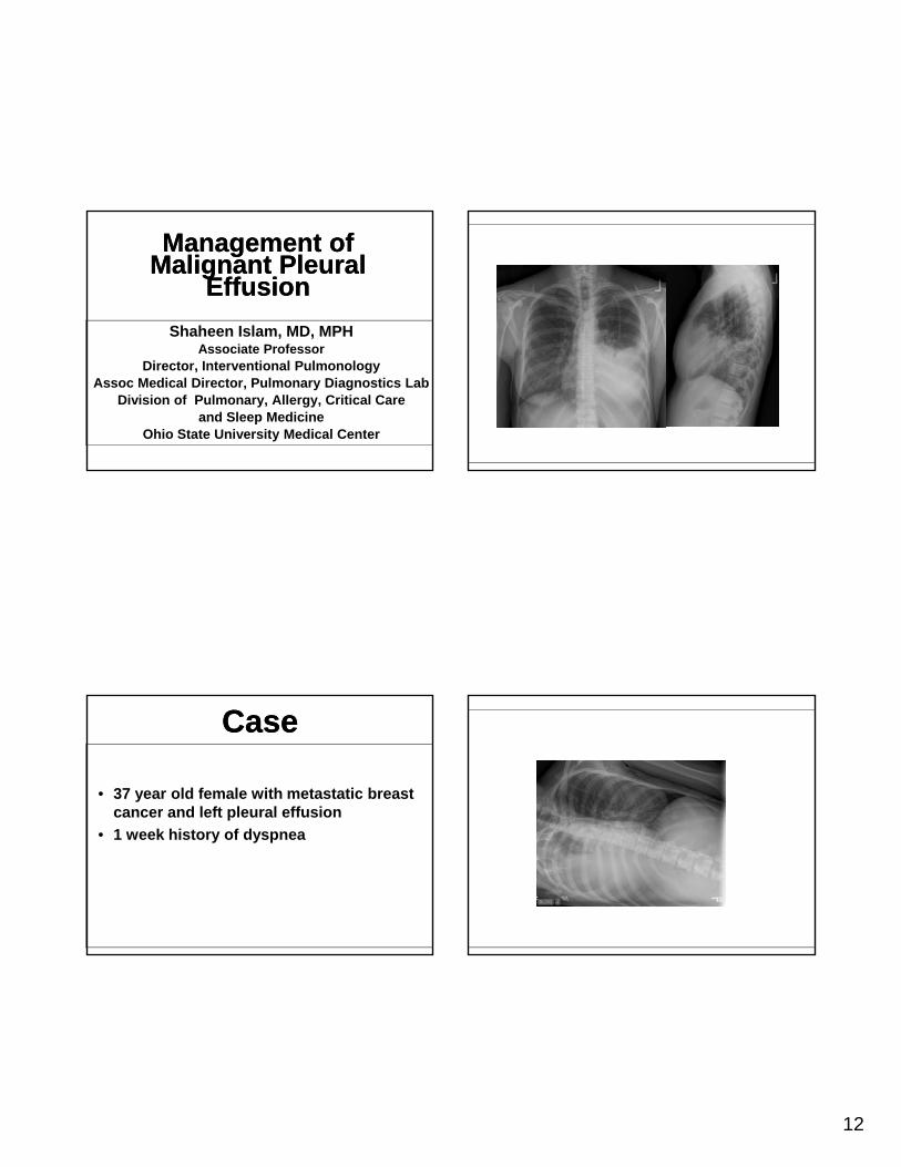
CaseCase
• 37 year old female with metastatic breast cancer and left pleural effusion
• 1 week history of dyspnea
13
Epidemiology of MPEEpidemiology of MPE
• Most common exudative effusion 40%~70%• Lung 32%, Breast 18%, Lymphoma 11%• More common in females <60 (50% vs 35%)• 15% of patients with Lung Cancer have MPE at
diagnosis
Gomez et al. CHEST 2007, 618SMarel M. Eur Respir Mon, 2002;22:146-156
EpidemiologyEpidemiology
• Primary tumor not identified 5-10% MPE• 20-30% of malignant lymphoma• 50% of patient with breast Ca• Ovarian, GI, mesothelioma ~15%• Usually <6 months survival except in
breast Ca
Marel M. Eur Respir Mon, 2002;22:146-156 Ruckdeschel JC Semin Oncol 1995; 22:58-63
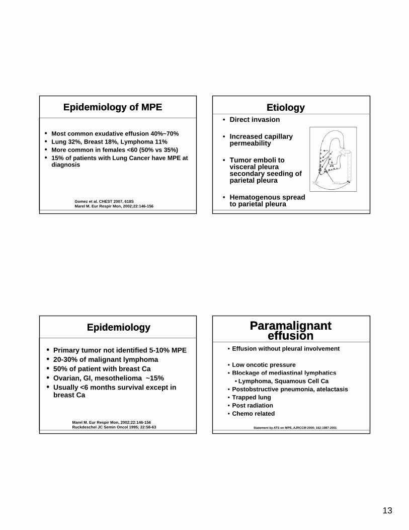
EtiologyEtiology• Direct invasion
• Increased capillary permeability
• Tumor emboli to visceral pleura secondary seeding of parietal pleura
• Hematogenous spread to parietal pleura
Paramalignanteffusion
Paramalignanteffusion
• Effusion without pleural involvement
• Low oncotic pressure• Blockage of mediastinal lymphaticsBlockage of mediastinal lymphatics
• Lymphoma, Squamous Cell Ca• Postobstructive pneumonia, atelactasis• Trapped lung• Post radiation• Chemo related
Statement by ATS on MPE, AJRCCM 2000; 162:1987-2001
14
Clinical featuresClinical features
• Progressive Dyspnea• Decreased chest wall compliance• Mediastinal shift• Increased shunt fraction from atelactaticIncreased shunt fraction from atelactatic
lung
• Dull chest pain• Malignant mesothelioma
• Cough
DiagnosisDiagnosis
History
Symptoms
ImagingImaging
CXR, CT chest, Ultrasound
Thoracentesis
Pleuroscopy/Pleural biopsy
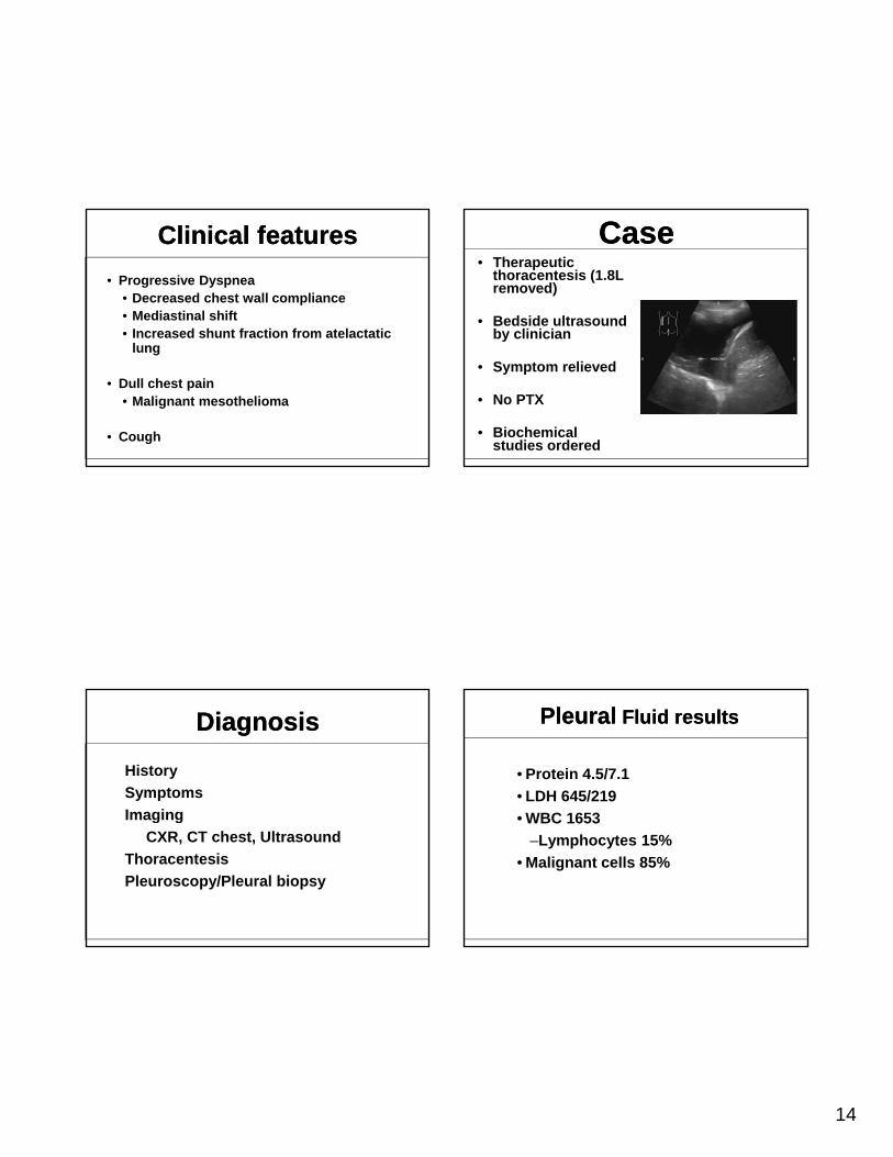
CaseCase• Therapeutic
thoracentesis (1.8L removed)
• Bedside ultrasound by clinicianby clinician
• Symptom relieved
• No PTX
• Biochemical studies ordered
Pleural Fluid resultsPleural Fluid results
• Protein 4.5/7.1
• LDH 645/219
• WBC 1653• WBC 1653
–Lymphocytes 15%
• Malignant cells 85%
15
Other Fluid StudiesOther Fluid Studies• Amylase
• Lymphoma, Ovarian Ca, Pancreatic Ca• CEA, B72.3, Leu-M1 • Calretinin and cytokeratin 5/6 identifies
mesothelioma but not benign mesothelial cells
• Flow cytometry • if lymphocytic effusion with possibility of
lymphoma
• Tumor markers• CEA (>10-12ng/mL)• Vascular Endothelial Growth Factor
(VEGF)
Goal for thoracentesisGoal for thoracentesis
• Diagnostic & therapeutic (large volume)
• Any relief of symptom?
• Will it recur?
• If so, how soon?
• Did the lung expand?
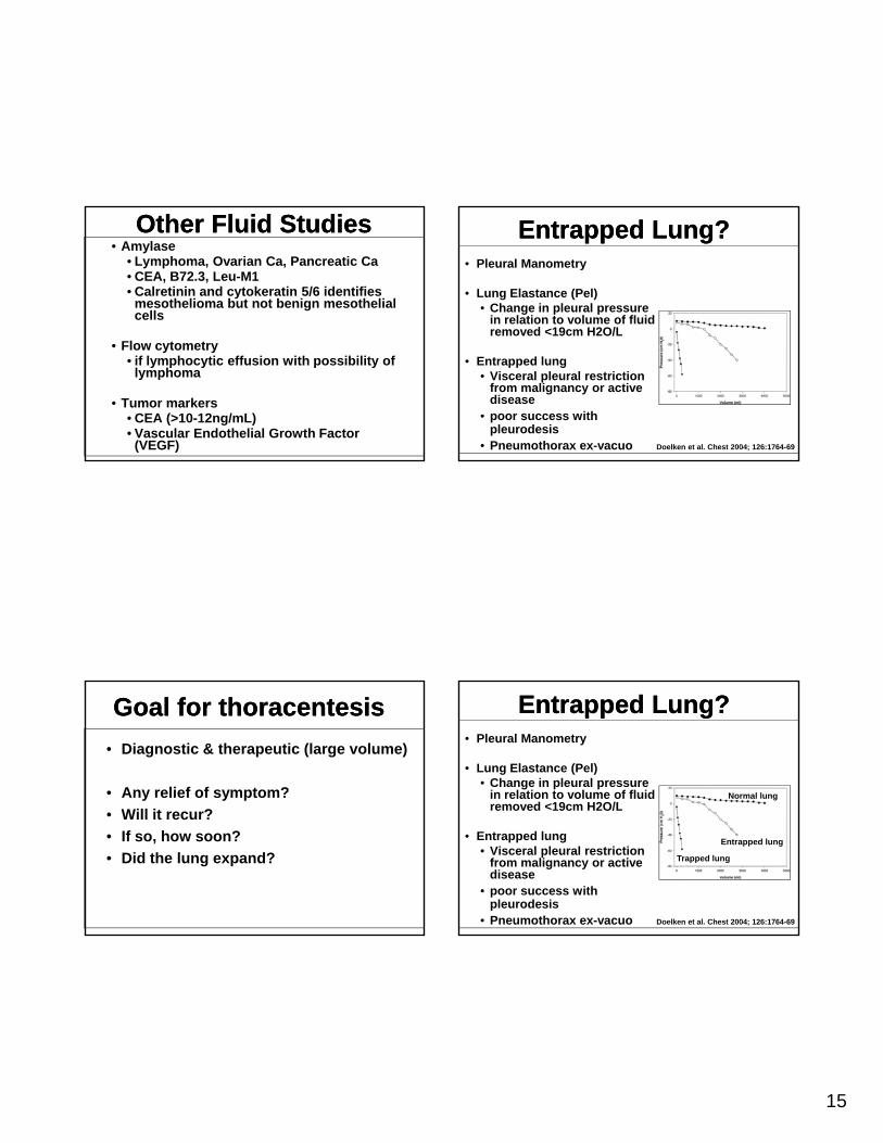
Entrapped Lung?Entrapped Lung?• Pleural Manometry
• Lung Elastance (Pel)• Change in pleural pressure
in relation to volume of fluid removed <19cm H2O/Lremoved <19cm H2O/L
• Entrapped lung• Visceral pleural restriction
from malignancy or active disease
• poor success with pleurodesis
• Pneumothorax ex-vacuo Doelken et al. Chest 2004; 126:1764-69
Entrapped Lung?Entrapped Lung?• Pleural Manometry
• Lung Elastance (Pel)• Change in pleural pressure
in relation to volume of fluid removed <19cm H2O/L
Normal lung
removed <19cm H2O/L
• Entrapped lung• Visceral pleural restriction
from malignancy or active disease
• poor success with pleurodesis
• Pneumothorax ex-vacuo
Entrapped lung
Trapped lung
Doelken et al. Chest 2004; 126:1764-69
16
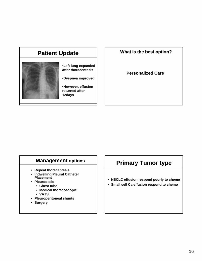
Patient UpdatePatient Update
•Left lung expanded after thoracentesis
•Dyspnea improved
•However, effusion returned after 12days
Management optionsManagement options
• Repeat thoracentesis• Indwelling Pleural Catheter
Placement• Pleurodesis
Ch b• Chest tube• Medical thoracoscopic• VATS
• Pleuroperitoneal shunts• Surgery
What is the best option?What is the best option?
Personalized Care
Primary Tumor typePrimary Tumor type
• NSCLC effusion respond poorly to chemo
• Small cell Ca effusion respond to chemo• Small cell Ca effusion respond to chemo
17
Pleural Fluid TestsPleural Fluid Tests
• Poor survival with
L H• Low pH
• Low glucose
• High LDH
• CEA
Performance StatusPerformance Status
• Karnofsky score <30, <1 month survival
Lung re-expansionLung re-expansion
• Extensive intrapleural deposition, multiple loculations, trapped lung, endobronchial airway obstruction will cause of failure of pleurodesisp
• Pneumothorax after large volume thoracentesis suggest trapped lung
Patient PreferencePatient Preference
• Duration of hospital stay• Invasiveness of procedures• Success of a definitive therapy• Success of a definitive therapy• Associated risks
18
Individual Management Options
Individual Management Options
Repeat thoracentesisRepeat thoracentesis
• Only if reaccumulation >30 days• Limited life expectancy• Poor performance status
• May trigger cytokine, fibrin and cause loculations
• Large volume thoracentesis with pleural manometry safe
Chemical PleurodesisChemical Pleurodesis
• Dyspneic and life expectancy more than 4-6 weeks
• Frequent recurrence with symptomssymptoms
• Success rate 71%~97%
Heffner J. Semin Respir Crit Care Med 2010; 31:723-733
• Doxycycline• Severe CP, thru Chest tube• Used with lidocaine
• Talc: • Slurry thru CT
• Lower success rate
Chemical AgentsChemical Agents
Lower success rate• Poudrage during thoracoscopy 90% success
rate *• No ARDS with larger callibrated particles
• Other Agents• Quinacrine• Bleomycin• Silver nitrate• IFN alpha-2b
19
Thoracoscopic vs Chest tube Pleurodesis
Thoracoscopic vs Chest tube Pleurodesis
• No large RCT available
• Cochrane review of 112 patients
• Slightly better with thoracoscopy
• Better success with thoracoscopy in breast and primary lung Ca
• Center dependent
• May be better with talc poudrage vs talc slurry
Shaw P et al. Cochrane database Systemic review 2004; CD002916Dressler et al Chest 2005; 127:909-915
Medical Thoracoscopy vs. VATS
Medical Thoracoscopy vs. VATS
• Similar
• Adhesions if present can be lysed
• Poudrage sprayed effectively under g p y yvision
• Sedation
• VATS more invasive
• Cost
• Requires 3-7days of hospital stay
Indwelling Pleural CatheterIndwelling Pleural Catheter
• Outpatient placement
• Patients may remain active with good QOL
• Can be placed in d l i f il dtrapped lung or in failed
pleurodesis cases
• Complications
• Obstruction, tumor seeding, infection
Indwelling Pleural CatheterIndwelling Pleural Catheter
• Outpatient placement
• Patients may remain active with good QOL
• Can be placed in d l i f il dtrapped lung or in failed
pleurodesis cases
• Complications
• Obstruction, tumor seeding, infection
20
Indwelling Pleural CatheterIndwelling Pleural Catheter
• Spontaneous pleurodesis in 40%
• Removed in 60% with resolution of effusion
• Sclerosants can be instilled through catheter
Putnam et al. Cancer 1999; 86:1992-1999Musani et al. Respiration 2004;71:559-566Warren et al. Eur J Cardiothoracic Surg 2008;33:89-94
SurgerySurgery
• Pleuroperitoneal Shunting with 95% efficacy if other options fail
• Parietal pleurectomy• Parietal pleurectomy
• Decortication
• Higher mortality
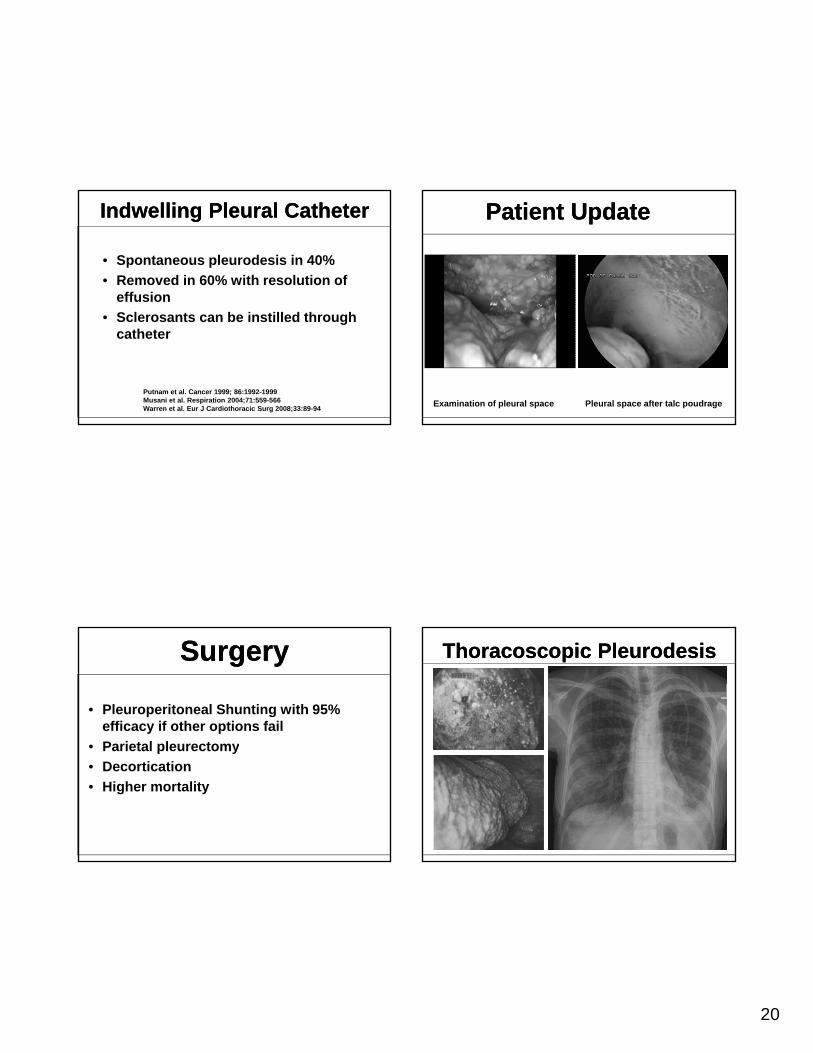
Patient Update Patient Update
Examination of pleural space Pleural space after talc poudrage
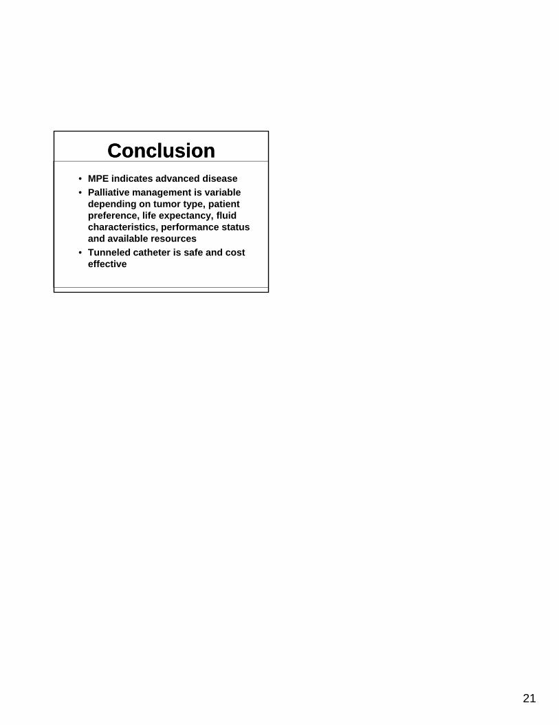
Thoracoscopic PleurodesisThoracoscopic Pleurodesis
21
ConclusionConclusion• MPE indicates advanced disease
• Palliative management is variable depending on tumor type, patient preference, life expectancy, fluid characteristics, performance status and available resources
• Tunneled catheter is safe and cost effective


![Management of Malignant Pleural Effusion...Asymptomatic patients with either a malignant or a paramalignant effusion need not be treated initially [9]. Malignant pleural effusion will](https://img.pdfslide.us/doc/110x75/5f8bc67cd3c5026bc44819fe/management-of-malignant-pleural-effusion-asymptomatic-patients-with-either-a.jpg)