Embed Size (px)
Citation preview
Int J Clin Exp Pathol 2016;9(12):12459-12469www.ijcep.com /ISSN:1936-2625/IJCEP0036593
Original ArticleKi20227 influences the morphology of microglia and neurons through inhibition of CSF1R during global ischemia
Boru Hou1,2*, Furong Ju1*, Xiumei Guo2*, Dong Wang2, Xiaofeng Cheng1, Akbar khan1, Shengxiang Zhang1, Haijun Ren2
1Gansu Key Laboratory of Biomonitoring and Bioremediation for Environmental Pollution, School of Life Sciences, Lanzhou University, Lanzhou 730000, China; 2Department of Neurosurgery, Lanzhou University Second Hospital, Lanzhou 730000, China. *Equal contributors and co-first authors.
Received July 24, 2016; Accepted October 17, 2016; Epub December 1, 2016; Published December 15, 2016
Abstract: Microglia are the main immune cells of the brain, and play a vital role in stroke pathophysiology. In the healthy brain, microglia depend on colony-stimulating factor 1 receptor (CSF1R) signaling for survival, and its phar-macological inhibition can cause morphological changes in microglia. How CSF1R influences microglia and neuro-nal structures after stroke remains unclear. Here we found that treatment with ki20227 during global ischemia led to a significant deficit in microglial density in the CNS in mice, and CSF1R-inhibition led to a significant reduction in the neuronal density of mice. Also, the dendritic spines of layer 5 pyramidal neurons showed a significant loss in the cortex over a period of 3 d. These findings demonstrated that microglial density and survival during global ischemia were dependent on CSF1R signaling, and inhibition of CSF1R receptors in microglia contributed to the loss of den-dritic spines and neurons in stroke. Thus, microglia may play a role in global ischemic pathology.
Keywords: Microglia, global ischemia, reperfusion, ki20227, dendritic spines
Introduction
Colony stimulating factor 1 receptor (CSF1R) is expressed on committed macrophage precur-sors, monocytes, and microglia [1-4]. In the brain, microglia are shown to be the only cell type that express CSF1R under normal condi-tions [2, 5]. Survival of microglia in the adult brain is shown to be fully dependent on CSF1R signaling [6]. It has two natural ligands: colony stimulating factor 1 (CSF1) and interleukin-34 (IL-34) [7-9]. CSF1 promotes the survival, prolif-eration and differentiation of bone marrow pro-genitor cells, monocytes, and macrophages, and regulates the response of this lineage to inflammatory challenges [10-15]. Mice lacking either CSF1 or CSF1R showed reduced densi-ties of macrophages in several tissues [16, 17]. Furthermore, CSF1R knockout mice would die before adulthood [5, 18]. Besides its homeo-static role in normal animals, it can play a role in pathologies such as arthritis that involve chronic activation of tissue macrophage popu-lations [10]. Recent research found that the ramified microglia perform tissue surveillance
and also have a purposeful role [19]. In the absence of pathological insult, interactions with synapses [20], while after ischemia, rami-fied changes in morphology may be associated with microglial function, and may contribute to the pathological process of stroke during isch-emia and reperfusion [21, 22]. The pleiotropic physiological functions of microglia confound our understanding of its role in neurodegenera-tive or neuroprotective actions after stroke [23-27]. In the present study, we inhibited CSF1R signaling in microglia in global ischemia using quantitative methods to analyze microglial den-sity in fixed tissue, and precisely characterize microglial morphological responses to injury. Our objective was to examine the changes in microglial morphology related to cerebral injury induced by global ischemia and reperfusion after inhibition of CSF1R signaling. We hypoth-esized that ischemia and reperfusion would lead to microglial morphological responses after inhibition of CSF1R signaling, and that a relationship existed between microglial mor-phology and brain injury after ischemia and reperfusion. To test these hypotheses, we ana-
Microglia in global ischemia
12460 Int J Clin Exp Pathol 2016;9(12):12459-12469
lyzed microglial density in a murine model of global ischemic stroke. The results revealed that microglial physiology in the ischemic brain was also dependent on CSF1R signaling. Inhibition of CSF1R signaling activity in isch-emic mice resulted in morphological changes in microglia, a remarkable loss of dendritic spines and neuronal deficits as compared to the con-trol group.
Materials and methods
Description of ki20227
Ki20227 (N-{4-[96, 7-dimethoxy-4-quinolyl) oxy]-2-methoxyphenyl}-N-[1-(1, 3-Thiazole-2-yl) ethyl] urea, which is a highly selective CSF1R inhibitor, was bought from Med Chem Express. It was suspended in vehicle (0.5% methyl cel-lulose in distilled water) in vivo.
Animals
Transgenic mice expressing yellow fluorescent protein (YFP)-H in the layer 5 pyramidal neu-rons (Thy1-YFP line H) were purchased from the Jackson Laboratory, and bred in the Labora- tory Centre for Medical Sciences, Lanzhou University. Transgenic mice aged 3-5 months and weighing 20-30 g were used in this study. All experiments were performed in accordance with the institutional animal guidelines.
Response of CSF1R to inhibitory effect of ki20227
The tyrosine kinase activity of CSF1R was inhib-ited by the administration of ki20227, and inhi-bition of protein kinases was determined as previously described. The mice were treated with ki20227 (n=16) by intragastric administra-tion (each mouse was given 2 mg/g/day), and after 2 days of treatment, a global ischemic model was developed. During reperfusion for 3 days, we also gave ki20227 daily. Vehicle was administrated intragastrically after ischemia for 60 minutes and reperfusion for 3 days (n=16).
Global ischemia model
Bilateral common carotid artery ligation (BCAL) model was used in all mice to induce global cerebral ischemia. Animals were deeply anes-thetized by intraperitoneal injection with 2% ketamine and 0.2% xylazine. Thereafter, ani-
mals were placed in supine position and the occlusion of carotid arteries were made through a midline incision. To confirm the effectiveness of surgery, the surgical mice were identified by their blood flow and dendritic structure through the cranial window with a fluorescent micro-scope. If the blood flow decreased obviously and the dendritic structure showed beading in 10 minutes, as expected, global ischemia was induced. Sutures were loosened after occlu-sion for 60 minutes to restore the brain blood flow for 3 days.
Image analysis of ki20227 efficacy on microg-lia
The CX3CR1GFP/+ mice that express green fluo-rescent protein in the microglia were selected to study the efficacy of ki20227. After global ischemia for 1 h and reperfusion for 1 d, 2 d and 3 d, the brain was fixed with 4% PFA, and then cut into slices (30 μm) by vibrating micro-tome (Leica). Confocal images (60-μm z-stack at 1 μm intervals, Olympus- FV1000MPE, 40× objective) were acquired at the somatosensory cortical region. On different reperfusion days, we used ImageJ software to analyze the density of microglia. SPSS software was used for all statistical analyses. Analysis of variance, two-tailed unpaired t-test, and one-way ANOVA were used for comparison of the four groups. Results were expressed as the mean ± standard error of the mean, *P<0.05, **P<0.01.
Immunofluorescent labeling and confocal microscopy
Brain sections were washed in phosphate buf-fer saline (PBS), and blocked with 10% goat serum in PBS for 0.5 h. Primary antibodies NeuN (Millipore, 1:200) were diluted in buffer with 0.01% Triton X-100 and 10% goat serum for 12 h at 4°C. For each confocal experiment, all brain sections were cut on the same day, immunostained together and imaged within 48 h of staining. SPSS software was used for all statistical analyses. Analysis of variance, two-tailed unpaired t-test, and one-way ANOVA were used for comparison of the four groups. Results were expressed as the mean ± standard error of the mean, *P<0.05, **P<0.01.
Image analysis of spines
We chose the mice that express yellow fluores-cent protein (YFP)-H in the layer 5 pyramidal
Microglia in global ischemia
12461 Int J Clin Exp Pathol 2016;9(12):12459-12469
neurons (Thy1-YFP line H), and after reperfu-sion for 3 d, the brain was fixed with PFA, and cut into slices (30 μm) by vibrating microtome (Leica). All the brain slices were imaged using confocal microscopy (30-μm z-stack at 1 μm intervals, Olympus- FV1000MPE, 100× oil objective), and the images were acquired at the cortex region. All images were analyzed using ImageJ software (http://imagej.net/ImageJ). To quantify dendritic spines after ischemia, we measured the total length of dendrites with the aid of a custom designed macros, which was used to mark the dendrites at 100 µm fixed length, and calculated the length of the select-ed dendritic segments. For each neuron, we chose apical shaft, apical oblique, and basilar dendrite. The apical shaft dendrite was divided into four parts from the top to bottom, each
part was 100 µm long; four parts of the apical oblique and basilar dendrite were chosen in each neuron, and on each side we chose two. Approximately 20 neurons were counted for each mouse to quantify the changes in dendrit-ic spines. SPSS software was used for all statis-tical analyses. Analysis of variance, two-tailed unpaired t-test, and one-way ANOVA were used for comparison of the four groups. Results were expressed as the mean ± standard error of the mean, *P<0.05, **P<0.01.
Results
Induction of ischemia and alteration of den-dritic structure in the BCAL model
In the BCAL model, neuronal activity is closely related to microcirculation and influences the
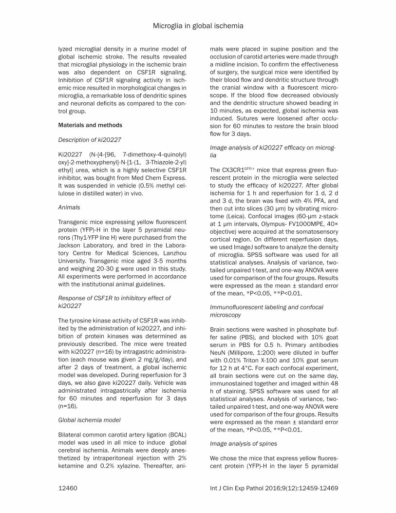
Figure 1. Two-photon imaging shows a murine model of global ischemia. A. BCAL model was used to create global ischemia. B. Cranial window under two-photon imaging shows the changes in dendritic structure (green) before isch-emia, 60 min after ischemia, and 3 d after reperfusion. In vivo two-photon imaging demonstrated the cessation of blood flow and obvious dendritic beading after 60 minutes BCAL, and most of the spines were lost, which reflected a successful BCAL. Two-photon imaging revealed that after reperfusion for 3 d, most of the beading dendritic struc-tures and spines recovered to their normal positions. C. Partial enlargement of B. Representative image showing structural changes in dendritic spines during ischemia/reperfusion.
Microglia in global ischemia
12462 Int J Clin Exp Pathol 2016;9(12):12459-12469
integrity of neural network. To create this model, we placed the animal in the supine posi-tion and blood flow was discontinued through common carotid arteries by ligation (Figure 1A). An effective and successful surgery can
reduce the blood flow, and passive flow of blood can be seen through the cranial window under a fluorescent microscope. We ceased blood flow effectively for 10 minutes. An obvious change in the dendritic structure demonstrated
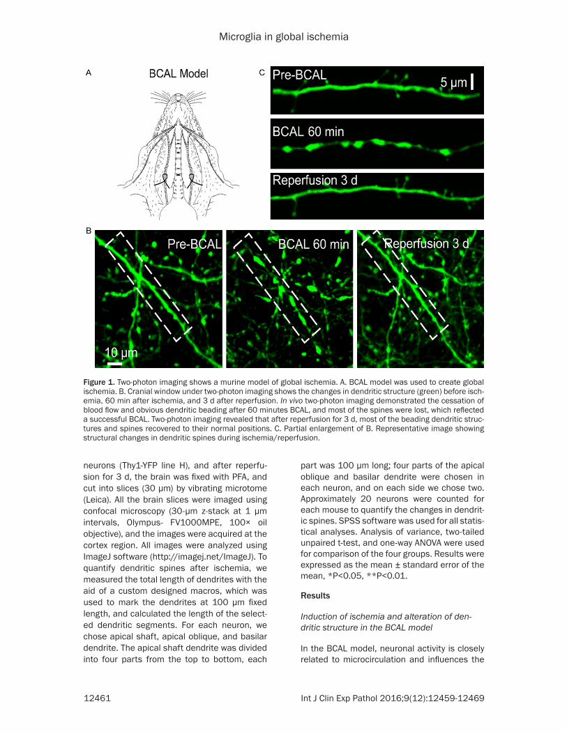
Figure 2. Microglial changes over time in the cerebral cortex of a mouse model of ischemic pathology. A. Confocal image of microglia in global ischemia. The images show microglial morphology (green) before ischemia, BCAL 1 h-reperfusion for 1 d and 3 d, respectively, in the drug- treated and control groups. B. Post stroke, drug-treated groups showed reduced density of microglia. Analysis of microglial density in the vehicle and ki20227 groups, respectively, after BCAL 1 h following days of reperfusion showed that the density of microglia decreased significantly after treat-ment with ki20227 during global ischemia. Two-way ANOVA was used to analyze statistical differences in the data: *P<0.05, **P<0.01. n=8.
Microglia in global ischemia
12463 Int J Clin Exp Pathol 2016;9(12):12459-12469
Microglia in global ischemia
12464 Int J Clin Exp Pathol 2016;9(12):12459-12469
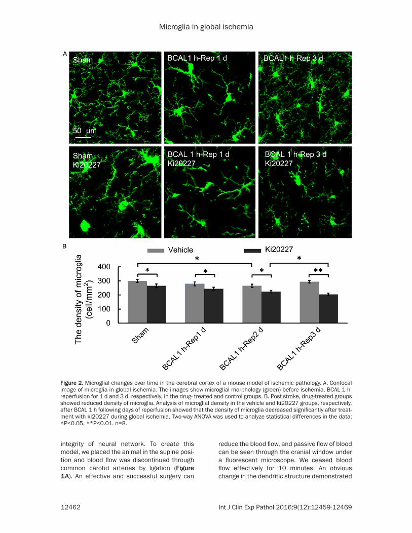
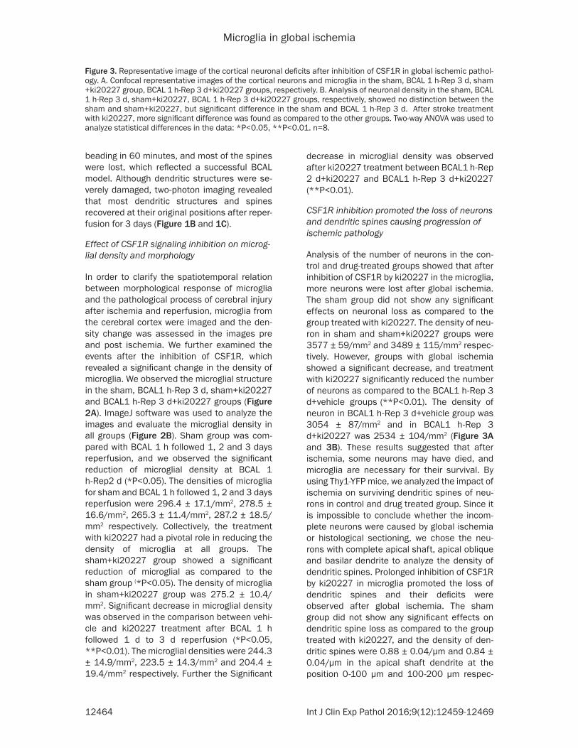
Figure 3. Representative image of the cortical neuronal deficits after inhibition of CSF1R in global ischemic pathol-ogy. A. Confocal representative images of the cortical neurons and microglia in the sham, BCAL 1 h-Rep 3 d, sham +ki20227 group, BCAL 1 h-Rep 3 d+ki20227 groups, respectively. B. Analysis of neuronal density in the sham, BCAL 1 h-Rep 3 d, sham+ki20227, BCAL 1 h-Rep 3 d+ki20227 groups, respectively, showed no distinction between the sham and sham+ki20227, but significant difference in the sham and BCAL 1 h-Rep 3 d. After stroke treatment with ki20227, more significant difference was found as compared to the other groups. Two-way ANOVA was used to analyze statistical differences in the data: *P<0.05, **P<0.01. n=8.
beading in 60 minutes, and most of the spines were lost, which reflected a successful BCAL model. Although dendritic structures were se- verely damaged, two-photon imaging revealed that most dendritic structures and spines recovered at their original positions after reper-fusion for 3 days (Figure 1B and 1C).
Effect of CSF1R signaling inhibition on microg-lial density and morphology
In order to clarify the spatiotemporal relation between morphological response of microglia and the pathological process of cerebral injury after ischemia and reperfusion, microglia from the cerebral cortex were imaged and the den-sity change was assessed in the images pre and post ischemia. We further examined the events after the inhibition of CSF1R, which revealed a significant change in the density of microglia. We observed the microglial structure in the sham, BCAL1 h-Rep 3 d, sham+ki20227 and BCAL1 h-Rep 3 d+ki20227 groups (Figure 2A). ImageJ software was used to analyze the images and evaluate the microglial density in all groups (Figure 2B). Sham group was com-pared with BCAL 1 h followed 1, 2 and 3 days reperfusion, and we observed the significant reduction of microglial density at BCAL 1 h-Rep2 d (*P<0.05). The densities of microglia for sham and BCAL 1 h followed 1, 2 and 3 days reperfusion were 296.4 ± 17.1/mm2, 278.5 ± 16.6/mm2, 265.3 ± 11.4/mm2, 287.2 ± 18.5/mm2 respectively. Collectively, the treatment with ki20227 had a pivotal role in reducing the density of microglia at all groups. The sham+ki20227 group showed a significant reduction of microglial as compared to the sham group (*P<0.05). The density of microglia in sham+ki20227 group was 275.2 ± 10.4/mm2. Significant decrease in microglial density was observed in the comparison between vehi-cle and ki20227 treatment after BCAL 1 h followed 1 d to 3 d reperfusion (*P<0.05, **P<0.01). The microglial densities were 244.3 ± 14.9/mm2, 223.5 ± 14.3/mm2 and 204.4 ± 19.4/mm2 respectively. Further the Significant
decrease in microglial density was observed after ki20227 treatment between BCAL1 h-Rep 2 d+ki20227 and BCAL1 h-Rep 3 d+ki20227 (**P<0.01).
CSF1R inhibition promoted the loss of neurons and dendritic spines causing progression of ischemic pathology
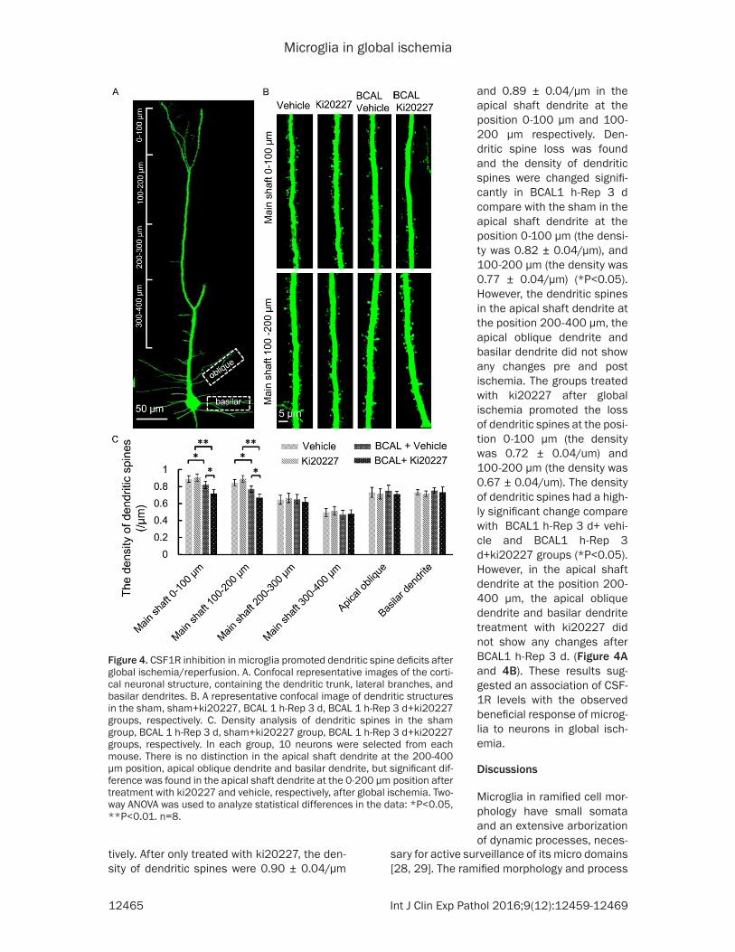
Analysis of the number of neurons in the con-trol and drug-treated groups showed that after inhibition of CSF1R by ki20227 in the microglia, more neurons were lost after global ischemia. The sham group did not show any significant effects on neuronal loss as compared to the group treated with ki20227. The density of neu-ron in sham and sham+ki20227 groups were 3577 ± 59/mm2 and 3489 ± 115/mm2 respec-tively. However, groups with global ischemia showed a significant decrease, and treatment with ki20227 significantly reduced the number of neurons as compared to the BCAL1 h-Rep 3 d+vehicle groups (**P<0.01). The density of neuron in BCAL1 h-Rep 3 d+vehicle group was 3054 ± 87/mm2 and in BCAL1 h-Rep 3 d+ki20227 was 2534 ± 104/mm2 (Figure 3A and 3B). These results suggested that after ischemia, some neurons may have died, and microglia are necessary for their survival. By using Thy1-YFP mice, we analyzed the impact of ischemia on surviving dendritic spines of neu-rons in control and drug treated group. Since it is impossible to conclude whether the incom-plete neurons were caused by global ischemia or histological sectioning, we chose the neu-rons with complete apical shaft, apical oblique and basilar dendrite to analyze the density of dendritic spines. Prolonged inhibition of CSF1R by ki20227 in microglia promoted the loss of dendritic spines and their deficits were observed after global ischemia. The sham group did not show any significant effects on dendritic spine loss as compared to the group treated with ki20227, and the density of den-dritic spines were 0.88 ± 0.04/μm and 0.84 ± 0.04/μm in the apical shaft dendrite at the position 0-100 μm and 100-200 μm respec-
Microglia in global ischemia
12465 Int J Clin Exp Pathol 2016;9(12):12459-12469
Figure 4. CSF1R inhibition in microglia promoted dendritic spine deficits after global ischemia/reperfusion. A. Confocal representative images of the corti-cal neuronal structure, containing the dendritic trunk, lateral branches, and basilar dendrites. B. A representative confocal image of dendritic structures in the sham, sham+ki20227, BCAL 1 h-Rep 3 d, BCAL 1 h-Rep 3 d+ki20227 groups, respectively. C. Density analysis of dendritic spines in the sham group, BCAL 1 h-Rep 3 d, sham+ki20227 group, BCAL 1 h-Rep 3 d+ki20227 groups, respectively. In each group, 10 neurons were selected from each mouse. There is no distinction in the apical shaft dendrite at the 200-400 μm position, apical oblique dendrite and basilar dendrite, but significant dif-ference was found in the apical shaft dendrite at the 0-200 μm position after treatment with ki20227 and vehicle, respectively, after global ischemia. Two-way ANOVA was used to analyze statistical differences in the data: *P<0.05, **P<0.01. n=8.
and 0.89 ± 0.04/μm in the apical shaft dendrite at the position 0-100 μm and 100-200 μm respectively. Den- dritic spine loss was found and the density of dendritic spines were changed signifi-cantly in BCAL1 h-Rep 3 d compare with the sham in the apical shaft dendrite at the position 0-100 μm (the densi-ty was 0.82 ± 0.04/μm), and 100-200 μm (the density was 0.77 ± 0.04/μm) (*P<0.05). However, the dendritic spines in the apical shaft dendrite at the position 200-400 μm, the apical oblique dendrite and basilar dendrite did not show any changes pre and post ischemia. The groups treated with ki20227 after global ischemia promoted the loss of dendritic spines at the posi-tion 0-100 μm (the density was 0.72 ± 0.04/um) and 100-200 μm (the density was 0.67 ± 0.04/um). The density of dendritic spines had a high-ly significant change compare with BCAL1 h-Rep 3 d+ vehi-cle and BCAL1 h-Rep 3 d+ki20227 groups (*P<0.05). However, in the apical shaft dendrite at the position 200-400 μm, the apical oblique dendrite and basilar dendrite treatment with ki20227 did not show any changes after BCAL1 h-Rep 3 d. (Figure 4A and 4B). These results sug-gested an association of CSF- 1R levels with the observed beneficial response of microg-lia to neurons in global isch- emia.
Discussions
Microglia in ramified cell mor-phology have small somata and an extensive arborization of dynamic processes, neces-
tively. After only treated with ki20227, the den-sity of dendritic spines were 0.90 ± 0.04/μm
sary for active surveillance of its micro domains [28, 29]. The ramified morphology and process
Microglia in global ischemia
12466 Int J Clin Exp Pathol 2016;9(12):12459-12469
activity vary across brain regions [30, 31]. Recent studies have elucidated the role of mediators that altered microglial morphological and functional activities in healthy as well as models of CNS injury [32-36]. The total progres-sive morphological and functional changes occurring during ischemia and reperfusion that contribute to cerebral injury after ischemic stroke are poorly understood [18, 37]. This study focused on the changes in the morpholo-gy of neurons and microglia in response to isch-emic stroke and reperfusion, which was further extended using ki20227. Our results confirmed the efficacy of CSF1R inhibition in reducing microglial density in ischemia. The inhibition of CSF1R kinase activity by ki20227 was used to modify microglia, which can influence neurons and loss of dendritic spines. Our results sup-ported a previous report that showed neuropro-tective function of ramified microglia in brain excitotoxicity [38-40]. Our findings support the relevance of CSF1R signaling of microglia in global ischemic stroke and validate the evalua-tion of CSF1R inhibitors in clinical trials for ischemic diseases. Microglia are distributed throughout brain parenchyma. However, cell density between brain regions is variable [35, 41, 42]. BCAL 1 h after reperfusion for 3 d and treatment with ki20227 to inhibit CSF1R showed reduced microglial density, suggesting that the CSF1R of microglia was heavily impli-cated in stroke, which was consistent with find-ing that mice lacking either of the two CSF1R ligands, CSF1 or IL-34, also have reduced den-sities of microglia throughout the CNS [43]. CSF1R plays a crucial role in microglial tissue homeostasis, and the adult brain has a remark-able capacity to regulate microglia population [6] . Recent research found that chronic deple-tion of >99% of all microglia for 3 or 8 weeks in adult mice resulted in no deficits in any behav-ioral cognitive task [44, 45]. But pharmacologi-cal inhibition of CSF1R in microglia prevented the progression of Alzheimer’s-like pathophysi-ology [46]. Microglia play an important role in cognition and behavior in healthy adult mice, and their depletion showed a significant reduc-tion in motor-learning-dependent synapse for-mation [36, 40, 47] . Groups treated with ki20227 after global ischemia showed reduc-tion in the number of neurons and dendritic spines, as compared to the global ischemic mice treated with vehicle. Our study found after reperfusion for 3 d, the microglial processes recovered to the normal state, but inhibiting the
CSF1R in microglia decreased the number of neurons and dendritic spines with progression of global ischemia-like pathology. Our study suggests that microglia are necessary for the stability of the neurons and dendritic spines after global ischemia. These features suggest that microglia are crucial for stroke, preferen-tially depend on CSF1R and actively modify morphology via CSF1R, to play a key role in the progression of ischemia-like pathophysiology.
In summary, the present data provided evi-dence of an important role of microglia in glob-al ischemic diseases, as well as their depen-dence upon CSF1R activation. Reduction of microglial density and processes in global isch-emic disease mice using a selective CSF1R inhibitor promoted neuronal decline and the loss of dendritic spines leading to exacerbated progression of global ischemic disease-like pathology. The efficacy of microglial inhibition in global ischemic disease-like pathology and reduction of the potentially damaging compo-nents to dendritic spines and neurons highlight the possible evaluation of microglia in clinical trials for global ischemic disease.
Acknowledgements
This study was supported by National Natural Science Foundation of Gansu, China (No.15- 06RJZA222) and Lanzhou Science and Tech- nology Bureau Project (2015-2-55). We thank L.P Guan at the core facility of School of Life Sciences for technical support, and X.X Xi for assistance with preliminary experiments.
Disclosure of conflict of interest
None.
Address correspondence to: Dr. Shengxiang Zhang, School of Life Sciences, Lanzhou University, No. 222 South Tianshui Road, Lanzhou 730000, Gansu, China. Tel: +86-931-8915607; Fax: +86-931-8915607; E-mail: [email protected]; Dr. Haijun Ren, Department of Neurosurgery, Lanzhou University Second Hospital, Lanzhou 730000, Gansu, China. Tel: +86 13893668727; E-mail: [email protected]
References
[1] Pixley FJ and Stanley ER. CSF-1 regulation of the wandering macrophage: complexity in ac-tion. Trends Cell Biol 2004; 14: 628-638.
Microglia in global ischemia
12467 Int J Clin Exp Pathol 2016;9(12):12459-12469
[2] Nandi S, Gokhan S, Dai XM, Wei S, Enikolopov G, Lin H, Mehler MF and Stanley ER. The CSF-1 receptor ligands IL-34 and CSF-1 exhibit dis-tinct developmental brain expression patterns and regulate neural progenitor cell mainte-nance and maturation. Dev Biol 2012; 367: 100-113.
[3] Patel S and Player MR. Colony-stimulating fac-tor-1 receptor inhibitors for the treatment of cancer and inflammatory disease. Curr Top Med Chem 2009; 9: 599-610.
[4] Kierdorf K, Erny D, Goldmann T, Sander V, Schulz C, Perdiguero EG, Wieghofer P, Heinrich A, Riemke P, Holscher C, Muller DN, Luckow B, Brocker T, Debowski K, Fritz G, Opdenakker G, Diefenbach A, Biber K, Heikenwalder M, Geiss-mann F, Rosenbauer F and Prinz M. Microglia emerge from erythromyeloid precursors via Pu.1- and Irf8-dependent pathways. Nat Neu-rosci 2013; 16: 273-280.
[5] Erblich B, Zhu L, Etgen AM, Dobrenis K and Pollard JW. Absence of colony stimulation fac-tor-1 receptor results in loss of microglia, dis-rupted brain development and olfactory defi-cits. PLoS One 2011; 6: e26317.
[6] Elmore MR, Najafi AR, Koike MA, Dagher NN, Spangenberg EE, Rice RA, Kitazawa M, Matu-sow B, Nguyen H, West BL and Green KN. Col-ony-stimulating factor 1 receptor signaling is necessary for microglia viability, unmasking a microglia progenitor cell in the adult brain. Neuron 2014; 82: 380-397.
[7] Blalock EM, Geddes JW, Chen KC, Porter NM, Markesbery WR and Landfield PW. Incipient Alzheimer’s disease: microarray correlation analyses reveal major transcriptional and tu-mor suppressor responses. Proc Natl Acad Sci U S A 2004; 101: 2173-2178.
[8] Huang HY, West RB, Tzeng CC, van de Rijn M, Wang JW, Chou SC, Huang WW, Eng HL, Lin CN, Yu SC, Wu JM, Lu CC and Li CF. Immunohisto-chemical and biogenetic features of diffuse-type tenosynovial giant cell tumors: the poten-tial roles of cyclin A, P53, and deletion of 15q in sarcomatous transformation. Clin Cancer Res 2008; 14: 6023-6032.
[9] Dower K, Ellis DK, Saraf K, Jelinsky SA and Lin LL. Innate immune responses to TREM-1 acti-vation: overlap, divergence, and positive and negative cross-talk with bacterial lipopolysac-charide. J Immunol 2008; 180: 3520-3534.
[10] Ohno H, Uemura Y, Murooka H, Takanashi H, Tokieda T, Ohzeki Y, Kubo K and Serizawa I. The orally-active and selective c-Fms tyrosine kinase inhibitor Ki20227 inhibits disease pro-gression in a collagen-induced arthritis mouse model. Eur J Immunol 2008; 38: 283-291.
[11] Nakase K, Kita K, Kyo T, Ueda T, Tanaka I and Katayama N. Prognostic relevance of cytokine
receptor expression in acute myeloid leuke-mia: interleukin-2 receptor alpha-chain (CD25) expression predicts a poor prognosis. PLoS One 2015; 10: e0128998.
[12] Shimada-Sugawara M, Sakai E, Okamoto K, Fukuda M, Izumi T, Yoshida N and Tsukuba T. Rab27A regulates transport of cell surface re-ceptors modulating multinucleation and lyso-some-related organelles in osteoclasts. Sci Rep 2015; 5: 9620.
[13] Sauter KA, Pridans C, Sehgal A, Bain CC, Scott C, Moffat L, Rojo R, Stutchfield BM, Davies CL, Donaldson DS, Renault K, McColl BW, Mowat AM, Serrels A, Frame MC, Mabbott NA and Hume DA. The MacBlue binary transgene (cs-f1r-gal4VP16/UAS-ECFP) provides a novel marker for visualisation of subsets of mono-cytes, macrophages and dendritic cells and responsiveness to CSF1 administration. PLoS One 2014; 9: e105429.
[14] Obba S, Hizir Z, Boyer L, Selimoglu-Buet D, Pfeifer A, Michel G, Hamouda MA, Goncalves D, Cerezo M, Marchetti S, Rocchi S, Droin N, Cluzeau T, Robert G, Luciano F, Robaye B, Foretz M, Viollet B, Legros L, Solary E, Auberger P and Jacquel A. The PRKAA1/AMPKalpha1 pathway triggers autophagy during CSF1-in-duced human monocyte differentiation and is a potential target in CMML. Autophagy 2015; 11: 1114-1129.
[15] Stutchfield BM, Antoine DJ, Mackinnon AC, Gow DJ, Bain CC, Hawley CA, Hughes MJ, Fran-cis B, Wojtacha D, Man TY, Dear JW, Devey LR, Mowat AM, Pollard JW, Park BK, Jenkins SJ, Simpson KJ, Hume DA, Wigmore SJ and Forbes SJ. CSF1 Restores Innate Immunity After Liver Injury in Mice and Serum Levels Indicate Out-comes of Patients With Acute Liver Failure. Gastroenterology 2015; 149: 1896-1909 e1814.
[16] Choudhury GG, Sylvia VL, Wang LM, Pierce J and Sakaguchi AY. The kinase insert domain of colony stimulating factor-1 receptor is dispens-able for CSF-1 induced phosphatidylcholine hydrolysis. FEBS Lett 1991; 282: 351-354.
[17] Li J, Chen K, Zhu L and Pollard JW. Conditional deletion of the colony stimulating factor-1 re-ceptor (c-fms proto-oncogene) in mice. Gene-sis 2006; 44: 328-335.
[18] Ginhoux F, Greter M, Leboeuf M, Nandi S, See P, Gokhan S, Mehler MF, Conway SJ, Ng LG, Stanley ER, Samokhvalov IM and Merad M. Fate mapping analysis reveals that adult mi-croglia derive from primitive macrophages. Sci-ence 2010; 330: 841-845.
[19] Wake H, Moorhouse AJ, Jinno S, Kohsaka S and Nabekura J. Resting microglia directly monitor the functional state of synapses in vivo and determine the fate of ischemic termi-nals. J Neurosci 2009; 29: 3974-3980.
Microglia in global ischemia
12468 Int J Clin Exp Pathol 2016;9(12):12459-12469
[20] Tremblay ME, Lowery RL and Majewska AK. Mi-croglial interactions with synapses are modu-lated by visual experience. PLoS Biol 2010; 8: e1000527.
[21] Morrison H, McKee D and Ritter L. Systemic neutrophil activation in a mouse model of isch-emic stroke and reperfusion. Biol Res Nurs 2011; 13: 154-163.
[22] Morrison HW and Filosa JA. A quantitative spa-tiotemporal analysis of microglia morphology during ischemic stroke and reperfusion. J Neu-roinflammation 2013; 10: 4.
[23] Chen S, Meng F, Chen Z, Tomlinson BN, Wesley JM, Sun GY, Whaley-Connell AT, Sowers JR, Cui J and Gu Z. Two-dimensional zymography dif-ferentiates gelatinase isoforms in stimulated microglial cells and in brain tissues of acute brain injuries. PLoS One 2015; 10: e0123852.
[24] Zhang ZY, Sun BL, Yang MF, Li DW, Fang J and Zhang S. Carnosine attenuates early brain in-jury through its antioxidative and anti-apoptot-ic effects in a rat experimental subarachnoid hemorrhage model. Cell Mol Neurobiol 2015; 35: 147-157.
[25] Zhang G, Xia F, Zhang Y, Zhang X, Cao Y, Wang L, Liu X, Zhao G and Shi M. Ginsenoside Rd Is Efficacious Against Acute Ischemic Stroke by Suppressing Microglial Proteasome-Mediated Inflammation. Mol Neurobiol 2016; 53: 2529-2540.
[26] Ueda H, Halder SK, Matsunaga H, Sasaki K and Maeda S. Neuroprotective impact of pro-thymosin alpha-derived hexapeptide against retinal ischemia-reperfusion. Neuroscience 2016; 318: 206-218.
[27] Yang X, Asakawa T, Han S, Liu L, Li W, Wu W, Luo Y, Cao W, Cheng X, Xiao B, Namba H, Lu C, Dong Q and Wang L. Neuroserpin protects rat neurons and microglia-mediated inflammatory response against oxygen-glucose deprivation- and reoxygenation treatments in an in vitro study. Cell Physiol Biochem 2016; 38: 1472-1482.
[28] Masuda T and Prinz M. Microglia: a inique ver-satile cell in the central nervous system. ACS Chem Neurosci 2016; 7: 428-434.
[29] Sajja VS, Hlavac N and VandeVord PJ. Role of glia in memory deficits following traumatic brain injury: biomarkers of glia dysfunction. Front Integr Neurosci 2016; 10: 7.
[30] Bisht K, Sharma KP, Lecours C, Gabriela San-chez M, El Hajj H, Milior G, Olmos-Alonso A, Gomez-Nicola D, Luheshi G, Vallieres L, Bran-chi I, Maggi L, Limatola C, Butovsky O and Tremblay ME. Dark microglia: A new phenotype predominantly associated with pathological states. Glia 2016; 64: 826-839.
[31] Lawson LJ, Perry VH, Dri P and Gordon S. Het-erogeneity in the distribution and morphology
of microglia in the normal adult mouse brain. Neuroscience 1990; 39: 151-170.
[32] Nimmerjahn A, Kirchhoff F and Helmchen F. Resting microglial cells are highly dynamic sur-veillants of brain parenchyma in vivo. Science 2005; 308: 1314-1318.
[33] Fontainhas AM, Wang M, Liang KJ, Chen S, Mettu P, Damani M, Fariss RN, Li W and Wong WT. Microglial morphology and dynamic be-havior is regulated by ionotropic glutamatergic and GABAergic neurotransmission. PLoS One 2011; 6: e15973.
[34] Haynes SE, Hollopeter G, Yang G, Kurpius D, Dailey ME, Gan WB and Julius D. The P2Y12 receptor regulates microglial activation by ex-tracellular nucleotides. Nat Neurosci 2006; 9: 1512-1519.
[35] Tremblay ME, Stevens B, Sierra A, Wake H, Bessis A and Nimmerjahn A. The role of mi-croglia in the healthy brain. J Neurosci 2011; 31: 16064-16069.
[36] Masuch A, Shieh CH, van Rooijen N, van Calker D and Biber K. Mechanism of microglia neuro-protection: Involvement of P2X7, TNFalpha, and valproic acid. Glia 2016; 64: 76-89.
[37] Moskowitz MA, Lo EH and Iadecola C. The sci-ence of stroke: mechanisms in search of treat-ments. Neuron 2010; 67: 181-198.
[38] Vecino E, Rodriguez FD, Ruzafa N, Pereiro X and Sharma SC. Glia-neuron interactions in the mammalian retina. Prog Retin Eye Res 2016; 51: 1-40.
[39] Zhang F, Nance E, Alnasser Y, Kannan R and Kannan S. Microglial migration and interac-tions with dendrimer nanoparticles are altered in the presence of neuroinflammation. J Neu-roinflammation 2016; 13: 65.
[40] Vinet J, Weering HR, Heinrich A, Kalin RE, Weg-ner A, Brouwer N, Heppner FL, Rooijen N, Bod-deke HW and Biber K. Neuroprotective func-tion for ramified microglia in hippocampal excitotoxicity. J Neuroinflammation 2012; 9: 27.
[41] Gelderblom M, Leypoldt F, Steinbach K, Beh-rens D, Choe CU, Siler DA, Arumugam TV, Orth-ey E, Gerloff C, Tolosa E and Magnus T. Tempo-ral and spatial dynamics of cerebral immune cell accumulation in stroke. Stroke 2009; 40: 1849-1857.
[42] Perry VH, Nicoll JA and Holmes C. Microglia in neurodegenerative disease. Nat Rev Neurol 2010; 6: 193-201.
[43] Wang Y, Szretter KJ, Vermi W, Gilfillan S, Ros-sini C, Cella M, Barrow AD, Diamond MS and Colonna M. IL-34 is a tissue-restricted ligand of CSF1R required for the development of Lang-erhans cells and microglia. Nat Immunol 2012; 13: 753-760.
[44] Monica RM, Delli Zotti GB, Spotti D and Sarno L. [Integration of a psychologist into nephrolo-
Microglia in global ischemia
12469 Int J Clin Exp Pathol 2016;9(12):12459-12469
gy-dialysis-hypertension operative unit: from needs evaluation to the definition of an inter-vention model]. G Ital Nefrol 2014; 31.
[45] Bellizzi MJ, Geathers JS, Allan KC and Gelbard HA. Platelet-Activating Factor Receptors Medi-ate Excitatory Postsynaptic Hippocampal Inju-ry in Experimental Autoimmune Encephalomy-elitis. J Neurosci 2016; 36: 1336-1346.
[46] Olmos-Alonso A, Schetters ST, Sri S, Askew K, Mancuso R, Vargas-Caballero M, Holscher C, Perry VH and Gomez-Nicola D. Pharmacologi-cal targeting of CSF1R inhibits microglial prolif-eration and prevents the progression of Al-zheimer’s-like pathology. Brain 2016; 139: 891-907.
[47] Wang X, Zhao L, Zhang J, Fariss RN, Ma W, Kretschmer F, Wang M, Qian HH, Badea TC, Diamond JS, Gan WB, Roger JE and Wong WT. Requirement for Microglia for the Maintenance of Synaptic Function and Integrity in the Ma-ture Retina. J Neurosci 2016; 36: 2827-2842.


![Passivation of Textured Silicon Wafers:Influence of Pyramid Size … · 2017. 1. 16. · IPA-free KOH containing solution influences the pyramid morphology [8,9]. Largest pyramids](https://img.pdfslide.us/doc/110x75/5ff2912062019c53bf06933e/passivation-of-textured-silicon-wafersinfluence-of-pyramid-size-2017-1-16.jpg)