Embed Size (px)
Citation preview
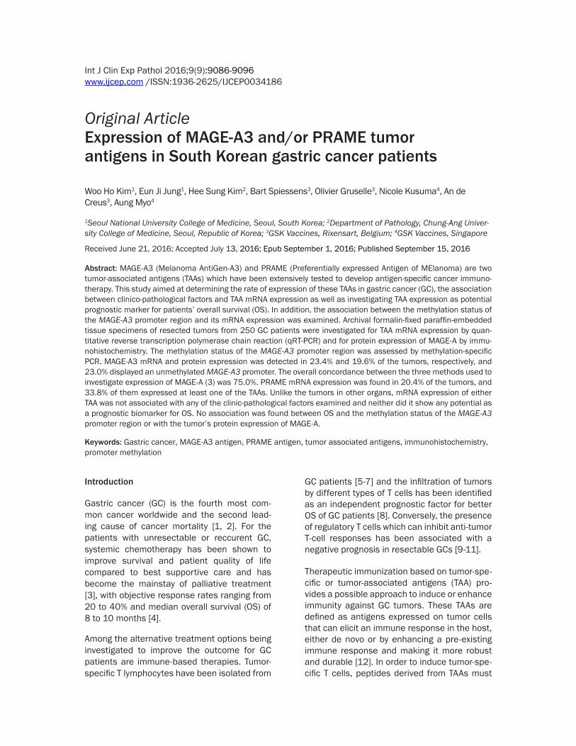
Int J Clin Exp Pathol 20169(9)9086-9096wwwijcepcom ISSN1936-2625IJCEP0034186
Original ArticleExpression of MAGE-A3 andor PRAME tumor antigens in South Korean gastric cancer patients
Woo Ho Kim1 Eun Ji Jung1 Hee Sung Kim2 Bart Spiessens3 Olivier Gruselle3 Nicole Kusuma4 An de Creus3 Aung Myo4
1Seoul National University College of Medicine Seoul South Korea 2Department of Pathology Chung-Ang Univer-sity College of Medicine Seoul Republic of Korea 3GSK Vaccines Rixensart Belgium 4GSK Vaccines Singapore
Received June 21 2016 Accepted July 13 2016 Epub September 1 2016 Published September 15 2016
Abstract MAGE-A3 (Melanoma AntiGen-A3) and PRAME (Preferentially expressed Antigen of MElanoma) are two tumor-associated antigens (TAAs) which have been extensively tested to develop antigen-specific cancer immuno-therapy This study aimed at determining the rate of expression of these TAAs in gastric cancer (GC) the association between clinico-pathological factors and TAA mRNA expression as well as investigating TAA expression as potential prognostic marker for patientsrsquo overall survival (OS) In addition the association between the methylation status of the MAGE-A3 promoter region and its mRNA expression was examined Archival formalin-fixed paraffin-embedded tissue specimens of resected tumors from 250 GC patients were investigated for TAA mRNA expression by quan-titative reverse transcription polymerase chain reaction (qRT-PCR) and for protein expression of MAGE-A by immu-nohistochemistry The methylation status of the MAGE-A3 promoter region was assessed by methylation-specific PCR MAGE-A3 mRNA and protein expression was detected in 234 and 196 of the tumors respectively and 230 displayed an unmethylated MAGE-A3 promoter The overall concordance between the three methods used to investigate expression of MAGE-A (3) was 750 PRAME mRNA expression was found in 204 of the tumors and 338 of them expressed at least one of the TAAs Unlike the tumors in other organs mRNA expression of either TAA was not associated with any of the clinic-pathological factors examined and neither did it show any potential as a prognostic biomarker for OS No association was found between OS and the methylation status of the MAGE-A3 promoter region or with the tumorrsquos protein expression of MAGE-A
Keywords Gastric cancer MAGE-A3 antigen PRAME antigen tumor associated antigens immunohistochemistry promoter methylation
Introduction
Gastric cancer (GC) is the fourth most com- mon cancer worldwide and the second lead- ing cause of cancer mortality [1 2] For the patients with unresectable or reccurent GC systemic chemotherapy has been shown to improve survival and patient quality of life compared to best supportive care and has become the mainstay of palliative treatment [3] with objective response rates ranging from 20 to 40 and median overall survival (OS) of 8 to 10 months [4]
Among the alternative treatment options being investigated to improve the outcome for GC patients are immune-based therapies Tumor-specific T lymphocytes have been isolated from
GC patients [5-7] and the infiltration of tumors by different types of T cells has been identified as an independent prognostic factor for better OS of GC patients [8] Conversely the presence of regulatory T cells which can inhibit anti-tumor T-cell responses has been associated with a negative prognosis in resectable GCs [9-11]
Therapeutic immunization based on tumor-spe-cific or tumor-associated antigens (TAA) pro-vides a possible approach to induce or enhance immunity against GC tumors These TAAs are defined as antigens expressed on tumor cells that can elicit an immune response in the host either de novo or by enhancing a pre-existing immune response and making it more robust and durable [12] In order to induce tumor-spe-cific T cells peptides derived from TAAs must
MAGE-A3 and PRAME expression in gastric cancer
9087 Int J Clin Exp Pathol 20169(9)9086-9096
be presented to T cells by antigen-presenting cells which can activate naiumlve and memory T cells Many studies have demonstrated the capability of immunogenic peptides derived from TAAs to lyse gastric tumor cells (review- ed in [12])
Numerous TAAs have been identified and partly characterized of which a large proportion have been assessed for their potential as targets in antigen-specific cancer immunotherapy A fun-damental preliminary step in assessing the potential of a specific TAA to be used as immu-notherapy against a particular type of cancer is to assess the prevalence of the TAA on the tumor cells of the patient population con-cerned Only patients with tumors expressing this particular TAA will be able to respond to immunotherapy specifically targeted at this TAA Tumor expression of a particular TAA may be detected and quantified by different meth-ods One is by means of quantitative reverse transcription polymerase chain reaction (qRT-PCR) assays on messenger RNA (mRNA) extracted from the tumor cells Another is by immunohistochemistry (IHC) staining of the protein (antigen) provided that an antigen-spe-cific antibody has been identified and can be used for the staining Alternatively gene expres-sion status may be assessed by methylation analysis of the promoter region
It is widely recognized that important contribu-tors to human carcinogenesis consist of epi-genetic alterations including hypermethylation of promoter CpG islands and hypomethylation of the global DNA [13 14] Hypermethylation of promoter CpG islands engenders transcription-al silencing of their downstream genes Many studies have thus reported that tumor suppre- ssor genes are silenced by hypermethylation of their promoter region during carcinogenesis [15] Conversely hypomethylation of the pro-moter region leads to activation of the gene and production of abnormally high levels of protein [13 14]
Investigation of the methylation status of the promoter region of the genes of interest may therefore be used to assess whether they are silenced or activated An unmethylated promot-er region of a specific gene would then indicate that the gene was expressed
Two TAAs that have been widely investigated as possible targets for antigen-specific im- munotherapy against cancer are melanoma-associated antigen A3 (MAGE-A3) and prefer-entially expressed antigen of melanoma (PR- AME) In addition to the therapeutic target for immunotherapeutics aberrant expression of PRAME in tumor cells have been found to be correlated with shorter survival in neuroblasto-ma [16] in ovarian cancer [17] and in breast cancer [18] The aim of the present study was to assess the prevalence of expression of MAGE-A3 and PRAME in tumors of GC patients their co-expression and the association be- tween expression of either antigen and clini- co-pathological factors or clinical outcomes MAGE-A3 expression was detected by qRT- PCR of mRNA by IHC of MAGE-A protein and by assessment of the methylation status of the MAGE-A3 gene promoter region PRAME expression was detected by qRT-PCR of mRNA
To the best of our knowledge this study is the first assessment of PRAME expression and of co-expression of MAGE-A3 and PRAME in GC
Materials and methods
Study cohort
Surgically resected formalin-fixed paraffin-em- bedded (FFPE) tissue specimens were obtain- ed from 250 consecutive GC patients under- going gastrectomy at the Seoul National Uni- versity Hospital in South Korea between 1 January and 31 December 2004 Patient and tumor characteristics were recorded from the hospital files and included patient demogra- phics (age gender) tumor characteristics (his-tologic type differentiation grade depth of in- vasion pathological stage (according to Edi- tion 7 of the AJCC Staging Manual) lymph no- de metastases) and patient survival time Pa- tient survival data including dates and causes of death were obtained from the Korean Cen- tral Cancer Registry at the Ministry of Health and Welfare South Korea
The study was approved by the Institutional Review Board of Seoul National University Hospital (H-1010-065-336)
Tissue array preparation
All of the specimens were assembled into tis-sue microarrays Three representative core tis-
MAGE-A3 and PRAME expression in gastric cancer
9088 Int J Clin Exp Pathol 20169(9)9086-9096
sue biopsy specimens (diameter 2 mm) were obtained from FFPE GCs (donor blocks) and arranged in triplicate sets of new recipient par-affin blocks (tissue array blocks) using a tre-phine apparatus (Superbiochips Laboratories) Each tissue array block contained up to 60 cores thus 24 array blocks were prepared dur-ing the study
mRNA antigen expression detected by qRT-PCR arrays
mRNA expression of MAGE-A3 and PRAME was detected and quantified as described in detail previously [19] In brief manual dissection of the FFPE tissue specimens was performed by Response Genetics Inc (RGI USA) to obtain the minimally required 50 mm2 of tumor tis- sue with 50-80 neoplastic cells Total RNA extraction was performed using the RNeasytrade FFPE kit (Qiagen USA) modified with an addi-tional DNAse digestion step added in order to improve the elimination of genomic DNA Com- plementary DNA (cDNA) was synthesized by mixing 15 microL cDNA master mix with 15 microL of RNA and incubating
MAGE-A3 and PRAME genes along with β-actin housekeeping gene were amplified using qRT-PCR TaqManreg chemistry (ThermoFischer) on the ABI 7900 system (Applied Biosystems) in 384-well plates 50 ng (100) cDNA and 05 ng (1) of total RNA extracted from the hu- man melanoma cell line MZ-2-30 (referred to as gene expression reference level (GERL) and provided by Ludwig Institute of Cancer Research Belgium) was included into the RT in parallel as a positive control
mRNA of a tumor specimen was determined in a semi-quantitative way relative to the GERL A
specimen was declared MAGE-A3-positive if its relative expression of MAGE-A3 to β-actin was ge 1 that in the GERL For PRAME the corre-sponding threshold was 03 of the relative expression of PRAME to β-actin in the GERL Probes and primers were used as previously described [19]
Methylation status of MAGE-A3 determined by methylation-specific PCR
For the methylation-specific PCR 1 microg of ge- nomic DNA extracted by a standard protein- ase-K digestion and phenolchloroform proce-dure was denatured with NaOH (final concen-tration 02 M) treated with 3 M sodium-bisulfite (Sigma USA) and 10 mM hydroquinone (pH 50 Sigma) and then incubated at 50degC for 16 hours After incubation DNA was purified using a Wizard DNA purification kit (Promega USA) and then treated with NaOH recovered in ethanol and resuspended in 20 microL of dis- tilled water After the sodium-bisulfite modifi- cation PCR amplification was performed in a thermal cycler for 1 cycle at 95degC for 5 min followed by 35 cycles each at 95degC for 30 s 62degC (unmethylated) or 64degC (methylated) for 30 s 72degC for 1 min and final extension at 72degC for 10 min The primer sequences are shown in Table S1 of the Supplementary Material
MAGE-A protein expression detected by immu-nohistochemistry
Sections (4 microm) from FFPE blocks were dewax- ed in xylene and rehydrated using a graded alcohol series For the antigen retrieval step slides were inserted in a rack in diluted re- trieval solution (pH 60 EDTA) and preheated to 100degC for 6 min and then further heated at 1000 Watt for 5 min The slides were then
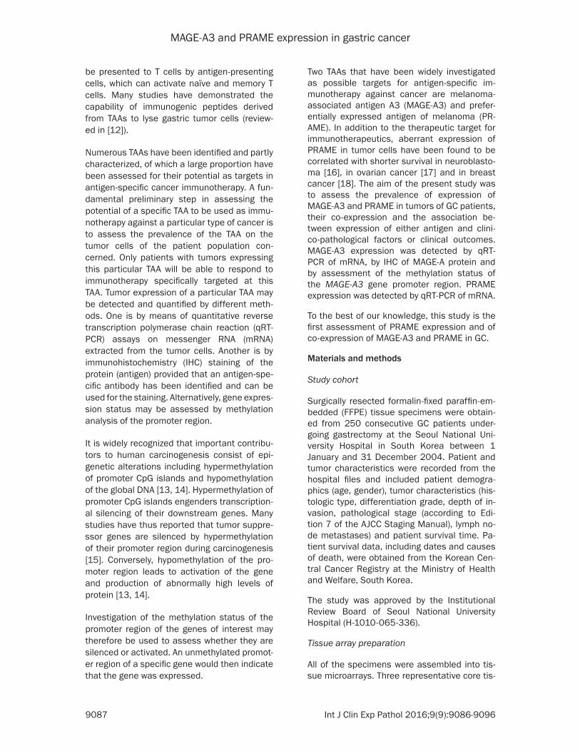
Figure 1 Representative images of MAGE-A positive (A) and MAGE-A-negative (B) tumors and normal gastric cells (C) The MAGE-A3 staining was found in the cytoplasm as well as in the nucleus (original magnification times100) (A)
MAGE-A3 and PRAME expression in gastric cancer
9089 Int J Clin Exp Pathol 20169(9)9086-9096
transferred to the Autostainer 360 (Lab Vision USA) and the program was run as follows 1) slide rinse in wash buffer and peroxidase block-ing solution 2) incubation with mouse anti-MAGE A monoclonal antibody 6C1 (Zymed La- boratories Inc USA) diluted at 1100 for 60 min labelled with polymer for 8 min incubated with DAB (Envision kit DAKO Denmark) for 10 min and then counterstained in Mayerrsquos hema-toxylin The 6C1 antibody can detect but not distinguish between the proteins MAGE-A1 -A2 -A3 -A4 -A6 -A10 and -A12
Three representative tumor cores were ob- tained from each tumor MAGE-A was stained in either the cytoplasma or nucleus and often both The result of the immunostaining ex- periment was considered positive for MAGE-A if at least one of the three tumor cores had an IHC score ge 50 (Figure 1) in either the cy- toplasma or the nucleus The IHC score was calculated as follows
IHC score = ΣIntensity (1 2 or 3) times Area (0-100) IHC MAGE-A3 results is shown in Table S2 of the Supplementary Materials
Statistical methods
This study was exploratory Thus the sample size was not calculated on the basis of any pre-specified hypotheses and for antigen expres-sion in subgroups determined by patient or tumor characteristics only descriptive statistics are presented The proportions of FFPE speci-mens with an antigen expression level above the cut-off value were estimated on the basis of the number of specimens with a valid as- say result for the respective antigen exclud- ing specimens with missing or invalid antigen expression results The percentages of anti- gen-positive specimens are presented with their exact 95 confidence intervals (CIs) The rates of co-expression of MAGE-A3 and PRA- ME were estimated on the basis of the num- ber of FFPE specimens with a valid assay re- sult for each antigen
Kaplan-Meier (KM) curves for OS time were estimated and compared using the log-rank test and unadjusted hazard ratios (HRs) with 95 CIs estimated by means of the Cox propor-tional hazard regression model
Table 1 Patient and tumor characteristics
Characteristics CategoriesTotal N = 250
n 95 CIAge Less than 45 years old 40 160 117-211
45-54 years old 58 232 181-289
55-64 years old 59 236 185-294
65-74 years old 73 292 236-353
75 years old or more 20 80 50-121
Gender Female 74 296 240-357
Male 176 704 643-760
Histology (Laureacuten) Intestinal 91 364 304-427
Diffused 110 440 378-504
Mixed 47 188 142-242
Undetermined 2 08 01-29
Histology (Ming) Infiltrative 230 920 879-950
Expanding 20 80 50-121
WHO 2000 classification and differentiation grade Papillary adenocarcinoma 2 08 01-29
Tubular adenocarcinoma well differentiated 6 24 09-52
Tubular adenocarcinoma moderately differentiated 85 340 281-402
Tubular adenocarcinoma poorly differentiated 101 404 343-468
Mucinous adenocarcinoma 12 48 25-82
Signet-ring cell carcinoma 38 152 110-203
Undifferentiated carcinoma 3 12 02-35
Others 3 12 02-35
TNM Stage (pooled) I 44 176 131-229
II 58 232 181-289
III 101 404 343-468
IV 47 188 142-242N = Number of patientssamples n = Number of patientssamples in a given category = nNumber of patientssamples with available results times 100 Others = Other types including squamous cell carcinomas small cell carcinoma etc LL UL for percentage = Exact 95 Lower and Upper confidence limits CI = Confidence interval
MAGE-A3 and PRAME expression in gastric cancer
9090 Int J Clin Exp Pathol 20169(9)9086-9096
Results
Two-hundred-and-fifty FFPE specimens were tested for antigen expression and MAGE-A3 promoter methylation status Patient demo-
196 for MAGE-A- IHC to 234 for MAGE-A3- mRNA expression A total of 75 of specimens had fully concordant results across the 3 tests (Table 4) Fourteen (103) specimens had MAGE-A3 methylation status results that were
Table 2 Validity of MAGE-A3 and PRAME expression tests using qRT-PCR assays
Characteristics CategoriesMAGE-A3 (N = 250) PRAME (N = 250)n 95 CI n 95 CI
Validity test Valid 137 604 537-668 142 626 559-689Invalid 52 229 176-289 47 207 156-266QNS 38 167 121-222 38 167 121-222
Missing 23 - - 23 - -N = Number of patientssamples n = Number of patientssamples in a given category QNS = Quantity not sufficient = nNumber of patientssamples with available results times 100
Table 3 Overall rates of expression of MAGE-A3 and PRAME
Characteristics CategoriesTotal (N = 250)
n 95 CIMAGE-A3 expression (mRNA) result Positive 32 234 166-313
Negative 105 766 687-834Missing 113 - -
MAGE-A3 methylation result Unmethylated 54 230 178-289Methylated 181 770 711-822
Missing 15 - -MAGE-A IHC result Positive 49 196 149-251
Negative 201 804 749-851PRAME expression (mRNA) result Positive 29 204 141-280
Negative 113 796 720-859Missing 108 - -
N = Number of patientssamples n = Number of patientssamples in a given category = nNumber of patientssamples with available results times 100 IHC = Immunohistochemistry
graphics and tumor charac-teristics are summarized in Table 1
Percentage of specimens giving valid test results
Each mRNA expression te- st of an FFPE specimen was done with two repli-cates Twenty-three speci-mens were not tested for mRNA expression (Table 2) and for 38 specimens the amount of tumor cells in the mRNA extracted was in- sufficient (3438) or com-pletely absent (438)
Valid results were obtain- ed for 604 of the speci-mens tested for mRNA ex- pression of MAGE-A3 The reasons for invalid assay results for MAGE-A3 were test results lsquoout-of-rangersquo (4752) inconsistent repli-cates (352) and contami- nation with genomic DNA (252) For mRNA express- ion of PRAME valid results were obtained for 626 of tested specimens Invalid test results for PRAME were lsquoout-of-rangersquo
All the 250 FFPE specimens gave valid IHC results for MAGE-A protein whereas MAGE-A3 methylation sta- tus could not be obtained for 15 specimens
Antigen expression
The three methods for de- tecting MAGE-A3 express- ion gave concordant re- sults overall (Table 3) with the percentage of speci- mens with positive results for MAGE-A3 ranging from
Table 4 Concordance between MAGE-A3 expression tests with the three methods
CategoriesN = 136
n 95 CImRNA-negativeMethylatedIHC-negative 87 640 553-720mRNA-negativeMethylatedIHC-positive 4 29 08-74mRNA-negativeUnmethylatedIHC-negative 9 66 31-122mRNA-negativeUnmethylatedIHC-positive 5 37 12-84mRNA-positiveMethylatedIHC-negative 6 44 16-94mRNA-positiveMethylatedIHC-positive 5 37 12-84mRNA-positiveUnmethylatedIHC-negative 5 37 12-84mRNA-positiveUnmethylatedIHC-positive 15 110 63-175N = Number of specimens n = Number of specimens in a given category = nNumber of specimens with available results times 100 CI = Confidence interval
MAGE-A3 and PRAME expression in gastric cancer
9091 Int J Clin Exp Pathol 20169(9)9086-9096
Table 5 Rates of co-expression of MAGE-A3 and PRAME
Characteristics CategoriesN = 136
n 95 CICo-expression MAGE-A3 and PRAME MAGE-A3-negativePRAME-negative 90 662 576-741
MAGE-A3-negativePRAME-positive 14 103 57-167MAGE-A3-positivePRAME-negative 18 132 80-201MAGE-A3-positivePRAME-positive 14 103 57-167
At least MAGE-A3 or PRAME MAGE-A3-negative and PRAME-negative 90 662 576-741MAGE-A3-positive or PRAME-positive 46 338 259-424
N = Number of patientssamples n = Number of patientssamples in a given category = nNumber of patientssamples with available results times 100
Table 6 Subgroup analyses of MAGE-A3 or PRAME expression (mRNA) according to age gender tumor histology and tumor stage
Variable CategoriesMAGE-A3 expression PRAME expression
N n 95 CI N n 95 CIAge 26-44 23 1 43 01-219 24 2 83 10-270
45-54 34 4 118 33-275 35 7 200 84-36955-64 25 6 240 94-451 28 4 143 40-32765-74 45 17 378 238-535 45 12 267 146-419ge 75 10 4 400 122-738 10 4 400 122-738
Gender Female 35 5 143 48-303 39 5 128 43-274Male 102 27 265 182-361 103 24 233 155-327
Histology (Lauren) Intestinal 55 16 291 176-429 55 16 291 176-429Diffused 64 7 109 45-212 67 8 119 53-222Mixed 18 9 500 260-740 20 5 250 87-491Undetermined 0 0 - - 0 0 - -
Histology (Ming) Infiltrative 124 29 234 163-318 128 24 188 124-266Expanding 13 3 231 50-538 14 5 357 128-649
Histology (WHO) Papillary AC 2 0 00 00-842 1 0 00 00-975Tubular AC well diff 5 1 200 05-716 5 1 200 05-716Tubular AC moderately diff 48 17 354 222-505 48 16 333 204-484Tubular AC poorly diff 53 9 170 81-298 56 8 143 64-262Mucinous AC 5 1 200 05-716 6 1 167 04-641Signet-ring cell carcinoma 23 3 130 28-336 24 1 42 01-211Undifferentiated carcinoma 0 0 - - 0 0 - -Others 1 1 100 25-100 2 2 100 158-100
Stage I 26 3 115 24-302 25 5 200 68-407II 35 13 371 215-551 36 8 222 101-392III 50 8 160 72-291 54 11 204 106-335IV 26 8 308 143-518 27 5 185 63-381
N = Number of FFPE specimens in this category giving a valid test result for the antigen concerned n = Number of specimens in a given category with a valid test result positive for the antigen concerned AC = Adenocarcinoma diff = Differentiated = (nN) times 100 CI = Confidence interval
discordant with the results for mRNA detection and IHC (nine with mRNA-negativeunmethyl-ationIHC-negative and five with mRNA-posi-tivemethylationIHC-positive)
Of the 142 specimens with valid PRAME assay results 29 (204) were PRAME-positive (Table 3) Of the 136 FFPE specimens with valid mRNA expression assay results for both antigens 14
MAGE-A3 and PRAME expression in gastric cancer
9092 Int J Clin Exp Pathol 20169(9)9086-9096
(103) expressed both while 46 (338) expressed at least one of them (Table 5)
The exploratory analyses of the mRNA ex- pression of the TAAs in subgroups determin- ed by patient and tumor characteristics show- ed similar results for MAGE-A3 and PRAME (Table 6) TAA mRNA expression was indepen-dent of tumor stage and there was no obvious association between tumor TAA mRNA expre- ssion and any of the other clinico-pathological factors investigated
Another study investigated MAGE-A express- ion in GC cell lines as well as primary gastric tumor specimens using both IHC and analy- sis of MAGE-A3 promoter methylation status for detection Of 1097 cancer tissues 158 expressed MAGE-A protein with no distinction between the subtypes of MAGE-A by the anti-body used for the IHC Of 52 randomly selected tumors expressing MAGE-A protein 28 (538) displayed unmethylation of the MAGE-A3 pro-moter [24] In the present study 20 of the 29 (690 recalculated from Table 4) tumors
Figure 2 Kaplan-Meier curves for OS by MAGE-A3 methylation status of the specimen HR for MAGE-A3 unmethylated 094 (95 CI 057-156 P = 082) (Wald test from Cox regression model)
Figure 3 Kaplan-Meier curves for OS by MAGE-A protein expression of the specimen HR for MAGE-A3-positive 146 (95 CI 092-232 P = 011) (Wald test from Cox regression model)
Overall survival by MAGE-A3 methylation status protein expression of MAGE-A and mRNA expression of the an-tigens
Kaplan-Meier curves for OS according to the tumorrsquos MAGE-A3 methylation status (Figure 2) overlap almost completely The Kaplan-Meier curves for OS according to the specimensrsquo expression of MAGE-A3 protein are more separated (Figure 3) but are not statistically significant- ly different (P = 011) Like- wise there were no indica-tions of an association be- tween OS and mRNA expres-sion of the antigens neither for MAGE-A3 nor for PRAME (Figures 4 and 5)
Discussion
This is the first study of PRAME expression in GC while mRNA MAGE-A3 expre- ssion has been reported in several previous studies with rates of MAGE-A3-positive GC tumors varying between 30 and 416 [20-23] None of these studies reported sta- tistically significant associa-tions between clinico-patho-logical tumor characteristics and mRNA expression of MAGE-A3 in line with findings in the current study
MAGE-A3 and PRAME expression in gastric cancer
9093 Int J Clin Exp Pathol 20169(9)9086-9096
expressing the MAGE-A protein displayed un- methylation of the MAGE-A3 promoter where- as this was displayed by 14 of the 107 (131) tumors not expressing the MAGE-A protein These results indicate that unmethylation of the MAGE-A3 promoter may be (partly) respon-sible for the expression of the MAGE-A pro- tein in gastric cancer
Honda et al [13] detected MAGE-A3 promoter demethylation in 66 of 84 GC specimens tumors displaying MAGE-A3 promoter demeth-
Honda et al [13] described a trend for worse survival for patients with tumors displaying demethylated MAGE-A3 in particular if their tumor also showed demethylated MAGE-A1 (P = 018)
Circulating tumor cell DNA can be detected in serum or blood even at early stages of the dis-ease as shown by Kounalakos et al [25] for melanoma patients This DNA may be derived from lysed cells of the primary tumor or from circulating tumor cells releasing their DNA With
Figure 4 Kaplan-Meier curves for overall survival by mRNA expression of MAGE-A3 (Total cohort) HR for MAGE-A3-positive 103 (95 CI 0538-1963 P = 093) (Wald test from Cox regression model)
Figure 5 Kaplan-Meier curves for overall survival by mRNA expression of PRAME (Total cohort) HR for PRAME-positive 097 (95 CI 0499-1885 P = 093) (Wald test from Cox regression model)
ylation were in a more ad- vanced state and were asso-ciated with a higher preva-lence of lymph-node metas- tases than those without de- methylation In the present study 230 of gastric tu- mors displayed an unmethyl-ated MAGE-A3 promoter
We did not observe any rela-tion between OS and mRNA TAA expression of the FFPE specimen for either MAGE-A3 or PRAME Similarly there was no statistically significant association between OS and MAGE-A protein expression (P = 011) In contrast Jung et al [24] reported a significant-ly worse prognosis for patients with tumors expressing the MAGE-A proteins (P lt 0001)
Recent studies investigating the molecular basis of GC have reported associations between aberrant DNA meth-ylation of several gene pro-moters and the prognosis for GC patients generally sug-gesting that hypermethylation of the identified gene promot-ers is an indicator of poor prognosis (reviewed in [15]) Our observations indicate that MAGE-A3 is not to be added to this list of genes as the methylation status of the MAGE-A3 promoter had no association with patient OS (P = 082) In contrast
MAGE-A3 and PRAME expression in gastric cancer
9094 Int J Clin Exp Pathol 20169(9)9086-9096
a prior understanding of a potential correlation between gene promoter methylation status and gene expression methylation testing of cir-culating DNA could be useful in detecting the expression of specific genes in tumor cells Such non-invasive detection methods that are not dependent on the availability of tumor tissue could increase the number of patients that can be screened for expression of tumor antigens
A rough assessment of the possibility of rely- ing on such non-invasive methods for gene expression screening based on the observa-tions of the present study was performed Due to the lack of MAGE-A3 specific antibodies IHC cannot distinguish MAGE-A3 from other members of the MAGE-A family MAGE-A3 ex- pression was thus generally screened for us- ing RT-PCR assays Here we found that 867 (91105) of the FFPE specimens with no mRNA MAGE-A3 expression showed methylation of the MAGE-A3 promoter whereas 645 (20 31) of specimens with mRNA MAGE-A3 expre- ssion were unmethylated in the MAGE-A3 pro-moter Arguably the concordance between the results obtained with the two methods may be considered too low to rely on testing of MAGE-A3 promoter methylation status for pa- tient screening According to these observa-tions screening based on methylation testing of the MAGE-A3 promoter would miss more than a third of the patients whose tumors show mRNA MAGE-A3 expression
The overall concordance between the three MAGE-A3 assays was relatively low compared to previous findings (75 vs nearly 100) reported in certain studies (eg [26]) The qRT-PCR assay is specific for MAGE-A3 and the cut-off value was defined as 1 of the GERL con-trol By contrast the IHC assay could also detect other members of the MAGE-A family (in particular MAGE-A6) and the cut-off values for these assays are linked to their detection lim-its Additional investigations are needed to assess the impact of MAGE-A3A6 expression and to homogenize the cut-off values of the assays
In summary the rate of mRNA expression of each of these TAAs in tumors from GC patients was found to be relatively low with 234 expressing MAGE-A3 204 expressing PRAME and 338 expressing at least one of them Neither MAGE-A3 nor PRAME would se-
rve as a prognostic biomarker for OS of GC patients
Acknowledgements
Niels Neymark provided scientific writing ser-vices and Veacuteronique Duquenne (XPE PHAR- MA and SCIENCES Wavre Belgium CO GSK) coordinated the manuscriptrsquos development This study was funded and coordinated by GlaxoSmithKline Biologicals SA GSK was in- volved in the design and conduct of the study collection management analysis and inter- pretation of the data and preparation and review of the manuscript
Disclosure of conflict of interest
Olivier Gruselle and Bart Spiessens are employ-ees of the GSK group of companies An de Creus Nicole Kusuma and Aung Myo were employees of the GSK group of companies at the time of the study and manuscript develop-ment Bart Spiessens own stockstock options in GSK group of companies Woo Ho Kim Eun Ji Jung and Kim Hee Sung have no conflict of interest
Authorsrsquo contribution
BS AM AdeC and EJJ were involved in study design AM AdeC NK were involved in study supervision OG AM AdeC NK WHK EJJ were involved in data acquisition HSK EJJ NK AM WHK and OG were involved in data collection OG AM NK and WHK were involved in data extraction and quality check (AdeC in quality check only and EJJ in data extraction only) OG WHK and EJJ were involved in provision of study material WHK and EJJ were involved in labora-tory testing BS (biostatistician) OG AM AdeC WHK and HSK performed or supervised the analyses BS OG AM AdC NK WHK and HSK were involved in results interpretation OG AdeC NK EJJ were involved in administrativetechnicallogistic support EJJ and AM were involved in centers recruitment AM and NK were involved in center coordination AM was involved in acquisition of funding
Address correspondence to Dr Woo Ho Kim De- partment of Pathology Seoul National University College of Medicine 28 Yeongeon-dong 110-799 Seoul Korea Tel +82 8 740 8269 Fax +82 2 765 5600 E-mail woohokimsnuackr
MAGE-A3 and PRAME expression in gastric cancer
9095 Int J Clin Exp Pathol 20169(9)9086-9096
References
[1] Crew KD Neugut AI The epidemiology of gas-tric cancer World J Gastroenterol 2006 12 354-62
[2] Toomey PG Vohra NA Ghansah T Sarnaik AA Pilon-Thomas SA Immunotherapy for gastroin-testinal malignancies Cancer Control 2013 20 32-42
[3] Bilici A Treatment options in patients with met-astatic gastric cancer Current status and fu-ture perspective World J Gastroenterol 2014 20 3905-15
[4] Amadei A Benagiano M della Bella C Nicolai E DrsquoElios MM Novel immunotherapeutic strat-egies of gastric cancer treatment J Biomed Biotech 2011 2011 437348
[5] Ikeda H Sato N Matsuura A Kikuchi K Analysis of T-cell receptor V region gene usage of cytotoxic T-lymphocytes and tumor-infiltrat-ing lymphocytes derived from human autolo-gous gastric signet ring cell carcinomas Cancer Res 1993 53 3078-84
[6] Kono K Ichihara F Lizuka H Sekikawa T Matsumoto Y Differences in the recognition of tumor-specific CD8+ T cells derived from solid tumor metastatic lymph nodes and ascites in patients with gastric cancer Int J Cancer 1997 71 978-81
[7] Amadei A Niccolai E Della Bella C Cianchi F Trallori G Benagiano M Bencini L Bernini M Farsi M Moretti R Del Prete G DrsquoElios MM Characterization of tumor antigen peptide-spe-cific T cells isolated from the neoplastic tissue of patients with gastric adenocarcinomas Cancer Immunol Immunother 2009 58 1819-30
[8] Lee HE Chae SW Lee YJ Kim MA Lee HS Lee BL Kim WH Prognostic implications of type and density of tumour-infiltrating lymp- hocytes in gastric cancer Br J Cancer 2008 99 1704-11
[9] Perrone G Ruffini PA Catalano V Spino C Santini D Muretto P Spoto C Zingaretti C Sisti V Alessandroni P Giordani P Cicetti A DrsquoEmidio S Morini S Ruzzo A Magnani M Tonini G Rabitti C Graziano F Intratumoural FOXP3-positive regulatory T cells are associat-ed with adverse prognosis in radically resected gastric cancer Eur J Cancer 2008 44 1875-82
[10] Shen Z Zhou S Wang Y Li RL Zhong C Liang C Sun Y Higher intratumoural infiltrated Foxp3+ Treg numbers and Foxp3+CD8+ ratio are associated with adverse prognosis in re-sectable gastric cancer J Cancer Res Clin Oncol 2010 136 1585-95
[11] Zhou S Shen Z Wang Y Ma H Xu S Qin J Chen L Tao H Zhen Z Chen G Zhang Z Li R
Xiao H Zhong C Yang Y Liang C CCR7 expres-sion and intratumoural FOXP3+ regulatory T cells are correlated with overall survival and lymph node metastatis in gastric cancer PLoS One 2013 8 e74430
[12] Matsueda S Graham DY Immunotherapy in gastric cancer World J Gatroenterol 2014 20 1657-66
[13] Honda T Tamura G Waki T Kawata S Tera- shima M Nishizuka S Motoyama T Deme- thylation of MAGE promoters during gastric cancer progression Br J Cancer 2004 90 838-43
[14] Kim KH Choi JS Kim IJ Ku JL Park JG Promoter hypomethylation and reactivation of MAGE-A1 and MAGE-A3 genes in colorectal cancer cell lines and cancer tissues World J Gastroenterol 2006 12 5651-57
[15] Nakamura J Tanaka T Kitajima Y Noshiro H Miyazaki K Methylation-mediated gene silenc-ing as biomarkers of gastric cancer A review World J Gastroenterology 2014 20 11991-12006
[16] Oberthuer A Hero B Spitz R Berthold F Fischer M The tumor-associated antigen PRA- ME is universally expressed in high-stage neu-roblastoma and associated with poor out- come Clin Cancer Res 2004 10 4307-13
[17] Partheen K Levan K Osterberg L Claesson I Fallenius G Sundfeldt K Horvath G Four po-tential biomarkers as prognostic factors in stage III serous ovarian adenocarcinomas Int J Cancer 2008 123 2130-7
[18] Epping MT Hart AA Glas AM Krijgsman O Bernards R PRAME expression and clinical outcome of breast cancer Br J Cancer 2008 99 398-403
[19] Lerut E Van Poppel H Joniau S Gruselle O Coche T Therasse P Rates of MAGE-A3 and PRAME expressing tumors in FFPE tissue spec-imens from bladder cancer patients potential targets for antigen-specific cancer immuno-therapeutics Int J Clin Exp Pathol 2015 8 9522-32
[20] Inoue H Mori M Honda M Li J Shibuta K Mimori K Ueo H Akiyoshi T The expression of tumor-rejection antigen ldquoMAGErdquo genes in human gastric carcinoma Gastroenterology 1995 109 1522-25
[21] Li J Yang Y Fujie T Baba K Ueo H Mori M Akiyoshi T Expression of BAGE GAGE and MAGE genes in human gastric carcinoma Clin Cancer Res 1996 2 1619-25
[22] Wang Y Wu XJ Zhao AL Yuan YH Chen YT Jungbluth AA Gnjatic S Santiago D Ritter G Chen WF Old LJ Ji JF Cancertestis antigen expression and autologous humoral immunity to NY-ESO-1 in gastric cancer Cancer Immun 2004 4 11
MAGE-A3 and PRAME expression in gastric cancer
9096 Int J Clin Exp Pathol 20169(9)9086-9096
[23] Wang WX Li YB Xie XL Shu XL Ouyang XH Detection of tumor cells in peripheral blood of patients with gastric cancer using mRNA of MAGE genes as markers Zhonghua Wei Chang Wai Ke Za Zhi 2009 12 611-14
[24] Jung EJ Kim MA Lee HS Yang HK Lee YM Lee BL Kim WH Expression of family A mela-noma antigen in human gastric carcinoma Anticancer Research 2005 25 2105-12
[25] Kounalakos N Goydos JS Tumor cells and cir-culating markers in melanoma diagnosis prognosis and management Curr Oncol Rep 2005 7 377-382
[26] Lehmann-Che J Amira-Bouhidel F Turpin E Antoine M Soliman H Legres L Bocquet C Bernoud R Flandre E Varna M de Roquan- court A Plassa LF Giacchetti S Espieacute M de Bazelaire C Cahen-Doidy L Bourstyn E Janin A de Theacute H Bertheau P Immunohistoche- mical and molecular analyses of HER2 sta- tus in breast cancers are highly concordant and complementary approaches Br J Cancer 2011 104 1739-46
MAGE-A3 and PRAME expression in gastric cancer
1
Table S1 Primer sequences for methylation specific PCRMethylation status Type SequenceUnmethylated Anti-sense 5rsquo-TGTTAGGATGTGATGTTATTGATTTGT-3rsquo
Sense 5rsquo-CCTCACCAAACCTAAACCAA-3rsquoMethylated Anti-sense 5rsquo-CCATCTGACGTTATTGATTTGC-3rsquo
Sense 5rsquo-CTCACCGAACCTAAACCGAC-3rsquo
Table S2 Distribution of immunohistochemistry MAGE-A3 results in gastric cancerIHC score 0-50 51-100 101-150 151-200 201-250 251-300 Total of cases 201 19 8 4 3 19 250 of total 804 76 32 16 12 76 100IHC result (Score gt 50) Negative Positive
804 196
MAGE-A3 and PRAME expression in gastric cancer
9087 Int J Clin Exp Pathol 20169(9)9086-9096
be presented to T cells by antigen-presenting cells which can activate naiumlve and memory T cells Many studies have demonstrated the capability of immunogenic peptides derived from TAAs to lyse gastric tumor cells (review- ed in [12])
Numerous TAAs have been identified and partly characterized of which a large proportion have been assessed for their potential as targets in antigen-specific cancer immunotherapy A fun-damental preliminary step in assessing the potential of a specific TAA to be used as immu-notherapy against a particular type of cancer is to assess the prevalence of the TAA on the tumor cells of the patient population con-cerned Only patients with tumors expressing this particular TAA will be able to respond to immunotherapy specifically targeted at this TAA Tumor expression of a particular TAA may be detected and quantified by different meth-ods One is by means of quantitative reverse transcription polymerase chain reaction (qRT-PCR) assays on messenger RNA (mRNA) extracted from the tumor cells Another is by immunohistochemistry (IHC) staining of the protein (antigen) provided that an antigen-spe-cific antibody has been identified and can be used for the staining Alternatively gene expres-sion status may be assessed by methylation analysis of the promoter region
It is widely recognized that important contribu-tors to human carcinogenesis consist of epi-genetic alterations including hypermethylation of promoter CpG islands and hypomethylation of the global DNA [13 14] Hypermethylation of promoter CpG islands engenders transcription-al silencing of their downstream genes Many studies have thus reported that tumor suppre- ssor genes are silenced by hypermethylation of their promoter region during carcinogenesis [15] Conversely hypomethylation of the pro-moter region leads to activation of the gene and production of abnormally high levels of protein [13 14]
Investigation of the methylation status of the promoter region of the genes of interest may therefore be used to assess whether they are silenced or activated An unmethylated promot-er region of a specific gene would then indicate that the gene was expressed
Two TAAs that have been widely investigated as possible targets for antigen-specific im- munotherapy against cancer are melanoma-associated antigen A3 (MAGE-A3) and prefer-entially expressed antigen of melanoma (PR- AME) In addition to the therapeutic target for immunotherapeutics aberrant expression of PRAME in tumor cells have been found to be correlated with shorter survival in neuroblasto-ma [16] in ovarian cancer [17] and in breast cancer [18] The aim of the present study was to assess the prevalence of expression of MAGE-A3 and PRAME in tumors of GC patients their co-expression and the association be- tween expression of either antigen and clini- co-pathological factors or clinical outcomes MAGE-A3 expression was detected by qRT- PCR of mRNA by IHC of MAGE-A protein and by assessment of the methylation status of the MAGE-A3 gene promoter region PRAME expression was detected by qRT-PCR of mRNA
To the best of our knowledge this study is the first assessment of PRAME expression and of co-expression of MAGE-A3 and PRAME in GC
Materials and methods
Study cohort
Surgically resected formalin-fixed paraffin-em- bedded (FFPE) tissue specimens were obtain- ed from 250 consecutive GC patients under- going gastrectomy at the Seoul National Uni- versity Hospital in South Korea between 1 January and 31 December 2004 Patient and tumor characteristics were recorded from the hospital files and included patient demogra- phics (age gender) tumor characteristics (his-tologic type differentiation grade depth of in- vasion pathological stage (according to Edi- tion 7 of the AJCC Staging Manual) lymph no- de metastases) and patient survival time Pa- tient survival data including dates and causes of death were obtained from the Korean Cen- tral Cancer Registry at the Ministry of Health and Welfare South Korea
The study was approved by the Institutional Review Board of Seoul National University Hospital (H-1010-065-336)
Tissue array preparation
All of the specimens were assembled into tis-sue microarrays Three representative core tis-
MAGE-A3 and PRAME expression in gastric cancer
9088 Int J Clin Exp Pathol 20169(9)9086-9096
sue biopsy specimens (diameter 2 mm) were obtained from FFPE GCs (donor blocks) and arranged in triplicate sets of new recipient par-affin blocks (tissue array blocks) using a tre-phine apparatus (Superbiochips Laboratories) Each tissue array block contained up to 60 cores thus 24 array blocks were prepared dur-ing the study
mRNA antigen expression detected by qRT-PCR arrays
mRNA expression of MAGE-A3 and PRAME was detected and quantified as described in detail previously [19] In brief manual dissection of the FFPE tissue specimens was performed by Response Genetics Inc (RGI USA) to obtain the minimally required 50 mm2 of tumor tis- sue with 50-80 neoplastic cells Total RNA extraction was performed using the RNeasytrade FFPE kit (Qiagen USA) modified with an addi-tional DNAse digestion step added in order to improve the elimination of genomic DNA Com- plementary DNA (cDNA) was synthesized by mixing 15 microL cDNA master mix with 15 microL of RNA and incubating
MAGE-A3 and PRAME genes along with β-actin housekeeping gene were amplified using qRT-PCR TaqManreg chemistry (ThermoFischer) on the ABI 7900 system (Applied Biosystems) in 384-well plates 50 ng (100) cDNA and 05 ng (1) of total RNA extracted from the hu- man melanoma cell line MZ-2-30 (referred to as gene expression reference level (GERL) and provided by Ludwig Institute of Cancer Research Belgium) was included into the RT in parallel as a positive control
mRNA of a tumor specimen was determined in a semi-quantitative way relative to the GERL A
specimen was declared MAGE-A3-positive if its relative expression of MAGE-A3 to β-actin was ge 1 that in the GERL For PRAME the corre-sponding threshold was 03 of the relative expression of PRAME to β-actin in the GERL Probes and primers were used as previously described [19]
Methylation status of MAGE-A3 determined by methylation-specific PCR
For the methylation-specific PCR 1 microg of ge- nomic DNA extracted by a standard protein- ase-K digestion and phenolchloroform proce-dure was denatured with NaOH (final concen-tration 02 M) treated with 3 M sodium-bisulfite (Sigma USA) and 10 mM hydroquinone (pH 50 Sigma) and then incubated at 50degC for 16 hours After incubation DNA was purified using a Wizard DNA purification kit (Promega USA) and then treated with NaOH recovered in ethanol and resuspended in 20 microL of dis- tilled water After the sodium-bisulfite modifi- cation PCR amplification was performed in a thermal cycler for 1 cycle at 95degC for 5 min followed by 35 cycles each at 95degC for 30 s 62degC (unmethylated) or 64degC (methylated) for 30 s 72degC for 1 min and final extension at 72degC for 10 min The primer sequences are shown in Table S1 of the Supplementary Material
MAGE-A protein expression detected by immu-nohistochemistry
Sections (4 microm) from FFPE blocks were dewax- ed in xylene and rehydrated using a graded alcohol series For the antigen retrieval step slides were inserted in a rack in diluted re- trieval solution (pH 60 EDTA) and preheated to 100degC for 6 min and then further heated at 1000 Watt for 5 min The slides were then
Figure 1 Representative images of MAGE-A positive (A) and MAGE-A-negative (B) tumors and normal gastric cells (C) The MAGE-A3 staining was found in the cytoplasm as well as in the nucleus (original magnification times100) (A)
MAGE-A3 and PRAME expression in gastric cancer
9089 Int J Clin Exp Pathol 20169(9)9086-9096
transferred to the Autostainer 360 (Lab Vision USA) and the program was run as follows 1) slide rinse in wash buffer and peroxidase block-ing solution 2) incubation with mouse anti-MAGE A monoclonal antibody 6C1 (Zymed La- boratories Inc USA) diluted at 1100 for 60 min labelled with polymer for 8 min incubated with DAB (Envision kit DAKO Denmark) for 10 min and then counterstained in Mayerrsquos hema-toxylin The 6C1 antibody can detect but not distinguish between the proteins MAGE-A1 -A2 -A3 -A4 -A6 -A10 and -A12
Three representative tumor cores were ob- tained from each tumor MAGE-A was stained in either the cytoplasma or nucleus and often both The result of the immunostaining ex- periment was considered positive for MAGE-A if at least one of the three tumor cores had an IHC score ge 50 (Figure 1) in either the cy- toplasma or the nucleus The IHC score was calculated as follows
IHC score = ΣIntensity (1 2 or 3) times Area (0-100) IHC MAGE-A3 results is shown in Table S2 of the Supplementary Materials
Statistical methods
This study was exploratory Thus the sample size was not calculated on the basis of any pre-specified hypotheses and for antigen expres-sion in subgroups determined by patient or tumor characteristics only descriptive statistics are presented The proportions of FFPE speci-mens with an antigen expression level above the cut-off value were estimated on the basis of the number of specimens with a valid as- say result for the respective antigen exclud- ing specimens with missing or invalid antigen expression results The percentages of anti- gen-positive specimens are presented with their exact 95 confidence intervals (CIs) The rates of co-expression of MAGE-A3 and PRA- ME were estimated on the basis of the num- ber of FFPE specimens with a valid assay re- sult for each antigen
Kaplan-Meier (KM) curves for OS time were estimated and compared using the log-rank test and unadjusted hazard ratios (HRs) with 95 CIs estimated by means of the Cox propor-tional hazard regression model
Table 1 Patient and tumor characteristics
Characteristics CategoriesTotal N = 250
n 95 CIAge Less than 45 years old 40 160 117-211
45-54 years old 58 232 181-289
55-64 years old 59 236 185-294
65-74 years old 73 292 236-353
75 years old or more 20 80 50-121
Gender Female 74 296 240-357
Male 176 704 643-760
Histology (Laureacuten) Intestinal 91 364 304-427
Diffused 110 440 378-504
Mixed 47 188 142-242
Undetermined 2 08 01-29
Histology (Ming) Infiltrative 230 920 879-950
Expanding 20 80 50-121
WHO 2000 classification and differentiation grade Papillary adenocarcinoma 2 08 01-29
Tubular adenocarcinoma well differentiated 6 24 09-52
Tubular adenocarcinoma moderately differentiated 85 340 281-402
Tubular adenocarcinoma poorly differentiated 101 404 343-468
Mucinous adenocarcinoma 12 48 25-82
Signet-ring cell carcinoma 38 152 110-203
Undifferentiated carcinoma 3 12 02-35
Others 3 12 02-35
TNM Stage (pooled) I 44 176 131-229
II 58 232 181-289
III 101 404 343-468
IV 47 188 142-242N = Number of patientssamples n = Number of patientssamples in a given category = nNumber of patientssamples with available results times 100 Others = Other types including squamous cell carcinomas small cell carcinoma etc LL UL for percentage = Exact 95 Lower and Upper confidence limits CI = Confidence interval
MAGE-A3 and PRAME expression in gastric cancer
9090 Int J Clin Exp Pathol 20169(9)9086-9096
Results
Two-hundred-and-fifty FFPE specimens were tested for antigen expression and MAGE-A3 promoter methylation status Patient demo-
196 for MAGE-A- IHC to 234 for MAGE-A3- mRNA expression A total of 75 of specimens had fully concordant results across the 3 tests (Table 4) Fourteen (103) specimens had MAGE-A3 methylation status results that were
Table 2 Validity of MAGE-A3 and PRAME expression tests using qRT-PCR assays
Characteristics CategoriesMAGE-A3 (N = 250) PRAME (N = 250)n 95 CI n 95 CI
Validity test Valid 137 604 537-668 142 626 559-689Invalid 52 229 176-289 47 207 156-266QNS 38 167 121-222 38 167 121-222
Missing 23 - - 23 - -N = Number of patientssamples n = Number of patientssamples in a given category QNS = Quantity not sufficient = nNumber of patientssamples with available results times 100
Table 3 Overall rates of expression of MAGE-A3 and PRAME
Characteristics CategoriesTotal (N = 250)
n 95 CIMAGE-A3 expression (mRNA) result Positive 32 234 166-313
Negative 105 766 687-834Missing 113 - -
MAGE-A3 methylation result Unmethylated 54 230 178-289Methylated 181 770 711-822
Missing 15 - -MAGE-A IHC result Positive 49 196 149-251
Negative 201 804 749-851PRAME expression (mRNA) result Positive 29 204 141-280
Negative 113 796 720-859Missing 108 - -
N = Number of patientssamples n = Number of patientssamples in a given category = nNumber of patientssamples with available results times 100 IHC = Immunohistochemistry
graphics and tumor charac-teristics are summarized in Table 1
Percentage of specimens giving valid test results
Each mRNA expression te- st of an FFPE specimen was done with two repli-cates Twenty-three speci-mens were not tested for mRNA expression (Table 2) and for 38 specimens the amount of tumor cells in the mRNA extracted was in- sufficient (3438) or com-pletely absent (438)
Valid results were obtain- ed for 604 of the speci-mens tested for mRNA ex- pression of MAGE-A3 The reasons for invalid assay results for MAGE-A3 were test results lsquoout-of-rangersquo (4752) inconsistent repli-cates (352) and contami- nation with genomic DNA (252) For mRNA express- ion of PRAME valid results were obtained for 626 of tested specimens Invalid test results for PRAME were lsquoout-of-rangersquo
All the 250 FFPE specimens gave valid IHC results for MAGE-A protein whereas MAGE-A3 methylation sta- tus could not be obtained for 15 specimens
Antigen expression
The three methods for de- tecting MAGE-A3 express- ion gave concordant re- sults overall (Table 3) with the percentage of speci- mens with positive results for MAGE-A3 ranging from
Table 4 Concordance between MAGE-A3 expression tests with the three methods
CategoriesN = 136
n 95 CImRNA-negativeMethylatedIHC-negative 87 640 553-720mRNA-negativeMethylatedIHC-positive 4 29 08-74mRNA-negativeUnmethylatedIHC-negative 9 66 31-122mRNA-negativeUnmethylatedIHC-positive 5 37 12-84mRNA-positiveMethylatedIHC-negative 6 44 16-94mRNA-positiveMethylatedIHC-positive 5 37 12-84mRNA-positiveUnmethylatedIHC-negative 5 37 12-84mRNA-positiveUnmethylatedIHC-positive 15 110 63-175N = Number of specimens n = Number of specimens in a given category = nNumber of specimens with available results times 100 CI = Confidence interval
MAGE-A3 and PRAME expression in gastric cancer
9091 Int J Clin Exp Pathol 20169(9)9086-9096
Table 5 Rates of co-expression of MAGE-A3 and PRAME
Characteristics CategoriesN = 136
n 95 CICo-expression MAGE-A3 and PRAME MAGE-A3-negativePRAME-negative 90 662 576-741
MAGE-A3-negativePRAME-positive 14 103 57-167MAGE-A3-positivePRAME-negative 18 132 80-201MAGE-A3-positivePRAME-positive 14 103 57-167
At least MAGE-A3 or PRAME MAGE-A3-negative and PRAME-negative 90 662 576-741MAGE-A3-positive or PRAME-positive 46 338 259-424
N = Number of patientssamples n = Number of patientssamples in a given category = nNumber of patientssamples with available results times 100
Table 6 Subgroup analyses of MAGE-A3 or PRAME expression (mRNA) according to age gender tumor histology and tumor stage
Variable CategoriesMAGE-A3 expression PRAME expression
N n 95 CI N n 95 CIAge 26-44 23 1 43 01-219 24 2 83 10-270
45-54 34 4 118 33-275 35 7 200 84-36955-64 25 6 240 94-451 28 4 143 40-32765-74 45 17 378 238-535 45 12 267 146-419ge 75 10 4 400 122-738 10 4 400 122-738
Gender Female 35 5 143 48-303 39 5 128 43-274Male 102 27 265 182-361 103 24 233 155-327
Histology (Lauren) Intestinal 55 16 291 176-429 55 16 291 176-429Diffused 64 7 109 45-212 67 8 119 53-222Mixed 18 9 500 260-740 20 5 250 87-491Undetermined 0 0 - - 0 0 - -
Histology (Ming) Infiltrative 124 29 234 163-318 128 24 188 124-266Expanding 13 3 231 50-538 14 5 357 128-649
Histology (WHO) Papillary AC 2 0 00 00-842 1 0 00 00-975Tubular AC well diff 5 1 200 05-716 5 1 200 05-716Tubular AC moderately diff 48 17 354 222-505 48 16 333 204-484Tubular AC poorly diff 53 9 170 81-298 56 8 143 64-262Mucinous AC 5 1 200 05-716 6 1 167 04-641Signet-ring cell carcinoma 23 3 130 28-336 24 1 42 01-211Undifferentiated carcinoma 0 0 - - 0 0 - -Others 1 1 100 25-100 2 2 100 158-100
Stage I 26 3 115 24-302 25 5 200 68-407II 35 13 371 215-551 36 8 222 101-392III 50 8 160 72-291 54 11 204 106-335IV 26 8 308 143-518 27 5 185 63-381
N = Number of FFPE specimens in this category giving a valid test result for the antigen concerned n = Number of specimens in a given category with a valid test result positive for the antigen concerned AC = Adenocarcinoma diff = Differentiated = (nN) times 100 CI = Confidence interval
discordant with the results for mRNA detection and IHC (nine with mRNA-negativeunmethyl-ationIHC-negative and five with mRNA-posi-tivemethylationIHC-positive)
Of the 142 specimens with valid PRAME assay results 29 (204) were PRAME-positive (Table 3) Of the 136 FFPE specimens with valid mRNA expression assay results for both antigens 14
MAGE-A3 and PRAME expression in gastric cancer
9092 Int J Clin Exp Pathol 20169(9)9086-9096
(103) expressed both while 46 (338) expressed at least one of them (Table 5)
The exploratory analyses of the mRNA ex- pression of the TAAs in subgroups determin- ed by patient and tumor characteristics show- ed similar results for MAGE-A3 and PRAME (Table 6) TAA mRNA expression was indepen-dent of tumor stage and there was no obvious association between tumor TAA mRNA expre- ssion and any of the other clinico-pathological factors investigated
Another study investigated MAGE-A express- ion in GC cell lines as well as primary gastric tumor specimens using both IHC and analy- sis of MAGE-A3 promoter methylation status for detection Of 1097 cancer tissues 158 expressed MAGE-A protein with no distinction between the subtypes of MAGE-A by the anti-body used for the IHC Of 52 randomly selected tumors expressing MAGE-A protein 28 (538) displayed unmethylation of the MAGE-A3 pro-moter [24] In the present study 20 of the 29 (690 recalculated from Table 4) tumors
Figure 2 Kaplan-Meier curves for OS by MAGE-A3 methylation status of the specimen HR for MAGE-A3 unmethylated 094 (95 CI 057-156 P = 082) (Wald test from Cox regression model)
Figure 3 Kaplan-Meier curves for OS by MAGE-A protein expression of the specimen HR for MAGE-A3-positive 146 (95 CI 092-232 P = 011) (Wald test from Cox regression model)
Overall survival by MAGE-A3 methylation status protein expression of MAGE-A and mRNA expression of the an-tigens
Kaplan-Meier curves for OS according to the tumorrsquos MAGE-A3 methylation status (Figure 2) overlap almost completely The Kaplan-Meier curves for OS according to the specimensrsquo expression of MAGE-A3 protein are more separated (Figure 3) but are not statistically significant- ly different (P = 011) Like- wise there were no indica-tions of an association be- tween OS and mRNA expres-sion of the antigens neither for MAGE-A3 nor for PRAME (Figures 4 and 5)
Discussion
This is the first study of PRAME expression in GC while mRNA MAGE-A3 expre- ssion has been reported in several previous studies with rates of MAGE-A3-positive GC tumors varying between 30 and 416 [20-23] None of these studies reported sta- tistically significant associa-tions between clinico-patho-logical tumor characteristics and mRNA expression of MAGE-A3 in line with findings in the current study
MAGE-A3 and PRAME expression in gastric cancer
9093 Int J Clin Exp Pathol 20169(9)9086-9096
expressing the MAGE-A protein displayed un- methylation of the MAGE-A3 promoter where- as this was displayed by 14 of the 107 (131) tumors not expressing the MAGE-A protein These results indicate that unmethylation of the MAGE-A3 promoter may be (partly) respon-sible for the expression of the MAGE-A pro- tein in gastric cancer
Honda et al [13] detected MAGE-A3 promoter demethylation in 66 of 84 GC specimens tumors displaying MAGE-A3 promoter demeth-
Honda et al [13] described a trend for worse survival for patients with tumors displaying demethylated MAGE-A3 in particular if their tumor also showed demethylated MAGE-A1 (P = 018)
Circulating tumor cell DNA can be detected in serum or blood even at early stages of the dis-ease as shown by Kounalakos et al [25] for melanoma patients This DNA may be derived from lysed cells of the primary tumor or from circulating tumor cells releasing their DNA With
Figure 4 Kaplan-Meier curves for overall survival by mRNA expression of MAGE-A3 (Total cohort) HR for MAGE-A3-positive 103 (95 CI 0538-1963 P = 093) (Wald test from Cox regression model)
Figure 5 Kaplan-Meier curves for overall survival by mRNA expression of PRAME (Total cohort) HR for PRAME-positive 097 (95 CI 0499-1885 P = 093) (Wald test from Cox regression model)
ylation were in a more ad- vanced state and were asso-ciated with a higher preva-lence of lymph-node metas- tases than those without de- methylation In the present study 230 of gastric tu- mors displayed an unmethyl-ated MAGE-A3 promoter
We did not observe any rela-tion between OS and mRNA TAA expression of the FFPE specimen for either MAGE-A3 or PRAME Similarly there was no statistically significant association between OS and MAGE-A protein expression (P = 011) In contrast Jung et al [24] reported a significant-ly worse prognosis for patients with tumors expressing the MAGE-A proteins (P lt 0001)
Recent studies investigating the molecular basis of GC have reported associations between aberrant DNA meth-ylation of several gene pro-moters and the prognosis for GC patients generally sug-gesting that hypermethylation of the identified gene promot-ers is an indicator of poor prognosis (reviewed in [15]) Our observations indicate that MAGE-A3 is not to be added to this list of genes as the methylation status of the MAGE-A3 promoter had no association with patient OS (P = 082) In contrast
MAGE-A3 and PRAME expression in gastric cancer
9094 Int J Clin Exp Pathol 20169(9)9086-9096
a prior understanding of a potential correlation between gene promoter methylation status and gene expression methylation testing of cir-culating DNA could be useful in detecting the expression of specific genes in tumor cells Such non-invasive detection methods that are not dependent on the availability of tumor tissue could increase the number of patients that can be screened for expression of tumor antigens
A rough assessment of the possibility of rely- ing on such non-invasive methods for gene expression screening based on the observa-tions of the present study was performed Due to the lack of MAGE-A3 specific antibodies IHC cannot distinguish MAGE-A3 from other members of the MAGE-A family MAGE-A3 ex- pression was thus generally screened for us- ing RT-PCR assays Here we found that 867 (91105) of the FFPE specimens with no mRNA MAGE-A3 expression showed methylation of the MAGE-A3 promoter whereas 645 (20 31) of specimens with mRNA MAGE-A3 expre- ssion were unmethylated in the MAGE-A3 pro-moter Arguably the concordance between the results obtained with the two methods may be considered too low to rely on testing of MAGE-A3 promoter methylation status for pa- tient screening According to these observa-tions screening based on methylation testing of the MAGE-A3 promoter would miss more than a third of the patients whose tumors show mRNA MAGE-A3 expression
The overall concordance between the three MAGE-A3 assays was relatively low compared to previous findings (75 vs nearly 100) reported in certain studies (eg [26]) The qRT-PCR assay is specific for MAGE-A3 and the cut-off value was defined as 1 of the GERL con-trol By contrast the IHC assay could also detect other members of the MAGE-A family (in particular MAGE-A6) and the cut-off values for these assays are linked to their detection lim-its Additional investigations are needed to assess the impact of MAGE-A3A6 expression and to homogenize the cut-off values of the assays
In summary the rate of mRNA expression of each of these TAAs in tumors from GC patients was found to be relatively low with 234 expressing MAGE-A3 204 expressing PRAME and 338 expressing at least one of them Neither MAGE-A3 nor PRAME would se-
rve as a prognostic biomarker for OS of GC patients
Acknowledgements
Niels Neymark provided scientific writing ser-vices and Veacuteronique Duquenne (XPE PHAR- MA and SCIENCES Wavre Belgium CO GSK) coordinated the manuscriptrsquos development This study was funded and coordinated by GlaxoSmithKline Biologicals SA GSK was in- volved in the design and conduct of the study collection management analysis and inter- pretation of the data and preparation and review of the manuscript
Disclosure of conflict of interest
Olivier Gruselle and Bart Spiessens are employ-ees of the GSK group of companies An de Creus Nicole Kusuma and Aung Myo were employees of the GSK group of companies at the time of the study and manuscript develop-ment Bart Spiessens own stockstock options in GSK group of companies Woo Ho Kim Eun Ji Jung and Kim Hee Sung have no conflict of interest
Authorsrsquo contribution
BS AM AdeC and EJJ were involved in study design AM AdeC NK were involved in study supervision OG AM AdeC NK WHK EJJ were involved in data acquisition HSK EJJ NK AM WHK and OG were involved in data collection OG AM NK and WHK were involved in data extraction and quality check (AdeC in quality check only and EJJ in data extraction only) OG WHK and EJJ were involved in provision of study material WHK and EJJ were involved in labora-tory testing BS (biostatistician) OG AM AdeC WHK and HSK performed or supervised the analyses BS OG AM AdC NK WHK and HSK were involved in results interpretation OG AdeC NK EJJ were involved in administrativetechnicallogistic support EJJ and AM were involved in centers recruitment AM and NK were involved in center coordination AM was involved in acquisition of funding
Address correspondence to Dr Woo Ho Kim De- partment of Pathology Seoul National University College of Medicine 28 Yeongeon-dong 110-799 Seoul Korea Tel +82 8 740 8269 Fax +82 2 765 5600 E-mail woohokimsnuackr
MAGE-A3 and PRAME expression in gastric cancer
9095 Int J Clin Exp Pathol 20169(9)9086-9096
References
[1] Crew KD Neugut AI The epidemiology of gas-tric cancer World J Gastroenterol 2006 12 354-62
[2] Toomey PG Vohra NA Ghansah T Sarnaik AA Pilon-Thomas SA Immunotherapy for gastroin-testinal malignancies Cancer Control 2013 20 32-42
[3] Bilici A Treatment options in patients with met-astatic gastric cancer Current status and fu-ture perspective World J Gastroenterol 2014 20 3905-15
[4] Amadei A Benagiano M della Bella C Nicolai E DrsquoElios MM Novel immunotherapeutic strat-egies of gastric cancer treatment J Biomed Biotech 2011 2011 437348
[5] Ikeda H Sato N Matsuura A Kikuchi K Analysis of T-cell receptor V region gene usage of cytotoxic T-lymphocytes and tumor-infiltrat-ing lymphocytes derived from human autolo-gous gastric signet ring cell carcinomas Cancer Res 1993 53 3078-84
[6] Kono K Ichihara F Lizuka H Sekikawa T Matsumoto Y Differences in the recognition of tumor-specific CD8+ T cells derived from solid tumor metastatic lymph nodes and ascites in patients with gastric cancer Int J Cancer 1997 71 978-81
[7] Amadei A Niccolai E Della Bella C Cianchi F Trallori G Benagiano M Bencini L Bernini M Farsi M Moretti R Del Prete G DrsquoElios MM Characterization of tumor antigen peptide-spe-cific T cells isolated from the neoplastic tissue of patients with gastric adenocarcinomas Cancer Immunol Immunother 2009 58 1819-30
[8] Lee HE Chae SW Lee YJ Kim MA Lee HS Lee BL Kim WH Prognostic implications of type and density of tumour-infiltrating lymp- hocytes in gastric cancer Br J Cancer 2008 99 1704-11
[9] Perrone G Ruffini PA Catalano V Spino C Santini D Muretto P Spoto C Zingaretti C Sisti V Alessandroni P Giordani P Cicetti A DrsquoEmidio S Morini S Ruzzo A Magnani M Tonini G Rabitti C Graziano F Intratumoural FOXP3-positive regulatory T cells are associat-ed with adverse prognosis in radically resected gastric cancer Eur J Cancer 2008 44 1875-82
[10] Shen Z Zhou S Wang Y Li RL Zhong C Liang C Sun Y Higher intratumoural infiltrated Foxp3+ Treg numbers and Foxp3+CD8+ ratio are associated with adverse prognosis in re-sectable gastric cancer J Cancer Res Clin Oncol 2010 136 1585-95
[11] Zhou S Shen Z Wang Y Ma H Xu S Qin J Chen L Tao H Zhen Z Chen G Zhang Z Li R
Xiao H Zhong C Yang Y Liang C CCR7 expres-sion and intratumoural FOXP3+ regulatory T cells are correlated with overall survival and lymph node metastatis in gastric cancer PLoS One 2013 8 e74430
[12] Matsueda S Graham DY Immunotherapy in gastric cancer World J Gatroenterol 2014 20 1657-66
[13] Honda T Tamura G Waki T Kawata S Tera- shima M Nishizuka S Motoyama T Deme- thylation of MAGE promoters during gastric cancer progression Br J Cancer 2004 90 838-43
[14] Kim KH Choi JS Kim IJ Ku JL Park JG Promoter hypomethylation and reactivation of MAGE-A1 and MAGE-A3 genes in colorectal cancer cell lines and cancer tissues World J Gastroenterol 2006 12 5651-57
[15] Nakamura J Tanaka T Kitajima Y Noshiro H Miyazaki K Methylation-mediated gene silenc-ing as biomarkers of gastric cancer A review World J Gastroenterology 2014 20 11991-12006
[16] Oberthuer A Hero B Spitz R Berthold F Fischer M The tumor-associated antigen PRA- ME is universally expressed in high-stage neu-roblastoma and associated with poor out- come Clin Cancer Res 2004 10 4307-13
[17] Partheen K Levan K Osterberg L Claesson I Fallenius G Sundfeldt K Horvath G Four po-tential biomarkers as prognostic factors in stage III serous ovarian adenocarcinomas Int J Cancer 2008 123 2130-7
[18] Epping MT Hart AA Glas AM Krijgsman O Bernards R PRAME expression and clinical outcome of breast cancer Br J Cancer 2008 99 398-403
[19] Lerut E Van Poppel H Joniau S Gruselle O Coche T Therasse P Rates of MAGE-A3 and PRAME expressing tumors in FFPE tissue spec-imens from bladder cancer patients potential targets for antigen-specific cancer immuno-therapeutics Int J Clin Exp Pathol 2015 8 9522-32
[20] Inoue H Mori M Honda M Li J Shibuta K Mimori K Ueo H Akiyoshi T The expression of tumor-rejection antigen ldquoMAGErdquo genes in human gastric carcinoma Gastroenterology 1995 109 1522-25
[21] Li J Yang Y Fujie T Baba K Ueo H Mori M Akiyoshi T Expression of BAGE GAGE and MAGE genes in human gastric carcinoma Clin Cancer Res 1996 2 1619-25
[22] Wang Y Wu XJ Zhao AL Yuan YH Chen YT Jungbluth AA Gnjatic S Santiago D Ritter G Chen WF Old LJ Ji JF Cancertestis antigen expression and autologous humoral immunity to NY-ESO-1 in gastric cancer Cancer Immun 2004 4 11
MAGE-A3 and PRAME expression in gastric cancer
9096 Int J Clin Exp Pathol 20169(9)9086-9096
[23] Wang WX Li YB Xie XL Shu XL Ouyang XH Detection of tumor cells in peripheral blood of patients with gastric cancer using mRNA of MAGE genes as markers Zhonghua Wei Chang Wai Ke Za Zhi 2009 12 611-14
[24] Jung EJ Kim MA Lee HS Yang HK Lee YM Lee BL Kim WH Expression of family A mela-noma antigen in human gastric carcinoma Anticancer Research 2005 25 2105-12
[25] Kounalakos N Goydos JS Tumor cells and cir-culating markers in melanoma diagnosis prognosis and management Curr Oncol Rep 2005 7 377-382
[26] Lehmann-Che J Amira-Bouhidel F Turpin E Antoine M Soliman H Legres L Bocquet C Bernoud R Flandre E Varna M de Roquan- court A Plassa LF Giacchetti S Espieacute M de Bazelaire C Cahen-Doidy L Bourstyn E Janin A de Theacute H Bertheau P Immunohistoche- mical and molecular analyses of HER2 sta- tus in breast cancers are highly concordant and complementary approaches Br J Cancer 2011 104 1739-46
MAGE-A3 and PRAME expression in gastric cancer
1
Table S1 Primer sequences for methylation specific PCRMethylation status Type SequenceUnmethylated Anti-sense 5rsquo-TGTTAGGATGTGATGTTATTGATTTGT-3rsquo
Sense 5rsquo-CCTCACCAAACCTAAACCAA-3rsquoMethylated Anti-sense 5rsquo-CCATCTGACGTTATTGATTTGC-3rsquo
Sense 5rsquo-CTCACCGAACCTAAACCGAC-3rsquo
Table S2 Distribution of immunohistochemistry MAGE-A3 results in gastric cancerIHC score 0-50 51-100 101-150 151-200 201-250 251-300 Total of cases 201 19 8 4 3 19 250 of total 804 76 32 16 12 76 100IHC result (Score gt 50) Negative Positive
804 196
MAGE-A3 and PRAME expression in gastric cancer
9088 Int J Clin Exp Pathol 20169(9)9086-9096
sue biopsy specimens (diameter 2 mm) were obtained from FFPE GCs (donor blocks) and arranged in triplicate sets of new recipient par-affin blocks (tissue array blocks) using a tre-phine apparatus (Superbiochips Laboratories) Each tissue array block contained up to 60 cores thus 24 array blocks were prepared dur-ing the study
mRNA antigen expression detected by qRT-PCR arrays
mRNA expression of MAGE-A3 and PRAME was detected and quantified as described in detail previously [19] In brief manual dissection of the FFPE tissue specimens was performed by Response Genetics Inc (RGI USA) to obtain the minimally required 50 mm2 of tumor tis- sue with 50-80 neoplastic cells Total RNA extraction was performed using the RNeasytrade FFPE kit (Qiagen USA) modified with an addi-tional DNAse digestion step added in order to improve the elimination of genomic DNA Com- plementary DNA (cDNA) was synthesized by mixing 15 microL cDNA master mix with 15 microL of RNA and incubating
MAGE-A3 and PRAME genes along with β-actin housekeeping gene were amplified using qRT-PCR TaqManreg chemistry (ThermoFischer) on the ABI 7900 system (Applied Biosystems) in 384-well plates 50 ng (100) cDNA and 05 ng (1) of total RNA extracted from the hu- man melanoma cell line MZ-2-30 (referred to as gene expression reference level (GERL) and provided by Ludwig Institute of Cancer Research Belgium) was included into the RT in parallel as a positive control
mRNA of a tumor specimen was determined in a semi-quantitative way relative to the GERL A
specimen was declared MAGE-A3-positive if its relative expression of MAGE-A3 to β-actin was ge 1 that in the GERL For PRAME the corre-sponding threshold was 03 of the relative expression of PRAME to β-actin in the GERL Probes and primers were used as previously described [19]
Methylation status of MAGE-A3 determined by methylation-specific PCR
For the methylation-specific PCR 1 microg of ge- nomic DNA extracted by a standard protein- ase-K digestion and phenolchloroform proce-dure was denatured with NaOH (final concen-tration 02 M) treated with 3 M sodium-bisulfite (Sigma USA) and 10 mM hydroquinone (pH 50 Sigma) and then incubated at 50degC for 16 hours After incubation DNA was purified using a Wizard DNA purification kit (Promega USA) and then treated with NaOH recovered in ethanol and resuspended in 20 microL of dis- tilled water After the sodium-bisulfite modifi- cation PCR amplification was performed in a thermal cycler for 1 cycle at 95degC for 5 min followed by 35 cycles each at 95degC for 30 s 62degC (unmethylated) or 64degC (methylated) for 30 s 72degC for 1 min and final extension at 72degC for 10 min The primer sequences are shown in Table S1 of the Supplementary Material
MAGE-A protein expression detected by immu-nohistochemistry
Sections (4 microm) from FFPE blocks were dewax- ed in xylene and rehydrated using a graded alcohol series For the antigen retrieval step slides were inserted in a rack in diluted re- trieval solution (pH 60 EDTA) and preheated to 100degC for 6 min and then further heated at 1000 Watt for 5 min The slides were then
Figure 1 Representative images of MAGE-A positive (A) and MAGE-A-negative (B) tumors and normal gastric cells (C) The MAGE-A3 staining was found in the cytoplasm as well as in the nucleus (original magnification times100) (A)
MAGE-A3 and PRAME expression in gastric cancer
9089 Int J Clin Exp Pathol 20169(9)9086-9096
transferred to the Autostainer 360 (Lab Vision USA) and the program was run as follows 1) slide rinse in wash buffer and peroxidase block-ing solution 2) incubation with mouse anti-MAGE A monoclonal antibody 6C1 (Zymed La- boratories Inc USA) diluted at 1100 for 60 min labelled with polymer for 8 min incubated with DAB (Envision kit DAKO Denmark) for 10 min and then counterstained in Mayerrsquos hema-toxylin The 6C1 antibody can detect but not distinguish between the proteins MAGE-A1 -A2 -A3 -A4 -A6 -A10 and -A12
Three representative tumor cores were ob- tained from each tumor MAGE-A was stained in either the cytoplasma or nucleus and often both The result of the immunostaining ex- periment was considered positive for MAGE-A if at least one of the three tumor cores had an IHC score ge 50 (Figure 1) in either the cy- toplasma or the nucleus The IHC score was calculated as follows
IHC score = ΣIntensity (1 2 or 3) times Area (0-100) IHC MAGE-A3 results is shown in Table S2 of the Supplementary Materials
Statistical methods
This study was exploratory Thus the sample size was not calculated on the basis of any pre-specified hypotheses and for antigen expres-sion in subgroups determined by patient or tumor characteristics only descriptive statistics are presented The proportions of FFPE speci-mens with an antigen expression level above the cut-off value were estimated on the basis of the number of specimens with a valid as- say result for the respective antigen exclud- ing specimens with missing or invalid antigen expression results The percentages of anti- gen-positive specimens are presented with their exact 95 confidence intervals (CIs) The rates of co-expression of MAGE-A3 and PRA- ME were estimated on the basis of the num- ber of FFPE specimens with a valid assay re- sult for each antigen
Kaplan-Meier (KM) curves for OS time were estimated and compared using the log-rank test and unadjusted hazard ratios (HRs) with 95 CIs estimated by means of the Cox propor-tional hazard regression model
Table 1 Patient and tumor characteristics
Characteristics CategoriesTotal N = 250
n 95 CIAge Less than 45 years old 40 160 117-211
45-54 years old 58 232 181-289
55-64 years old 59 236 185-294
65-74 years old 73 292 236-353
75 years old or more 20 80 50-121
Gender Female 74 296 240-357
Male 176 704 643-760
Histology (Laureacuten) Intestinal 91 364 304-427
Diffused 110 440 378-504
Mixed 47 188 142-242
Undetermined 2 08 01-29
Histology (Ming) Infiltrative 230 920 879-950
Expanding 20 80 50-121
WHO 2000 classification and differentiation grade Papillary adenocarcinoma 2 08 01-29
Tubular adenocarcinoma well differentiated 6 24 09-52
Tubular adenocarcinoma moderately differentiated 85 340 281-402
Tubular adenocarcinoma poorly differentiated 101 404 343-468
Mucinous adenocarcinoma 12 48 25-82
Signet-ring cell carcinoma 38 152 110-203
Undifferentiated carcinoma 3 12 02-35
Others 3 12 02-35
TNM Stage (pooled) I 44 176 131-229
II 58 232 181-289
III 101 404 343-468
IV 47 188 142-242N = Number of patientssamples n = Number of patientssamples in a given category = nNumber of patientssamples with available results times 100 Others = Other types including squamous cell carcinomas small cell carcinoma etc LL UL for percentage = Exact 95 Lower and Upper confidence limits CI = Confidence interval
MAGE-A3 and PRAME expression in gastric cancer
9090 Int J Clin Exp Pathol 20169(9)9086-9096
Results
Two-hundred-and-fifty FFPE specimens were tested for antigen expression and MAGE-A3 promoter methylation status Patient demo-
196 for MAGE-A- IHC to 234 for MAGE-A3- mRNA expression A total of 75 of specimens had fully concordant results across the 3 tests (Table 4) Fourteen (103) specimens had MAGE-A3 methylation status results that were
Table 2 Validity of MAGE-A3 and PRAME expression tests using qRT-PCR assays
Characteristics CategoriesMAGE-A3 (N = 250) PRAME (N = 250)n 95 CI n 95 CI
Validity test Valid 137 604 537-668 142 626 559-689Invalid 52 229 176-289 47 207 156-266QNS 38 167 121-222 38 167 121-222
Missing 23 - - 23 - -N = Number of patientssamples n = Number of patientssamples in a given category QNS = Quantity not sufficient = nNumber of patientssamples with available results times 100
Table 3 Overall rates of expression of MAGE-A3 and PRAME
Characteristics CategoriesTotal (N = 250)
n 95 CIMAGE-A3 expression (mRNA) result Positive 32 234 166-313
Negative 105 766 687-834Missing 113 - -
MAGE-A3 methylation result Unmethylated 54 230 178-289Methylated 181 770 711-822
Missing 15 - -MAGE-A IHC result Positive 49 196 149-251
Negative 201 804 749-851PRAME expression (mRNA) result Positive 29 204 141-280
Negative 113 796 720-859Missing 108 - -
N = Number of patientssamples n = Number of patientssamples in a given category = nNumber of patientssamples with available results times 100 IHC = Immunohistochemistry
graphics and tumor charac-teristics are summarized in Table 1
Percentage of specimens giving valid test results
Each mRNA expression te- st of an FFPE specimen was done with two repli-cates Twenty-three speci-mens were not tested for mRNA expression (Table 2) and for 38 specimens the amount of tumor cells in the mRNA extracted was in- sufficient (3438) or com-pletely absent (438)
Valid results were obtain- ed for 604 of the speci-mens tested for mRNA ex- pression of MAGE-A3 The reasons for invalid assay results for MAGE-A3 were test results lsquoout-of-rangersquo (4752) inconsistent repli-cates (352) and contami- nation with genomic DNA (252) For mRNA express- ion of PRAME valid results were obtained for 626 of tested specimens Invalid test results for PRAME were lsquoout-of-rangersquo
All the 250 FFPE specimens gave valid IHC results for MAGE-A protein whereas MAGE-A3 methylation sta- tus could not be obtained for 15 specimens
Antigen expression
The three methods for de- tecting MAGE-A3 express- ion gave concordant re- sults overall (Table 3) with the percentage of speci- mens with positive results for MAGE-A3 ranging from
Table 4 Concordance between MAGE-A3 expression tests with the three methods
CategoriesN = 136
n 95 CImRNA-negativeMethylatedIHC-negative 87 640 553-720mRNA-negativeMethylatedIHC-positive 4 29 08-74mRNA-negativeUnmethylatedIHC-negative 9 66 31-122mRNA-negativeUnmethylatedIHC-positive 5 37 12-84mRNA-positiveMethylatedIHC-negative 6 44 16-94mRNA-positiveMethylatedIHC-positive 5 37 12-84mRNA-positiveUnmethylatedIHC-negative 5 37 12-84mRNA-positiveUnmethylatedIHC-positive 15 110 63-175N = Number of specimens n = Number of specimens in a given category = nNumber of specimens with available results times 100 CI = Confidence interval
MAGE-A3 and PRAME expression in gastric cancer
9091 Int J Clin Exp Pathol 20169(9)9086-9096
Table 5 Rates of co-expression of MAGE-A3 and PRAME
Characteristics CategoriesN = 136
n 95 CICo-expression MAGE-A3 and PRAME MAGE-A3-negativePRAME-negative 90 662 576-741
MAGE-A3-negativePRAME-positive 14 103 57-167MAGE-A3-positivePRAME-negative 18 132 80-201MAGE-A3-positivePRAME-positive 14 103 57-167
At least MAGE-A3 or PRAME MAGE-A3-negative and PRAME-negative 90 662 576-741MAGE-A3-positive or PRAME-positive 46 338 259-424
N = Number of patientssamples n = Number of patientssamples in a given category = nNumber of patientssamples with available results times 100
Table 6 Subgroup analyses of MAGE-A3 or PRAME expression (mRNA) according to age gender tumor histology and tumor stage
Variable CategoriesMAGE-A3 expression PRAME expression
N n 95 CI N n 95 CIAge 26-44 23 1 43 01-219 24 2 83 10-270
45-54 34 4 118 33-275 35 7 200 84-36955-64 25 6 240 94-451 28 4 143 40-32765-74 45 17 378 238-535 45 12 267 146-419ge 75 10 4 400 122-738 10 4 400 122-738
Gender Female 35 5 143 48-303 39 5 128 43-274Male 102 27 265 182-361 103 24 233 155-327
Histology (Lauren) Intestinal 55 16 291 176-429 55 16 291 176-429Diffused 64 7 109 45-212 67 8 119 53-222Mixed 18 9 500 260-740 20 5 250 87-491Undetermined 0 0 - - 0 0 - -
Histology (Ming) Infiltrative 124 29 234 163-318 128 24 188 124-266Expanding 13 3 231 50-538 14 5 357 128-649
Histology (WHO) Papillary AC 2 0 00 00-842 1 0 00 00-975Tubular AC well diff 5 1 200 05-716 5 1 200 05-716Tubular AC moderately diff 48 17 354 222-505 48 16 333 204-484Tubular AC poorly diff 53 9 170 81-298 56 8 143 64-262Mucinous AC 5 1 200 05-716 6 1 167 04-641Signet-ring cell carcinoma 23 3 130 28-336 24 1 42 01-211Undifferentiated carcinoma 0 0 - - 0 0 - -Others 1 1 100 25-100 2 2 100 158-100
Stage I 26 3 115 24-302 25 5 200 68-407II 35 13 371 215-551 36 8 222 101-392III 50 8 160 72-291 54 11 204 106-335IV 26 8 308 143-518 27 5 185 63-381
N = Number of FFPE specimens in this category giving a valid test result for the antigen concerned n = Number of specimens in a given category with a valid test result positive for the antigen concerned AC = Adenocarcinoma diff = Differentiated = (nN) times 100 CI = Confidence interval
discordant with the results for mRNA detection and IHC (nine with mRNA-negativeunmethyl-ationIHC-negative and five with mRNA-posi-tivemethylationIHC-positive)
Of the 142 specimens with valid PRAME assay results 29 (204) were PRAME-positive (Table 3) Of the 136 FFPE specimens with valid mRNA expression assay results for both antigens 14
MAGE-A3 and PRAME expression in gastric cancer
9092 Int J Clin Exp Pathol 20169(9)9086-9096
(103) expressed both while 46 (338) expressed at least one of them (Table 5)
The exploratory analyses of the mRNA ex- pression of the TAAs in subgroups determin- ed by patient and tumor characteristics show- ed similar results for MAGE-A3 and PRAME (Table 6) TAA mRNA expression was indepen-dent of tumor stage and there was no obvious association between tumor TAA mRNA expre- ssion and any of the other clinico-pathological factors investigated
Another study investigated MAGE-A express- ion in GC cell lines as well as primary gastric tumor specimens using both IHC and analy- sis of MAGE-A3 promoter methylation status for detection Of 1097 cancer tissues 158 expressed MAGE-A protein with no distinction between the subtypes of MAGE-A by the anti-body used for the IHC Of 52 randomly selected tumors expressing MAGE-A protein 28 (538) displayed unmethylation of the MAGE-A3 pro-moter [24] In the present study 20 of the 29 (690 recalculated from Table 4) tumors
Figure 2 Kaplan-Meier curves for OS by MAGE-A3 methylation status of the specimen HR for MAGE-A3 unmethylated 094 (95 CI 057-156 P = 082) (Wald test from Cox regression model)
Figure 3 Kaplan-Meier curves for OS by MAGE-A protein expression of the specimen HR for MAGE-A3-positive 146 (95 CI 092-232 P = 011) (Wald test from Cox regression model)
Overall survival by MAGE-A3 methylation status protein expression of MAGE-A and mRNA expression of the an-tigens
Kaplan-Meier curves for OS according to the tumorrsquos MAGE-A3 methylation status (Figure 2) overlap almost completely The Kaplan-Meier curves for OS according to the specimensrsquo expression of MAGE-A3 protein are more separated (Figure 3) but are not statistically significant- ly different (P = 011) Like- wise there were no indica-tions of an association be- tween OS and mRNA expres-sion of the antigens neither for MAGE-A3 nor for PRAME (Figures 4 and 5)
Discussion
This is the first study of PRAME expression in GC while mRNA MAGE-A3 expre- ssion has been reported in several previous studies with rates of MAGE-A3-positive GC tumors varying between 30 and 416 [20-23] None of these studies reported sta- tistically significant associa-tions between clinico-patho-logical tumor characteristics and mRNA expression of MAGE-A3 in line with findings in the current study
MAGE-A3 and PRAME expression in gastric cancer
9093 Int J Clin Exp Pathol 20169(9)9086-9096
expressing the MAGE-A protein displayed un- methylation of the MAGE-A3 promoter where- as this was displayed by 14 of the 107 (131) tumors not expressing the MAGE-A protein These results indicate that unmethylation of the MAGE-A3 promoter may be (partly) respon-sible for the expression of the MAGE-A pro- tein in gastric cancer
Honda et al [13] detected MAGE-A3 promoter demethylation in 66 of 84 GC specimens tumors displaying MAGE-A3 promoter demeth-
Honda et al [13] described a trend for worse survival for patients with tumors displaying demethylated MAGE-A3 in particular if their tumor also showed demethylated MAGE-A1 (P = 018)
Circulating tumor cell DNA can be detected in serum or blood even at early stages of the dis-ease as shown by Kounalakos et al [25] for melanoma patients This DNA may be derived from lysed cells of the primary tumor or from circulating tumor cells releasing their DNA With
Figure 4 Kaplan-Meier curves for overall survival by mRNA expression of MAGE-A3 (Total cohort) HR for MAGE-A3-positive 103 (95 CI 0538-1963 P = 093) (Wald test from Cox regression model)
Figure 5 Kaplan-Meier curves for overall survival by mRNA expression of PRAME (Total cohort) HR for PRAME-positive 097 (95 CI 0499-1885 P = 093) (Wald test from Cox regression model)
ylation were in a more ad- vanced state and were asso-ciated with a higher preva-lence of lymph-node metas- tases than those without de- methylation In the present study 230 of gastric tu- mors displayed an unmethyl-ated MAGE-A3 promoter
We did not observe any rela-tion between OS and mRNA TAA expression of the FFPE specimen for either MAGE-A3 or PRAME Similarly there was no statistically significant association between OS and MAGE-A protein expression (P = 011) In contrast Jung et al [24] reported a significant-ly worse prognosis for patients with tumors expressing the MAGE-A proteins (P lt 0001)
Recent studies investigating the molecular basis of GC have reported associations between aberrant DNA meth-ylation of several gene pro-moters and the prognosis for GC patients generally sug-gesting that hypermethylation of the identified gene promot-ers is an indicator of poor prognosis (reviewed in [15]) Our observations indicate that MAGE-A3 is not to be added to this list of genes as the methylation status of the MAGE-A3 promoter had no association with patient OS (P = 082) In contrast
MAGE-A3 and PRAME expression in gastric cancer
9094 Int J Clin Exp Pathol 20169(9)9086-9096
a prior understanding of a potential correlation between gene promoter methylation status and gene expression methylation testing of cir-culating DNA could be useful in detecting the expression of specific genes in tumor cells Such non-invasive detection methods that are not dependent on the availability of tumor tissue could increase the number of patients that can be screened for expression of tumor antigens
A rough assessment of the possibility of rely- ing on such non-invasive methods for gene expression screening based on the observa-tions of the present study was performed Due to the lack of MAGE-A3 specific antibodies IHC cannot distinguish MAGE-A3 from other members of the MAGE-A family MAGE-A3 ex- pression was thus generally screened for us- ing RT-PCR assays Here we found that 867 (91105) of the FFPE specimens with no mRNA MAGE-A3 expression showed methylation of the MAGE-A3 promoter whereas 645 (20 31) of specimens with mRNA MAGE-A3 expre- ssion were unmethylated in the MAGE-A3 pro-moter Arguably the concordance between the results obtained with the two methods may be considered too low to rely on testing of MAGE-A3 promoter methylation status for pa- tient screening According to these observa-tions screening based on methylation testing of the MAGE-A3 promoter would miss more than a third of the patients whose tumors show mRNA MAGE-A3 expression
The overall concordance between the three MAGE-A3 assays was relatively low compared to previous findings (75 vs nearly 100) reported in certain studies (eg [26]) The qRT-PCR assay is specific for MAGE-A3 and the cut-off value was defined as 1 of the GERL con-trol By contrast the IHC assay could also detect other members of the MAGE-A family (in particular MAGE-A6) and the cut-off values for these assays are linked to their detection lim-its Additional investigations are needed to assess the impact of MAGE-A3A6 expression and to homogenize the cut-off values of the assays
In summary the rate of mRNA expression of each of these TAAs in tumors from GC patients was found to be relatively low with 234 expressing MAGE-A3 204 expressing PRAME and 338 expressing at least one of them Neither MAGE-A3 nor PRAME would se-
rve as a prognostic biomarker for OS of GC patients
Acknowledgements
Niels Neymark provided scientific writing ser-vices and Veacuteronique Duquenne (XPE PHAR- MA and SCIENCES Wavre Belgium CO GSK) coordinated the manuscriptrsquos development This study was funded and coordinated by GlaxoSmithKline Biologicals SA GSK was in- volved in the design and conduct of the study collection management analysis and inter- pretation of the data and preparation and review of the manuscript
Disclosure of conflict of interest
Olivier Gruselle and Bart Spiessens are employ-ees of the GSK group of companies An de Creus Nicole Kusuma and Aung Myo were employees of the GSK group of companies at the time of the study and manuscript develop-ment Bart Spiessens own stockstock options in GSK group of companies Woo Ho Kim Eun Ji Jung and Kim Hee Sung have no conflict of interest
Authorsrsquo contribution
BS AM AdeC and EJJ were involved in study design AM AdeC NK were involved in study supervision OG AM AdeC NK WHK EJJ were involved in data acquisition HSK EJJ NK AM WHK and OG were involved in data collection OG AM NK and WHK were involved in data extraction and quality check (AdeC in quality check only and EJJ in data extraction only) OG WHK and EJJ were involved in provision of study material WHK and EJJ were involved in labora-tory testing BS (biostatistician) OG AM AdeC WHK and HSK performed or supervised the analyses BS OG AM AdC NK WHK and HSK were involved in results interpretation OG AdeC NK EJJ were involved in administrativetechnicallogistic support EJJ and AM were involved in centers recruitment AM and NK were involved in center coordination AM was involved in acquisition of funding
Address correspondence to Dr Woo Ho Kim De- partment of Pathology Seoul National University College of Medicine 28 Yeongeon-dong 110-799 Seoul Korea Tel +82 8 740 8269 Fax +82 2 765 5600 E-mail woohokimsnuackr
MAGE-A3 and PRAME expression in gastric cancer
9095 Int J Clin Exp Pathol 20169(9)9086-9096
References
[1] Crew KD Neugut AI The epidemiology of gas-tric cancer World J Gastroenterol 2006 12 354-62
[2] Toomey PG Vohra NA Ghansah T Sarnaik AA Pilon-Thomas SA Immunotherapy for gastroin-testinal malignancies Cancer Control 2013 20 32-42
[3] Bilici A Treatment options in patients with met-astatic gastric cancer Current status and fu-ture perspective World J Gastroenterol 2014 20 3905-15
[4] Amadei A Benagiano M della Bella C Nicolai E DrsquoElios MM Novel immunotherapeutic strat-egies of gastric cancer treatment J Biomed Biotech 2011 2011 437348
[5] Ikeda H Sato N Matsuura A Kikuchi K Analysis of T-cell receptor V region gene usage of cytotoxic T-lymphocytes and tumor-infiltrat-ing lymphocytes derived from human autolo-gous gastric signet ring cell carcinomas Cancer Res 1993 53 3078-84
[6] Kono K Ichihara F Lizuka H Sekikawa T Matsumoto Y Differences in the recognition of tumor-specific CD8+ T cells derived from solid tumor metastatic lymph nodes and ascites in patients with gastric cancer Int J Cancer 1997 71 978-81
[7] Amadei A Niccolai E Della Bella C Cianchi F Trallori G Benagiano M Bencini L Bernini M Farsi M Moretti R Del Prete G DrsquoElios MM Characterization of tumor antigen peptide-spe-cific T cells isolated from the neoplastic tissue of patients with gastric adenocarcinomas Cancer Immunol Immunother 2009 58 1819-30
[8] Lee HE Chae SW Lee YJ Kim MA Lee HS Lee BL Kim WH Prognostic implications of type and density of tumour-infiltrating lymp- hocytes in gastric cancer Br J Cancer 2008 99 1704-11
[9] Perrone G Ruffini PA Catalano V Spino C Santini D Muretto P Spoto C Zingaretti C Sisti V Alessandroni P Giordani P Cicetti A DrsquoEmidio S Morini S Ruzzo A Magnani M Tonini G Rabitti C Graziano F Intratumoural FOXP3-positive regulatory T cells are associat-ed with adverse prognosis in radically resected gastric cancer Eur J Cancer 2008 44 1875-82
[10] Shen Z Zhou S Wang Y Li RL Zhong C Liang C Sun Y Higher intratumoural infiltrated Foxp3+ Treg numbers and Foxp3+CD8+ ratio are associated with adverse prognosis in re-sectable gastric cancer J Cancer Res Clin Oncol 2010 136 1585-95
[11] Zhou S Shen Z Wang Y Ma H Xu S Qin J Chen L Tao H Zhen Z Chen G Zhang Z Li R
Xiao H Zhong C Yang Y Liang C CCR7 expres-sion and intratumoural FOXP3+ regulatory T cells are correlated with overall survival and lymph node metastatis in gastric cancer PLoS One 2013 8 e74430
[12] Matsueda S Graham DY Immunotherapy in gastric cancer World J Gatroenterol 2014 20 1657-66
[13] Honda T Tamura G Waki T Kawata S Tera- shima M Nishizuka S Motoyama T Deme- thylation of MAGE promoters during gastric cancer progression Br J Cancer 2004 90 838-43
[14] Kim KH Choi JS Kim IJ Ku JL Park JG Promoter hypomethylation and reactivation of MAGE-A1 and MAGE-A3 genes in colorectal cancer cell lines and cancer tissues World J Gastroenterol 2006 12 5651-57
[15] Nakamura J Tanaka T Kitajima Y Noshiro H Miyazaki K Methylation-mediated gene silenc-ing as biomarkers of gastric cancer A review World J Gastroenterology 2014 20 11991-12006
[16] Oberthuer A Hero B Spitz R Berthold F Fischer M The tumor-associated antigen PRA- ME is universally expressed in high-stage neu-roblastoma and associated with poor out- come Clin Cancer Res 2004 10 4307-13
[17] Partheen K Levan K Osterberg L Claesson I Fallenius G Sundfeldt K Horvath G Four po-tential biomarkers as prognostic factors in stage III serous ovarian adenocarcinomas Int J Cancer 2008 123 2130-7
[18] Epping MT Hart AA Glas AM Krijgsman O Bernards R PRAME expression and clinical outcome of breast cancer Br J Cancer 2008 99 398-403
[19] Lerut E Van Poppel H Joniau S Gruselle O Coche T Therasse P Rates of MAGE-A3 and PRAME expressing tumors in FFPE tissue spec-imens from bladder cancer patients potential targets for antigen-specific cancer immuno-therapeutics Int J Clin Exp Pathol 2015 8 9522-32
[20] Inoue H Mori M Honda M Li J Shibuta K Mimori K Ueo H Akiyoshi T The expression of tumor-rejection antigen ldquoMAGErdquo genes in human gastric carcinoma Gastroenterology 1995 109 1522-25
[21] Li J Yang Y Fujie T Baba K Ueo H Mori M Akiyoshi T Expression of BAGE GAGE and MAGE genes in human gastric carcinoma Clin Cancer Res 1996 2 1619-25
[22] Wang Y Wu XJ Zhao AL Yuan YH Chen YT Jungbluth AA Gnjatic S Santiago D Ritter G Chen WF Old LJ Ji JF Cancertestis antigen expression and autologous humoral immunity to NY-ESO-1 in gastric cancer Cancer Immun 2004 4 11
MAGE-A3 and PRAME expression in gastric cancer
9096 Int J Clin Exp Pathol 20169(9)9086-9096
[23] Wang WX Li YB Xie XL Shu XL Ouyang XH Detection of tumor cells in peripheral blood of patients with gastric cancer using mRNA of MAGE genes as markers Zhonghua Wei Chang Wai Ke Za Zhi 2009 12 611-14
[24] Jung EJ Kim MA Lee HS Yang HK Lee YM Lee BL Kim WH Expression of family A mela-noma antigen in human gastric carcinoma Anticancer Research 2005 25 2105-12
[25] Kounalakos N Goydos JS Tumor cells and cir-culating markers in melanoma diagnosis prognosis and management Curr Oncol Rep 2005 7 377-382
[26] Lehmann-Che J Amira-Bouhidel F Turpin E Antoine M Soliman H Legres L Bocquet C Bernoud R Flandre E Varna M de Roquan- court A Plassa LF Giacchetti S Espieacute M de Bazelaire C Cahen-Doidy L Bourstyn E Janin A de Theacute H Bertheau P Immunohistoche- mical and molecular analyses of HER2 sta- tus in breast cancers are highly concordant and complementary approaches Br J Cancer 2011 104 1739-46
MAGE-A3 and PRAME expression in gastric cancer
1
Table S1 Primer sequences for methylation specific PCRMethylation status Type SequenceUnmethylated Anti-sense 5rsquo-TGTTAGGATGTGATGTTATTGATTTGT-3rsquo
Sense 5rsquo-CCTCACCAAACCTAAACCAA-3rsquoMethylated Anti-sense 5rsquo-CCATCTGACGTTATTGATTTGC-3rsquo
Sense 5rsquo-CTCACCGAACCTAAACCGAC-3rsquo
Table S2 Distribution of immunohistochemistry MAGE-A3 results in gastric cancerIHC score 0-50 51-100 101-150 151-200 201-250 251-300 Total of cases 201 19 8 4 3 19 250 of total 804 76 32 16 12 76 100IHC result (Score gt 50) Negative Positive
804 196
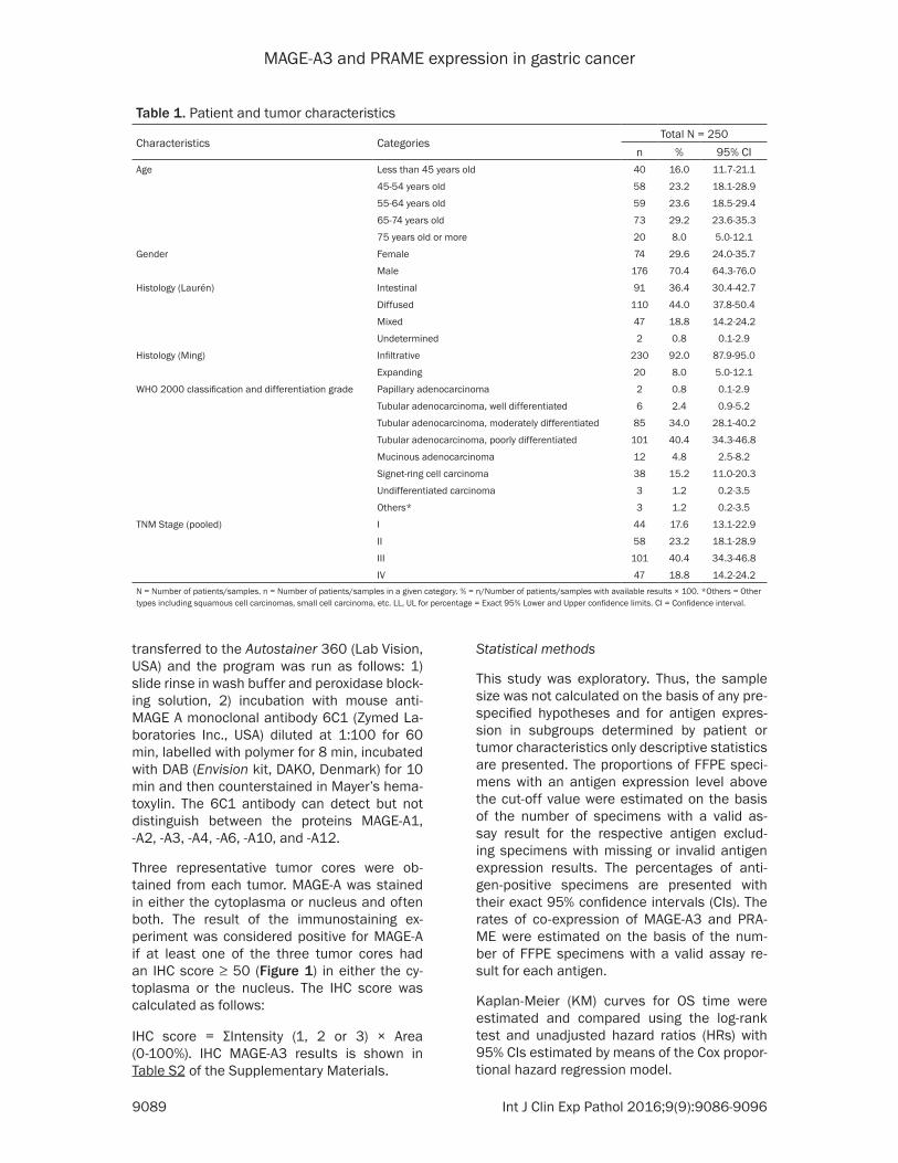
MAGE-A3 and PRAME expression in gastric cancer
9089 Int J Clin Exp Pathol 20169(9)9086-9096
transferred to the Autostainer 360 (Lab Vision USA) and the program was run as follows 1) slide rinse in wash buffer and peroxidase block-ing solution 2) incubation with mouse anti-MAGE A monoclonal antibody 6C1 (Zymed La- boratories Inc USA) diluted at 1100 for 60 min labelled with polymer for 8 min incubated with DAB (Envision kit DAKO Denmark) for 10 min and then counterstained in Mayerrsquos hema-toxylin The 6C1 antibody can detect but not distinguish between the proteins MAGE-A1 -A2 -A3 -A4 -A6 -A10 and -A12
Three representative tumor cores were ob- tained from each tumor MAGE-A was stained in either the cytoplasma or nucleus and often both The result of the immunostaining ex- periment was considered positive for MAGE-A if at least one of the three tumor cores had an IHC score ge 50 (Figure 1) in either the cy- toplasma or the nucleus The IHC score was calculated as follows
IHC score = ΣIntensity (1 2 or 3) times Area (0-100) IHC MAGE-A3 results is shown in Table S2 of the Supplementary Materials
Statistical methods
This study was exploratory Thus the sample size was not calculated on the basis of any pre-specified hypotheses and for antigen expres-sion in subgroups determined by patient or tumor characteristics only descriptive statistics are presented The proportions of FFPE speci-mens with an antigen expression level above the cut-off value were estimated on the basis of the number of specimens with a valid as- say result for the respective antigen exclud- ing specimens with missing or invalid antigen expression results The percentages of anti- gen-positive specimens are presented with their exact 95 confidence intervals (CIs) The rates of co-expression of MAGE-A3 and PRA- ME were estimated on the basis of the num- ber of FFPE specimens with a valid assay re- sult for each antigen
Kaplan-Meier (KM) curves for OS time were estimated and compared using the log-rank test and unadjusted hazard ratios (HRs) with 95 CIs estimated by means of the Cox propor-tional hazard regression model
Table 1 Patient and tumor characteristics
Characteristics CategoriesTotal N = 250
n 95 CIAge Less than 45 years old 40 160 117-211
45-54 years old 58 232 181-289
55-64 years old 59 236 185-294
65-74 years old 73 292 236-353
75 years old or more 20 80 50-121
Gender Female 74 296 240-357
Male 176 704 643-760
Histology (Laureacuten) Intestinal 91 364 304-427
Diffused 110 440 378-504
Mixed 47 188 142-242
Undetermined 2 08 01-29
Histology (Ming) Infiltrative 230 920 879-950
Expanding 20 80 50-121
WHO 2000 classification and differentiation grade Papillary adenocarcinoma 2 08 01-29
Tubular adenocarcinoma well differentiated 6 24 09-52
Tubular adenocarcinoma moderately differentiated 85 340 281-402
Tubular adenocarcinoma poorly differentiated 101 404 343-468
Mucinous adenocarcinoma 12 48 25-82
Signet-ring cell carcinoma 38 152 110-203
Undifferentiated carcinoma 3 12 02-35
Others 3 12 02-35
TNM Stage (pooled) I 44 176 131-229
II 58 232 181-289
III 101 404 343-468
IV 47 188 142-242N = Number of patientssamples n = Number of patientssamples in a given category = nNumber of patientssamples with available results times 100 Others = Other types including squamous cell carcinomas small cell carcinoma etc LL UL for percentage = Exact 95 Lower and Upper confidence limits CI = Confidence interval
MAGE-A3 and PRAME expression in gastric cancer
9090 Int J Clin Exp Pathol 20169(9)9086-9096
Results
Two-hundred-and-fifty FFPE specimens were tested for antigen expression and MAGE-A3 promoter methylation status Patient demo-
196 for MAGE-A- IHC to 234 for MAGE-A3- mRNA expression A total of 75 of specimens had fully concordant results across the 3 tests (Table 4) Fourteen (103) specimens had MAGE-A3 methylation status results that were
Table 2 Validity of MAGE-A3 and PRAME expression tests using qRT-PCR assays
Characteristics CategoriesMAGE-A3 (N = 250) PRAME (N = 250)n 95 CI n 95 CI
Validity test Valid 137 604 537-668 142 626 559-689Invalid 52 229 176-289 47 207 156-266QNS 38 167 121-222 38 167 121-222
Missing 23 - - 23 - -N = Number of patientssamples n = Number of patientssamples in a given category QNS = Quantity not sufficient = nNumber of patientssamples with available results times 100
Table 3 Overall rates of expression of MAGE-A3 and PRAME
Characteristics CategoriesTotal (N = 250)
n 95 CIMAGE-A3 expression (mRNA) result Positive 32 234 166-313
Negative 105 766 687-834Missing 113 - -
MAGE-A3 methylation result Unmethylated 54 230 178-289Methylated 181 770 711-822
Missing 15 - -MAGE-A IHC result Positive 49 196 149-251
Negative 201 804 749-851PRAME expression (mRNA) result Positive 29 204 141-280
Negative 113 796 720-859Missing 108 - -
N = Number of patientssamples n = Number of patientssamples in a given category = nNumber of patientssamples with available results times 100 IHC = Immunohistochemistry
graphics and tumor charac-teristics are summarized in Table 1
Percentage of specimens giving valid test results
Each mRNA expression te- st of an FFPE specimen was done with two repli-cates Twenty-three speci-mens were not tested for mRNA expression (Table 2) and for 38 specimens the amount of tumor cells in the mRNA extracted was in- sufficient (3438) or com-pletely absent (438)
Valid results were obtain- ed for 604 of the speci-mens tested for mRNA ex- pression of MAGE-A3 The reasons for invalid assay results for MAGE-A3 were test results lsquoout-of-rangersquo (4752) inconsistent repli-cates (352) and contami- nation with genomic DNA (252) For mRNA express- ion of PRAME valid results were obtained for 626 of tested specimens Invalid test results for PRAME were lsquoout-of-rangersquo
All the 250 FFPE specimens gave valid IHC results for MAGE-A protein whereas MAGE-A3 methylation sta- tus could not be obtained for 15 specimens
Antigen expression
The three methods for de- tecting MAGE-A3 express- ion gave concordant re- sults overall (Table 3) with the percentage of speci- mens with positive results for MAGE-A3 ranging from
Table 4 Concordance between MAGE-A3 expression tests with the three methods
CategoriesN = 136
n 95 CImRNA-negativeMethylatedIHC-negative 87 640 553-720mRNA-negativeMethylatedIHC-positive 4 29 08-74mRNA-negativeUnmethylatedIHC-negative 9 66 31-122mRNA-negativeUnmethylatedIHC-positive 5 37 12-84mRNA-positiveMethylatedIHC-negative 6 44 16-94mRNA-positiveMethylatedIHC-positive 5 37 12-84mRNA-positiveUnmethylatedIHC-negative 5 37 12-84mRNA-positiveUnmethylatedIHC-positive 15 110 63-175N = Number of specimens n = Number of specimens in a given category = nNumber of specimens with available results times 100 CI = Confidence interval
MAGE-A3 and PRAME expression in gastric cancer
9091 Int J Clin Exp Pathol 20169(9)9086-9096
Table 5 Rates of co-expression of MAGE-A3 and PRAME
Characteristics CategoriesN = 136
n 95 CICo-expression MAGE-A3 and PRAME MAGE-A3-negativePRAME-negative 90 662 576-741
MAGE-A3-negativePRAME-positive 14 103 57-167MAGE-A3-positivePRAME-negative 18 132 80-201MAGE-A3-positivePRAME-positive 14 103 57-167
At least MAGE-A3 or PRAME MAGE-A3-negative and PRAME-negative 90 662 576-741MAGE-A3-positive or PRAME-positive 46 338 259-424
N = Number of patientssamples n = Number of patientssamples in a given category = nNumber of patientssamples with available results times 100
Table 6 Subgroup analyses of MAGE-A3 or PRAME expression (mRNA) according to age gender tumor histology and tumor stage
Variable CategoriesMAGE-A3 expression PRAME expression
N n 95 CI N n 95 CIAge 26-44 23 1 43 01-219 24 2 83 10-270
45-54 34 4 118 33-275 35 7 200 84-36955-64 25 6 240 94-451 28 4 143 40-32765-74 45 17 378 238-535 45 12 267 146-419ge 75 10 4 400 122-738 10 4 400 122-738
Gender Female 35 5 143 48-303 39 5 128 43-274Male 102 27 265 182-361 103 24 233 155-327
Histology (Lauren) Intestinal 55 16 291 176-429 55 16 291 176-429Diffused 64 7 109 45-212 67 8 119 53-222Mixed 18 9 500 260-740 20 5 250 87-491Undetermined 0 0 - - 0 0 - -
Histology (Ming) Infiltrative 124 29 234 163-318 128 24 188 124-266Expanding 13 3 231 50-538 14 5 357 128-649
Histology (WHO) Papillary AC 2 0 00 00-842 1 0 00 00-975Tubular AC well diff 5 1 200 05-716 5 1 200 05-716Tubular AC moderately diff 48 17 354 222-505 48 16 333 204-484Tubular AC poorly diff 53 9 170 81-298 56 8 143 64-262Mucinous AC 5 1 200 05-716 6 1 167 04-641Signet-ring cell carcinoma 23 3 130 28-336 24 1 42 01-211Undifferentiated carcinoma 0 0 - - 0 0 - -Others 1 1 100 25-100 2 2 100 158-100
Stage I 26 3 115 24-302 25 5 200 68-407II 35 13 371 215-551 36 8 222 101-392III 50 8 160 72-291 54 11 204 106-335IV 26 8 308 143-518 27 5 185 63-381
N = Number of FFPE specimens in this category giving a valid test result for the antigen concerned n = Number of specimens in a given category with a valid test result positive for the antigen concerned AC = Adenocarcinoma diff = Differentiated = (nN) times 100 CI = Confidence interval
discordant with the results for mRNA detection and IHC (nine with mRNA-negativeunmethyl-ationIHC-negative and five with mRNA-posi-tivemethylationIHC-positive)
Of the 142 specimens with valid PRAME assay results 29 (204) were PRAME-positive (Table 3) Of the 136 FFPE specimens with valid mRNA expression assay results for both antigens 14
MAGE-A3 and PRAME expression in gastric cancer
9092 Int J Clin Exp Pathol 20169(9)9086-9096
(103) expressed both while 46 (338) expressed at least one of them (Table 5)
The exploratory analyses of the mRNA ex- pression of the TAAs in subgroups determin- ed by patient and tumor characteristics show- ed similar results for MAGE-A3 and PRAME (Table 6) TAA mRNA expression was indepen-dent of tumor stage and there was no obvious association between tumor TAA mRNA expre- ssion and any of the other clinico-pathological factors investigated
Another study investigated MAGE-A express- ion in GC cell lines as well as primary gastric tumor specimens using both IHC and analy- sis of MAGE-A3 promoter methylation status for detection Of 1097 cancer tissues 158 expressed MAGE-A protein with no distinction between the subtypes of MAGE-A by the anti-body used for the IHC Of 52 randomly selected tumors expressing MAGE-A protein 28 (538) displayed unmethylation of the MAGE-A3 pro-moter [24] In the present study 20 of the 29 (690 recalculated from Table 4) tumors
Figure 2 Kaplan-Meier curves for OS by MAGE-A3 methylation status of the specimen HR for MAGE-A3 unmethylated 094 (95 CI 057-156 P = 082) (Wald test from Cox regression model)
Figure 3 Kaplan-Meier curves for OS by MAGE-A protein expression of the specimen HR for MAGE-A3-positive 146 (95 CI 092-232 P = 011) (Wald test from Cox regression model)
Overall survival by MAGE-A3 methylation status protein expression of MAGE-A and mRNA expression of the an-tigens
Kaplan-Meier curves for OS according to the tumorrsquos MAGE-A3 methylation status (Figure 2) overlap almost completely The Kaplan-Meier curves for OS according to the specimensrsquo expression of MAGE-A3 protein are more separated (Figure 3) but are not statistically significant- ly different (P = 011) Like- wise there were no indica-tions of an association be- tween OS and mRNA expres-sion of the antigens neither for MAGE-A3 nor for PRAME (Figures 4 and 5)
Discussion
This is the first study of PRAME expression in GC while mRNA MAGE-A3 expre- ssion has been reported in several previous studies with rates of MAGE-A3-positive GC tumors varying between 30 and 416 [20-23] None of these studies reported sta- tistically significant associa-tions between clinico-patho-logical tumor characteristics and mRNA expression of MAGE-A3 in line with findings in the current study
MAGE-A3 and PRAME expression in gastric cancer
9093 Int J Clin Exp Pathol 20169(9)9086-9096
expressing the MAGE-A protein displayed un- methylation of the MAGE-A3 promoter where- as this was displayed by 14 of the 107 (131) tumors not expressing the MAGE-A protein These results indicate that unmethylation of the MAGE-A3 promoter may be (partly) respon-sible for the expression of the MAGE-A pro- tein in gastric cancer
Honda et al [13] detected MAGE-A3 promoter demethylation in 66 of 84 GC specimens tumors displaying MAGE-A3 promoter demeth-
Honda et al [13] described a trend for worse survival for patients with tumors displaying demethylated MAGE-A3 in particular if their tumor also showed demethylated MAGE-A1 (P = 018)
Circulating tumor cell DNA can be detected in serum or blood even at early stages of the dis-ease as shown by Kounalakos et al [25] for melanoma patients This DNA may be derived from lysed cells of the primary tumor or from circulating tumor cells releasing their DNA With
Figure 4 Kaplan-Meier curves for overall survival by mRNA expression of MAGE-A3 (Total cohort) HR for MAGE-A3-positive 103 (95 CI 0538-1963 P = 093) (Wald test from Cox regression model)
Figure 5 Kaplan-Meier curves for overall survival by mRNA expression of PRAME (Total cohort) HR for PRAME-positive 097 (95 CI 0499-1885 P = 093) (Wald test from Cox regression model)
ylation were in a more ad- vanced state and were asso-ciated with a higher preva-lence of lymph-node metas- tases than those without de- methylation In the present study 230 of gastric tu- mors displayed an unmethyl-ated MAGE-A3 promoter
We did not observe any rela-tion between OS and mRNA TAA expression of the FFPE specimen for either MAGE-A3 or PRAME Similarly there was no statistically significant association between OS and MAGE-A protein expression (P = 011) In contrast Jung et al [24] reported a significant-ly worse prognosis for patients with tumors expressing the MAGE-A proteins (P lt 0001)
Recent studies investigating the molecular basis of GC have reported associations between aberrant DNA meth-ylation of several gene pro-moters and the prognosis for GC patients generally sug-gesting that hypermethylation of the identified gene promot-ers is an indicator of poor prognosis (reviewed in [15]) Our observations indicate that MAGE-A3 is not to be added to this list of genes as the methylation status of the MAGE-A3 promoter had no association with patient OS (P = 082) In contrast
MAGE-A3 and PRAME expression in gastric cancer
9094 Int J Clin Exp Pathol 20169(9)9086-9096
a prior understanding of a potential correlation between gene promoter methylation status and gene expression methylation testing of cir-culating DNA could be useful in detecting the expression of specific genes in tumor cells Such non-invasive detection methods that are not dependent on the availability of tumor tissue could increase the number of patients that can be screened for expression of tumor antigens
A rough assessment of the possibility of rely- ing on such non-invasive methods for gene expression screening based on the observa-tions of the present study was performed Due to the lack of MAGE-A3 specific antibodies IHC cannot distinguish MAGE-A3 from other members of the MAGE-A family MAGE-A3 ex- pression was thus generally screened for us- ing RT-PCR assays Here we found that 867 (91105) of the FFPE specimens with no mRNA MAGE-A3 expression showed methylation of the MAGE-A3 promoter whereas 645 (20 31) of specimens with mRNA MAGE-A3 expre- ssion were unmethylated in the MAGE-A3 pro-moter Arguably the concordance between the results obtained with the two methods may be considered too low to rely on testing of MAGE-A3 promoter methylation status for pa- tient screening According to these observa-tions screening based on methylation testing of the MAGE-A3 promoter would miss more than a third of the patients whose tumors show mRNA MAGE-A3 expression
The overall concordance between the three MAGE-A3 assays was relatively low compared to previous findings (75 vs nearly 100) reported in certain studies (eg [26]) The qRT-PCR assay is specific for MAGE-A3 and the cut-off value was defined as 1 of the GERL con-trol By contrast the IHC assay could also detect other members of the MAGE-A family (in particular MAGE-A6) and the cut-off values for these assays are linked to their detection lim-its Additional investigations are needed to assess the impact of MAGE-A3A6 expression and to homogenize the cut-off values of the assays
In summary the rate of mRNA expression of each of these TAAs in tumors from GC patients was found to be relatively low with 234 expressing MAGE-A3 204 expressing PRAME and 338 expressing at least one of them Neither MAGE-A3 nor PRAME would se-
rve as a prognostic biomarker for OS of GC patients
Acknowledgements
Niels Neymark provided scientific writing ser-vices and Veacuteronique Duquenne (XPE PHAR- MA and SCIENCES Wavre Belgium CO GSK) coordinated the manuscriptrsquos development This study was funded and coordinated by GlaxoSmithKline Biologicals SA GSK was in- volved in the design and conduct of the study collection management analysis and inter- pretation of the data and preparation and review of the manuscript
Disclosure of conflict of interest
Olivier Gruselle and Bart Spiessens are employ-ees of the GSK group of companies An de Creus Nicole Kusuma and Aung Myo were employees of the GSK group of companies at the time of the study and manuscript develop-ment Bart Spiessens own stockstock options in GSK group of companies Woo Ho Kim Eun Ji Jung and Kim Hee Sung have no conflict of interest
Authorsrsquo contribution
BS AM AdeC and EJJ were involved in study design AM AdeC NK were involved in study supervision OG AM AdeC NK WHK EJJ were involved in data acquisition HSK EJJ NK AM WHK and OG were involved in data collection OG AM NK and WHK were involved in data extraction and quality check (AdeC in quality check only and EJJ in data extraction only) OG WHK and EJJ were involved in provision of study material WHK and EJJ were involved in labora-tory testing BS (biostatistician) OG AM AdeC WHK and HSK performed or supervised the analyses BS OG AM AdC NK WHK and HSK were involved in results interpretation OG AdeC NK EJJ were involved in administrativetechnicallogistic support EJJ and AM were involved in centers recruitment AM and NK were involved in center coordination AM was involved in acquisition of funding
Address correspondence to Dr Woo Ho Kim De- partment of Pathology Seoul National University College of Medicine 28 Yeongeon-dong 110-799 Seoul Korea Tel +82 8 740 8269 Fax +82 2 765 5600 E-mail woohokimsnuackr
MAGE-A3 and PRAME expression in gastric cancer
9095 Int J Clin Exp Pathol 20169(9)9086-9096
References
[1] Crew KD Neugut AI The epidemiology of gas-tric cancer World J Gastroenterol 2006 12 354-62
[2] Toomey PG Vohra NA Ghansah T Sarnaik AA Pilon-Thomas SA Immunotherapy for gastroin-testinal malignancies Cancer Control 2013 20 32-42
[3] Bilici A Treatment options in patients with met-astatic gastric cancer Current status and fu-ture perspective World J Gastroenterol 2014 20 3905-15
[4] Amadei A Benagiano M della Bella C Nicolai E DrsquoElios MM Novel immunotherapeutic strat-egies of gastric cancer treatment J Biomed Biotech 2011 2011 437348
[5] Ikeda H Sato N Matsuura A Kikuchi K Analysis of T-cell receptor V region gene usage of cytotoxic T-lymphocytes and tumor-infiltrat-ing lymphocytes derived from human autolo-gous gastric signet ring cell carcinomas Cancer Res 1993 53 3078-84
[6] Kono K Ichihara F Lizuka H Sekikawa T Matsumoto Y Differences in the recognition of tumor-specific CD8+ T cells derived from solid tumor metastatic lymph nodes and ascites in patients with gastric cancer Int J Cancer 1997 71 978-81
[7] Amadei A Niccolai E Della Bella C Cianchi F Trallori G Benagiano M Bencini L Bernini M Farsi M Moretti R Del Prete G DrsquoElios MM Characterization of tumor antigen peptide-spe-cific T cells isolated from the neoplastic tissue of patients with gastric adenocarcinomas Cancer Immunol Immunother 2009 58 1819-30
[8] Lee HE Chae SW Lee YJ Kim MA Lee HS Lee BL Kim WH Prognostic implications of type and density of tumour-infiltrating lymp- hocytes in gastric cancer Br J Cancer 2008 99 1704-11
[9] Perrone G Ruffini PA Catalano V Spino C Santini D Muretto P Spoto C Zingaretti C Sisti V Alessandroni P Giordani P Cicetti A DrsquoEmidio S Morini S Ruzzo A Magnani M Tonini G Rabitti C Graziano F Intratumoural FOXP3-positive regulatory T cells are associat-ed with adverse prognosis in radically resected gastric cancer Eur J Cancer 2008 44 1875-82
[10] Shen Z Zhou S Wang Y Li RL Zhong C Liang C Sun Y Higher intratumoural infiltrated Foxp3+ Treg numbers and Foxp3+CD8+ ratio are associated with adverse prognosis in re-sectable gastric cancer J Cancer Res Clin Oncol 2010 136 1585-95
[11] Zhou S Shen Z Wang Y Ma H Xu S Qin J Chen L Tao H Zhen Z Chen G Zhang Z Li R
Xiao H Zhong C Yang Y Liang C CCR7 expres-sion and intratumoural FOXP3+ regulatory T cells are correlated with overall survival and lymph node metastatis in gastric cancer PLoS One 2013 8 e74430
[12] Matsueda S Graham DY Immunotherapy in gastric cancer World J Gatroenterol 2014 20 1657-66
[13] Honda T Tamura G Waki T Kawata S Tera- shima M Nishizuka S Motoyama T Deme- thylation of MAGE promoters during gastric cancer progression Br J Cancer 2004 90 838-43
[14] Kim KH Choi JS Kim IJ Ku JL Park JG Promoter hypomethylation and reactivation of MAGE-A1 and MAGE-A3 genes in colorectal cancer cell lines and cancer tissues World J Gastroenterol 2006 12 5651-57
[15] Nakamura J Tanaka T Kitajima Y Noshiro H Miyazaki K Methylation-mediated gene silenc-ing as biomarkers of gastric cancer A review World J Gastroenterology 2014 20 11991-12006
[16] Oberthuer A Hero B Spitz R Berthold F Fischer M The tumor-associated antigen PRA- ME is universally expressed in high-stage neu-roblastoma and associated with poor out- come Clin Cancer Res 2004 10 4307-13
[17] Partheen K Levan K Osterberg L Claesson I Fallenius G Sundfeldt K Horvath G Four po-tential biomarkers as prognostic factors in stage III serous ovarian adenocarcinomas Int J Cancer 2008 123 2130-7
[18] Epping MT Hart AA Glas AM Krijgsman O Bernards R PRAME expression and clinical outcome of breast cancer Br J Cancer 2008 99 398-403
[19] Lerut E Van Poppel H Joniau S Gruselle O Coche T Therasse P Rates of MAGE-A3 and PRAME expressing tumors in FFPE tissue spec-imens from bladder cancer patients potential targets for antigen-specific cancer immuno-therapeutics Int J Clin Exp Pathol 2015 8 9522-32
[20] Inoue H Mori M Honda M Li J Shibuta K Mimori K Ueo H Akiyoshi T The expression of tumor-rejection antigen ldquoMAGErdquo genes in human gastric carcinoma Gastroenterology 1995 109 1522-25
[21] Li J Yang Y Fujie T Baba K Ueo H Mori M Akiyoshi T Expression of BAGE GAGE and MAGE genes in human gastric carcinoma Clin Cancer Res 1996 2 1619-25
[22] Wang Y Wu XJ Zhao AL Yuan YH Chen YT Jungbluth AA Gnjatic S Santiago D Ritter G Chen WF Old LJ Ji JF Cancertestis antigen expression and autologous humoral immunity to NY-ESO-1 in gastric cancer Cancer Immun 2004 4 11
MAGE-A3 and PRAME expression in gastric cancer
9096 Int J Clin Exp Pathol 20169(9)9086-9096
[23] Wang WX Li YB Xie XL Shu XL Ouyang XH Detection of tumor cells in peripheral blood of patients with gastric cancer using mRNA of MAGE genes as markers Zhonghua Wei Chang Wai Ke Za Zhi 2009 12 611-14
[24] Jung EJ Kim MA Lee HS Yang HK Lee YM Lee BL Kim WH Expression of family A mela-noma antigen in human gastric carcinoma Anticancer Research 2005 25 2105-12
[25] Kounalakos N Goydos JS Tumor cells and cir-culating markers in melanoma diagnosis prognosis and management Curr Oncol Rep 2005 7 377-382
[26] Lehmann-Che J Amira-Bouhidel F Turpin E Antoine M Soliman H Legres L Bocquet C Bernoud R Flandre E Varna M de Roquan- court A Plassa LF Giacchetti S Espieacute M de Bazelaire C Cahen-Doidy L Bourstyn E Janin A de Theacute H Bertheau P Immunohistoche- mical and molecular analyses of HER2 sta- tus in breast cancers are highly concordant and complementary approaches Br J Cancer 2011 104 1739-46
MAGE-A3 and PRAME expression in gastric cancer
1
Table S1 Primer sequences for methylation specific PCRMethylation status Type SequenceUnmethylated Anti-sense 5rsquo-TGTTAGGATGTGATGTTATTGATTTGT-3rsquo
Sense 5rsquo-CCTCACCAAACCTAAACCAA-3rsquoMethylated Anti-sense 5rsquo-CCATCTGACGTTATTGATTTGC-3rsquo
Sense 5rsquo-CTCACCGAACCTAAACCGAC-3rsquo
Table S2 Distribution of immunohistochemistry MAGE-A3 results in gastric cancerIHC score 0-50 51-100 101-150 151-200 201-250 251-300 Total of cases 201 19 8 4 3 19 250 of total 804 76 32 16 12 76 100IHC result (Score gt 50) Negative Positive
804 196
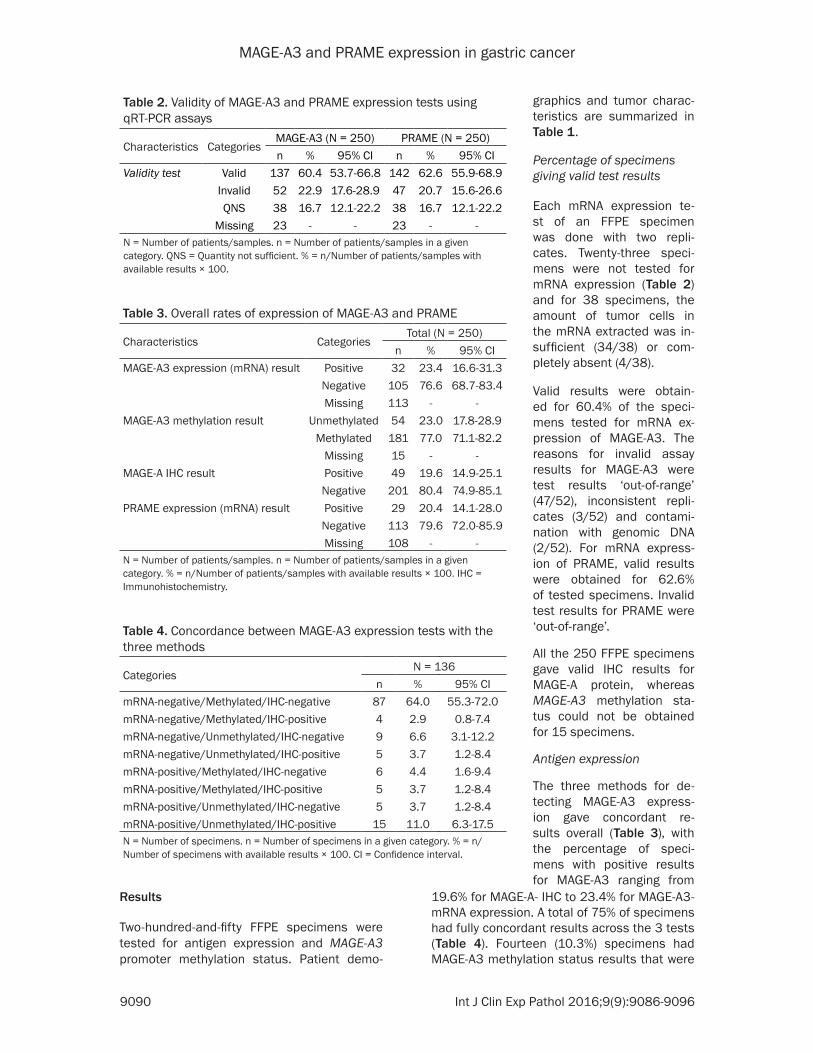
MAGE-A3 and PRAME expression in gastric cancer
9090 Int J Clin Exp Pathol 20169(9)9086-9096
Results
Two-hundred-and-fifty FFPE specimens were tested for antigen expression and MAGE-A3 promoter methylation status Patient demo-
196 for MAGE-A- IHC to 234 for MAGE-A3- mRNA expression A total of 75 of specimens had fully concordant results across the 3 tests (Table 4) Fourteen (103) specimens had MAGE-A3 methylation status results that were
Table 2 Validity of MAGE-A3 and PRAME expression tests using qRT-PCR assays
Characteristics CategoriesMAGE-A3 (N = 250) PRAME (N = 250)n 95 CI n 95 CI
Validity test Valid 137 604 537-668 142 626 559-689Invalid 52 229 176-289 47 207 156-266QNS 38 167 121-222 38 167 121-222
Missing 23 - - 23 - -N = Number of patientssamples n = Number of patientssamples in a given category QNS = Quantity not sufficient = nNumber of patientssamples with available results times 100
Table 3 Overall rates of expression of MAGE-A3 and PRAME
Characteristics CategoriesTotal (N = 250)
n 95 CIMAGE-A3 expression (mRNA) result Positive 32 234 166-313
Negative 105 766 687-834Missing 113 - -
MAGE-A3 methylation result Unmethylated 54 230 178-289Methylated 181 770 711-822
Missing 15 - -MAGE-A IHC result Positive 49 196 149-251
Negative 201 804 749-851PRAME expression (mRNA) result Positive 29 204 141-280
Negative 113 796 720-859Missing 108 - -
N = Number of patientssamples n = Number of patientssamples in a given category = nNumber of patientssamples with available results times 100 IHC = Immunohistochemistry
graphics and tumor charac-teristics are summarized in Table 1
Percentage of specimens giving valid test results
Each mRNA expression te- st of an FFPE specimen was done with two repli-cates Twenty-three speci-mens were not tested for mRNA expression (Table 2) and for 38 specimens the amount of tumor cells in the mRNA extracted was in- sufficient (3438) or com-pletely absent (438)
Valid results were obtain- ed for 604 of the speci-mens tested for mRNA ex- pression of MAGE-A3 The reasons for invalid assay results for MAGE-A3 were test results lsquoout-of-rangersquo (4752) inconsistent repli-cates (352) and contami- nation with genomic DNA (252) For mRNA express- ion of PRAME valid results were obtained for 626 of tested specimens Invalid test results for PRAME were lsquoout-of-rangersquo
All the 250 FFPE specimens gave valid IHC results for MAGE-A protein whereas MAGE-A3 methylation sta- tus could not be obtained for 15 specimens
Antigen expression
The three methods for de- tecting MAGE-A3 express- ion gave concordant re- sults overall (Table 3) with the percentage of speci- mens with positive results for MAGE-A3 ranging from
Table 4 Concordance between MAGE-A3 expression tests with the three methods
CategoriesN = 136
n 95 CImRNA-negativeMethylatedIHC-negative 87 640 553-720mRNA-negativeMethylatedIHC-positive 4 29 08-74mRNA-negativeUnmethylatedIHC-negative 9 66 31-122mRNA-negativeUnmethylatedIHC-positive 5 37 12-84mRNA-positiveMethylatedIHC-negative 6 44 16-94mRNA-positiveMethylatedIHC-positive 5 37 12-84mRNA-positiveUnmethylatedIHC-negative 5 37 12-84mRNA-positiveUnmethylatedIHC-positive 15 110 63-175N = Number of specimens n = Number of specimens in a given category = nNumber of specimens with available results times 100 CI = Confidence interval
MAGE-A3 and PRAME expression in gastric cancer
9091 Int J Clin Exp Pathol 20169(9)9086-9096
Table 5 Rates of co-expression of MAGE-A3 and PRAME
Characteristics CategoriesN = 136
n 95 CICo-expression MAGE-A3 and PRAME MAGE-A3-negativePRAME-negative 90 662 576-741
MAGE-A3-negativePRAME-positive 14 103 57-167MAGE-A3-positivePRAME-negative 18 132 80-201MAGE-A3-positivePRAME-positive 14 103 57-167
At least MAGE-A3 or PRAME MAGE-A3-negative and PRAME-negative 90 662 576-741MAGE-A3-positive or PRAME-positive 46 338 259-424
N = Number of patientssamples n = Number of patientssamples in a given category = nNumber of patientssamples with available results times 100
Table 6 Subgroup analyses of MAGE-A3 or PRAME expression (mRNA) according to age gender tumor histology and tumor stage
Variable CategoriesMAGE-A3 expression PRAME expression
N n 95 CI N n 95 CIAge 26-44 23 1 43 01-219 24 2 83 10-270
45-54 34 4 118 33-275 35 7 200 84-36955-64 25 6 240 94-451 28 4 143 40-32765-74 45 17 378 238-535 45 12 267 146-419ge 75 10 4 400 122-738 10 4 400 122-738
Gender Female 35 5 143 48-303 39 5 128 43-274Male 102 27 265 182-361 103 24 233 155-327
Histology (Lauren) Intestinal 55 16 291 176-429 55 16 291 176-429Diffused 64 7 109 45-212 67 8 119 53-222Mixed 18 9 500 260-740 20 5 250 87-491Undetermined 0 0 - - 0 0 - -
Histology (Ming) Infiltrative 124 29 234 163-318 128 24 188 124-266Expanding 13 3 231 50-538 14 5 357 128-649
Histology (WHO) Papillary AC 2 0 00 00-842 1 0 00 00-975Tubular AC well diff 5 1 200 05-716 5 1 200 05-716Tubular AC moderately diff 48 17 354 222-505 48 16 333 204-484Tubular AC poorly diff 53 9 170 81-298 56 8 143 64-262Mucinous AC 5 1 200 05-716 6 1 167 04-641Signet-ring cell carcinoma 23 3 130 28-336 24 1 42 01-211Undifferentiated carcinoma 0 0 - - 0 0 - -Others 1 1 100 25-100 2 2 100 158-100
Stage I 26 3 115 24-302 25 5 200 68-407II 35 13 371 215-551 36 8 222 101-392III 50 8 160 72-291 54 11 204 106-335IV 26 8 308 143-518 27 5 185 63-381
N = Number of FFPE specimens in this category giving a valid test result for the antigen concerned n = Number of specimens in a given category with a valid test result positive for the antigen concerned AC = Adenocarcinoma diff = Differentiated = (nN) times 100 CI = Confidence interval
discordant with the results for mRNA detection and IHC (nine with mRNA-negativeunmethyl-ationIHC-negative and five with mRNA-posi-tivemethylationIHC-positive)
Of the 142 specimens with valid PRAME assay results 29 (204) were PRAME-positive (Table 3) Of the 136 FFPE specimens with valid mRNA expression assay results for both antigens 14
MAGE-A3 and PRAME expression in gastric cancer
9092 Int J Clin Exp Pathol 20169(9)9086-9096
(103) expressed both while 46 (338) expressed at least one of them (Table 5)
The exploratory analyses of the mRNA ex- pression of the TAAs in subgroups determin- ed by patient and tumor characteristics show- ed similar results for MAGE-A3 and PRAME (Table 6) TAA mRNA expression was indepen-dent of tumor stage and there was no obvious association between tumor TAA mRNA expre- ssion and any of the other clinico-pathological factors investigated
Another study investigated MAGE-A express- ion in GC cell lines as well as primary gastric tumor specimens using both IHC and analy- sis of MAGE-A3 promoter methylation status for detection Of 1097 cancer tissues 158 expressed MAGE-A protein with no distinction between the subtypes of MAGE-A by the anti-body used for the IHC Of 52 randomly selected tumors expressing MAGE-A protein 28 (538) displayed unmethylation of the MAGE-A3 pro-moter [24] In the present study 20 of the 29 (690 recalculated from Table 4) tumors
Figure 2 Kaplan-Meier curves for OS by MAGE-A3 methylation status of the specimen HR for MAGE-A3 unmethylated 094 (95 CI 057-156 P = 082) (Wald test from Cox regression model)
Figure 3 Kaplan-Meier curves for OS by MAGE-A protein expression of the specimen HR for MAGE-A3-positive 146 (95 CI 092-232 P = 011) (Wald test from Cox regression model)
Overall survival by MAGE-A3 methylation status protein expression of MAGE-A and mRNA expression of the an-tigens
Kaplan-Meier curves for OS according to the tumorrsquos MAGE-A3 methylation status (Figure 2) overlap almost completely The Kaplan-Meier curves for OS according to the specimensrsquo expression of MAGE-A3 protein are more separated (Figure 3) but are not statistically significant- ly different (P = 011) Like- wise there were no indica-tions of an association be- tween OS and mRNA expres-sion of the antigens neither for MAGE-A3 nor for PRAME (Figures 4 and 5)
Discussion
This is the first study of PRAME expression in GC while mRNA MAGE-A3 expre- ssion has been reported in several previous studies with rates of MAGE-A3-positive GC tumors varying between 30 and 416 [20-23] None of these studies reported sta- tistically significant associa-tions between clinico-patho-logical tumor characteristics and mRNA expression of MAGE-A3 in line with findings in the current study
MAGE-A3 and PRAME expression in gastric cancer
9093 Int J Clin Exp Pathol 20169(9)9086-9096
expressing the MAGE-A protein displayed un- methylation of the MAGE-A3 promoter where- as this was displayed by 14 of the 107 (131) tumors not expressing the MAGE-A protein These results indicate that unmethylation of the MAGE-A3 promoter may be (partly) respon-sible for the expression of the MAGE-A pro- tein in gastric cancer
Honda et al [13] detected MAGE-A3 promoter demethylation in 66 of 84 GC specimens tumors displaying MAGE-A3 promoter demeth-
Honda et al [13] described a trend for worse survival for patients with tumors displaying demethylated MAGE-A3 in particular if their tumor also showed demethylated MAGE-A1 (P = 018)
Circulating tumor cell DNA can be detected in serum or blood even at early stages of the dis-ease as shown by Kounalakos et al [25] for melanoma patients This DNA may be derived from lysed cells of the primary tumor or from circulating tumor cells releasing their DNA With
Figure 4 Kaplan-Meier curves for overall survival by mRNA expression of MAGE-A3 (Total cohort) HR for MAGE-A3-positive 103 (95 CI 0538-1963 P = 093) (Wald test from Cox regression model)
Figure 5 Kaplan-Meier curves for overall survival by mRNA expression of PRAME (Total cohort) HR for PRAME-positive 097 (95 CI 0499-1885 P = 093) (Wald test from Cox regression model)
ylation were in a more ad- vanced state and were asso-ciated with a higher preva-lence of lymph-node metas- tases than those without de- methylation In the present study 230 of gastric tu- mors displayed an unmethyl-ated MAGE-A3 promoter
We did not observe any rela-tion between OS and mRNA TAA expression of the FFPE specimen for either MAGE-A3 or PRAME Similarly there was no statistically significant association between OS and MAGE-A protein expression (P = 011) In contrast Jung et al [24] reported a significant-ly worse prognosis for patients with tumors expressing the MAGE-A proteins (P lt 0001)
Recent studies investigating the molecular basis of GC have reported associations between aberrant DNA meth-ylation of several gene pro-moters and the prognosis for GC patients generally sug-gesting that hypermethylation of the identified gene promot-ers is an indicator of poor prognosis (reviewed in [15]) Our observations indicate that MAGE-A3 is not to be added to this list of genes as the methylation status of the MAGE-A3 promoter had no association with patient OS (P = 082) In contrast
MAGE-A3 and PRAME expression in gastric cancer
9094 Int J Clin Exp Pathol 20169(9)9086-9096
a prior understanding of a potential correlation between gene promoter methylation status and gene expression methylation testing of cir-culating DNA could be useful in detecting the expression of specific genes in tumor cells Such non-invasive detection methods that are not dependent on the availability of tumor tissue could increase the number of patients that can be screened for expression of tumor antigens
A rough assessment of the possibility of rely- ing on such non-invasive methods for gene expression screening based on the observa-tions of the present study was performed Due to the lack of MAGE-A3 specific antibodies IHC cannot distinguish MAGE-A3 from other members of the MAGE-A family MAGE-A3 ex- pression was thus generally screened for us- ing RT-PCR assays Here we found that 867 (91105) of the FFPE specimens with no mRNA MAGE-A3 expression showed methylation of the MAGE-A3 promoter whereas 645 (20 31) of specimens with mRNA MAGE-A3 expre- ssion were unmethylated in the MAGE-A3 pro-moter Arguably the concordance between the results obtained with the two methods may be considered too low to rely on testing of MAGE-A3 promoter methylation status for pa- tient screening According to these observa-tions screening based on methylation testing of the MAGE-A3 promoter would miss more than a third of the patients whose tumors show mRNA MAGE-A3 expression
The overall concordance between the three MAGE-A3 assays was relatively low compared to previous findings (75 vs nearly 100) reported in certain studies (eg [26]) The qRT-PCR assay is specific for MAGE-A3 and the cut-off value was defined as 1 of the GERL con-trol By contrast the IHC assay could also detect other members of the MAGE-A family (in particular MAGE-A6) and the cut-off values for these assays are linked to their detection lim-its Additional investigations are needed to assess the impact of MAGE-A3A6 expression and to homogenize the cut-off values of the assays
In summary the rate of mRNA expression of each of these TAAs in tumors from GC patients was found to be relatively low with 234 expressing MAGE-A3 204 expressing PRAME and 338 expressing at least one of them Neither MAGE-A3 nor PRAME would se-
rve as a prognostic biomarker for OS of GC patients
Acknowledgements
Niels Neymark provided scientific writing ser-vices and Veacuteronique Duquenne (XPE PHAR- MA and SCIENCES Wavre Belgium CO GSK) coordinated the manuscriptrsquos development This study was funded and coordinated by GlaxoSmithKline Biologicals SA GSK was in- volved in the design and conduct of the study collection management analysis and inter- pretation of the data and preparation and review of the manuscript
Disclosure of conflict of interest
Olivier Gruselle and Bart Spiessens are employ-ees of the GSK group of companies An de Creus Nicole Kusuma and Aung Myo were employees of the GSK group of companies at the time of the study and manuscript develop-ment Bart Spiessens own stockstock options in GSK group of companies Woo Ho Kim Eun Ji Jung and Kim Hee Sung have no conflict of interest
Authorsrsquo contribution
BS AM AdeC and EJJ were involved in study design AM AdeC NK were involved in study supervision OG AM AdeC NK WHK EJJ were involved in data acquisition HSK EJJ NK AM WHK and OG were involved in data collection OG AM NK and WHK were involved in data extraction and quality check (AdeC in quality check only and EJJ in data extraction only) OG WHK and EJJ were involved in provision of study material WHK and EJJ were involved in labora-tory testing BS (biostatistician) OG AM AdeC WHK and HSK performed or supervised the analyses BS OG AM AdC NK WHK and HSK were involved in results interpretation OG AdeC NK EJJ were involved in administrativetechnicallogistic support EJJ and AM were involved in centers recruitment AM and NK were involved in center coordination AM was involved in acquisition of funding
Address correspondence to Dr Woo Ho Kim De- partment of Pathology Seoul National University College of Medicine 28 Yeongeon-dong 110-799 Seoul Korea Tel +82 8 740 8269 Fax +82 2 765 5600 E-mail woohokimsnuackr
MAGE-A3 and PRAME expression in gastric cancer
9095 Int J Clin Exp Pathol 20169(9)9086-9096
References
[1] Crew KD Neugut AI The epidemiology of gas-tric cancer World J Gastroenterol 2006 12 354-62
[2] Toomey PG Vohra NA Ghansah T Sarnaik AA Pilon-Thomas SA Immunotherapy for gastroin-testinal malignancies Cancer Control 2013 20 32-42
[3] Bilici A Treatment options in patients with met-astatic gastric cancer Current status and fu-ture perspective World J Gastroenterol 2014 20 3905-15
[4] Amadei A Benagiano M della Bella C Nicolai E DrsquoElios MM Novel immunotherapeutic strat-egies of gastric cancer treatment J Biomed Biotech 2011 2011 437348
[5] Ikeda H Sato N Matsuura A Kikuchi K Analysis of T-cell receptor V region gene usage of cytotoxic T-lymphocytes and tumor-infiltrat-ing lymphocytes derived from human autolo-gous gastric signet ring cell carcinomas Cancer Res 1993 53 3078-84
[6] Kono K Ichihara F Lizuka H Sekikawa T Matsumoto Y Differences in the recognition of tumor-specific CD8+ T cells derived from solid tumor metastatic lymph nodes and ascites in patients with gastric cancer Int J Cancer 1997 71 978-81
[7] Amadei A Niccolai E Della Bella C Cianchi F Trallori G Benagiano M Bencini L Bernini M Farsi M Moretti R Del Prete G DrsquoElios MM Characterization of tumor antigen peptide-spe-cific T cells isolated from the neoplastic tissue of patients with gastric adenocarcinomas Cancer Immunol Immunother 2009 58 1819-30
[8] Lee HE Chae SW Lee YJ Kim MA Lee HS Lee BL Kim WH Prognostic implications of type and density of tumour-infiltrating lymp- hocytes in gastric cancer Br J Cancer 2008 99 1704-11
[9] Perrone G Ruffini PA Catalano V Spino C Santini D Muretto P Spoto C Zingaretti C Sisti V Alessandroni P Giordani P Cicetti A DrsquoEmidio S Morini S Ruzzo A Magnani M Tonini G Rabitti C Graziano F Intratumoural FOXP3-positive regulatory T cells are associat-ed with adverse prognosis in radically resected gastric cancer Eur J Cancer 2008 44 1875-82
[10] Shen Z Zhou S Wang Y Li RL Zhong C Liang C Sun Y Higher intratumoural infiltrated Foxp3+ Treg numbers and Foxp3+CD8+ ratio are associated with adverse prognosis in re-sectable gastric cancer J Cancer Res Clin Oncol 2010 136 1585-95
[11] Zhou S Shen Z Wang Y Ma H Xu S Qin J Chen L Tao H Zhen Z Chen G Zhang Z Li R
Xiao H Zhong C Yang Y Liang C CCR7 expres-sion and intratumoural FOXP3+ regulatory T cells are correlated with overall survival and lymph node metastatis in gastric cancer PLoS One 2013 8 e74430
[12] Matsueda S Graham DY Immunotherapy in gastric cancer World J Gatroenterol 2014 20 1657-66
[13] Honda T Tamura G Waki T Kawata S Tera- shima M Nishizuka S Motoyama T Deme- thylation of MAGE promoters during gastric cancer progression Br J Cancer 2004 90 838-43
[14] Kim KH Choi JS Kim IJ Ku JL Park JG Promoter hypomethylation and reactivation of MAGE-A1 and MAGE-A3 genes in colorectal cancer cell lines and cancer tissues World J Gastroenterol 2006 12 5651-57
[15] Nakamura J Tanaka T Kitajima Y Noshiro H Miyazaki K Methylation-mediated gene silenc-ing as biomarkers of gastric cancer A review World J Gastroenterology 2014 20 11991-12006
[16] Oberthuer A Hero B Spitz R Berthold F Fischer M The tumor-associated antigen PRA- ME is universally expressed in high-stage neu-roblastoma and associated with poor out- come Clin Cancer Res 2004 10 4307-13
[17] Partheen K Levan K Osterberg L Claesson I Fallenius G Sundfeldt K Horvath G Four po-tential biomarkers as prognostic factors in stage III serous ovarian adenocarcinomas Int J Cancer 2008 123 2130-7
[18] Epping MT Hart AA Glas AM Krijgsman O Bernards R PRAME expression and clinical outcome of breast cancer Br J Cancer 2008 99 398-403
[19] Lerut E Van Poppel H Joniau S Gruselle O Coche T Therasse P Rates of MAGE-A3 and PRAME expressing tumors in FFPE tissue spec-imens from bladder cancer patients potential targets for antigen-specific cancer immuno-therapeutics Int J Clin Exp Pathol 2015 8 9522-32
[20] Inoue H Mori M Honda M Li J Shibuta K Mimori K Ueo H Akiyoshi T The expression of tumor-rejection antigen ldquoMAGErdquo genes in human gastric carcinoma Gastroenterology 1995 109 1522-25
[21] Li J Yang Y Fujie T Baba K Ueo H Mori M Akiyoshi T Expression of BAGE GAGE and MAGE genes in human gastric carcinoma Clin Cancer Res 1996 2 1619-25
[22] Wang Y Wu XJ Zhao AL Yuan YH Chen YT Jungbluth AA Gnjatic S Santiago D Ritter G Chen WF Old LJ Ji JF Cancertestis antigen expression and autologous humoral immunity to NY-ESO-1 in gastric cancer Cancer Immun 2004 4 11
MAGE-A3 and PRAME expression in gastric cancer
9096 Int J Clin Exp Pathol 20169(9)9086-9096
[23] Wang WX Li YB Xie XL Shu XL Ouyang XH Detection of tumor cells in peripheral blood of patients with gastric cancer using mRNA of MAGE genes as markers Zhonghua Wei Chang Wai Ke Za Zhi 2009 12 611-14
[24] Jung EJ Kim MA Lee HS Yang HK Lee YM Lee BL Kim WH Expression of family A mela-noma antigen in human gastric carcinoma Anticancer Research 2005 25 2105-12
[25] Kounalakos N Goydos JS Tumor cells and cir-culating markers in melanoma diagnosis prognosis and management Curr Oncol Rep 2005 7 377-382
[26] Lehmann-Che J Amira-Bouhidel F Turpin E Antoine M Soliman H Legres L Bocquet C Bernoud R Flandre E Varna M de Roquan- court A Plassa LF Giacchetti S Espieacute M de Bazelaire C Cahen-Doidy L Bourstyn E Janin A de Theacute H Bertheau P Immunohistoche- mical and molecular analyses of HER2 sta- tus in breast cancers are highly concordant and complementary approaches Br J Cancer 2011 104 1739-46
MAGE-A3 and PRAME expression in gastric cancer
1
Table S1 Primer sequences for methylation specific PCRMethylation status Type SequenceUnmethylated Anti-sense 5rsquo-TGTTAGGATGTGATGTTATTGATTTGT-3rsquo
Sense 5rsquo-CCTCACCAAACCTAAACCAA-3rsquoMethylated Anti-sense 5rsquo-CCATCTGACGTTATTGATTTGC-3rsquo
Sense 5rsquo-CTCACCGAACCTAAACCGAC-3rsquo
Table S2 Distribution of immunohistochemistry MAGE-A3 results in gastric cancerIHC score 0-50 51-100 101-150 151-200 201-250 251-300 Total of cases 201 19 8 4 3 19 250 of total 804 76 32 16 12 76 100IHC result (Score gt 50) Negative Positive
804 196
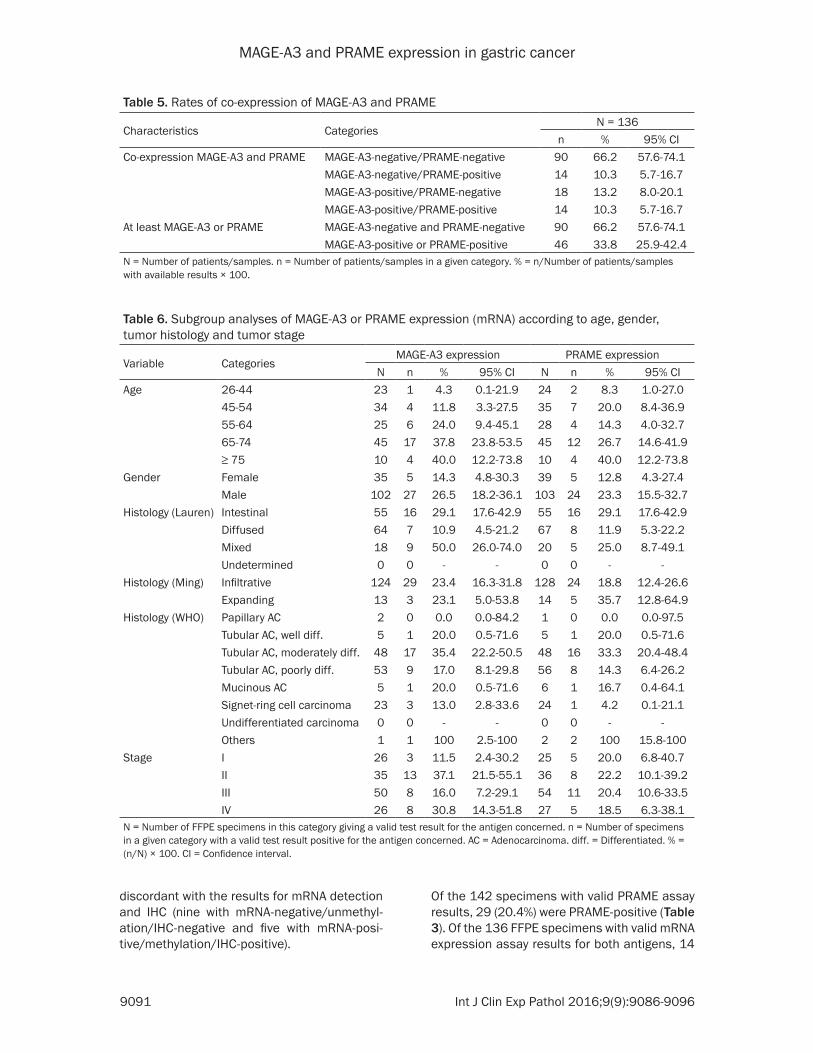
MAGE-A3 and PRAME expression in gastric cancer
9091 Int J Clin Exp Pathol 20169(9)9086-9096
Table 5 Rates of co-expression of MAGE-A3 and PRAME
Characteristics CategoriesN = 136
n 95 CICo-expression MAGE-A3 and PRAME MAGE-A3-negativePRAME-negative 90 662 576-741
MAGE-A3-negativePRAME-positive 14 103 57-167MAGE-A3-positivePRAME-negative 18 132 80-201MAGE-A3-positivePRAME-positive 14 103 57-167
At least MAGE-A3 or PRAME MAGE-A3-negative and PRAME-negative 90 662 576-741MAGE-A3-positive or PRAME-positive 46 338 259-424
N = Number of patientssamples n = Number of patientssamples in a given category = nNumber of patientssamples with available results times 100
Table 6 Subgroup analyses of MAGE-A3 or PRAME expression (mRNA) according to age gender tumor histology and tumor stage
Variable CategoriesMAGE-A3 expression PRAME expression
N n 95 CI N n 95 CIAge 26-44 23 1 43 01-219 24 2 83 10-270
45-54 34 4 118 33-275 35 7 200 84-36955-64 25 6 240 94-451 28 4 143 40-32765-74 45 17 378 238-535 45 12 267 146-419ge 75 10 4 400 122-738 10 4 400 122-738
Gender Female 35 5 143 48-303 39 5 128 43-274Male 102 27 265 182-361 103 24 233 155-327
Histology (Lauren) Intestinal 55 16 291 176-429 55 16 291 176-429Diffused 64 7 109 45-212 67 8 119 53-222Mixed 18 9 500 260-740 20 5 250 87-491Undetermined 0 0 - - 0 0 - -
Histology (Ming) Infiltrative 124 29 234 163-318 128 24 188 124-266Expanding 13 3 231 50-538 14 5 357 128-649
Histology (WHO) Papillary AC 2 0 00 00-842 1 0 00 00-975Tubular AC well diff 5 1 200 05-716 5 1 200 05-716Tubular AC moderately diff 48 17 354 222-505 48 16 333 204-484Tubular AC poorly diff 53 9 170 81-298 56 8 143 64-262Mucinous AC 5 1 200 05-716 6 1 167 04-641Signet-ring cell carcinoma 23 3 130 28-336 24 1 42 01-211Undifferentiated carcinoma 0 0 - - 0 0 - -Others 1 1 100 25-100 2 2 100 158-100
Stage I 26 3 115 24-302 25 5 200 68-407II 35 13 371 215-551 36 8 222 101-392III 50 8 160 72-291 54 11 204 106-335IV 26 8 308 143-518 27 5 185 63-381
N = Number of FFPE specimens in this category giving a valid test result for the antigen concerned n = Number of specimens in a given category with a valid test result positive for the antigen concerned AC = Adenocarcinoma diff = Differentiated = (nN) times 100 CI = Confidence interval
discordant with the results for mRNA detection and IHC (nine with mRNA-negativeunmethyl-ationIHC-negative and five with mRNA-posi-tivemethylationIHC-positive)
Of the 142 specimens with valid PRAME assay results 29 (204) were PRAME-positive (Table 3) Of the 136 FFPE specimens with valid mRNA expression assay results for both antigens 14
MAGE-A3 and PRAME expression in gastric cancer
9092 Int J Clin Exp Pathol 20169(9)9086-9096
(103) expressed both while 46 (338) expressed at least one of them (Table 5)
The exploratory analyses of the mRNA ex- pression of the TAAs in subgroups determin- ed by patient and tumor characteristics show- ed similar results for MAGE-A3 and PRAME (Table 6) TAA mRNA expression was indepen-dent of tumor stage and there was no obvious association between tumor TAA mRNA expre- ssion and any of the other clinico-pathological factors investigated
Another study investigated MAGE-A express- ion in GC cell lines as well as primary gastric tumor specimens using both IHC and analy- sis of MAGE-A3 promoter methylation status for detection Of 1097 cancer tissues 158 expressed MAGE-A protein with no distinction between the subtypes of MAGE-A by the anti-body used for the IHC Of 52 randomly selected tumors expressing MAGE-A protein 28 (538) displayed unmethylation of the MAGE-A3 pro-moter [24] In the present study 20 of the 29 (690 recalculated from Table 4) tumors
Figure 2 Kaplan-Meier curves for OS by MAGE-A3 methylation status of the specimen HR for MAGE-A3 unmethylated 094 (95 CI 057-156 P = 082) (Wald test from Cox regression model)
Figure 3 Kaplan-Meier curves for OS by MAGE-A protein expression of the specimen HR for MAGE-A3-positive 146 (95 CI 092-232 P = 011) (Wald test from Cox regression model)
Overall survival by MAGE-A3 methylation status protein expression of MAGE-A and mRNA expression of the an-tigens
Kaplan-Meier curves for OS according to the tumorrsquos MAGE-A3 methylation status (Figure 2) overlap almost completely The Kaplan-Meier curves for OS according to the specimensrsquo expression of MAGE-A3 protein are more separated (Figure 3) but are not statistically significant- ly different (P = 011) Like- wise there were no indica-tions of an association be- tween OS and mRNA expres-sion of the antigens neither for MAGE-A3 nor for PRAME (Figures 4 and 5)
Discussion
This is the first study of PRAME expression in GC while mRNA MAGE-A3 expre- ssion has been reported in several previous studies with rates of MAGE-A3-positive GC tumors varying between 30 and 416 [20-23] None of these studies reported sta- tistically significant associa-tions between clinico-patho-logical tumor characteristics and mRNA expression of MAGE-A3 in line with findings in the current study
MAGE-A3 and PRAME expression in gastric cancer
9093 Int J Clin Exp Pathol 20169(9)9086-9096
expressing the MAGE-A protein displayed un- methylation of the MAGE-A3 promoter where- as this was displayed by 14 of the 107 (131) tumors not expressing the MAGE-A protein These results indicate that unmethylation of the MAGE-A3 promoter may be (partly) respon-sible for the expression of the MAGE-A pro- tein in gastric cancer
Honda et al [13] detected MAGE-A3 promoter demethylation in 66 of 84 GC specimens tumors displaying MAGE-A3 promoter demeth-
Honda et al [13] described a trend for worse survival for patients with tumors displaying demethylated MAGE-A3 in particular if their tumor also showed demethylated MAGE-A1 (P = 018)
Circulating tumor cell DNA can be detected in serum or blood even at early stages of the dis-ease as shown by Kounalakos et al [25] for melanoma patients This DNA may be derived from lysed cells of the primary tumor or from circulating tumor cells releasing their DNA With
Figure 4 Kaplan-Meier curves for overall survival by mRNA expression of MAGE-A3 (Total cohort) HR for MAGE-A3-positive 103 (95 CI 0538-1963 P = 093) (Wald test from Cox regression model)
Figure 5 Kaplan-Meier curves for overall survival by mRNA expression of PRAME (Total cohort) HR for PRAME-positive 097 (95 CI 0499-1885 P = 093) (Wald test from Cox regression model)
ylation were in a more ad- vanced state and were asso-ciated with a higher preva-lence of lymph-node metas- tases than those without de- methylation In the present study 230 of gastric tu- mors displayed an unmethyl-ated MAGE-A3 promoter
We did not observe any rela-tion between OS and mRNA TAA expression of the FFPE specimen for either MAGE-A3 or PRAME Similarly there was no statistically significant association between OS and MAGE-A protein expression (P = 011) In contrast Jung et al [24] reported a significant-ly worse prognosis for patients with tumors expressing the MAGE-A proteins (P lt 0001)
Recent studies investigating the molecular basis of GC have reported associations between aberrant DNA meth-ylation of several gene pro-moters and the prognosis for GC patients generally sug-gesting that hypermethylation of the identified gene promot-ers is an indicator of poor prognosis (reviewed in [15]) Our observations indicate that MAGE-A3 is not to be added to this list of genes as the methylation status of the MAGE-A3 promoter had no association with patient OS (P = 082) In contrast
MAGE-A3 and PRAME expression in gastric cancer
9094 Int J Clin Exp Pathol 20169(9)9086-9096
a prior understanding of a potential correlation between gene promoter methylation status and gene expression methylation testing of cir-culating DNA could be useful in detecting the expression of specific genes in tumor cells Such non-invasive detection methods that are not dependent on the availability of tumor tissue could increase the number of patients that can be screened for expression of tumor antigens
A rough assessment of the possibility of rely- ing on such non-invasive methods for gene expression screening based on the observa-tions of the present study was performed Due to the lack of MAGE-A3 specific antibodies IHC cannot distinguish MAGE-A3 from other members of the MAGE-A family MAGE-A3 ex- pression was thus generally screened for us- ing RT-PCR assays Here we found that 867 (91105) of the FFPE specimens with no mRNA MAGE-A3 expression showed methylation of the MAGE-A3 promoter whereas 645 (20 31) of specimens with mRNA MAGE-A3 expre- ssion were unmethylated in the MAGE-A3 pro-moter Arguably the concordance between the results obtained with the two methods may be considered too low to rely on testing of MAGE-A3 promoter methylation status for pa- tient screening According to these observa-tions screening based on methylation testing of the MAGE-A3 promoter would miss more than a third of the patients whose tumors show mRNA MAGE-A3 expression
The overall concordance between the three MAGE-A3 assays was relatively low compared to previous findings (75 vs nearly 100) reported in certain studies (eg [26]) The qRT-PCR assay is specific for MAGE-A3 and the cut-off value was defined as 1 of the GERL con-trol By contrast the IHC assay could also detect other members of the MAGE-A family (in particular MAGE-A6) and the cut-off values for these assays are linked to their detection lim-its Additional investigations are needed to assess the impact of MAGE-A3A6 expression and to homogenize the cut-off values of the assays
In summary the rate of mRNA expression of each of these TAAs in tumors from GC patients was found to be relatively low with 234 expressing MAGE-A3 204 expressing PRAME and 338 expressing at least one of them Neither MAGE-A3 nor PRAME would se-
rve as a prognostic biomarker for OS of GC patients
Acknowledgements
Niels Neymark provided scientific writing ser-vices and Veacuteronique Duquenne (XPE PHAR- MA and SCIENCES Wavre Belgium CO GSK) coordinated the manuscriptrsquos development This study was funded and coordinated by GlaxoSmithKline Biologicals SA GSK was in- volved in the design and conduct of the study collection management analysis and inter- pretation of the data and preparation and review of the manuscript
Disclosure of conflict of interest
Olivier Gruselle and Bart Spiessens are employ-ees of the GSK group of companies An de Creus Nicole Kusuma and Aung Myo were employees of the GSK group of companies at the time of the study and manuscript develop-ment Bart Spiessens own stockstock options in GSK group of companies Woo Ho Kim Eun Ji Jung and Kim Hee Sung have no conflict of interest
Authorsrsquo contribution
BS AM AdeC and EJJ were involved in study design AM AdeC NK were involved in study supervision OG AM AdeC NK WHK EJJ were involved in data acquisition HSK EJJ NK AM WHK and OG were involved in data collection OG AM NK and WHK were involved in data extraction and quality check (AdeC in quality check only and EJJ in data extraction only) OG WHK and EJJ were involved in provision of study material WHK and EJJ were involved in labora-tory testing BS (biostatistician) OG AM AdeC WHK and HSK performed or supervised the analyses BS OG AM AdC NK WHK and HSK were involved in results interpretation OG AdeC NK EJJ were involved in administrativetechnicallogistic support EJJ and AM were involved in centers recruitment AM and NK were involved in center coordination AM was involved in acquisition of funding
Address correspondence to Dr Woo Ho Kim De- partment of Pathology Seoul National University College of Medicine 28 Yeongeon-dong 110-799 Seoul Korea Tel +82 8 740 8269 Fax +82 2 765 5600 E-mail woohokimsnuackr
MAGE-A3 and PRAME expression in gastric cancer
9095 Int J Clin Exp Pathol 20169(9)9086-9096
References
[1] Crew KD Neugut AI The epidemiology of gas-tric cancer World J Gastroenterol 2006 12 354-62
[2] Toomey PG Vohra NA Ghansah T Sarnaik AA Pilon-Thomas SA Immunotherapy for gastroin-testinal malignancies Cancer Control 2013 20 32-42
[3] Bilici A Treatment options in patients with met-astatic gastric cancer Current status and fu-ture perspective World J Gastroenterol 2014 20 3905-15
[4] Amadei A Benagiano M della Bella C Nicolai E DrsquoElios MM Novel immunotherapeutic strat-egies of gastric cancer treatment J Biomed Biotech 2011 2011 437348
[5] Ikeda H Sato N Matsuura A Kikuchi K Analysis of T-cell receptor V region gene usage of cytotoxic T-lymphocytes and tumor-infiltrat-ing lymphocytes derived from human autolo-gous gastric signet ring cell carcinomas Cancer Res 1993 53 3078-84
[6] Kono K Ichihara F Lizuka H Sekikawa T Matsumoto Y Differences in the recognition of tumor-specific CD8+ T cells derived from solid tumor metastatic lymph nodes and ascites in patients with gastric cancer Int J Cancer 1997 71 978-81
[7] Amadei A Niccolai E Della Bella C Cianchi F Trallori G Benagiano M Bencini L Bernini M Farsi M Moretti R Del Prete G DrsquoElios MM Characterization of tumor antigen peptide-spe-cific T cells isolated from the neoplastic tissue of patients with gastric adenocarcinomas Cancer Immunol Immunother 2009 58 1819-30
[8] Lee HE Chae SW Lee YJ Kim MA Lee HS Lee BL Kim WH Prognostic implications of type and density of tumour-infiltrating lymp- hocytes in gastric cancer Br J Cancer 2008 99 1704-11
[9] Perrone G Ruffini PA Catalano V Spino C Santini D Muretto P Spoto C Zingaretti C Sisti V Alessandroni P Giordani P Cicetti A DrsquoEmidio S Morini S Ruzzo A Magnani M Tonini G Rabitti C Graziano F Intratumoural FOXP3-positive regulatory T cells are associat-ed with adverse prognosis in radically resected gastric cancer Eur J Cancer 2008 44 1875-82
[10] Shen Z Zhou S Wang Y Li RL Zhong C Liang C Sun Y Higher intratumoural infiltrated Foxp3+ Treg numbers and Foxp3+CD8+ ratio are associated with adverse prognosis in re-sectable gastric cancer J Cancer Res Clin Oncol 2010 136 1585-95
[11] Zhou S Shen Z Wang Y Ma H Xu S Qin J Chen L Tao H Zhen Z Chen G Zhang Z Li R
Xiao H Zhong C Yang Y Liang C CCR7 expres-sion and intratumoural FOXP3+ regulatory T cells are correlated with overall survival and lymph node metastatis in gastric cancer PLoS One 2013 8 e74430
[12] Matsueda S Graham DY Immunotherapy in gastric cancer World J Gatroenterol 2014 20 1657-66
[13] Honda T Tamura G Waki T Kawata S Tera- shima M Nishizuka S Motoyama T Deme- thylation of MAGE promoters during gastric cancer progression Br J Cancer 2004 90 838-43
[14] Kim KH Choi JS Kim IJ Ku JL Park JG Promoter hypomethylation and reactivation of MAGE-A1 and MAGE-A3 genes in colorectal cancer cell lines and cancer tissues World J Gastroenterol 2006 12 5651-57
[15] Nakamura J Tanaka T Kitajima Y Noshiro H Miyazaki K Methylation-mediated gene silenc-ing as biomarkers of gastric cancer A review World J Gastroenterology 2014 20 11991-12006
[16] Oberthuer A Hero B Spitz R Berthold F Fischer M The tumor-associated antigen PRA- ME is universally expressed in high-stage neu-roblastoma and associated with poor out- come Clin Cancer Res 2004 10 4307-13
[17] Partheen K Levan K Osterberg L Claesson I Fallenius G Sundfeldt K Horvath G Four po-tential biomarkers as prognostic factors in stage III serous ovarian adenocarcinomas Int J Cancer 2008 123 2130-7
[18] Epping MT Hart AA Glas AM Krijgsman O Bernards R PRAME expression and clinical outcome of breast cancer Br J Cancer 2008 99 398-403
[19] Lerut E Van Poppel H Joniau S Gruselle O Coche T Therasse P Rates of MAGE-A3 and PRAME expressing tumors in FFPE tissue spec-imens from bladder cancer patients potential targets for antigen-specific cancer immuno-therapeutics Int J Clin Exp Pathol 2015 8 9522-32
[20] Inoue H Mori M Honda M Li J Shibuta K Mimori K Ueo H Akiyoshi T The expression of tumor-rejection antigen ldquoMAGErdquo genes in human gastric carcinoma Gastroenterology 1995 109 1522-25
[21] Li J Yang Y Fujie T Baba K Ueo H Mori M Akiyoshi T Expression of BAGE GAGE and MAGE genes in human gastric carcinoma Clin Cancer Res 1996 2 1619-25
[22] Wang Y Wu XJ Zhao AL Yuan YH Chen YT Jungbluth AA Gnjatic S Santiago D Ritter G Chen WF Old LJ Ji JF Cancertestis antigen expression and autologous humoral immunity to NY-ESO-1 in gastric cancer Cancer Immun 2004 4 11
MAGE-A3 and PRAME expression in gastric cancer
9096 Int J Clin Exp Pathol 20169(9)9086-9096
[23] Wang WX Li YB Xie XL Shu XL Ouyang XH Detection of tumor cells in peripheral blood of patients with gastric cancer using mRNA of MAGE genes as markers Zhonghua Wei Chang Wai Ke Za Zhi 2009 12 611-14
[24] Jung EJ Kim MA Lee HS Yang HK Lee YM Lee BL Kim WH Expression of family A mela-noma antigen in human gastric carcinoma Anticancer Research 2005 25 2105-12
[25] Kounalakos N Goydos JS Tumor cells and cir-culating markers in melanoma diagnosis prognosis and management Curr Oncol Rep 2005 7 377-382
[26] Lehmann-Che J Amira-Bouhidel F Turpin E Antoine M Soliman H Legres L Bocquet C Bernoud R Flandre E Varna M de Roquan- court A Plassa LF Giacchetti S Espieacute M de Bazelaire C Cahen-Doidy L Bourstyn E Janin A de Theacute H Bertheau P Immunohistoche- mical and molecular analyses of HER2 sta- tus in breast cancers are highly concordant and complementary approaches Br J Cancer 2011 104 1739-46
MAGE-A3 and PRAME expression in gastric cancer
1
Table S1 Primer sequences for methylation specific PCRMethylation status Type SequenceUnmethylated Anti-sense 5rsquo-TGTTAGGATGTGATGTTATTGATTTGT-3rsquo
Sense 5rsquo-CCTCACCAAACCTAAACCAA-3rsquoMethylated Anti-sense 5rsquo-CCATCTGACGTTATTGATTTGC-3rsquo
Sense 5rsquo-CTCACCGAACCTAAACCGAC-3rsquo
Table S2 Distribution of immunohistochemistry MAGE-A3 results in gastric cancerIHC score 0-50 51-100 101-150 151-200 201-250 251-300 Total of cases 201 19 8 4 3 19 250 of total 804 76 32 16 12 76 100IHC result (Score gt 50) Negative Positive
804 196
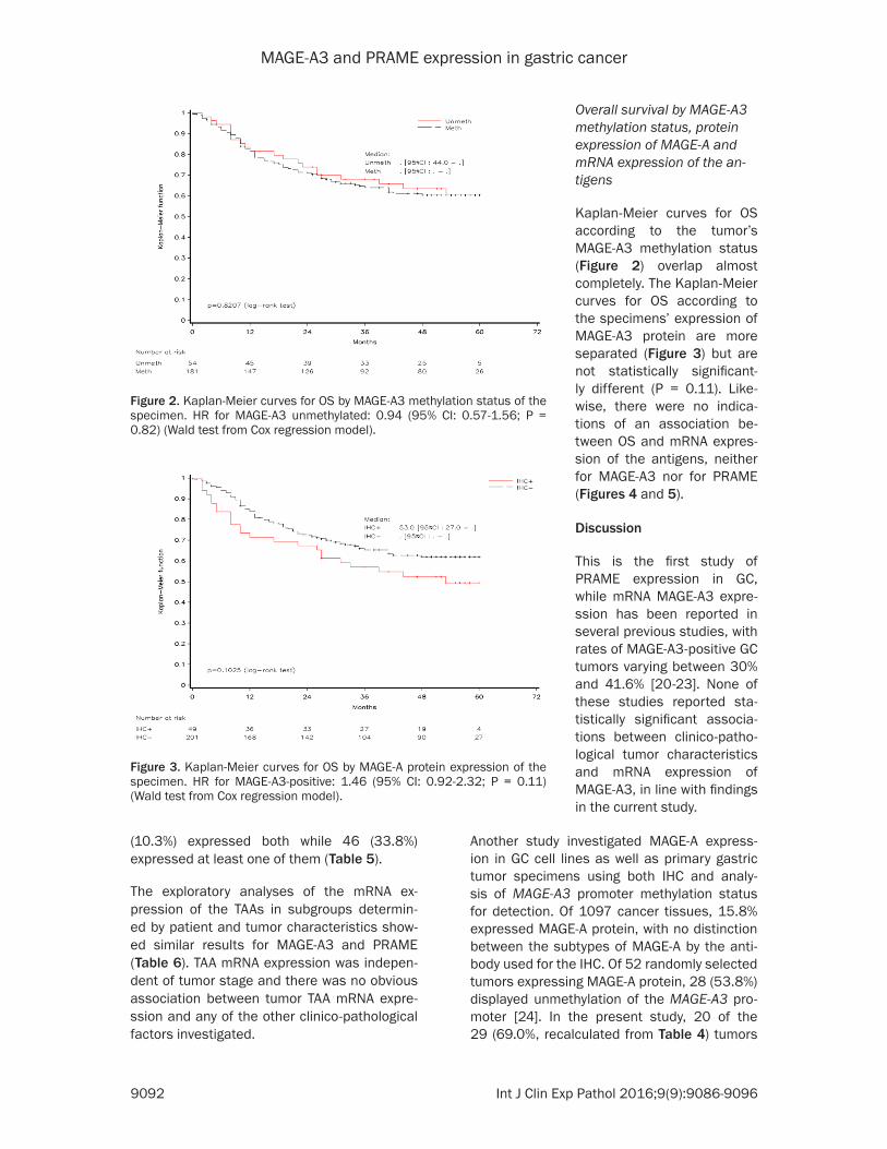
MAGE-A3 and PRAME expression in gastric cancer
9092 Int J Clin Exp Pathol 20169(9)9086-9096
(103) expressed both while 46 (338) expressed at least one of them (Table 5)
The exploratory analyses of the mRNA ex- pression of the TAAs in subgroups determin- ed by patient and tumor characteristics show- ed similar results for MAGE-A3 and PRAME (Table 6) TAA mRNA expression was indepen-dent of tumor stage and there was no obvious association between tumor TAA mRNA expre- ssion and any of the other clinico-pathological factors investigated
Another study investigated MAGE-A express- ion in GC cell lines as well as primary gastric tumor specimens using both IHC and analy- sis of MAGE-A3 promoter methylation status for detection Of 1097 cancer tissues 158 expressed MAGE-A protein with no distinction between the subtypes of MAGE-A by the anti-body used for the IHC Of 52 randomly selected tumors expressing MAGE-A protein 28 (538) displayed unmethylation of the MAGE-A3 pro-moter [24] In the present study 20 of the 29 (690 recalculated from Table 4) tumors
Figure 2 Kaplan-Meier curves for OS by MAGE-A3 methylation status of the specimen HR for MAGE-A3 unmethylated 094 (95 CI 057-156 P = 082) (Wald test from Cox regression model)
Figure 3 Kaplan-Meier curves for OS by MAGE-A protein expression of the specimen HR for MAGE-A3-positive 146 (95 CI 092-232 P = 011) (Wald test from Cox regression model)
Overall survival by MAGE-A3 methylation status protein expression of MAGE-A and mRNA expression of the an-tigens
Kaplan-Meier curves for OS according to the tumorrsquos MAGE-A3 methylation status (Figure 2) overlap almost completely The Kaplan-Meier curves for OS according to the specimensrsquo expression of MAGE-A3 protein are more separated (Figure 3) but are not statistically significant- ly different (P = 011) Like- wise there were no indica-tions of an association be- tween OS and mRNA expres-sion of the antigens neither for MAGE-A3 nor for PRAME (Figures 4 and 5)
Discussion
This is the first study of PRAME expression in GC while mRNA MAGE-A3 expre- ssion has been reported in several previous studies with rates of MAGE-A3-positive GC tumors varying between 30 and 416 [20-23] None of these studies reported sta- tistically significant associa-tions between clinico-patho-logical tumor characteristics and mRNA expression of MAGE-A3 in line with findings in the current study
MAGE-A3 and PRAME expression in gastric cancer
9093 Int J Clin Exp Pathol 20169(9)9086-9096
expressing the MAGE-A protein displayed un- methylation of the MAGE-A3 promoter where- as this was displayed by 14 of the 107 (131) tumors not expressing the MAGE-A protein These results indicate that unmethylation of the MAGE-A3 promoter may be (partly) respon-sible for the expression of the MAGE-A pro- tein in gastric cancer
Honda et al [13] detected MAGE-A3 promoter demethylation in 66 of 84 GC specimens tumors displaying MAGE-A3 promoter demeth-
Honda et al [13] described a trend for worse survival for patients with tumors displaying demethylated MAGE-A3 in particular if their tumor also showed demethylated MAGE-A1 (P = 018)
Circulating tumor cell DNA can be detected in serum or blood even at early stages of the dis-ease as shown by Kounalakos et al [25] for melanoma patients This DNA may be derived from lysed cells of the primary tumor or from circulating tumor cells releasing their DNA With
Figure 4 Kaplan-Meier curves for overall survival by mRNA expression of MAGE-A3 (Total cohort) HR for MAGE-A3-positive 103 (95 CI 0538-1963 P = 093) (Wald test from Cox regression model)
Figure 5 Kaplan-Meier curves for overall survival by mRNA expression of PRAME (Total cohort) HR for PRAME-positive 097 (95 CI 0499-1885 P = 093) (Wald test from Cox regression model)
ylation were in a more ad- vanced state and were asso-ciated with a higher preva-lence of lymph-node metas- tases than those without de- methylation In the present study 230 of gastric tu- mors displayed an unmethyl-ated MAGE-A3 promoter
We did not observe any rela-tion between OS and mRNA TAA expression of the FFPE specimen for either MAGE-A3 or PRAME Similarly there was no statistically significant association between OS and MAGE-A protein expression (P = 011) In contrast Jung et al [24] reported a significant-ly worse prognosis for patients with tumors expressing the MAGE-A proteins (P lt 0001)
Recent studies investigating the molecular basis of GC have reported associations between aberrant DNA meth-ylation of several gene pro-moters and the prognosis for GC patients generally sug-gesting that hypermethylation of the identified gene promot-ers is an indicator of poor prognosis (reviewed in [15]) Our observations indicate that MAGE-A3 is not to be added to this list of genes as the methylation status of the MAGE-A3 promoter had no association with patient OS (P = 082) In contrast
MAGE-A3 and PRAME expression in gastric cancer
9094 Int J Clin Exp Pathol 20169(9)9086-9096
a prior understanding of a potential correlation between gene promoter methylation status and gene expression methylation testing of cir-culating DNA could be useful in detecting the expression of specific genes in tumor cells Such non-invasive detection methods that are not dependent on the availability of tumor tissue could increase the number of patients that can be screened for expression of tumor antigens
A rough assessment of the possibility of rely- ing on such non-invasive methods for gene expression screening based on the observa-tions of the present study was performed Due to the lack of MAGE-A3 specific antibodies IHC cannot distinguish MAGE-A3 from other members of the MAGE-A family MAGE-A3 ex- pression was thus generally screened for us- ing RT-PCR assays Here we found that 867 (91105) of the FFPE specimens with no mRNA MAGE-A3 expression showed methylation of the MAGE-A3 promoter whereas 645 (20 31) of specimens with mRNA MAGE-A3 expre- ssion were unmethylated in the MAGE-A3 pro-moter Arguably the concordance between the results obtained with the two methods may be considered too low to rely on testing of MAGE-A3 promoter methylation status for pa- tient screening According to these observa-tions screening based on methylation testing of the MAGE-A3 promoter would miss more than a third of the patients whose tumors show mRNA MAGE-A3 expression
The overall concordance between the three MAGE-A3 assays was relatively low compared to previous findings (75 vs nearly 100) reported in certain studies (eg [26]) The qRT-PCR assay is specific for MAGE-A3 and the cut-off value was defined as 1 of the GERL con-trol By contrast the IHC assay could also detect other members of the MAGE-A family (in particular MAGE-A6) and the cut-off values for these assays are linked to their detection lim-its Additional investigations are needed to assess the impact of MAGE-A3A6 expression and to homogenize the cut-off values of the assays
In summary the rate of mRNA expression of each of these TAAs in tumors from GC patients was found to be relatively low with 234 expressing MAGE-A3 204 expressing PRAME and 338 expressing at least one of them Neither MAGE-A3 nor PRAME would se-
rve as a prognostic biomarker for OS of GC patients
Acknowledgements
Niels Neymark provided scientific writing ser-vices and Veacuteronique Duquenne (XPE PHAR- MA and SCIENCES Wavre Belgium CO GSK) coordinated the manuscriptrsquos development This study was funded and coordinated by GlaxoSmithKline Biologicals SA GSK was in- volved in the design and conduct of the study collection management analysis and inter- pretation of the data and preparation and review of the manuscript
Disclosure of conflict of interest
Olivier Gruselle and Bart Spiessens are employ-ees of the GSK group of companies An de Creus Nicole Kusuma and Aung Myo were employees of the GSK group of companies at the time of the study and manuscript develop-ment Bart Spiessens own stockstock options in GSK group of companies Woo Ho Kim Eun Ji Jung and Kim Hee Sung have no conflict of interest
Authorsrsquo contribution
BS AM AdeC and EJJ were involved in study design AM AdeC NK were involved in study supervision OG AM AdeC NK WHK EJJ were involved in data acquisition HSK EJJ NK AM WHK and OG were involved in data collection OG AM NK and WHK were involved in data extraction and quality check (AdeC in quality check only and EJJ in data extraction only) OG WHK and EJJ were involved in provision of study material WHK and EJJ were involved in labora-tory testing BS (biostatistician) OG AM AdeC WHK and HSK performed or supervised the analyses BS OG AM AdC NK WHK and HSK were involved in results interpretation OG AdeC NK EJJ were involved in administrativetechnicallogistic support EJJ and AM were involved in centers recruitment AM and NK were involved in center coordination AM was involved in acquisition of funding
Address correspondence to Dr Woo Ho Kim De- partment of Pathology Seoul National University College of Medicine 28 Yeongeon-dong 110-799 Seoul Korea Tel +82 8 740 8269 Fax +82 2 765 5600 E-mail woohokimsnuackr
MAGE-A3 and PRAME expression in gastric cancer
9095 Int J Clin Exp Pathol 20169(9)9086-9096
References
[1] Crew KD Neugut AI The epidemiology of gas-tric cancer World J Gastroenterol 2006 12 354-62
[2] Toomey PG Vohra NA Ghansah T Sarnaik AA Pilon-Thomas SA Immunotherapy for gastroin-testinal malignancies Cancer Control 2013 20 32-42
[3] Bilici A Treatment options in patients with met-astatic gastric cancer Current status and fu-ture perspective World J Gastroenterol 2014 20 3905-15
[4] Amadei A Benagiano M della Bella C Nicolai E DrsquoElios MM Novel immunotherapeutic strat-egies of gastric cancer treatment J Biomed Biotech 2011 2011 437348
[5] Ikeda H Sato N Matsuura A Kikuchi K Analysis of T-cell receptor V region gene usage of cytotoxic T-lymphocytes and tumor-infiltrat-ing lymphocytes derived from human autolo-gous gastric signet ring cell carcinomas Cancer Res 1993 53 3078-84
[6] Kono K Ichihara F Lizuka H Sekikawa T Matsumoto Y Differences in the recognition of tumor-specific CD8+ T cells derived from solid tumor metastatic lymph nodes and ascites in patients with gastric cancer Int J Cancer 1997 71 978-81
[7] Amadei A Niccolai E Della Bella C Cianchi F Trallori G Benagiano M Bencini L Bernini M Farsi M Moretti R Del Prete G DrsquoElios MM Characterization of tumor antigen peptide-spe-cific T cells isolated from the neoplastic tissue of patients with gastric adenocarcinomas Cancer Immunol Immunother 2009 58 1819-30
[8] Lee HE Chae SW Lee YJ Kim MA Lee HS Lee BL Kim WH Prognostic implications of type and density of tumour-infiltrating lymp- hocytes in gastric cancer Br J Cancer 2008 99 1704-11
[9] Perrone G Ruffini PA Catalano V Spino C Santini D Muretto P Spoto C Zingaretti C Sisti V Alessandroni P Giordani P Cicetti A DrsquoEmidio S Morini S Ruzzo A Magnani M Tonini G Rabitti C Graziano F Intratumoural FOXP3-positive regulatory T cells are associat-ed with adverse prognosis in radically resected gastric cancer Eur J Cancer 2008 44 1875-82
[10] Shen Z Zhou S Wang Y Li RL Zhong C Liang C Sun Y Higher intratumoural infiltrated Foxp3+ Treg numbers and Foxp3+CD8+ ratio are associated with adverse prognosis in re-sectable gastric cancer J Cancer Res Clin Oncol 2010 136 1585-95
[11] Zhou S Shen Z Wang Y Ma H Xu S Qin J Chen L Tao H Zhen Z Chen G Zhang Z Li R
Xiao H Zhong C Yang Y Liang C CCR7 expres-sion and intratumoural FOXP3+ regulatory T cells are correlated with overall survival and lymph node metastatis in gastric cancer PLoS One 2013 8 e74430
[12] Matsueda S Graham DY Immunotherapy in gastric cancer World J Gatroenterol 2014 20 1657-66
[13] Honda T Tamura G Waki T Kawata S Tera- shima M Nishizuka S Motoyama T Deme- thylation of MAGE promoters during gastric cancer progression Br J Cancer 2004 90 838-43
[14] Kim KH Choi JS Kim IJ Ku JL Park JG Promoter hypomethylation and reactivation of MAGE-A1 and MAGE-A3 genes in colorectal cancer cell lines and cancer tissues World J Gastroenterol 2006 12 5651-57
[15] Nakamura J Tanaka T Kitajima Y Noshiro H Miyazaki K Methylation-mediated gene silenc-ing as biomarkers of gastric cancer A review World J Gastroenterology 2014 20 11991-12006
[16] Oberthuer A Hero B Spitz R Berthold F Fischer M The tumor-associated antigen PRA- ME is universally expressed in high-stage neu-roblastoma and associated with poor out- come Clin Cancer Res 2004 10 4307-13
[17] Partheen K Levan K Osterberg L Claesson I Fallenius G Sundfeldt K Horvath G Four po-tential biomarkers as prognostic factors in stage III serous ovarian adenocarcinomas Int J Cancer 2008 123 2130-7
[18] Epping MT Hart AA Glas AM Krijgsman O Bernards R PRAME expression and clinical outcome of breast cancer Br J Cancer 2008 99 398-403
[19] Lerut E Van Poppel H Joniau S Gruselle O Coche T Therasse P Rates of MAGE-A3 and PRAME expressing tumors in FFPE tissue spec-imens from bladder cancer patients potential targets for antigen-specific cancer immuno-therapeutics Int J Clin Exp Pathol 2015 8 9522-32
[20] Inoue H Mori M Honda M Li J Shibuta K Mimori K Ueo H Akiyoshi T The expression of tumor-rejection antigen ldquoMAGErdquo genes in human gastric carcinoma Gastroenterology 1995 109 1522-25
[21] Li J Yang Y Fujie T Baba K Ueo H Mori M Akiyoshi T Expression of BAGE GAGE and MAGE genes in human gastric carcinoma Clin Cancer Res 1996 2 1619-25
[22] Wang Y Wu XJ Zhao AL Yuan YH Chen YT Jungbluth AA Gnjatic S Santiago D Ritter G Chen WF Old LJ Ji JF Cancertestis antigen expression and autologous humoral immunity to NY-ESO-1 in gastric cancer Cancer Immun 2004 4 11
MAGE-A3 and PRAME expression in gastric cancer
9096 Int J Clin Exp Pathol 20169(9)9086-9096
[23] Wang WX Li YB Xie XL Shu XL Ouyang XH Detection of tumor cells in peripheral blood of patients with gastric cancer using mRNA of MAGE genes as markers Zhonghua Wei Chang Wai Ke Za Zhi 2009 12 611-14
[24] Jung EJ Kim MA Lee HS Yang HK Lee YM Lee BL Kim WH Expression of family A mela-noma antigen in human gastric carcinoma Anticancer Research 2005 25 2105-12
[25] Kounalakos N Goydos JS Tumor cells and cir-culating markers in melanoma diagnosis prognosis and management Curr Oncol Rep 2005 7 377-382
[26] Lehmann-Che J Amira-Bouhidel F Turpin E Antoine M Soliman H Legres L Bocquet C Bernoud R Flandre E Varna M de Roquan- court A Plassa LF Giacchetti S Espieacute M de Bazelaire C Cahen-Doidy L Bourstyn E Janin A de Theacute H Bertheau P Immunohistoche- mical and molecular analyses of HER2 sta- tus in breast cancers are highly concordant and complementary approaches Br J Cancer 2011 104 1739-46
MAGE-A3 and PRAME expression in gastric cancer
1
Table S1 Primer sequences for methylation specific PCRMethylation status Type SequenceUnmethylated Anti-sense 5rsquo-TGTTAGGATGTGATGTTATTGATTTGT-3rsquo
Sense 5rsquo-CCTCACCAAACCTAAACCAA-3rsquoMethylated Anti-sense 5rsquo-CCATCTGACGTTATTGATTTGC-3rsquo
Sense 5rsquo-CTCACCGAACCTAAACCGAC-3rsquo
Table S2 Distribution of immunohistochemistry MAGE-A3 results in gastric cancerIHC score 0-50 51-100 101-150 151-200 201-250 251-300 Total of cases 201 19 8 4 3 19 250 of total 804 76 32 16 12 76 100IHC result (Score gt 50) Negative Positive
804 196
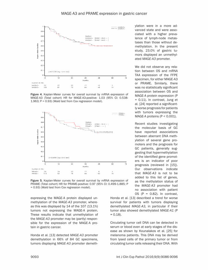
MAGE-A3 and PRAME expression in gastric cancer
9093 Int J Clin Exp Pathol 20169(9)9086-9096
expressing the MAGE-A protein displayed un- methylation of the MAGE-A3 promoter where- as this was displayed by 14 of the 107 (131) tumors not expressing the MAGE-A protein These results indicate that unmethylation of the MAGE-A3 promoter may be (partly) respon-sible for the expression of the MAGE-A pro- tein in gastric cancer
Honda et al [13] detected MAGE-A3 promoter demethylation in 66 of 84 GC specimens tumors displaying MAGE-A3 promoter demeth-
Honda et al [13] described a trend for worse survival for patients with tumors displaying demethylated MAGE-A3 in particular if their tumor also showed demethylated MAGE-A1 (P = 018)
Circulating tumor cell DNA can be detected in serum or blood even at early stages of the dis-ease as shown by Kounalakos et al [25] for melanoma patients This DNA may be derived from lysed cells of the primary tumor or from circulating tumor cells releasing their DNA With
Figure 4 Kaplan-Meier curves for overall survival by mRNA expression of MAGE-A3 (Total cohort) HR for MAGE-A3-positive 103 (95 CI 0538-1963 P = 093) (Wald test from Cox regression model)
Figure 5 Kaplan-Meier curves for overall survival by mRNA expression of PRAME (Total cohort) HR for PRAME-positive 097 (95 CI 0499-1885 P = 093) (Wald test from Cox regression model)
ylation were in a more ad- vanced state and were asso-ciated with a higher preva-lence of lymph-node metas- tases than those without de- methylation In the present study 230 of gastric tu- mors displayed an unmethyl-ated MAGE-A3 promoter
We did not observe any rela-tion between OS and mRNA TAA expression of the FFPE specimen for either MAGE-A3 or PRAME Similarly there was no statistically significant association between OS and MAGE-A protein expression (P = 011) In contrast Jung et al [24] reported a significant-ly worse prognosis for patients with tumors expressing the MAGE-A proteins (P lt 0001)
Recent studies investigating the molecular basis of GC have reported associations between aberrant DNA meth-ylation of several gene pro-moters and the prognosis for GC patients generally sug-gesting that hypermethylation of the identified gene promot-ers is an indicator of poor prognosis (reviewed in [15]) Our observations indicate that MAGE-A3 is not to be added to this list of genes as the methylation status of the MAGE-A3 promoter had no association with patient OS (P = 082) In contrast
MAGE-A3 and PRAME expression in gastric cancer
9094 Int J Clin Exp Pathol 20169(9)9086-9096
a prior understanding of a potential correlation between gene promoter methylation status and gene expression methylation testing of cir-culating DNA could be useful in detecting the expression of specific genes in tumor cells Such non-invasive detection methods that are not dependent on the availability of tumor tissue could increase the number of patients that can be screened for expression of tumor antigens
A rough assessment of the possibility of rely- ing on such non-invasive methods for gene expression screening based on the observa-tions of the present study was performed Due to the lack of MAGE-A3 specific antibodies IHC cannot distinguish MAGE-A3 from other members of the MAGE-A family MAGE-A3 ex- pression was thus generally screened for us- ing RT-PCR assays Here we found that 867 (91105) of the FFPE specimens with no mRNA MAGE-A3 expression showed methylation of the MAGE-A3 promoter whereas 645 (20 31) of specimens with mRNA MAGE-A3 expre- ssion were unmethylated in the MAGE-A3 pro-moter Arguably the concordance between the results obtained with the two methods may be considered too low to rely on testing of MAGE-A3 promoter methylation status for pa- tient screening According to these observa-tions screening based on methylation testing of the MAGE-A3 promoter would miss more than a third of the patients whose tumors show mRNA MAGE-A3 expression
The overall concordance between the three MAGE-A3 assays was relatively low compared to previous findings (75 vs nearly 100) reported in certain studies (eg [26]) The qRT-PCR assay is specific for MAGE-A3 and the cut-off value was defined as 1 of the GERL con-trol By contrast the IHC assay could also detect other members of the MAGE-A family (in particular MAGE-A6) and the cut-off values for these assays are linked to their detection lim-its Additional investigations are needed to assess the impact of MAGE-A3A6 expression and to homogenize the cut-off values of the assays
In summary the rate of mRNA expression of each of these TAAs in tumors from GC patients was found to be relatively low with 234 expressing MAGE-A3 204 expressing PRAME and 338 expressing at least one of them Neither MAGE-A3 nor PRAME would se-
rve as a prognostic biomarker for OS of GC patients
Acknowledgements
Niels Neymark provided scientific writing ser-vices and Veacuteronique Duquenne (XPE PHAR- MA and SCIENCES Wavre Belgium CO GSK) coordinated the manuscriptrsquos development This study was funded and coordinated by GlaxoSmithKline Biologicals SA GSK was in- volved in the design and conduct of the study collection management analysis and inter- pretation of the data and preparation and review of the manuscript
Disclosure of conflict of interest
Olivier Gruselle and Bart Spiessens are employ-ees of the GSK group of companies An de Creus Nicole Kusuma and Aung Myo were employees of the GSK group of companies at the time of the study and manuscript develop-ment Bart Spiessens own stockstock options in GSK group of companies Woo Ho Kim Eun Ji Jung and Kim Hee Sung have no conflict of interest
Authorsrsquo contribution
BS AM AdeC and EJJ were involved in study design AM AdeC NK were involved in study supervision OG AM AdeC NK WHK EJJ were involved in data acquisition HSK EJJ NK AM WHK and OG were involved in data collection OG AM NK and WHK were involved in data extraction and quality check (AdeC in quality check only and EJJ in data extraction only) OG WHK and EJJ were involved in provision of study material WHK and EJJ were involved in labora-tory testing BS (biostatistician) OG AM AdeC WHK and HSK performed or supervised the analyses BS OG AM AdC NK WHK and HSK were involved in results interpretation OG AdeC NK EJJ were involved in administrativetechnicallogistic support EJJ and AM were involved in centers recruitment AM and NK were involved in center coordination AM was involved in acquisition of funding
Address correspondence to Dr Woo Ho Kim De- partment of Pathology Seoul National University College of Medicine 28 Yeongeon-dong 110-799 Seoul Korea Tel +82 8 740 8269 Fax +82 2 765 5600 E-mail woohokimsnuackr
MAGE-A3 and PRAME expression in gastric cancer
9095 Int J Clin Exp Pathol 20169(9)9086-9096
References
[1] Crew KD Neugut AI The epidemiology of gas-tric cancer World J Gastroenterol 2006 12 354-62
[2] Toomey PG Vohra NA Ghansah T Sarnaik AA Pilon-Thomas SA Immunotherapy for gastroin-testinal malignancies Cancer Control 2013 20 32-42
[3] Bilici A Treatment options in patients with met-astatic gastric cancer Current status and fu-ture perspective World J Gastroenterol 2014 20 3905-15
[4] Amadei A Benagiano M della Bella C Nicolai E DrsquoElios MM Novel immunotherapeutic strat-egies of gastric cancer treatment J Biomed Biotech 2011 2011 437348
[5] Ikeda H Sato N Matsuura A Kikuchi K Analysis of T-cell receptor V region gene usage of cytotoxic T-lymphocytes and tumor-infiltrat-ing lymphocytes derived from human autolo-gous gastric signet ring cell carcinomas Cancer Res 1993 53 3078-84
[6] Kono K Ichihara F Lizuka H Sekikawa T Matsumoto Y Differences in the recognition of tumor-specific CD8+ T cells derived from solid tumor metastatic lymph nodes and ascites in patients with gastric cancer Int J Cancer 1997 71 978-81
[7] Amadei A Niccolai E Della Bella C Cianchi F Trallori G Benagiano M Bencini L Bernini M Farsi M Moretti R Del Prete G DrsquoElios MM Characterization of tumor antigen peptide-spe-cific T cells isolated from the neoplastic tissue of patients with gastric adenocarcinomas Cancer Immunol Immunother 2009 58 1819-30
[8] Lee HE Chae SW Lee YJ Kim MA Lee HS Lee BL Kim WH Prognostic implications of type and density of tumour-infiltrating lymp- hocytes in gastric cancer Br J Cancer 2008 99 1704-11
[9] Perrone G Ruffini PA Catalano V Spino C Santini D Muretto P Spoto C Zingaretti C Sisti V Alessandroni P Giordani P Cicetti A DrsquoEmidio S Morini S Ruzzo A Magnani M Tonini G Rabitti C Graziano F Intratumoural FOXP3-positive regulatory T cells are associat-ed with adverse prognosis in radically resected gastric cancer Eur J Cancer 2008 44 1875-82
[10] Shen Z Zhou S Wang Y Li RL Zhong C Liang C Sun Y Higher intratumoural infiltrated Foxp3+ Treg numbers and Foxp3+CD8+ ratio are associated with adverse prognosis in re-sectable gastric cancer J Cancer Res Clin Oncol 2010 136 1585-95
[11] Zhou S Shen Z Wang Y Ma H Xu S Qin J Chen L Tao H Zhen Z Chen G Zhang Z Li R
Xiao H Zhong C Yang Y Liang C CCR7 expres-sion and intratumoural FOXP3+ regulatory T cells are correlated with overall survival and lymph node metastatis in gastric cancer PLoS One 2013 8 e74430
[12] Matsueda S Graham DY Immunotherapy in gastric cancer World J Gatroenterol 2014 20 1657-66
[13] Honda T Tamura G Waki T Kawata S Tera- shima M Nishizuka S Motoyama T Deme- thylation of MAGE promoters during gastric cancer progression Br J Cancer 2004 90 838-43
[14] Kim KH Choi JS Kim IJ Ku JL Park JG Promoter hypomethylation and reactivation of MAGE-A1 and MAGE-A3 genes in colorectal cancer cell lines and cancer tissues World J Gastroenterol 2006 12 5651-57
[15] Nakamura J Tanaka T Kitajima Y Noshiro H Miyazaki K Methylation-mediated gene silenc-ing as biomarkers of gastric cancer A review World J Gastroenterology 2014 20 11991-12006
[16] Oberthuer A Hero B Spitz R Berthold F Fischer M The tumor-associated antigen PRA- ME is universally expressed in high-stage neu-roblastoma and associated with poor out- come Clin Cancer Res 2004 10 4307-13
[17] Partheen K Levan K Osterberg L Claesson I Fallenius G Sundfeldt K Horvath G Four po-tential biomarkers as prognostic factors in stage III serous ovarian adenocarcinomas Int J Cancer 2008 123 2130-7
[18] Epping MT Hart AA Glas AM Krijgsman O Bernards R PRAME expression and clinical outcome of breast cancer Br J Cancer 2008 99 398-403
[19] Lerut E Van Poppel H Joniau S Gruselle O Coche T Therasse P Rates of MAGE-A3 and PRAME expressing tumors in FFPE tissue spec-imens from bladder cancer patients potential targets for antigen-specific cancer immuno-therapeutics Int J Clin Exp Pathol 2015 8 9522-32
[20] Inoue H Mori M Honda M Li J Shibuta K Mimori K Ueo H Akiyoshi T The expression of tumor-rejection antigen ldquoMAGErdquo genes in human gastric carcinoma Gastroenterology 1995 109 1522-25
[21] Li J Yang Y Fujie T Baba K Ueo H Mori M Akiyoshi T Expression of BAGE GAGE and MAGE genes in human gastric carcinoma Clin Cancer Res 1996 2 1619-25
[22] Wang Y Wu XJ Zhao AL Yuan YH Chen YT Jungbluth AA Gnjatic S Santiago D Ritter G Chen WF Old LJ Ji JF Cancertestis antigen expression and autologous humoral immunity to NY-ESO-1 in gastric cancer Cancer Immun 2004 4 11
MAGE-A3 and PRAME expression in gastric cancer
9096 Int J Clin Exp Pathol 20169(9)9086-9096
[23] Wang WX Li YB Xie XL Shu XL Ouyang XH Detection of tumor cells in peripheral blood of patients with gastric cancer using mRNA of MAGE genes as markers Zhonghua Wei Chang Wai Ke Za Zhi 2009 12 611-14
[24] Jung EJ Kim MA Lee HS Yang HK Lee YM Lee BL Kim WH Expression of family A mela-noma antigen in human gastric carcinoma Anticancer Research 2005 25 2105-12
[25] Kounalakos N Goydos JS Tumor cells and cir-culating markers in melanoma diagnosis prognosis and management Curr Oncol Rep 2005 7 377-382
[26] Lehmann-Che J Amira-Bouhidel F Turpin E Antoine M Soliman H Legres L Bocquet C Bernoud R Flandre E Varna M de Roquan- court A Plassa LF Giacchetti S Espieacute M de Bazelaire C Cahen-Doidy L Bourstyn E Janin A de Theacute H Bertheau P Immunohistoche- mical and molecular analyses of HER2 sta- tus in breast cancers are highly concordant and complementary approaches Br J Cancer 2011 104 1739-46
MAGE-A3 and PRAME expression in gastric cancer
1
Table S1 Primer sequences for methylation specific PCRMethylation status Type SequenceUnmethylated Anti-sense 5rsquo-TGTTAGGATGTGATGTTATTGATTTGT-3rsquo
Sense 5rsquo-CCTCACCAAACCTAAACCAA-3rsquoMethylated Anti-sense 5rsquo-CCATCTGACGTTATTGATTTGC-3rsquo
Sense 5rsquo-CTCACCGAACCTAAACCGAC-3rsquo
Table S2 Distribution of immunohistochemistry MAGE-A3 results in gastric cancerIHC score 0-50 51-100 101-150 151-200 201-250 251-300 Total of cases 201 19 8 4 3 19 250 of total 804 76 32 16 12 76 100IHC result (Score gt 50) Negative Positive
804 196
MAGE-A3 and PRAME expression in gastric cancer
9096 Int J Clin Exp Pathol 20169(9)9086-9096
[23] Wang WX Li YB Xie XL Shu XL Ouyang XH Detection of tumor cells in peripheral blood of patients with gastric cancer using mRNA of MAGE genes as markers Zhonghua Wei Chang Wai Ke Za Zhi 2009 12 611-14
[24] Jung EJ Kim MA Lee HS Yang HK Lee YM Lee BL Kim WH Expression of family A mela-noma antigen in human gastric carcinoma Anticancer Research 2005 25 2105-12
[25] Kounalakos N Goydos JS Tumor cells and cir-culating markers in melanoma diagnosis prognosis and management Curr Oncol Rep 2005 7 377-382
[26] Lehmann-Che J Amira-Bouhidel F Turpin E Antoine M Soliman H Legres L Bocquet C Bernoud R Flandre E Varna M de Roquan- court A Plassa LF Giacchetti S Espieacute M de Bazelaire C Cahen-Doidy L Bourstyn E Janin A de Theacute H Bertheau P Immunohistoche- mical and molecular analyses of HER2 sta- tus in breast cancers are highly concordant and complementary approaches Br J Cancer 2011 104 1739-46
MAGE-A3 and PRAME expression in gastric cancer
1
Table S1 Primer sequences for methylation specific PCRMethylation status Type SequenceUnmethylated Anti-sense 5rsquo-TGTTAGGATGTGATGTTATTGATTTGT-3rsquo
Sense 5rsquo-CCTCACCAAACCTAAACCAA-3rsquoMethylated Anti-sense 5rsquo-CCATCTGACGTTATTGATTTGC-3rsquo
Sense 5rsquo-CTCACCGAACCTAAACCGAC-3rsquo
Table S2 Distribution of immunohistochemistry MAGE-A3 results in gastric cancerIHC score 0-50 51-100 101-150 151-200 201-250 251-300 Total of cases 201 19 8 4 3 19 250 of total 804 76 32 16 12 76 100IHC result (Score gt 50) Negative Positive
804 196
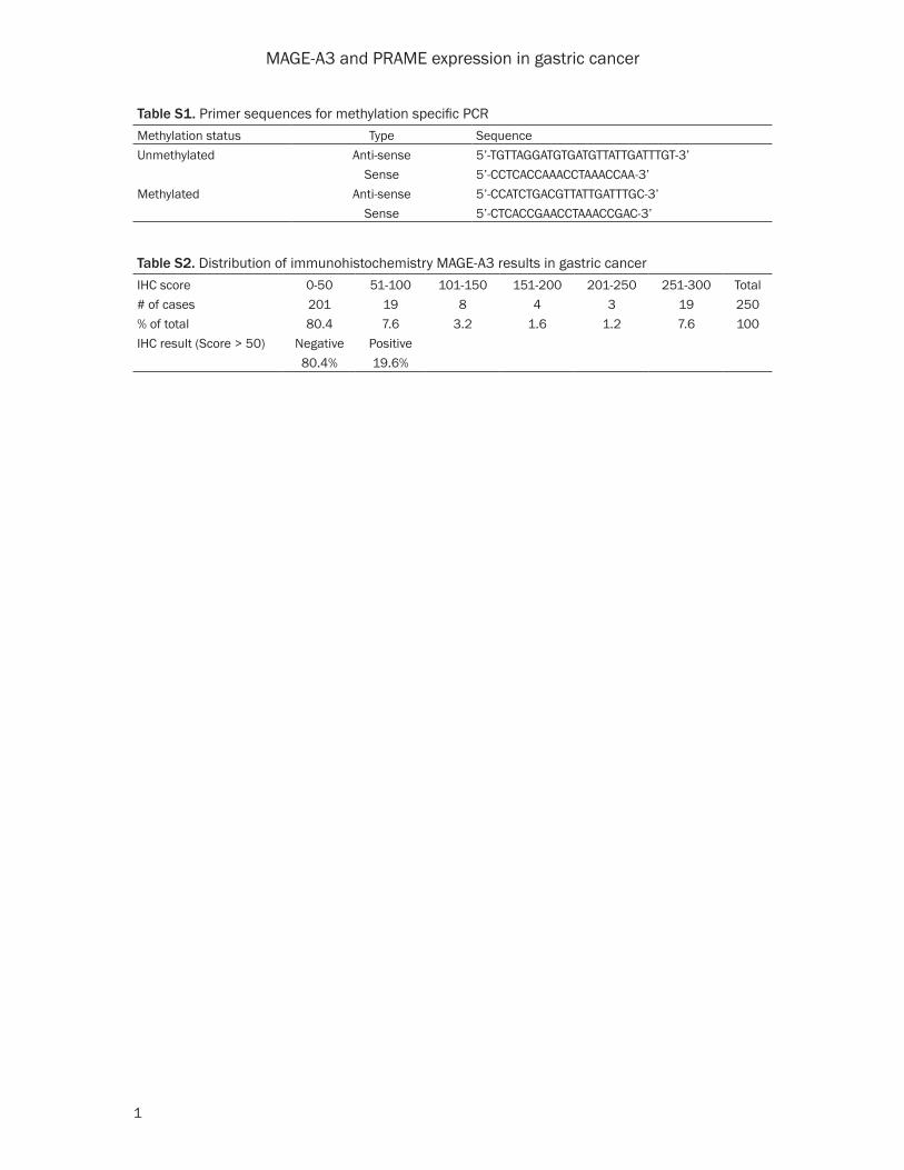


![mage l'ascension [VF]](https://img.pdfslide.us/doc/110x75/56d6beac1a28ab30169319a4/mage-lascension-vf.jpg)