Embed Size (px)
Citation preview
of August 31, 2018.This information is current as
Superfamily in Several Types of CancerMultiple Epitopes of the MAGE-A Antigen Restricted Epitope of MAGE-A3 Recognizes
−A TCR Targeting the HLA-A*0201
Rosenberg and Richard A. MorganSchrump, Nicholas P. Restifo, Paul F. Robbins, Steven A. Hong, Maria R. Parkhurst, Steven A. Feldman, David S.Mahadev Rao, Timothy L. Frankel, John P. Riley, Jenny J. Nachimuthu Chinnasamy, Jennifer A. Wargo, Zhiya Yu,
http://www.jimmunol.org/content/186/2/685doi: 10.4049/jimmunol.1001775December 2010;
2011; 186:685-696; Prepublished online 13J Immunol
Referenceshttp://www.jimmunol.org/content/186/2/685.full#ref-list-1
, 35 of which you can access for free at: cites 62 articlesThis article
average*
4 weeks from acceptance to publicationFast Publication! •
Every submission reviewed by practicing scientistsNo Triage! •
from submission to initial decisionRapid Reviews! 30 days* •
Submit online. ?The JIWhy
Subscriptionhttp://jimmunol.org/subscription
is online at: The Journal of ImmunologyInformation about subscribing to
Permissionshttp://www.aai.org/About/Publications/JI/copyright.htmlSubmit copyright permission requests at:
Email Alertshttp://jimmunol.org/alertsReceive free email-alerts when new articles cite this article. Sign up at:
Print ISSN: 0022-1767 Online ISSN: 1550-6606. All rights reserved.1451 Rockville Pike, Suite 650, Rockville, MD 20852The American Association of Immunologists, Inc.,
is published twice each month byThe Journal of Immunology
by guest on August 31, 2018
http://ww
w.jim
munol.org/
Dow
nloaded from
by guest on August 31, 2018
http://ww
w.jim
munol.org/
Dow
nloaded from
The Journal of Immunology
A TCR Targeting the HLA-A*0201–Restricted Epitope ofMAGE-A3 Recognizes Multiple Epitopes of the MAGE-AAntigen Superfamily in Several Types of Cancer
Nachimuthu Chinnasamy, Jennifer A. Wargo, Zhiya Yu, Mahadev Rao, Timothy L. Frankel,
John P. Riley, Jenny J. Hong, Maria R. Parkhurst, Steven A. Feldman, David S. Schrump,
Nicholas P. Restifo, Paul F. Robbins, Steven A. Rosenberg, and Richard A. Morgan
Adoptive immunotherapy using TCR-engineered PBLs against melanocyte differentiation Ags mediates objective tumor re-
gression but is associated with on-target toxicity. To avoid toxicity to normal tissues, we targeted cancer testis Ag (CTA)
MAGE-A3, which is widely expressed in a range of epithelial malignancies but is not expressed in most normal tissues. To
generate high-avidity TCRs against MAGE-A3, we employed a transgenic mouse model that expresses the human HLA-
A*0201 molecule. Mice were immunized with two HLA-A*0201–restricted peptides of MAGE-A3: 112–120 (KVAELVHFL)
or MAGE-A3: 271–279 (FLWGPRALV), and T cell clones were generated. MAGE-A3–specific TCR a- and b-chains were
isolated and cloned into a retroviral vector. Expression of both TCRs in human PBLs demonstrated Ag-specific reactivity
against a range of melanoma and nonmelanoma tumor cells. The TCR against MAGE-A3: 112–120 was selected for further
development based on superior reactivity against tumor target cells. Interestingly, peptide epitopes from MAGE-A3 and
MAGE-A12 (and to a lesser extent, peptides from MAGE-A2 and MAGE-A6) were recognized by PBLs engineered to express
this TCR. To further improve TCR function, single amino acid variants of the CDR3 a-chain were generated. Substitution of
alanine to threonine at position 118 of the a-chain in the CDR3 region of the TCR improved its functional avidity in CD4 and
CD8 cells. On the basis of these results, a clinical trial is planned in which patients bearing a variety of tumor histologies will
receive autologous PBLs that have been transduced with this optimized anti–MAGE-A3 TCR. The Journal of Immunology,
2011, 186: 685–696.
In the past two decades, fundamental advances in immunologyand tumor biology combined with the identification of a largenumber of tumor Ags have led to significant progress in
the field of cell-based immunotherapy (1–4). Adoptive cellularimmunotherapy involving transfer of tumor-reactive T cells hasshown some notable antitumor responses in patients with meta-static melanoma. The administration of naturally occurring tumorinfiltrating lymphocytes (TILs) expanded ex vivo mediated anobjective response rate ranging from 50–70% in melanomapatients, including bulky invasive tumors at multiple sites in-volving liver, lung, soft tissue, and brain (3, 5). A major limitationto the widespread application of TIL therapy is the difficulty ingenerating human T cells with antitumor potential. It has beenreported that approximately only half of melanomas reproduciblygive rise to antitumor TILs (6). As an alternative approach, high-affinity TCRs can be introduced into normal T cells of thepatients, and the adoptive transfer of these cells into lymphode-pleted patients has been shown to mediate cancer regression (7, 8).
Adoptive transfer of TCR-transduced PBLs targeting melanomadifferentiation Ags such as MART-1 and gp100 resulted in ob-jective cancer regression in up to 30% of patients (7). However,patients also exhibited significant toxicity associated with de-struction of normal melanocytes in the skin, eye, and ear (7). Thistrial revealed that T cells expressing highly reactive TCRs mediatecancer regression and also target cognate Ag-expressing cellsthroughout the body. Efforts to enhance adoptive immunotherapyresponse rates may hinge on targeting tumor Ags with little or noexpression in normal tissue. In an effort to overcome the on-targettoxicities associated with immunotherapies directed against Agsexpressed on normal tissues, we and others have focused on gen-erating TCRs targeting cancer testis Ags (CTAs). CTAs are im-munogenic proteins, which are normally expressed in non-MHC-expressing germ cells of testis yet are aberrantly expressed in manytumors; thus, CTAs may represent ideal targets for tumor immu-notherapy. More than 110 CTA genes or gene families have beenidentified that are expressed in multiple tumor types (9–11). Theseimmunogenic proteins are being vigorously pursued as targets fortherapeutic cancer vaccines and TCR-based adoptive immunother-apy (12–14). In theory, targeting T cells against tumor-associatedCTAs might selectively eliminate tumor cells while avoiding tox-icity to normal tissue.Since the identification of the first human MAGE CTA gene
in 1991, the number MAGE family genes have grown to .25members (15, 16). MAGE-A is a multigene family consisting of12 homologous genes MAGE-A1 to MAGE-A12 located at chro-mosome Xq28 (11). The precise function and biological role ofMAGE proteins have not been completely elucidated. However,members of MAGE-A, MAGE-B, and MAGE-C proteins have
Surgery Branch, National Cancer Institute, National Institutes of Health, Bethesda,MD 20892
Received for publication May 27, 2010. Accepted for publication November 8, 2010.
This work was supported by the Intramural Research Program of the Center forCancer Research, National Cancer Institute, National Institutes of Health.
Address correspondence and reprint requests to Dr. Richard A. Morgan, SurgeryBranch, National Cancer Institute, National Institutes of Health, Building 10, CRCRoom 3-5940, 10 Center Drive, Bethesda, MD 20892. E-mail address: [email protected]
Abbreviations used in this article: CTA, cancer testis Ag; NSCLC, non-small cell lungcarcinoma; rhIL-2, recombinant human IL-2; TIL, tumor infiltrating lymphocyte.
www.jimmunol.org/cgi/doi/10.4049/jimmunol.1001775
by guest on August 31, 2018
http://ww
w.jim
munol.org/
Dow
nloaded from
been implicated in the suppression of p53-dependent apoptosis
(17, 18), and MAGE-A3 has been attributed to mediate fibro-nectin-controlled progression and metastasis (19). Genomic clus-tering, restricted expression pattern, and single exon open readingframe of the MAGE genes are consistent with the possibility thatthese genes evolved from retrotransposition and subsequent du-plication (20). Expression of CTAs including MAGE genes intumor cells has been attributed to global DNA demethylation andother mechanisms that normally silence these genes in somaticcells (21). MAGE-A3 is one of the more frequently expressedCTAs in human tumors, including melanoma (22), non-small celllung carcinoma (NSCLC) (23), head and neck squamous cellcarcinoma (24), hepatocellular carcinoma, (25) and multiple my-eloma (26). The expression of MAGE-A3 has been shown to behigher in more advanced stages of the disease and is associatedwith poor disease prognosis (27–29). Several antigenic peptidesthat bind to HLA class I or class II molecules on tumor cells havebeen reported (30–36).Because of its high expression in a wide array of tumor types,
MAGE-A3 is an attractive target for cancer immunotherapy. Inan effort to generate TCRs against MAGE-A3, we immunizedtransgenic mice expressing the HLA-A*0201 molecule with twoMAGE-A3 peptides. In this report, we describe a TCR with a highavidity for similar peptides derived from MAGE-A3 and MAGE-A12 that also cross-reacts with related peptides from MAGE-A2and MAGE-A6. Normal PBLs engineered with this TCR demon-strated potent cytolytic activity and secreted high levels of IFN-gin response to HLA-A*0201+/MAGE+ tumor cells from multiplehistologies. These findings suggest that the use of this TCR foradoptive immunotherapy may be valuable for the treatment ofpatients with cancers that express these gene products.
Materials and MethodsCell lines and human PBLs
HLA-A*0201+/MAGE-A3+ melanoma cell lines 526, 624.38, 1300, and2984 and non-HLA-A*0201 cell lines 397, 888, 938, 1359, and 1088were established from surgically resected metastatic melanoma tumorsand maintained at the Surgery Branch, National Cancer Institute, Na-tional Institutes of Health (Bethesda, MD). The HLA-A*0201+/MAGE-A32 2361R cell line was isolated from a surgically resected metastaticrenal cell carcinoma. NSCLC line H1299 (HLA-A*02012/MAGE-A3+);small cell lung carcinoma lines H2721 (HLA-A*0201+/MAGE-A32),H2122 (HLA-A*02012/MAGE-A3+), and H1250 (HLA-A*0201+/MAGE-A3+); and esophageal cancer cell line BE-3 (HLA-A*0201+/MAGE-A3+)were obtained from the laboratory of Dr. David Schrump (SurgeryBranch, National Cancer Institute, National Institutes of Health). Breastcancer cell lines MDA-MB-453S (HLA-A*02012/MAGE-A3+) werefrom American Type Culture Collection (Manassas, VA). Glioma cellline U251 (HLA-A*0201+/MAGE-A3+) was obtained from the Division ofCancer Treatment and Diagnosis Tumor Repository, National Cancer In-stitute, National Institutes of Health (Frederick, MD). The COS7-A*0201,293-A*0201, 397-A*0201, 1359-A*0201, MDA-MB-453S-A*0201, andH1299-A*0201 cells were retrovirally engineered to express HLA-A*0201as described previously (14, 37). COS7-A*0201-MAGE-A3, COS7-A*0201-MAGE-A12, 293-A*0201-MAGE-A3, and 293-A*0201-MAGE-A12 cells were transduced with a retroviral vector expressing the re-spective MAGE genes. T2 is a lymphoblastoid cell line lacking TAPfunction, whose HLA class I proteins can be easily loaded with exogenouspeptides.
All of the PBLs used in this study were obtained from melanoma patientstreated in the Surgery Branch, National Cancer Institute, National Institutes ofHealth, on Institutional Review Board-approved protocols. Human lym-phocytes were cultured in AIM-V medium (Invitrogen, Carlsbad, CA) sup-plemented with 5% human serum from donors of blood group AB (ValleyBiomedical, Winchester, VA), 50 U/ml penicillin, 50 mg/ml streptomycin(Invitrogen), and 300 IU/ml IL-2 and maintained at 37˚C with 5% CO2.Murine lymphocytes were cultured in RPMI 1640 supplemented with 10%FBS, 2 mmol/l L-glutamine, 1 mmol/l sodium pyruvate, MEM nonessentialamino acids, 55 mmol/l 2-ME, 50 U/ml penicillin, 50 mg/ml streptomycin,and 30 IU/ml recombinant human IL-2 (rhIL-2) (R10) (Invitrogen).
Quantitative real-time PCR for MAGE-A3/6 and MAGE-A12expression
Total RNA from tumor cell lines was isolated using an RNeasy kit (Qiagen,Valencia, CA) according to the manufacturer’s instructions. A total of 1 mgof RNA was then used for cDNA synthesis using oligo(dT) with the Su-perScript First-Strand Synthesis kit (Invitrogen). The cDNA was used asthe templates for subsequent real-time PCR with primers designed spe-cifically for MAGE-A3/6 (38), MAGE-A12, and b-actin (TaqMan GeneExpression Assays; Applied Biosystems, Foster City, CA). Each experi-ment was performed in duplicate using a TaqMan 7900 (Applied Bio-systems) real-time PCR machine according to the manufacturer’s in-structions. Absolute numbers of copies were estimated using standardcurves with plasmid DNA expressing respective genes.
Synthetic peptides
The following peptides were used in this study: MAGE-A1: 105–113(KVADLVGFL), MAGE-A2: 112–120 (KMVELVHFL), MAGE-A3: 112–120 (KVAELVHFL), MAGE-A3: 271–279 (FLWGPRALV), MAGE-A4:113–121 (KVDELAHFL), MAGE-A6: 112–120 (KVAKLVHFL), MAGE-A8: 115–123 (KVAELVRFL), MAGE-A12: 112–120 (KMAELVHFL),and MAGE-C2: 144–152 (KVAELVEFL). The peptides were synthesizedusing a solid-phase method based on Fmoc chemistry with one of twomultiple peptide synthesizers (model AMS 422; Gilson, or Pioneer; Ap-plied Biosystems). Purity of the peptides was verified by mass spectrom-etry. Peptides were dissolved in DMSO and diluted in RPMI 1640medium.
Immunization of HLA-A*0201 transgenic mice and isolation ofTCRs
Transgenic mice expressing the full-length human HLA-A*0201 gene wereobtained from The Jackson Laboratory. Mouse studies were conductedaccording to the protocols approved by the National Cancer InstituteAnimal Care and Use Committee as described previously (37). Briefly, 8-to 12-wk-old mice were immunized s.c. at the base of the tail with100 mg of MAGE-A3: 112–120 (KVAELVHFL) or MAGE-A3: 271–279(FLWGPRALV) plus 120 mg of hepatitis B virus core: 128–140 helperpeptide (TPPAYRPPNAPIL) emulsified in 100 ml of IFA (Montanide ISA-51). A booster immunization was given 1 wk later. One week after thebooster immunization, mice were euthanized and splenocytes were har-vested and stimulated in vitro with irradiated T2 cells (18,000 rad) loadedwith 0.01, 0.1, or 1 mg/ml of the immunizing peptide in R10 mediumcontaining 30 IU/ml rhIL-2. Cultures were set up in 24-well plates with 1–3 million splenocytes and 0.2–0.4 million peptide-loaded T2 cells. Oneweek after stimulation, bulk murine T cell cultures were tested in cocultureassays for peptide-specific reactivity using T2 cells and tumor cell rec-ognition using 1300 melanoma and H1299-A*0201 cells. Ag-specificIFN-g secretion was measured by ELISA (Thermo Scientific, Rockford,IL). Peptide-reactive bulk cultures were cloned at 10 cells per well in U-bottom 96-well plates, with 5 3 104 peptide-pulsed irradiated T2 cells and5 3 104 irradiated (3000 rad) C57BL/6 feeder splenocytes in medium con-taining 30 IU/ml recombinant human IL-2. Two to three weeks later,growth-positive wells were identified, and the T lymphocytes from thosewells were transferred into 48-well plates and stimulated with 2 3 105
peptide-pulsed irradiated T2 cells and 1 3 106 irradiated C57BL/6 spleno-cytes in medium containing 30 IU/ml rhIL-2. One to 2 wk later, theseT cell cultures were evaluated for specific recognition of peptide and tumorcells by means of specific IFN-g secretion. T cells from each of the tumor-reactive clones were expanded by restimulation as described previously,and the total RNA was extracted for TCR isolation.
Cloning of MAGE-A3–specific, HLA-A*0201–restricted TCRs
Total RNAwas extracted from tumor-reactive T cell clones using an RNeasymini kit (Qiagen). TCR a- and b-chains from each tumor-reactive T cellclone were cloned using a SMART RACE cDNA amplification kit (Clontec,Mountain View, CA). For the amplification of TCRs, gene-specific primerswere made from the C region of mouse TCR a- and b-chains, TCR a Cregion-59-ACTGGTACACAGCAGGTTCTGG-39 and TCR b C region-59-AAGGAGACCTTGGGTGGAGTC-39. The PCR products of the 59-RACEwere cloned into the PCR2.1 TOPO vector (Invitrogen Life Technologies),and the insert DNA fragments were sequenced. The DNA sequence datawere analyzed using The International Immunogenetics Information System(http://imgt.cines.fr/IMGT_vquest/vquest?livret=0&Option=mouseTcR) forthe identification of mouse TCR a- and b-chains. After the identification ofthe variable regions of a- and b-chains and the identification of the C region
686 TCR RECOGNIZING MULTIPLE MAGE GENES FOR CANCER GENE THERAPY
by guest on August 31, 2018
http://ww
w.jim
munol.org/
Dow
nloaded from
of the b-chain (CB1 or CB2), specific primers were used to amplify the full-length TCR a- and b-chains from the cDNA.
Electroporation of TCRs into PBLs
The full-length a- and b-chains were individually cloned into the RNAexpression vector pGEM-4Z/64A. In vitro-transcribed mRNA encoding a-and b-chains were generated using mMESSAGE mMACHINE (Ambion,Austin, TX) and electroporated into OKT3-stimulated human lymphocyteswith an ElectroSquarePorator ECM-830 (BTX, San Diego, CA) as de-scribed previously (39). Electroporated PBLs were tested for Ag-specificreactivity using peptide-loaded T2 cells and tumor cell lines 2361-RCC,938 melanoma, H1299-A*0201, and 1300 melanoma. The Ag-specific re-
sponse of TCR-electroporated T cells was evaluated by coculture withrespective MAGE-A3 peptide-loaded T2 cells and tumor cell lines, and theculture supernatants were tested for IFN-g levels by ELISA.
Construction of retroviral vectors expressing MAGE-A3–specificHLA-A*0201–restricted TCRs
Two MSGV1-based retroviral vectors were constructed using the over-lapping PCR method with the transgene construct arranged in the fol-lowing order of configuration: TCR a-chain, linker peptide furin-SGSG-P2A (13), and TCR b-chain. The cloned TCR inserts were verified byrestriction enzyme digestion and DNA sequencing.
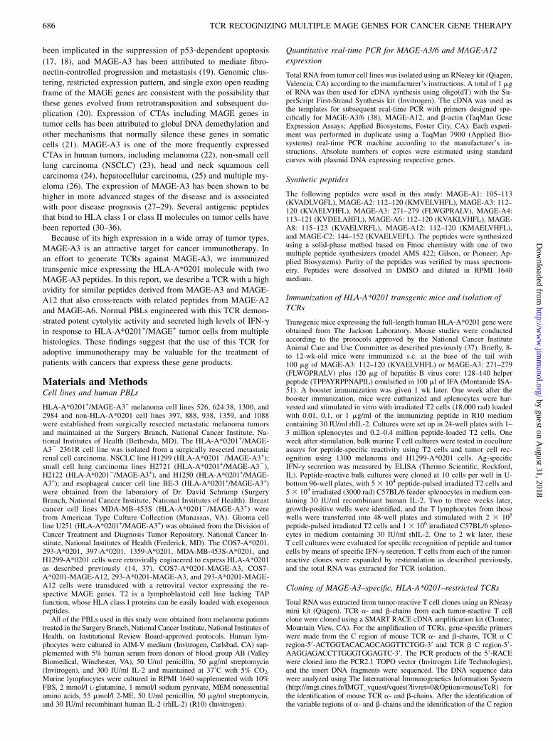
FIGURE 1. A, Schematic illustration of the MSGV1-based retroviral vector encoding anti–MAGE-A3 TCR expression cassette. TCR a- and b-chains are
linked with furin-spacer (SGSG)-P2A ribosomal skip peptide sequence. B and C, Flow cytometric analysis of MAGE-A3 TCR-transduced PBLs. B, Dot
plots showing the FACS profile of PBLs stained with anti–human-CD8-PE and anti–mouse-TCR b-FITC Abs. C, Dot plots showing the FACS profile of
PBLs stained with anti–human-CD8-FITC Ab and PE-conjugated MAGE-A3: 112–120 or MAGE-A3: 271–279 HLA-A*0201 tetramers.
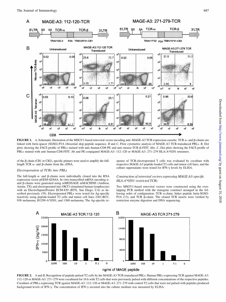
FIGURE 2. A and B, Recognition of peptide-pulsed T2 cells by the MAGE-A3 TCR-transduced PBLs. Human PBLs expressing TCR against MAGE-A3:
112–120 or MAGE-A3: 271–279 were cocultured for 16 h with T2 cells that were previously pulsed with different concentrations of the respective peptides.
Coculture of PBLs expressing TCR against MAGE-A3: 112–120 or MAGE-A3: 271–279 with control T2 cells that were not pulsed with peptides produced
background levels of IFN-g. The concentration of IFN-g secreted into the culture medium was measured by ELISA.
The Journal of Immunology 687
by guest on August 31, 2018
http://ww
w.jim
munol.org/
Dow
nloaded from
Transduction of PBLs
Retroviral supernatants were generated by transfecting respective MSGV1-MAGE-TCR vector DNA from each of the constructs with a plasmidencoding RD114 envelope into 293-GP cells using the Lipofectamine 2000reagent (Invitrogen) in Opti-MEM medium (Invitrogen) (13). Retroviralvector expressing TCR against NY ESO-1 was used as a positive control inall of the experiments (12). MSGV1 vector expressing GFP was also gen-erated. Viral supernatants were then loaded onto RetroNectin-coated (TakaraBio, Japan) non-tissue culture-treated six-well plates. PBLs were stimulatedwith OKT3 (50 ng/ml) and rhIL-2 (300 IU/ml) 48 h prior to transduction,and the transduction was carried out as described previously (13, 40).
Tetramer staining
HLA-A*0201–restricted MAGE-A3–derived peptides MAGE-A3: 112–120 (KVAELVHFL) and MAGE-A3: 271–279 (FLWGPRALV) were usedby the National Institutes of Health Tetramer Core Facility at EmoryUniversity to produce tetramers linking PE as the fluorophore. MAGE-A3TCR-transduced T cells were stained with a FITC-labeled anti-CD8 (BDPharmingen, San Jose, CA) and with PE-labeled HLA-A*0201 tetramers.FITC-conjugated mAb against the C region of the murine b-chain(eBioscience, San Diego, CA) and PE-conjugated anti-CD8 Abs were alsoused to detect the expression of MAGE-A3 TCRs in the human PBLs.Cells were analyzed using a FACScan flow cytometer with CellQuestsoftware (BD Biosciences) or FlowJo software (Tree Star, Ashland, OR).
Intracellular cytokine staining
Intracellular cytokine staining was performed using a BDCytofix/Cytopermkit (BD Biosciences) according to the manufacturer’s instructions. Briefly,cells were first stained with cell surface markers CD3 and CD8 and thenstained with FITC-conjugated anti–IFN-g and allophycocyanin-conjugatedanti–IL-2 Abs for intracellular detection of the cytokines. All of the Abs aswell as isotype controls were purchased from BD Biosciences. Cells wereanalyzed using a FACSCanto II flow cytometer with CellQuest software(BD Biosciences) or FlowJo software (Tree Star).
CD107a mobilization assay
The cell surface mobilization of the CD107a molecule was determined asa measure of degranulation and functional reactivity after Ag recognitionby the TCR. In these assays, 1 3 105 H1299-A2 or H1299 cells werecocultured with an equal number of PBLs at 37˚C for 2 h. The cells werethen stained with mouse anti-human Abs against CD107a and CD8 (BDBiosciences) and analyzed by FACS.
Cytokine release assay
TCR-engineered PBLs were tested for Ag-specific reactivity in cytokinerelease assays using peptide-loaded T2 cells and tumor cells. In theseassays, effector cells (1 3 105) were cocultured with an equal number of
target cells in AIM-V medium in a final volume of 0.2 ml in duplicatewells of a 96-well U-bottom microplate. Culture supernatants were har-vested 18–24 h after the initiation of coculture and assayed for IFN-g andGM-CSF by ELISA (Thermo Scientific).
[51Cr] release assay
The ability of the transduced PBLs to lyse HLA-A*0201+/MAGE-A3+ tumorcells was measured using a [51Cr] release assay as described previously (12,41). In these assays, TCR-engineered PBLs were coincubated with decreasingratios of 51Cr-labeled target cells (E:T ratio) in AIM-V medium in 96-well U-bottom plates at 37˚C for 4 h. Lysis was measured by [51Cr] release in themedium: percent lysis = (sample release 2 minimum release)/(maximumrelease 2 minimum release) 3 100%, average of duplicate samples.
CD4/CD8 separation
MAGE TCR-engineered CD4+ and CD8+ populations were separated usingmagnetic bead-based BD IMag human CD4 or CD8 T lymphocyte enrich-ment set DM kit for negative selection of those subsets (BD Biosciences).
Lymphocyte proliferation assay
TCR-transduced PBLs were tested for Ag-specific proliferation using the[3H]thymidine incorporation assay. Briefly, effector cells (1 3 105) werecocultured with equal number of irradiated (18,000 rad, cesium source)H1299 or H1299-A2 target cells in AIM-V medium in a final volume of0.2 ml in triplicate wells of a 96-well U-bottom microplate. The cells werecocultured for 3 d and pulsed with 1 mCi [3H]thymidine (DuPont, NewEngland Nuclear, Shelton, CT) per well and cultured for an additional18 h. The cells were then harvested onto a glass fiber filter (Wallac Oy, Turku,Finland), and radionucleotide incorporation was measured using a Perkin-Elmer Microbeta Trilux counter (Shelton, CT). Results expressed as cpm.
Generation of single amino acid variants of the CDR3 a-chainMAGE-A3: 112–120 TCR
We generated 85 single amino acid variant TCRs in four stages. 1) Site-directed mutagenesis using a QuikChange Lightning Site-Directed Muta-genesis kit (Stratagene, La Jolla, CA) was used to substitute all of the other 19aa at position D115 introducing appropriate nucleotide changes in the PCRprimer. 2) Alanine substitutions at eight of the residues F114, D115, T116,N117, Y119, K120, V121, and I122 were introduced except at position A118that already had an alanine residue. 3) Conservative amino acid substitutionswere introduced at positionsN117 toQ/K/R,A118 to V/L/I, andY119 toR/K/Q/N by site-directed mutagenesis. 4) Single amino acid substitutions at thesethree residues were synthesized (GENEART, Regensburg, Germany) toproduce a 16 aa substitution library at each position. Retroviral vectorsupernatants expressing all of the above-mentioned single amino acidvariant TCRs were generated by transient transfection into 293-GP cells;PBLs from donors were transduced and tested for tetramer binding andIFN-g production in coculture assays with tumor cell lines.
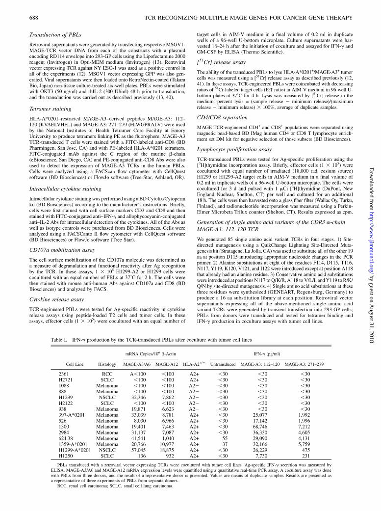
Table I. IFN-g production by the TCR-transduced PBLs after coculture with tumor cell lines
mRNA Copies/106 b-Actin IFN-g (pg/ml)
Cell Line Histology MAGE-A3/A6 MAGE-A12 HLA-A2+/2 Untransduced MAGE-A3: 112–120 MAGE-A3: 271–279
2361 RCC A,100 ,100 A2+ ,30 ,30 ,30H2721 SCLC ,100 ,100 A2+ ,30 ,30 ,301088 Melanoma ,100 ,100 A22 ,30 ,30 ,30888 Melanoma ,100 ,100 A22 ,30 ,30 ,30H1299 NSCLC 32,346 7,862 A22 ,30 ,30 ,30H2122 SCLC ,100 ,100 A22 ,30 ,30 ,30938 Melanoma 19,871 6,623 A22 ,30 ,30 ,30397-A*0201 Melanoma 33,039 8,781 A2+ ,30 25,077 1,992526 Melanoma 8,030 6,966 A2+ ,30 17,142 1,9961300 Melanoma 19,401 7,463 A2+ ,30 68,746 7,2122984 Melanoma 31,137 7,087 A2+ ,30 36,330 4,605624.38 Melanoma 41,541 1,040 A2+ 55 29,090 4,1311359-A*0201 Melanoma 20,766 10,977 A2+ 37 32,166 5,759H1299-A*0201 NSCLC 57,045 18,875 A2+ ,30 26,229 475H1250 SCLC 136 932 A2+ ,30 7,730 231
PBLs transduced with a retroviral vector expressing TCRs were cocultured with tumor cell lines. Ag-specific IFN-g secretion was measured byELISA. MAGE-A3/A6 and MAGE-A12 mRNA expression levels were quantified using a quantitative real-time PCR assay. A coculture assay was donewith PBLs from three donors, and the result of a representative donor is presented. Values are means of duplicate samples. Results are presented asa representative of three experiments of PBLs from separate donors.
RCC, renal cell carcinoma; SCLC, small cell lung carcinoma.
688 TCR RECOGNIZING MULTIPLE MAGE GENES FOR CANCER GENE THERAPY
by guest on August 31, 2018
http://ww
w.jim
munol.org/
Dow
nloaded from
ResultsGeneration of MAGE-A3–reactive murine T cell clones fromHLA-A*0201 transgenic mice
Transgenic mice expressing the full-length HLA-A*0201 moleculewere immunized with one of the two previously identified natu-rally processed and presented HLA-A*0201–restricted peptidesfrom MAGE-A3 [MAGE-A3: 112–120 (KVAELVHFL) (35) or
MAGE-A3: 271–279 (FLWGPRALV) (42)] along with a helper
peptide, hepatitis B virus core: 128–140. After two immuniza-
tions, murine T cells were harvested from spleen and lymph nodes
and stimulated in vitro with the respective peptide and IL-2. Bulk
T cell cultures from mice immunized with the MAGE-A3 peptides
demonstrated specific reactivity against T2 cells pulsed with the
relevant peptide and the HLA-A*0201+/MAGE-A3+ tumor cell
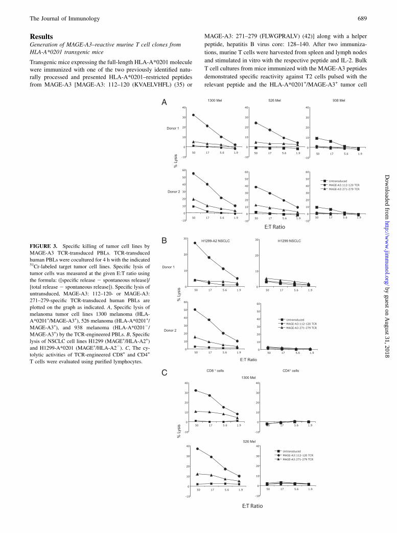
FIGURE 3. Specific killing of tumor cell lines by
MAGE-A3 TCR-transduced PBLs. TCR-transduced
human PBLs were cocultured for 4 h with the indicated51Cr-labeled target tumor cell lines. Specific lysis of
tumor cells was measured at the given E:T ratio using
the formula: ([specific release 2 spontaneous release]/
[total release 2 spontaneous release]). Specific lysis of
untransduced, MAGE-A3: 112–120- or MAGE-A3:
271–279-specific TCR-transduced human PBLs are
plotted on the graph as indicated. A, Specific lysis of
melanoma tumor cell lines 1300 melanoma (HLA-
A*0201+/MAGE-A3+), 526 melanoma (HLA-A*0201+/
MAGE-A3+), and 938 melanoma (HLA-A*02012/
MAGE-A3+) by the TCR-engineered PBLs. B, Specific
lysis of NSCLC cell lines H1299 (MAGE+/HLA-A2+)
and H1299-A*0201 (MAGE+/HLA-A22). C, The cy-
tolytic activities of TCR-engineered CD8+ and CD4+
T cells were evaluated using purified lymphocytes.
The Journal of Immunology 689
by guest on August 31, 2018
http://ww
w.jim
munol.org/
Dow
nloaded from
lines H1299-A*0201, 1300 melanoma, and 624 melanoma aftertwo in vitro stimulations (data not shown). Reactive T cells frompositive wells were cloned by limiting dilution and tested for Ag-specific reactivity. Five clones derived from the mice immunizedwith the MAGE-A3: 112–120 peptide and six clones derived fromthe mice immunized with the MAGE-A3: 271–279 peptide thatsecreted high levels of IFN-g in response to tumor cells andpeptide-loaded T2 cells were expanded and further characterized.
Cloning of MAGE-reactive TCRs
TCR a- and b-chains from each tumor-reactive T cell clone werecloned using a SMART RACE cDNA amplification kit with gene-specific primers in the C region of mouse TCR a- and b-chains.After the identification of the variable regions of the a- and b-chainsand the specific C region of the b-chain, specific primers were usedto amplify the full-length TCR a- and b-chains from the cDNA. TheTCR a- and b-chains were then cloned into the RNA expressionvector pGEM. In vitro-transcribed RNA of TCR a- and b-chainswere electroporated into human PBLs and tested for Ag-specificreactivity as described previously (39) using peptide-loaded T2cells, H1299-A*0201, and 1300 melanoma tumor cell lines. On thebasis of the specific reactivity, we selected a TCR against MAGE-A3:112–120 peptide (TCR a-TRAV12D-3, TCR b-TRBV29*01, andCB1) and a TCR against MAGE-A3: 271–279 peptide (TRAV17*02,TRBV15*01, and CB2) for further evaluation.
Construction of a MAGE-A3 TCR-expressing retroviral vectorand transduction of PBLs
Two MSGV1-based retroviral vectors with expression cassettesconsisting of TCR a-TRAV12D-3 and TCR b-TRBV29*01-CB1
and TRAV17*02 and TRBV15*01-CB2 were constructed (Fig.1A). The TCR expression in these vectors is driven by the virallong terminal repeat, and a- and b-chains are expressed as a singleopen reading frame using the 2A linker peptide (13, 43). HumanPBLs were stimulated for 2 d and then transduced. FACS analysisof transduced PBLs using the anti-mouse TCR b-chain revealedthat both CD8+ and CD4+ cells had been transduced with theseTCR vectors (Fig. 1B); however, specific tetramer binding wasobserved only in transduced CD8+ and not CD4+ T cells (Fig. 1C).
Evaluation of the function of MAGE TCR-engineered PBLs
To evaluate the recognition of the respective MAGE-A3 TCRs,transduced PBLs were subjected to coculture assay with peptide-pulsed T2 cells. TCR-transduced PBLs specifically secreted IFN-gupon encounter with the antigenic peptide in a dose-dependentmanner (Fig. 2). PBLs transduced with either MAGE-A3: 112–120 or MAGE-A3: 271–279 TCRs recognized T2 cells pulsedwith as little as 0.1 ng/ml MAGE-A3 peptides, indicating that bothof the TCRs were relatively high-avidity receptors. Coculture ofPBLs expressing TCRs against MAGE-A3: 112–120 or MAGE-A3: 271–279 with control T2 cells that were not pulsed with anypeptides produced background levels of IFN-g. To assess the spe-cific recognition of tumor cells, TCR-engineered PBLs were co-cultured with a panel of HLA-A*0201+ and HLA-A*02012 mel-anoma- and lung tumor-derived cell lines. Specific release ofIFN-g was observed when the TCR-engineered PBLs were co-cultured with HLA-A*0201+/MAGE-A3+ cell lines but not HLA-A*02012/MAGE-A3+ or HLA-A*0201+/MAGE-A32 cell lines(Table I). A comparison of the two TCRs revealed that T cellstransduced with the MAGE-A3: 112–120 TCR released ∼10-foldhigher levels of IFN-g in response to HLA-A*0201+/MAGE-A3+
tumor cell targets (Table I). These responses were specific becauselow levels of IFN-g were released in response to MAGE+/HLA-A*02012 cell lines and MAGE2/HLA-A*0201+ cell lines. Al-though MAGE-A3: 271–279 TCR-transduced PBLs efficientlyrecognized the peptide loaded on T2 cells, the recognition ofMAGE-A3+/HLA-A*0201+ tumor cells as measured by the re-lease of IFN-g production was relatively weak (Table I).We next measured the specific lysis of melanoma cell lines by the
TCR-engineered PBLs. MAGE-A3: 112–120 TCR-transducedPBLs demonstrated superior lytic function against MAGE-A3+/HLA-A*0201+ tumor cell lines 1300 melanoma and 526 mela-noma cells compared with that of MAGE-A3: 271–279 TCR-transduced PBLs (Fig. 3A). There was little or no lysis of theHLA-A*02012 cell line 938 melanoma, and the untransduced PBLs
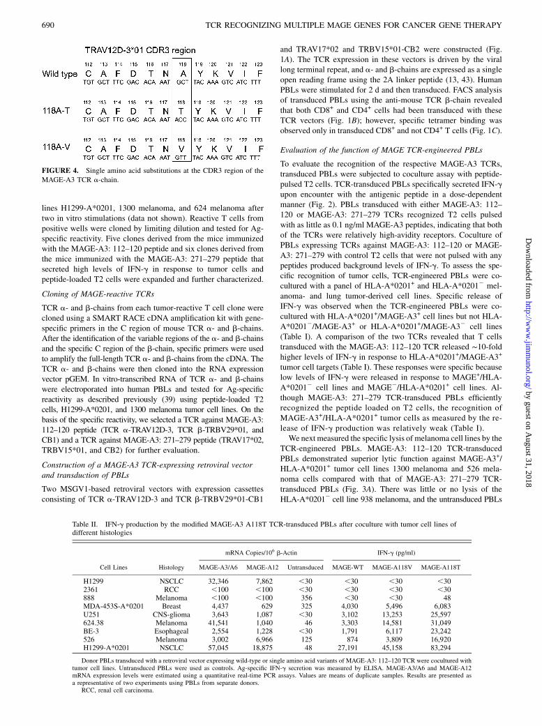
FIGURE 4. Single amino acid substitutions at the CDR3 region of the
MAGE-A3 TCR a-chain.
Table II. IFN-g production by the modified MAGE-A3 A118T TCR-transduced PBLs after coculture with tumor cell lines ofdifferent histologies
mRNA Copies/106 b-Actin IFN-g (pg/ml)
Cell Lines Histology MAGE-A3/A6 MAGE-A12 Untransduced MAGE-WT MAGE-A118V MAGE-A118T
H1299 NSCLC 32,346 7,862 ,30 ,30 ,30 ,302361 RCC ,100 ,100 ,30 ,30 ,30 ,30888 Melanoma ,100 ,100 356 ,30 ,30 48MDA-453S-A*0201 Breast 4,437 629 325 4,030 5,496 6,083U251 CNS-glioma 3,643 1,087 ,30 3,102 13,253 25,597624.38 Melanoma 41,541 1,040 46 3,303 14,581 31,049BE-3 Esophageal 2,554 1,228 ,30 1,791 6,117 23,242526 Melanoma 3,002 6,966 125 874 3,809 16,920H1299-A*0201 NSCLC 57,045 18,875 48 27,191 45,158 83,294
Donor PBLs transduced with a retroviral vector expressing wild-type or single amino acid variants of MAGE-A3: 112–120 TCR were cocultured withtumor cell lines. Untransduced PBLs were used as controls. Ag-specific IFN-g secretion was measured by ELISA. MAGE-A3/A6 and MAGE-A12mRNA expression levels were estimated using a quantitative real-time PCR assays. Values are means of duplicate samples. Results are presented asa representative of two experiments using PBLs from separate donors.
RCC, renal cell carcinoma.
690 TCR RECOGNIZING MULTIPLE MAGE GENES FOR CANCER GENE THERAPY
by guest on August 31, 2018
http://ww
w.jim
munol.org/
Dow
nloaded from
showed little reactivity against any of the target cells (Fig. 3A).MAGE-A3: 112–120 TCR-transduced PBLs also showed superiorlytic function against NSCLC cell line H1299-A*0201+ and didnot recognize the parental non-HLA-A*0201 cell line H1299 (Fig.3B). Because tetramer binding was observed only in MAGE-A3TCR-transduced CD8+ cells and not in CD4+ cells, we investi-gated IFN-g production and cytolytic activity using purifiedlymphocytes. As shown in Fig. 3C, Ag-specific lysis of melanomacell lines 1300 melanoma and 526 melanoma was evident in CD8+
cells but not seen in CD4+ cells. Similarly, IFN-g productionafter coculture with tumor cells was observed only in CD8+ cellsand not seen in CD4+ cells (data not shown). On the basis ofthese data, the MAGE-A3: 112–120 TCR was chosen for furtheranalysis.
Single amino acid substitution variants within CDR3 of theTCR a-chain enhance the function of TCR-engineered PBLs
Previous studies by our group have shown that it is possible toimprove the function of the TCR by engineering single or multipleamino acid changes in the CDR3 region of the TCR a-chain (14,37, 44). We generated retroviral vectors expressing 85 singleamino acid variants in the CDR3 region of the TCR a-chain andtested their function in PBLs. Preliminary screening experimentswere carried out by single amino acid residue alanine substitutionin the CDR3 of the a-chain. Alanine substitution at F114, D115,T116, N117, Y119, K120, and V121 completely abolished theactivity of the TCR. This was seen by the complete loss of tet-ramer binding as well as a lack of production of IFN-g after co-culture with peptide-pulsed T2 cells or MAGE+/HLA-A*0201+
tumor cells. We next focused on position 115 and created 19 aasubstitutions at this at position. Complete loss of TCR activity, asseen by the complete lack of tetramer binding and loss of IFN-gproduction, was observed when aspartic acid at position 115 wassubstituted with any of the other amino acids. Finally, we createda retroviral vector library of single amino acid variants at positions117, 118, and 119 of the a-chain. During this screening, we foundthat a substitution of valine or threonine for the alanine residuepresent at position 118 in the wild-type a-chain retained TCRfunction (Fig. 4).Table II shows the IFN-g secretion results of a coculture assay
with tumor cells of different histologies using the modified TCRs.Ag-specific HLA-A*0201–restricted recognition of tumor cellsfrom diverse histologies were observed after coculture of TCR-engineered PBLs with breast cancer line MDA-454S-A2, gliomaline U251, melanoma lines 624 and 526, esophageal line BE-3,and NSCLC line H1299-A*0201. The results demonstrated thatT cells transduced with the A118V and A118T TCR variants se-creted higher levels of IFN-g than cells transduced with the wild-type TCR. T cells transduced with the A118T variant TCR secretedhigher levels of IFN-g than the A118V variant when tested against
multiple MAGE+/HLA-A*0201+ cells. The HLA-A*02012 celllines H1299 and 888 as well as MAGE2/HLA-A*0201+ cell line2361-RCC were not recognized by the PBLs engineered to expresseither the wild-type or the A118T variant, indicating that this aminoacid alteration did not alter the specificity of this TCR.To further test the function of this improved MAGE TCR, we
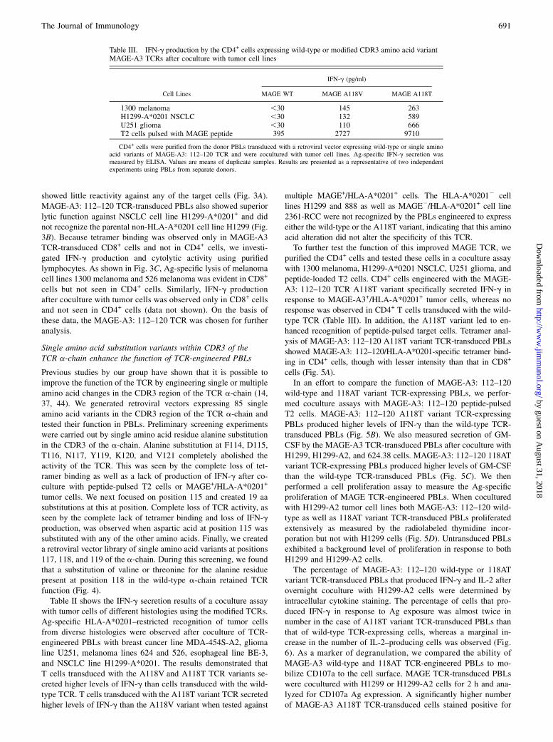
purified the CD4+ cells and tested these cells in a coculture assaywith 1300 melanoma, H1299-A*0201 NSCLC, U251 glioma, andpeptide-loaded T2 cells. CD4+ cells engineered with the MAGE-A3: 112–120 TCR A118T variant specifically secreted IFN-g inresponse to MAGE-A3+/HLA-A*0201+ tumor cells, whereas noresponse was observed in CD4+ T cells transduced with the wild-type TCR (Table III). In addition, the A118T variant led to en-hanced recognition of peptide-pulsed target cells. Tetramer anal-ysis of MAGE-A3: 112–120 A118T variant TCR-transduced PBLsshowed MAGE-A3: 112–120/HLA-A*0201-specific tetramer bind-ing in CD4+ cells, though with lesser intensity than that in CD8+
cells (Fig. 5A).In an effort to compare the function of MAGE-A3: 112–120
wild-type and 118AT variant TCR-expressing PBLs, we perfor-med coculture assays with MAGE-A3: 112–120 peptide-pulsedT2 cells. MAGE-A3: 112–120 A118T variant TCR-expressingPBLs produced higher levels of IFN-g than the wild-type TCR-transduced PBLs (Fig. 5B). We also measured secretion of GM-CSF by the MAGE-A3 TCR-transduced PBLs after coculture withH1299, H1299-A2, and 624.38 cells. MAGE-A3: 112–120 118ATvariant TCR-expressing PBLs produced higher levels of GM-CSFthan the wild-type TCR-transduced PBLs (Fig. 5C). We thenperformed a cell proliferation assay to measure the Ag-specificproliferation of MAGE TCR-engineered PBLs. When coculturedwith H1299-A2 tumor cell lines both MAGE-A3: 112–120 wild-type as well as 118AT variant TCR-transduced PBLs proliferatedextensively as measured by the radiolabeled thymidine incor-poration but not with H1299 cells (Fig. 5D). Untransduced PBLsexhibited a background level of proliferation in response to bothH1299 and H1299-A2 cells.The percentage of MAGE-A3: 112–120 wild-type or 118AT
variant TCR-transduced PBLs that produced IFN-g and IL-2 afterovernight coculture with H1299-A2 cells were determined byintracellular cytokine staining. The percentage of cells that pro-duced IFN-g in response to Ag exposure was almost twice innumber in the case of A118T variant TCR-transduced PBLs thanthat of wild-type TCR-expressing cells, whereas a marginal in-crease in the number of IL-2–producing cells was observed (Fig.6). As a marker of degranulation, we compared the ability ofMAGE-A3 wild-type and 118AT TCR-engineered PBLs to mo-bilize CD107a to the cell surface. MAGE TCR-transduced PBLswere cocultured with H1299 or H1299-A2 cells for 2 h and ana-lyzed for CD107a Ag expression. A significantly higher numberof MAGE-A3 A118T TCR-transduced cells stained positive for
Table III. IFN-g production by the CD4+ cells expressing wild-type or modified CDR3 amino acid variantMAGE-A3 TCRs after coculture with tumor cell lines
IFN-g (pg/ml)
Cell Lines MAGE WT MAGE A118V MAGE A118T
1300 melanoma ,30 145 263H1299-A*0201 NSCLC ,30 132 589U251 glioma ,30 110 666T2 cells pulsed with MAGE peptide 395 2727 9710
CD4+ cells were purified from the donor PBLs transduced with a retroviral vector expressing wild-type or single aminoacid variants of MAGE-A3: 112–120 TCR and were cocultured with tumor cell lines. Ag-specific IFN-g secretion wasmeasured by ELISA. Values are means of duplicate samples. Results are presented as a representative of two independentexperiments using PBLs from separate donors.
The Journal of Immunology 691
by guest on August 31, 2018
http://ww
w.jim
munol.org/
Dow
nloaded from
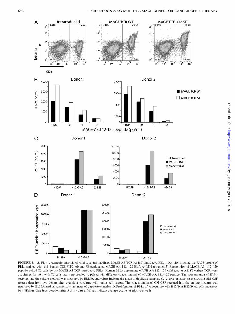
FIGURE 5. A, Flow cytometric analysis of wild-type and modified MAGE-A3 TCR-A118T-transduced PBLs. Dot blot showing the FACS profile of
PBLs stained with anti–human-CD8-FITC Ab and PE-conjugated MAGE-A3: 112–120-HLA-A*0201 tetramer. B, Recognition of MAGE-A3: 112–120
peptide-pulsed T2 cells by the MAGE-A3 TCR-transduced PBLs. Human PBLs expressing MAGE-A3: 112–120 wild-type or A118T variant TCR were
cocultured for 16 h with T2 cells that were previously pulsed with different concentrations of MAGE-A3: 112–120 peptide. The concentration of IFN-g
secreted into the culture medium was measured by ELISA, and values indicate the mean of duplicate samples. C, A representative assay showing GM-CSF
release data from two donors after overnight coculture with tumor cell targets. The concentration of GM-CSF secreted into the culture medium was
measured by ELISA, and values indicate the mean of duplicate samples. D, Proliferation of PBLs after coculture with H1299 or H1299-A2 cells measured
by [3H]thymidine incorporation after 3 d in culture. Values indicate average counts of triplicate wells.
692 TCR RECOGNIZING MULTIPLE MAGE GENES FOR CANCER GENE THERAPY
by guest on August 31, 2018
http://ww
w.jim
munol.org/
Dow
nloaded from
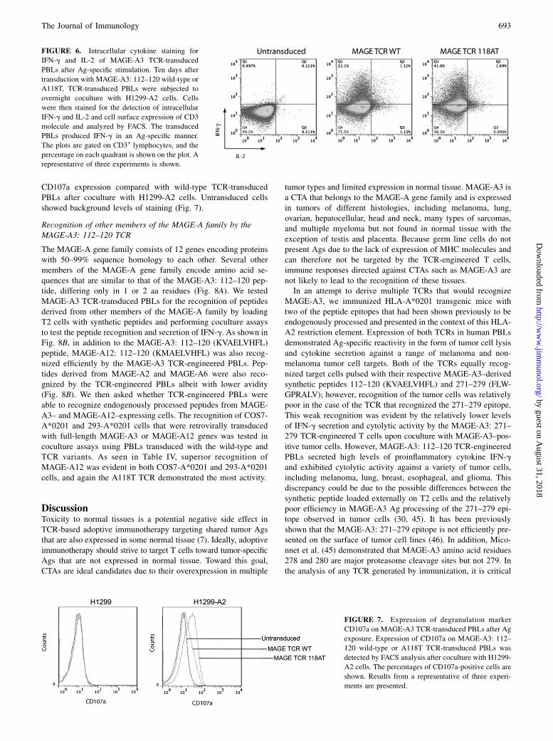
CD107a expression compared with wild-type TCR-transducedPBLs after coculture with H1299-A2 cells. Untransduced cellsshowed background levels of staining (Fig. 7).
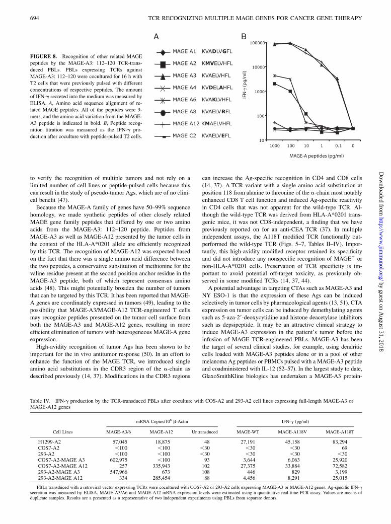
Recognition of other members of the MAGE-A family by theMAGE-A3: 112–120 TCR
The MAGE-A gene family consists of 12 genes encoding proteinswith 50–99% sequence homology to each other. Several othermembers of the MAGE-A gene family encode amino acid se-quences that are similar to that of the MAGE-A3: 112–120 pep-tide, differing only in 1 or 2 aa residues (Fig. 8A). We testedMAGE-A3 TCR-transduced PBLs for the recognition of peptidesderived from other members of the MAGE-A family by loadingT2 cells with synthetic peptides and performing coculture assaysto test the peptide recognition and secretion of IFN-g. As shown inFig. 8B, in addition to the MAGE-A3: 112–120 (KVAELVHFL)peptide, MAGE-A12: 112–120 (KMAELVHFL) was also recog-nized efficiently by the MAGE-A3 TCR-engineered PBLs. Pep-tides derived from MAGE-A2 and MAGE-A6 were also reco-gnized by the TCR-engineered PBLs albeit with lower avidity(Fig. 8B). We then asked whether TCR-engineered PBLs wereable to recognize endogenously processed peptides from MAGE-A3– and MAGE-A12–expressing cells. The recognition of COS7-A*0201 and 293-A*0201 cells that were retrovirally transducedwith full-length MAGE-A3 or MAGE-A12 genes was tested incoculture assays using PBLs transduced with the wild-type andTCR variants. As seen in Table IV, superior recognition ofMAGE-A12 was evident in both COS7-A*0201 and 293-A*0201cells, and again the A118T TCR demonstrated the most activity.
DiscussionToxicity to normal tissues is a potential negative side effect inTCR-based adoptive immunotherapy targeting shared tumor Agsthat are also expressed in some normal tissue (7). Ideally, adoptiveimmunotherapy should strive to target T cells toward tumor-specificAgs that are not expressed in normal tissue. Toward this goal,CTAs are ideal candidates due to their overexpression in multiple
tumor types and limited expression in normal tissue. MAGE-A3 isa CTA that belongs to the MAGE-A gene family and is expressedin tumors of different histologies, including melanoma, lung,ovarian, hepatocellular, head and neck, many types of sarcomas,and multiple myeloma but not found in normal tissue with theexception of testis and placenta. Because germ line cells do notpresent Ags due to the lack of expression of MHC molecules andcan therefore not be targeted by the TCR-engineered T cells,immune responses directed against CTAs such as MAGE-A3 arenot likely to lead to the recognition of these tissues.In an attempt to derive multiple TCRs that would recognize
MAGE-A3, we immunized HLA-A*0201 transgenic mice withtwo of the peptide epitopes that had been shown previously to beendogenously processed and presented in the context of this HLA-A2 restriction element. Expression of both TCRs in human PBLsdemonstrated Ag-specific reactivity in the form of tumor cell lysisand cytokine secretion against a range of melanoma and non-melanoma tumor cell targets. Both of the TCRs equally recog-nized target cells pulsed with their respective MAGE-A3–derivedsynthetic peptides 112–120 (KVAELVHFL) and 271–279 (FLW-GPRALV); however, recognition of the tumor cells was relativelypoor in the case of the TCR that recognized the 271–279 epitope.This weak recognition was evident by the relatively lower levelsof IFN-g secretion and cytolytic activity by the MAGE-A3: 271–279 TCR-engineered T cells upon coculture with MAGE-A3–pos-itive tumor cells. However, MAGE-A3: 112–120 TCR-engineeredPBLs secreted high levels of proinflammatory cytokine IFN-gand exhibited cytolytic activity against a variety of tumor cells,including melanoma, lung, breast, esophageal, and glioma. Thisdiscrepancy could be due to the possible differences between thesynthetic peptide loaded externally on T2 cells and the relativelypoor efficiency in MAGE-A3 Ag processing of the 271–279 epi-tope observed in tumor cells (30, 45). It has been previouslyshown that the MAGE-A3: 271–279 epitope is not efficiently pre-sented on the surface of tumor cell lines (46). In addition, Mico-nnet et al. (45) demonstrated that MAGE-A3 amino acid residues278 and 280 are major proteasome cleavage sites but not 279. Inthe analysis of any TCR generated by immunization, it is critical
FIGURE 6. Intracellular cytokine staining for
IFN-g and IL-2 of MAGE-A3 TCR-transduced
PBLs after Ag-specific stimulation. Ten days after
transduction with MAGE-A3: 112–120 wild-type or
A118T, TCR-transduced PBLs were subjected to
overnight coculture with H1299-A2 cells. Cells
were then stained for the detection of intracellular
IFN-g and IL-2 and cell surface expression of CD3
molecule and analyzed by FACS. The transduced
PBLs produced IFN-g in an Ag-specific manner.
The plots are gated on CD3+ lymphocytes, and the
percentage on each quadrant is shown on the plot. A
representative of three experiments is shown.
FIGURE 7. Expression of degranulation marker
CD107a on MAGE-A3 TCR-transduced PBLs after Ag
exposure. Expression of CD107a on MAGE-A3: 112–
120 wild-type or A118T TCR-transduced PBLs was
detected by FACS analysis after coculture with H1299-
A2 cells. The percentages of CD107a-positive cells are
shown. Results from a representative of three experi-
ments are presented.
The Journal of Immunology 693
by guest on August 31, 2018
http://ww
w.jim
munol.org/
Dow
nloaded from
to verify the recognition of multiple tumors and not rely on alimited number of cell lines or peptide-pulsed cells because thiscan result in the study of pseudo-tumor Ags, which are of no clini-cal benefit (47).Because the MAGE-A family of genes have 50–99% sequence
homology, we made synthetic peptides of other closely relatedMAGE gene family peptides that differed by one or two aminoacids from the MAGE-A3: 112–120 peptide. Peptides fromMAGE-A3 as well as MAGE-A12 presented by the tumor cells inthe context of the HLA-A*0201 allele are efficiently recognizedby this TCR. The recognition of MAGE-A12 was expected basedon the fact that there was a single amino acid difference betweenthe two peptides, a conservative substitution of methionine for thevaline residue present at the second position anchor residue in theMAGE-A3 peptide, both of which represent consensus aminoacids (48). This might potentially broaden the number of tumorsthat can be targeted by this TCR. It has been reported that MAGE-A genes are coordinately expressed in tumors (49), leading to thepossibility that MAGE-A3/MAGE-A12 TCR-engineered T cellsmay recognize peptides presented on the tumor cell surface fromboth the MAGE-A3 and MAGE-A12 genes, resulting in moreefficient elimination of tumors with heterogeneous MAGE-A geneexpression.High-avidity recognition of tumor Ags has been shown to be
important for the in vivo antitumor response (50). In an effort toenhance the function of the MAGE TCR, we introduced singleamino acid substitutions in the CDR3 region of the a-chain asdescribed previously (14, 37). Modifications in the CDR3 regions
can increase the Ag-specific recognition in CD4 and CD8 cells(14, 37). A TCR variant with a single amino acid substitution atposition 118 from alanine to threonine of the a-chain most notablyenhanced CD8 T cell function and induced Ag-specific reactivityin CD4 cells that was not apparent for the wild-type TCR. Al-though the wild-type TCR was derived from HLA-A*0201 trans-genic mice, it was not CD8-independent, a finding that we havepreviously reported on for an anti-CEA TCR (37). In multipleindependent assays, the A118T modified TCR functionally out-performed the wild-type TCR (Figs. 5–7, Tables II–IV). Impor-tantly, this high-avidity modified receptor retained its specificityand did not introduce any nonspecific recognition of MAGE2 ornon-HLA-A*0201 cells. Preservation of TCR specificity is im-portant to avoid potential off-target toxicity, as previously ob-served in some modified TCRs (14, 37, 44).A potential advantage in targeting CTAs such as MAGE-A3 and
NY ESO-1 is that the expression of these Ags can be inducedselectively in tumor cells by pharmacological agents (13, 51). CTAexpression on tumor cells can be induced by demethylating agentssuch as 5-aza-29-deoxycytidine and histone deacetylase inhibitorssuch as depsipeptide. It may be an attractive clinical strategy toinduce MAGE-A3 expression in the patient’s tumor before theinfusion of MAGE TCR-engineered PBLs. MAGE-A3 has beenthe target of several clinical studies, for example, using dendriticcells loaded with MAGE-A3 peptides alone or in a pool of othermelanoma Ag peptides or PBMCs pulsed with a MAGE-A3 peptideand coadministered with IL-12 (52–57). In the largest study to date,GlaxoSmithKline biologics has undertaken a MAGE-A3 protein-
FIGURE 8. Recognition of other related MAGE
peptides by the MAGE-A3: 112–120 TCR-trans-
duced PBLs. PBLs expressing TCRs against
MAGE-A3: 112–120 were cocultured for 16 h with
T2 cells that were previously pulsed with different
concentrations of respective peptides. The amount
of IFN-g secreted into the medium was measured by
ELISA. A, Amino acid sequence alignment of re-
lated MAGE peptides. All of the peptides were 9-
mers, and the amino acid variation from the MAGE-
A3 peptide is indicated in bold. B, Peptide recog-
nition titration was measured as the IFN-g pro-
duction after coculture with peptide-pulsed T2 cells.
Table IV. IFN-g production by the TCR-transduced PBLs after coculture with COS-A2 and 293-A2 cell lines expressing full-length MAGE-A3 orMAGE-A12 genes
mRNA Copies/106 b-Actin IFN-g (pg/ml)
Cell Lines MAGE-A3/6 MAGE-A12 Untransduced MAGE-WT MAGE-A118V MAGE-A118T
H1299-A2 57,045 18,875 48 27,191 45,158 83,294COS7-A2 ,100 ,100 ,30 ,30 ,30 69293-A2 ,100 ,100 ,30 ,30 ,30 ,30COS7-A2-MAGE A3 602,975 ,100 93 3,644 6,063 25,920COS7-A2-MAGE A12 257 335,943 102 27,375 33,884 72,582293-A2-MAGE A3 547,966 673 108 446 829 3,199293-A2-MAGE A12 334 285,454 88 4,456 8,291 25,015
PBLs transduced with a retroviral vector expressing TCRs were cocultured with COS7-A2 or 293-A2 cells expressing MAGE-A3 or MAGE-A12 genes. Ag-specific IFN-gsecretion was measured by ELISA. MAGE-A3/A6 and MAGE-A12 mRNA expression levels were estimated using a quantitative real-time PCR assay. Values are means ofduplicate samples. Results are a presented as a representative of two independent experiments using PBLs from separate donors.
694 TCR RECOGNIZING MULTIPLE MAGE GENES FOR CANCER GENE THERAPY
by guest on August 31, 2018
http://ww
w.jim
munol.org/
Dow
nloaded from
based tumor vaccine multinational Phase III clinical trial to treatNSCLC patients in the adjuvant setting to prevent disease relapse(58, 59). MAGE-A3 recombinant protein vaccines have been shownto be generally well tolerated in patients, though only modest clini-cal responses were reported (60). Unlike peptide vaccines, recom-binant protein vaccines have the potential to induce a broad array ofimmune responses. No significant toxicity has been reported in anyof the patients treated with protein vaccines (58–61). In animalmodels of adoptive cell therapy, vaccination was found to be es-sential for effective tumor treatment (62). As we move forward withthe TCR-based adoptive immunotherapy targeting MAGE-A3, com-bining it with a MAGE-A3 vaccine may be a useful clinical strat-egy to stimulate the transferred T cells in vivo after adoptive celltransfer therapy.The MAGE-A3 TCR may be an ideal candidate for tumor
immunotherapy for several reasons. First, TCR-engineered T cellscan be directed against a very large variety of tumor types. Second,considering the high percentage of tumors that express MAGEgenes, (for example.60% of melanomas and.50% of NSCLCs),a large number of patients can be eligible for treatment. Third,coordinated expression of MAGE genes are reported, and theTCR-engineered PBLs can recognize MAGE-A3 and MAGE-A12epitopes. Finally, because MAGE is not expressed in normal tissueexcept testis, this may limit the risk of on-target toxicity to normaltissue. In summary, the data presented in this article have signif-icant implications to MAGE TCR-based adoptive immunotherapyfor melanoma and nonmelanoma epithelial tumors, where we canpotentially administer large numbers of Ag-specific T cells with-out toxicity. In addition, MAGE TCR-engineered T cells could becombined with TCRs targeted against other CTAs, such as NY-ESO-1, to enhance tumor killing.
AcknowledgmentsWe thank Arnold Mixon and Shawn Farid for technical support for FACS
analysis and Yong Li for technical help.
DisclosuresThe authors have no financial conflicts of interest.
References1. Blattman, J. N., and P. D. Greenberg. 2004. Cancer immunotherapy: a treatment
for the masses. Science 305: 200–205.2. Rosenberg, S. A. 1999. A new era for cancer immunotherapy based on the genes
that encode cancer antigens. Immunity 10: 281–287.3. Rosenberg, S. A., N. P. Restifo, J. C. Yang, R. A. Morgan, and M. E. Dudley.
2008. Adoptive cell transfer: a clinical path to effective cancer immunotherapy.Nat. Rev. Cancer 8: 299–308.
4. Rosenberg, S. A. 2001. Progress in human tumour immunology and immuno-therapy. Nature 411: 380–384.
5. Dudley, M. E., J. C. Yang, R. Sherry, M. S. Hughes, R. Royal, U. Kammula,P. F. Robbins, J. Huang, D. E. Citrin, S. F. Leitman, et al. 2008. Adoptive celltherapy for patients with metastatic melanoma: evaluation of intensive mye-loablative chemoradiation preparative regimens. J. Clin. Oncol. 26: 5233–5239.
6. Dudley, M. E., J. R. Wunderlich, T. E. Shelton, J. Even, and S. A. Rosenberg.2003. Generation of tumor-infiltrating lymphocyte cultures for use in adoptivetransfer therapy for melanoma patients. J. Immunother. 26: 332–342.
7. Johnson, L. A., R. A. Morgan, M. E. Dudley, L. Cassard, J. C. Yang,M. S. Hughes, U. S. Kammula, R. E. Royal, R. M. Sherry, J. R. Wunderlich,et al. 2009. Gene therapy with human and mouse T-cell receptors mediatescancer regression and targets normal tissues expressing cognate antigen. Blood114: 535–546.
8. Morgan, R. A., M. E. Dudley, J. R. Wunderlich, M. S. Hughes, J. C. Yang,R. M. Sherry, R. E. Royal, S. L. Topalian, U. S. Kammula, N. P. Restifo, et al.2006. Cancer regression in patients after transfer of geneticallyengineered lymphocytes. Science 314: 126–129.
9. Suri, A. 2006. Cancer testis antigens—their importance in immunotherapy and inthe early detection of cancer. Expert Opin. Biol. Ther. 6: 379–389.
10. Simpson, A. J., O. L. Caballero, A. Jungbluth, Y. T. Chen, and L. J. Old. 2005.Cancer/testis antigens, gametogenesis and cancer. Nat. Rev. Cancer 5: 615–625.
11. Caballero, O. L., and Y. T. Chen. 2009. Cancer/testis (CT) antigens: potentialtargets for immunotherapy. Cancer Sci. 100: 2014–2021.
12. Zhao, Y., Z. Zheng, P. F. Robbins, H. T. Khong, S. A. Rosenberg, andR. A. Morgan. 2005. Primary human lymphocytes transduced with NY-ESO-1antigen-specific TCR genes recognize and kill diverse human tumor cell lines. J.Immunol. 174: 4415–4423.
13. Wargo, J. A., P. F. Robbins, Y. Li, Y. Zhao, M. El-Gamil, D. Caragacianu,Z. Zheng, J. A. Hong, S. Downey, D. S. Schrump, et al. 2009. Recognition ofNY-ESO-1+ tumor cells by engineered lymphocytes is enhanced by improvedvector design and epigenetic modulation of tumor antigen expression. CancerImmunol. Immunother. 58: 383–394.
14. Robbins, P. F., Y. F. Li, M. El-Gamil, Y. Zhao, J. A. Wargo, Z. Zheng, H. Xu,R. A. Morgan, S. A. Feldman, L. A. Johnson, et al. 2008. Single and dual aminoacid substitutions in TCR CDRs can enhance antigen-specific T cell functions. J.Immunol. 180: 6116–6131.
15. Barker, P. A., and A. Salehi. 2002. The MAGE proteins: emerging roles in cellcycle progression, apoptosis, and neurogenetic disease. J. Neurosci. Res. 67:705–712.
16. van der Bruggen, P., C. Traversari, P. Chomez, C. Lurquin, E. De Plaen, B. Vanden Eynde, A. Knuth, and T. Boon. 1991. A gene encoding an antigen recog-nized by cytolytic T lymphocytes on a human melanoma. Science 254: 1643–1647.
17. Yang, B., S. M. O’Herrin, J. Wu, S. Reagan-Shaw, Y. Ma, K. M. Bhat,C. Gravekamp, V. Setaluri, N. Peters, F. M. Hoffmann, et al. 2007. MAGE-A,mMage-b, and MAGE-C proteins form complexes with KAP1 and suppress p53-dependent apoptosis in MAGE-positive cell lines. Cancer Res. 67: 9954–9962.
18. Monte, M., M. Simonatto, L. Y. Peche, D. R. Bublik, S. Gobessi, M. A. Pierotti,M. Rodolfo, and C. Schneider. 2006. MAGE-A tumor antigens target p53transactivation function through histone deacetylase recruitment and confer re-sistance to chemotherapeutic agents. Proc. Natl. Acad. Sci. USA 103: 11160–11165.
19. Liu, W., S. Cheng, S. L. Asa, and S. Ezzat. 2008. The melanoma-associatedantigen A3 mediates fibronectin-controlled cancer progression and metastasis.Cancer Res. 68: 8104–8112.
20. Chomez, P., O. De Backer, M. Bertrand, E. De Plaen, T. Boon, and S. Lucas.2001. An overview of the MAGE gene family with the identification of all hu-man members of the family. Cancer Res. 61: 5544–5551.
21. De Smet, C., C. Lurquin, B. Lethe, V. Martelange, and T. Boon. 1999. DNAmethylation is the primary silencing mechanism for a set of germ line- andtumor-specific genes with a CpG-rich promoter. Mol. Cell. Biol. 19: 7327–7335.
22. Roeder, C., B. Schuler-Thurner, S. Berchtold, G. Vieth, P. Driesch, G. Schuler,and M. Luftl. 2005. MAGE-A3 is a frequent tumor antigen of metastasizedmelanoma. Arch. Dermatol. Res. 296: 314–319.
23. Tajima, K., Y. Obata, H. Tamaki, M. Yoshida, Y. T. Chen, M. J. Scanlan,L. J. Old, H. Kuwano, T. Takahashi, T. Takahashi, and T. Mitsudomi. 2003.Expression of cancer/testis (CT) antigens in lung cancer. Lung Cancer 42: 23–33.
24. Filho, P. A., A. Lopez-Albaitero, L. Xi, W. Gooding, T. Godfrey, andR. L. Ferris. 2009. Quantitative expression and immunogenicity of MAGE-3 and-6 in upper aerodigestive tract cancer. Int. J. Cancer 125: 1912–1920.
25. Luo, G., S. Huang, X. Xie, E. Stockert, Y. T. Chen, B. Kubuschok, andM. Pfreundschuh. 2002. Expression of cancer-testis genes in human hepatocel-lular carcinomas. Cancer Immun. 2: 11.
26. Jungbluth, A. A., S. Ely, M. DiLiberto, R. Niesvizky, B. Williamson, D. Frosina,Y. T. Chen, N. Bhardwaj, S. Chen-Kiang, L. J. Old, and H. J. Cho. 2005. Thecancer-testis antigens CT7 (MAGE-C1) and MAGE-A3/6 are commonlyexpressed in multiple myeloma and correlate with plasma-cell proliferation.Blood 106: 167–174.
27. Bolli, M., T. Kocher, M. Adamina, U. Guller, P. Dalquen, P. Haas, M. Mirlacher,F. Gambazzi, F. Harder, M. Heberer, et al. 2002. Tissue microarray evaluation ofMelanoma antigen E (MAGE) tumor-associated antigen expression: potentialindications for specific immunotherapy and prognostic relevance in squamouscell lung carcinoma. Ann. Surg. 236: 785–793, discussion 793.
28. Gure, A. O., R. Chua, B. Williamson, M. Gonen, C. A. Ferrera, S. Gnjatic,G. Ritter, A. J. Simpson, Y. T. Chen, L. J. Old, and N. K. Altorki. 2005. Cancer-testis genes are coordinately expressed and are markers of poor outcome in non-small cell lung cancer. Clin. Cancer Res. 11: 8055–8062.
29. Dhodapkar, M. V., K. Osman, J. Teruya-Feldstein, D. Filippa, C. V. Hedvat,K. Iversen, D. Kolb, M. D. Geller, H. Hassoun, T. Kewalramani, et al. 2003.Expression of cancer/testis (CT) antigens MAGE-A1, MAGE-A3, MAGE-A4,CT-7, and NY-ESO-1 in malignant gammopathies is heterogeneous and corre-lates with site, stage and risk status of disease. Cancer Immun. 3: 9.
30. Valmori, D., D. Lienard, G. Waanders, D. Rimoldi, J. C. Cerottini, andP. Romero. 1997. Analysis of MAGE-3-specific cytolytic T lymphocytes inhuman leukocyte antigen-A2 melanoma patients. Cancer Res. 57: 735–741.
31. Chaux, P., V. Vantomme, V. Stroobant, K. Thielemans, J. Corthals, R. Luiten,A. M. Eggermont, T. Boon, and P. van der Bruggen. 1999. Identification ofMAGE-3 epitopes presented by HLA-DR molecules to CD4(+) T lymphocytes.J. Exp. Med. 189: 767–778.
32. Manici, S., T. Sturniolo, M. A. Imro, J. Hammer, F. Sinigaglia, C. Noppen,G. Spagnoli, B. Mazzi, M. Bellone, P. Dellabona, and M. P. Protti. 1999. Mel-anoma cells present a MAGE-3 epitope to CD4(+) cytotoxic T cells in associ-ation with histocompatibility leukocyte antigen DR11. J. Exp. Med. 189: 871–876.
33. Schultz, E. S., B. Lethe, C. L. Cambiaso, J. Van Snick, P. Chaux, J. Corthals,C. Heirman, K. Thielemans, T. Boon, and P. van der Bruggen. 2000. A MAGE-A3 peptide presented by HLA-DP4 is recognized on tumor cells by CD4+ cy-tolytic T lymphocytes. Cancer Res. 60: 6272–6275.
The Journal of Immunology 695
by guest on August 31, 2018
http://ww
w.jim
munol.org/
Dow
nloaded from
34. Kobayashi, H., Y. Song, D. S. Hoon, E. Appella, and E. Celis. 2001. Tumor-reactive T helper lymphocytes recognize a promiscuous MAGE-A3 epitopepresented by various major histocompatibility complex class II alleles. CancerRes. 61: 4773–4778.
35. Kawashima, I., S. J. Hudson, V. Tsai, S. Southwood, K. Takesako, E. Appella,A. Sette, and E. Celis. 1998. The multi-epitope approach for immunotherapy forcancer: identification of several CTL epitopes from various tumor-associatedantigens expressed on solid epithelial tumors. Hum. Immunol. 59: 1–14.
36. Graff-Dubois, S., O. Faure, D. A. Gross, P. Alves, A. Scardino, S. Chouaib,F. A. Lemonnier, and K. Kosmatopoulos. 2002. Generation of CTL recognizingan HLA-A*0201-restricted epitope shared by MAGE-A1, -A2, -A3, -A4, -A6,-A10, and -A12 tumor antigens: implication in a broad-spectrum tumor immu-notherapy. J. Immunol. 169: 575–580.
37. Parkhurst, M. R., J. Joo, J. P. Riley, Z. Yu, Y. Li, P. F. Robbins, andS. A. Rosenberg. 2009. Characterization of genetically modified T-cell receptorsthat recognize the CEA:691-699 peptide in the context of HLA-A2.1 on humancolorectal cancer cells. Clin. Cancer Res. 15: 169–180.
38. Riker, A. I., U. S. Kammula, M. C. Panelli, E. Wang, G. A. Ohnmacht,S. M. Steinberg, S. A. Rosenberg, and F. M. Marincola. 2000. Threshold levelsof gene expression of the melanoma antigen gp100 correlate with tumor cellrecognition by cytotoxic T lymphocytes. Int. J. Cancer 86: 818–826.
39. Zhao, Y., Z. Zheng, C. J. Cohen, L. Gattinoni, D. C. Palmer, N. P. Restifo,S. A. Rosenberg, and R. A. Morgan. 2006. High-efficiency transfection of pri-mary human and mouse T lymphocytes using RNA electroporation. Mol. Ther.13: 151–159.
40. Hughes, M. S., Y. Y. Yu, M. E. Dudley, Z. Zheng, P. F. Robbins, Y. Li,J. Wunderlich, R. G. Hawley, M. Moayeri, S. A. Rosenberg, and R. A. Morgan.2005. Transfer of a TCR gene derived from a patient with a marked antitumorresponse conveys highly active T-cell effector functions. Hum. Gene Ther. 16:457–472.
41. Morgan, R. A., M. E. Dudley, Y. Y. Yu, Z. Zheng, P. F. Robbins, M. R. Theoret,J. R. Wunderlich, M. S. Hughes, N. P. Restifo, and S. A. Rosenberg. 2003. Highefficiency TCR gene transfer into primary human lymphocytes affords avidrecognition of melanoma tumor antigen glycoprotein 100 and does not alter therecognition of autologous melanoma antigens. J. Immunol. 171: 3287–3295.
42. van der Bruggen, P., J. Bastin, T. Gajewski, P. G. Coulie, P. Boel, C. De Smet,C. Traversari, A. Townsend, and T. Boon. 1994. A peptide encoded by humangene MAGE-3 and presented by HLA-A2 induces cytolytic T lymphocytes thatrecognize tumor cells expressing MAGE-3. Eur. J. Immunol. 24: 3038–3043.
43. Yang, S., C. J. Cohen, P. D. Peng, Y. Zhao, L. Cassard, Z. Yu, Z. Zheng, S. Jones,N. P. Restifo, S. A. Rosenberg, and R. A. Morgan. 2008. Development of optimalbicistronic lentiviral vectors facilitates high-level TCR gene expression and ro-bust tumor cell recognition. Gene Ther. 15: 1411–1423.
44. Zhao, Y., A. D. Bennett, Z. Zheng, Q. J. Wang, P. F. Robbins, L. Y. Yu, Y. Li,P. E. Molloy, S. M. Dunn, B. K. Jakobsen, et al. 2007. High-affinity TCRsgenerated by phage display provide CD4+ T cells with the ability to recognizeand kill tumor cell lines. J. Immunol. 179: 5845–5854.
45. Miconnet, I., C. Servis, J. C. Cerottini, P. Romero, and F. Levy. 2000. Aminoacid identity and/or position determines the proteasomal cleavage of the HLA-A*0201-restricted peptide tumor antigen MAGE-3271-279. J. Biol. Chem. 275:26892–26897.
46. Valmori, D., U. Gileadi, C. Servis, P. R. Dunbar, J. C. Cerottini, P. Romero,V. Cerundolo, and F. Levy. 1999. Modulation of proteasomal activity requiredfor the generation of a cytotoxic T lymphocyte-defined peptide derived from thetumor antigen MAGE-3. J. Exp. Med. 189: 895–906.
47. Parkhurst, M. R., J. P. Riley, T. Igarashi, Y. Li, P. F. Robbins, andS. A. Rosenberg. 2004. Immunization of patients with the hTERT:540-548peptide induces peptide-reactive T lymphocytes that do not recognize tumorsendogenously expressing telomerase. Clin. Cancer Res. 10: 4688–4698.
48. Parker, K. C., M. A. Bednarek, and J. E. Coligan. 1994. Scheme for rankingpotential HLA-A2 binding peptides based on independent binding of individualpeptide side-chains. J. Immunol. 152: 163–175.
49. Bredenbeck, A., V. M. Hollstein, U. Trefzer, W. Sterry, P. Walden, andF. O. Losch. 2008. Coordinated expression of clustered cancer/testis genesencoded in a large inverted repeat DNA structure. Gene 415: 68–73.
50. Zeh, H. J., III, D. Perry-Lalley, M. E. Dudley, S. A. Rosenberg, and J. C. Yang.1999. High avidity CTLs for two self-antigens demonstrate superior in vitro andin vivo antitumor efficacy. J. Immunol. 162: 989–994.
51. Weiser, T. S., G. A. Ohnmacht, Z. S. Guo, M. R. Fischette, G. A. Chen,J. A. Hong, D. M. Nguyen, and D. S. Schrump. 2001. Induction of MAGE-3expression in lung and esophageal cancer cells. Ann. Thorac. Surg. 71: 295–301,discussion 301–302.
52. Schuler-Thurner, B., E. S. Schultz, T. G. Berger, G. Weinlich, S. Ebner, P. Woerl,A. Bender, B. Feuerstein, P. O. Fritsch, N. Romani, and G. Schuler. 2002. Rapidinduction of tumor-specific type 1 T helper cells in metastatic melanoma patientsby vaccination with mature, cryopreserved, peptide-loaded monocyte-deriveddendritic cells. J. Exp. Med. 195: 1279–1288.
53. Sadanaga, N., H. Nagashima, K. Mashino, K. Tahara, H. Yamaguchi, M. Ohta,T. Fujie, F. Tanaka, H. Inoue, K. Takesako, et al. 2001. Dendritic cell vaccinationwith MAGE peptide is a novel therapeutic approach for gastrointestinal carci-nomas. Clin. Cancer Res. 7: 2277–2284.
54. Mackensen, A., B. Herbst, J. L. Chen, G. Kohler, C. Noppen, W. Herr,G. C. Spagnoli, V. Cerundolo, and A. Lindemann. 2000. Phase I study in mel-anoma patients of a vaccine with peptide-pulsed dendritic cells generated in vitrofrom CD34(+) hematopoietic progenitor cells. Int. J. Cancer 86: 385–392.
55. Gajewski, T. F., F. Fallarino, A. Ashikari, and M. Sherman. 2001. Immunizationof HLA-A2+ melanoma patients with MAGE-3 or MelanA peptide-pulsed au-tologous peripheral blood mononuclear cells plus recombinant human in-terleukin 12. Clin. Cancer Res. 7(3, Suppl.): 895s–901s.
56. Carrasco, J., A. Van Pel, B. Neyns, B. Lethe, F. Brasseur, N. Renkvist, P. van derBruggen, N. van Baren, R. Paulus, K. Thielemans, et al. 2008. Vaccination ofa melanoma patient with mature dendritic cells pulsed with MAGE-3 peptidestriggers the activity of nonvaccine anti-tumor cells. J. Immunol. 180: 3585–3593.
57. Banchereau, J., A. K. Palucka, M. Dhodapkar, S. Burkeholder, N. Taquet,A. Rolland, S. Taquet, S. Coquery, K. M. Wittkowski, N. Bhardwaj, et al. 2001.Immune and clinical responses in patients with metastatic melanoma to CD34(+)progenitor-derived dendritic cell vaccine. Cancer Res. 61: 6451–6458.
58. Tyagi, P., and B. Mirakhur. 2009. MAGRIT: the largest-ever phase III lungcancer trial aims to establish a novel tumor-specific approach to therapy. Clin.Lung Cancer 10: 371–374.
59. Brichard, V. G., and D. Lejeune. 2007. GSK’s antigen-specific cancer immu-notherapy programme: pilot results leading to Phase III clinical development.Vaccine 25(Suppl. 2): B61–B71.
60. Marchand, M., C. J. Punt, S. Aamdal, B. Escudier, W. H. Kruit, U. Keilholz,L. Hakansson, N. van Baren, Y. Humblet, P. Mulders, et al. 2003. Immunisationof metastatic cancer patients with MAGE-3 protein combined with adjuvantSBAS-2: a clinical report. Eur. J. Cancer 39: 70–77.
61. Atanackovic, D., N. K. Altorki, Y. Cao, E. Ritter, C. A. Ferrara, G. Ritter,E. W. Hoffman, C. Bokemeyer, L. J. Old, and S. Gnjatic. 2008. Booster vacci-nation of cancer patients with MAGE-A3 protein reveals long-term immuno-logical memory or tolerance depending on priming. Proc. Natl. Acad. Sci. USA105: 1650–1655.
62. Overwijk, W. W., M. R. Theoret, S. E. Finkelstein, D. R. Surman, L. A. de Jong,F. A. Vyth-Dreese, T. A. Dellemijn, P. A. Antony, P. J. Spiess, D. C. Palmer, et al.2003. Tumor regression and autoimmunity after reversal of a functionally tol-erant state of self-reactive CD8+ T cells. J. Exp. Med. 198: 569–580.
696 TCR RECOGNIZING MULTIPLE MAGE GENES FOR CANCER GENE THERAPY
by guest on August 31, 2018
http://ww
w.jim
munol.org/
Dow
nloaded from