Embed Size (px)
Citation preview

Kyung-Hee Kim, Jin-Sung Choi, Il-Jin Kim, Ja-Lok Ku, Jae-Gahb Park
genes in colorectal cancer cell lines and cancer tissues. World J Gastroenterol 2006; 12(35): 5651-5657
http://www.wjgnet.com/1007-9327/12/5651.asp
INTRODUCTIONHuman tumors often display changes in DNA methylation, which include both genome-wide hypomethylation and site-specific hypermethylation. Global hypomethylation and CpG island hypermethylation have been recognized as important contributors to the development of carcinogenesis in humans. Hypermethylation of promoter CpG islands is the signature of transcriptional silencing of their downstream genes, including RB, p16, VHL, BRCA1, E-cadherin, APC, hMLH1, FHIT, COX2, and CDX1 in various human cancers; and is as effective as inactivation by gene mutation or deletion[1-6]. Global DNA hypomethylation has been implicated in the activation of oncogenes such as c-myc, k-ras, and it may also contribute to tumor progression by the induction of genome instability[7,8].
The MAGE family of genes belongs to a group of germ line-specifi c genes that are activated in different types of tumors. This family of genes was reported to direct the expression of a tumor-specifi c antigen that was recognized in a melanoma cell by cytolytic T lymphocytes[9]. The MAGE-A1 gene has a CpG-rich promoter, which, unlike classical CpG-rich promoters, is methylated in all normal somatic tissues, except for the placenta and testis. In contrast, the promoter region of MAGE-A1 is completely unmethylated in testicular germ cells and in tumor cells that express the gene[10]. Demethylation, and therefore, activation of MAGE-A1 in tumors appears to be a consequence of the genome-wide demethylation process, since the expression of this gene in tumor cells correlates with a decreased level of overall DNA methylation[11]. A correlation between MAGE-A1 and MAGE-A3 expression and genome-wide hypomethylation has been observed in some types of carcinomas[12,13]. The human MAGE-A1 and MAGE-A3 genes, which are located on chromosome X, are expressed in 29% and 66%, respectively, of human gastric cancer cells due to the hypomethylation of the promoter region[12]. However, it is unknown if this relationship is present in colorectal carcinomas.
In this study, we investigated the promoter methylation
COLORECTAL CANCER
Promoter hypomethylation and reactivation of MAGE-A1 and MAGE-A3 genes in colorectal cancer cell lines and cancer tissues
www.wjgnet.com
Kyung-Hee Kim, Jin-Sung Choi, Il-Jin Kim, Ja-Lok Ku, Jae-Gahb Park, Laboratory of Cell Biology, Korean Cell Line Bank, Cancer Research Institute, Seoul National University College of Medicine, Seoul 110-744, KoreaSupported by the Korea Research Foundation Grant, No.KRF-2003-03-E00199Correspondence to: Ja-Lok Ku, Laboratory of Cell Biology, Cancer Research Institute, Seoul National University College of Medicine, 28 Yongon-dong, Chongno-gu, Seoul 110-744, Korea. [email protected]: +82-2-36687919 Fax: +82-2-7420021Received: 2006-05-10 Accepted: 2006-06-14
AbstractAIM: To verify the expression and methylation status of the MAGE-A1 and MAGE-A3 genes in colorectal cancer tissues and cancer cell lines.
METHODS: We evaluated promoter demethylation status of the MAGE-A1 and MAGE-A3 genes by RT-PCR analysis and methylation-specifi c PCR (MS-PCR), as well as sequencing analysis, after sodium bisulfi te modifi ca-tion in 32 colorectal cancer cell lines and 87 cancer tis-sues.
RESULTS: Of the 32 cell lines, MAGE-A1 and MAGE-A3 expressions were observed in 59% and 66%, respective-ly. Subsequent to sodium bisulfi te modifi cation and MS-PCR analysis, the promoter hypomethylation of MAGE-A1 and MAGE-A3 was confirmed in both at 81% each. Promoter hypomethylation of MAGE-A1 and MAGE-A3 in colorectal cancer tissues was observed in 43% and 77%, respectively. Hypomethylation of MAGE-A1 and MAGE-A3 genes in corresponding normal tissues were observed in 2% and 6%, respectively.
CONCLUSION: The promoter hypomethylation of MAGE genes up-regulates its expression in colorectal carcino-mas as well as in gastric cancers and might play a signif-icant role in the development and progression of human colorectal carcinomas.
© 2006 The WJG Press. All rights reserved.
Key words: MAGE-A1; MAGE-A3; Promoter; Hypomethy-lation; Colorectal cancer
Kim KH, Choi JS, Kim IJ, Ku JL, Park JG. Promoter hypo-methylation and reactivation of MAGE-A1 and MAGE-A3
PO Box 2345, Beijing 100023, China World J Gastroenterol 2006 September 21; 12(35): 5651-5657www.wjgnet.com World Journal of Gastroenterology ISSN [email protected] © 2006 The WJG Press. All rights reserved.

www.wjgnet.com
status of MAGE-A1 and MAGE-A3 genes. A total of 32 colorectal cancer cell lines were tested for hypomethylation of the MAGE-A1 and MAGE-A3 genes promoter. In addition, we screened the methylation status of the MAGE-A1 and MAGE-A3 genes promoter in 87 paired colorectal cancers and normal mucosal tissue samples.
MATERIALS AND METHODSCell culturesA total of 32 colorectal cancer cell lines (Table 1) and 2 gastric cancer cell lines (SNU-1 and SNU-5) were obtained from either the Korean Cell Line Bank (KCLB; Seoul, Korea) or the American Type Culture Collection (ATCC; Manassas, VA, USA). Sixteen SNU-colorectal cancer cell lines were established and were reported upon previously by this laboratory [14,15]. SNU-1 and SNU-5 gastric carcinoma cell lines were used as methylation positive (SNU-1) and negative (SNU-5) controls for MAGE gene expression[12]. All the cell lines were maintained in RPMI1640, which was supplemented with 10% FBS, 100 kU/L penicillin, and 0.1 g/L streptomycin. The cultures were maintained in humidifi ed incubators at 37℃ in a 5% CO2 and 95% ambient air atmosphere.
Nucleic acid isolation and cDNA synthesis from the cell linesGenomic DNA and total RNA were isolated from washed-cell pellets. Total genomic DNA was extracted in accordance with the standard SDS-proteinase K procedure; and total cellular RNA was extracted based on the manufacturer’s instructions (Intron Biotechnology; Seoul, Korea). For cDNA synthesis, 2 μg of total RNA was reverse transcribed with a random hexamer, dNTPs, and 1 μL (200 U) of SuperscriptTM II reverse transcriptase (Life Technologies; Gaithersburg, MD, USA) in a final volume of 20 μL for 1 h and 15 min at 42℃ after a 10-min denaturation at 70℃. Eighty microliters of distilled water were added subsequent to the reverse-transcription reaction.
Expression of MAGE-A1 and MAGE-A3 genesFor mRNA expression analysis, the cDNA was amplifi ed in 25 μL of a PCR reaction mix with 1 μL of the reverse-transcription reaction, the primers and 0.5 U of Taq DNA polymerase. The PCR conditions consisted of 10 min at 94℃ for the initial denaturation, followed by 35 cycles of 94℃ for 30 s, 54℃ for 60 s, and 72℃ for 60 s, and a fi nal elongation of 7 min at 72℃. The primer sequences are as follows. MAGE-A1 cDNA was amplifi ed by PCR with MG1 RT primers; MG1 RT sense, 5’-TGTGGGCAGGAGCTGGGCAA-3’, MG1 RT antisense, 5’- GCCGAAGGAACCTGACCCAG -3’. For the MAGE-A3 cDNA, the MG3 RT primers were used; MG3 RT sense, 5’-AAGCCGGCCCAGGCTCGGT-3’, MG3 RT antisense, 5’-GCTGGGCAATGGAGACCCAC-3’. PCR amplifi cation was performed in a programmable thermal cycler (PCR System 9700, Applied Biosystems; Foster City, CA, USA). Primers for β -actin were used to confi rm RNA integrity. Both MAGE-A1 and MAGE-A3 and β -actin RT-PCR reactions used the same cDNA synthesis. The
amplifi ed DNA fragments were fractionated in 2% agarose gel and stained with ethidium bromide.
Tissue sample collection and DNA extractionA total of 87 paired tumor and normal mucosal tissue samples were obtained from 87 patients, who had primary colorectal adenocarcinoma. The normal mucosal tissue specimens were collected from each patient 10 cm or more away from the tumor areas. Approximately 2 g of the surgically removed tissues were frozen immediately and then stored in liquid nitrogen. The remaining sections of the samples were fi xed with formalin and used for further histological examination in order to confi rm the diagnosis postoperatively. Genomic DNA was isolated from the frozen-tissue biopsies with the standard SDS-proteinase K procedure.
Methylation specifi c PCR With respect to the MS-PCR, the sodium bisulfite modification of genomic DNA was performed as reported previously[16]. A total of 2 μg of genomic DNA obtained from colorectal cancer cell lines, was denatured with NaOH and hydroquinone. Then, 3 mmol/L sodium bisulfi te was added and the mixture was incubated at 55℃ for 16 h. Following the bisulfi te modifi cation, the DNA was purified with a Wizard DNA purification system (Promega; Madison, WI, USA), ethanol precipitated, dried, and resuspended in 100 μL distilled water. The PCR was performed using the PCR primers that were described previously[12]. The amplified DNA fragments were fractionated in 2% agarose gel that was stained with ethidium bromide and visualized under UV light.
5-aza-2’-deoxycytidine treatment and RT-PCRFor 5-aza-2’-deoxycytidine treatment, the cells were seeded in two 2 × 10⁵ cells/75 cm2 culture fl asks on d 0. The cells were treated with and without 1-5 μmol/L of 5-aza-2’-deoxycytidine (Sigma Chemical Co.) for 24 h on d 2 and 5, and the medium was changed 24 h after addition of 5-aza-2’-deoxycytidine. The cells were harvested on d 8 for the analysis of the MAGEs expression. Subsequently, the RNA was prepared, and RT-PCR was performed to detect MAGE-A1 and MAGE-A3 expression with the MAGE-A1 and MAGE-A3 RT-PCR primers as described above.
RESULTSExpression of MAGE-A1 and MAGE–A3 in colorectal cancer cell linesExpression of MAGE-A1 and MAGE-A3 mRNA in 32 colorectal cancer cell lines was analyzed by RT-PCR, and MAGE-A1 and MAGE-A3 expressions were observed in 19 (59%) and 21 (66%) of the cell lines, respectively (Figure 1 and Table 1). PCR for β -actin confirmed the integrity of the RNA.
Clinico-pathological featuresOf the 87 colorectal carcinomas, 57 (66%) were obtained from the proximal colon (cecum to splenic fl exure), and 30 (34%) from the distal colorectum (splenic flexure to
5652 ISSN 1007-9327 CN 14-1219/R World J Gastroenterol September 21, 2006 Volume 12 Number 35

rectum). Randomly selected patients aged 16-81 years, including 55 males and 32 females. Of the 32 colorectal cancer cell lines, 7 originated from the proximal colon and 8 from the distal colorectum. The origin of the remaining 17 colorectal cancer cell lines was unknown.
Analysis of MAGE-A1 and MAGE-A3 methylation by MS-PCRBy using primers for unmethylated MAGE-A1 DNA amplification on bisulfite modified DNA, amplified DNA fragments were observed in 26 (81%) cell lines (SNU-61, SNU-175, SNU-283, SNU-503, SNU-769A, SNU-769B, SNU-1033, SNU-1197A, SNU-C1, SNU-C2A, SNU-C5, Caco2, COLO201, COLO205, COLO320, DLD-1, HCT-15, HCT-116, HT-29, Lovo, LS174T, NCI-H716, SW403, SW480, SW1116, and WiDR) (Figure 2 and Table 1). And by using primers for unmethylated MAGE-A3 DNA amplification on bisulfite modified DNA, amplified DNA fragments were found also in 26 (81%) cell lines (SNU-61, SNU-175, SNU-407, SNU-503, SNU-769A, SNU-769B, SNU-1033, SNU-1047, SNU-
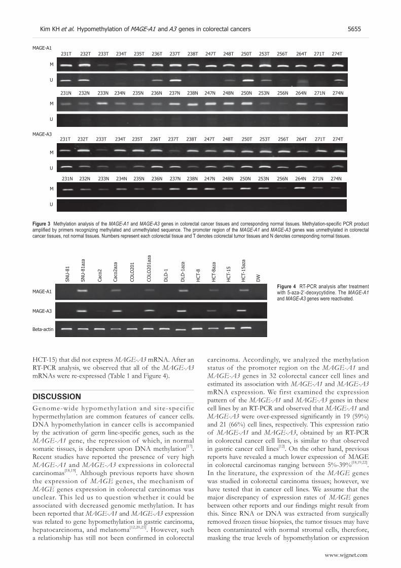
1197A, SNU-C1, SNU-C2A, SNU-C4, Caco2, COLO201, COLO205, COLO320, DLD-1, HCT-15, HCT-116, HT-29, Lovo, LS174T, NCI-H716, SW403, SW1116, and WiDR) (Figure 2 and Table 1). By using primers for amplification of unmethylated or methylated DNA, amplifi ed DNA fragments were found in all 32 cell lines. MAGE-A1 unmethylated DNA products were observed in 37 out of 87 tumor tissue samples (43%; Figure 3). In the normal tissue samples, the methylated DNA was amplified in all 87 samples. However, the unmethylated DNA was amplifi ed in 2 normal tissues (2%). MAGE-A3 unmethylated DNA products were observed in 67 out of 87 tumor tissue samples (77%; Figure 3). In the normal tissue samples, the methylated DNA was amplifi ed in all 87 samples. However, the unmethylated DNA was amplifi ed in 5 normal tissues (6%).
Reexpression of MAGE-A1 and MAGE-A3 after treatment with 5-aza-2’-deoxycytidine We investigated whether MAGE-A1 mRNA was re-expressed after 5-aza-2’-deoxycytidine treatment in 22 cell
Table 1 Methylation status of the promoter region of MAGE-A1 and MAGE-A3 genes in 32 colorectal cancer cell lines
Cell line
MAGEs expression MS-PCR
MAGE-A1 MAGE-A3 MAGE-A1 MAGE-A3
-5-aza/+5-aza - 5-aza/+5-aza M/U M/U
1 SNU-61 - ++ ± ++ + + - + 2 SNU-81 - ++ - +++ + - + - 3 SNU-175 ++ ++ +++ +++ + + + + 4 SNU-283 +++ NT +++ NT + + + - 5 SNU-407 +++ + +++ ++ + - + + 6 SNU-503 ++ NT - NT + + + + 7 SNU-769A +++ ++ +++ +++ - + - + 8 SNU-769B +++ +++ +++ +++ - + - + 9 SNU-1033 ++ + ++ +++ + + + +10 SNU-1040 - - ± ++ + - + -11 SNU-1047 +++ ++ ++ ++ + - - +12 SNU-1197 ++ +++ ++ +++ + + + +13 SNU-C1 ± NT ++ NT + + - +14 SNU-C2A ++ NT +++ NT + + + +15 SNU-C4 - NT - NT + - + +16 SNU-C5 ± ++ - +++ + + + -17 Caco-2 - ++ ++ +++ + + + +18 COLO201 - ++ - +++ - + - +19 COLO205 - - - ++ + + + +20 COLO320 +++ NT +++ NT + + - +21 DLD-1 - +++ - +++ + + + +22 HCT-8 - ++ - +++ + - + -23 HCT-15 - ++ - +++ + + + +24 HCT-116 +++ NT +++ NT + + + +25 HT-29 +++ ++ +++ +++ + + + +26 Lovo +++ +++ +++ +++ + + + +27 LS174T +++ NT +++ NT + + - +28 NCI-H716 +++ +++ +++ +++ + + - +29 SW403 +++ ++ +++ +++ + + + +30 SW480 +++ NT +++ NT + + + -31 SW1116 - NT ++ NT + + - +32 WiDR ++ +++ +++ +++ + + - +
5-aza: 5-aza-2’-deoxycytidine; NT: Not tested; M: The amplifi ed product with primers recognizing methylated sequence; U: The amplifi ed product with primers recognizing unmethylated sequence; +: Amplifi ed product; -: Not amplifi ed product.
Kim KH et al. Hypomethylation of MAGE-A1 and A3 genes in colorectal cancers 5653
www.wjgnet.com

lines, including 10 cell lines (SNU-61, SNU-81, SNU-1040, SNU-C5, Caco-2, COLO201, COLO205, DLD1, HCT-8, and HCT-15) that did not express MAGE-A1 mRNA. After an RT-PCR analysis, we observed that all of the MAGE-A1 mRNAs were re-expressed, except for the SNU-1040 and COLO205 cell lines (Table 1).
This suggested that the inactivation of MAGE-A1 expression was caused by another mechanism. Further, we investigated whether MAGE-A3 mRNA was re-expressed after 5-aza-2’-deoxycytidine treatment in 22 cell lines, including 9 cell lines (SNU-61, SNU-81, SNU-1040, SNU-C5, COLO201, COLO205, DLD1, HCT-8, and
MAGE-A1
MAGE-A3
β-actin
1 2 3 4 5 6 7 8 9 10 11 12 13 14 15 16
17 18 19 20 21 22 23 24 25 26 27 28 29 30 31 32
MAGE-A1
MAGE-A3
β-actin
33 34 DW
MAGE-A1
MAGE-A3
β-actin
Figure 1 RT-PCR analysis of the MAGE-A1 and MAGE-A3 genes in 32 colorectal cancer cell lines. β-actin was amplifi ed as an internal control. The MAGE-A1 gene was signifi cantly expressed in 19 colorectal cancer cell lines (Lanes, 3, 4, 5, 6, 7, 8, 9, 11, 12, 14, 20, 24, 25, 26, 27, 28, 29, 30 and 32). The MAGE-A3 gene was expressed in 21 colorectal cancer cell lines (Lanes, 3, 4, 5, 7, 8, 9, 11, 12, 13, 14, 17, 20, 24, 25, 26, 27, 28, 29, 30, 31 and 32). Lane numbers 1 to 34 show cell lines SNU-61, SNU-81, SNU-175, SNU-283, SNU-407, SNU-503, SNU-769A, SNU-769B, SNU-1033, SNU-1040, SNU-1047, SNU-1197, SNU-C1, SNU-C2A, SNU-C4, SNU-C5, Caco-2, COLO201, COLO205, COLO320, DLD1, HCT-8, HCT-15, HCT-116, HT-29, LOVO, LS174T, NCI-H716, SW403, SW480, SW1116, WiDr, SNU-1, and SNU-5 respectively.
1 2 3 4 5 6 7 8 9 10 11 12 13 14 15 16
M
U
17 18 19 20 21 22 23 24 25 26 27 28 29 30 31 32 DW
MAGE-A1
1 2 3 4 5 6 7 8 9 10 11 12 13 14 15 16
M
U
M
U
17 18 19 20 21 22 23 24 25 26 27 28 29 30 31 32 DW
M
U
MAGE-A3
Figure 2 Methylation analysis of the MAGE-A1 and MAGE-A3 genes in colorectal cancer cell lines. The promoter region of the MAGE-A1 gene was unmethylated in 26 cell lines (SNU-61, SNU-175, SNU-283, SNU-503, SNU-769A, SNU-769B, SNU-1033, SNU-1197A, SNU-C1, SNU-C2A, SNU-C5, Caco2, COLO201, COLO205, COLO320, DLD-1, HCT-15, HCT-116, HT-29, Lovo, LS174T, NCI-H716, SW403, SW1116, and WiDR cell lines). Unmethylated MAGE-A3 DNA amplifi cations were found in 26 cell lines (SNU-61, SNU-175, SNU-407, SNU-503, SNU-769A, SNU-769B, SNU-1033, SNU-1047, SNU-1197A, SNU-C1, SNU-C2A, SNU-C4, Caco2, COLO201, COLO205, COLO320, DLD-1, HCT-15, HCT-116, HT-29, Lovo, LS174T, NCI-H716, SW403, SW1116, and WiDR cell lines). Lane M denotes product amplifi ed by primers recognizing a methylated sequence and Lane U denotes the product amplifi ed by primers recognizing an unmethylated sequence, respectively.
www.wjgnet.com
5654 ISSN 1007-9327 CN 14-1219/R World J Gastroenterol September 21, 2006 Volume 12 Number 35
HCT-15) that did not express MAGE-A3 mRNA. After an RT-PCR analysis, we observed that all of the MAGE-A3 mRNAs were re-expressed (Table 1 and Figure 4).
DISCUSSIONGenome-wide hypomethylat ion and s i te-specif ic hypermethylation are common features of cancer cells. DNA hypomethylation in cancer cells is accompanied by the activation of germ line-specifi c genes, such as the MAGE-A1 gene, the repression of which, in normal somatic tissues, is dependent upon DNA methylation[17]. Recent studies have reported the presence of very high MAGE-A1 and MAGE-A3 expressions in colorectal carcinomas[18,19]. Although previous reports have shown the expression of MAGE genes, the mechanism of MAGE genes expression in colorectal carcinomas was unclear. This led us to question whether it could be associated with decreased genomic methylation. It has been reported that MAGE-A1 and MAGE-A3 expression was related to gene hypomethylation in gastric carcinoma, hepatocarcinoma, and melanoma[12,20,21]. However, such a relationship has still not been confirmed in colorectal
carcinoma. Accordingly, we analyzed the methylation status of the promoter region on the MAGE-A1 and MAGE-A3 genes in 32 colorectal cancer cell lines and estimated its association with MAGE-A1 and MAGE-A3 mRNA expression. We first examined the expression pattern of the MAGE-A1 and MAGE-A3 genes in these cell lines by an RT-PCR and observed that MAGE-A1 and MAGE-A3 were over-expressed signifi cantly in 19 (59%) and 21 (66%) cell lines, respectively. This expression ratio of MAGE-A1 and MAGE-A3, obtained by an RT-PCR in colorectal cancer cell lines, is similar to that observed in gastric cancer cell lines[12]. On the other hand, previous reports have revealed a much lower expression of MAGE in colorectal carcinomas ranging between 5%-39%[18,19,22]. In the literature, the expression of the MAGE genes was studied in colorectal carcinoma tissues; however, we have tested that in cancer cell lines. We assume that the major discrepancy of expression rates of MAGE genes between other reports and our fi ndings might result from this. Since RNA or DNA was extracted from surgically removed frozen tissue biopsies, the tumor tissues may have been contaminated with normal stromal cells, therefore, masking the true levels of hypomethylation or expression
M
U
MAGE-A1231T 232T 233T 234T 235T 236T 237T 238T 247T 248T 250T 253T 256T 264T 271T 274T
M
U
231N 232N 233N 234N 235N 236N 237N 238N 247N 248N 250N 253N 256N 264N 271N 274N
M
U
MAGE-A3231T 232T 233T 234T 235T 236T 237T 238T 247T 248T 250T 253T 256T 264T 271T 274T
M
U
231N 232N 233N 234N 235N 236N 237N 238N 247N 248N 250N 253N 256N 264N 271N 274N
Figure 3 Methylation analysis of the MAGE-A1 and MAGE-A3 genes in colorectal cancer tissues and corresponding normal tissues. Methylation-specifi c PCR product amplifi ed by primers recognizing methylated and unmethylated sequence. The promoter region of the MAGE-A1 and MAGE-A3 genes was unmethylated in colorectal cancer tissues, not normal tissues. Numbers represent each colorectal tissue and T denotes colorectal tumor tissues and N denotes corresponding normal tissues.
MAGE-A1
MAGE-A3
Beta-actin
SNU
-81
SNU
-81a
za
Caco
2
Caco
2aza
COLO
201
COLO
201a
za
DLD
-1
DLD
-1az
a
HCT
-8
HCT
-8az
a
HCT
-15
HCT
-15a
za
DW
Figure 4 RT-PCR analysis after treatment with 5-aza-2’-deoxycytidine. The MAGE-A1 and MAGE-A3 genes were reactivated.
Kim KH et al. Hypomethylation of MAGE-A1 and A3 genes in colorectal cancers 5655
www.wjgnet.com
of the MAGE genes in cancer tissues[23]. For further analysis of the methylation status of the MAGE genes in colorectal cancer tissues, laser capture microdissection techniques would allow more precise isolation of cancer cells and normal cells. It has already been reported that cancer cell lines have much higher levels of CpG island hypermethylation than corresponding malignant tissues, which may explain our lower incidence of hypomethylation in tissues versus cell lines. Moreover, cancer cells might be clonally selected with growth advantages over cancer cell lines. However, cancer cell lines often preserve hypermethylation or hypomethylation from the tumors they originate, thus they are indeed useful tools to study methylation status.
We ana lyzed promoter unmethy la t ion of the MAGE-A1 and MAGE-A3 genes with a methylation-specific PCR after sodium-bisulfite modification and by direct sequencing analysis. Of the 32 cell lines analyzed, the promoter hypomethylation of MAGE-A1 and MAGE-A3 was observed in both at 26 cell lines each. Further, 23 cell lines (SNU-61, SNU-175, SNU-503, SNU-769A, SNU-769B, SNU-1033, SNU-1197A, SNU-C1, SNU-C2A, Caco2, COLO201, COLO205, COLO320, DLD-1, HCT-15, HCT-116, HT-29, Lovo, LS174T, NCI-H716, SW403, SW1116, and WiDR) were simultaneously unmethylated in both the MAGE-A1 and MAGE-A3 genes. With exception, there were two cell lines (SNU-61, COLO201) with negative gene expression for either MAGE-A1 or MAGE-A3, but unmethylated MAGE-A1 or MAGE-A3 promoter was detected. On the contrary, there were four cell lines (SNU-283, SNU-407, SNU-1047 and SW480) in which MAGE genes were strongly expressed, although no unmethylated MAGE promoter could be detected, suggesting the activation or inactivation of MAGE expression by another mechanism.
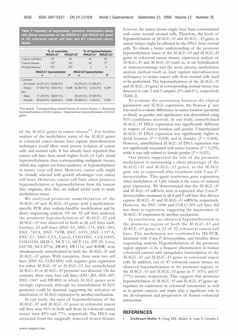
In our study, the rates of hypomethylation of the MAGE-A1 and MAGE-A3 genes in colorectal cancer cell lines were 81% in both and those in colorectal cancer tissues were 43% and 77%, respectively. The DNA was extracted from the surgically removed frozen-tissues;
however, the tumor tissues might have been contaminated with some normal stromal cells. Therefore, the levels of hypomethylation of MAGE-A1 and MAGE- A3 genes in cancer tissues might be affected by the DNA from normal cells. To obtain a better understanding of the promoter hypomethylation status of the MAGE-A1 and MAGE-A3 genes in colorectal cancer tissues, expression analysis of MAGE-A1 and MAGE-A3 (such as, in situ hybridization or immunostaining) and the more precise methylation analysis method (such as, laser capture microdissection techniques) to isolate cancer cells from normal cells need to be performed. The hypomethylation of the MAGE-A1 and MAGE-A3 genes in corresponding normal tissues was detected in only 2 and 5 samples (2% and 6%), respectively (Table 2).
To evaluate the association between the clinical parameters and MAGE expression, the Pearson χ2 test was used to evaluate differences in tumor location (proximal or distal) or gender, and signifi cance was determined using 95% confidence intervals. In our study, unmethylated MAGE-A1 DNA expression was significantly different in respect of tumor location and gender. Unmethylated MAGE-A1 DNA expression was significantly higher in distal location (P = 0.028) and in females (P = 0.004). However, unmethylated MAGE-A3 DNA expression was not signifi cantly associated with tumor location (P = 0.239), while it was only related to female gender (P = 0.021).
Our results supported the role of the promoter methylation in maintaining a silent phenotype of the MAGE-A1 and MAGE-A3 genes, as the MAGE gene was re-expressed after treatment with 5-aza-2'-deoxycytidine. This agent reactivates gene expression when methylation of CpG islands is the cause of reduced gene expression. We demonstrated that the MAGE-A1 and MAGE-A3 mRNAs were re-expressed after 5-aza-2'-deoxycytidine treatment in all 8 and 9 cell lines that did not express MAGE-A1 and MAGE-A3 mRNAs, respectively. However, the SNU-1040 and COLO 205 cell lines did not show re-expression, suggesting the inactivation of MAGE-A1 expression by another mechanism.
In conclusion, we observed hypomethylation in the promoter region of both the MAGE-A1 and MAGE-A3 genes in 23 of 32 colorectal cancer cell lines. This methylation was confirmed by MS-PCR, treatment with 5-aza-2’-deoxycytidine, and bisulfi te direct sequencing analysis. Hypomethylation of the promoter region appears to be a frequent phenomenon in human colorectal cancers and upregulates transcription of the MAGE-A1 and MAGE-A3 genes in colorectal cancer cells. In addition, out of 87 colorectal cancer tissues, we observed hypomethylation in the promoter regions of the MAGE-A1 and MAGE-A3 genes in 37 (43%) and 67 (77%) tissues, respectively. This suggests that promoter hypomethylation of MAGE-A1 and MAGE-A3 genes up-regulates its expression in colorectal carcinomas as well as in gastric cancers, and might play a signifi cant role in the development and progression of human colorectal carcinomas.
REFERENCES1 Zochbauer-Muller S, Fong KM, Maitra A, Lam S, Geradts J,
Table 2 Summary of expressions, promoter methylation status and clinical associations of the MAGE-A1 and MAGE-A3 genes in 32 colorectal cancer cell lines and 87 colorectal cancer tissues
% of expression % of hypomethylation MAGE-A1 MAGE-A3 MAGE-A1 MAGE-A3 Cancer cell lines 59 66 81 81Cancer tissues NT1 NT 43 77 Normal tissues2 NT NT 2 6
MAGE-A1 hypomethylation MAGE-A3 hypomethylation + - P + - P Location Proximal 14 (31.1%) 31(68.9%) 34 (75.6%) 11 (24.4%) Distal 17 (56.7%) 13(43.3%) 0.028 26 (86.7%) 4 (13.3%) 0.239Sex Male 17 (30.9%) 38(69.1%) 38 (69.1%) 17 (30.9%) Female 20 (62.5%) 12(18.4%) 0.004 29 (90.6%) 3 (9.4%) 0.021
1Not tested; 2Corresponding normal tissues of cancer tissues; +: Represent hypomethylation of MAGE genes; -: Represent no hypomethylation of MAGE genes.
www.wjgnet.com
5656 ISSN 1007-9327 CN 14-1219/R World J Gastroenterol September 21, 2006 Volume 12 Number 35

Ashfaq R, Virmani AK, Milchgrub S, Gazdar AF, Minna JD. 5’ CpG island methylation of the FHIT gene is correlated with loss of gene expression in lung and breast cancer. Cancer Res 2001; 61: 3581-3585
2 Song SH, Jong HS, Choi HH, Inoue H, Tanabe T, Kim NK, Bang YJ. Transcriptional silencing of Cyclooxygenase-2 by hyper-methylation of the 5’ CpG island in human gastric carcinoma cells. Cancer Res 2001; 61: 4628-4635
3 Suh ER, Ha CS, Rankin EB, Toyota M, Traber PG. DNA methylation down-regulates CDX1 gene expression in colorectal cancer cell lines. J Biol Chem 2002; 277: 35795-35800
4 Melki JR, Vincent PC, Brown RD, Clark SJ. Hypermethylation of E-cadherin in leukemia. Blood 2000; 95: 3208-3213
5 Yang Q, Nakamura M, Nakamura Y, Yoshimura G, Suzuma T, Umemura T, Shimizu Y, Mori I, Sakurai T, Kakudo K. Two-hit inactivation of FHIT by loss of heterozygosity and hypermethylation in breast cancer. Clin Cancer Res 2002; 8: 2890-2893
6 Ku JL, Kang SB, Shin YK, Kang HC, Hong SH, Kim IJ, Shin JH, Han IO, Park JG. Promoter hypermethylation downregulates RUNX3 gene expression in colorectal cancer cell lines. Oncogene 2004; 23: 6736-6742
7 Feinberg AP, Vogelstein B. Hypomethylation of ras oncogenes in primary human cancers. Biochem Biophys Res Commun 1983; 111: 47-54
8 Fang JY, Zhu SS, Xiao SD, Jiang SJ, Shi Y, Chen XY, Zhou XM, Qian LF. Studies on the hypomethylation of c-myc, c-Ha-ras oncogenes and histopathological changes in human gastric carcinoma. J Gastroenterol Hepatol 1996; 11: 1079-1082
9 Van Der Bruggen P , Zhang Y, Chaux P, Stroobant V, Panichelli C, Schultz ES, Chapiro J, Van Den Eynde BJ, Brasseur F, Boon T. Tumor-specifi c shared antigenic peptides recognized by human T cells. Immunol Rev 2002; 188: 51-64
10 De Smet C, Lurquin C, Lethe B, Martelange V, Boon T. DNA methylation is the primary silencing mechanism for a set of germ line- and tumor-specific genes with a CpG-rich promoter. Mol Cell Biol 1999; 19: 7327-7335
11 De Smet C, De Backer O, Faraoni I, Lurquin C, Brasseur F, Boon T. The activation of human gene MAGE-1 in tumor cells is correlated with genome-wide demethylation. Proc Natl Acad Sci USA 1996; 93: 7149-7153
12 Honda T , Tamura G, Waki T, Kawata S, Terashima M, Nishizuka S, Motoyama T. Demethylation of MAGE promoters during gastric cancer progression. Br J Cancer 2004;
90: 838-84313 Liu J, Wang G, Okutomi T, Chen Z. Expression of MAGE-A1
and MAGE-A3 genes in human salivary gland carcinomas. Chin Med J (Engl) 2003; 116: 897-900
14 Park JG, Oie HK, Sugarbaker PH, Henslee JG, Chen TR, Johnson BE, Gazdar A. Characteristics of cell lines established from human colorectal carcinoma. Cancer Res 1987; 47: 6710-6718
15 Oh JH , Ku JL, Yoon KA, Kwon HJ, Kim WH, Park HS, Yeo KS, Song SY, Chung JK, Park JG. Establishment and characterization of 12 human colorectal-carcinoma cell lines. Int J Cancer 1999; 81: 902-910
16 Herman JG, Graff JR, Myohanen S, Nelkin BD, Baylin SB. Methylation-specifi c PCR: a novel PCR assay for methylation status of CpG islands. Proc Natl Acad Sci USA 1996; 93: 9821-9826
17 De Smet C, Loriot A, Boon T. Promoter-dependent mechanism leading to selective hypomethylation within the 5’region of gene MAGE-A1 in tumor cells. Mol Cell Biol 2004; 24: 4781-4790
18 Park MS, Park JW, Jeon CH, Lee KD, Chang HK. Expression of melanoma antigen-encoding genes (MAGE) by common primers for MAGE-A1 to -A6 in colorectal carcinomas among Koreans. J Korean Med Sci 2002; 17: 497-501
19 Koketsu S, Watanabe T, Kazama S, Ishihara S, Komuro Y, Nagawa H. What types of colorectal cancer overexpress the MAGE protein? Hepatogastroenterology 2004; 51: 1648-1652
20 Xiao J, Chen HS, Fei R, Cong X, Wang LP, Wang Y, Jiang D, Wei L, Wang Y. Expression of MAGE-A1 mRNA is associated with gene hypomethylation in hepatocarcinoma cell lines. J Gastroenterol 2005; 40: 716-721
21 Zendman AJ, de Wit NJ, van Kraats AA, Weidle UH, Ruiter DJ, Van Muijen GN. Expression profile of genes coding for melanoma differentiation antigens and cancer/testis antigens in metastatic lesions of human cutaneous melanoma. Melanoma Res 2001; 11: 451-459
22 Li M , Yuan YH, Han Y, Liu YX, Yan L, Wang Y, Gu J. Expression profile of cancer-testis genes in 121 human colorectal cancer tissue and adjacent normal tissue. Clin Cancer Res 2005; 11: 1809-1814
23 Ku JL, Kang SB, Shin YK, Kang HC, Hong SH, Kim IJ, Shin JH, Han IO, Park JG. Promoter hypermethylation downregulates RUNX3 gene expression in colorectal cancer cell lines. Oncogene 2004; 23: 6736-6742
S- Editor Pan BR L- Editor Zhu LH E- Editor Liu WF
Kim KH et al. Hypomethylation of MAGE-A1 and A3 genes in colorectal cancers 5657
www.wjgnet.com