Embed Size (px)
Citation preview
Int J Clin Exp Pathol 2015;8(9):10475-10481www.ijcep.com /ISSN:1936-2625/IJCEP0012656
Original ArticleClinicopathological features of cryoglobulinemic glomerulonephritis associated with HBV infection: a retrospective analysis of 8 cases in China
Chen Wang1*, Zi-Yin Ye2*, De-Hua Zeng3, Fei-Lai Xie3, Li-Juan Qu3, Zhi-Yong Zheng3
1Department of Pathology, Fujian Provincial Hospital, Fujian Provincial Clinical College of Fujian Medical Univer-sity, Fuzhou, China; 2Department of Pathology, The First Affiliated Hospital of Sun Yat-sen University, Guangzhou, China; 3Department of Pathology, Dongfang Hospital, Fujian Medical University, Fuzhou, China. *Equal contribu-tors and co-first authors.
Received July 9, 2015; Accepted August 21, 2015; Epub September 1, 2015; Published September 15, 2015
Abstract: Aims: We retrospectively analyzed clinicopathologic features of 8 cases of hepatitis B virus-associated glomerulonephritis with hyaline thrombi, to confirm the diagnosis of cryoglobulinemic glomerulonephritis (CRYGN) associated with HBV infection. Methods: Retrospective analysis was carried out with demographic information, clini-cal manifestations, laboratory parameters, pathological and prognostic features. Results: The median age of 8 pa-tients was 30.5 years (range, 21-75 years), including 6 males and 2 femles (M:F = 3:1). One patient had Raynaud’s syndrome. Cryoglobulin testing was performed in 4 cases of our series, and 3 cases had elevated cryocrit (>256). Serum C4 decreased in all detected cases. Histopathologically, all cases showed hyaline thrombi occluded in capil-lary lumina; Co-deposit of IgG, IgM, IgA, Fib, C3d, C4d, C1q, HBsAg and HBcAb were identified in hyaline deposit/hyaline thrombi with polyclonal Igκ and Igλ staining. Ultrastructural examination confirmed the hyaline thrombi to be huge electron-dense bodies, which were a homogeneous texture. Conclusions: The results suggest that 8 cases in the series are CRYGN associated with HBV infection. The incidence of CRYGN associated HBV was extremely low. Our series suggested that prognosis of CRYGN associated HBV was better in patients with mild symptoms, but it was poor in elder patients with severe vasculitis.
Keywords: Hyaline thrombi, cryoglobulinemia, glomerulonephritis, hepatitis B virus
Introduction
Mixed cryoglobulinemia is occasionally seen in patients with hepatitis B virus (HBV) infection, which present as protracted purpura, ulcer-ative skin lesions, etc [1, 2]. It may share similar mechanism as HCV infection commonly with a poly-oligoclonal B-lymphocyte expansion, which are responsible for the production of different autoantibodies and immune-complexes, such as mixed cryoglobulins [3].
Kidney may be affected by HBV infection. Although pathology of hepatitis B virus-associ-ated glomerulonephritis (HBV-GN) has not been clearly defined so far, it is believed to be an immune complex-mediated process that is most often associated membranous glomeru-lopathy or membranoproliferative glomerulone-phritis types I and III [4]. We have found a few
cases of HBV-GN showed membranous nephropathy and membranoproliferative glo-merulonephritis with hyaline thrombi occluded in capillary lumina, which was believed to be mixed cryoglobulins. HBV infection is an uncom-mon cause of cryoglobulinemia. Renal cryoglob-ulinemia has been rarely reported in the setting of chronic hepatitis B infection. We retrospec-tively analyzed clinicopathologic features of a series of HBV-GN cases with hyaline thrombi, which thought to be cryoglobulinemic glomeru-lonephritis (CRYGN) associated with HBV infec-tion, may expand our understanding of the disease.
Materials and methods
Patients
Renal biopsy records from 2006, Jan to 2014; Jun had been retrieved from the archive of
Cryoglobulinemic glomerulonephritis associated with HBV infection
10476 Int J Clin Exp Pathol 2015;8(9):10475-10481
Department of pathology, Fujian DongFang Hospital, China. In a total of 34117 consecutive cases of renal biopsy, there were 1970 cases (5.77%) with hepatitis B virus markers (HBsAg or HBcAg) deposition, and 8 cases (0.023%) of HBV-GN with hyaline thrombi.
Clinical information and laboratory values were obtained by review of medical records, includ-ing sex, age, disease course (onset to renal biopsy time), clinical diagnosis, proteinuria (g/24 hours), microscopic hematuria (+, >10 red blood cells/HPF; 2+, >20 red blood cells/HPF; 3+, >30 red blood cells/HPF), serum cre-atinine (SCr) (normal range 53-124 umol/L), blood urea nitrogen (BUN) (normal range 2.9-8.9 nmol/L), serum C3 (normal range 0.90~1.80 g/L), serum C4 (normal range 0.10-0.40 g/L), blood pressure (hypertension was defined as systolic pressure >140 mmHg or diastolic blood pressure >90 mmHg), HBV marks (HBsAg, HBeAg, anti-HBs, anti-HBe and anti-HBc), Serum cryoprecipitate (normal titer range 0-32 at 4°C, pathological titer is greater than or equal to 256 at 4°C).
With/without liver dysfunction, arthritis, myal-gia or Raynaud’s syndrome, bone marrow biop-sies were performed in all patients. Follow up time was recorded from the time of discharge. Patient progressing to end-stage renal disease (ESRD) was defined as end point of study.
This study was approved by the Human Ethics Committee of The DongFang Provincial Hos- pital.
Methods
All renal biopsy specimens were submitted for light microscopy examination. Specimens for light microscopy were fixed in 10% formalde-hyde (formalin), embedded in paraffin and sec-tions were cut at four microns and stained with hematoxylin and eosin (H&E), periodicacid-Schiff (PAS) and periodic acid-silver methena-mine and Masson’s trichrom (PAM-Masson).
The 3rd and 6th cases were submitted for elec-tron microscopy examination.
Formalin-fixed, paraffin-embedded sections of the biopsied tissue were stained with primary antibody used for immunohistochemical study by the Elivision system, which were IgG (DAKO),
IgA (DAKO), IgM (DAKO), C3d (Abcam), C4d (Biomedica), C1q (DAKO), Fib (DAKO), CD68 (PGM-1) (EliVision), CD61 (EliVision), Igκ (DAKO), Igλ (DAKO), HBsAg (EliVision), anti-HBc (EliVision) and Apo E (DAKO). IgG, IgA, IgM, C3d, C1q, Fib, Igκ and Igλ were retrieved with high-pressure heating method in 0.01 mol/L pH6.0 citrate buffer and 0.4% gastric enzyme (Anresco) (9 minutes for IgG, Igκ, Igλ and C3d; 10 minutes for IgA, IgM and Fib [5]). HBsAg and HBcAb were retrieved with high-pressure heat-ing method with 0.05% type-ΧΧIV protease for 7 minutes). CD68 (PGM-1) and CD61 were retrieved with high-pressure heating method. Apo E was not retrieved. The remaining steps were performed according to the kit manual.
Evaluation for immunohistochemical results: A semiquantitative grading of the extent and intensity of all the antibodies staining was per-formed. A score of 0 to 3 was defined as fol-lows: 0, no staining (negative); 1, less than 25% of arterioles stained (+, weak); 2, 25% to 50% arterioles stained (2+, moderate); 3, more than 50% arterioles stained (3+, strong). The total scores were scores of extent multiply by those of intensity.
Results
Clinical features (Table 1)
There were 8 cases of HBV-GN in the study, including 6 male and 2 females (M:F = 3:1). The median age was 30.5 years (range, 21-75 years). All patients had proteinuria and micro-scopic hematuria to different extents. One patient had mild elevated BUN. The serum cre-atinine was normal in all the patients. Two patients had hypertension. The 3rd and 4th cases had chronic hepatitis B. Seven cases had arthralgia and myoalgia to different extents, One case had Raynaud’s syndrome. Serum cryoglobulin testing was performed in 4 cases, 3 cases of which were positive (the titer is greater than 256). Serum C3 and serum C4 was detected in 5 cases: serum C3 deceased in 2 cases; serum C4 decreased in all detected cases. Serum HBsAg, HBeAg and anti-HBcAb were positive in all cases. Serum HCV antibody and Bence-Jones protein were negative in all cases. Bone marrow biopsies were negative for plasmacytosis or tumor involvement in all
Cryoglobulinemic glomerulonephritis associated with HBV infection
10477 Int J Clin Exp Pathol 2015;8(9):10475-10481
Table 1. Clinical features of cases with HBV-GN with hyaline thrombi
NO Gender Age Disease course
Serum cryopre-cipitate (>256)
Urine protein (g/24 hr)
Microscopic hematuria
SCr (umol/L)
BUN (mmol/L)
Hyper-tension
Serum C3 (g/L)
Serum C4 (g/L)
Liver dysfuction
Raynaud’s syndrome
Follow up time (months) ESRD
1 M 32 3 Months positive 1.47 3+ 69 5.39 No - - No No Loss to follow up -
2 M 29 2 Months - 2.13 3+ 75 5.77 No - - No No Loss to follow up -
3 F 35 2weeks positive 1.07 1+ 63 5.24 No 1.2 0.08 Yes No 49 No
4 M 29 1 year - 0.75 3+ 86 3.33 No 0.56 0.07 Yes No 40 No
5 M 21 2 years negative 0.75 3+ 82 5.56 No 1.2 0.04 No No 39 No
6 F 25 4 months - 0.61 2+ 57 6.72 Yes 1.29 0.05 No No 32 No
7 M 59 2 months - 8.07 2+ 79 4.34 No - - No No 23 No
8 M 75 6 months positive 1.11 3+ 81 9.76 Yes 0.41 0.03 No Yes 13 Died
Table 2. Pathologic features of cases with HBV-GN with hyaline thrombiImmunohistochemical staining
No. Morphologic patterns IgG* IgM* IgA* C3d* C4d* C1q* Fib* Igκ* Igλ* HBsAg* HBcAg* PGM-1**
1 Membranous nephropathy 9 4 1 9 9 4 0 2 2 4 1 02 Membranous nephropathy 4 1 0 6 9 1 0 1 1 4 4 13 Membranous nephropathy 6 4 1 9 9 0 1 2 2 4 4 04 Membranous nephropathy 2 4 0 9 9 1 4 1 1 4 4 25 Membranous nephropathy 9 1 1 9 9 2 4 1 1 4 0 16 Diffuse mesangial proliferative nephropathy with focal membranoproliferation 4 9 1 9 9 6 4 2 2 9 1 17 Membranous nephropathy 4 4 1 4 6 4 2 2 2 4 0 18 Diffuse mesangial proliferative nephropathy with focal membranoproliferation 1 9 1 6 6 2 4 1 2 1 1 3*Results of immunohistochemical staining were evaluated by total scores. **CD68 (PGM1) was average numbers of macrophages within each glomerular capillary.
Cryoglobulinemic glomerulonephritis associated with HBV infection
10478 Int J Clin Exp Pathol 2015;8(9):10475-10481
cases. SLE and IgM monoclonal gammopathy had also been ruled out.
Light microscopy
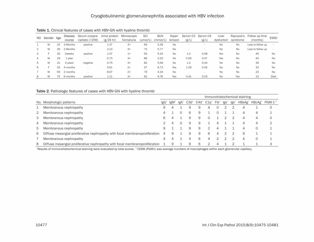
Six cases exhibited diffuse membranous nephropathy and two cases exhibited diffuse mesangial proliferative nephropathy with focal membranoproliferation (Table 2). All cases showed round or oval homogenous huge hya-line deposit in mesangial areas, with hyaline thrombi occluded in capillary lumina. Hyaline thrombi deposit was stained purple with PAS and red with PASM-Masson (Figure 1). There were focal segmental or global glomerular scle-rosis, tubular atrophy and interstitial fibrosis. Congo read staining was negative.
Immunohistochemical study
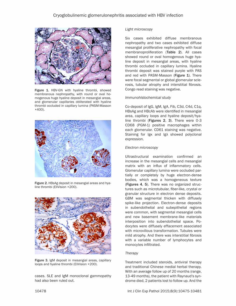
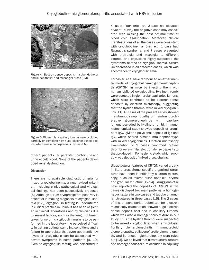
Co-deposit of IgG, IgM, IgA, Fib, C3d, C4d, C1q, HBsAg and HBcAb were identified in mesangial area, capillary loops and hyaline deposit/hya-line thrombi (Figures 2, 3). There were 0-3 CD68 (PGM-1) positive macrophages within each glomerular. CD61 staining was negative. Staining for Igκ and Igλ showed polyclonal expression.
Electron microscopy
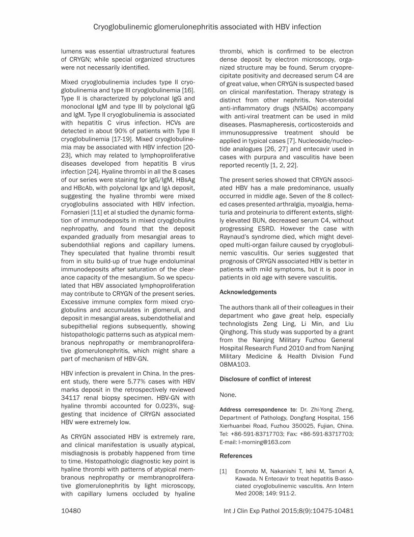
Ultrastructural examination confirmed an increase in the mesangial cells and mesangial matrix with an influx of inflammatory cells. Glomerular capillary lumina were occluded par-tially or completely by huge electron-dense bodies, which was a homogeneous texture (Figures 4, 5). There was no organized struc-tures such as microtubular, fiber-like, crystal or granular structure in electron dense deposits. GBM was segmental thicken with diffusely spike-like projection. Electron-dense deposits in subendothelial and subepithelial regions were common, with segmental mesangial cells and new basement membrane-like materials interposition into subendothelial space. Po- docytes were diffusely effacement associated with microvillous transformation. Tubules were mild atrophy. And there was interstitial fibrosis with a variable number of lymphocytes and monocytes infiltrated.
Therapy
Treatment included steroids, antiviral therapy and traditional Chinese medial herbal therapy. With an average follow up of 20 months (range, 13-49 months), the patient with Raynaud’s syn-drome died, 2 patients lost to follow up. And the
Figure 1. HBV-GN with hyaline thrombi, showed membranous nephropathy, with round or oval ho-mogenous huge hyaline deposit in mesangial areas, and glomerular capillaries obliterated with hyaline thrombi occluded in capillary lumina (PASM-Masson ×400).
Figure 2. HBsAg deposit in mesangial areas and hya-line thrombi (EliVision ×200).
Figure 3. IgM deposit in mesangial areas, capillary loops and hyaline thrombi (EliVision ×200).
Cryoglobulinemic glomerulonephritis associated with HBV infection
10479 Int J Clin Exp Pathol 2015;8(9):10475-10481
other 5 patients had persistent proteinuria and urine occult blood. None of the patients devel-oped renal dysfunction.
Discussion
There are no available diagnostic criteria for mixed cryoglobulinemia; a new revised criteri-on, including clinico-pathological and virologi-cal findings, has been successively proposed [6]. Although serum cryoprecipitate positivity is essential in making diagnosis of cryoglobuline-mia [6-8], cryoglobulin testing is underutilized in clinical practice in China. It has been neglect-ed in clinical laboratories and by clinicians due to several factors, such as the length of time it takes for serum cryoglobulin analysis to be per-formed in the laboratory, the perceived difficul-ty in getting optimal sampling conditions and a failure to appreciate that even apparently low levels of cryoglobulin can be associated with severe symptoms in some patients [9, 10]. Even so cryoglobulin testing was performed in
4 cases of our series, and 3 cases had elevated cryocrit (>256), the negative case may associ-ated with missing the best optimal time of blood cold agalutination. Moreover, clinical manifestations of all the cases were consistent with cryoglobulinemia [6-9], e.g. 1 case had Raynaud’s syndrome, and 7 cases presented with arthralgia and myoalgia to different extents, and physicians highly suspected the symptoms related to cryoglobulinemia. Serum C4 decreased in all detected cases, which was accordance to cryoglobulinemia.
Fornasieri et al have reproduced an experimen-tal model of cryoglobulinemic glomerulonephri-tis (CRYGN) in mice by injecting them with human IgMk-IgG cryoglobulins. Hyaline thrombi were detected in glomerular capillaries lumens, which were confirmed to be electron-dense deposits by electron microscopy, suggesting that the hyaline thrombi were mixed cryoglobu-lins [11]. All cases of the present series showed membranous nephropathy or membranoprolif-erative glomerulonephritis with capillary lumens occluded by hyaline thrombi. Immuno- histochemical study showed deposit of promi-nent IgG/IgM and polyclonal deposit of Igκ and Igλ, which shared similar immunophenotype with mixed cryoglobulins. Electron microscopy examination of 2 cases confirmed hyaline thrombi were similar electron dense deposits to that produced in Fornasieri’s study, which prob-ably was deposit of mixed cryoglobulins.
Ultrastuctural features of CRYGN varied greatly in literatures. Some specific organized struc-tures have been identified by electron micros-copy, such as microtubular, fiber-like, crystal and granular structure [12-14]. Faraggiana et al have reported the deposits of CRYGN in five cases displayed two main patterns; a homoge-neous texture in two cases and tubular or annu-lar structures in three cases [15]. The 2 cases of the present series submitted for electron microscopy examination showed huge electron dense deposit occluded in capillary lumens, which was also a homogeneous texture in our study. Thus the hyaline thrombi were suspected to be mixed cryoglobulins, when amyloidosis, fibrillary glomerulonephritis, immunotactoid glomerulopathy, collagenofibrotic glomerulopa-thy and fibronectin glomerulopathy were ruled out [13]. We believed that ultrastuctural feature of a homogeneous texture occluded in capillary
Figure 4. Electron-dense deposits in subendothelial and subepithelial and mesangial areas (EM).
Figure 5. Glomerular capillary lumina were occluded partially or completely by huge electron-dense bod-ies, which was a homogeneous texture (EM).
Cryoglobulinemic glomerulonephritis associated with HBV infection
10480 Int J Clin Exp Pathol 2015;8(9):10475-10481
lumens was essential ultrastructural features of CRYGN; while special organized structures were not necessarily identified.
Mixed cryoglobulinemia includes type II cryo-globulinemia and type III cryoglobulinemia [16]. Type II is characterized by polyclonal IgG and monoclonal IgM and type III by polyclonal IgG and IgM. Type II cryoglobulinemia is associated with hepatitis C virus infection. HCVs are detected in about 90% of patients with Type II cryoglobulinemia [17-19]. Mixed cryoglobuline-mia may be associated with HBV infection [20-23], which may related to lymphoproliferative diseases developed from hepatitis B virus infection [24]. Hyaline thrombi in all the 8 cases of our series were staining for IgG/IgM, HBsAg and HBcAb, with polyclonal Igκ and Igλ deposit, suggesting the hyaline thrombi were mixed cryoglobulins associated with HBV infection. Fornasieri [11] et al studied the dynamic forma-tion of immunodeposits in mixed cryoglobulins nephropathy, and found that the deposit expanded gradually from mesangial areas to subendothlial regions and capillary lumens. They speculated that hyaline thrombi result from in situ build-up of true huge endoluminal immunodeposits after saturation of the clear-ance capacity of the mesangium. So we specu-lated that HBV associated lymphoproliferation may contribute to CRYGN of the present series. Excessive immune complex form mixed cryo-globulins and accumulates in glomeruli, and deposit in mesangial areas, subendothelial and subepithelial regions subsequently, showing histopathologic patterns such as atypical mem-branous nephropathy or membranoprolifera-tive glomerulonephritis, which might share a part of mechanism of HBV-GN.
HBV infection is prevalent in China. In the pres-ent study, there were 5.77% cases with HBV marks deposit in the retrospectively reviewed 34117 renal biopsy specimen. HBV-GN with hyaline thrombi accounted for 0.023%, sug-gesting that incidence of CRYGN associated HBV were extremely low.
As CRYGN associated HBV is extremely rare, and clinical manifestation is usually atypical, misdiagnosis is probably happened from time to time. Histopathologic diagnostic key point is hyaline thrombi with patterns of atypical mem-branous nephropathy or membranoprolifera-tive glomerulonephritis by light microscopy, with capillary lumens occluded by hyaline
thrombi, which is confirmed to be electron dense deposit by electron microscopy, orga-nized structure may be found. Serum cryopre-cipitate positivity and decreased serum C4 are of great value, when CRYGN is suspected based on clinical manifestation. Therapy strategy is distinct from other nephritis. Non-steroidal anti-inflammatory drugs (NSAIDs) accompany with anti-viral treatment can be used in mild diseases. Plasmapheresis, corticosteroids and immunosuppressive treatment should be applied in typical cases [7]. Nucleoside/nucleo-tide analogues [26, 27] and entecavir used in cases with purpura and vasculitis have been reported recently [1, 2, 22].
The present series showed that CRYGN associ-ated HBV has a male predominance, usually occurred in middle age. Seven of the 8 collect-ed cases presented arthralgia, myoalgia, hema-turia and proteinuria to different extents, slight-ly elevated BUN, decreased serum C4, without progressing ESRD. However the case with Raynaud’s syndrome died, which might devel-oped multi-organ failure caused by cryoglobuli-nemic vasculitis. Our series suggested that prognosis of CRYGN associated HBV is better in patients with mild symptoms, but it is poor in patients in old age with severe vasculitis.
Acknowledgements
The authors thank all of their colleagues in their department who gave great help, especially technologists Zeng Ling, Li Min, and Liu Qinghong. This study was supported by a grant from the Nanjing Military Fuzhou General Hospital Research Fund 2010 and from Nanjing Military Medicine & Health Division Fund 08MA103.
Disclosure of conflict of interest
None.
Address correspondence to: Dr. Zhi-Yong Zheng, Department of Pathology, Dongfang Hospital, 156 Xierhuanbei Road, Fuzhou 350025, Fujian, China. Tel: +86-591-83717703; Fax: +86-591-83717703; E-mail: [email protected]
References
[1] Enomoto M, Nakanishi T, Ishii M, Tamori A, Kawada. N Entecavir to treat hepatitis B-asso-ciated cryoglobulinemic vasculitis. Ann Intern Med 2008; 149: 911-2.
Cryoglobulinemic glomerulonephritis associated with HBV infection
10481 Int J Clin Exp Pathol 2015;8(9):10475-10481
[2] D’Amico E, Pace-Palitti V, Di Lembo E, Palazzi C. Successful treatment of hepatitis B virus in-fection and related cryoglobulinaemic purpura with nucleoside/nucleotide analogues. Clin Exp Rheumatol 2013; 31: 155.
[3] Vladareanu AM, Ciufu C, Neagu AM, Onisai M, Bumbea H, Vintilescu AM, Dobrea C, Arama V, Mihailescu R, Arama S. The impact of hepatitis viruses on chronic lymphoproliferative disor-ders-preliminary results. J Med Life 2010; 3: 320-9.
[4] Bhimma R, Coovadia HM. Hepatitis B. virus-associated nephropathy. Am J Nephrol 2004; 24: 198-211.
[5] Zhang R, Lin J, Qu L, Zheng F, Zheng Z. C3d deposition in the media of renal arterioles is a useful marker for arteriolosclerosis in IgA ne-phropathy. Ann Diagn Pathol 2014; 18: 104-108.
[6] Ferri C, Zignego AL, Pileri SA. Review. Cryoglob-ulins. J Clin Pathol 2002; 55: 4-13.
[7] Takada S, Shimizu T, Hadano Y, Matsumoto K, Kataoka Y, Arima Y, Inoue T, Sorano S. Cryo-globulinemia. Mol Med Rep 2012; 6: 3-8.
[8] Sargur R, White P, Egner W. Cryoglobulin evalu-ation: best practice? Ann Clin Biochem 2010; 47: 8-16.
[9] Mascia MT, Ferrari D, Campioli D, Sandri G, Mussini C, Ferri C. Non HCV-related mixed cryo-globulinemia. Dig Liver Dis 2007; 39: S61-S64.
[10] Monti G, Saccardo F, Castelnovo L, Novati P, Sollima S, Riva A, Sarzi-Puttini P, Quartuccio L, De Vita S, Galli M. Prevalence of mixed cryo-globulinaemia syndrome and circulating cryo-globulins in a population-based survey: the Origgio study. Autoimmun Rev 2014; 13: 609-14.
[11] Fornasieri A, Tazzari S, Li M, Armelloni S, Tarel-li LT, Sessa A, D’Amico G. Electron microscopy study of genesis and dynamics of immunode-position in IgMk-IgG cryoglobulin-induced glo-merulonephritis in mice. Am J Kidney Dis 1998; 31: 435-42.
[12] Ojemakinde K, Turbat-Herrera EA, Zeng X, Gu X, Herrera GA. The many faces of cryoglobulin-emic nephropathy: a clinico-pathologic study of 47 cases with emphasis on the value of electron microscopy. Ultrastruct Pathol 2014; 38: 367-76.
[13] Iskandar SS, Herrera GA. Glomerulopathies with organized deposits. Semin Diagn Pathol 2002; 19: 116-32.
[14] Monga G, Mazzucco G, Casanova S, Boero R, Cagnoli L, Barbiano di Belgiojoso G, Confaloni-eri R. Ultrastructural glomerular findings in cryoglobulinemic glomerulonephritis. Appl Pathol 1987; 5: 108-15.
[15] Faraggiana T, Parolini C, Previato G, Lupo A. Light and electron microscopic findings in five cases of cryoglobulinemic glomerulonephritis.
Virchows Arch A Pathol Anat Histol 1979; 384: 29-44.
[16] Della Rossa A, Marchi F, Catarsi E, Tavoni A, Bombardieri S. Mixed cryoglobulinemia and mortality: a review of the literature. Clin Exp Rheum 2008; 26: S105-S108.
[17] Fabrizi F, Pozzi C, Farina M, Dattolo P, Lunghi G, Badalamenti S, Pagano A, Locatelli F. Hepatitis C virus infection and acute or chronic glomeru-lonephritis: an epidemiological and clinical ap-praisal. Nephrol Dial Transplant 1998; 13: 1991-7.
[18] Agnello V, Chung RT, Kaplan LM. A role for hep-atitis C virus infection in Type II cryoglobuline-mia. New Engl J Med 1992; 327: 1490-1495.
[19] Cacoub P, Fabiani FL, Musset L, Perrin M, Frangeul L, Leger JM, Huraux JM, Piette JC, Go-deau P. Mixed cryoglobulinemia and hepatitis C virus. Am J Med 1994; 9: 124-132.
[20] Yamazaki T, Akimoto T, Okuda K, Sugase T, Takeshima E, Numata A, Morishita Y, Iwazu Y, Yoshizawa H, Komada T, Iwazu K, Saito O, Takemoto F, Muto S,Kusano E. Purpura with ulcerative skin lesions and mixed cryoglobuli-nemia in a quiescent hepatitis B virus carrier. Intern Med 2014; 53: 115-9.
[21] Löhr H, Goergen B, Weber W, Gödderz W, Mey-er zum Büschenfelde KH, Gerken G. Mixed cryoglobulinemia type II in chronic hepatitis B associated with HBe-minus HBV mutant: cel-lular immune reactions and response to inter-feron treatment. J Med Virol 1994; 44: 330-335.
[22] Levo Y, Gorevic PD, Kassab HJ, Zucker-Franklin D, Franklin EC. Association between hepatitis B virus and essential mixed cryoglobulinemia. N Engl J Med 1977; 296: 1501-1504.
[23] Enríquez R, Sirvent AE, Andrada E, Escolano C, Rodríguez JC, Millán I, Gutiérrez F, Amorós F. Cryoglobulinemic glomerulonephritis in chron-ic hepatitis B infection. Ren Fail 2010; 32: 518-22.
[24] Vladareanu AM, Ciufu C, Neagu AM, Onisai M, Bumbea H, Vintilescu AM, Dobrea C, Arama V, Mihailescu R, Arama S. The impact of hepatitis viruses on chronic lymphoproliferative disor-ders-preliminary results. J Med Life 2010; 3: 320-9.
[25] Kawakami T, Ooka S, Mizoguchi M, Soma Y, Yamazaki M. Remission of hepatitis Bvirus-re-lated cryoglobulinemic vasculitis with enteca-vir. Ann Intern Med 2008; 149: 911-912.
[26] Enomoto M, Nakanishi T, Ishii M, Tamori A, Kawada N. Entecavir to treat hepatitis B-asso-ciated cryoglobulinemic vasculitis. Ann Intern Med 2008; 149: 912-3.
[27] Viganò M, Martin P, Cappelletti M, Fabrizi F. HBV-associated cryoglobulinemic vasculitis: remission after antiviral therapy with entecavir. Kidney Blood Press Res 2014; 39: 65-73.